manual: lambda zap® ii premade library2 lambda zap® ii premade library notice to purchaser the...
TRANSCRIPT

Lambda ZAP® II Premade Library
INSTRUCTION MANUAL Revision A
BN #946306-11
For In Vitro Use Only 946306-11

LIMITED PRODUCT WARRANTY This warranty limits our liability to replacement of this product. No other warranties of any kind, express or implied, including without limitation, implied warranties of merchantability or fitness for a particular purpose, are provided by Stratagene. Stratagene shall have no liability for any direct, indirect, consequential, or incidental damages arising out of the use, the results of use, or the inability to use this product.
ORDERING INFORMATION AND TECHNICAL SERVICES
United States and Canada Stratagene 11011 North Torrey Pines Road La Jolla, CA 92037 Telephone (858) 373-6300 Order Toll Free (800) 424-5444 Technical Services (800) 894-1304 Internet [email protected] World Wide Web www.stratagene.com
Stratagene European Contacts Location Telephone Fax Technical Services
Austria 0800 292 499 0800 292 496 0800 292 498
00800 7000 7000 00800 7001 7001 00800 7400 7400 Belgium
0800 15775 0800 15740 0800 15720
00800 7000 7000 00800 7001 7001 00800 7400 7400 France
0800 919 288 0800 919 287 0800 919 289
00800 7000 7000 00800 7001 7001 00800 7400 7400 Germany
0800 182 8232 0800 182 8231 0800 182 8234
00800 7000 7000 00800 7001 7001 00800 7400 7400 Netherlands
0800 023 0446 +31 (0)20 312 5700 0800 023 0448
00800 7000 7000 00800 7001 7001 00800 7400 7400 Switzerland
0800 563 080 0800 563 082 0800 563 081
00800 7000 7000 00800 7001 7001 00800 7400 7400 United Kingdom
0800 917 3282 0800 917 3283 0800 917 3281
All Other Countries Please contact your local distributor. A complete list of distributors is available at www.stratagene.com.

Lambda ZAP® II Premade Library
CONTENTS Materials Provided.............................................................................................................................. 1
Storage Conditions.............................................................................................................................. 1
Additional Materials Required .......................................................................................................... 1
Notice to Purchaser ............................................................................................................................. 2
Introduction......................................................................................................................................... 3
Overview of the Lambda ZAP® II Vector System ................................................................ 3
Lambda ZAP® II Vector Map................................................................................................ 3
pBluescript® SK(–) Vector Map............................................................................................ 4
cDNA Synthesis .................................................................................................................... 5
Bacterial Host Strains ......................................................................................................................... 6
Host Strain Genotypes........................................................................................................... 6
XL1-Blue MRF’ Bacterial Strain Description....................................................................... 6
Recommended Media............................................................................................................ 7
Establishing an Agar Plate Bacterial Stock ........................................................................... 7
Preparing a –80°C Bacterial Glycerol Stock ......................................................................... 8
Growth of Cells for Plating Phage......................................................................................... 8
Determining Background by Color Selection with IPTG and X-gal..................................... 8
Helper Phage ....................................................................................................................................... 9
Storing the Helper Phage....................................................................................................... 9
Titering the Helper Phage...................................................................................................... 9
Amplifying the Helper Phage.............................................................................................. 10
Titering the Library.......................................................................................................................... 11
Preparing the Host Bacteria................................................................................................. 11
Titering Protocol.................................................................................................................. 11
Amplifying the Library..................................................................................................................... 13
Performing Plaque Lifts ................................................................................................................... 15
Hybridizing and Screening............................................................................................................... 16
Antibody Screening Protocol ........................................................................................................... 16
In Vivo Excision of the pBluescript® SK(–) Phagemid from the Lambda ZAP II Vector.......... 17


In Vivo Excision Protocols Using the ExAssist® Interference-Resistant Helper Phage with
SOLR™ Strain .......................................................................................................................... 18
Single-Clone Excision Protocol .......................................................................................... 18
Mass Excision Protocol ....................................................................................................... 20
Appendix: Recovery of Single-Stranded DNA from Cells Containing pBluescript® Phagemids22
Single-Stranded Rescue Protocol ........................................................................................ 23
Troubleshooting ................................................................................................................................ 24
Preparation of Media and Reagents ................................................................................................ 25
References .......................................................................................................................................... 27
Endnotes............................................................................................................................................. 27
MSDS Information............................................................................................................................ 27


Lambda ZAP® II Premade Library 1
Lambda ZAP® II Premade Library
MATERIALS PROVIDED Materials provided Quantity
Amplified premade library constructed in the Lambda ZAP® II vectora 1 ml
Host strainsb
XL1-Blue MRF´ strain 0.5-ml bacterial glycerol stock
SOLR™ strain 0.5-ml bacterial glycerol stock
f1 helper phagec
ExAssist® interference-resistant helper phage 1 ml
VCSM13 interference-resistant helper phage 1 ml a Premade libraries have been amplified one time and frozen in the presence of 7% DMSO. Upon arrival, store at –80°C.
Do not pass through more than two freeze–thaw cycles. b Use the SOLR strain for plating excised phagemids and the XL1-Blue MRF´ strain for all other manipulations. For host
strain storage conditions, see Bacterial Host Strains. c Retiter after 1 month. (Take care not to contaminate the Lambda ZAP II vector with this high-titer filamentous helper
phage.) Store at –80°C. Stratagene recommends VCSM13 interference-resistant helper phage for single-stranded rescue. The ExAssist interference-resistant helper phage is recommended for excision of the pBluescript phagemid from the Lambda ZAP II vector. It should not be used for single-stranded rescue in general, because this f1 helper phage possesses α-complementing β-galactosidase sequences which may interfere with sequencing or site-directed mutagenesis where oligonucleotide primers hybridize to β-galactosidase sequences (e.g., M13–20 primer).
STORAGE CONDITIONS Premade Library: –80°C Bacterial Glycerol Stocks: –80°C Helper Phage: –80°C
ADDITIONAL MATERIALS REQUIRED Isopropyl-1-thio-β-D-galactopyranoside (IPTG) 5-Bromo-4-chloro-3-indoyl-β-D-galactopyranoside (X-gal)
Revision A Copyright © 2006 by Stratagene.

2 Lambda ZAP® II Premade Library
NOTICE TO PURCHASER The Lambda ZAP® II vector is covered by Stratagene's United States Patent No. 5,128,256. The purchase of this vector includes a limited, nonexclusive license under such patent rights to use the vector for the cloning, expression and characterization of genes. This license does not grant rights to (1) use the Lambda ZAP II vector for the reproduction, amplification or modification of the vector; (2) offer the Lambda ZAP II vector or any derivative thereof for resale; (3) distribute or transfer the Lambda ZAP II vector or any derivative thereof to any third party; or (4) incorporate the Lambda ZAP II vector or any derivative thereof in any genomic or cDNA library for resale, distribution or transfer to any third party. No other license, express, implied or by estoppel, is granted. For information concerning the availability of licenses to reproduce and/or modify the Lambda ZAP II vector, please contact Stratagene's Technical Services Department at 1-800-424-5444.

Lambda ZAP® II Premade Library 3
INTRODUCTION
Overview of the Lambda ZAP® II Vector System The Lambda ZAP® II system combines the high efficiency of lambda library construction and the convenience of a plasmid system with improved blue–white color selection (see Figure 1). The original Lambda ZAP vector contains the Sam100 mutation, thus limiting the choice of suitable host strains to those containing a supF genotype. Lambda ZAP II, a new variation of the Lambda ZAP vector, no longer contains the Sam100 mutation, therefore, highly efficient growth can be obtained on many non-supF strains, including XL1-Blue MRF´ cells. Use of the XL1-Blue MRF´ host strain with Lambda ZAP II enhances the blue color produced by nonrecombinant phage, thereby improving blue–white color selection. As with the original Lambda ZAP vector, the Lambda ZAP II vector has six unique cloning sites that will accommodate DNA inserts from 0 to 10 kb in length. Clones in the Lambda ZAP II vector can be screened with either DNA probes or antibody probes and allows in vivo rapid excision of the pBluescript® phagemid, allowing your insert to be characterized in a plasmid system (see Figure 2).1 The polylinker of the pBluescript phagemid, derived from pUC19, has 21 unique cloning sites flanked by T3 and T7 promoters and a choice of 6 different primer sites for DNA sequencing. The phagemid has the bacteriophage f1 origin of replication, allowing rescue of single-stranded DNA, which can be used for DNA sequencing or site-directed mutagenesis. Unidirectional deletions can be made with exonuclease III and mung bean nuclease by taking advantage of the unique positioning of 5´ and 3´ restriction sites. Transcripts made from the T3 and T7 promoters generate riboprobes useful in Southern and Northern blotting, and the lacZ promoter may be used to drive expression of fusion proteins suitable for Western blot analysis or protein purification. The pBluescript SK(–) phagemid in the Lambda ZAP II vector contains the N-terminus of the lacZ gene, which can be α-complemented by the specific host strain used. There are 36 amino acids from the MET sequence to the EcoR I site. A total of 131 amino acids are coded for, but this is interrupted by the large polylinker.
Lambda ZAP® II Vector Map
FIGURE 1 Map of the Lambda ZAP® II insertion vector.
A - J att int xis c1857 (nin5)
pBluescript®MCS
Sma
I 34
.89
Hin
d III
32.
47
Left
End
0 k
b
Pvu
I 11
.93
Mlu
I 1
7.79
Hin
d III
22.
48
Mlu
I 1
5.37
Mlu
I 5
.5
Bam
H I
22.
97
Sac
I 22
.03
Kpn
I 22
.14
Mlu
I 0
.46
Bgl I
I 33
.81
Bam
H I
29.
50
Righ
t End
40.
82 k
b
Bgl I
I 0.
42
Apa
I 10
.09
SnaB
I 1
2.19
Kpn
I 17
.05
Kpn
I 18
.56
Sma
I 26
.62
Nar
I 3
8.00
Bgl I
I 33
.10
Bgl I
I 33
.75
Hin
d III
36.
46
Hin
d III
31.
90
Bgl I
I 30
.71

4 Lambda ZAP® II Premade Library
pBluescript® SK(–) Vector Map
Feature Nucleotide Position
f1 (–) origin of ss-DNA replication 24–330
β-galactosidase α-fragment coding sequence (lacZ’) 463–816
T7 promoter transcription initiation site 643
multiple cloning site 653–760
T3 promoter transcription initiation site 774
lac promoter 817–938
pUC origin of replication 1158–1825
ampicillin resistance (bla) ORF 1976–2833
FIGURE 2 Circular map and polylinker sequence of the pBluescript SK(–) phagemid. The complete sequence and list of restriction sites are available from www.stratagene.com or from the GenBank® database (#X52324).
f1 (-) ori
lacZ'
MCS
P lac
pUC ori
ampicillin
Kpn I
Sac IpBluescript SK-
3.0 kb
pBluescript SK (–) Multiple Cloning Site Region(sequence shown 601–826)
M13 Reverse primer binding site
β α-gal -fragment
...GCTTTTGTTCCCTTTAGTGAGGGTTAATTTCGAGCTTGGCGTAATCATGGTCATAGCTGTTTCC
T3 Promoter
T3 primer binding site
T7 Promoter
T7 primer binding siteM13 –20 primer binding site KS primer binding site...TTGTAAAACGACGGCCAGTGAATTGTAATACGACTCACTATAGGGCGAATTGGGTACCGGGCCCCCCCTCGAGGTCGACGGT...
Kpn I
SK primer binding site...KS primer binding site...ATCGATAAGCTTGATATCGAATTCCTGCAGCCCGGGGGATCCACTAGTTCTAGAGCGGCCGCCACCGCGGTGGAGCTCCA...
Xho I
Sac IBstX IEcoR IEcoR V Sac IISpe ISma I Xba IPst IHind IIIBsp106 ICla I BamH I
Not IEag I
EcoO109 IDra II
Apa IAcc IHinc II
Sal I

Lambda ZAP® II Premade Library 5
cDNA Synthesis Premade Lambda Zap II libraries are primed with an oligo poly(dt) linker-primer, digested to create blunt ends, and ligated with EcoR I adaptors. The adaptors are comprised of 9- and 13-mer oligonucleotides, which are complementary to each other and have an EcoR I cohesive end. The adaptors have the following sequence:
5´ AATTCGGCACGAG 3´ 3´ GCCGTGCTC 5´

6 Lambda ZAP® II Premade Library
BACTERIAL HOST STRAINS
Host Strain Genotypes Host strain Genotype
SOLR™ straina e14–(McrA–) Δ(mcrCB-hsdSMR-mrr)171 sbcC recB recJ uvrC umuC::Tn5 (Kanr) lac gyrA96 relA1 thi-1 endA1 λR [F´ proAB lacIqZΔM15] Su– (nonsuppressing)
XL1-Blue MRF´ strain Δ(mcrA)183 Δ(mcrCB-hsdSMR-mrr)173 endA1 supE44 thi-1 recA1 gyrA96 relA1 lac [F´ proAB lacIqZΔM15 Tn10 (Tetr)]
a Use the SOLR strain for excision only.
XL1-Blue MRF’ Bacterial Strain Description The RecA– E. coli host strain XL1-Blue MRF´ is supplied with the Lambda ZAP II library. Because the Lambda ZAP II vector does not require a supF genotype, the amplified library grows very efficiently on the XL1-Blue MRF´ strain.2 In addition, use of the correct host strain is important when working with the Lambda ZAP II vector as the F´ episome present in the XL1-Blue MRF´ strain serves three purposes. First, the ΔM15 lacZ gene present on the F´ episome is required for the β-galactosidase-based nonrecombinant selection strategy. When cDNA is present in the polylinker, expression from the lacZ gene is disrupted and white plaques are produced. In contrast, without insert in the polylinker, the amino terminus of β-galactosidase is expressed and nonrecombinants can be scored visually by the presence of blue plaques. To produce an enzymatically active β-galactosidase protein, two domains are required: the α-region expressed by the vector and the ΔM15 lacZ domain expressed by the F´ episome. These two domains fold to form a functional protein, the α-region complementing the missing amino acids resulting from the ΔM15 mutation. Therefore, in order to utilize the nonrecombinant selection strategy, the correct host strain must be used to produce a functional β-galactosidase protein. Second, the F´ episome expresses the genes forming the F´ pili found on the surface of the bacteria. Without pili formation, filamentous phage (i.e., M13 or f1) infection could not occur. Because the conversion of a recombinant Lambda ZAP II clone to a pBluescript phagemid requires superinfection with a filamentous helper phage, the F´ episome is required for in vivo excision (see In Vivo Excision of the pBluescript® Phagemid from the Lambda ZAP II Vector).
Lambda ZAP® II Premade Library 7
Third, the F´ episome contains the lac repressor (lacIq gene), which blocks transcription from the lacZ promoter in the absence of the inducer isopropyl-1-thio-β-D-galactopyranoside (IPTG). This repressor is important for controlling expression of fusion proteins which may be toxic to the E. coli. Because the presence of the lacIq repressor in the E. coli host strain can potentially increase the representation or completeness of the library, XL1-Blue MRF´ is useful for screening the amplified library.
Note The strains used for the Lambda gt11 vector (i.e., Y1088, Y1089, and Y1090) are not suitable for use with the Lambda ZAP II vector because these strains contain the plasmid pMC9, a pBR322 derivative, which contains many of the same sequences as those found in the phagemid portion of the Lambda ZAP II vector. Using these strains with the Lambda ZAP II vector could result in recombination between the homologous sequences.
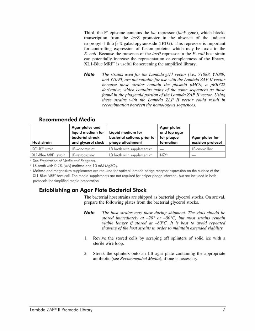
Recommended Media Host strain
Agar plates and liquid medium for bacterial streak and glycerol stock
Liquid medium for bacterial cultures prior to phage attachment
Agar plates and top agar for plaque formation
Agar plates for excision protocol
SOLR™ strain LB-kanamycina LB broth with supplementsa-c — LB-ampicillina
XL1-Blue MRF´ strain LB-tetracyclinea LB broth with supplementsa-c NZYa — a See Preparation of Media and Reagents. b LB broth with 0.2% (w/v) maltose and 10 mM MgSO4. c Maltose and magnesium supplements are required for optimal lambda phage receptor expression on the surface of the
XL1-Blue MRF’ host cell. The media supplements are not required for helper phage infection, but are included in both protocols for simplified media preparation.
Establishing an Agar Plate Bacterial Stock The bacterial host strains are shipped as bacterial glycerol stocks. On arrival, prepare the following plates from the bacterial glycerol stocks.
Note The host strains may thaw during shipment. The vials should be stored immediately at –20° or –80°C, but most strains remain viable longer if stored at –80°C. It is best to avoid repeated thawing of the host strains in order to maintain extended viability.
1. Revive the stored cells by scraping off splinters of solid ice with a sterile wire loop.
2. Streak the splinters onto an LB agar plate containing the appropriate antibiotic (see Recommended Media), if one is necessary.

8 Lambda ZAP® II Premade Library
3. Incubate the plate overnight at 37°C.
4. Seal the plate with Parafilm® laboratory film and store the plate at 4°C for up to 1 week.
5. Restreak the cells onto a fresh plate every week.
Preparing a –80°C Bacterial Glycerol Stock
1. In a sterile 50-ml conical tube, inoculate 10 ml of LB broth with the appropriate antibiotic (see Recommended Media) with one colony from the plate. Grow the cells to late log phase.
2. Add 4.5 ml of a sterile glycerol-liquid medium solution (prepared by mixing 5 ml of glycerol + 5 ml of the appropriate medium) to the bacterial culture from step 1. Mix well.
3. Aliquot into sterile centrifuge tubes (1 ml/tube). This preparation may be stored at –20°C for 1–2 years or at –80°C for more than 2 years.
Growth of Cells for Plating Phage Bacterial cultures for plating phage should be started from a fresh plate using a single colony and should be grown overnight with vigorous shaking at 30°C in 50 ml of LB broth supplemented with 0.2% (w/v) maltose and 10 mM MgSO4. (Do not use tetracycline in the presence of magnesium.) The lower temperature ensures that the cells will not overgrow. The cells should be spun at 1000 × g for 10 minutes then gently resuspended in 10 ml of 10 mM MgSO4. Before use, dilute cells to an OD600 of 0.5 with 10 mM MgSO4. Bacterial cells prepared in this manner can be used for all phage manipulations described within the manual. Highest efficiencies are obtained from freshly prepared cells.
Determining Background by Color Selection with IPTG and X-gal The color selection by α-complementation with the Lambda ZAP II vector requires higher amounts of IPTG and X-gal for generation of the blue color. Transcription and translation of the fusion protein are normal, but the large polylinker present within the pBluescript phagemid, which is present in the Lambda ZAP II vector, is partly responsible for the reduced activity of the β-galactosidase protein. As would be expected, the copy number of the Lambda ZAP II vector is much less per cell than the copy number of pBluescript phagemids. However, it is important to note that the color assay is used only for determining the ratio of recombinants to nonrecombinants within a newly constructed library and is not used for any other manipulations.

Lambda ZAP® II Premade Library 9
HELPER PHAGE Two different helper phages are provided with the Lambda ZAP II library: (1) the ExAssist® interference-resistant helper phage with SOLR™ strain3 and (2) the VCSM13 helper phage. The ExAssist interference-resistant helper phage with SOLR strain is designed to allow efficient in vivo excision of the pBluescript phagemid from the Lambda ZAP II vector while preventing the problems that can be associated with helper phage co-infection. The ExAssist helper phage contains an amber mutation that prevents replication of the phage genome in a nonsuppressing E. coli strain (e.g., SOLR cells). Only the excised phagemid can replicate in the host, removing the possibility of co-infection from the ExAssist helper phage. The ExAssist helper phage cannot be used for single-stranded rescue due to its inability to replicate in the SOLR strain. The other helper phage, VCSM13 helper phage, is recommended for single-stranded rescue procedures from the excised pBluescript phagemids.
Storing the Helper Phage The ExAssist helper phage and the VCSM13 helper phage are supplied in 7% dimethylsulfoxide (DMSO) and should be stored at –80°C. The helper phage may be stored for short periods of time at –20°C or 4°C. It is important to titer the helper phage prior to each use. Expect titers of approximately 1010 pfu/ml for the ExAssist helper phage or 1011 pfu/ml for the VCSM13 helper phage. If the titer drops over time, prepare a fresh high-titer stock of the helper phage as outlined in Amplifying the Helper Phage.
Titering the Helper Phage
1. Transfer a colony of XL1-Blue MRF´ cells into 10 ml of LB broth with supplements in a 50-ml conical tube. Incubate the conical tube with shaking at 37°C until growth reaches an OD600 of 1.0.
2. Dilute the phage (10–4–10–7) in SM buffer (See Preparation of Media and Reagents) and combine 1 μl of each dilution with 200 μl of XL1-Blue MRF´ cells (OD600 = 1.0).
3. Incubate the helper phage and the XL1-Blue MRF´ cells for 15 minutes at 37°C to allow the phage to attach to the cells.
4. Add 3 ml of NZY top agar, melted and cooled to ~48°C, and plate immediately onto dry, prewarmed NZY agar plates. Allow the plates to set for 10 minutes.
10 Lambda ZAP® II Premade Library
5. Invert the plates and incubate overnight at 37°C.
Note ExAssist and VCSM13 plaques will have a cloudier appearance than lambda phage plaques.
6. To determine the titer [in plaque-forming units per milliliter (pfu/ml)], use the following formula:
( )( )
Number of plaques pfu dilution factor
Volume plated l 1000 l / ml
×⎡
⎣⎢
⎤
⎦⎥ ×
μμ
where the volume plated (in microliters) refers to the volume of the helper phage solution added to the cells.
Amplifying the Helper Phage
1. Transfer a colony of XL1-Blue MRF´ cells into 10 ml of LB broth with supplements in a 50-ml conical tube. Incubate the conical tube with shaking at 37°C until growth reaches an OD600 of 0.3.
Note An OD600 of 0.3 corresponds to 2.5 × 10⁸ cells/ml.
2. Add the helper phage at a multiplicity of infection (MOI) of 20:1 (phage-to-cells ratio).
3. Incubate the conical tube at 37°C for 15 minutes to allow the phage to attach to the cells.
4. Incubate the conical tube with shaking at 37°C for 8 hours.
Note When amplifying VCSM13 helper phage, add kanamycin to a final concentration of 25 μg/ml after 30 minutes of growth.
5. Heat the conical tube at 65°C for 15 minutes.
6. Spin down the cell debris and transfer the supernatant to a fresh conical tube.
7. The titer of the supernatant should be between 7.5 × 10¹⁰ and 1.0 × 10¹² pfu/ml for ExAssist helper phage or between 1.0 × 10¹¹ and 1.0 × 10¹² pfu/ml for VCSM13 helper phage.
Note ExAssist and VCSM13 plaques will have a cloudier appearance than lambda phage plaques.
8. Add dimethylsulfoxide (DMSO) to a final concentration of 7% (v/v) and store at –80°C.
9. For further details about helper phage titering or amplification, please see Titering the Helper Phage or Reference 4.

Lambda ZAP® II Premade Library 11
TITERING THE LIBRARY
Preparing the Host Bacteria
1. Streak the XL1-Blue MRF’ cells onto an LB-tetracycline agar plate. Incubate the plate overnight at 37°C.
2. Inoculate 50 ml of LB broth with supplements in a sterile flask with a single colony of the XL1-Blue MRF’ host.
Note Do not add antibiotic to the overnight culture or to the titering plates. The antibiotic will bind to the bacterial cell wall and will inhibit the ability of the phage to infect the cell.
3. Incubate with shaking at 37°C for 4–6 hours (do not grow past an OD600 of 1.0). Alternatively, grow overnight at 30°C, shaking at 200 rpm.
Note The lower temperature keeps the bacteria from overgrowing, thus reducing the number of nonviable cells. Phage can adhere to nonviable cells resulting in a decreased titer.
4. Pellet the bacteria at 1000 × g for 10 minutes.
5. Gently resuspend the cell pellet in 25 ml sterile 10 mM MgSO4.
Note For later use, store the cells at 4°C overnight in 10 mM MgSO4.
Titering Protocol A background test can be completed by plating several hundred plaques on a plate [see Determining Background by Color Selection with IPTG and X-gal]. Add 15 μl of 0.5 M IPTG (in water) and 50 μl of 250 mg/ml X-gal [in dimethylformamide (DMF)] to 2–3 ml of NZY top agar, melted and cooled to ~48°C. The higher concentrations of IPTG and X-gal used in the plating often result in the formation of a precipitate, which disappears after incubation. Stratagene suggests adding the IPTG and X-gal to the NZY top agar separately, with mixing in between additions, to minimize the formation of this precipitate. Plate immediately on NZY agar plates. Plaques are visible after incubation for 12 hours at 37°C, although color detection requires overnight incubation. Background plaques are blue, while recombinant plaques are white.
1. Dilute the XL1-Blue MRF’ cells (from step 5 of Preparing the Host Bacteria in Titering the Library) to an OD600 of 0.5 with sterile 10 mM MgSO4.
Note The bacteria should be used immediately following dilution.

12 Lambda ZAP® II Premade Library
2. To determine the titer of the library, mix the following components: 1 μl of the library aliquot 200 μl of XL1-Blue MRF’ cells at an OD600 of 0.5
and 1 μl of a 1:10 dilution of the library aliquot 200 μl of XL1-Blue MRF’ cells at an OD600 of 0.5
For amplified library titering, first dilute the amplified phage stock in SM buffer by the following amounts: 1:10,000, 1:100,000, 1:1,000,000. Add 1 μl of each dilution to 200 μl of host cells.
3. Incubate the phage and the bacteria at 37°C for 15 minutes to allow the phage to attach to the cells.
4. Add the following components:
2–3 ml of NZY top agar (melted and cooled to ~48°C). 15 μl of 0.5M IPTG (in water) 50 μl of X-gal [250 mg/ml (in DMF)]
5. Plate immediately onto dry, prewarmed NZY agar plates and allow the plates to set for 10 minutes. Invert the plates and incubate at 37°C.
6. Plaques should be visible after 12 hours, although color detection requires overnight incubation. Background plaques are blue and should be < 1× 10⁵ pfu/μg of arms, while recombinant plaques will be white (clear) and should be 10–100-fold above the background.

Lambda ZAP® II Premade Library 13
AMPLIFYING THE LIBRARY It is usually desirable to amplify libraries prepared in lambda vectors to make a large, stable quantity of a high-titer stock of the library. However, more than one round of amplification is not recommended, since slower growing clones may be significantly underrepresented.
Note The premade library has been through one round of amplification. The following protocol is recommended for amplifying the Lambda ZAP II library:
Day 1
1. Grow a 50-ml overnight culture of XL1-Blue MRF’ cells in LB broth with supplements at 30°C with shaking.
Day 2
2. Gently spin down the XL1-Blue MRF’ cells (1000 × g). Resuspend the cell pellet in 25 ml of 10 mM MgSO4. Measure the OD600 of the cell suspension, then dilute the cells to an OD600 of 0.5 in 10 mM MgSO4.
3. Combine aliquots of the library suspension containing ~5 × 10⁴ pfu of bacteriophage with 600 μl of XL1-Blue MRF’ cells at an OD600 of 0.5 in Falcon® 2059 polypropylene tubes. To amplify 1 × 10⁶ plaques, use a total of 20 aliquots (each aliquot contains 5 × 10⁴ plaques/150-mm plate).
Note Do not add more than 300 μl of phage/600 μl of cells.
4. Incubate the tubes containing the phage and host cells for 15 minutes at 37°C to allow the phage to attach to the cells.
5. Mix 6.5 ml of NZY top agar, melted and cooled to ~48°C, with each aliquot of infected bacteria and spread evenly onto a freshly poured 150-mm NZY agar plate. Allow the plates to set for 10 minutes.
6. Invert the plates and incubate at 37°C for 6–8 hours. Do not allow the plaques to get larger than 1–2 mm. On completion, the plaques should be touching.
7. Overlay the plates with ~8–10 ml of SM buffer. Store the plates at 4°C overnight (with gentle rocking if possible). This allows the phage to diffuse into the SM buffer.

14 Lambda ZAP® II Premade Library
Day 3
8. Recover the bacteriophage suspension from each plate and pool it into a sterile polypropylene container. Rinse the plates with an additional 2 ml of SM buffer and pool. Add chloroform to a 5% (v/v) final concentration. Mix well and incubate for 15 minutes at room temperature.
9. Remove the cell debris by centrifugation for 10 minutes at 500 × g.
10. Recover the supernatant and transfer it to a sterile polypropylene container. If the supernatant appears cloudy or has a high amount of cell debris, repeat steps 8 and 9. If the supernatant is clear, add chloroform to a 0.3% (v/v) final concentration and store at 4°C. Stratagene recommends storing aliquots of the amplified library in 7% (v/v) DMSO at –80°C.
11. Check the titer of the amplified library using host cells and serial dilutions of the library. (Assume ~109–1011 pfu/ml.)
Note Briefly spin the lambda phage stock to ensure that the chloroform is separated completely before removing the aliquot for titering.

Lambda ZAP® II Premade Library 15
PERFORMING PLAQUE LIFTS
1. Titer the library suspension to determine the concentration using XL1-Blue MRF’ cells.
2. Combine the equivalent of 5 × 10⁴ pfu/plate and 600 μl of freshly prepared XL1-Blue MRF’ cells at an OD600 of 0.5.
3. Incubate the bacteria and phage mixture at 37°C for 15 minutes to allow the phage to attach to the cells.
4. Add 6.5 ml of NZY top agar (~48°C) to the bacteria and phage mixture.
5. Quickly pour the plating culture onto a dry, prewarmed 150-mm NZY agar plate, which is at least 2 days old. Carefully swirl the plate to distribute the cells evenly. Allow the plates to set for 10 minutes. (Use 20 plates to screen 1 × 10⁶ pfu.)
6. Invert the plates and incubate at 37°C for ~8 hours.
7. Chill the plates for 2 hours at 4°C to prevent the top agar from sticking to the nitrocellulose membrane.
Note Use forceps and wear gloves for the following steps.
8. Place a nitrocellulose membrane onto each NZY agar plate for 2 minutes to allow the transfer of the phage particles to the membrane. Use a needle to prick through the membrane and agar for orientation. (If desired, waterproof ink in a syringe needle may be used.)
Notes If making duplicate nitrocellulose membranes, allow the second membrane to transfer for ~4 minutes.
Pyrex® dishes are convenient for the following steps. All solutions should be at room temperature.
a. Denature the nitrocellulose-bound DNA after lifting by submerging the membrane in a 1.5 M NaCl and 0.5 M NaOH denaturation solution for 2 minutes.
Note If using charged nylon, wash with gloved fingertips to remove the excess top agar.
b. Neutralize the nitrocellulose membrane for 5 minutes by submerging the membrane in a 1.5 M NaCl and 0.5 M Tris-HCl (pH 8.0) neutralization solution.
c. Rinse the nitrocellulose membrane for no more than 30 seconds by submerging the membrane in a 0.2 M Tris-HCl (pH 7.5) and 2× SSC buffer solution (see Preparation of Media and Reagents).

16 Lambda ZAP® II Premade Library
9. Blot briefly on a Whatman® 3MM paper.
10. Crosslink the DNA to the membranes using the autocrosslink setting on the Stratalinker® UV crosslinker* (120,000 μJ of UV energy) for ~30 seconds. Alternatively, oven bake at 80°C for ~1.5–2 hours.
11. Store the stock agar plates of the transfers at 4°C to use after screening.
HYBRIDIZING AND SCREENING Following the preparation of the membranes for hybridization, perform prehybridization, probe preparation, hybridization, and washes for either oligonucleotide probes or double-stranded probes and then expose the membranes to film as outlined in standard methodology texts.4, 5 Following these procedures, perform secondary and tertiary screenings also as outlined in the standard methodology texts.4, 5 After an isolate is obtained, refer to Sambrook et al.4 for suggested phage miniprep and maxiprep procedures.
ANTIBODY SCREENING PROTOCOL A complete manual for immunoscreening is supplied with Stratagene's picoBlue™ immunoscreening kit. This kit is available with goat anti-rabbit antibodies and goat anti-mouse antibodies [Catalog #200371 (goat anti-rabbit) and #200372 (goat anti-mouse)]. * Available from Stratagene, Catalog #400071 (1800) and #400075 (2400).

Lambda ZAP® II Premade Library 17
In Vivo EXCISION OF THE pBLUESCRIPT® SK(–) PHAGEMID FROM THE LAMBDA ZAP II VECTOR
The Lambda ZAP II vector is designed to allow simple, efficient in vivo excision and recircularization of any cloned insert contained within the lambda vector to form a phagemid containing the cloned insert (see Figure 3). This in vivo excision depends on the placement of the DNA sequences within the lambda phage genome and on the presence of a variety of proteins, including f1 bacteriophage-derived proteins. The f1 phage proteins recognize a region of DNA normally serving as the f1 bacteriophage origin of replication. This origin of replication can be divided into two overlying parts: (1) the site of initiation and (2) the site of termination for DNA synthesis.6 These two regions are subcloned separately into the Lambda ZAP II vector. The lambda phage (target) is made accessible to the f1-derived proteins by simultaneously infecting a strain of E. coli with both the lambda vector and the f1 bacteriophage. Inside E. coli, the "helper" proteins (i.e., proteins from f1 or M13 phage) recognize the initiator DNA that is within the lambda vector. One of these proteins then nicks one of the two DNA strands. At the site of this nick, new DNA synthesis begins and duplicates whatever DNA exists in the lambda vector "downstream" (3´) of the nicking site. DNA synthesis of a new single strand of DNA continues through the cloned insert until a termination signal, positioned 3´ of the initiator signal, is encountered within the constructed lambda vector. The single-stranded DNA molecule is circularized by the gene II product from the f1 phage, forming a circular DNA molecule containing the DNA between the initiator and terminator. In the case of the Lambda ZAP II vector, this includes all sequences of the pBluescript SK(–) phagemid and the insert, if one is present. This conversion is the "subcloning" step, since all sequences associated with normal lambda vectors are positioned outside of the initiator and terminator signals and are not contained within the circularized DNA. In addition, the circularizing of the DNA automatically recreates a functional f1 origin as found in f1 bacteriophage or phagemids.

18 Lambda ZAP® II Premade Library
In Vivo EXCISION PROTOCOLS USING THE EXASSIST® INTERFERENCE-RESISTANT HELPER PHAGE WITH SOLR™ STRAIN
The ExAssist helper phage with SOLR strain is designed to allow efficient excision of the pBluescript phagemid from the Lambda ZAP II vector, while eliminating problems associated with helper phage co-infection. The ExAssist helper phage contains an amber mutation that prevents replication of the phage genome in a nonsuppressing E. coli strain such as SOLR cells. This allows only the excised phagemid to replicate in the host, removing the possibility of co-infection from the ExAssist helper phage. Since the ExAssist helper phage cannot replicate in the SOLR strain, single-stranded rescue cannot be performed in this strain using this helper phage. Mass excision can be used to generate subtraction libraries and subtracted DNA probes.
Single-Clone Excision Protocol
Day 1
1. Core the plaque of interest from the agar plate and transfer the plaque to a sterile microcentrifuge tube containing 500 μl of SM buffer and 20 μl of chloroform. Vortex the microcentrifuge tube to release the phage particles into the SM buffer. Incubate the microcentrifuge tube for 1–2 hours at room temperature or overnight at 4°C. (This phage stock is stable for up to 6 months at 4°C.)
2. Grow separate 50-ml overnight cultures of XL1-Blue MRF´ and SOLR cells in LB broth with supplements at 30°C.
Day 2
3. Gently spin down the XL1-Blue MRF´ and SOLR cells (1000 × g). Resuspend each of the cell pellets in 25 ml of 10 mM MgSO4. Measure the OD600 of the cell suspensions, then adjust the concentration of the cells to an OD600 of 1.0 (8 × 10⁸ cells/ml) in 10 mM MgSO4.
4. Combine the following components in a Falcon 2059 polypropylene tube:
200 μl of XL1-Blue MRF´ cells at an OD600 of 1.0 250 μl of phage stock (containing >1 × 10⁵ phage particles) 1 μl of the ExAssist helper phage (>1 × 10⁶ pfu/μl)
Note Briefly spin the lambda phage stock to ensure that the chloroform is separated completely before removing the aliquot used in the excision reaction.
5. Incubate the Falcon 2059 polypropylene tube at 37°C for 15 minutes to allow the phage to attach to the cells.

Lambda ZAP® II Premade Library 19
6. Add 3 ml of LB broth with supplements and incubate the Falcon 2059 polypropylene tube for 2.5–3 hours at 37°C with shaking. Because clonal representation is not relevant, single-clone excision reactions can be safely performed overnight.
Note The turbidity of the media is not indicative of the success of the excision.
7. Heat the Falcon 2059 polypropylene tube at 65–70°C for 20 minutes to lyse the lambda phage particles and the cells. Spin the tube at 1000 × g for 15 minutes to pellet the cell debris.
8. Decant the supernatant into a sterile Falcon 2059 polypropylene tube. This stock contains the excised pBluescript phagemid packaged as filamentous phage particles. (This stock may be stored at 4°C for 1–2 months.)
9. To plate the excised phagemids, add 200 μl of freshly grown SOLR cells from step 3 (OD600 = 1.0) to two 1.5-ml microcentrifuge tubes. Add 100 μl of the phage supernatant (from step 8 above) to one microcentrifuge tube and 10 μl of the phage supernatant to the other microcentrifuge tube.
10. Incubate the microcentrifuge tubes at 37°C for 15 minutes.
11. Plate 200 μl of the cell mixture from each microcentrifuge tube on LB-ampicillin agar plates (100 μg/ml) and incubate the plates overnight at 37°C.
Due to the high-efficiency of the excision process, it may be necessary to titrate the supernatant to achieve single-colony isolation. Colonies appearing on the plate contain the pBluescript double-stranded phagemid with the cloned DNA insert. Helper phage will not grow, since helper phage is unable to replicate in the Su– (nonsuppressing) SOLR strain and does not contain ampicillin-resistance genes. SOLR cells are also resistant to lambda phage infection, thus preventing lambda phage contamination after excision. To maintain the pBluescript phagemid, streak the colony on a new LB-ampicillin agar plate. For long-term storage, prepare a bacterial glycerol stock and store at –80°C. VCSM13 helper phage is recommended for the single-stranded rescue procedure. The single-stranded rescue procedure can be found in Appendix: Recovery Of Single-Stranded DNA From Cells Containing pBluescript® Phagemids.

20 Lambda ZAP® II Premade Library
Mass Excision Protocol
Day 1
1. Grow separate 50-ml overnight cultures of XL1-Blue MRF´ and SOLR cells in LB broth with supplements at 30°C.
Day 2
2. Gently spin down the XL1-Blue MRF´ and SOLR cells (1000 × g). Resuspend each of the cell pellets in 25 ml of 10 mM MgSO4. Measure the OD600 of the cell suspensions, then adjust the concentration of the cells to an OD600 of 1.0 (8 × 10⁸ cells/ml) in 10 mM MgSO4.
3. In a 50-ml conical tube, combine a portion of the amplified lambda bacteriophage library with XL1-Blue MRF´ cells at a MOI of 1:10 lambda phage-to-cell ratio. Excise 10- to 100-fold more lambda phage than the size of the primary library to ensure statistical representation of the excised clones. Add ExAssist helper phage at a 10:1 helper phage-to-cells ratio to ensure that every cell is co-infected with lambda phage and helper phage.
For example, use
107 pfu of the lambda phage (i.e., 10- to 100-fold above the primary library size)
108 XL1-Blue MRF´ cells (1:10 lambda phage-to-cell ratio, noting that an OD600 of 1.0 corresponds to 8 × 10⁸ cells/ml)
109 pfu of ExAssist helper phage (10:1 helper phage-to-cells ratio)
Note Briefly spin the lambda phage stock to ensure that the chloroform is separated completely before removing the aliquot used in the excision reaction.
4. Incubate the conical tube at 37°C for 15 minutes to allow the phage to attach to the cells.
5. Add 20 ml of LB broth with supplements and incubate the conical tube for 2.5–3 hours at 37°C with shaking.
Notes Incubation times for mass excision in excess of 3 hours may alter the clonal representation.
The turbidity of the media is not indicative of the success of the excision.
6. Heat the conical tube at 65–70°C for 20 minutes to lyse the lambda phage particles and the cells.
7. Spin the conical tube at 1000 × g for 10 minutes to pellet the cell debris and then decant the supernatant into a sterile conical tube.

Lambda ZAP® II Premade Library 21
8. To titer the excised phagemids, combine 1 μl of this supernatant with 200 μl of SOLR cells from step 2 in a 1.5-ml microcentrifuge tube.
9. Incubate the microcentrifuge tube at 37°C for 15 minutes.
10. Plate 100 μl of the cell mixture onto LB–ampicillin agar plates (100 μg/ml) and incubate the plates overnight at 37°C.
Note It may be necessary to further dilute the cell mixture to achieve single-colony isolation.
At this stage, colonies may be selected for plasmid preps, or the cell mixture may be plated directly onto filters for colony screening.

22 Lambda ZAP® II Premade Library
APPENDIX: RECOVERY OF SINGLE-STRANDED DNA FROM CELLS CONTAINING PBLUESCRIPT® PHAGEMIDS
pBluescript is a phagemid that can be secreted as single-stranded DNA in the presence of M13 helper phage. These phagemids contain the intergenic (IG) region of a filamentous f1 phage. This region encodes all of the cis-acting functions of the phage required for packaging and replication. In E. coli with the F+ phenotype (containing an F´ episome), pBluescript phagemids will be secreted as single-stranded f1 "packaged" phage when the bacteria has been infected by a helper phage. Since these filamentous helper phages (M13, f1) will not infect E. coli without an F´ episome coding for pili, it is essential to use XL1-Blue MRF´ or a similar strain containing the F´ episome.7, 8 Stratagene offers helper phages that preferentially package pBluescript phagemids. Typically, 30–50 pBluescript molecules are packaged/helper phage DNA molecule. pBluescript phagemids are offered with the IG region in either of two orientations: pBluescript (+) is replicated such that the sense strand of the β-galactosidase gene is secreted within the phage particles; pBluescript (–) is replicated such that the antisense strand of the β-galactosidase gene is secreted in the phage particles. Yields of single-stranded (ss)DNA depend on the specific insert sequence. For most inserts, over 1 μg of ssDNA can be obtained from a 1.5-ml miniprep if grown in XL1-Blue MRF´. A faint single-strand helper phage band may appear on a gel at ~6 kb for VCSM13. This DNA mixture can be sequenced with primers that are specific for pBluescript and do not hybridize to the helper phage genome. Site-specific mutagenesis is also possible using standard techniques. The advantages of using pBluescript phagemids for either purpose are as follows: (1) pBluescript phagemids do not replicate via the M13 cycle, lessening the tendency to delete DNA inserts, therefore it is unlikely that even 10-kb inserts will be deleted. (2) "Packaging" of pBluescript phagemids containing inserts is efficient since the pBluescript vector is significantly smaller than wild-type M13. (3) Oligonucleotide mutagenesis in pBluescript vectors is advantageous because the mutagenized insert is located between the T3 and T7 promoters. The resultant mutant transcripts can be synthesized in vitro without further subcloning. VCSM13 (single-strand size ~6 kb), is efficient at single-stranded DNA rescue and provides good yields of single-stranded phagemid; however it can revert to wild-type (more frequently than R408 helper phage, for example). This difficulty can be addressed by periodically propagating VCSM13 in the presence of kanamycin. [VCSM13 (a derivative of M13KO7) has a kanamycin gene inserted into the intergenic region.]

Lambda ZAP® II Premade Library 23
Single-Stranded Rescue Protocol
1. Inoculate a single colony into 5 ml of 2× YT broth§ containing 100 μg/ml ampicillin and VCSM13 helper phage at 107–108 pfu/ml (MOI ~10).
2. Grow the culture at 37°C with vigorous aeration for 1–2 hours.
3. Add kanamycin to 70 μg/ml to select for infected cells.
4. Continue growth at 37°C with vigorous aeration for 16–24 hours, or until growth has reached saturation.
5. Centrifuge 1.5 ml of the cell culture for 5 minutes in a microcentrifuge.
6. Remove 1 ml of the supernatant to a fresh tube, then add 150 μl of a solution containing 20% PEG8000 and 2.5 M NaCl. Allow phage particles to precipitate on ice for 15 minutes.
Note For increased yield, perform the PEG precipitation overnight at 4°C.
7. Centrifuge for 5 minutes in a microcentrifuge. (A pellet should be obvious.)
8. Remove supernatant. Centrifuge the PEG pellets a few seconds more to collect residual liquid, then remove and discard the residual liquid.
9. Resuspend the pellet in 400 μl of 0.3 M NaOAc (pH 6.0) and 1 mM EDTA by vortexing vigorously.
10. Extract with 1 volume phenol–chloroform and centrifuge for 1–2 minutes to separate phases.
11. Transfer the aqueous phase to a fresh tube and add 1 ml of ethanol. Centrifuge for 5 minutes.
12. Remove ethanol and dry the DNA pellet.
13. Dissolve the pellet in 25 μl of TE buffer§.
14. Analyze 1–2 μl on an agarose gel.
§ See Preparation of Media and Reagents.

24 Lambda ZAP® II Premade Library
TROUBLESHOOTING Observation Suggestions
The molar ratios of lambda phage to cells to helper phage is critical. Verify that the titer on the tubes is current and correct and use only calibrated pipettor
Excision efficiencies are directly related to the Lambda ZAP II phage titer. If an excision is unsuccessful, prepare a high-titer stock of the phage and repeat the excision procedure
Poor rescue may be a result of toxic cDNA clones which can be isolated in lambda vectors but not in plasmid vectors. The ABLE® C strain* and the ABLE® K strain* reduce the copy number of common cloning vectors by ~4- and 10-fold, respectively, enhancing the probability that a toxic clone will be propagated. Positive clones observed on initial screening as lambda plaques can be excised and introduced into the ABLE strains. Excised phagemid libraries can also be screened directly in the ABLE strains
The number of colonies is too low
The lambda phage stock aliquot used for in vivo excision cannot contain chloroform, as chloroform lyses the E. coli cells. Briefly spin the lambda phage stock to ensure that the chloroform is separated completely before removing the aliquot
* ABLE competent cells (Catalog #200170–200172) and ABLE electroporation competent cells (Catalog #200160–200162) are available separately from Stratagene.

Lambda ZAP® II Premade Library 25
PREPARATION OF MEDIA AND REAGENTS LB Agar (per Liter)
10 g of NaCl 10 g of tryptone 5 g of yeast extract 20 g of agar Add deionized H2O to a final volume of 1
liter Adjust pH to 7.0 with 5 N NaOH Autoclave Pour into petri dishes (~25 ml/100-mm plate)
LB–Ampicillin Agar (per Liter) Prepare 1 liter of LB agar Autoclave Cool to 55°C Add 10 ml of 10 mg/ml ampicillin
(filter-sterilized) Pour into petri dishes (~25 ml/100-mm plate)
LB Broth 10 g of NaCl 10 g of tryptone 5 g of yeast extract Add deionized H2O to a final volume of 1
liter Adjust pH to 7.0 with 5 N NaOH Autoclave
LB Broth with Supplements Prepare 1 liter of LB broth Autoclave Add the following filter-sterilized
supplements prior to use 10 ml of 1 M MgSO4 3 ml of a 2 M maltose solution or 10 ml
of 20% (w/v) maltose
LB–Tetracycline Broth (per Liter) Prepare 1 liter of LB broth Autoclave Cool to 55°C Add 1.5 ml of 10 mg/ml tetracycline (filter-
sterilized) Store broth in a dark, cool place as
tetracycline is light-sensitive
LB–Tetracycline Agar (per Liter) Prepare 1 liter of LB agar Autoclave Cool to 55°C Add 1.5 ml of 10 mg/ml tetracycline (filter-
sterilized) Pour into petri dishes (~25 ml/100-mm plate) Store plates in a dark, cool place or cover
plates with foil if left out at room temperature for extended time periods as tetracycline is light-sensitive
LB–Kanamycin Broth (per Liter) Prepare 1 liter of LB broth Autoclave Cool to 55°C Add 7.5 ml of 10 mg/ml kanamycin (filter-
sterilized)
LB–Kanamycin Agar (per Liter) Prepare 1 liter of LB agar Autoclave Cool to 55°C Add 5 ml of 10-mg/ml filter-sterilized
kanamycin Pour into petri dishes (~25 ml/100-mm plate)
2× YT Broth (per Liter) 10 g of NaCl 10 g of yeast extract 16 g of tryptone Add deionized H2O to a final volume of 1
liter Adjust to pH 7.5 with NaOH Autoclave

26 Lambda ZAP® II Premade Library
NZY Broth (per Liter) 5 g of NaCl 2 g of MgSO4 . 7H2O 5 g of yeast extract 10 g of NZ amine (casein hydrolysate) Add deionized H2O to a final volume of 1
liter Adjust the pH to 7.5 with NaOH Autoclave
NZY Agar (per Liter) 5 g of NaCl 2 g of MgSO4 . 7H2O 5 g of yeast extract 10 g of NZ amine (casein hydrolysate) 15 g of agar Add deionized H2O to a final volume of
1 liter Adjust the pH to 7.5 with NaOH Autoclave Pour into petri dishes (~80 ml/150-mm plate)
TE Buffer 5 mM Tris-HCl (pH 7.5) 0.1 mM EDTA
NZY Top Agar (per Liter) Prepare 1 liter of NZY broth Add 0.7% (w/v) agarose Autoclave
SM Buffer (per Liter) 5.8 g of NaCl 2.0 g of MgSO4 · 7H2O 50.0 ml of 1 M Tris-HCl (pH 7.5) 5.0 ml of 2% (w/v) gelatin Add deionized H2O to a final volume of 1
liter Autoclave
20× SSC Buffer (per Liter) 175.3 g of NaCl 88.2 g of sodium citrate 800.0 ml of deionized H2O Adjust to pH 7.0 with a few drops of 10 N
NaOH Add deionized H2O to a final volume of
1 liter

Lambda ZAP® II Premade Library 27
REFERENCES 1. Short, J. M. and Sorge, J. A. (1992) Methods Enzymol 216:495-508. 2. Bullock, W. O., Fernandez, J. M. and Short, J. M. (1987) Biotechniques 5(4):376–378. 3. Hay, B. and Short, J. M. (1992) Strategies 5(1):16–18. 4. Sambrook, J., Fritsch, E. F. and Maniatis, T. (1989). Molecular Cloning: A Laboratory
Manual. Cold Spring Harbor Laboratory Press, Cold Spring Harbor, NY. 5. Ausubel, F. M., Brent, R., Kingston, R. E., Moore, D. D., Seidman, J. G. et al. (1987).
Current Protocols in Molecular Biology. John Wiley and Sons, New York. 6. Dotto, G. P., Horiuchi, K. and Zinder, N. D. (1984) J Mol Biol 172(4):507-21. 7. Dente, L., Cesareni, G. and Cortese, R. (1983) Nucleic Acids Res 11(6):1645-55. 8. Mead, D. A., Skorupa, E. S. and Kemper, B. (1985) Nucleic Acids Res 13(4):1103-18.
ENDNOTES ABLE®, ExAssist®, Lambda ZAP®, pBluescript®, and Stratalinker® are registered
trademarks of Stratagene in the United States. picoBlue and SOLR are trademarks of Stratagene. Falcon® is a registered trademark of Becton Dickinson Labware. GenBank® is a registered trademark of the U.S. Department of Health and Human Services. Parafilm® is a registered trademark of American Can Company. Pyrex® is a registered trademark of Corning Glass Works. Whatman® is a trademark of Whatman Paper Ltd.
MSDS INFORMATION The Material Safety Data Sheet (MSDS) information for Stratagene products is provided on Stratagene’s website at http://www.stratagene.com/MSDS/. Simply enter the catalog number to retrieve any associated MSDS’s in a print-ready format. MSDS documents are not included with product shipments.