manual on case management and surveillance of the ... · abstract this manual makes recommendations...
TRANSCRIPT


MANUAL ON CASE MANAGEMENT
AND SURVEILLANCE OF THE LEISHMANIASES
IN THE WHO EUROPEAN REGION

ABSTRACT
This manual makes recommendations on a standardized approach to the case management and epi-
demiological surveillance of the leishmaniases across the WHO European Region. It was conceived as a
practical guide for health workers dealing with the difficult task of diagnosing and treating different
clinical forms of leishmaniasis, and for public health workers involved in surveillance systems for
infectious diseases. Stepwise decision algorithms are presented for clinical and laboratory diagnosis, as
well as for the treatment of various leishmaniasis entities endemic or frequently imported in the Region.
The manual provides case and treatment outcome definitions for epidemiological surveillance.
Particular attention is given to establishing monitoring and evaluation systems that provide sets of
indicators allowing the performance of leishmaniasis control strategies to be properly assessed. Annexes
include epidemiological information, antileishmanial drug information, and detailed standard operating
procedures for diagnosis and treatment.
KEYWORDS
Leishmaniasis - diagnosis
Leishmaniasis - epidemiology
Leishmaniasis - parasitology
Leishmaniasis - prevention and control
Leishmaniasis, Cutaneous - diagnosis
Leishmaniasis, Cutaneous - epidemiology
Leishmaniasis, Cutaneous - parasitology
ISBN 978 92 89052 51 1
© World Health Organization 2017
All rights reserved. The Regional Office for Europe of the World Health Organization welcomes requests for permission to
reproduce or translate its publications, in part or in full.
The designations employed and the presentation of the material in this publication do not imply the expression of any opinion
whatsoever on the part of the World Health Organization concerning the legal status of any country, territory, city or area or of its
authorities, or concerning the delimitation of its frontiers or boundaries. Dotted lines on maps represent approximate border lines for
which there may not yet be full agreement.
The mention of specific companies or of certain manufacturers’ products does not imply that they are endorsed or recommended by
the World Health Organization in preference to others of a similar nature that are not mentioned. Errors and omissions excepted, the
names of proprietary products are distinguished by initial capital letters.
All reasonable precautions have been taken by the World Health Organization to verify the information contained in this publication.
However, the published material is being distributed without warranty of any kind, either express or implied. The responsibility for
the interpretation and use of the material lies with the reader. In no event shall the World Health Organization be liable for damages
arising from its use. The views expressed by authors, editors, or expert groups do not necessarily represent the decisions or the stated
policy of the World Health Organization.
Address requests about publications of the WHO Regional Office for Europe to:
Publications
WHO Regional Office for Europe
UN City, Marmorvej 51
DK-2100 Copenhagen Ø, Denmark
Alternatively, complete an online request form for documentation, health information, or for permission to quote or
translate, on the Regional Office website (http://www.euro.who.int/pubrequest).
Leishmaniasis, Cutaneous - prevention and control
Leishmaniasis, Visceral - diagnosis
Leishmaniasis, Visceral - epidemiology
Leishmaniasis, Visceral - parasitology
Leishmaniasis, Visceral - prevention and control
Europe

iii
Contents
Acknowledgements............................................................................................................................ iv
Contributors ....................................................................................................................................... v
Abbreviations .................................................................................................................................... vi
1. Introduction ................................................................................................................................ 1
2. The leishmaniases in the WHO European Region .................................................................. 2 2.1 Epidemiology and geographical distribution ........................................................................ 2 2.2 VL burden.............................................................................................................................. 2 2.3 CL burden .............................................................................................................................. 5
3. Case management of the leishmaniases in the WHO European Region ............................... 7 3.1 Case management of VL ....................................................................................................... 7
3.2 Case management of VL in HIV-coinfected patients ......................................................... 15
3.3 Case management of VL in other special categories of patients ......................................... 21
3.4 Case management of CL ..................................................................................................... 23 3.5 Case management of CL in special categories of patients .................................................. 31 3.6 Imported cases of CL and MCL .......................................................................................... 32
4. Surveillance............................................................................................................................... 33 4.1 General purpose and components........................................................................................ 33 4.2 Recommended case and treatment outcome definitions for epidemiological surveillance . 34
4.3 Leishmaniasis case detection strategies .............................................................................. 35 4.4 Strategies for the detection of leishmaniasis in the canine reservoir .................................. 36 4.5 Recommended types of surveillance ................................................................................... 37
5. Monitoring and evaluation of leishmaniasis control ............................................................. 40 5.1 Leishmaniasis control strategies .......................................................................................... 40
5.2 Indicators for monitoring and evaluation of leishmaniasis control ..................................... 42
Bibliography ..................................................................................................................................... 45
Annex 1. Country information and annual VL incidence ....................................................... 48
Annex 2. Information on drugs used for treating VL .............................................................. 51
Annex 3. Standard operating procedure for conventional parasitological diagnosis ........... 55
Annex 4. Standard operating procedure for cryotherapy and intralesional injection
of antimony..................................................................................................................57
Annex 5. Standard operating procedure for thermotherapy .................................................. 59
Annex 6. Systemic treatment of CL with pentavalent antimonials ........................................ 60
Annex 7. Drug options for imported CL and MCL cases ........................................................ 62
iv
Acknowledgements
The World Health Organization would like to thank all the experts who provided input to this
manual through participation in the Expert Group Meeting on Finalization of the WHO Regional
Manual on Surveillance and Case Management of Leishmaniasis, which was held on 19–
21 September 2016 in Tirana, Albania.
The WHO Regional Office for Europe also thanks all country representatives and experts who
provided input to the manual through their participation at the regional meeting on surveillance and
control of leishmaniasis in the WHO European Region, held in Sofia, Bulgaria, on 10–12 April
2017.
All figures and photographs in this manual are used with the kind permission of Dr Luigi Gradoni
of Istituto Superiore di Sanità, Rome; Professor Rogelio López-Vélez of Ramon y Cajal University
Hospital, Madrid; Professor Mourad Mokni of La Rabta Hospital, Tunis, and of Tunis El Manar
University; and the Oxford University Press/on behalf of Infectious Diseases Society of America.

v
Contributors
Authors
Luigi Gradoni, Research Director, Unit of Vector-borne Diseases and International Health, Istituto
Superiore di Sanità, Rome, Italy
Rogelio López-Vélez, Associate Professor, National Referral Unit for Tropical Diseases, Infectious
Diseases Department, Ramon y Cajal University Hospital, Madrid, Spain
Mourad Mokni, Professor, Faculty of Medicine, El Manar University, Tunis, and Head of
Department of Dermatology and Research Unit, La Rabta Hospital, Tunis, Tunisia
External peer reviewers
Johannes Blum, Swiss Tropical and Public Health Institute and University of Basel, Basel,
Switzerland
Pierre Buffet, National Institute for Blood Transfusion, Faculty of Medicine, Université Paris
Descartes, Paris, France
Charles L. Jaffe, National Center for Leishmaniasis, Kuvin Center for Study of Tropical and
Infectious Diseases, Hebrew University-Hadassah Medical School, Jerusalem, Israel
Nora Kokaia, Institute of Medical Parasitology and Tropical Medicine, S. Virsaladze, Tbilisi,
Georgia
Javier Moreno Nuncio, Unit for Leishmaniasis and Chagas Disease, WHO Collaborating Centre for
Leishmaniasis, National Centre for Microbiology, Instituto de Salud Carlos III, Majadahonda,
Madrid, Spain
WHO representatives
Elkhan Gasimov, Technical Officer, Malaria and Other Vector-borne and Parasitic Diseases, WHO
Regional Office for Europe, Copenhagen, Denmark
José Antonio Ruiz Postigo, Leishmaniasis Control Programme, Department of Neglected Tropical
Diseases, WHO headquarters, Geneva, Switzerland
Daniel Argaw Dagne, Coordinator, Innovative and Intensified Disease Management, Department of
Neglected Tropical Diseases, WHO headquarters, Geneva, Switzerland

vi
Abbreviations
ABD amphotericin B deoxycholate
ABLC amphotericin B lipid complex
ACD active case detection
ART antiretroviral therapy
BUN blood urea nitrogen
CBC complete blood count
CL cutaneous leishmaniasis
Dx diagnosis
ECG electrocardiogram
ELISA enzyme-linked immunosorbent assay
IFAT immunofluorescence antibody test
IM intramuscular
IV intravenous
LAB liposomal amphotericin B
LCL localized cutaneous leishmaniasis
MA meglumine antimoniate
MCL mucocutaneous leishmaniasis
ML mucosal leishmaniasis
N number (of patients)
NGO nongovernmental organization
PAHO Pan American Health Organization
PCD passive case detection
PCR polymerase chain reaction
qPCR quantitative polymerase chain
reaction
RDT rapid diagnostic test
SAE serious adverse event
Sb5+
pentavalent antimony
SLA soluble Leishmania antigens
SSG sodium stibogluconate
TOC test of cure
VL visceral leishmaniasis

1
1. Introduction
Leishmaniasis is a protozoan disease caused by members of the genus Leishmania, parasites that
infect numerous mammal species including humans, and transmitted by the bite of phlebotomine
sandflies. Clinical manifestations of human leishmaniasis, caused by some 20 Leishmania species,
are largely diverse and can be grouped into two main clinical forms: visceral leishmaniasis (VL), a
severe condition that results from the dissemination of Leishmania in the phagocytes, mainly
macrophages, and which is fatal in almost all cases if left untreated; and cutaneous leishmaniasis
(CL), a benign but often disfiguring condition that is caused by the multiplication of Leishmania
in the phagocytes of the skin and which has a tendency towards spontaneous resolution. The
coexistence of these clinical forms in the same patient is rare.
Leishmaniases are endemic in over 98 countries, with more than 350 million people at risk. It is
estimated that 1.3 million new cases of leishmaniasis (0.3 million VL and 1 million CL) occur
every year. Like other neglected tropical diseases, leishmaniasis has the characteristics that it is
not recognized and prioritized politically, and its visibility is not proportionate to its burden; that
national strategies for its control are lacking; and that accurate information on its extent and
distribution is often missing. Although estimated to cause the ninth largest disease burden among
infectious diseases, leishmaniasis is largely ignored because of its complex epidemiology and
ecology, lack of practical tools for its case management, and the inadequacy of current sur-
veillance systems.
Systematic collection and analysis of data associated with leishmaniasis occurrence in populations
are necessary for planning, implementation and evaluation of public health practice. Among other
things, surveillance data are essential to determine disease trends over time (incidence) and space
(spread) in endemic countries; to monitor disease importation into non-endemic countries; to
identify boundaries of autochthonous transmission within territories; to detect epidemic clusters;
and to monitor and evaluate efforts towards appropriate case management and control.

2
2. The leishmaniases in the WHO European Region
2.1 Epidemiology and geographical distribution
The leishmaniases are neglected and poorly reported diseases with underestimated or undetermined
incidence in most countries of the WHO European Region. In general, according to recent WHO
estimates, the regional incidence of leishmaniasis is estimated at less than 2% of the global burden.
However, the regional epidemiology of leishmaniasis is complex, since it comprises various
diseases that are caused by distinct Leishmania species adapted to various hosts and transmitted by
different phlebotomine vectors. All these factors determine the prevalence of a particular disease
and the extent to which it is zoonotic or anthroponotic in nature.
The two main clinical forms of leishmaniasis, VL and CL, are endemic and geographically
widespread in the WHO European Region. Other clinical types occur more rarely and include
localized mucosal and lymph node leishmaniasis. The sole agent of autochthonous VL throughout
the Region is Leishmania infantum, which has domestic dogs as its main reservoir host and several
phlebotomine species of the subgenus Phlebotomus (Larroussius) as competent vectors. By
contrast, three entities of CL are endemic to the Region; these are caused by L. tropica, assumed to
be anthroponotic in most of its range; L. major, a natural parasite of wild rodents; and L. infantum,
frequently detected as genetically different from the typical VL agent. Only a fraction of those
individuals infected by these Leishmania species will eventually develop clinical VL or CL in
endemic settings. Many more react positively to immunological and/or molecular tests without
developing clinical signs and symptoms, and therefore are not included in the epidemiological
surveillance and do not require treatment.
The distribution of the leishmaniases in the WHO European Region is shown in Table 1.
2.2 VL burden
Underreporting is considered mild to moderate in all endemic countries of the WHO European
Region. The estimated annual incidence is around 1100 to 1900 cases. Georgia, Spain, Albania,
Italy, Turkey, Tajikistan and Azerbaijan are the most affected countries (Annex 1). The incidence of
VL has been declining in many foci where living standards have improved. VL associated with HIV
infection has also been declining in Europe in the past few years, thanks to antiretroviral therapies
(ART). There are no locally acquired VL cases in Andorra, Austria, Belarus, Belgium, Czech
Republic, Denmark, Estonia, Finland, Germany, Hungary, Iceland, Ireland, Latvia, Lithuania,
Luxembourg, Netherlands, Norway, Poland, Republic of Moldova, Russian Federation, Serbia,
Slovakia, Sweden, Switzerland and United Kingdom (Fig. 1, overleaf). Cases reported in these
countries are assumed to be imported cases in travellers and migrants; however, there are recent
reports suggesting autochthonous transmission of VL agents among canines in fringe countries
between the endemic areas and areas free of the disease, such as Hungary and Serbia.

3
Table 1. Distribution of the leishmaniases in the WHO European Region1
Country Leishmania
species
Clinical form Proven or suspected
Phlebotomus vector2
Proven or suspected
animal reservoir
Albania L. infantum VL, CL P. neglectus, P. perfiliewi,
P. tobbi
Dog
Armenia L. infantum VL P. kandelakii, P. balcanicus Dog
Azerbaijan L. infantum VL P. kandelakii, P.
transcaucasicus
Dog, Vulpes vulpes
L. major CL P. papatasi Rhombomys opimus
L. tropica CL P. sergenti Human
Bosnia and
Herzegovina
L. infantum VL, CL P. neglectus, P. tobbi Dog
Bulgaria L. infantum VL, CL P. neglectus, P. perfiliewi,
P. tobbi
Dog
Croatia L. infantum VL, CL P. neglectus, P. tobbi, P.
perfiliewi, P. perniciosus
Dog
Cyprus L. infantum VL, CL P. tobbi Dog
L. donovani VL, CL Unknown
France L. infantum VL, CL P. perniciosus, P. ariasi Dog, V. vulpes
Georgia L. infantum VL P. kandelakii, P.
balcanicus, P. halepensis
Dog
Unknown CL Unknown Unknown
Greece L. infantum VL, CL P. neglectus, P. tobbi, P.
perfiliewi
Dog, V. vulpes
L. tropica CL P. sergenti Human
Israel L. major CL P. papatasi Psammomys obesus, Meriones
crassus, Meriones tristami,
Gerbillus dasyurus, Microtus
guntheri3
L. tropica CL P. sergenti, P. arabicus Human, Procavia capensis
L. infantum VL, CL P. syriacus, P. tobbi, P.
perfiliewi
Dog
Italy L. infantum VL, CL P. perniciosus, P. perfiliewi,
P. neglectus, P. ariasi
Dog, V. vulpes
1 All data are adapted from: Control of the leishmaniases. Report of a meeting of the WHO Expert Committee on the Control of
Leishmaniases, Geneva, 22–26 March 2010. Geneva: World Health Organization; 2010 (WHO Technical Report Series, No. 949; http://www.euro.who.int/en/health-topics/communicable-diseases/vector-borne-and-parasitic-diseases/leishmaniasis/policy, accessed 26 May 2017). 2 Data on vectors have been updated with the aid of the phlebotomine distribution maps produced as part of the VectorNet project
by the European Centre for Disease Prevention and Control (http://ecdc.europa.eu/en/healthtopics/vectors/vector-maps/Pages/VBORNET_maps_sandflies.aspx, accessed 26 May 2017). 3 Faiman R, Abbasi I, Jaffe C, Motro Y, Nasereddin A, Schnur LF et al. A newly emerged cutaneous leishmaniasis focus in northern
Israel and two new reservoir hosts of Leishmania major. PLoS Negl Trop Dis. 2013;7(2):e2058. doi:10.1371/journal.pntd.0002058. Epub 2013 February 21.

4
Country Leishmania
species
Clinical form Proven or suspected
Phlebotomus vector
Proven or suspected
animal reservoir
Kazakhstan L. infantum VL P. longiductus, P. smirnovi Dog
L. major CL P. papatasi,
P. mongolensis
R. opimus
Kyrgyzstan L. infantum VL, CL P. longiductus Dog
Malta L. infantum VL, CL P. perniciosus Dog
Monaco L. infantum VL, CL P. perniciosus Dog
Montenegro L. infantum VL, CL P. neglectus, P. tobbi Dog
Portugal L. infantum VL, CL P. perniciosus, P. ariasi Dog, V. vulpes
Romania L. infantum VL P. neglectus, P. perfiliewi Dog
Slovenia L. infantum VL, CL P. neglectus, P. perniciosus Dog
Spain L. infantum VL, CL P. perniciosus, P. ariasi Dog
Tajikistan1 Unknown CL Unknown Unknown
L. infantum VL P. longiductus,
P. turanicus, P. kandelakii
Dog, Canis aureus
The former
Yugoslav Republic
of Macedonia
L. infantum VL, CL P. neglectus, P. tobbi,
P. perfiliewi
Dog
Turkey L. infantum VL, CL P. tobbi, P. neglectus,
P. syriacus, P. alexandri
Dog
L. tropica CL P. sergenti Human
L. major2 CL P. papatasi Unknown
Turkmenistan L. major CL P. papatasi R. opimus
L. tropica CL P. sergenti Human
L. infantum VL Unknown Dog
Ukraine L. infantum VL P. neglectus, P. longiductus Dog
Uzbekistan
L. infantum VL P. longiductus Dog
L. major CL P. papatasi R. opimus
L. tropica CL P. sergenti Human
1 Strelkova MV, Ponirovsky EN, Morozov EN, Zhirenkina EN, Razakov SA, Kovalenko DA et al. A narrative review of visceral
leishmaniasis in Armenia, Azerbaijan, Georgia, Kazakhstan, Kyrgyzstan, Tajikistan, Turkmenistan, Uzbekistan, the Crimean Peninsula and Southern Russia. Parasit Vectors. 2015 June 16;8:330. doi:10.1186/s13071-015-0925-z. 2 Özbilgin A, Çulha G, Uzun S, Harman M, Topal SG, Okudan F et al. Leishmaniasis in Turkey: first clinical isolation of Leishmania
major from 18 autochthonous cases of cutaneous leishmaniasis in four geographical regions. Trop Med Int Health. 2016 Jun;21(6):783-91. doi:10.1111/tmi.12698. Epub 2016 April 28.

5
Fig. 1. Countries endemic for VL in the WHO European Region1
2.3 CL burden
Because of the benign nature of CL, which rarely requires hospitalization, underreporting is more
frequent than for VL. The geographical distribution of agents and the estimated incidence of disease
across the Region is uneven and patchy; underreporting is considered moderate to severe in most
countries. The estimated annual incidence is around 10 000 to 17 000. Turkey, Israel, Tajikistan,
Turkmenistan and Uzbekistan are the most affected countries. Other countries reporting
autochthonous CL include Albania, Armenia, Azerbaijan, Bosnia and Herzegovina, Bulgaria,
Croatia, Cyprus, France, Georgia, Greece, Italy, Kazakhstan, Kyrgyzstan, Malta, Monaco,
Montenegro, Portugal, Slovenia, Spain and the former Yugoslav Republic of Macedonia. In the
southern part of the European Union and in Balkan countries, sporadic CL is caused mostly by L.
infantum, although cases of L. tropica have long been reported in Greece. Recently, L. donovani
was identified as a CL agent in Cyprus. In Turkey, the causative agents are L. tropica in south-
eastern Anatolia, L. tropica or L. infantum in the eastern Mediterranean, and L. infantum on the
Aegean coast. In Israel, the main CL agent has been L. major historically; however, illness caused
by L. tropica has recently emerged with elevated incidences in the centre and north of the country.
In Uzbekistan and Turkmenistan, at present, only zoonotic CL caused by L. major is recorded; no
cases of anthroponotic CL due to L. tropica have been reported in these countries since the 1980s.
Localized cutaneous leishmaniasis (LCL) is the most common clinical form of imported
leishmaniasis, of which over 80% of cases involve returning travellers or migrants. Limited
information is available on the incidence of CL importation in non-endemic countries, and even
1 Data source and map production: WHO Regional Office for Europe 2014. All rights reserved.
6
more limited information on the importation of exotic CL entities in endemic countries. Reports
from the United Kingdom, Netherlands and Italy show an increase in imported CL in recent years.
Immunosuppressive conditions due either to comorbidities (for example, HIV infection) or to
therapies (for example, organ transplantation or treatment of immunological disorders) may result
in the reactivation of latent or proliferative infections. In this regard, it should be emphasized that
dermotropic L. infantum genotypes – the usual agents of benign CL – may spread to cause severe
disseminated CL or VL in immunosuppressed individuals.

7
3. Case management of the leishmaniases in the WHO European Region
3.1 Case management of VL
Screening, diagnosis, treatment and follow-up features of VL are considered here. Management of
VL in HIV-coinfected patients is presented in section 3.2; in other special categories of patients, in
section 3.3.
Technical information is largely based on previous WHO reports,1 as well as an extensive review of
the relevant literature. Whenever possible, recommendations for treatment are based on randomized
clinical trials. Nevertheless, observational studies, anecdotal data and expert opinion have also been
taken into account in order to give final recommendations based on graded evidence. The quality of
evidence is classified as follows:
high based on evidence from one or more randomized clinical trials;
moderate based on evidence from one or more well-designed clinical trials, without
randomization;
low based on evidence from cohort or case-controlled analytic studies, from multiple
time series; or from dramatic results from uncontrolled experiments; and
very low based on evidence from opinions of respected authorities, on the basis of clinical
experience, descriptive studies or reports from expert committees.
3.1.1 Screening for leishmaniasis infection in healthy populations
In Europe, a large number of Leishmania infections are asymptomatic. Some patients with
subclinical infection can harbour viable parasites throughout life and may develop reactivation to
full-blown VL if immunosuppression occurs thereafter. Screening for leishmaniasis infection in
healthy populations can be performed by the following techniques.
• The leishmanin skin test (Montenegro test) assesses the degree of exposure to parasites by
measuring the delayed immune response to intradermal injection of Leishmania antigens. The
test has no role in the diagnosis and clinical management of VL. Nowadays, good
manufacturing practice-grade reagents are not available and the test has been replaced by
serological, molecular or ex vivo cell stimulation assays.
• Serological tests are employed to detect antileishmanial antibodies. They can be informative,
although different methods may produce varying results.
• Molecular techniques such as polymerase chain reaction (PCR) are very sensitive and can be
used in peripheral blood sample material. PCR detects the presence of Leishmania DNA but
does not provide information on whether parasites are viable and actively multiplying.
• Ex vivo whole blood or peripheral blood mononuclear cells stimulated with soluble
Leishmania antigens (SLA) combined with specific cytokine release assay allow the detection
of specific cellular responses against Leishmania species and may have the potential to
replace the leishmanin skin test.
1 Control of the leishmaniases. Report of a meeting of the WHO Expert Committee on the Control of Leishmaniases, Geneva, 22–
26 March 2010. Geneva: World Health Organization; 2010 (WHO Technical Report Series, No. 949).

8
3.1.2 Clinical manifestations
The incubation period is usually two to six months but can be up to several years. Onset of
symptoms is usually subacute, with slow progression of malaise, fever, weight loss and abdominal
pain over the splenic area, extending over a period of months. Nevertheless, there are cases of acute
rapidly progressive illness, especially in children. The patient often looks pale, because of anaemia.
The spleen is enlarged and usually firm and minimally tender, but can be painful if the size is very
big. Hepatomegaly is usually less marked (Fig. 2). Lymph node enlargement may be observed in
Mediterranean VL. Isolated lymph node leishmaniasis, without systemic involvement, was
observed in up to 20% of patients with VL during an outbreak of leishmaniasis in Madrid (Spain) in
2009–2012.
Laboratory findings of VL include: pancytopenia (anaemia, neutropenia, and thrombocytopenia);
elevated liver enzymes and bilirubin; polyclonal hypergammaglobulinaemia; and mild renal impair-
ment. Neutrophilia could suggest
secondary bacterial infection.
Advanced VL is associated with marked
cachexia, oedema and ascites. Patients
may have spontaneous bleeding from
the digestive tract or from the gingival
or nasal mucosae. Occasionally, chronic
diarrhoea and malabsorption can occur
as well as bacterial superinfections.
VL is generally lethal without treatment.
Wasting, severe anaemia, concomitant
tuberculosis and HIV coinfection are
associated with increased mortality.
3.1.3 Laboratory diagnosis
3.1.3.1 Sampling
Since parasites multiply in macrophages, macrophage-rich tissues such as spleen, bone marrow,
liver, lymph node or peripheral blood are the main sites to perform aspiration or biopsy for
demonstration of Leishmania by microscopic examination of Giemsa-stained smears, culture and
molecular diagnostic techniques. Bone marrow aspiration is preferred over splenic aspiration
because of the risk of splenic haemorrhage or bowel perforation.
3.1.3.2 Microscopy on stained smears
Visualization of the amastigote form of the parasite by microscopy is the classical means of VL
diagnosis. Usually bone marrow or spleen samples are used, with a global sensitivity of 53–86%
and 93–99%, respectively. Aspirated material should be used to prepare routine slide smears stained
with Giemsa, Wright-Giemsa or haematoxylin–eosin. Under an oil-immersion 1000x magnification
microscope, amastigotes are seen as spherical or ovoid bodies measuring 2–5 μm, with a large
nucleus, a densely stained kinetoplast and a plasma membrane. Parasites are found inside intact
macrophages or in extracellular position if the sample smear provoked host-cell rupture. The
parasite load can be quantified on a scale from 0 (no parasites in 1000 microscopic fields) to 6+
Fig. 2. VL patient with extensive hepatosplenomegaly (left); bone marrow biopsy needle (top right); bone
marrow biopsy showing amastigotes of Leishmania species (bottom right)

9
(over 100 parasites per microscopic field), using a 10x eyepiece and 100x oil-immersion lens. The
average amastigote density is graded as follows:
6+ over 100 parasites per field
5+ 10–100 parasites per field
4+ 1–10 parasites per field
3+ 1–10 parasites per 10 fields
2+ 1–10 parasites per 100 fields
1+ 1–10 parasites per 1000 fields
0 no parasites per 1000 fields.
Microscopical parasite grading has several uses and can be performed in any laboratory, although
better-equipped ones tend to use quantitative PCR (qPCR) (see section 3.1.3.4). Accurate grading
increases the sensitivity of parasite detection, provides an objective measure of the speed of
response to treatment, distinguishes quickly between slow responders and nonresponders, and
provides an indication of parasite load that can be useful to clinical research.
3.1.3.3 Parasite culture
Drops of buffy coat from peripheral blood, bone marrow or splenic aspirate material are inoculated
into vials containing appropriate culture media (Novy-McNeal-Nicolle or similar blood–agar-based
media). It is worth noting that Leishmania culture media must be prepared in the laboratory as they
are not commercially available. Cultures should be checked weekly by microscopy for up to four
weeks after inoculation for the presence of promastigotes. The sensitivity of culture is about 60–
85%, but it depends on the parasite load in the sample. Diagnosis value can be limited due to the
time needed for parasites to grow when they are scarce.
3.1.3.4 Molecular techniques
The detection of parasite DNA by PCR in clinical samples, including peripheral blood, is
substantially more sensitive than microscopy or culture. Less invasive samples, such as peripheral
blood or saliva, also show high sensitivity depending on the amount of circulating parasites. PCR
techniques are being used increasingly for diagnosis of VL and Leishmania species identification in
Europe.
Quantitative measures of parasite DNA (qPCR) in peripheral blood can be helpful for measuring the
initial parasitic load and for monitoring responses to treatment.
There is no standard PCR technique for identification of Leishmania species.
3.1.3.5 Serum antibody tests
Specific antileishmanial antibodies are detectable in almost any immunocompetent individual with
clinical VL, while they may be undetectable or present at very low titres in people with VL and
concurrent HIV/AIDS, or other severe immunosuppressive conditions.
In well-equipped laboratories, quantitative serological tests, based on the immunofluorescence
antibody test (IFAT) or the enzyme-linked immunosorbent assay (ELISA), are considered the tests
of choice, having high sensitivity and specificity in immunocompetent patients. Antibody titration
is useful both for initial diagnosis, since detection of high antibody titres may not require

10
parasitological confirmation, and for patient follow-up, because successful treatment is followed by
constant decline of titres over a long period.
The immunochromatographic strip test using rK39 antigen is a rapid diagnostic test (RDT): it is
easy to perform and cheap, and can be used for early diagnosis of VL at both peripheral and central
levels. The sensitivity of rK39 antigen varies depending on geographical area, but it is considered to
be high.
3.1.4 Recommended diagnostic protocol
The diagnosis of VL is made by combining anamnestic information with clinical manifestations and
laboratory diagnosis. As there is currently no single “gold standard” test for the diagnosis of VL,
use of multiple diagnostic tests is recommended for less experienced laboratories to increase the
likelihood of correct diagnosis.
VL laboratory criteria for diagnosis include a positive parasitology with demonstration of parasite
or its DNA, and detection of elevated titres of circulating specific antibodies against Leishmania.
When a VL suspect patient is first seen, a careful anamnesis and physical examination should be
performed in order to rule out diseases other than VL. The following algorithm is recommended
(see also Fig. 3).
• If an immunocompetent patient fulfils the clinical criteria of a new VL case, perform an RDT
(rK39 RDT). Because rK39 RDT has high positive predictive value in Europe, VL is very
probable when the test result is positive. Wherever available, confirmatory parasitology
methods should be performed before a treatment decision is reached. If these methods are not
available and referral to reference centres is not possible, antileishmanial treatment should be
started.
• If rK39 RDT results are negative, a second serological test (IFAT, ELISA) or qPCR on
peripheral blood should be performed wherever available. If the second test is positive,
stained smears from clinical samples should be performed to confirm VL by microscopy. If
confirmed, treatment should be started. If the results of rK39 RDT and a second quantitative
test (IFAT, ELISA, qPCR) are both negative, the probability of VL is very low (very high
negative predictive value).
• If combined serological and parasitological tests are negative, reconsider the diagnosis and
assess the conditions mentioned in the next section (3.1.5).
• If clinical suspicion is very high and diagnostic tests are either unreliable or not available,
empirical treatment should be considered.
3.1.5 Differential diagnosis
The differential diagnosis of VL includes, principally but not exclusively: typhoid fever, malaria,
disseminated tuberculosis, brucellosis, histoplasmosis, hepatosplenic schistosomiasis, hyperreactive
malarial splenomegaly, hepatosplenic cat scratch disease, subacute bacterial endocarditis,
lymphoma, myeloproliferative diseases, haemophagocytic syndrome, Castleman’s disease, or
cirrhosis with portal hypertension. Isolated lymphatic leishmaniasis can mimic cat scratch disease,
lymphoma, toxoplasmosis, mononucleosis or lymph node tuberculosis.

11
Fig. 3. Algorithm for VL diagnosis*
1 The most common clinical sample is bone marrow aspiration, but lymph node and other tissue biopsy and leucoconcentration of peripheral blood may be considered in specific situations.
12
3.1.6 Treatment
Since the late 1940s, the traditional drugs for VL treatment have been pentavalent antimonials
(Sb5+
). Pentamidine was introduced in 1952 and mainly used as a second-line drug until its use was
discouraged because of toxicity. In the 1980s, conventional amphotericin B deoxycholate (ABD)
was introduced, followed by lipid formulations of amphotericin B showing high efficacy and low
toxicity. Miltefosine was developed as an oral drug option for VL in 2003, and paromomycin
(aminosidine) was then incorporated in 2005 as a cheap and effective parenteral drug which can be
easily administered intramuscularly (IM).
Antimonials are associated with a high toxicity in adults and with clear higher mortality in patients
suffering from malnourishment, HIV coinfection and other underlying diseases. Liposomal
amphotericin B (LAB) has much lower toxicity, but the price is still very high in Europe as most
European countries do not benefit from the preferential price given to WHO for low- and middle-
income endemic countries. Injectable paromomycin is commercialized in India and available in
East Africa, but not in countries of the WHO European Region. Miltefosine efficacy has not been
definitely proven in the treatment of European VL.
Individuals newly diagnosed with VL should also be assessed for concurrent HIV/AIDS or other
causes of cell-mediated immunosuppression. Every VL case should be treated under the supervision
of medical personnel. Besides antileishmanial treatment, nutritional support, treatment of other
infectious diseases and administration of blood products may be needed.
The treatment schedule decision is based on the risk‒benefit analysis of the intervention for each
patient. Several factors, such as drug access and facilities, should be taken into account to choose
the best treatment option for the patient, in such a way as to minimize the occurrence of parasite
resistance and to decrease the duration of hospitalization.
Detailed information on drugs used to treat VL is shown in Annex 2. WHO recommendations for
treatment of VL caused by L. infantum in the Mediterranean Basin, Middle East and central Asia
were:1
first choice LAB: 3–5 mg/kg per daily dose by intravenous (IV) infusion given over 3–
6 days, up to a total dose of 18–21 mg/kg;
second choice pentavalent antimonials: 20 mg Sb5+
/kg per day IM or IV for 28 days; or
third choice ABD: 0.75–1.0 mg/kg per day by IV infusion, daily or on alternate days for
20–30 doses, for a total dose of 2–3 g.
3.1.6.1 Current recommendations for treatment of VL in the WHO European Region
Recommendations for treatment are updated on the basis of an exhaustive review of past and recent
literature on VL treatment. A summary of evidence, including the most relevant trials, is shown in
Table 2.
1 Control of the leishmaniases. Report of a meeting of the WHO Expert Committee on the Control of Leishmaniases, Geneva, 22–
26 March 2010. Geneva: World Health Organization; 2010 (WHO Technical Report Series, No. 949).

13
Table 2. Summary of evidence for the treatment of VL in the WHO European Region
Treatment
Evidence and relevant trials
Pentavalent antimonials
Reference: Gradoni L, Soteriadou K, Louzir H, Dakkak A, Toz SO, Jaffe C et al. Drug regimens for visceral leishmaniasis in Mediterranean countries. Trop Med Int Health. 2008;13:1272–6.
Country: 11 endemic Mediterranean countries.
Type of study: Retrospective study collecting information in 2005–2007 regarding efficacy of pentavalent antimonials at 12-month follow-up.
Regimen administered and cure rate: Greece (N = 20): MA 20 mg Sb5+
/kg/day for 20–30 days. Cure rate: ≥95%. Israel (N = 12): SSG 20 mg Sb
5+/kg/day for 28 days. Cure rate: > 95%.
Morocco (N = 55): MA 20 mg Sb5+
/kg/day for 28 days. Cure rate: > 95%. Palestine (N = 15): SSG 20 mg Sb
5+/kg/day for 28 days. Cure rate: > 95%. Portugal (N not specified): MA 20 mg
Sb5+
/kg/day for 20–30 days. Cure rate: > 95%. Spain (N not specified): MA 20 mg Sb5+
/kg/day for 28 days. Cure rate: > 95%. Tunisia (N = 52): MA 20 mg Sb
5+/kg/day for 21–28 days. Cure
rate: 95%. Turkey (N = 36): MA 20 mg Sb5+
/kg/day for 30 days. Cure rate: 95%. Reference: Petrela R, Kuneshka L, Foto E, Zavalani F, Gradoni L. Pediatric visceral leishmaniasis in Albania: a retrospective analysis of 1210 consecutive hospitalized patients (1995–2009). PLoS Negl Trop Dis. 2010;4:e814.
Country: Albania.
Type of study: Retrospective study collecting information on children (aged 0–14 years) in 1995–2009.
Regimen administered and cure rate: N = 1210. SSG 20 mg Sb5+
/kg/day IM for 21–28 days. Cure rate at 6–12-month follow-up: 99.3%.
LAB Reference: Davidson RN, Di Martino L, Gradoni L, Giacchino R, Russo R, Gaeta GB et al. Liposomal amphotericin B (AmBisome) in Mediterranean visceral leishmaniasis: a multicentre trial. Q J Med. 1994;87:75–81.
Country: Italy.
Type of study: Multicentre noncomparative study of LAB.
Regimen administered and cure rate: Group 1 (N = 10): 1–1.38 mg/kg/day IV for 21 days. Group 2 (N = 10): 3 mg/kg/day IV for 10 days. Group 3 (N = 11 immune-compromised patients): 1.38–1.85 mg/kg/day IV for 21 days. Cure rate at 24-month follow-up: 100% in groups 1 and 2; eight of 11 patients in Group 3 had relapsed.
Reference: Davidson RN, Di Martino L, Gradoni L, Giacchino R, Gaeta GB, Pempinello R et al. Short-course treatment of visceral leishmaniasis with liposomal amphotericin B (AmBisome). Clin Infect Dis. 1996;22:938–43.
Country: Italy.
Type of study: Open-label dose-finding study of different schedules of LAB.
Regimen administered and cure rate: Group 1 (N = 13): 4 mg/kg/day IV on days 1–5 and 10. Group 2 (N = 42): 3 mg/kg/day IV on days 1–5 and 10. Group 3 (N = 32): 3 mg/kg/day IV on days 1–4 and 10. Group 4 (N = 1): 3 mg/kg/day IV on days 1–3 and 10. Cure rate at 12-month follow-up: 100%, 97.6%, 90.6% and 100%, respectively.
Reference: Syriopoulou V, Daikos GL, Theodoridou M, Pavlopoulou I, Manolaki AG, Sereti E et al. Two doses of a lipid formulation of amphotericin B for the treatment of Mediterranean visceral leishmaniasis. Clin Infect Dis. 2003;36:560–6.
Country: Greece.
Type of study: Open-label study in children, with historical controls.
Regimen administered and cure rate: Group 1 (N = 41): LAB 10 mg/kg/day IV for 2 days. Group 2 (N = 30): LAB 4 mg/kg/day IV for 5 days. Group 3 (N = 52): MA 20 mg Sb
5+/kg/day for
30 days. Cure rate at 6-month follow-up: 97.6%, 90% and 90.4%, respectively.

14
• LAB
(IV) 3 mg/kg/day for seven doses (total dose 21 mg/kg)*
[STRONG recommendation, HIGH quality of evidence]
• Sodium stibogluconate (SSG) or meglumine antimoniate (MA)
(IM or IV) 20 mg Sb5+
/kg/day for 28–30 days
[STRONG recommendation, MODERATE quality of evidence]
• ABD
(IV) 0.7–1 mg/kg/day, on alternate days, for 15–20 doses
[STRONG recommendation, VERY LOW quality of evidence]
• Combination therapy: LAB plus miltefosine
[STRONG recommendation, VERY LOW quality of evidence]
• Miltefosine
(orally) for 28 days: 150 mg/day in those aged ≥12 years with bodyweight ≥50 kg
[WEAK recommendation, VERY LOW quality of evidence]
• Paromomycin
(IM) 15–20 mg (11–15 mg base)/kg/day for 21–28 days
[WEAK recommendation, VERY LOW quality of the evidence]
* Common scheme: 1–5, 14 and 21 days.
3.1.7 Follow-up of VL-treated patients and test of cure (TOC)
While VL resistance to pentavalent antimonials treatment is very common in Bihar, India, (over
60%) and in Nepal, due to intrinsic L. donovani acquired resistance, clinical resistance to drugs used
to treat VL in the WHO European Region is rare. Cure of VL goes beyond the drug used to treat it,
as host factors such as development of an efficient cell response against the parasite play a very
important role in clinical cure. Resistance to ABD has been selected experimentally in vitro. While
resistance to LAB has not been documented, it is suspected in some circumstances, such as in cases
of severely immunosuppressed patients with multiple relapses repeatedly treated with the same
drug. Miltefosine resistance has been proven using genetic markers in some human patients not
responding to treatment; indeed, failure of miltefosine therapy in Nepal is frequent (up to 20%).
Experimental selection of strains resistant to paromomycin sulphate has been induced, but no cases
resistant to treatment were reported.
3.1.7.1 Clinical cure
Clinical cure correlates well with parasitological responses to treatment. A good clinical response is
suggested by normalization of temperature (usually in one week); disappearance of symptoms;
decrease in liver and spleen size (usually in two weeks, but big spleens can take up to six months to
decrease in size); rise in peripheral blood leukocyte, haemoglobin and platelet values (usually
within one month, but resolution of anaemia can take several months); and increased appetite and
weight.
3.1.7.2 Parasitological cure
Parasitological cure is defined as no Leishmania detection by microscopy and culture in tissue
aspirates from spleen, bone marrow or lymph nodes. Detection of parasite DNA in tissues by PCR
15
is substantially more sensitive than conventional parasitological techniques, but it can give false
positive results when performed too early because of persistence of nonviable Leishmania material.
Serological tests are not useful for a rapid evaluation as antibody titres, though falling, tend to do so
over many months. Semi-quantitative and qPCR assays show rapid clearance of Leishmania DNA
from the peripheral blood during effective VL treatment. These tests are not standardized and are
not widely available for clinical use.
In clinical practice, parasitological TOC is generally not recommended in patients showing a timely
clinical response; it is usually restricted to patients in clinical trials. Patients are clinically evaluated
at the end of treatment, and at one and six months post-treatment. At the last visit, they should be
informed that relapses, though rare, may occur beyond that period.
TOC is sometimes performed one month after the last dose of treatment (for example, in
investigational drugs trials), which may be too early, as residual parasites can be demonstrated in
those patients who had a very high parasite load at diagnosis. This may be the case with HIV/AIDS
patients, for whom aspiration/biopsy of tissues should be postponed and performed at least two or
three months after treatment.
3.1.7.3 Treatment of VL relapses
Relapse – return of clinical signs or symptoms in concert with parasitological confirmation – may
occur within the first six to 12 months after treatment, sometimes later. When a VL relapse is
suspected, other diseases should be considered and excluded.
There are insufficient data to formulate a firm recommendation for retreating relapsing patients. The
patient can be treated with an alternative drug, the same drug at higher doses or for longer periods,
or a combination of drugs.
3.2 Case management of VL in HIV-coinfected patients
3.2.1 VL–HIV coinfection burden
To date, coinfection of Leishmania and HIV has been reported in more than 35 countries. Initially,
in the early 1990s, a rapid increase in the incidence of VL–HIV coinfection was noticed in the
Mediterranean basin, coinciding with the peak of the HIV epidemic. In fact, of the first cases
reported, nearly 85% came from the Mediterranean area, mainly from Spain. The number of cases
of coinfection reached a peak in 1997, and between 1998 and 2001 the incidence reached a plateau.
Since 2001, the incidence of VL–HIV coinfection has fallen significantly, thanks to the
implementation of antiretroviral therapy (ART) in the Mediterranean area. Currently, there are other
geographical areas, particularly northwest Ethiopia, where incidence rates of VL–HIV coinfection
are very high.
There is an interaction between VL and HIV infections. VL hampers the immunological competence
of HIV patients and causes an increase in HIV load. HIV infection increases the risk of developing
clinically manifest VL; even infection that has been dormant for years may reactivate after
immunosuppression. Typically, VL is diagnosed when the CD4 cell count is below 200 cells/mm3.
Concomitant VL–HIV infection is characterized by significantly higher rates of drug toxicity, lower
cure rates, and higher relapse and mortality rates when compared with HIV-negative VL patients. The
introduction of ART in Europe has led to an improvement in the quality of life of coinfected patients,

16
reducing the number of relapses as well as mortality, and has significantly decreased the number of
new cases of coinfection. Nevertheless, VL relapses still occur in patients on ART despite increasing
CD4 counts and undetectable HIV loads.
3.2.2 Screening for latent Leishmania infection in HIV-infected patients
Even though HIV-positive patients have a high risk of progression to clinical VL when harbouring a
Leishmania infection, a variable proportion of cases will remain in asymptomatic or subclinical
condition. It is questionable, therefore, whether screening for latent Leishmania infection in HIV-
coinfected patients is useful and whether pre-emptive therapy should be administered.
Screening for latent Leishmania infection in HIV individuals presents several limitations, and no
drug is available that meets all the requirements of a good therapeutic option suitable for primary
prophylaxis. A “screen and treat” strategy, therefore, is not recommended, as there is not enough
data to support it.
3.2.3 Clinical manifestations and diagnosis
Clinical manifestations of patients with VL–HIV coinfection are the typical manifestations of VL,
although splenomegaly may be absent. On the other hand, Leishmania parasites may be found in
several organs, including the gastrointestinal tract and lungs, with or without clinical
manifestations. Coinfected patients commonly develop dermatologic or mucosal involvement.
Diffuse and disseminated cutaneous forms have also been associated with L. infantum–HIV
coinfection.
There are no accurate methods for serodiagnosis in coinfected patients owing to limited sensitivity.
The available evidence indicates that serological tests should not be used to rule out VL in HIV-
infected patients. Classical diagnostic methods, therefore, such as bone marrow/spleen aspirate
culture and microscopy, are used. Peripheral blood or buffy-coat smears and cultures are much
more sensitive in these patients than in immunocompetent individuals. Nevertheless, molecular
detection of parasite DNA in peripheral blood or bone marrow aspirates by PCR increases
sensitivity and specificity when compared with conventional methods.
3.2.4 Treatment
The management of coinfected patients is more complex than that of immunocompetent VL
patients. However, most of the principles regarding treatment of VL are applicable to VL–HIV
patients. One of the major challenges in VL–HIV coinfection is the development of an effective
therapy that not only addresses the first VL episode but also prevents relapse. There have been few
clinical trials focusing on the efficacy of treatment in VL patients coinfected with HIV, and most of
these were carried out in Europe. There are still many unanswered questions, including the
preferred drug of choice, the appropriate dose, duration and maintenance of therapy, prophylaxis
and efficacy of combined therapies in coinfected patients.
In various European case series, pentavalent antimonials have been used in coinfected patients at a
dose of 20 mg Sb5+
/kg/day for 28–30 days, with response rates in a range between 33% and 82%,
and with frequent relapses. In two clinical trials conducted in Spain, in which MA was compared
with ABD and amphotericin B lipid complex (ABLC), response rates recorded for antimonials were
65.9% and 37%, respectively. Moreover, antimonials should be avoided as a first-line treatment for

17
patients with VL–HIV coinfection, as higher levels of toxicity and mortality have been reported
than in patients not infected with HIV.
Although ABD is one of the first-line drugs for treatment of VL, there has been only one
comparative study in HIV-infected patients, which was carried out in Spain. This study
demonstrated that, at a dose of 0.7 mg/kg/day for 28 days (20 mg/kg total dose), ABD was as
effective in the initial cure and prevention of relapses as antimonials (cure rate 62.6%).
A total dose of 30 mg/kg of ABLC proved to be slightly superior to a total dose of 15 mg/kg ABLC
and to antimonials (20 mg Sb5+
/kg/day for 28 days) in coinfected patients in Spain. However, the
response rate did not exceed 42%.
Regimens including LAB to achieve a total cumulative dose of approximately 40 mg/kg (range: 20–
60 mg/kg) have been used with variable response rates. As prospective comparative clinical studies
have not been conducted in Europe, we are obliged to rely on studies carried out in India and
Ethiopia, where anthroponotic VL is caused by L. donovani. In these endemic settings, a final long-
term cure rate in excess of 80% was obtained in patients treated with LAB regimens.
Evidence from a systematic review of data regarding therapy of VL–HIV coinfection has concluded
that treatment with any amphotericin B formulation is superior to treatment with pentavalent
antimonial compounds.
There is scant information about the efficacy of second-line drugs such as pentamidine, paro-
momycin, miltefosine, allopurinol or fluconazole in patients with VL–HIV coinfection. Published
data are based on clinical cases where these drugs were used mostly in combination with others and
not as first-line treatment.
Combination therapy might increase the efficacy of treatment and also reduce the emergence of
resistance parasites. However, there are no published clinical trials assessing the effectiveness of
combination therapy in VL–HIV-coinfected patients in the WHO European Region, and
information is based on case series or case reports.
WHO recommendations for treatment of VL–HIV coinfection in Europe were as follows.1 LAB is
recommended as the preferred treatment for VL–HIV patients based on its safety profile. ABD or
any of the amphotericin B lipid formulations are the first option, while pentavalent antimonials can
be used in areas of no significant antimony resistance and where amphotericin B lipid formulations
are unavailable or unaffordable. Lipid formulations infused at a dose of 3–5 mg/kg daily or
intermittently for 10 doses (days 1–5, 10, 17, 24, 31 and 38) up to a total dose of 40 mg/kg are
recommended. Experience with miltefosine is limited. Combination regimens may improve
treatment efficacy and reduce toxicity.
The US Food and Drug Administration recommends LAB as the drug of choice for treatment of VL
at a dosage regimen of 4 mg/kg, IV, on days 1–5, 10, 17, 24, 31 and 38, for a total dose of
40 mg/kg, for immunocompromised patients.
1 Control of the leishmaniases. Report of a meeting of the WHO Expert Committee on the Control of Leishmaniases, Geneva, 22–
26 March 2010. Geneva: World Health Organization; 2010 (WHO Technical Report Series, No. 949).

18
3.2.4.1 Current recommendations for treatment of VL–HIV coinfection in the WHO European Region
A summary of evidence, including the most relevant trials, is shown in Table 3.
Table 3. Summary of evidence for the treatment and maintenance of VL–HIV coinfection in the WHO European Region
Treatment
Evidence and relevant trials
Pentavalent antimonials
Reference: Laguna F, López-Vélez R, Pulido F, Salas A, Torre-Cisneros J, Torres E et al. Treatment of visceral leishmaniasis in HIV-infected patients: a randomized trial comparing meglumine antimoniate with amphotericin B. Spanish HIV-Leishmania Study Group. AIDS. 1999;13:1063–9.
Country: Spain.
Type of study: Multicentre open prospective randomized trial.
Regimen administered and cure rate: Group 1 (N = 44): MA 20 mg/kg/day parenterally for 28 days. Group 2 (N = 45): ABD 0.7 mg/kg/day IV for 28 day. Cure rate at the end of treatment: 65.9% and 62.6%, respectively.
Reference: Laguna F, Videla S, Jiménez-Mejías ME, Sirera G, Torre-Cisneros J, Ribera E et al. Amphotericin B lipid complex versus meglumine antimoniate in the treatment of visceral leishmaniasis in patients infected with HIV: a randomized pilot study. J Antimicrob Chemother. 2003;52:464–8.
Country: Spain.
Type of study: Multicentre open-label blinded centrally randomized parallel trial.
Regimen administered and cure rate: Group 1 (N = 18): ABLC 3 mg/kg/day IV for 5 days. Group 2 (N = 20): ABLC 3 mg/kg/day IV for 10 days. Group 3 (N = 19): MA 20 mg Sb
5+/kg/day parenterally for 28 days. Cure rate at the end of treatment: 33%, 42% and
37%, respectively.
ABD and ABLC Reference: Laguna F, López-Vélez R, Pulido F, Salas A, Torre-Cisneros J, Torres E et al. Treatment of visceral leishmaniasis in HIV-infected patients: a randomized trial comparing meglumine antimoniate with amphotericin B. Spanish HIV-Leishmania Study Group. AIDS. 1999;13:1063–9.
Country: Spain.
Type of study: Multicentre open prospective randomized trial.
Regimen administered and cure rate: Group 1 (N = 44): MA 20 mg/kg/day parenterally for 28 days. Group 2 (N = 45): AB 0.7 mg/kg/day IV for 28 days. Cure rate at the end of treatment: 65.9% and 62.6%, respectively.
Reference: Laguna F, Videla S, Jiménez-Mejías ME, Sirera G, Torre-Cisneros J, Ribera E et al. Amphotericin B lipid complex versus meglumine antimoniate in the treatment of visceral leishmaniasis in patients infected with HIV: a randomized pilot study. J Antimicrob Chemother. 2003;52:464–8.
Country: Spain.
Type of study: Multicentre open-label blinded centrally randomized parallel trial.
Regimen administered and cure rate: Group 1 (N = 18): ABLC 3 mg/kg/day IV for 5 days. Group 2 (N = 20): ABLC 3 mg/kg/day IV for 10 days. Group 3 (N = 19): MA 20 mg/kg/day IM/IV for 28 days. Cure rate after treatment: 33%, 42% and 37%, respectively.

19
Treatment
Evidence and relevant trials
Maintenance therapy
Reference: Ribera E, Ocaña I, de Otero J, Cortes E, Gasser I, Pahissa A. Prophylaxis of visceral leishmaniasis in human immunodeficiency virus-infected patients. Am J Med. 1996;100:496–501.
Country: Spain.
Type of study: Open-label retrospective nonrandomized trial.
Regimen administered and cure rate: Group 1 (N = 20): no treatment. Group 2 (N = 9): oral allopurinol 300 mg/8 h. Group 3 (N = 17): MA 850 mg Sb
5+ IM once monthly. Freedom
from relapse at 12-month follow-up: 9%, 21% and 93%, respectively. Reference: López-Vélez R, Videla S, Márquez M, Boix V, Jiménez-Mejías ME, Górgolas M et al. Amphotericin B lipid complex versus no treatment in the secondary prophylaxis of visceral leishmaniasis in HIV-infected patients. J Antimicrob Chemother. 2004;53:540–3.
Country: Spain.
Type of study: Multicentre open-label blinded randomized trial.
Regimen administered and cure rate: Group 1 (N = 8): ABLC 3–5 mg/kg/day IV every 3 weeks for 12 months. Group 2 (N = 9): no treatment. Freedom from relapse at 12-month follow-up: 50% and 22.2%, respectively.
Reference: Molina I, Falcó V, Crespo M, Riera C, Ribera E, Curran A et al. Efficacy of liposomal amphotericin B for secondary prophylaxis of visceral leishmaniasis in HIV-infected patients. J Antimicrob Chemother. 2007;60:837–42.
Country: Spain.
Type of study: Prospective nonrandomized noncontrolled study.
Regimen administered and cure rate: N = 17: LAB 4 mg/kg/day IV for 5 days and once weekly for 5 more weeks (total 10 doses). Freedom from relapse at 12-month follow-up: 79.1%.
• LAB
(IV) total dose 40 mg/kg
[STRONG recommendation, VERY LOW quality of evidence]
• ABLC
(IV) total dose 30 mg/kg
[STRONG recommendation, MODERATE quality of evidence]
• ABD
(IV) 0.7 mg/kg/day for 28 days
[STRONG recommendation, MODERATE quality of evidence]
• Pentavalent antimonials: SSG or MA
(IM or IV) 20 mg Sb5+
/kg/day (without an upper limit of 850 mg/day) for 28 days
[WEAK recommendation, MODERATE quality of evidence]
• Miltefosine
(orally) 150 mg/day for 28 days
[WEAK recommendation, LOW quality of evidence]

20
3.2.5 Follow-up of patients treated for VL–HIV coinfection
The identified risk factors for VL relapses in coinfected patients include a CD4 cell count below
100 cells/mm3 when VL was initially diagnosed, a poor increase in CD4 cell count in response to
ART, lack of secondary prophylaxis, and history of a previous relapse.
Relapses may occur even among those patients who have been treated with LAB and ART, so even
under secondary prophylaxis, it seems that these measures can only partially protect the patients.
Hence, patients need to be monitored indefinitely until a sustained immune reconstitution has
occurred, by evaluating clinical data suggesting relapses. When a relapse is suspected, it should be
confirmed parasitologically by microscopy/culture of bone marrow or buffy-coat material, or by
demonstrating through qPCR that the Leishmania DNA burden has increased. In fact, frequent
measurement of parasite load by qPCR has been shown to be a useful marker to predict the risk of
relapse after treatment of a VL episode. The evidence of a positive qualitative PCR alone is not
enough to confirm a VL relapse. Serology is not useful in this context.
When a VL relapse is detected or strongly suspected, and other conditions have been ruled out,
treatment is indicated.
For those patients who have been treated with amphotericin B formulations, relapses can be treated
again with amphotericin B or with alternative drugs on monotherapy or in combinations. It should
be remembered that relapses could occur even several years after a successful treatment of VL.
3.2.6 Secondary prophylaxis (maintenance therapy)
Secondary prophylaxis significantly reduces the relapse rate and should be initiated after the end of
the initial treatment course. However, data about which is the best regimen (drug, dose and dosing
interval) have not been defined, and comparative data regarding different regimens are not
available.
3.2.6.1 Drugs used for secondary prophylaxis
• ABLC
3–5 mg/kg/day IV every 3 weeks
[STRONG recommendation, HIGH quality of evidence]
• LAB
3–5 mg/kg IV every 3–4 weeks
[STRONG recommendation, LOW quality of evidence]
• Pentavalent antimonials
20 mg/kg/day IM or IV administered every 3–4 weeks
[STRONG recommendation, MODERATE quality of evidence]
• Pentamidine
4 mg/kg/day IM every 2–4 weeks
[STRONG recommendation, LOW quality of evidence]
• Miltefosine (oral); or itraconazole/fluconazole (oral) alone or combined with allopurinol
(oral)
[WEAK recommendation, VERY LOW quality of evidence]

21
3.2.6.2 Discontinuation of secondary prophylaxis
Drug discontinuation can be considered in coinfected patients who do not have clinical evidence of
active Leishmania infection and whose CD4 cell counts on ART have been higher than 200–
350 cells/mm3 for at least six months [STRONG recommendation, HIGH quality of evidence].
Recent use of the SLA-cell stimulation test to follow up VL–HIV patients has been a useful tool
that can allow secondary prophylaxis to be withdrawn in the case of patients with low CD4 counts
(below 200 cells/mm3) under clinical supervision, reducing risk of toxicity and cost.
3.2.7 ART
Implementation of effective ART for HIV infection can improve immunity, decrease the likelihood
of progression from asymptomatic leishmaniasis infections to active diseases, and reduce the
relapse rate after treatment.
ART should be started as soon as the patient is sufficiently stable to tolerate it. Coinfected patients
often respond poorly and slowly to ART, with persistently low CD4 T-lymphocyte cell counts.
Standard regimens of ART should be used, although in vitro data suggest that certain HIV-1
protease inhibitors might have direct inhibitory effects against Leishmania. For treatment of
HIV/AIDS, the updated WHO or national guidelines should be followed.
3.3 Case management of VL in other special categories of patients
3.3.1 Non-HIV-immunocompromised patients
In individuals medically immunosuppressed by chemical or biological drugs, or presenting
primitive immunodeficiencies, clinical presentation of VL often resembles typical VL. However,
such patients are prone to VL relapses, and atypical or disseminated clinical forms may occur. In
these cases, serological tests work much better for diagnosis of VL than in HIV-coinfected patients.
Immunocompromised patients generally respond better to initial treatment and have lower
recurrence rates than HIV-coinfected patients. Many patients remain relapse-free without
maintenance therapy despite ongoing use of immunosuppressive medication.
LAB (total dose of 40 mg/kg) is the drug of choice. Doses of immunosuppressive drugs should be
decreased during VL treatment whenever possible.
Secondary drug prophylaxis is not recommended and should be left to the discretion of the
physician. Immunocompromised patients treated for VL should be monitored for years in order to
detect possible relapses.
Routine serological screening of organ donors from areas endemic for leishmaniasis is not usually
indicated, as the risk of transmission through organs appears to be low. Similarly, patients who are
considered for initiation of immunosuppressive therapies and have lived in, or travelled to, endemic
regions are not usually screened for asymptomatic VL.
Although pre-emptive treatment for VL in immunosuppressed individuals found to be
asymptomatically infected with Leishmania is not recommended, close monitoring for progression
to clinical VL is advisable.

22
Protective measures to prevent exposure to sandfly bites are recommended for immuno-
compromised travellers to leishmaniasis-endemic regions.
3.3.2 Children
In general, children treated for VL tend to have fewer and less severe adverse effects compared to
adults.
• LAB is efficient in treating infantile VL at doses used for adults in Europe and should be
considered as the first choice.
• Antimonials have traditionally been the drug of choice but should now be used when LAB
is unavailable or shown to be toxic.
• Miltefosine is considered safe and effective for paediatric VL, but higher doses are needed
to avoid treatment failures, as miltefosine drug exposure is lower in children. Children
should be treated with an optimal allometric miltefosine dosing regimen. WHO
recommends 2.5 mg/kg/day for children aged 2–11 years; for those 12 years and over, but
less than 25 kg, 50 mg/day; for those 25–50 kg, 100 mg/day; and for those over 50 kg,
150 mg/day, all for 28 days.1
3.3.3 Pregnancy
There are no data to support that VL is more common during pregnancy. Nevertheless, several
cases have been reported in the literature; VL has been associated with increased rates of abortion/
miscarriage in pregnant women.
WHO suggests that LAB is the best choice for systemic therapy of VL during pregnancy and
lactation.*
As for other antileishmanial drugs, contraindications are as follows.
• Antimonials have been associated with spontaneous abortion, preterm delivery and hepatic
encephalopathy.
• Paromomycin has been associated with fetal and maternal ototoxicity.
• Pentamidine is contraindicated in the first trimester.
• Miltefosine is contraindicated, as it is potentially embryotoxic and teratogenic; female
patients should use effective contraception during treatment and for six months thereafter.
3.3.4 Elderly patients or patients with comorbidities
The quality of evidence for VL treatment in elderly patients or in patients with comorbidities is very
low. Nevertheless, antimonial drugs have been associated with damage to kidneys, pancreas and
heart, with an increased mortality rate in elderly patients. It seems reasonable to treat this category
of patients with LAB, with careful monitoring of renal function.
1 Control of the leishmaniases. Report of a meeting of the WHO Expert Committee on the Control of Leishmaniases, Geneva, 22–
26 March 2010. Geneva: World Health Organization; 2010 (WHO Technical Report Series, No. 949).

23
3.4 Case management of CL
CL diagnosis, treatment and follow-up are considered here. Recommendations for special
populations are given in section 3.5.
3.4.1 Clinical manifestations
3.4.1.1 LCL
The incubation period of LCL is generally two
to eight weeks, but in exceptional cases it may
be as long as three years. Typical skin lesions
have three stages.
Papular stage
The first sign of LCL is often an unnoticed
area of erythema at the site of the sandfly bite
that slowly becomes an inflammatory papule
(Fig. 4).
Nodular or nodulo-ulcerative stage
After a few days or weeks, the papule may
progress to an indolent nodule (Fig. 5) or a
plaque (Fig. 6), with the surface becoming
covered with fine, papery scales, which are white and dry at first, but later become moist and form
an adherent crust (Fig. 7), revealing a shallow ulcer as they fall off. A raised, indurated area with a
characteristic dusky discoloration surrounds the edge of the ulcer. In some species, satellite lesions
may merge with the parent lesion (Fig. 8).
Fig. 5. Nodule Fig. 6. Plaque Fig. 7. Ulcero-crusted nodule
Fig. 8. Satellite papules
The clinical appearance of the lesions may vary from small papules to non-ulcerated plaques to
large ulcers with well-defined, raised, indurated margins. When multiple lesions are present, more
frequently they are similar in appearance and tend to enlarge and heal together (Fig. 9). Less
frequently, patients may have two or three large lesions together with several small ones. When the
number of lesions is more than 10, it is known as disseminated CL (Fig. 10).
Fig. 4. Early papular stage

24
Scar
From a few months to more than a year after the
beginning of skin lesions, healing begins with central
granulation tissue that spreads peripherally. The
resultant scar is white or pink and depressed (atrophic);
it is often cosmetically disfiguring and a substantial
stigma for affected individuals, especially when on the
face (Fig. 11).
The infecting Leishmania species can influence the
lesion aspect.
LCL caused by L. tropica
Previously known as urban anthroponotic CL, this
frequently appears as a painless, dry skin ulcer with a
thick crust (Fig. 12). It usually heals spontaneously
within about one year, sometimes longer, often leading
to a disfiguring scar. The incubation period is usually
two to eight months.
Fig. 9. Multiple lesions Fig. 10. Disseminated CL
Fig. 11. Atrophic scar
Fig. 12. L. tropica lesion
25
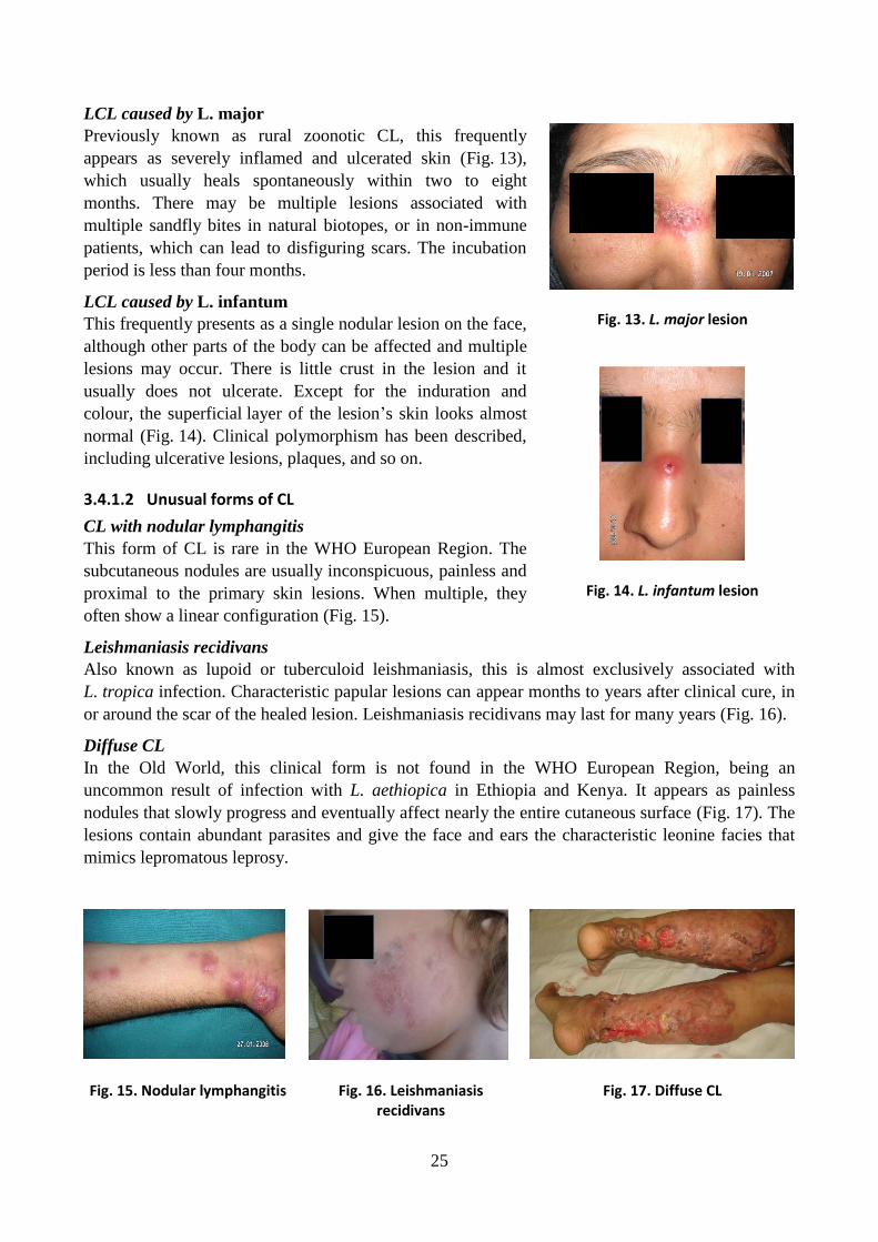
LCL caused by L. major
Previously known as rural zoonotic CL, this frequently
appears as severely inflamed and ulcerated skin (Fig. 13),
which usually heals spontaneously within two to eight
months. There may be multiple lesions associated with
multiple sandfly bites in natural biotopes, or in non-immune
patients, which can lead to disfiguring scars. The incubation
period is less than four months.
LCL caused by L. infantum
This frequently presents as a single nodular lesion on the face,
although other parts of the body can be affected and multiple
lesions may occur. There is little crust in the lesion and it
usually does not ulcerate. Except for the induration and
colour, the superficial layer of the lesion’s skin looks almost
normal (Fig. 14). Clinical polymorphism has been described,
including ulcerative lesions, plaques, and so on.
3.4.1.2 Unusual forms of CL
CL with nodular lymphangitis
This form of CL is rare in the WHO European Region. The
subcutaneous nodules are usually inconspicuous, painless and
proximal to the primary skin lesions. When multiple, they
often show a linear configuration (Fig. 15).
Leishmaniasis recidivans
Also known as lupoid or tuberculoid leishmaniasis, this is almost exclusively associated with
L. tropica infection. Characteristic papular lesions can appear months to years after clinical cure, in
or around the scar of the healed lesion. Leishmaniasis recidivans may last for many years (Fig. 16).
Diffuse CL
In the Old World, this clinical form is not found in the WHO European Region, being an
uncommon result of infection with L. aethiopica in Ethiopia and Kenya. It appears as painless
nodules that slowly progress and eventually affect nearly the entire cutaneous surface (Fig. 17). The
lesions contain abundant parasites and give the face and ears the characteristic leonine facies that
mimics lepromatous leprosy.
Fig. 15. Nodular lymphangitis Fig. 16. Leishmaniasis recidivans
Fig. 17. Diffuse CL
Fig. 13. L. major lesion
Fig. 14. L. infantum lesion

26
3.4.1.3 Mucocutaneous leishmaniasis (MCL)
This clinical form of tegumentary leishmaniasis indicates a condition in which, following a primary
ulcerative cutaneous lesion, parasites disseminate towards nasal and oropharyngeal mucosae. MCL
is not endemic in the WHO European Region and should not be confused with mucosal
leishmaniasis (ML), covered in the next section. MCL is more commonly seen in Bolivia, Brazil
and Peru, and is much less common in other countries of Latin America. Mucosal lesions may
appear concurrently with the primary skin ulcer, but more often they become evident several
months to many years after healing of the initial cutaneous lesion. Between 1% and 5% of
individuals infected with the Neotropical species L. braziliensis develop mucosal involvement.
Recent data suggest that the risk of developing MCL is higher when lesions are (i) infected with
L. braziliensis or L. panamensis (the condition is less commonly seen with L. guyanensis
infections); (ii) acquired in Bolivia; (iii) multiple (more than four) and large (greater than 4–6 cm2);
(iv) present for over four months; (v) localized above the waist; (vi) associated with acquired or
induced immunosuppression; and (vii) treated inappropriately.
The nasal mucosa is the first site involved. Initially, patients may
complain of nasal stuffiness, difficulty in breathing through their
nose and occasional bleeding, as their first symptoms are
associated with erythema and oedema of the involved nasal
mucosa. This proceeds to septum ulceration covered with a
mucopurulent exudate. In severe MCL there is often mutilating
destruction of the nasal septum (Fig. 18), palate, lips, pharynx and
larynx. The lesions are chronic and progressive, and death can be
caused by aspiration or inanition. There is currently no way to
predict the development of mucosal involvement in a person with a
primary CL ulcer. However, risk factors for the development of
MCL are male gender, older age, severe malnutrition and lesion
duration of over four months. Cases of imported MCL in the WHO
European Region, although underreported or poorly documented,
are increasing.
3.4.1.4 ML
ML is a condition in which localized Leishmania lesions in buccal, pharyngeal or laryngeal mucosa
occur without primary skin involvement. ML can be seen in immunocompetent individuals, in
individuals under local or systemic immunosuppressive therapies, or as a result of secondary
dissemination of VL in HIV/AIDS patients. L. infantum is the usual agent, although L. major and
L. tropica can also cause ML in elderly and immunocompromised patients.
3.4.2 Laboratory diagnosis
Presumptive diagnosis of CL is often made on the basis of clinical presentation in some highly
endemic areas, where clinical accuracy has been evaluated to be as high as 93%. A diagnostic
challenge arises when Leishmania lesions present to medical attention in low-endemic or in non-
endemic areas (for example, in travellers); when the clinical picture is distorted; or when atypical
variants are seen. Serological methods are of limited use because of low sensitivity and variable
specificity, so they are not recommended for laboratory diagnosis of CL.
Fig. 18. MCL
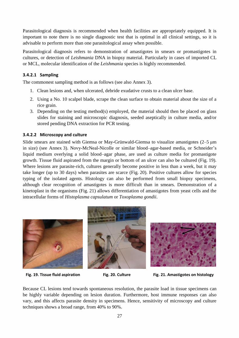
27
Parasitological diagnosis is recommended when health facilities are appropriately equipped. It is
important to note there is no single diagnostic test that is optimal in all clinical settings, so it is
advisable to perform more than one parasitological assay when possible.
Parasitological diagnosis refers to demonstration of amastigotes in smears or promastigotes in
cultures, or detection of Leishmania DNA in biopsy material. Particularly in cases of imported CL
or MCL, molecular identification of the Leishmania species is highly recommended.
3.4.2.1 Sampling
The commonest sampling method is as follows (see also Annex 3).
1. Clean lesions and, when ulcerated, debride exudative crusts to a clean ulcer base.
2. Using a No. 10 scalpel blade, scrape the clean surface to obtain material about the size of a
rice grain.
3. Depending on the testing method(s) employed, the material should then be placed on glass
slides for staining and microscopic diagnosis, seeded aseptically in culture media, and/or
stored pending DNA extraction for PCR testing.
3.4.2.2 Microscopy and culture
Slide smears are stained with Giemsa or May-Grünwald-Giemsa to visualize amastigotes (2–5 μm
in size) (see Annex 3). Novy-McNeal-Nicolle or similar blood–agar-based media, or Schneider’s
liquid medium overlying a solid blood–agar phase, are used as culture media for promastigote
growth. Tissue fluid aspirated from the margin or bottom of an ulcer can also be cultured (Fig. 19).
Where lesions are parasite-rich, cultures generally become positive in less than a week, but it may
take longer (up to 30 days) when parasites are scarce (Fig. 20). Positive cultures allow for species
typing of the isolated agents. Histology can also be performed from small biopsy specimens,
although clear recognition of amastigotes is more difficult than in smears. Demonstration of a
kinetoplast in the organisms (Fig. 21) allows differentiation of amastigotes from yeast cells and the
intracellular forms of Histoplasma capsulatum or Toxoplasma gondii.
Fig. 19. Tissue fluid aspiration Fig. 20. Culture Fig. 21. Amastigotes on histology
Because CL lesions tend towards spontaneous resolution, the parasite load in tissue specimens can
be highly variable depending on lesion duration. Furthermore, host immune responses can also
vary, and this affects parasite density in specimens. Hence, sensitivity of microscopy and culture
techniques shows a broad range, from 40% to 90%.

28
3.4.2.3 Molecular techniques
Both qualitative and quantitative PCR methods can be used. They are the most sensitive techniques
for CL diagnosis (96%). From positive specimens, DNA target sequences can be analysed by
restriction fragment length polymorphism or by direct sequencing to identify the leishmanial agent
at species level.
A patient is considered to be a confirmed CL case if at least one of the techniques – smear, culture,
PCR or histology – is positive. A compatible histology is defined as a histopathologic picture
compatible with leishmaniasis but with no direct demonstration of parasites.
3.4.3 Differential diagnosis
LCL must be differentiated from other skin conditions such as lupus vulgaris (Fig. 22), chronic
pyoderma, leprosy, tropical ulcer, deep mycosis as blastomycosis (Fig. 23), sarcoidosis (Fig. 24)
and different forms of skin cancer (Fig. 25 and 26). Subacute infections with gram-positive bacteria,
atypical mycobacteria and bacillary angiomatosis are reported as the most frequent differential
diagnoses in imported CL. To ensure correct diagnosis, a comprehensive study of the patient is
needed, including microbiological, histopathological and immunological investigation methods.
Fig. 22. Lupus vulgaris Fig. 23. Blastomycosis
Fig. 24. Sarcoidosis
Fig. 25. Squamous cell carcinoma Fig. 26. Basal cell carcinoma
3.4.4 Treatment
It is not possible, or achievable in the short term, to recommend a single treatment regimen that
would be safe and effective for all forms of CL from all geographical regions. For each patient, the
therapeutic decision should be based on the risk–benefit ratio and the causative Leishmania species.
In general, systemic treatment is less commonly employed in clinical forms from the Old World,
including the WHO European Region, as compared with the New World.
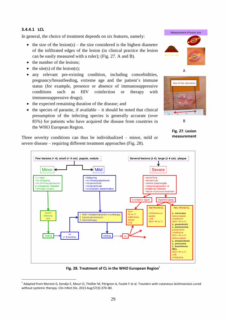
29
3.4.4.1 LCL
In general, the choice of treatment depends on six features, namely:
the size of the lesion(s) – the size considered is the highest diameter
of the infiltrated edges of the lesion (in clinical practice the lesion
can be easily measured with a ruler); (Fig. 27. A and B).
the number of the lesions;
the site(s) of the lesion(s);
any relevant pre-existing condition, including comorbidities,
pregnancy/breastfeeding, extreme age and the patient’s immune
status (for example, presence or absence of immunosuppressive
conditions such as HIV coinfection or therapy with
immunosuppressive drugs);
the expected remaining duration of the disease; and
the species of parasite, if available – it should be noted that clinical
presumption of the infecting species is generally accurate (over
85%) for patients who have acquired the disease from countries in
the WHO European Region.
Three severity conditions can thus be individualized – minor, mild or
severe disease – requiring different treatment approaches (Fig. 28).
Fig. 28. Treatment of CL in the WHO European Region1
1 Adapted from Morizot G, Kendjo E, Mouri O, Thellier M, Pérignon A, Foulet F et al. Travelers with cutaneous leishmaniasis cured
without systemic therapy. Clin Infect Dis. 2013 Aug;57(3):370–80.
A
B
Fig. 27. Lesion measurement
Measurement of lesion size
Size of the ulceration
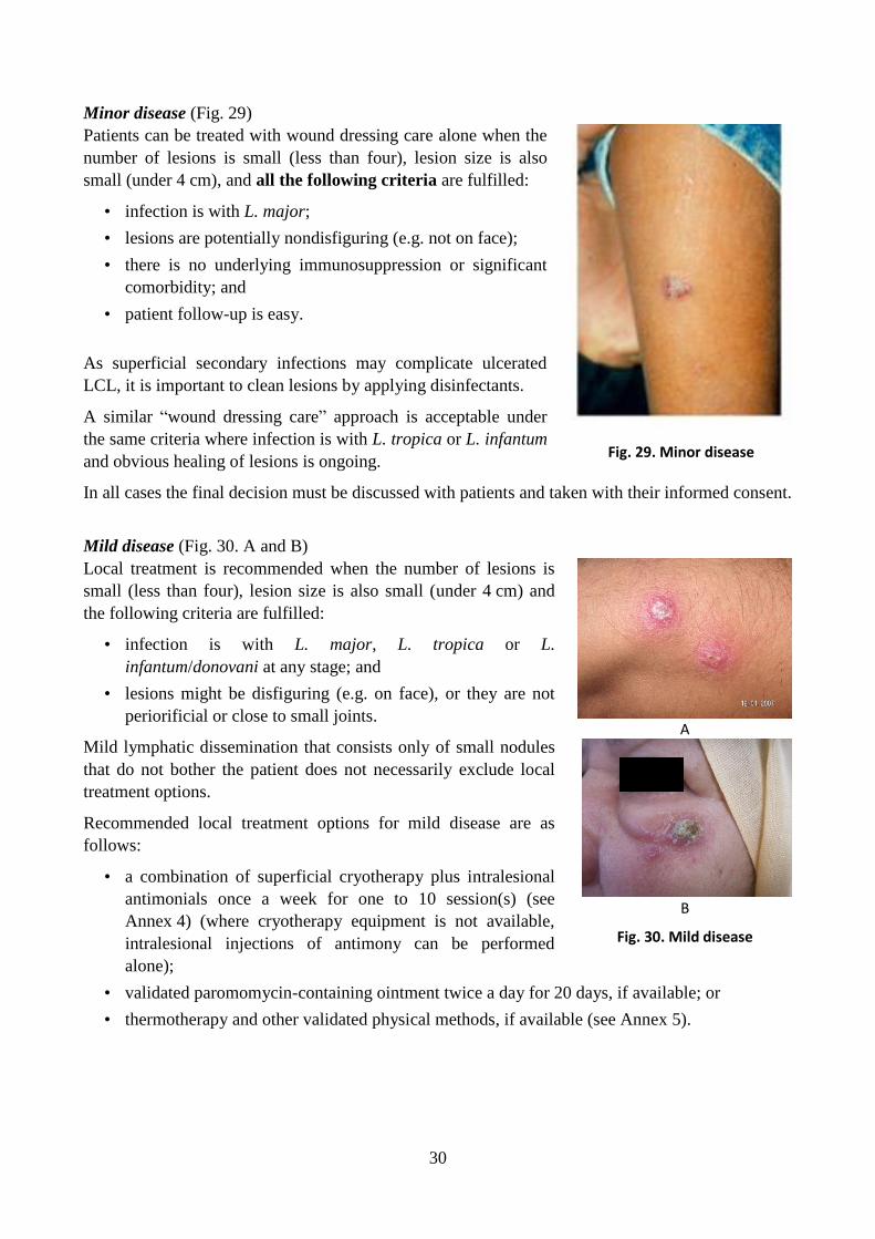
30
Minor disease (Fig. 29)
Patients can be treated with wound dressing care alone when the
number of lesions is small (less than four), lesion size is also
small (under 4 cm), and all the following criteria are fulfilled:
• infection is with L. major;
• lesions are potentially nondisfiguring (e.g. not on face);
• there is no underlying immunosuppression or significant
comorbidity; and
• patient follow-up is easy.
As superficial secondary infections may complicate ulcerated
LCL, it is important to clean lesions by applying disinfectants.
A similar “wound dressing care” approach is acceptable under
the same criteria where infection is with L. tropica or L. infantum
and obvious healing of lesions is ongoing.
In all cases the final decision must be discussed with patients and taken with their informed consent.
Mild disease (Fig. 30. A and B)
Local treatment is recommended when the number of lesions is
small (less than four), lesion size is also small (under 4 cm) and
the following criteria are fulfilled:
• infection is with L. major, L. tropica or L.
infantum/donovani at any stage; and
• lesions might be disfiguring (e.g. on face), or they are not
periorificial or close to small joints.
Mild lymphatic dissemination that consists only of small nodules
that do not bother the patient does not necessarily exclude local
treatment options.
Recommended local treatment options for mild disease are as
follows:
• a combination of superficial cryotherapy plus intralesional
antimonials once a week for one to 10 session(s) (see
Annex 4) (where cryotherapy equipment is not available,
intralesional injections of antimony can be performed
alone);
• validated paromomycin-containing ointment twice a day for 20 days, if available; or
• thermotherapy and other validated physical methods, if available (see Annex 5).
Fig. 29. Minor disease
A
B
Fig. 30. Mild disease

31
Severe disease (Fig. 31. A–D)
Systemic treatment is recommended when at
least one of the following criteria is fulfilled:
• lesion(s) size is over 4 cm (e.g. a plaque);
• the number of lesions is over four;
• lesions are periorificial or close to small
joints;
• clinically significant lymphatic
dissemination is present;
• there is no significant comorbidity for
which systemic treatments could be
contraindicated in a benefit–risk
assessment (e.g. unbalanced diabetes,
malnutrition, and so on);
• underlying immunosuppressive conditions
are present; or
• treatment approaches adopted in previous
situations have failed.
Recommended systemic treatment options for severe disease that can be adopted depending on drug
availability, benefit–risk assessment, and appropriate clinical and biological patient monitoring are
as follows:
• pentavalent antimonials: 20 mg Sb5+/kg IM or IV for 10–20 days (see Annex 6);
• miltefosine: orally, 50 mg three times a day for 28 days (female patients must use effective
contraception during treatment and for three months thereafter);
• fluconazole if suspected/confirmed L. major: orally, 200 to 400 mg/day for six weeks;
• itraconazole if suspected/confirmed L. tropica: orally, 200 to 400 mg/day for six weeks; or
• LAB: 3mg/kg/day IV on days 1–5 and at day 10; total dose 18 mg/kg.
3.5 Case management of CL in special categories of patients
3.5.1 Children
In general, the above guidelines on clinical and parasitological diagnosis and treatment of CL also
apply to children. Intralesional MA and cryotherapy are widely used. Thermotherapy can be used in
children over 5 years of age. Fluconazole has been used in children at a dose of 3 mg/kg/day
(50 mg/day), for six to eight weeks. For systemic treatment of children under 18 months, MA is not
recommended.
3.5.2 Pregnancy
CL manifestations during pregnancy are characterized by larger lesions with a highly atypical and
severe appearance and may increase the risk of fetal complications. Cryotherapy is probably the
best alternative therapy in pregnancy. Miltefosine has exhibited teratogenicity and should therefore
A B
C D
Fig. 31. Severe disease

32
not be administered to pregnant women. In situations where the location, number and size of
lesions, and their persistence in spite of local therapy, require systemic treatment, LAB treatment
probably has the best benefit–risk ratio.
3.5.3 Breastfeeding
Cryotherapy is probably the best alternative for breastfeeding women. However, intralesional or
systemic MA is compatible with breastfeeding. The Sb5+
concentration in breast milk is low
(3.5 mg Sb5+
/ml) and has little effect on the newborn.
3.6 Imported cases of CL and MCL
Imported leishmaniasis cases in endemic and non-endemic countries of the WHO European Region
are becoming more frequent as a result of increased travel and migration. Treatment for individuals
found to be infected by parasites and clinical forms that are not endemic to the Region can be
challenging. Drug options for imported CL/MCL cases found infected with Old and New World
parasites non-endemic to the WHO European Region are listed in Annex 7.

33
4. Surveillance
4.1 General purpose and components
This manual is intended to help countries to design or improve their health information system for
monitoring the burden and trends of leishmaniasis, and for evaluating the effectiveness of control
measures. The aim of this manual is to help countries:
• to determine the burden and distribution of leishmaniasis;
• to observe changes in the trend of the disease;
• to detect outbreaks early and respond accordingly;
• to establish appropriate prevention measures, control planning and resource allocation; and
• to evaluate the effectiveness of strategies and interventions in control programmes.
The components of a surveillance system include:
• health facilities such as clinics and hospitals – public, private or set up by nongovernmental
organizations (NGOs) – capable of collecting primary data, namely, the number of diagnosed
cases based on established case definitions;
• structured reporting forms, paper-based or in electronic format;
• officially mandated routes and frequency of reporting, collation, validation and monitoring;
• defined triggers for public health action – for example, the occurrence of an unexpected
number of cases within a geographically or otherwise defined catchment area that triggers an
investigation and appropriate response;
• continuous monitoring and evaluation of the surveillance system for timeliness and
effectiveness; and
• regular analysis of collated data for trends over time, space and population.
Because of their diverse and dynamic nature, the leishmaniases should be compulsorily notifiable in
countries where the disease is endemic – where autochthonous transmission has been proven; and
where the burden of imported disease represents a challenging medical and epidemiological issue –
for example, where case management is difficult or receptive vectors are present. When disease
notification is made compulsory and a well-structured reporting system is in place, surveillance is
likely to be far more efficient than when data collection and reporting are on a voluntary basis. Even
so, surveillance is unlikely to capture all cases, as can be seen in countries of the WHO European
Region where leishmaniasis notification is mandatory. For a correct interpretation of trends in
reported cases, therefore, it is essential to keep track of the extent of underreporting.
When a surveillance system functions well, the information it generates should lead to meaningful
action of various kinds. Surveillance data can help health authorities:
• to evaluate the real extent of the problem and the populations principally at risk;
• to improve the clinical management and follow-up of patients, based on appropriate
indicators; and
• to propose implementation of active case detection (ACD) where and when necessary.
Surveillance data can also help to identify and solve technical and operational difficulties faced by
the disease control programme and to facilitate evaluation of the impact of interventions.

34
4.2 Recommended case and treatment outcome definitions for epidemiological surveillance
Case and treatment outcome definitions are based on epidemiological, clinical and diagnostic
criteria, which vary according to the clinical forms of leishmaniasis.
4.2.1 VL
Case definitions include:
suspect case – a person living in or having travelled to endemic areas who is suffering from fever
lasting more than one week and showing clinical signs of VL; there may, as well or instead, be
high laboratory-based suspicion of disease (see section 3.1.2).
confirmed case – a person living in or having travelled to endemic areas who shows clinical signs
of VL, with serological and/or parasitological confirmation of disease (see section 3.1.3).
Definitions of treatment outcomes at initial assessment, determined at two to four weeks after start
of treatment, include:
initial cure – the drug course has been completed and the patient has improved clinically; clinical
criteria for initial cure are defined as absence of fever, splenomegaly regression, return of
appetite and/or gain in body weight (see section 3.1.7.1).
probable nonresponse – signs and symptoms persist or recur during treatment without
parasitological confirmation.
confirmed nonresponse – signs and symptoms persist or recur during treatment with
parasitological confirmation.
death – any death, whether or not related to VL.
default – the patient does not complete treatment.
lost to initial follow-up – the patient does not present for assessment after completion of treatment.
Definitions of treatment outcomes at final assessment, determined at six months after treatment
completion, include:
final cure – a patient who, following initial cure, remains symptom-free at six months after the end
of treatment.
relapse – a patient who experiences recurrence of VL symptoms with parasitological confirmation
at any time after initial cure.
death – any death, whether or not related to VL.
lost to follow-up – the patient does not present for assessment at six months.
4.2.2 CL
Case definitions include:
suspect case – a person living in or having travelled to endemic areas who shows clinical signs of
CL, without parasitological confirmation (see section 3.4.1).

35
confirmed case – a person living in or having travelled to endemic areas who shows clinical signs
of CL, with parasitological confirmation (see section 3.4.2).
Definitions of treatment outcomes include:
cure – flattening of lesions and complete re-epithelialization of ulcers at three months from start of
treatment.
nonresponse – no clinical improvement (no change or worsening) of the lesions after one month of
treatment, or no cure at three months from start of treatment.
probable relapse – reappearance of lesion(s) after cure.
confirmed relapse – reappearance of lesion(s) after cure with parasitological confirmation.
4.3 Leishmaniasis case detection strategies
Case detection can be passive, when patients seek care at health facilities on their own initiative; or
active, when the control programme reaches out to the community to actively screen and find cases.
Different possible approaches to case detection in VL and CL are described below.
4.3.1 Passive case detection (PCD)
PCD is triggered when patients seek care for their illness from clinicians working in health
facilities. Clinicians who manage a case should notify it to the appropriate epidemiological sur-
veillance system.
PCD relates to cases of VL, CL and Leishmania–HIV coinfection that are detected in public
hospitals, private medical services and health facilities run by NGOs. This method does not require
many additional efforts or resources, as epidemiological surveillance is already part of the existing
health system. The reporting formats should, as far as possible, be integrated in existing systems,
although they should include indicators specific to leishmaniasis case management.
4.3.2 ACD
ACD involves health staff actively reaching out to the community and systematically screening the
population to find cases of leishmaniasis. In general, earlier diagnosis and treatment will improve
treatment outcomes for patients. For anthroponotic CL caused by L. tropica, particularly in urban
settings, ACD helps to reduce disease transmission by shortening the infectious period of patients.
Among several ACD approaches that have been validated for their utility in leishmaniasis detection
worldwide, three are relevant to the WHO European Region.
House-to-house approach
This approach is recommended when PCD data indicate that a leishmaniasis outbreak is possible.
This is rare in the case of zoonotic VL (although there was a recent L. infantum VL/CL outbreak in
the Madrid area of Spain), whereas outbreaks of anthroponotic or zoonotic CL may be not
uncommon in the WHO European Region. This approach works by conducting house-to-house
visits of the entire at-risk community to detect leishmaniasis cases. The visits are made by health
professionals trained to record skin conditions suggestive of CL or systemic signs suggestive of VL.
In general, suspect cases should be referred to a health centre for diagnosis confirmation and

36
treatment. Where there is strong suspicion of VL, however, serological diagnosis on site is
recommended, using a rapid assay such as the rK39 RDT, which is highly specific and sensitive in
patients suffering from symptomatic VL caused by L. infantum (see section 3.1.4). The house-to-
house approach is considered the gold standard of ACD, but the high cost of this method is a
limitation.
Camp approach
This approach may not be appropriate in most territories of the WHO European Region, where
basic health facilities are widespread. In some rural and remote settings, however, and in recent
refugee camp situations, the camp approach has proved useful – for example, in CL outbreaks in
which care-seeking by the affected population was limited because of the benign nature of the
disease. This approach involves the organization of health camps, set up in a basic health facility or
in public buildings such as schools, in defined communities on fixed dates. Mobile teams of
medical staff, nurses and laboratory technicians carry out screening for disease. Before the camp
takes place, it is essential that the population is duly informed about the team composition, its
purpose, and the time, date and place of the team’s activities.
Index case approach
This approach is useful when attempting to control or eliminate anthroponotic CL in a rural
territory; it is the preferred ACD method in such circumstances because households are scattered. It
is also employed when an imported CL case has been diagnosed and measures are taken to prevent
the disease spreading in receptive non-endemic areas. The approach involves an active search for
new cases among household members through house-to-house visits in the vicinity (such as in a
range of 50–300 m) of a house where a CL case has recently been detected by PCD. In elimination
programmes, the index case approach must be organized on a permanent basis throughout the year.
4.4 Strategies for the detection of leishmaniasis in the canine reservoir
Dogs are the main reservoir hosts of VL throughout the WHO European Region, but also have the
role of family members in many communities. Following the principles of the One Health concept,
leishmaniasis surveillance should include canine leishmaniasis for several reasons.
• The detection of L. infantum infection or disease in domestic dogs has great epidemiological
value, as they represent the most suitable sentinel species for identification of potential areas
of VL transmission. For example, occurrence of human VL cases in newly endemic areas of
northern Italy and Spain was predicted on the basis of earlier detection in infected dogs.
• Early detection of infected dogs, followed by various public health measures (which depend
on the social context of affected countries), allows the infectious burden carried by these hosts
to be reduced.
• Periodic monitoring of leishmaniasis prevalence in dogs is important in evaluating measures
for control of VL.
• Veterinary drugs for leishmaniasis treatment in pets are available in western Europe and used
following widely available veterinary guidelines. Early treatment of infected dogs contributes
to the prevention of infection transmission to humans.
Cooperation and/or integration of public health activities with veterinary services are fundamental,
and such arrangements are currently operating in several countries of the WHO European Region.

37
Strategies are similar to those explained above for human case detection. They are usually planned
in areas where human VL cases are recurrent or outbreaks have been detected.
PCD is performed at private veterinary clinics when owners seek care for their pets. Veterinarians,
who manage cases with parasitological and/or serological confirmation, should report to the local
public veterinary service.
ACD is mainly performed through a camp approach and the preferred screening method is by
serology. The most suitable period for ACD is the winter months, which takes into account the long
serological incubation period that follows Leishmania transmission in the warm season. Sampling
for canine leishmaniasis is done by mobile teams of veterinarians, laboratory technicians and health
workers (Fig. 32). Dog owners should be informed about the purpose, time, date and place of the
team’s activities.
Fig. 32. A camp operated by a veterinary team in a village endemic for VL (top left) and dogs with clinical signs of leishmaniasis
4.5 Recommended types of surveillance
4.5.1 VL
In the WHO European Region, VL is usually sporadic in nature, and although it is a deadly disease,
human patients suffering from VL are dead-end hosts – they are not a source of infection. There are
no universally agreed threshold values of VL incidence that suggest implementation of a mandatory
notification system at country level. However, when VL cases are recurrent or outbreaks have been

38
reported, a surveillance system with compulsory notification of individual VL cases is recom-
mended. At peripheral level, individual data collection on VL patients is needed for case
management and case notification. Health workers should be aware of the standard case definition
for VL adopted by the national surveillance system, and should report their case data to the
intermediate or national level as appropriate. Routine periodic reporting (at least monthly) of
aggregated VL data to the central level is essential. During outbreaks, weekly reporting from the
periphery to intermediate and national levels is recommended in order to coordinate a timely and
efficient response. The central level will report aggregated VL data annually to WHO. It is
recommended that VL outbreaks are reported immediately to WHO, as it can assist with outbreak
response.
In countries where the incidence of VL is too low for inclusion in the list of notifiable diseases,
sentinel surveillance or periodic surveys provide alternative means to estimate its burden.
Minimal individual data required for a VL case record are: clinical features, HIV status, date of
diagnosis, type of diagnosis (parasitological/immunological), treatment completion, and treatment
outcome.
Identification data should include a single identifier (for example, a citizenship ID such as social
security number, driving licence or tax reference number, or a specifically generated patient
number), age, sex, usual place of residence, probable location(s) of exposure to transmission, and
travel history.
Additional individual information includes pregnancy, comorbidities, presence of other VL cases
in the household, and presence of canine leishmaniasis cases in the vicinity of the place of resid-
ence.
Minimal aggregated data for reporting include number of cases by age, sex and month/year.
Analysis and presentation of data should also include number of cases and incidence rate by
geographical area, and treatment outcome.
4.5.2 CL
In the WHO European Region, epidemiological entities of CL are largely diverse, as human patients
can be dead-end hosts (in the case of zoonotic CL caused by L. major, L. tropica and L. infantum)
or the main reservoir host of infection (in the case of anthroponotic CL caused by L. tropica). CL
should be included in the list of notifiable diseases at country level. All probable cases of CL, as
well as confirmed cases, should be reported individually.
At peripheral level, individual data collection on CL patients is needed for case management and
case notification. Health workers should be aware of the standard case definition for CL adopted by
the national surveillance system, and should report their case data to the intermediate or national
level as appropriate. Routine periodic reporting (at least quarterly) of aggregated data on CL cases
to the central level is essential. The central level will report aggregated CL data annually to WHO.
In countries where prevalence of CL is too low for inclusion in the list of notifiable diseases,
sentinel surveillance or periodic surveys provide alternative means to estimate its burden.
Minimal individual data required for a CL case record are: clinical features, date of diagnosis,
type of diagnosis (parasitological/molecular), treatment completion, and treatment outcome.
Leishmania species identification would be desirable, as this can discriminate between

39
anthroponotic and zoonotic entities of CL; it is most important in the case of imported CL from the
New World, but this is not always achievable.
Identification data should include a single identifier (for example, a citizenship ID such as social
security number, driving licence or tax reference number, or a specifically generated patient
number), age, sex, usual place of residence, probable location(s) of exposure to transmission, and
travel history.
Additional individual information includes presence of other CL cases in the household.
Minimal aggregated data for reporting include number of cases by age, sex and month/year.
Analysis and presentation of data should also include the number of cases and incidence rate by
geographical area, and treatment outcome.
4.5.3 Leishmania–HIV coinfection
In all areas where VL and HIV are coendemic, WHO recommends provider-initiated counselling
and HIV testing to be carried out on all VL patients to screen for HIV coinfection. Coendemic
countries should establish sentinel surveillance sites to collect detailed information on socio-
demographic characteristics, risk factors and other variables to detect and monitor trends and
changes in risk factors and the overall burden. HIV testing is beneficial to coinfected patients as
HIV treatment can be started early and improves the overall response to VL treatment. Furthermore,
secondary chemoprophylaxis can be established after the initial treatment course has ended, which
significantly reduces the relapse rate.
During the 1990s, WHO coordinated a dedicated network for surveillance of Leishmania–HIV co-
infection. Worldwide information was collected, processed and disseminated by a central registry
set up at WHO. Based on the lessons learned at that time, we recommend that specific surveillance
is set up at country level. Hospitals and laboratories should maintain individual patient records and
use the WHO guidelines for diagnosis and a standardized case report form for HIV-coinfected
patients.
Minimal individual data required for a Leishmania–HIV coinfection case record are: clinical
features, date and type of leishmaniasis diagnosis, type of episode (new infection/relapse), and
chemoprophylaxis. HIV data include date of diagnosis, CD4/mm³, risk group, AIDS-defining
diseases, and treatment outcome.
Identification data should include a single identifier (for example, a citizenship ID such as social
security number, driving licence or tax reference number, or a specifically generated patient
number), age, sex, usual place of residence, probable location(s) of exposure to Leishmania
transmission, and travel history.
Minimal aggregated data for reporting include number of cases by age, sex, month/year, and risk
group.
Analysis and presentation of data should focus on geographic distribution, sex and age
distribution, risk groups, dates of HIV and leishmaniasis diagnosis, immunological parameters, and
AIDS-defining diseases.

40
5. Monitoring and evaluation of leishmaniasis control
5.1 Leishmaniasis control strategies
Prevention and control of leishmaniasis in the WHO European Region require a combination of
intervention strategies because transmission occurs in a complex biological system that involves the
human host, parasite, phlebotomine vector and animal reservoir host. Key strategies are
summarized below.1
Early diagnosis and effective case management reduce the prevalence of the disease and prevent
disabilities and death. There are now highly effective and safe antileishmanial medicines,
particularly for VL, and access to these medicines has significantly improved. The therapeutic
options for CL are more limited and clinical research is badly needed in this area.
The aim of vector control is to reduce the transmission of the parasite by reducing vector density or
by decreasing the contact rate between vector and human host. Sandfly control depends on the
behaviour of the target vector, which may be endophilic, peridomestic or both. Strictly endophilic
and peridomestic species can be targeted by indoor residual spraying with insecticides. When
peridomestic or exophilic sandfly species are involved, the outer surfaces of domestic animal
shelters and structures close to such dwellings, which are potential resting sites, must be sprayed.
Different classes of insecticides can be used for this purpose, although the spectrum of
susceptibility of sandflies to insecticides is not completely known. Insecticide-impregnated bednets
are a relatively inexpensive and sustainable method of reducing contact between infectious sandflies
and humans, and they have proved effective in protecting against CL. Covering CL skin lesions is
another way of reducing sandfly–Leishmania contact, and is especially important in the case of
long-lasting lesions such as those caused by L. tropica. Sleeping behaviour is critical to the
effectiveness of insecticide-impregnated bednets, as is people’s readiness to use them. Use of vector
control measures, singly or in combination, should be guided by consideration of the extent to
which they are technically feasible, operationally applicable, cost-effective and sustainable.
Control of animal reservoir is fundamental in zoonotic leishmaniases, as control strategies can be
directed at these hosts. For zoonotic VL, culling of infected pets, considered unethical by many
communities, is a controversial strategy, not least because culled dogs are rapidly replaced by
young susceptible ones. Control measures include use of topical insecticides to prevent sandfly bites
in dogs, and treatment of infected dogs using veterinary drugs or drugs not employed in human VL
therapy (for example, allopurinol). In areas where CL is caused by L. major, reduction of the
density of rodents by physical destruction of burrows, followed by planting, has proved to be an
effective and sustainable method of controlling CL where Psammomys obesus is the known
reservoir. Poisoning with wheat grains and vegetable oil is effective for other migrant rodents such
as Meriones.
Environmental management may result in a reduction in sandfly–human contact or sandfly
populations. This strategy may include relocation of human settlements away from sandfly habitats
and physical modification of their habitats. Environmental management measures should be
1 For a full discussion of control strategies, see Control of the leishmaniases. Report of a meeting of the WHO Expert Committee on
the Control of Leishmaniases, Geneva, 22–26 March 2010. Geneva: World Health Organization; 2010 (WHO Technical Report Series, No. 949).

41
preceded by careful studies of local ecology and environmental impact. Physical modification of
Phlebotomus papatasi breeding and resting sites by destruction of rodent burrows was used
successfully in the republics of central Asia. Actual or potential sandfly breeding sites, such as
rubble and rubbish disposal sites, can be eliminated in sanitation programmes involving the local
community, especially in urban areas. It is important that any modification of vector habitats takes
into account environmental conservation and does not create local ecological conflicts.
Operational research, with assistance from research institutions, is needed to define the actual
burden of leishmaniasis and the population at risk based on eco-epidemiological data. Ideally, this
should be carried out at local level, rather than using data at administrative division level, which –
given the focal nature of the disease – is often much broader than the precise endemic area.
Operational research should use data collected through routine surveillance that allows
identification of descriptive epidemiological aspects of the disease (person, place and time) and
seasonality patterns to accurately identify where and when transmission occurs and how control
strategies should be focused. In addition to research addressing the efficacy and effectiveness of
applied control measures, evaluation of measures for vector and reservoir control, surveillance tools
and methods is recommended. A better understanding of the barriers facing target populations will
be important in improving access to all patients.
Participation and health education of communities, and their partnerships with the formal and
informal health sectors, are crucial in empowering them in their own health development.
Leishmaniasis prevention must go hand in hand with community participation. Unless individuals
in communities see the merits of disease control and prevention, even the best-designed preventive
strategies will not succeed. There is a desperate need to understand how a local community
perceives the disease and why it is important for them, and what existing patterns of behaviour may
help or hinder preventive measures. Both the community and patients should be educated about
leishmaniasis and its control and prevention, and have access to adequate health care facilities.
Existing practices to detect, diagnose and treat leishmaniasis should be improved by developing and
disseminating clear messages about the disease and its diagnosis and treatment.
Intersectoral collaboration is best developed through a shared understanding of the underlying
problems to be addressed. The Ministry of Health should encourage non-health sectors, including
national and local veterinary services and the environmental and agricultural sectors, to actively
collaborate in leishmaniasis control at central, as well as intermediate and local, level.
Capacity-building is a key component of any programme dealing with leishmaniasis. Health staff,
including physicians, entomologists, parasitologists, veterinarians and others engaged in
leishmaniasis control, need special training to become familiar with the epidemiological and
biological characteristics of the diseases, risk factors, diagnosis and treatment, and preventive and
control measures. Laboratory staff require training in the use of various diagnostic methods,
including parasite isolation by culture, and molecular biology tools.
Cross-border cooperation is becoming more and more important in tackling the problem of
leishmaniasis moving between countries through migration, labour and tourism. Exotic Leishmania
species may become established by participating in unexpected life cycles with endemic sandfly
and wild animal populations – with outcomes that are impossible to predict.

42
5.2 Indicators for monitoring and evaluation of leishmaniasis control
Monitoring involves routine tracking of programme performance by record-keeping, regular
reporting, surveillance and periodic surveys. The objectives of monitoring are to verify the progress
or status of implementation; to ensure accountability; to detect problems and constraints; to promote
evidence-based planning; and to provide timely feedback so that adjustments can be made as
needed.
Monitoring indicators include input, process and output indicators. Common indicators are those
related to epidemiological data, diagnosis performance, and treatment outcome.
Evaluation involves periodic assessment of changes in targeted outcomes or impact that can be
attributed to a programme. The objectives of evaluation are to relate a particular outcome or impact
directly to a particular intervention after a certain time; to determine the value or worth of a
particular project or programme; to link any two parts of the monitoring and evaluation framework;
to measure the effectiveness of the programme; and to provide reliable information on progress in
controlling leishmaniasis that can be used at local, national or international level.
Countries should evaluate their leishmaniasis control programmes regularly to assess their
compliance with the original objectives and targets. Parameters should be set for all key elements of
the programme – not only for case detection and treatment, but also for vector and reservoir
management and other strategies if relevant. Information on programme coverage, diagnosis
performances, rates of morbidity and mortality, therapeutic responses to drugs, insecticide
effectiveness and detection of new transmission foci is particularly important. Data for these
indicators are usually collected through the national epidemiological surveillance system (or at
sentinel sites) and health management information systems.
The main objectives of monitoring and evaluating leishmaniasis control programmes are:
• to collect, process, analyse and report or disseminate information relevant to leishmaniasis
and its control;
• to verify that activities have been implemented as planned so as to ensure quality and
accountability and to address problems in a timely manner;
• to improve community participation and community-based monitoring;
• to provide feedback to relevant authorities and the community to improve future planning;
and
• to document whether planned strategies have achieved expected outcomes and impact.
Indicators for monitoring a control programme should be SMART: specific, measurable, action-
oriented, relevant and time-bound. There is no point in calculating a large number of indicators if no
action follows.
5.2.1 Indicators used in VL control
In general, control programmes should primarily monitor epidemiological data such as:
43
• incidence rate of VL: number of new cases of VL per geographical unit and population size
detected in a given time unit (month and year) by gender and age group;1
• record of new VL foci, that is, the number of geographical units reporting cases for the first
time;
• HIV–VL coinfection rates, separately as new cases or relapses, calculated by the same
parameters as above; and
• HIV prevalence rate in VL patients screened for HIV.
Diagnosis performance in VL suspected cases should be monitored through the following
indicators:
• number of individuals passively or actively screened for VL;
• number of VL cases diagnosed by serology (for example, rK39 RTD), direct parasitology
(slide smears, culture, PCR), or clinical signs and symptoms; and
• proportion of positive diagnoses by each of the above methods out of the total number of
examinations performed.
Treatment outcome in VL patients should be monitored by the following indicators:
• initial cure rate
• failure rate
• case fatality rate.
These are calculated as the number of patients fulfilling the respective features reported in section
4.2 over the total patients treated with each drug included in the programme. The routine
monitoring of treatment outcomes in VL may be complex, as it requires an assessment six months
after the last day of treatment.
Additionally, to assess the coverage and quality of the programme, the following human indicators
are useful:
• patient delay – length of time between onset of symptoms and presentation at the health
centre;
• time to diagnosis – length of time between presentation at the health centre and diagnosis;
• time to treatment (patient and doctor delay) – length of time between onset of disease and
start of treatment;
• time from diagnosis to treatment – length of time between diagnosis and administration of
first treatment; and
• percentage of serious adverse events (SAE) cases in patients treated with each drug included
in the programme.
1 Examples of such geographical units are the territorial units for statistics developed by the European Union. Note that the number
of VL relapses should also be part of incidence data but they must be recorded separately.

44
This information may not be easy to collect, however, unless specific mention is made on the case
record forms; ad hoc operational research may be needed to collect these data.
In the context of zoonotic VL control, periodic (yearly) monitoring should include:
• leishmaniasis prevalence in dogs.
5.2.2 Indicators for monitoring CL control
Control programmes should primarily monitor epidemiological data such as:
• incidence rate of new CL cases calculated by the same geographical, temporal and population
parameters as VL, but separately for the main CL entities (for example, zoonotic and
anthroponotic CL) where they coexist;1 and
• recording of new CL foci, that is, the number of geographical units reporting cases for the
first time.
Diagnosis performance in CL suspected cases should be monitored through the following
indicators:
• number of individuals passively or actively screened for CL;
• number of CL cases diagnosed by direct parasitology (slide smears, culture, PCR) or by
clinical signs; and
• proportion of positive diagnoses by each of the above methods out of the total examinations
performed.
Treatment outcome in CL patients should be monitored by the following indicator:
• failure rate, calculated by the number of nonresponding or relapsing patients, over the total
number of patients treated with each therapy method included in the programme.
The following indicators are useful but information may not be easy to collect:
• time to diagnosis – length of time between presentation at the health centre and diagnosis;
• time to treatment (patient and doctor delay) – length of time between onset of disease and
start of treatment;
• time from diagnosis to treatment – length of time between diagnosis and administration of
first treatment; and
• percentage of SAE cases in patients treated with systemic drugs included in the programme.
1 The number of CL relapses should also be part of incidence data but they must be recorded separately.

45
Bibliography
Alvar J, Aparicio P, Aseffa A et al. The relationship between leishmaniasis and AIDS: the second
10 years. Clin Microbiol Rev 2008;21:334.
Alvar J, Velez ID, Bern C, Herrero M, Desjeux P, Cano J et al. Leishmaniasis worldwide and
global estimates of its incidence. PLoS One 2012;7:e35671.
Antoniou M, Gramiccia M, Molina R, Dvorak V, Volf P. The role of indigenous phlebotomine
sandflies and mammals in the spreading of leishmaniasis agents in the Mediterranean region.
Eurosurveill 2013;18:20540.
Arce A, Estirado A, Ordobas M, Sevilla S, García N, Moratilla L et al. Re-emergence of
leishmaniasis in Spain: community outbreak in Madrid, Spain, 2009 to 2012. Eurosurveill
2013;18:20546.
Aronson N, Herwaldt BL, Libman M, Pearson R, Lopez-Velez R, Weina P et al. Diagnosis and
treatment of leishmaniasis: clinical practice guidelines by the Infectious Diseases Society of
America (IDSA) and the American Society of Tropical Medicine and Hygiene (ASTMH). Clin
Infect Dis 2016;63:1539–57.
Bailey MS, Green AD, Ellis CJ, O’Dempsey TJ, Beeching NJ, Lockwood DN et al. Clinical
guidelines for the management of cutaneous leishmaniasis in British military personnel. J R Army
Med Corps 2005;151:73–80.
Ben Salah A, Ben Messaoud N, Guedri E, Zaatour A, Ben Alaya N, Bettaieb J et al. Topical
paromomycin with or without gentamicin for cutaneous leishmaniasis. N Engl J Med
2013;368:524–32.
Blum J, Buffet P, Visser L, Harms G, Bailey MS, Caumes E et al. LeishMan recommendations for
treatment of cutaneous and mucosal leishmaniasis in travelers. J Travel Med 2014;21:116–29.
Blum J, Lockwood DNJ, Visser L, Harms G, Bailey MS, Caumes E et al. Local or systemic
treatment for New World cutaneous leishmaniasis? Re-evaluating the evidence for the risk of
mucosal leishmaniasis. Int Health 2012;4:153–63.
Boecken G, Sunderkotter C, Bogdan C, Weitzel T, Fischer M, Müller A et al. [Diagnosis and
therapy of cutaneous and mucocutaneous leishmaniasis in Germany]. J Dtsch Dermatol Ges
2011;9(Suppl 8):1–51.
Buffet PA, Rosenthal E, Gangneux JP, Lightburne E, Couppié P, Morizot G et al. [Therapy of
leishmaniasis in France: consensus on proposed guidelines]. Presse Med 2011;40:173–84.
Castro A, Carrillo E, San Martín JV, Botana L, Molina L, Matía B et al. Lymphoproliferative
response after stimulation with soluble leishmania antigen (SLA) as a predictor of visceral
leishmaniasis (VL) relapse in HIV+ patients. Acta Trop 2016;164:345–51. PMID:27693332.
Chappuis F, Rijal S, Soto A, Menten J, Boelaert M. A meta-analysis of the diagnostic performance
of the direct agglutination test and rK39 dipstick for visceral leishmaniasis. BMJ 2006;333:723.
Copeland NK, Aronson NE. Leishmaniasis: treatment updates and clinical practice guidelines
review. Curr Opin Infect Dis 2015;28:426–37.

46
Cota GF, de Sousa MR, Fereguetti TO, Rabello A. Efficacy of anti-leishmania therapy in visceral
leishmaniasis among HIV infected patients: a systematic review with indirect comparison. PLoS
Negl Trop Dis 2013;7:e2195.
Davidson RN, Di Martino L, Gradoni L, Giacchino R, Russo R, Gaeta GB et al. Liposomal
amphotericin B (AmBisome) in Mediterranean visceral leishmaniasis: a multicentre trial. Q J Med
1994;87:75–81.
Dujardin JC, Campino L, Cañavate C, Dedet JP, Gradoni L, Soteriadou K et al. Spread of vector-
borne diseases and neglect of leishmaniasis, Europe. Emerg Infect Dis 2008;14:1013–18.
Eiras DP, Kirkman LA, Murray HW. Cutaneous leishmaniasis: current treatment practices in the
USA for returning travelers. Curr Treat Options Infect Dis 2015;7:52–62.
Ejov M, Dagne D. Strategic framework for leishmaniasis control in the WHO European Region
2014–2020. Copenhagen: WHO Regional Office for Europe; 2014.
Franco AO, Davies CR, Mylne A, Dedet JP, Gállego M, Ballart C et al. Predicting the distribution
of canine leishmaniasis in western Europe based on environmental variables. Parasitology
2011;138:1878–91.
Gandacu D, Glazer Y, Anis E, Karakis I, Warshavsky B, Slater P, Grotto I. Resurgence of
cutaneous leishmaniasis in Israel, 2001–2012. Emerg Infect Dis 2014;20:1605–11.
Gradoni L. Epidemiological surveillance of leishmaniasis in the European Union: operational and
research challenges. Eurosurveill 2013;18:20539.
Gradoni L, Soteriadou K, Louzir H, Dakkak A, Toz SO, Jaffe C et al. Drug regimens for visceral
leishmaniasis in Mediterranean countries. Trop Med Int Health 2008;13:1272–6.
Gürel MS, Yeşilova Y, Olgen MK, Ozbel Y. [Cutaneous leishmaniasis in Turkey]. Turkiye
Parazitol Derg 2012;36:121–9.
Hodiamont CJ, Kager PA, Bart A, de Vries HJC, van Thiel PP, Leenstra T et al. Species-directed
therapy for leishmaniasis in returning travellers: a comprehensive guide. PLoS Negl Trop Dis
2014;8:e2832.
Magill AJ. Leishmaniasis. In: Magill AJ, Ryan ET, Hill D, Solomon T, editors. Hunter’s tropical
medicine and emerging infectious diseases. Philadelphia (PA): Elsevier; 2013:740–58.
Mansueto P, Seidita A, Vitale G, Cascio A. Leishmaniasis in travelers: a literature review. Travel
Med Infect Dis 2014;12:563–81.
Millán J, Ferroglio E, Solano-Gallego L. Role of wildlife in the epidemiology of Leishmania
infantum infection in Europe. Parasitol Res 2014;113:2005–14.
Monge-Maillo B, López-Vélez R. Miltefosine for visceral and cutaneous leishmaniasis: drug
characteristics and evidence-based treatment recommendations. Clin Infect Dis 2015;60:1398–404.
Monge-Maillo B, López-Vélez R. Therapeutic options for Old World cutaneous leishmaniasis and
New World cutaneous and mucocutaneous leishmaniasis. Drugs 2013;73:1889–920.
Monge-Maillo B, López-Vélez R. Therapeutic options for visceral leishmaniasis. Drugs
2013;73:1863–88.
Monge-Maillo B, López-Vélez R. Treatment options for visceral leishmaniasis and HIV
coinfection. AIDS Rev 2016;18:32–43.
47
Monge-Maillo B, Norman FF, Cruz I, Alvar J, López-Vélez R. Visceral leishmaniasis and HIV
coinfection in the Mediterranean region. PLoS Negl Trop Dis 2014;8:e3021.
Morizot G, Kendjo E, Mouri O, Thellier M, Pérignon A, Foulet F et al; Cutaneous Leishmaniasis
French Study Group. Travelers with cutaneous leishmaniasis cured without systemic therapy. Clin
Infect Dis 2013;57(3):370–80.
Pagliano P, Carannante N, Rossi M, Gramiccia M, Gradoni L. Faella FS et al. Visceral
leishmaniasis in pregnancy: a case series and a systematic review of the literature. J Antimicrob
Chemother 2005;55:229.
Pan American Health Organization. Leishmaniasis en las Américas: recomendaciones para el
tratamiento [Leishmaniasis in the Americas: recommendations for treatment]. Washington (DC):
Pan American Health Organization; 2013.
Petrela R, Kuneshka L, Foto E, Zavalani F, Gradoni L. Pediatric visceral leishmaniasis in Albania:
a retrospective analysis of 1210 consecutive hospitalized patients (1995–2009). PLoS Negl Trop
Dis 2010;4:e814.
Ready PD. Leishmaniasis emergence in Europe. Eurosurveill 2010;15:19505.
Solano-Gallego L, Koutinas A, Miró G, Cardoso L, Pennisi MG, Ferrer L et al. Directions for the
diagnosis, clinical staging, treatment and prevention of canine leishmaniosis. Vet Parasitol
2009;165:1–18.
Sundar S, Singh A. Recent developments and future prospects in the treatment of visceral
leishmaniasis. Ther Adv Infect Dis 2016;3:98–109.
Syriopoulou V, Daikos GL, Theodoridou M, Pavlopoulou I, Manolaki AG, Sereti E et al. Two
doses of a lipid formulation of amphotericin B for the treatment of Mediterranean visceral
leishmaniasis. Clin Infect Dis 2003;36:560.
Van Griensven J, Carrillo E, López-Vélez R et al. Leishmaniasis in immunosuppressed individuals.
Clin Microbiol Infect 2014;20:286.
Van Griensven J, Diro E, López-Vélez R, Ritmeijer K, Boelaert M, Zijlstra EE et al. A screen-and-
treat strategy targeting visceral leishmaniasis in HIV-infected individuals in endemic East African
countries: the way forward? PLoS Negl Trop Dis 2014;8:e3011.
WHO. Control of the leishmaniases. Report of a meeting of the WHO Expert Committee on the
Control of Leishmaniases, Geneva, 22–26 March 2010. Geneva: World Health Organization; 2011.
WHO. Manual for case management of cutaneous leishmaniasis in the WHO Eastern
Mediterranean Region. Cairo: WHO Regional Office for the Eastern Mediterranean; 2014.
WHO. Outlining a strategic framework on leishmaniasis control. Report of a WHO meeting,
Tbilisi, Georgia, 16–18 April 2013. Copenhagen: WHO Regional Office for Europe; 2014.
WHO recommended surveillance standards, 2nd edition. Geneva: World Health Organization;
1999. WHO/CDS/CSR/ISR/99.2.
Zeegelaar JE, Steketee WH, van Thiel PP, Wetsteyn JC, Kager PA, Faber WR. Changing pattern of
imported cutaneous leishmaniasis in the Netherlands. Clin Exp Dermatol 2005;30:1–5.

48
Annex 1. Country information and annual VL incidence1
Albania VL is a frequently occurring disease. The estimated annual VL incidence is 140 to 210. It
is a typical childhood disease, but VL is also recorded in adults. This is different from other
European countries, where most cases now occur in adults. Leishmania–HIV coinfections have
been reported. Most frequent treatment first-line is with antimonials (20 mg Sb5+
/kg/day for 28
days, cure rate = 98.2%), second-line with LAB (3 mg/kg/day at days 1–5, 17 and 21).
Armenia A resurgence has occurred since 1999. Most cases are young children and occur in areas
bordering Azerbaijan, Georgia, Iran and Turkey. The estimated annual VL cases are 10 to 30.
Leishmania–HIV coinfections have not been reported. Treatment first-line is with antimonials
(20 mg Sb5+
/kg/day for 28 days, relapses reported).
Azerbaijan Incidence increased after the break-up of the Soviet Union. Most foci of VL are
located in the foothill areas and 86% of cases occur in very young children. The estimated
annual VL incidence is 60 to 110. Leishmania–HIV coinfections are very infrequent. Treatment
first-line is with antimonials (20 mg Sb5+
/kg/day for 28 days, cure rate = 90%).
Bosnia and Herzegovina VL is sporadic and hypoendemic in south and southeast Herzegovina.
The estimated annual VL incidence is below five. Leishmania–HIV coinfections are very
infrequent. Treatment first-line is with antimonials; second-line with conventional ABD.
Bulgaria Most cases were formerly from southern Bulgaria (the Thracian Lowlands and the valley
of the Struma river). Currently, however, the most highly endemic region is the municipality of
Petrich. Cases occur in all ages. The estimated annual VL incidence is eight to 12. Leishmania–
HIV coinfections are very infrequent. Treatment first-line is with antimonials (20 mg
Sb5+
/kg/day for 20–28 days).
Croatia Only sporadic cases occur. The estimated annual VL incidence is six to eight.
Leishmania–HIV coinfections are very infrequent. Treatment first-line is with antimonials (20
Sb5+
/mg/kg/day for 28 days, cure rate = 91%); second-line with conventional or lipid
formulations of ABD.
Cyprus Only sporadic cases occur. The estimated annual VL incidence is below five.
Leishmania–HIV coinfections are very infrequent. Treatment first-line is with ABLC or LAB
(3 mg/kg/day at days 1–5 and 10, cure rate > 95%).
France 97–99% of all cases are from the Mediterranean area (Cévennes, Provence, Côte d’Azur,
Pyrenées Orientales, Alpes Maritimes and Corsica). The estimated annual VL incidence is 20
to 30. The majority of cases occur in immunocompromised adults. Cases of Leishmania–HIV
coinfection are diagnosed in the Mediterranean area and Paris, and represent 38% of all VL
cases. Treatment first-line is with LAB (3 mg/kg/day for 6 days, cure rate > 95%); second-line
with miltefosine (2.5 mg/kg/day for 28 days) or antimonials (20 mg Sb5+
/kg/day for 28 days).
Georgia The number of cases has been increasing since 1996. The main endemic area is between
the capital Tbilisi and the Armenian border, with many cases occurring in the capital itself.
Most cases are recorded in children, but there is also a relatively high number of adult cases,
indicating that the disease seems to re-emerge from an endemic to an epidemic situation. The
1 WHO. Leishmaniasis: country profiles (http://www.who.int/leishmaniasis/burden/Country_profiles/en).

49
estimated annual VL incidence is 50 to 100. Cases of Leishmania–HIV coinfections are very
infrequent. Treatment first-line is with antimonials (20 mg Sb5+
/kg/day for 28 days, cure
rate = 96%).
Greece VL is still frequent in several areas of Greece. Recently, a shift occurred from infantile to
adult disease. The estimated annual VL incidence is 50 to 80. Cases of Leishmania–HIV
coinfections represent 0.6%. Treatment first-line is with antimonials (20 mg Sb5+
/kg/day for
20–30 days, cure rate > 95%); second-line with LAB (3 mg/kg/day for 6–10 days for adults and
5 mg/kg/day for 2 days for children).
Israel Only sporadic cases of human VL occur. The estimated annual VL incidence is < 5. Of all
VL cases, 21% are Leishmania–HIV coinfections. Treatment first-line is with antimonials
(20 mg Sb5+
/kg/day for 28 days, cure rate > 95%); second-line with LAB (3 mg/kg/day at days
1–5, 10, 14 and 21).
Italy The most important foci are in Tuscany, Sicily, Campania and Sardinia, with the highest
incidence in Sicily and the Naples region. Italy provided the first evidence in Europe of the
emergence and northward spreading of VL transmission as a probable result of climatic
modifications. The estimated annual VL incidence is 160 to 240. Half of the cases occur in
children. Leishmania–HIV coinfections rose steadily until 1997, after which the incidence
decreased sharply as highly active antiretroviral therapy was introduced. Treatment first-line is
with LAB (3 mg/kg/day for 6–7 days, cure rate > 95%); second-line with miltefosine
(2.5 mg/kg/day for 28 days).
Kazakhstan VL almost disappeared after 1960. Isolated cases occurred in the central part of
Kyzylorda Province after 2000. Between 2002 and 2012, 13 cases of VL were recorded,
including eight deaths. The estimated annual VL incidence is below five. Leishmania–HIV
coinfections are very infrequent. Treatment first-line is with antimonials (20 mg Sb5+
/kg/day,
cure rate = 100%).
Kyrgyzstan No cases have been reported since 1968, but existence of natural foci in the bordering
States makes local transmission very likely.
Malta VL was once highly prevalent but the incidence has declined significantly since the 1960s.
Adult cases have become more prevalent since that time. The estimated annual VL incidence is
below five. Leishmania–HIV coinfections are very infrequent. Treatment first-line is with
antimonials (20 mg Sb5+
/kg/day for 30 days, cure rate = 94%); second-line with LAB.
Monaco VL is relatively rare in humans.
Montenegro The estimated annual VL incidence is less than 5. 67% in children under 15 years of
age. Leishmania–HIV coinfections are very infrequent. Treatment first-line is with antimonials
(20 mg Sb5+
/kg/day).
Portugal The main endemic foci are the Alto Douro region, the metropolitan Lisbon region and the
Algarve in south Portugal. The estimated annual VL incidence is 20 to 30. Most of the
Leishmania–HIV coinfections are concentrated in Lisbon. Treatment first-line is with
antimonials (20 mg Sb5+
/kg/day for 20–28 days, cure rate > 95%) or LAB (3–5 mg/kg/day for
5–10 days, cure rate > 95%).
Romania The first case of VL was reported in 1912. An outbreak of VL was described in Oltenia
region in 1954. No further cases were documented until 1989. However, since growing

50
numbers of Romanians have become seasonal migrant workers in southern Mediterranean
countries, imported VL is now seen in Romania. Treatment first-line is with ABD.
Spain VL is endemic mostly along the Mediterranean coast and in Madrid. The estimated annual
VL incidence is 140 to 210. One third are Leishmania–HIV coinfections. In 2013, an outbreak
in Madrid affecting more than 500 patients (38% VL), 17% with some grade of
immunosuppression, was reported. Treatment first-line is with LAB (3–5 mg/kg/day for 3–10
days, cure rate = 90%), or antimonials (20 mg Sb5+
/kg/day for 28 days).
Tajikistan The civil war in 1992 had a very negative effect on the spread of leishmaniasis to new
areas. Areas endemic for VL include Gorno-Badakhshan Autonomous Province and Sughd
Province. The estimated annual VL incidence is 100. Leishmania–HIV coinfections have not
been reported. Treatment first-line is with antimonials (15–20 mg Sb5+
/kg/day for 28 days, cure
rate = 95%).
The former Yugoslav Republic of Macedonia More than half of the VL cases come from the
central part of the country, with 52% of these cases being between 20 and 40 years old. The
estimated annual VL incidence is nine to 13. Leishmania–HIV coinfections are very infrequent.
Treatment first-line is with antimonials (20 mg Sb5+
/kg/day for 28 days, cure rate = 98%);
second-line with ABD (0.5 mg/kg/day for 20 days).
Turkey The estimated annual VL incidence is 30 to 35. Leishmania–HIV coinfections are very
infrequent. Treatment first-line is with antimonials (20 mg Sb5+
/kg/day for 28 days, cure
rate = 95%); second-line with LAB.
Turkmenistan VL was reported in the past, but no cases have been reported in recent times.
Ukraine No locally acquired cases of VL have been reported. From 1995 to 2010, 41 cases of VL
were imported.
Uzbekistan VL had been practically eliminated, but the number of cases is on the rise again,
mainly in children. The estimated annual VL incidence is 30. Leishmania–HIV coinfections are
very infrequent. Treatment first-line is with antimonials (20 mg Sb5+
/kg/day for 28 days, cure
rate = 100%).
51
Annex 2. Information on drugs used for treating VL
Pentavalent antimonials
These are currently considered the first-line drugs in different parts of the world, except in those
areas where resistance has developed. In the state of Bihar (India), where resistance rates are
approaching 60% of all cases, they are no longer recommended. Antimonial resistance (up to 8%) is
now spreading to neighbouring Nepal.
Antimonials are very efficient for treating VL cases caused by L. donovani in Bangladesh and East
Africa. VL in the New World is caused by L. infantum chagasi. Brazil is the most affected country
and antimonials yield a high cure rate. In fact, the recent Pan American Health Organization
(PAHO) guidelines for the treatment of leishmaniasis in the Americas have established MA as one
of the first-line drugs for treatment of VL, at a dose of 20 mg Sb5+
/kg/day IM or IV for 30 days.
In the Mediterranean countries, therapeutic evidence is less solid, and within the same zone
therapeutic attitudes vary from country to country. During the 1990s, antimonials were the first-line
treatment in France, Greece, Italy, Malta, Spain, Portugal, Morocco, Algeria and Tunisia, with cure
rates of 95% in immunocompetent patients. However, information collected in the 21st century
from 11 countries in southern Europe, northern Africa and the Middle East reflects certain
variations in treatment recommendations: in Morocco, Tunisia, Turkey and Palestine, antimonials at
a dose of 20 mg Sb5+
/kg/day were the first-line treatment; in Portugal, Spain and Greece, anti-
monials or ABD preparations were the two options for first-line treatment (although antimonials
were not administered to patients with severe immunosuppression, and preparations of LAB were
recommended for treatment of relapses after antimonials); and in France, Italy and Cyprus, LAB
was the first-line treatment, and relapses were treated with different regimens of the same drug. A
recently published study collected, in Albania between 1995 and 2009, a total of 1210 cases of VL
in children aged 0–14 years; it demonstrated that antimonials at a dose of 20 mg Sb5+
/kg/day for
21–28 days continue to be effective, with a cure rate of 99%.
MA (Glucantime, 81 mg/Sb5+
/ml) and SSG (Pentostam, 100 mg Sb5+
/ml) are the two available
formulations containing pentavalent antimonials. MA is the formulation used in the majority of
Mediterranean countries, except Libya and the Syrian Arab Republic, where SSG is used. After
several trials considering dose and duration, in 1992 the final recommendation was parenteral
administration of 20 mg Sb5+
/kg without a limit of 850 mg/day, for 28–30 days. It can be
administered either IM or IV (slowly, over five minutes). Antimonials have a short life and are
rapidly excreted in the urine.
Myalgia, large-joint arthralgia, headache, malaise, fatigue, anorexia and nausea are commonly
noted as treatment course progresses. Nonsteroidal anti-inflammatory drugs may be used for
symptomatic therapy. Increased aminotransferase, lipase and amylase values as well as
electrocardiogram (ECG) abnormalities are frequent. Patients with advanced immunosuppression
may have life-threatening pancreatitis or cardiotoxicity. Serum chemistry values of
aminotransferases, lipase/amylase, potassium, creatinine, blood urea nitrogen (BUN) and glucose
should be checked weekly, and a complete blood count (CBC) and ECG are recommended. Drugs
linked to QTc prolongation should be avoided. Therapy must be interrupted if QTc exceeds 0.50
second, or there are concave ST segments, clinically relevant arrhythmias, or moderate to severe
clinical pancreatitis. Antimonials should be avoided in patients with severe renal impairment, and in
renal failure modification of the dose (decreasing the daily dose or increasing the dosing interval) is

52
required. They should also be used with caution in patients with concomitant cardiac arrhythmias or
if patients are taking drugs that affect the QTc interval.
ABD
ABD has been used for treatment of VL in the New World. The PAHO guidelines for treatment of
leishmaniasis in the Americas have established ABD as one first-line drug (1 mg/kg/day IV up to
800 mg total dose).
Clinical trials undertaken for treatment of VL in India (L. donovani) have demonstrated response
rates of 98–100% as first-line treatment (regimens of 1 mg/kg/day given daily or on alternate days
for up to 20 doses, or even at lower doses of 0.5 mg/kg/day, given on alternate days for up to
14 days).
A retrospective study of five cases of VL in Tunisia treated with ABD at a dose of 0.5–1 mg/kg/day
for an average of 25 days obtained a 100% response rate. However, there are no studies examining
use of ABD in treating VL in the WHO European Region, since in most of the countries LAB is
used instead.
ABD is administered IV at 0.7–1 mg/kg/day, on alternate days, for 15–20 doses. Infusion-related
reactions, electrolyte abnormalities (hypokalaemia, hypomagnesaemia), nephrotoxicity and anaemia
are frequent secondary effects. Premedication with saline loading and slow infusions over two to six
hours minimize adverse effects occurring during infusion. Serum chemistry values and CBC should
be checked weekly.
LAB
LAB has been extensively tested in India and Bangladesh, where cure rates of over 95% have been
achieved when the drug is administered at doses of 3–5 mg/kg/day for three to five doses (total dose
15 mg/kg) and even at lower doses of 10 mg/kg for one or two doses. Other noncomparative studies
found that a single dose of 7.5 mg/kg achieved a cure rate of 96%. LAB is now the first-choice drug
for treating VL in the Indian subcontinent. On the other hand, it seems that higher doses are needed
to cure VL in East Africa.
In Brazil, doses of 20 mg/kg have proved to be effective. The PAHO guidelines for treatment of
leishmaniasis in the Americas have established LAB (3–5 mg/kg/day IV for 3–6 days, with a total
dose of 20 mg) as one of the first-line therapeutic options.
In southern Europe, doses of 3–5 mg/kg/day, up to a total dose of 18–21 mg/kg in different
regimens, have been demonstrated to be effective in up to 99–100% of patients. Total doses of 15,
18 and 24 mg/kg were tested in Italy, with response rates of 91%, 98% and 100%, respectively. In
Greece, LAB administered at a total dose of 20 mg/kg in a short regimen of two days obtained a
cure rate of 98%; by contrast, when administered over five days, the cure rate was 90%. There is an
important accumulation of evidence regarding the use of LAB in paediatric populations in Europe,
with response rates of over 97% with total doses of 18–24 mg/kg in different regimens. It has been
shown that LAB reduces the average duration of hospitalization when compared with antimonials
and that it is effective in those cases where antimonials have previously failed. For all these reasons,
and despite the absence of randomized clinical trials, LAB is considered a reference treatment for
VL in the countries of the WHO European Region in adults as well as in children.
LAB is administered IV (slow infusion over two hours) at 3–5 mg/kg/day for 3–10 doses (total dose
18–30 mg/kg) in adults; and at 3–5 mg/kg/day for 3–5 doses (total dose 18–20 mg/kg) in children.

53
LAB is better tolerated than ABD but has similar types of toxicity. Infusion-related reactions to
LAB can also be caused by liposome-induced complement activation-related pseudoallergy. Serum
chemistry values and CBC should be checked weekly. LAB suffers no extensive metabolism, with
very little renal and biliary excretion. No dose modification is needed in renal insufficiency.
Pentamidine
There is little literature about the use of pentamidine for VL in the WHO European Region or in
Latin America. Because of lowered efficacy, serious and sometimes irreversible toxicity, and the
availability of other therapeutic options, pentamidine has practically been abandoned in recent
years. Nevertheless, it can still be used in the maintenance therapy of VL–HIV-coinfected patients
to prevent relapses.
Pentamidine isethionate can be administered either IM or IV at 4 mg/kg/day on alternate days or
three times weekly for 15–20 doses. Toxicity includes nausea, vomiting, dysgeusia, headache,
hypo- or hyperglycaemia, insulin-dependent diabetes mellitus, pancreatitis, hypotension, QTc
prolongation, nephrotoxicity, hyperkalaemia, hypocalcaemia, hepatoxicity and cytopenia, and pain
and sterile abscesses at IM injection sites. To minimize risk for hypotension, infuse drug over 1–2
hours; keep patient supine; and check vital signs before, during and after infusion until stable.
Serum chemistry values, CBC and ECG should be checked during and after therapy. Monitor
fasting glucose level (and urinalysis) before each dose, and about three weeks and 2–3 months post-
treatment.
Paromomycin (aminosidine)
This aminoglycoside antibiotic, administered IM, has been shown to be very effective in India at
doses of 15 mg/kg/day for 21 days. However, higher doses are needed in East Africa, with a lower
response rate in Sudan. Combined with antimonials, it has been shown to be very effective in East
Africa. There are no data for the treatment of VL by L. infantum either in the Mediterranean or in
Latin America.
Miltefosine
Several clinical trials conducted in the Indian subcontinent based on miltefosine regimens of
2.5 mg/kg/day for 28 days have led to cure rates of over 90% at six-month follow-up in adults.
However, the cure rate in children is lower (85%), as they need higher doses. There is a major
concern about resistance to miltefosine, since it is easily induced in vitro. In East Africa the cure
rate is about 75%. Reliable data on the efficacy of miltefosine in the treatment of VL in the
Mediterranean region and Latin America have not been published. Miltefosine is an excellent
candidate to be combined with other antileishmanial drugs.
Miltefosine is administered orally for 28 days: 2.5 mg/kg/day in children aged 2–11 years;
50 mg/day in those aged 12 years and over, with bodyweight under 25 kg; 100 mg/day in those
aged 12 years and over, with bodyweight of 25 kg or more; and 150 mg/day in those aged 12 years
and over, with bodyweight of 50 kg or more.
Nausea, vomiting and diarrhoea are frequent early in the treatment course. It is recommended that
the drug is taken with food. Other observed secondary effects are dizziness, scrotal pain, nephro-
toxicity and hepatoxicity. Female patients of reproductive age should have a pregnancy test pre-
treatment, should use effective contraception during treatment and for three months thereafter, and
should not rely on hormonal contraception if suffering from vomiting or diarrhoea. Breastfeeding is

54
not recommended during treatment or for five months thereafter. Check baseline and weekly
assessment of renal function, hepatic function and CBC.
Combination therapy
Most combination therapy trials have been performed in Asia (India, Bangladesh), Africa (Sudan,
Ethiopia) and the Americas (Brazil). Excellent results, with cure rates similar to those achieved by
longer treatments, have been obtained by combining a single dose of LAB of 5 mg/kg with
miltefosine for 7–14 days; a single dose of LAB of 5 mg/kg with paromomycin 15 mg/kg/day for
10 days; or miltefosine with paromomycin, both for 10 days. In East Africa, combination therapy of
antimonials 20 mg Sb5+
/kg/day plus paromomycin 15 mg/kg/day, both for 17 days, increased the
response rate in comparison with SSG as monotherapy, obtaining cure rates of over 90%. No
clinical trials using combination therapy for VL have been performed in the WHO European
Region.
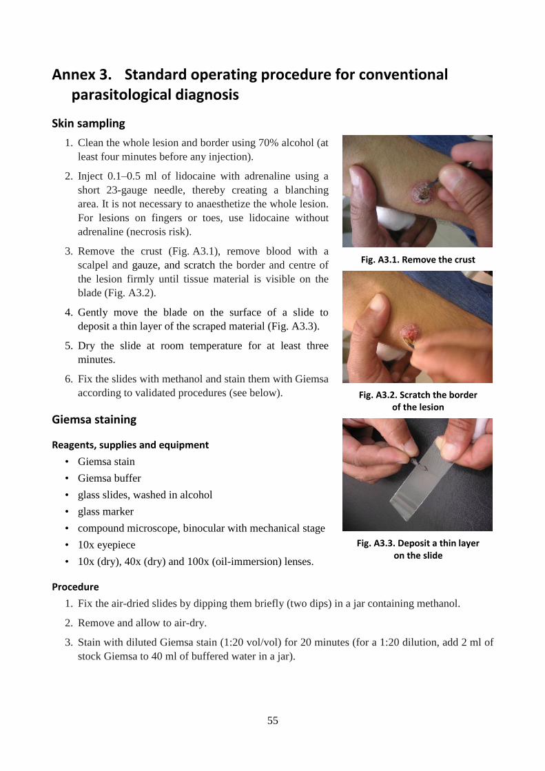
55
Annex 3. Standard operating procedure for conventional parasitological diagnosis
Skin sampling
1. Clean the whole lesion and border using 70% alcohol (at
least four minutes before any injection).
2. Inject 0.1–0.5 ml of lidocaine with adrenaline using a
short 23-gauge needle, thereby creating a blanching
area. It is not necessary to anaesthetize the whole lesion.
For lesions on fingers or toes, use lidocaine without
adrenaline (necrosis risk).
3. Remove the crust (Fig. A3.1), remove blood with a
scalpel and gauze, and scratch the border and centre of
the lesion firmly until tissue material is visible on the
blade (Fig. A3.2).
4. Gently move the blade on the surface of a slide to
deposit a thin layer of the scraped material (Fig. A3.3).
5. Dry the slide at room temperature for at least three
minutes.
6. Fix the slides with methanol and stain them with Giemsa
according to validated procedures (see below).
Giemsa staining
Reagents, supplies and equipment
• Giemsa stain
• Giemsa buffer
• glass slides, washed in alcohol
• glass marker
• compound microscope, binocular with mechanical stage
• 10x eyepiece
• 10x (dry), 40x (dry) and 100x (oil-immersion) lenses.
Procedure
1. Fix the air-dried slides by dipping them briefly (two dips) in a jar containing methanol.
2. Remove and allow to air-dry.
3. Stain with diluted Giemsa stain (1:20 vol/vol) for 20 minutes (for a 1:20 dilution, add 2 ml of
stock Giemsa to 40 ml of buffered water in a jar).
Fig. A3.1. Remove the crust
Fig. A3.2. Scratch the border of the lesion
Fig. A3.3. Deposit a thin layer on the slide

56
4. Wash by briefly dipping the slides in a jar of buffered
water (one or two dips).
5. Allow to air-dry.
6. Examine the slides under the microscope (40x and
100x oil-immersion lenses).
7. Read smears for at least 20 minutes (1000 fields) at
400x or 1000x magnification.
8. A smear can be reported positive when at least two
amastigotes are observed (Fig. A3.4). For valid
identification, an amastigote form must show a
nucleus, a kinetoplast and a plasma membrane.
Fig. A3.4. Amastigotes within the macrophages

57
Annex 4. Standard operating procedure for cryotherapy and intralesional injection of antimony
Swab the lesion with antiseptics several minutes before starting the procedure. Repeat the procedure
once a week, until healing of lesions is complete. Generally, three to five sessions are sufficient to
cure most lesions.
Cryotherapy
Apply liquid nitrogen (–195 oC) to the lesion (Fig. A4.1. A–D) and up to 2 mm outside the lesion
margin (Fig. A4.2), until a 10-second blanching is obtained. Ideally, a sprayer should be used;
otherwise, use a cotton-tipped applicator.
Fig. A4.2. Complete
blanching of the lesion
When cryotherapy is applied before an
intralesional injection of antimony, one 10-
second blanching is sufficient. When
cryotherapy is applied alone, the procedure
should be repeated two or three times at short
intervals, resulting in a total time of 30
seconds.
Intralesional injection
1. Aseptically withdraw the product directly from the ampule of antimony as formulated for
parenteral administration by the manufacturer.
2. Inject the antimony (immediately after liquid nitrogen application) into three peripheral
points of the lesion and induce blanching of the borders (Fig. A4.3. A–E), until the lesion is
entirely swollen.
A B
C D
Fig. A4.1. Application of nitrogen liquid

58
A B C
Fig. A4.3. Intralesional infiltration of antimony
D E
The appearance of the lesion before the procedure is shown in Fig. A4.4; its appearance at the end
of the procedure in Fig. A4.5.
Fig. A4.4. Aspect before procedure Fig. A4.5. Aspect after procedure

59
Annex 5. Standard operating procedure for thermotherapy
Thermotherapy is a technique available for the treatment of CL patients by application of local heat
at the site of the lesion with a portable, battery-operated device known as a localized current field
radiofrequency generator.
Indication
• The papule, nodule or ulcer is less than 4 cm in size.
• There are fewer than four lesions.
• The lesions are not located close to eyes, nose or lips.
Method
A single thermotherapy treatment consists of one or more applications of localized heat of 50 °C for
30 seconds, depending on lesion size. The area between the electrodes covers 49–73 mm2. Several
thermotherapy applications, therefore, may be required to cover a lesion.
Procedure
1. Disinfect the lesion and a 2 cm border of healthy skin
around the lesion with antiseptic (e.g. 0.1% chlorine
dioxide solution).
2. Anaesthetize the lesion with 1% lidocaine hydrochloride.
3. Moisturize the lesion with sterile saline solution.
4. Apply heat locally for 30 seconds (Fig. A5.1).
5. Apply chlorine dioxide gel to the lesion and cover after
treatment.
Patient follow-up
Follow-up after completion of treatment should be scheduled at 14, 30, 45 and 180 days. It is
important to explain to patients that, if the lesion does not improve, they should return to the health
facility at any time.
Fig. A5.1. Thermotherapy application
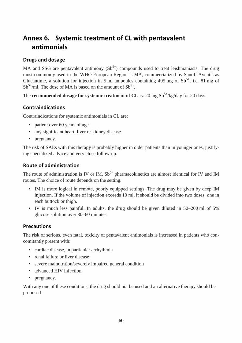
60
Annex 6. Systemic treatment of CL with pentavalent antimonials
Drugs and dosage
MA and SSG are pentavalent antimony (Sb5+
) compounds used to treat leishmaniasis. The drug
most commonly used in the WHO European Region is MA, commercialized by Sanofi-Aventis as
Glucantime, a solution for injection in 5 ml ampoules containing 405 mg of Sb5+
, i.e. 81 mg of
Sb5+
/ml. The dose of MA is based on the amount of Sb5+
.
The recommended dosage for systemic treatment of CL is: 20 mg Sb5+
/kg/day for 20 days.
Contraindications
Contraindications for systemic antimonials in CL are:
• patient over 60 years of age
• any significant heart, liver or kidney disease
• pregnancy.
The risk of SAEs with this therapy is probably higher in older patients than in younger ones, justify-
ing specialized advice and very close follow-up.
Route of administration
The route of administration is IV or IM. Sb5+
pharmacokinetics are almost identical for IV and IM
routes. The choice of route depends on the setting.
• IM is more logical in remote, poorly equipped settings. The drug may be given by deep IM
injection. If the volume of injection exceeds 10 ml, it should be divided into two doses: one in
each buttock or thigh.
• IV is much less painful. In adults, the drug should be given diluted in 50–200 ml of 5%
glucose solution over 30–60 minutes.
Precautions
The risk of serious, even fatal, toxicity of pentavalent antimonials is increased in patients who con-
comitantly present with:
• cardiac disease, in particular arrhythmia
• renal failure or liver disease
• severe malnutrition/severely impaired general condition
• advanced HIV infection
• pregnancy.
With any one of these conditions, the drug should not be used and an alternative therapy should be
proposed.

61
Patient monitoring
A CBC should be done before treatment and weekly during treatment.
Hepatic function tests – hepatic function determinations, including serum alanine aminotransferase
(serum glutamic pyruvic transaminase), serum alkaline aminotransferase, and serum aspartate
aminotransferase (also called serum glutamic oxaloacetic transaminase) – may be required before
treatment and weekly during therapy. If the value of one of the serum aminotransferases reaches
three to four times the normal upper limit, antimonial therapy should be discontinued.
Serum amylase and lipase should be monitored before treatment, at day 2–3 and weekly during
treatment. If the serum amylase value reaches over four times the normal upper limit, or if the
serum lipase value reaches over 15 times the normal upper limit, and if these rises in enzyme levels
occur rapidly or are associated with abdominal pain, nausea and vomiting, antimonial therapy
should be temporarily interrupted.
Renal function tests – renal function determinations, including BUN and serum creatinine – are
required before treatment and weekly during treatment.
ECG monitoring is recommended and should be done twice weekly. Treatment must be suspended
if QTc is greater than 0.5 second, or if there are concave ST segments or clinically relevant
arrhythmias. In the event that Stokes–Adams attacks develop and sudden cardiac collapse occurs,
treatment with antimonials must be stopped and atropine should be started. Atropine should be
administered IV at a dose of 0.5–1.5 mg, followed by IM administration of 0.5–1.0 mg every three
hours. If treatment with atropine is unsuccessful, isoprenaline or atrial pacing should be considered.
Toxicity and side-effects
Minor side-effects
symptoms: nausea, anorexia, arthralgias, myalgias, injection site pain (minimized in some but not
all patients by slow and deep injection and use of lidocaine), fatigue and abdominal pain.
laboratory toxicity: elevated amylase (biochemical pancreatitis), elevated liver enzymes
(biochemical hepatitis), leukopenia/anaemia/thrombocytopenia; occasionally, renal failure
occurs.
ECG changes: prolongation of ST segment and alteration of T wave, essentially concave ST
segment.
nausea and anorexia: substantial problems where patients are already malnourished and
dehydrated; they subside somewhat in the later weeks of treatment.
Serious toxicity
Severe vomiting and abdominal pain (possibly due to pancreatitis) can be treated with antiemetic
medications. When antiemetic treatment fails, Sb5+
treatment should be interrupted. If pancreatitis is
confirmed by elevated serum amylase and/or lipase, Sb5+
treatment should also be interrupted when
amylase is more than four times the normal upper limit and/or lipase is 12 times the normal upper
limit.

62
Annex 7. Drug options for imported CL and MCL cases
The following drug options are available for treatment of imported CL and MCL cases found
infected with Old and New World parasites non-endemic in the WHO European Region.
Treatment of diffuse CL (L. aethiopica)
• pentavalent antimonials (20 mg Sb5+
/kg/day for 20 days)
• LAB (18 mg/kg total dose: 3 mg/kg/day, days 1–5 and at day 10)
• pentamidine isethionate (4 mg/kg: three infusions over five days) or miltefosine (50 mg three
times a day for 28 days)
Treatment of MCL (L. braziliensis, L. panamensis and L. guyanensis)
• pentavalent antimonials (20 mg Sb5+
/kg/day for 28 days) with or without oral pentoxifylline
(400 mg three time a day for 30 days)
• LAB (18 mg/kg total dose: 3 mg/kg/day, days 1–5 and at day 10)
• miltefosine (only Bolivia and Brazil) (50 mg three times a day for 28 days)
Treatment of New World CL by parasite species
Treatment of L. mexicana
• ketoconazole (600 mg/day for 28 days)
• miltefosine (50 mg three times a day for 28 days)
• pentavalent antimonials (20 mg Sb5+
/kg/day for 20 days)
Treatment of L. guyanensis and L. panamensis
• miltefosine (50 mg three times a day for 28 days)
• pentamidine isethionate (4 mg/kg: three infusions over five days)
• pentavalent antimonials (20 mg Sb5+
/kg/day for 20 days)
• ketoconazole (600 mg for 28 days) (for L. panamensis)
Treatment of L. braziliensis, L. peruviana and L. amazonensis
• pentavalent antimonials (20 mg Sb5+
/kg/day for 20 days)
• LAB (18 mg/kg total dose: 3 mg/kg/day, days 1–5 and at day 10)
• miltefosine (only Bolivia and Brazil) (50 mg three times a day for 28 days)
