mapping rna-protein interactions in ribonuclease p from
TRANSCRIPT
Article No. jmbi.1999.3443 available online at http://www.idealibrary.com on J. Mol. Biol. (2000) 296, 19±31
Mapping RNA-Protein Interactions in Ribonuclease Pfrom Escherichia coli using Disulfide-linked EDTA-Fe
Roopa Biswas1, David W. Ledman2,3, Robert O. Fox3, Sidney Altman4
and Venkat Gopalan1*
1Department of BiochemistryThe Ohio State UniversityColumbus, OH, 43210, USA2Marketing Department, PEBiosystems, Inc., FraminghamMA 01701, USA3Department of HumanBiological Chemistry andGenetics, The Sealy Center forStructural Biology, Universityof Texas Medical BranchGalveston, TX, 77555, USA4Department of MolecularCellular and DevelopmentalBiology, Yale University, NewHaven, CT, 06520-8103, USA
E-mail address of the [email protected]
Abbreviations used: BABE, 1-(p-bromoacetamidobenzyl)-EDTA; CDCAP, catabolite activator protein; Eaminoethyl-2-pyridyl disul®de; RNP
0022-2836/00/60019±13 $35.00/0
The protein subunit of Escherichia coli ribonuclease P (which has acysteine residue at position 113) and its single cysteine-substitutedmutant derivatives (S16C/C113S, K54C/C113S and K66C/C113S) havebeen modi®ed using a sulfhydryl-speci®c iron complex of EDTA-2-aminoethyl 2-pyridyl disul®de (EPD-Fe). This reaction converts C5 pro-tein, or its single cysteine-substituted mutant derivatives, into chemicalnucleases which are capable of cleaving the cognate RNA ligand, M1RNA, the catalytic RNA subunit of E. coli RNase P, in the presence ofascorbate and hydrogen peroxide. Cleavages in M1 RNA are expected tooccur at positions proximal to the site of contact between the modi®edresidue (in C5 protein) and the ribose units in M1 RNA. When EPD-Fewas used to modify residue Cys16 in C5 protein, hydroxyl radical-mediated cleavages occurred predominantly in the P3 helix of M1 RNApresent in the reconstituted holoenzyme. C5 Cys54-EDTA-Fe producedcleavages on the 50 strand of the P4 pseudoknot of M1 RNA, while thecleavages promoted by C5 Cys66-EDTA-Fe were in the loop connectinghelices P18 and P2 (J18/2) and the loop (J2/4) preceding the 30 strand ofthe P4 pseudoknot. However, hydroxyl radical-mediated cleavages in M1RNA were not evident with Cys113-EDTA-Fe, perhaps indicative ofCys113 being distal from the RNA-protein interface in the RNase Pholoenzyme. Our directed hydroxyl radical-mediated footprinting exper-iments indicate that conserved residues in the RNA and protein subunitof the RNase-P holoenzyme are adjacent to each other and provide struc-tural information essential for understanding the assembly of RNase P.
# 2000 Academic Press
Keywords: footprinting; RNase P protein subunit; RNA-proteininteractions; EPD-Fe; EDTA-Fe
*Corresponding authorIntroduction
Ribonuclease P (RNase P) is a member of theensemble of ribonucleases involved in the matu-ration of tRNA molecules (Altman & Kirsebom,1999; Deutscher, 1995; Frank & Pace, 1998). TheRNase P holoenzyme in Escherichia coli is a ribonu-cleoprotein (RNP) complex consisting of M1 RNA(377 nucleotides) and C5 protein (119 amino acidresidues) (Altman & Kirsebom, 1999; Frank &Pace, 1998). Under certain conditions in vitro, M1
ing author:
, circular dichroism;PD, EDTA-2-, ribonucleoprotein.
RNA (the catalytic subunit) can cleave its sub-strates even in the absence of its protein cofactor.The processing of precursor tRNA molecules byM1 RNA is signi®cantly enhanced by C5 proteinin vitro (Altman & Kirsebom, 1999), consistent withthe observation that RNase P functions as an RNPcomplex in vivo (Sakano et al., 1974). Recent studiesindicate that the protein subunit enhances the af®-nity of the RNase P holoenzyme for the substrateperhaps by promoting speci®c interactions withthe leader sequence in the substrate (Crary et al.,1998; Kurz et al., 1998; Niranjanakumari et al., 1998;Krummel & Altman, 1999).
Although various biochemical and geneticstudies have provided insights into structure-func-tion relationships of both subunits of RNase Pfrom E. coli (Altman & Kirsebom, 1999; Frank &Pace, 1998), further progress in elucidating the
# 2000 Academic Press
20 RNA-Protein Interactions in Ribonuclease P
mechanism of action of RNase P cannot be madewithout detailed structural data. To date, structuralcharacterization of the RNA subunit in the absenceand presence of its ptRNA substrate is based oncumulative information from low resolution tech-niques such as crosslinking and footprinting (Chenet al., 1998; Christian et al., 1998; Guerrier-Takadaet al., 1989; Harris et al. 1994, 1998; Kufel &Kirsebom, 1996). Similar studies with the RNase Pholoenzyme would be vital for understanding itsassembly and mechanism of action. Here, we usean EDTA-Fe-based footprinting approach as a ®rststep in the determination of the spatial orientationof speci®c parts of the protein cofactor in relationto various domains in the catalytic RNA subunit.
Results
Rationale
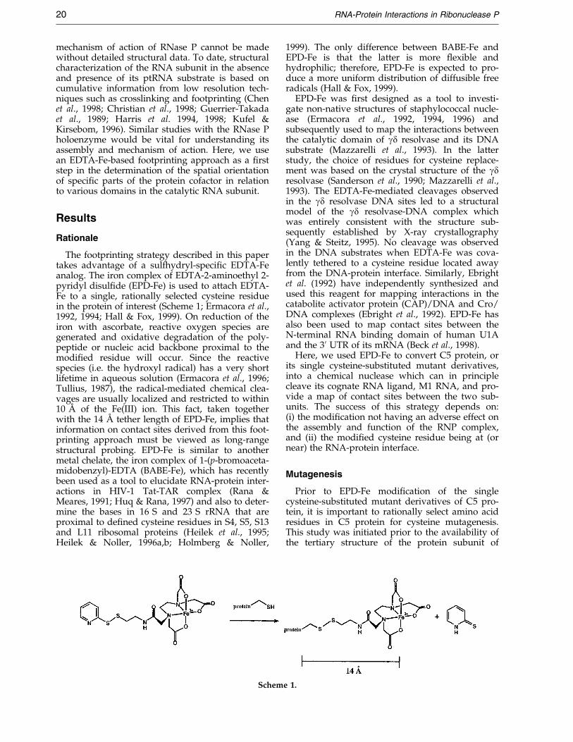
The footprinting strategy described in this papertakes advantage of a sulfhydryl-speci®c EDTA-Feanalog. The iron complex of EDTA-2-aminoethyl 2-pyridyl disul®de (EPD-Fe) is used to attach EDTA-Fe to a single, rationally selected cysteine residuein the protein of interest (Scheme 1; Ermacora et al.,1992, 1994; Hall & Fox, 1999). On reduction of theiron with ascorbate, reactive oxygen species aregenerated and oxidative degradation of the poly-peptide or nucleic acid backbone proximal to themodi®ed residue will occur. Since the reactivespecies (i.e. the hydroxyl radical) has a very shortlifetime in aqueous solution (Ermacora et al., 1996;Tullius, 1987), the radical-mediated chemical clea-vages are usually localized and restricted to within10 AÊ of the Fe(III) ion. This fact, taken togetherwith the 14 AÊ tether length of EPD-Fe, implies thatinformation on contact sites derived from this foot-printing approach must be viewed as long-rangestructural probing. EPD-Fe is similar to anothermetal chelate, the iron complex of 1-(p-bromoaceta-midobenzyl)-EDTA (BABE-Fe), which has recentlybeen used as a tool to elucidate RNA-protein inter-actions in HIV-1 Tat-TAR complex (Rana &Meares, 1991; Huq & Rana, 1997) and also to deter-mine the bases in 16 S and 23 S rRNA that areproximal to de®ned cysteine residues in S4, S5, S13and L11 ribosomal proteins (Heilek et al., 1995;Heilek & Noller, 1996a,b; Holmberg & Noller,
Schem
1999). The only difference between BABE-Fe andEPD-Fe is that the latter is more ¯exible andhydrophilic; therefore, EPD-Fe is expected to pro-duce a more uniform distribution of diffusible freeradicals (Hall & Fox, 1999).
EPD-Fe was ®rst designed as a tool to investi-gate non-native structures of staphylococcal nucle-ase (Ermacora et al., 1992, 1994, 1996) andsubsequently used to map the interactions betweenthe catalytic domain of gd resolvase and its DNAsubstrate (Mazzarelli et al., 1993). In the latterstudy, the choice of residues for cysteine replace-ment was based on the crystal structure of the gdresolvase (Sanderson et al., 1990; Mazzarelli et al.,1993). The EDTA-Fe-mediated cleavages observedin the gd resolvase DNA sites led to a structuralmodel of the gd resolvase-DNA complex whichwas entirely consistent with the structure sub-sequently established by X-ray crystallography(Yang & Steitz, 1995). No cleavage was observedin the DNA substrates when EDTA-Fe was cova-lently tethered to a cysteine residue located awayfrom the DNA-protein interface. Similarly, Ebrightet al. (1992) have independently synthesized andused this reagent for mapping interactions in thecatabolite activator protein (CAP)/DNA and Cro/DNA complexes (Ebright et al., 1992). EPD-Fe hasalso been used to map contact sites between theN-terminal RNA binding domain of human U1Aand the 30 UTR of its mRNA (Beck et al., 1998).
Here, we used EPD-Fe to convert C5 protein, orits single cysteine-substituted mutant derivatives,into a chemical nuclease which can in principlecleave its cognate RNA ligand, M1 RNA, and pro-vide a map of contact sites between the two sub-units. The success of this strategy depends on:(i) the modi®cation not having an adverse effect onthe assembly and function of the RNP complex,and (ii) the modi®ed cysteine residue being at (ornear) the RNA-protein interface.
Mutagenesis
Prior to EPD-Fe modi®cation of the singlecysteine-substituted mutant derivatives of C5 pro-tein, it is important to rationally select amino acidresidues in C5 protein for cysteine mutagenesis.This study was initiated prior to the availability ofthe tertiary structure of the protein subunit of
e 1.
RNA-Protein Interactions in Ribonuclease P 21
RNase P. In addition to Cys113 that is present inwild-type C5 protein, the residues Ser16, Lys54and Lys66 were selected as sites to engineer Cysresidues for subsequent modi®cation with EPD-Fe.Ser16 was selected for mutagenesis because of itsproximity to the highly conserved residue Phe18.When M1 RNA is reconstituted with the mutantC5 F18A, the resultant mutant holoenzyme dis-plays decreased activity and altered substratespeci®city compared to the wild-type holoenzyme(Gopalan et al., 1997). Moreover, C5 F18A dis-played weaker binding to M1 RNA compared tothe wild-type C5 protein (V.G. & S.A., unpublishedresults). Since Ser16 was only two residues awayfrom Phe18 and because a Ser to Cys residue sub-stitution causes modest alterations in the chemicalcharacter, we chose to replace Ser16 with a Cysresidue. Based on the results from our recent EPRspectroscopic studies of M1 RNA-C5 protein inter-actions, positions 54 and 66 were also selected forCys mutagenesis modi®cation with EPD-Fe(Gopalan et al., 1999). Although the EPR spec-troscopy-based approach facilitated identi®cationof cysteine residues, engineered or otherwise, thatmight be part of the RNA-protein interface in anRNP complex, it did not yield any information onwhich residues in M1 RNA are involved in theseinteractions (Gopalan et al., 1999). The EPD-Fe foot-printing experiments reported here were per-formed to identify nucleotide positions in M1 RNAwhich are proximal to speci®c residues (such as 16,54 and 66) on C5 protein.
Mass spectrometry
The single cysteine residue at position 113 inwild-type C5 protein (Hansen et al., 1985;Figure 1(a)) and in the three single cysteine-sub-stituted mutants (S16C/C113S, K54C/C113S andK66C/C113S) were modi®ed with EPD-Fe(Scheme 1). To examine if the derivatization pro-cedure resulted in covalently tethering Fe-EDTA toC5 protein and its mutant derivatives, electrosprayionization mass spectrometry was used to deter-mine the molecular masses of the various proteinsamples before and after derivatization with EPD-Fe. The molecular masses observed are in goodagreement with the expected values (Table 1).Although we have reported the masses for the pro-tein minus the N-terminal methionine residue, wedo observe some species whose masses indicatethat the N-terminal methionine residue is present(data not shown). It is likely, therefore, that there issome heterogeneity at the N terminus, as has beenobserved with other proteins overexpressed inE. coli (Dalboge et al., 1990; Sandman et al., 1995).
The extent of derivatization with EPD-Fe wasalso examined by amino acid analysis. Two of thederivatized protein samples were oxidized withperformic acid and hydrolyzed with 6 N HCl foramino acid analysis at the W. M. Keck FoundationBiotechnology Resource Laboratory, Yale Univer-sity. The oxidation of an EPD-modi®ed cysteine
residue results in formation of cysteic acid andtaurine. For a protein sample that is completelyderivatized, equimolar amounts of cysteic acid andtaurine are expected. Amino acid analysis per-formed with the protein samples in which residuesCys16 and Cys113 were modi®ed with EPDyielded taurine/cysteic acid ratios of 1.3 and 0.9,respectively. The near-unit ratio of taurine/cysteicacid in our modi®ed protein samples indicatealmost complete derivatization.
Circular dichroism spectroscopy
It is conceivable that the cysteine replacementsin C5 protein (i.e. S16C, K54C and K66C) and theirsubsequent modi®cation with EPD-Fe might affectprotein folding. CD spectroscopic studies of theunmodi®ed and the EPD-modi®ed mutant deriva-tives of C5 protein revealed that there were nodramatic alterations in the secondary structure.Although some differences were observed in theCD spectra (Figure 2) between the unmodi®ed andmodi®ed C5 protein mutants, the spectra are quali-tatively similar. The minor differences between thespectra are likely due to inaccuracies in determin-ing protein concentration, a problem accentuatedby a modest, albeit consistent, precipitation ofsome proteins during the CD measurement. More-over, deconvolution of the CD spectra of theunmodi®ed and modi®ed proteins yielded similarpercentages for the various secondary structuralelements (data not shown).
Activity assays
The unmodi®ed and EPD-modi®ed samples ofthe wild-type C5 protein and its single cysteine-substituted mutants were reconstituted with wild-type M1 RNA and the ability of these holoenzymesto cleave precursor tRNATyr (ptRNATyr) was exam-ined as described elsewhere (Gopalan et al., 1997).The results of the RNase P assays demonstratedthat neither the introduced mutations nor themodi®cation with EPD-Fe signi®cantly alter pro-tein function (Table 2). The wild-type C5 proteinwhen reconstituted with M1 RNA exhibits a cataly-tic turnover number of 30 minÿ1. The activitiesobserved with the various mutants are normalizedbased on the turnover number of the wild-typeRNase P holoenzyme. Modi®cation of the C5 pro-tein mutant, K66C/C113S, with EDTA-Fe led to asigni®cant decrease in activity relative to the wild-type protein (Table 2); however, it should be notedthat the unmodi®ed mutant protein itself, exhibitsa slightly lower activity in comparison to wild-typeC5 protein. Nevertheless, this result would implythat some caution is required while interpretingthe footprinting results observed with Cys66-EDTA-Fe.

Figure 1. (a) Alignment of the amino acid sequences of the protein subunits of RNase P from E. coli and B. subtilis.The residues marked by an asterisk indicate sites of cysteine replacement and EPD-Fe modi®cation in the proteinsubunit of E. coli RNase P. The respective secondary structural elements are indicated below the sequence alignment.(b) Tertiary structure of the protein subunit of RNase P from B. subtilis (Stams et al., 1998). The program RASMOLwas used to depict the a-carbon backbone of the protein structure as a ribbon. The various secondary structuralelements are identi®ed using the same color scheme as in (a). The positions where Cys residues were introduced andmodi®ed with EPD-Fe are indicated in blue; the numbering is based on the sequence of the protein subunit of E. coliRNase P. The program QUANTA'97 Protein Design (Molecular Simulations Inc., San Diego) was used to generate thestructure of the RNase P protein subunit in which the native amino acid residues at the indicated positions in theB. subtilis protein were replaced with a cysteine residue; the coordinates deposited in the PDB by Stams et al. (1998)served as the starting template. (c) Electrostatic potential mapped to the molecular surface of the protein depicted in(b); this Figure was prepared using GRASP software (Nicholls, 1993). To reveal the putative RNA-binding centralcleft, the view in (c) was obtained by turning the molecule in (b) in a clockwise direction. Blue and red shadingrepresent regions of the surface where basic and acidic residue side-chains would map, respectively. Moreover, K54and K66 are indicated in (c) to orient the reader while comparing (b) and (c).
22 RNA-Protein Interactions in Ribonuclease P
Footprinting studies
The footprinting experiments consisted of recon-stituting the unmodi®ed and modi®ed protein
Table 1. Molecular masses of C5 mutants before and after Emass spectrometry
Before modificationPredicteda Observ
1. Wild-type C5 (C113) 13,658 13,658 (2. C5 S16C/C113S 13,658 13,659 (3. C5 K54C/C113S 13,617 13,616 (4. C5 K66C/C113S 13,617 13,616 (
a The molecular mass corresponds to that of C5 protein lacking th
samples with either 50 or 30 end-labeled M1 RNAand immediately incubating the holoenzyme com-plexes individually with ascorbate and hydrogenperoxide. To detect the positions where the
PD modi®cation as measured by electrospray ionization
After modificationed Predicted Observed
�1) 14,060 14,061 (�1)�2) 14,060 14,061 (�2)�1) 14,019 14,021 (�2)�2) 14,019 14,021 (�3)
e N-terminal methionine residue.

Table 2. Relative RNase P activity of wild-type C5protein and its mutant derivatives
C5 Protein Relative initial velocity (%)b
Wild-type C5 100Wild-type-EDTA-Fe 68(�1)Cys16a 90(�4)Cys16-EDTA-Fe 74(�2)Cys54 89(�2)Cys54-EDTA-Fe 62(�1)Cys66 79(�5)Cys66-EDTA-Fe 28(�3)
a All cysteine mutants in this Table have an additionalchange (i.e. C113S).
b Average of values obtained from two independent experi-ments.
RNA-Protein Interactions in Ribonuclease P 23
hydroxyl radical has cleaved the M1 RNA back-bone, we used polyacrylamide gel electrophoresisfollowed by autoradiography. The three EPD-Fe-derivatized proteins, C5 Cys16-EDTA-Fe, Cys54-EDTA-Fe and Cys66-EDTA-Fe, produced speci®ccleavages in M1 RNA (Figure 3). The failure to
Figure 2. CD spectra of the unmodi®ed (open boxes) andtuted derivatives of C5 protein: (a) wild-type C5 protein, (b)
observe a speci®c hydroxyl radical-mediated clea-vage with C5 Cys113-EDTA-Fe implies that resi-due 113 is distal from the RNA-protein interface inthe RNase P holoenzyme (data not shown).
The cleavages in 50 end-labeled M1 RNAobserved upon incubation with C5 Cys16-EDTA-Fe, ascorbate and hydrogen peroxide, wererestricted to the P3 helix in M1 RNA (Figure 3(a)).After six minutes of incubation of the M1 RNA-C5Cys16-EDTA-Fe complex with ascorbate andhydrogen peroxide, the ®rst cleavages wereobserved at positions 49, 50, 51 and 52 in M1 RNAand, albeit weakly, at positions 29, 30 and 31. Aftera longer incubation (12 minutes), additional clea-vages were observed at nucleotide positions 46through to 48 as well as 53 in M1 RNA (these pos-itions ¯ank the major cleavage sites between 49and 52). Moreover, there are also cleavages fromnucleotide positions 29 through 33. A phosphor-imager scan of the dried gel enabled us to quantifythe cleavages at individual backbone positions inM1 RNA observed with the underivatized and
EDTA-Fe-modi®ed (®lled boxes) single cysteine-substi-S16C/C113S, (c) K54C/C113S, and (d) K66C/C113S.
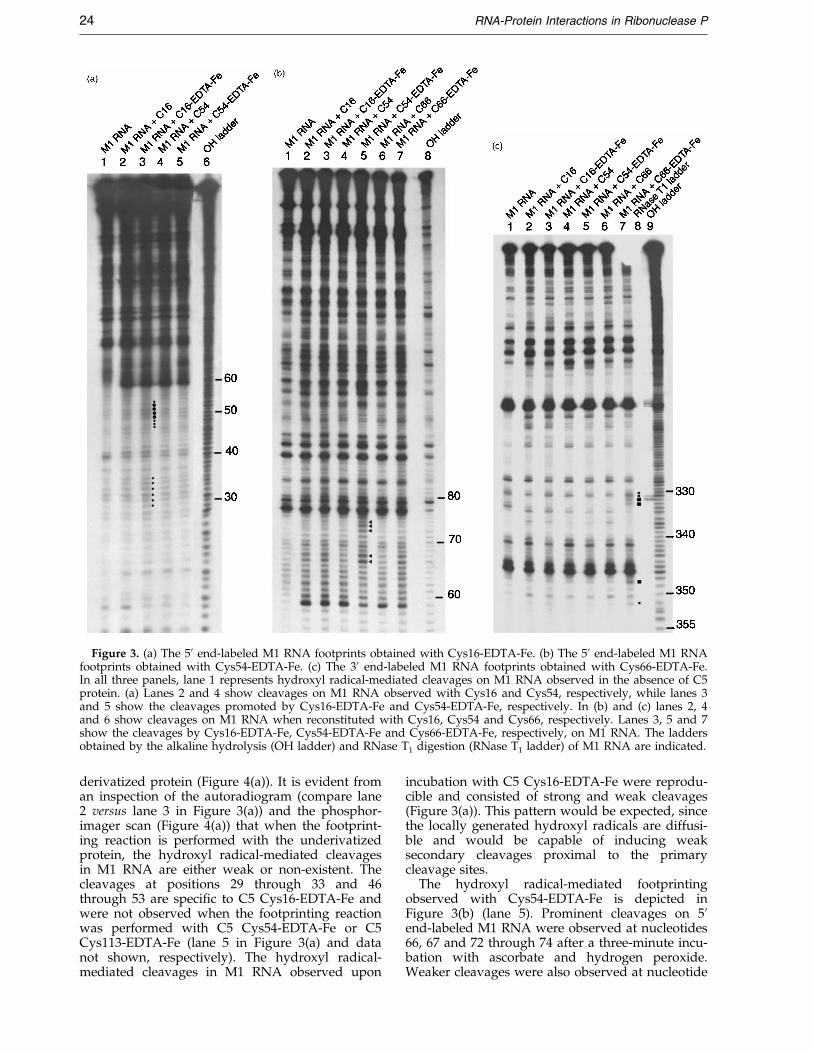
Figure 3. (a) The 50 end-labeled M1 RNA footprints obtained with Cys16-EDTA-Fe. (b) The 50 end-labeled M1 RNAfootprints obtained with Cys54-EDTA-Fe. (c) The 30 end-labeled M1 RNA footprints obtained with Cys66-EDTA-Fe.In all three panels, lane 1 represents hydroxyl radical-mediated cleavages on M1 RNA observed in the absence of C5protein. (a) Lanes 2 and 4 show cleavages on M1 RNA observed with Cys16 and Cys54, respectively, while lanes 3and 5 show the cleavages promoted by Cys16-EDTA-Fe and Cys54-EDTA-Fe, respectively. In (b) and (c) lanes 2, 4and 6 show cleavages on M1 RNA when reconstituted with Cys16, Cys54 and Cys66, respectively. Lanes 3, 5 and 7show the cleavages by Cys16-EDTA-Fe, Cys54-EDTA-Fe and Cys66-EDTA-Fe, respectively, on M1 RNA. The laddersobtained by the alkaline hydrolysis (OH ladder) and RNase T1 digestion (RNase T1 ladder) of M1 RNA are indicated.
24 RNA-Protein Interactions in Ribonuclease P
derivatized protein (Figure 4(a)). It is evident froman inspection of the autoradiogram (compare lane2 versus lane 3 in Figure 3(a)) and the phosphor-imager scan (Figure 4(a)) that when the footprint-ing reaction is performed with the underivatizedprotein, the hydroxyl radical-mediated cleavagesin M1 RNA are either weak or non-existent. Thecleavages at positions 29 through 33 and 46through 53 are speci®c to C5 Cys16-EDTA-Fe andwere not observed when the footprinting reactionwas performed with C5 Cys54-EDTA-Fe or C5Cys113-EDTA-Fe (lane 5 in Figure 3(a) and datanot shown, respectively). The hydroxyl radical-mediated cleavages in M1 RNA observed upon
incubation with C5 Cys16-EDTA-Fe were reprodu-cible and consisted of strong and weak cleavages(Figure 3(a)). This pattern would be expected, sincethe locally generated hydroxyl radicals are diffusi-ble and would be capable of inducing weaksecondary cleavages proximal to the primarycleavage sites.
The hydroxyl radical-mediated footprintingobserved with Cys54-EDTA-Fe is depicted inFigure 3(b) (lane 5). Prominent cleavages on 50end-labeled M1 RNA were observed at nucleotides66, 67 and 72 through 74 after a three-minute incu-bation with ascorbate and hydrogen peroxide.Weaker cleavages were also observed at nucleotide
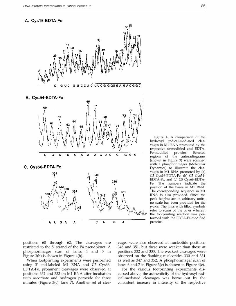
Figure 4. A comparison of thehydroxyl radical-mediated clea-vages in M1 RNA promoted by therespective unmodi®ed and EDTA-Fe-modi®ed proteins. Selectedregions of the autoradiograms(shown in Figure 3) were scannedwith a phosphorimager (MolecularDynamics) to illustrate the clea-vages in M1 RNA promoted by (a)C5 Cys16-EDTA-Fe, (b) C5 Cys54-EDTA-Fe, and (c) C5 Cys66-EDTA-Fe. The numbers indicate theposition of the bases in M1 RNA.The corresponding sequence in M1RNA is also provided. Since thepeak heights are in arbitrary units,no scale has been provided for they-axis. The lines with ®lled symbolsrefer to scans of the lanes whereinthe footprinting reaction was per-formed with the EDTA-Fe-modi®edproteins.
RNA-Protein Interactions in Ribonuclease P 25
positions 60 through 62. The cleavages arerestricted to the 50 strand of the P4 pseudoknot. Aphosphorimager scan of lanes 4 and 5 inFigure 3(b) is shown in Figure 4(b).
When footprinting experiments were performedusing 30 end-labeled M1 RNA and C5 Cys66-EDTA-Fe, prominent cleavages were observed atpositions 332 and 333 on M1 RNA after incubationwith ascorbate and hydrogen peroxide for threeminutes (Figure 3(c), lane 7). Another set of clea-
vages were also observed at nucleotide positions348 and 351, but these were weaker than those atpositions 332 and 333. The weakest cleavages wereobserved on the ¯anking nucleotides 330 and 331as well as 347 and 352. A phosphorimager scan oflanes 6 and 7 in Figure 3(c) is shown in Figure 4(c).
For the various footprinting experiments dis-cussed above, the authenticity of the hydroxyl rad-ical-mediated cleavages was borne out by theconsistent increase in intensity of the respective

26 RNA-Protein Interactions in Ribonuclease P
cleavage products upon prolonged incubation ofthe reconsituted holoenzymes with ascorbate andhydrogen peroxide (data not shown).
In contrast to the speci®c and distinct cleavagesobtained with 30 end-labeled M1 RNA, incubationof 50 end-labeled M1 RNA with Cys66-EDTA-Feled to weak, random cleavages at several positionsproximal to the 50 end of M1 RNA (data notshown). Although there appears to be no majorsecondary structural alterations in the modi®edprotein (Cys66-EDTA-Fe) compared to its unmodi-®ed counterpart (Figure 2(d)), it is possible that themutation and modi®cation of a highly conservedresidue (i.e. K66C) compromises RNase P holo-enyzme assembly and this underlies the non-speci®c cleavage pattern observed with C5 Cys-66-EDTA-Fe and 50 end-labeled M1 RNA.
Discussion
A footprinting strategy involving site-speci®cmodi®cation of C5 protein into a chemical nucleasehas been used to map its contact sites with M1RNA, the catalytic RNA subunit of E. coli RNase P.
Selected residues on C5 protein were mutated tocysteine and reacted with the thiol-speci®c chemi-cal reagent EPD-Fe. The modi®cation of singlecysteine-substituted mutant derivatives of C5 pro-tein with EPD-Fe was con®rmed by mass spec-trometry (Table 1). To ensure that the mutation ofselected residues to Cys residues and the sub-sequent modi®cation of these Cys residues withEPD-Fe did not interfere with the structure andfunction of the protein, CD spectroscopic analysesand RNase P activity assays were performed. Theobservation that the CD spectra of the unmodi®edand the respective EDTA-Fe-modi®ed mutantderivatives of C5 protein are similar con®rmedthat the EDTA-Fe modi®cation did not alter thestructure signi®cantly (Figure 2). The RNase Pactivity assays revealed a noticeable decrease inactivity in only one case; upon covalently tetheringEDTA-Fe to Cys66, the RNase P activity was one-quarter that observed with the wild-type C5 pro-tein (Table 2). This result highlights a limitation ofour footprinting strategy. To obtain information oncontact sites in an RNP complex, it is necessary tointroduce EDTA-Fe proximal to the RNA-proteininterface; however, the introduction of EDTA-Feproximal to residues (in the protein cofactor) thatare important for RNA-protein interactions inRNase P might annul interactions essential forRNase P activity. Since there was signi®cantactivity even after modi®cation of Cys66, we haveproceeded with footprinting experiments using C5Cys66-EDTA-Fe (see below). However, it would bedesirable if the results obtained with C5 Cys66-EDTA-Fe are independently con®rmed withanother single cysteine-substituted mutant deriva-tive in which the Cys residue is introduced at aposition proximal to Lys66.
The EPD-modi®ed single cysteine-substitutedmutants of C5 protein were reconstituted with M1RNA. The hydroxyl radicals generated upon incu-bation of ascorbate and hydrogen peroxide withthe EDTA-Fe-modi®ed RNase P holoenzymes wereexpected to cleave the M1 RNA backbone proximalto the site of modi®cation in C5 protein. The M1RNA cleavage patterns observed with Cys16-,Cys54-, and Cys66-EDTA-Fe are summarized inFigure 5. As cautioned by Hall & Fox (1999), thesehydroxyl radical-mediated cleavage sites in theRNA do not correspond to sites of direct inter-action with the protein subunit. Since the EDTA-Femoiety and the ribose units in the RNA must bewithin 10 AÊ and appropriately oriented for clea-vage, it is reasonable to conclude that the cleavageswe observed in M1 RNA map nucleotides whichare proximal to residues 16, 54 and 66 in C5 pro-tein (when the two subunits are assembled into theRNase P holoenzyme). Cys113-EDTA-Fe failed topromote hydroxyl radical-mediated cleavages inM1 RNA, indicating that this residue is not at theRNA-protein interface in the RNase P holoenzymecomplex (data not shown). Consistent with thisobservation, the tertiary structure of C5 proteinplaces Cys113 (in a3-helix) behind the putativeRNA-binding central cleft and predicts it to be dis-tal from the RNA-protein interface (Figure 1(b); seediscussion below).
Our studies indicate that C5 Cys16-EDTA-Fecleaved nucleotides 49, 50, 51 and 52 in the P3helix of M1 RNA (Figure 5(a)). Phylogeneticsequence analysis has led to inclusion of the P3helix in the minimal consensus structure of theRNase P RNA subunit. However, there are no con-served nucleotides in the P3 helix. It is possiblethat the hydroxyl radical-mediated cleavages in theP3 region that we observed with Cys16-EDTA-Femight merely re¯ect the physical juxtaposition ofa1-helix in C5 protein and the P3 paired region inM1 RNA, rather than speci®c RNA-protein inter-actions between these regions. A separate photo-chemical crosslinking experiment has also yieldedresults suggesting that the P3 helix of M1 RNAmight be adjacent to the protein in the RNase Pholoenzyme. M1 RNA with randomly incorporatedphotoactivatible nucleotides was crosslinked to C5protein to determine the sites of contact betweenM1 RNA and C5 protein (C. Guerrier-Takada &S.A., unpublished results). When the crosslinkedcomplex was isolated and subjected to a primerextension analysis with reverse transcriptase, thecrosslinks between M1 RNA and C5 proteininvolved bases in the P3 helix of M1 RNA. More-over, the observation that a 4-thiouridine residueat position ÿ10 in the 50 leader of the ptRNA sub-strate crosslinks to C50 in the P3 helix suggests thephysical vicinity of P3 in M1 RNA and the sub-strate during RNase P catalysis (Christian &Harris, 1999).
When Cys54-EDTA-Fe was used in the footprint-ing experiments, the hydroxyl radical-mediatedcleavages were localized to nucleotides 66, 67 and
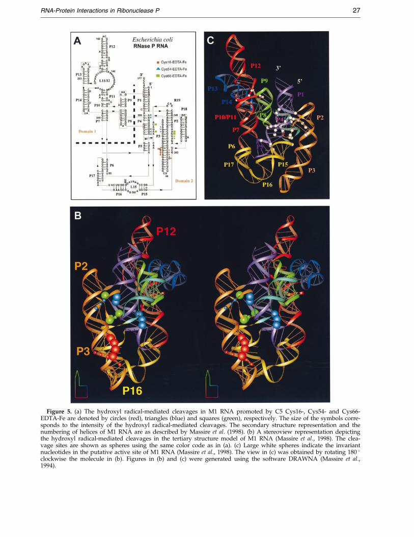
Figure 5. (a) The hydroxyl radical-mediated cleavages in M1 RNA promoted by C5 Cys16-, Cys54- and Cys66-EDTA-Fe are denoted by circles (red), triangles (blue) and squares (green), respectively. The size of the symbols corre-sponds to the intensity of the hydroxyl radical-mediated cleavages. The secondary structure representation and thenumbering of helices of M1 RNA are as described by Massire et al. (1998). (b) A stereoview representation depictingthe hydroxyl radical-mediated cleavages in the tertiary structure model of M1 RNA (Massire et al., 1998). The clea-vage sites are shown as spheres using the same color code as in (a). (c) Large white spheres indicate the invariantnucleotides in the putative active site of M1 RNA (Massire et al., 1998). The view in (c) was obtained by rotating 180 �clockwise the molecule in (b). Figures in (b) and (c) were generated using the software DRAWNA (Massire et al.,1994).
RNA-Protein Interactions in Ribonuclease P 27
28 RNA-Protein Interactions in Ribonuclease P
72 to 74 of the 50 strand of the P4 pseudoknot(Figure 5(a)). When the EDTA-Fe probe wasattached to Cys66, prominent cleavages wereobserved at positions 332 and 333 (in J18/2) aswell as 348 and 351 (in J2/4) (Figure 5(a)). Intra-molecular crosslinking studies have con®rmed thatthe J18/2 region is close to J2/4 (Harris et al.,1994). Since residues Lys54 and Lys66 are proximalin the tertiary structure of C5 protein, it is not sur-prising to ®nd that directed hydroxyl radical-mediated cleavages by Cys54- and Cys66-EDTA-Feare at adjacent positions in the proposed three-dimensional structure of M1 RNA (Figure 5(b)).This structure represents a computer-aided modelbased in part on conclusions from phylogeneticcovariation analysis and crosslinking data (Massireet al., 1998).
It is instructive to interpret the footprintingresults with Cys54- and Cys66-EDTA-Fe based on:(i) results from chemical-modi®cation interference,as well as crosslinking studies that have sought toestablish the active site in M1 RNA; and (ii) thethree-dimensional structure of the protein subunitof Bacillus subtilis RNase P.
Using Rp-phosphorothioates in modi®cation-interference experiments, Harris & Pace (1995)determined that replacement of a non-bridgingoxygen atom with a sulfur atom at the phosphates50 of M1 RNA nucleotides A67, G68, U69 and A352(in the P4 psuedoknot) led to a dramatic reductionin catalytic activity and that these phosphategroups were therefore likely to represent metal-binding sites important for RNase P catalysis. Asimilar approach was used by Hardt et al. (1995)while deriving their conclusion that phosphates 50of M1 RNA nucleotides A67, G68, U69, C70, C71(in the 50-strand of P4), A130, A132, A248, A249,G300, A317, A330 (in J18/2), A352 (in J2/4), C353and C354 (in the 30 strand of P4) were importantfor tRNA binding. Dimethylsulfate-based chemicalfootprinting experiments have revealed that G332in M1 RNA is protected from chemical modi®-cation by a precursor tRNA substrate which con-tains a four-nucleotide sequence (LaGrandeur et al.,1994). This observation was further supportedby the observation that a long-range photoagent(azidophenacyl bromide) at position 332 of M1RNA led to intermolecular crosslinking withnucleotides ÿ4 and ÿ5 in the leader sequence ofptRNA (Harris et al., 1994). In a complementaryapproach, short-range crosslinking experimentsusing 4-thiouridine at ÿ1 in the leader sequencedemonstrated convincingly that G332 and A333 onM1 RNA must be in close contact with the nucleo-tide at the ÿ1 position of the ptRNA substrate(Kufel & Kirsebom, 1996). Although these studiesused the catalytic RNA subunit (i.e. M1 RNA) andnot the RNase P holoenzyme complex (i.e. M1RNA and C5 protein), it is evident that the J18/2region, which includes nucleotides 332 and 333,interacts with the 50 leader in the ES complex.
The results from various experimentalapproaches mentioned above attest to the pivotal
role of the regions P4, J18/2 and J2/4 (in M1 RNA)in RNase P catalysis and lend signi®cance to thedirected hydroxyl radical-mediated footprints inP4, J18/2 and J2/4 that were observed with C5Cys54- and Cys66-EDTA-Fe.
Recently, the tertiary structure of the RNase Pprotein subunit from B. subtilis has been deter-mined (Stams et al., 1998). The overall topology isabbbaba and it includes the presence of an uncom-mon bab left-handed crossover connection from b3to a2 to b4 (Figure 1(b)). The structure of the pro-tein subunit of RNase P reveals that most of theconserved residues are present in two regions:(i) the large central cleft (20 AÊ long and 10 AÊ wide)which is formed by packing of the helix a1 againstthe b-sheet; and (ii) helix a2 as well as the loopwhich precedes it in the bab left-handed crossovermotif (Figure 1(b)). The surface electrostatic poten-tial map of this protein reveals a cleft rich in posi-tively charged residues (Figure 1(c)). Therefore,this large cleft could play a role in binding eitherthe catalytic RNA subunit or the ptRNA substrateor both. Since there is a high degree of sequencehomology between the protein subunits of RNaseP from E. coli and B. subtilis, it is likely that thetwo proteins will adopt a similar tertiary structure.Therefore, the highly conserved residues Lys54and Lys66 will likely line the basic cleft in the ter-tiary structure of the protein subunit of E. coliRNase P. Since the hydroxyl radical-mediated clea-vages promoted by Cys54- and Cys66-EDTA-Feoccur adjacent to or in conserved nucleotides (suchas A66, G332 and A351) that are part of the puta-tive active site of M1 RNA, RNA-protein inter-actions in RNase P likely involve conservedresidues in both M1 RNA and C5 protein.
Results from kinetic studies have led Fierke andco-workers to conclude that the protein componentof RNase P functions mainly to facilitate substratebinding by the RNase P holoenzyme (Kurz et al.,1998). A recent crosslinking study using the RNaseP holoenzyme from B. subtilis demonstrated thatthe residues in the central cleft of the protein cofac-tor are proximal to the ptRNA leader sequenceunder certain conditions in vitro (Niranjanakumariet al., 1998). Our observations show that Lys54 andLys66 (which line the central cleft) are proximal toP4 and J18/2 in M1 RNA, respectively, suggestingthat the binding cleft in C5 protein is adjacent tothe active site in M1 RNA and presumably to thecleavage site in the ptRNA substrate during RNaseP catalysis. Speci®c interactions between the RNAsubunit and the protein cofactor might constrainconserved nucleotides in M1 RNA, perhaps essen-tial for metal binding, and thus position them foroptimal interactions with the ptRNA substrate.
Our directed hydroxyl radical-mediated foot-printing experiments using the RNase P holoen-zyme have identi®ed nucleotides in M1 RNA thatare proximal to speci®c sites in C5 protein and pro-vide a framework for understanding the assemblyof the RNase P holoenzyme. Extending this foot-printing approach to the ternary complex formed
RNA-Protein Interactions in Ribonuclease P 29
by the RNase P holoenzyme and the ptRNA sub-strate will provide structural information on theholoenzyme-substrate complex.
Materials and Methods
Mutagenesis, purification and EPD-Fe modificationof mutant derivatives of C5 protein
Details of site-directed mutagenesis of C5 protein,overexpression and puri®cation of mutant derivatives inBL21 (DE3) or T7 A49 E. coli cells are provided else-where (Gopalan et al., 1997, 1999). After puri®cation ofthe various mutant derivatives of C5 protein, we veri®edthat the sulfhydryl groups in the various protein sampleswere accessible to modi®cation with the Ellman reagent(Means & Feeney, 1971). Subsequently, the modi®cationreactions were performed in 50 mM sodium acetate,10 mM magnesium acetate, 7 M urea (pH 7.2), to ensurecomplete derivatization of the cysteine residues. Thereaction was carried out at 25 �C for 60 minutes using atwofold excess of EPD-Fe over the protein concentration.The samples were dialyzed (after modi®cation) toremove any unreacted EPD-Fe.
Mass spectrometry
Mass spectra were acquired using a Fisons VG plat-form single quadrupole mass spectrometer with an elec-trospray ionization source. Instrument con®guration,voltage settings, resolution settings, and data processingparameters were as described (Ledman & Fox, 1997;Platis et al., 1993). The sample preparation, calibrationmethod and acquisition parameters are provided else-where (Bhat et al., 1997). The only difference between thelatter report and this study was that the protein samplescontained 250 mM acetic acid and protein concentrationsranged from 1 to 8 pmol/ml. For HPLC-ESI-MS, thesource temperature was increased to 150 �C and the con-tinuum data acquisition mode was used. A total of 1 mgof protein was injected into the HPLC system and runover a Vydac 218Tp52 C18 reversed-phase column at0.2 ml/minute and eluted in a linear gradient from 20 %to 60 % solvent B. Solvents A and B were 0.1 % formicacid with 10 mM ammonium acetate and 0.1 % formicacid with acetonitrile, respectively.
Circular dichroism spectroscopy
The unmodi®ed protein samples were treated withtenfold excess DTT at 4 �C for one hour. Both the EDTA-Fe-modi®ed and unmodi®ed samples (500 ml) at concen-trations of 24 mM were dialyzed against 10 mM potass-ium phosphate buffer (pH 7.0) and 400 mM potassiumchloride. The freshly dialyzed samples were immediatelyused for CD spectroscopic analysis. A cuvette of 1 mmpath length was used. The measurements were per-formed using an Aviv Instruments CD spectropho-tometer. The wavelength scans were performed from 200to 260 nm, with a scan speed of 0.5 nm every second.For each sample, the average values obtained from ®vescans were used to derive the ®nal spectra. Subsequentto blank corrections, the raw ellipticity values were con-verted to molar ellipticity values by using the appropri-ate protein concentrations.
Footprinting experiments
The ®rst step in our experiments involves renaturationof M1 RNA. The in vitro transcribed, end-labeled sampleof M1 RNA was subjected to a ®ve minute denaturationprocedure at 65 �C in 10 mM Hepes, 10 mM magnesiumacetate, 400 mM ammonium acetate, 0.01 % (v/v) NP-40(pH 7.5), and then allowed to renature by cooling thesample to room temperature. The footprinting reactionmixtures (50 ml) consisted of 4 nM 50 or 30 end-labeled(50,000 cpm), renatured M1 RNA and 40 nM of unmodi-®ed or modi®ed C5 protein. After a ten minute preincu-bation at 37 �C to promote RNase P holoenzymecomplex formation, the complex was kept on ice for tenminutes. Meanwhile, stocks of ascorbate (ALDRICH)and thiourea (SIGMA) were freshly prepared. A freshstock of hydrogen peroxide (3 %; MALLINCKRODT)was used for these footprinting experiments. The clea-vage reactions were initiated by addition of 5.5 ml of amixture of 25 mM ascorbate and 1 % (v/v) hydrogenperoxide yielding ®nal concentrations of 2.5 mM and0.1 %, respectively. The cleavage reactions were carriedout on ice. After six minutes, an aliquot of 27 ml waswithdrawn and added to a 1.5 ml tube containing 3 ml of0.2 M thiourea, vortexed and quickly immersed in a dryice/acetone bath. This procedure was followed to obtainthe samples after 12 minutes of incubation on ice. TheRNA samples were then precipitated using sodium acet-ate and ethanol and washed twice with 70 % (v/w) etha-nol. The dried RNA was resuspended in urea dye andloaded on an 8 % or 10 % denaturing polyacrylamide gel.The gels were dried, scanned using a phosphorimager(Molecular Dynamics, model 445 SI) and quanti®edusing the software ImageQuaNT (version 4.1).
Acknowledgments
We are grateful to Dr Cecilia Guerrier-Takada for gen-erously supplying reagents, for helpful discussions andfor kindly consenting to our citing her unpublishedresults. We thank Professor Eric Westhof, CNRS, Stras-bourg, for his generous assistance in preparation ofFigure 5. We appreciate the help of Dr Ramesh Kekudawith protein puri®cation and Tim Eubank with graphics.Research in the laboratory of S.A. is supported byNational Institutes of Health grant GM19422 and aHuman Frontier Science Program grant RG 0291.Research in the laboratory of R.O.F. is supported byNational Institutes of Health grants GM51332 andGM55851, the Welch Foundation and the Department ofHuman Biological Chemistry and Genetics StructuralBiology Program, University of Texas Medical Branch.Research in the laboratory of V.G. is supported by grantsfrom the Ohio-W. Virginia Af®liate of the AmericanHeart Association and by a Seed Grant from the OSURF.R.B. is supported by a postdoctoral fellowship fromthe Ohio-W. Virginia Af®liate of the American HeartAssociation.
References
Altman, S. & Kirsebom, L. (1999). Ribonuclease P. InThe RNA World (Gesteland, R. F., Cech, T. &Atkins, J. F., eds), pp. 351-380, Cold Spring HarborLaboratory Press, Cold Spring Harbor, NY.
30 RNA-Protein Interactions in Ribonuclease P
Beck, D. L., Stump, W. T. & Hall, K. B. (1998). De®ningthe orientation of the human U1A RBD1 on its UTRby tethered-EDTA(Fe) cleavage. RNA, 4, 331-339.
Bhat, M. G., Ganley, L. M., Ledman, D. W., Goodman,M. A. & Fox, R. O. (1997). Stability studies ofamino acid substitutions at tyrosine 27 of thestaphylococcal nuclease b-barel. Biochemistry, 36,12167-12174.
Chen, J. L., Nolan, J. M., Harris, M. E. & Pace, N. R.(1998). Comparative photocross-linking analysis ofthe tertiary structures of Escherichia coli and Bacillussubtilis RNase P RNAs. EMBO J. 17, 1515-1525.
Christian, E. L. & Harris, M. E. (1999). The track of thepre-tRNA 50 leader in the ribonuclease P ribozyme-substrate complex. Biochemistry, 38, 12629-12638.
Christian, E. L., McPheeters, D. S. & Harris, M. E.(1998). Identi®cation of individual nucleotides inthe bacterial ribonuclease P ribozyme adjacent tothe pre-tRNA cleavage site by short-range cross-linking. Biochemistry, 37, 17618-17628.
Crary, S. M., Niranjanakumari, S. & Fierke, C. A. (1998).The protein component of Bacillus subtilis RNase Pincreases catalytic ef®ciency by enhancing inter-actions with the 50 leader sequence of pre-tRNAAsp.Biochemistry, 37, 9409-9416.
Dalboge, H., Byane, S. & Pedersen, J. (1990). In vivo pro-cessing of N-terminal methionine in Escherichia coli.FEBS Letters, 266, 1-3.
Deutscher, M. P. (1995). tRNA processing nucleases. IntRNA: Structure, Biosynthesis and Function (SoÈ ll, D. &Raj Bhandary, U., eds), pp. 51-65, American Societyfor Microbiology, Washington, DC.
Ebright, Y. W., Chen, Y., Pendergrast, P. S. & Ebright,R. H. (1992). Incorporation of an EDTA-metal com-plex at a rationally selected site within a protein:application to EDTA-iron DNA af®nity cleavingwith catabolite activator protein (CAP) and Cro.Biochemistry, 31, 10664-10670.
Ermacora, M. R., Del®no, J. M., Cuenoud, B., Schepartz,A. & Fox, R. O. (1992). Conformation-dependentcleavage of staphylococcal nuclease with a disul-®de-linked iron chelate. Proc. Natl Acad. Sci. USA,89, 6383-6387.
Ermacora, M. R., Ledman, D. W., Hellinga, H. W., Hsu,G. W. & Fox, R. O. (1994). Mapping staphylococcalnuclease conformation using an EDTA-Fe derivativeattached to genetically engineered cysteine residues.Biochemistry, 33, 13625-13641.
Ermacora, M. R., Ledman, D. W. & Fox, R. O. (1996).Mapping the structure of a non-native state ofstaphylococcal nuclease. Nature Struct. Biol. 3, 59-66.
Frank, D. N. & Pace, N. R. (1998). Ribonuclease P: unityand diversity in a tRNA processing ribozyme.Annu. Rev. Biochem. 67, 153-180.
Gopalan, V., Baxevanis, A., Landsman, D. & Altman, S.(1997). Functional analysis of conserved amino acidresidues in the protein subunit of ribonuclease Pfrom Escherichia coli. J. Mol. Biol. 267, 818-829.
Gopalan, V., KuÈ hne, H., Biswas, R., Li, H., Brudvig,G. W. & Altman, S. (1999). Mapping RNA-proteininteractions in ribonuclease P from Escherichia coliusing electron paramagnetic resonance spec-troscopy. Biochemistry, 38, 1705-1714.
Guerrier-Takada, C. & Altman, S. (1992). Reconstitutionof enzymatic activity from fragments of M1 RNA.Proc. Natl Acad. Sci. USA, 89, 6383-6387.
Guerrier-Takada, C., Lumelsky, N. & Altman, S. (1989).Speci®c interactions in RNA enzyme-substrate com-plexes. Science, 286, 1578-1584.
Haas, E. S., Brown, J. W., Pitulle, C. & Pace, N. R.(1994). Further perspective on the catalytic core andsecondary structure of ribonuclease P RNA. Proc.Natl Acad. Sci. USA, 91, 2527-2531.
Hall, K. B. & Fox, R. O. (1999). Directed cleavage ofRNA with protein-tethered EDTA-Fe. Methods:Comp. Methods Enzymol. 18, 78-84.
Hansen, F. G., Hansen, E. G. & Atlung, T. (1985). Physi-cal mapping and nucleotide sequence of the rnpAgene that encodes the protein component of ribonu-clease P in Escherichia coli. Gene, 38, 85-93.
Hardt, W.-D., Warnecke, J. M., Erdmann, V. A. &Hartmann, R. K. (1995). Rp-phosphorothioate modi-®cations in RNAse P RNA that interfere with tRNAbinding. EMBO J. 14, 2935-2944.
Harris, M. E. & Pace, N. R. (1995). Identi®cation ofphosphates involved in catalysis by the ribozymeRNase P RNA. RNA, 1, 210-218.
Harris, M. E., Nolan, J. M., Malhotra, A., Brown, J. W.,Harvey, S. C. & Pace, N. R. (1994). Use of photoaf®-nity crosslinking and molecular modeling to ana-lyze the global architecture of ribonuclease P RNA.EMBO J. 13, 3953-3963.
Harris, M. E., Frank, D. N. & Pace, N. R. (1998). Struc-ture and catalytic function of the bacterial ribonu-clease P ribozyme. In RNA Structure and Function(Simons, R. W. & Grunberg-Manago, M., eds), ColdSpring Harbor Laboratory Press, Cold SpringHarbor, NY.
Heilek, G. & Noller, H. F. (1996a). Site-directed hydroxylradical probing of the rRNA neighborhood of ribo-somal protein S5. Science, 272, 1659-1662.
Heilek, G. & Noller, H. F. (1996b). Directed hydroxylradical probing of the rRNA neighborhood of ribo-somal protein S13 using tethered Fe(II). RNA, 2,597-602.
Heilek, G., Marousak, R., Meares, C. F. & Noller, H. F.(1995). Directed hydroxyl radical probing of 16SrRNA using Fe(II) tethered to ribosomal protein S4.Proc. Natl Acad. Sci. USA, 92, 1113-1116.
Holmberg, L. & Noller, H. F. (1999). Mapping the ribo-somal RNA neighborhood of protein L11 bydirected hydroxyl radical probing. J. Mol. Biol. 289,223-233.
Huq, I. & Rana, T. M. (1997). Probing the proximityof the core domain of HIV-1 Tat fragment in aTat-TAR complex by af®nity cleaving. Biochemistry,36, 12592-12599.
Krummel, D. A. P. & Altman, S. (1999). Multiple bind-ing modes of substrate to the catalytic RNA subunitof RNase P from Escherichia coli. RNA, 5, 1021-1033.
Kufel, J. & Kirsebom, L. A. (1996). Different cleavagesites are aligned differently in the active site of M1RNA, the catalytic subunit of Escherichia coli RNaseP. Proc. Natl Acad. Sci. USA, 93, 6085-6090.
Kurz, J. C., Niranjanakumari, S. & Fierke, C. A. (1998).Protein component of Bacillus subtilis RNase Pspeci®cally enhances the af®nity for precursor-tRNAAsp. Biochemistry, 37, 2393-2400.
LaGrandeur, T. E., Huttenhofer, A., Noller, H. F. &Pace, N. R. (1994). Phylogenetic comparative chemi-cal footprint analysis of the interaction betweenribonuclease P RNA and tRNA. EMBO J. 13, 3945-3952.
Ledman, D. W. & Fox, R. O. (1997). Water clustercalibration reduces mass error in electrosprayionization mass spectrometry of proteins. J. Am.Soc. Mass. Spectrom. 8, 1158-1164.
RNA-Protein Interactions in Ribonuclease P 31
Massire, C., Gaspin, C. & Westhof, E. (1994). DRAWNA:a program for drawing schematic views of nucleicacids. J. Mol. Graph. 12, 201-206.
Massire, C., Jaeger, L. & Westhof, E. (1998). Derivationof the three-dimensional architecture of bacterialribonuclease P RNAs from comparative sequenceanalysis. J. Mol. Biol. 279, 773-793.
Mazzarelli, J. M., Ermacora, M. R., Fox, R. O. &Grindley, N. D. F. (1993). Mapping interactionsbetween the catalytic domain of resolvase and itsDNA substrate using cysteine-coupled EDTA-iron.Biochemistry, 32, 2979-2986.
Means, G. E. & Feeney, R. E. (1971). In Chemical Modi®-cation of Proteins, pp. 155-157, Holden-Day, SanFrancisco.
Nicholls, A. (1993). GRASP: Graphical Representation andAlignment of Surface Properties, Columbia University,New York.
Niranjanakumari, S., Stams, T., Crary, S. M.,Christianson, D. W. & Fierke, C. (1998). Proteincomponent of the ribozyme ribonuclease P alterssubstrate recognition by directly contacting precur-sor tRNA. Proc. Natl Acad. Sci. USA, 95, 15212-15217.
Platis, I. E., Ermacora, M. R. & Fox, R. O. (1993). Oxi-dative polypeptide cleavage mediated by EDTA-Fecovalently linked to cysteine residues. Biochemistry,32, 12761-12767.
Rana, T. M. & Meares, C. F. (1991). Transfer of oxygenfrom an arti®cial protease to peptide carbon duringproteolysis. Proc. Natl Acad. Sci. USA, 88, 10578-10582.
Sakano, H., Yamada, S., Ikemura, T., Shimura, Y. &Ozaki, H. (1974). Temperature-sensitive mutants ofEscherichia coli for tRNA biosynthesis. Nucl. AcidsRes. 1, 355-371.
Sanderson, M. R., Freemont, P. S., Rice, P. A., Goldman,A., Hatfull, G. F., Grindley, N. D. & Steitz, T. A.(1990). The crystal structure of the catalytic domainof the site-speci®c recombination enzyme gammadelta resolvase at 2.7 AÊ resolution. Cell, 63, 1323-1329.
Sandman, K., Grayling, R. A. & Reeve, J. N. (1995).Improved N-terminal processing of recombinantproteins synthesized in Escherichia coli. Biotechnology,13, 504-506.
Stams, T., Niranjanakumari, S., Fierke, C. &Christianson, D. W. (1998). Ribonuclease P proteinstructure: evolutionary origins in the translationalapparatus. Science, 280, 752-756.
Talbot, S. J. & Altman, S. (1994). Gel retardation analysisof the interaction between C5 protein and M1 RNAin the formation of the ribonuclease P holoenzymefrom Escherichia coli. Biochemistry, 33, 1399-1405.
Tullius, T. D. (1987). Chemical `snapshots' of DNA:using the hydroxyl radical to study the structure ofDNA and DNA-protein complexes. Trends Biochem.Sci. 12, 297-300.
Yang, W. & Steitz, T. A. (1995). Crystal structure of thesite-speci®c recombinase gamma delta resolvasecomplexed with a 34-bp cleavage site. Cell, 82, 193-207.
Edited by K. Nagai
(Received 26 August 1999; received in revised form 30 November 1999; accepted 8 December 1999)