marcelo dias catarino - estudogeral.sib.uc.pt catarino.pdf · orac. uma vez que estes produtos...
TRANSCRIPT
UN
IVER
SID
AD
E D
E C
OIM
BR
A
Marcelo Dias Catarino
Dissertação de Mestrado em Biotecnologia Farmacêutica, orientada pela Doutora Susana Maria de Almeida Cardoso (Universidade de Aveiro) e pela Prof. Doutora Teresa Rosete (Universidade de Coimbra)
e apresentada à Faculdade de Farmácia da Universidade de Coimbra.
PHENOLIC CHARACTERIZATION AND EVALUATION OF THE ANTI-OXIDANT AND ANTI-INFLAMMATORY PROPERTIES OF ERIOCEPH-ALUS AFRICANUS
AND GERANIUM ROBERTIANUM EXTRACTS
PHEN
OLIC
CHAR
ACTE
RIZATIO
N AN
D EV
ALUA
TION
OF THE
ANT
I-OXID
ANT AN
D AN
TI-INFLAM
MATO
RY P
ROPE
RTIES
OF E
RIOC
EPH-AL
US AFR
ICANU
S AN
D GE
RANIUM
ROB
ERTIA
NUM
EXTR
ACTS
Marce
lo Dias C
atarino
September 2015

Marcelo Dias Catarino
Phenolic characterization and evaluation of the antioxi-
dant and anti-inflammatory properties of Eriocephalus
africanus and Geranium robertianum extracts
Dissertação de Mestrado em Biotecnologia Farmacêutica, orientada pela Doutora Susana Maria
de Almeida Cardoso (Universidade de Aveiro) e pela Prof. Doutora Teresa Rosete (Universidade de
Coimbra) e apresentada à Faculdade de Farmácia da Universidade de Coimbra
2015


“A person who never made a mistake
never tried anything new.”
Albert Einstein


AGRADECIMENTOS
Tendo chegado o final de mais uma etapa da minha vida, a gratidão que tenho para com
todos os que direta ou indiretamente contribuíram para que eu pudesse alcançar esta meta é
de uma tal dimensão que torna própria palavra “obrigado” numa forma ingrata de expressar
todo o meu agradecimento.
Assim, sem mais demoras, tenho que agradecer em primeiro lugar à Doutora Susana M.
Cardoso não só pela disponibilidade, apoio e orientação que me prestou durante esta jornada
mas também por todos os ensinamentos, conselhos, motivação e boa disposição; por todas as
chamadas de atenção, pelas oportunidades, amizade e momentos de descontração; mas so-
bretudo pela confiança depositada e por ter apostado em mim quando eu era ainda um passa-
rinho no ninho à procura do meu primeiro estágio de licenciatura. Um sincero obrigado por
me ter ajudado a crescer ao longo desta relação que já conta com alguns anitos, e por me
mostrar como a ciência pode ter tanto de frustrante como emocionante.
Quero ainda estender os meus agradecimentos à Doutora Maria Teresa Cruz, também
ela minha orientadora, por me ter acolhido junto da sua equipa e disponibilizado condições
para realizar parte deste trabalho, pela transmissão de novos conhecimentos e também pela
orientação, disponibilidade, simpatia e atenção.
Aos meus colegas do laboratório do IBILI, nomeadamente ao João Martins, Bruno Ne-
ves, Ana Rufino, Ana Silva, João Couto, Joana Liberal, Mónica Zuzarte, Margarida Neves, Cátia
Sousa e Isabel Ferreira, quero agradecer também pela facilidade com que me integraram neste
grupo, pelo apoio e prestabilidade quando precisei de ajuda, e também pelo companheirismo
e constante boa disposição.
Quero deixar um especial agradecimento:
Ao João Couto e Joana Liberal por todas as dicas e conselhos uteis que me deram, e
por andarem sempre a espalhar bom humor;
À Mónica Zuzarte e à Cátia Sousa tenho de agradecer por várias vezes me terem dispo-
nibilizado o material de que precisei, partilhado comigo os seus conhecimentos e pelo jeito
autoritário, que no fundo não deixava de ser preocupação, com que se certificavam que eu
executasse corretamente as minhas tarefas;

À Margarida Neves pela amizade, e por ter sido a minha companheira dos blots. Foste
uma grande ajuda quando tudo era novo para mim, e sem dúvida que a aprendizagem não teria
sido tão rápida sem ti.
À Isabel Ferreira tenho de agradecer pois foi ela quem me recebeu, introduziu, ensinou
e acompanhou durante o meu percurso neste laboratório. Por me teres ajudado dar a volta
quando os resultados pareciam maus quando na verdade era eu que precisava de ter uma
perspetiva mais critica sobre eles, pelo tempo e paciência que tiveste comigo quando eu insistia
na mesma tecla porque não conseguia entender, pela simpatia, disponibilidade e constante boa
disposição.
Não posso deixar passar o meu profundo agradecimento ao Pedro Fernandes e à minha
conterrânea Sónia Ferreira, meus colegas e amigos da UA, com quem tive a honra e enorme
prazer de conjuntamente trabalhar, discutir ideias e resultados, explorar pontos de vista e
fazer soar os alarmes do laboratório a altas horas da madrugada. Por me terem ajudado tão
prontamente sempre que precisei de algum favor, pelos bons e divertidos momentos que
partilhamos, pelo companheirismo, cumplicidade, apoio e amizade de curta data mas grande
dimensão, um grande obrigado de coração.
Quero agradecer ainda aos meus colegas de mestrado Inês Barata, Luís Martinho, João
Calmeiro, Jorge Silva, Helena Pereira, Vânia Vieira, Joana Martins e também, à nossa efémera
colega por umas horas Patrícia Pereira. Todos nós entrámos no mesmo barco e remámos
juntos a para alcançar um objetivo comum. Graças a vocês nunca me faltou motivação para
que em todos os finais de 6ª-feira e sábados inteiros fosse até à faculdade de sorriso estam-
pado. Pelas maratonas de estudo e trabalhos de grupo debaixo de pressão, mas também mo-
mentos de loucura e de diversão, pela cumplicidade, apoio e amizade, pelos sorrisos, chatices,
alegrias, tristezas e todas as vivências partilhadas, um obrigado a todos os meus camaradas.
Quero ainda salientar a minha companheira Joana Martins com quem partilhei grande
parte do tempo nesta reta final. A empatia contigo foi imediata desde início e apesar de umas
vezes estar mais presente, outras vezes menos, contigo posso sempre contar. Pela tua pron-
tidão, pelo teu sorriso rasgado, pela valiosa companhia nesta última maratona de trabalho por
toda ajuda e favores que nunca recusaste, quero-te agradecer especialmente.
Devo também um profundo agradecimento aos meus amigos do ginásio Mauro Coelho,
Priscila Santos, Ema Monteiro, Inês Lopes, Tatiana Pires, Fábio Teixeira, Rita Montez, Ana
Nunes, Lucas Roos, Margarida Azenha, Sónia Russo e Thomas Martins, por fazerem desse
espaço um excelente refúgio quando precisei de descomprimir. Por me terem promovido a

“quase staff”, por todas atividades e vivências que partilhámos, pelo carinho, preocupação,
interesse e apoio, no fundo por se terem tornado numa segunda família, um happy obrigado.
Não posso deixar de expressar o meu especial agradecimento ao Lucas Roos, à Marga-
rida Azenha e à Sónia Russo, por terem sido companheiros constantes nesta Coimbra deserta
de Agosto. Pelas bochechas e barriga doridas da compulsividade das nossas gargalhadas, pela
simpatia, amizade, preocupação, motivação, prestabilidade e confiança, um sentido obrigado.
Ao meu grande amigo “Zé Master” Thomas Martins, por todas as conversas parvas e
travessuras que engendrámos testando o limite da paciência das pessoas. Por seres das pessoas
mais engraçadas, autênticas e bem-dispostas que conheço, por perguntares constantemente
como corre o meu trabalho, por não me deixares desamparado e pela prontidão com que te
dispões a ajudar, pela amizade, camaradagem e incentivo, por seres como um irmão, mes plus
sincères remerciements.
Por último quero agradecer à minha família e em especial à minha mãe e irmã, pelo
apoio, preocupação e carinho incondicionais. Pelo incentivo, coragem, paciência e acompanha-
mento nos momentos bons e menos bons. Por todo o esforço, empenho, dedicação e por
sempre acreditarem em mim. Por serem os alicerces da pessoa que me tornei, a elas dedico
todo este trabalho.
Apesar de ainda pequenininhas, quero expressar a minha gratidão para com as minhas
sobrinhas pela pureza e genuinidade do amor com que me presenteiam. Um dia compreende-
rão esta mensagem.
A todos o meu muito obrigado!


LIST OF RELATED PUBLICATIONS
Papers in international scientific periodicals with referees
Catarino MD, Silva AM, Saraiva S, Sobral AJFN, Cardoso SM. Characterization of phenolic
constituents and evaluation of antioxidant properties of leaves and stems of Eriocephalus
africanus. Arabian J Chem DOI: 10.1016/j.arabjc.2015.04.018.
Papers in conference proceedings
Catarino MD, Silva J de A, Sobral, AJFN, Cardoso SM (2014). Eriocephalus africanus: derivados
de ácido cafeico e suas potencialidades. Atas do XII Encontro de Química dos Alimentos
(ISBN: 978-989-98541-6-1) p. 23-25.
Abstracts in international scientific periodicals with referees
Catarino MD, Silva JD, Sobral, AJFN, Cardoso SM (2014). Novel phenolics in Eriocephalus ge-
nus. Planta Medica, 80 (16), P1L130.
Santos SF, Catarino MD, Marcos M, Ferreira FM, Sobral AJFN, Cardoso SM (2013). Antioxi-
dant properties of Geranium robertianum L. Eur J Clin Invest 43(Supl1), 62.
Saraiva SC, Catarino MD, Marcos M, Sobral AJFN, Cardoso SM (2013). Eriocephalus africanus:
a potential source of antioxidant compounds. Eur J Clin Invest 43(Supl1), 62-63.


I
Resumo
Introdução: As plantas medicinais têm vindo a ser reconhecidas e utilizadas ao longo
da história da humanidade pelos seus efeitos terapêuticos e curativos. Estas plantas conseguem
sintetizar uma grande variedade de fitoquímicos biologicamente ativos, dos quais se podem
destacar os compostos fenólicos. Visto que estes constituintes têm sido intimamente associa-
dos a propriedades benéficas para a saúde, um crescente interesse tem-lhes sido prestado por
parte de vários investigadores. Dentro dos diferentes benefícios atribuídos aos compostos
fenólicos, podem destacar-se as atividades antioxidantes e anti-inflamatórias. De acordo com
o que tem vindo a ser descrito, estes compostos conseguem atenuar os efeitos negativos em
condições de stress oxidativo através de mecanismos de sequestro de diferentes espécies
reativas e atividade quelante de metais pesados, bem como intensificação da atividade de várias
enzimas antioxidantes endógenas tais como a superóxido dismutase (SOD), glutationa pero-
xidase (GSH-px) e a catalase (CAT), ou ainda inibindo vias de sinalização pró-inflamatórias que
incluem o óxido nítrico (NO●), oxido nítrico sintase induzível (iNOS), cicloxigenase-2 (COX-
2), lipoxigenase (LOX) e fator nuclear-κB (NF-κB).
Eriocephalus africanus e Geranium robertianum são duas espécies de plantas que têm sido
muito utilizadas na medicina tradicional devido às suas aclamadas propriedades benéficas.
Como tal, neste trabalho pretendeu-se avaliar as suas capacidades antioxidantes e anti-infla-
matórias, com o intuito de perceber se existe potencial para que estas plantas possam vir a
ser exploradas com fins terapêuticos ou como formulações para nutracêuticos.
Métodos: As propriedades antioxidantes dos extratos hidroetanólicos das folhas e cau-
les de Eriocephalus africanus (EAL e EAS, respetivamente) e Geranium robertianum (GRL e GRS,
respetivamente) foram avaliadas pelos métodos DPPH●, ABTS●+, OH●, NO●, TBARS, FRAP e
ORAC. Uma vez que estes produtos podem vir a ser integrados na dieta humana, possíveis
efeitos citotóxicos foram avaliados em linhas celulares de hepatócitos. Para a análise das pro-
priedades anti-inflamatórias dos extratos, testaram-se as amostras investigando os seus efeitos
inibidores da atividade da 5-LOX, através da medição espectrofotométrica da taxa de oxidação
do linoleato, e produção de NO● pelos macrófagos RAW 264.7 usando o método de Griess.
A concentração de amostra que revelou uma melhor inibição dos níveis de nitritos foi também
testada no que diz respeito aos seus efeitos sobre as enzimas ativadas durante a inflamação,
nomeadamente iNOS e COX-2, através do método de Western blot recorrendo a anticorpos
específicos. A identificação e quantificação dos compostos fenólicos de cada extrato foram
conseguidas recorrendo ao método de Folin-Ciocalteu e através da análise por HPLC-DAD-
ESI/MSn, respetivamente.

II
Resultados: A atividade antioxidante mais promissora foi registada para o extrato GRL,
tendo obtido os menores valores de IC50 para quase todos os ensaios (7.6±0.6, 3.9±0.6,
45.1±2.4, 20.0±0.9, 115.8±16.1 and 63.3±5.4 μg/mL para DPPH●, ABTS●+, OH●, NO●, TBARS
e FRAP, respetivamente). Verificou-se uma exceção no método ORAC no qual o extrato EAS
demonstrou possuir o dobro do poder antioxidante das restantes amostras (4.01±0.3 μM ET).
Numa perspetiva geral, a segunda amostra mais promissora foi o extrato GRS seguido do
extrato EAS. Globalmente, o extrato EAL foi o que obteve resultados menos promissores, e
por isso foi excluído das análises posteriores. Nenhum dos extratos de G. robertianum e EAS
revelou hepatotoxicidade sugerindo que as concentrações usadas (25 – 100 e 50 – 200 μg/mL,
respetivamente) são inócuas. Na concentração de 75 µg/mL, ambos os extratos de G. roberti-
anum inibiram aproximadamente 65% da atividade da 5-LOX enquanto o extrato EAS demons-
trou uma inibição inferior (30%) para a mesma concentração. Mais ainda, este último extrato
não revelou efeito na produção de nitritos em macrófagos estimulados com LPS para concen-
trações de 50 – 200 µg/mL. Por outro lado, ambos os extratos de G. robertianum, numa con-
centração de 100 µg/mL, demonstraram efeito inibidor na produção de NO● por parte dos
macrófagos estimulados com LPS. No entanto, para esta mesma concentração verificou-se um
decréscimo da viabilidade nas células tratadas com o extrato GRL, pelo que apenas o extrato
GRS foi sujeito à análise por Western blot. No entanto, os dados recolhidos permitiram con-
cluir que o extrato GRS (a 100 µg/mL) não causou inibição significativa nos níveis de iNOS ou
COX-2.
Adicionalmente, a quantificação do teor total de compostos fenólicos de todos os qua-
tro extratos revelou que a amostra com maior abundância destes compostos foi o extrato
GRL (462 mg EAG/g de matéria seca), seguido pelos extratos EAS>GRS=EAL. Apesar das
ligeiras variações nas intensidades dos picos, os perfis cromatográficos entre extratos da
mesma planta revelaram-se semelhantes. Os extratos de E. africanus são particularmente ricos
em ácidos clorogénicos e seus derivados, sendo que os dois maiores picos de ambos os ex-
tratos desta planta correspondem aos ácidos 3-cafeioilquínico e 3,5-dicaffeioilquínico. Um ter-
ceiro pico demonstrou-se relevante apenas no extrato EAL, tendo sido identificado como
eriodictiol-hexuronídeo. Outros compostos minoritários foram também identificados nestes
extratos, nomeadamente derivados de hesperetina, eriodictiol, ácidos cafeico, ferúlico e pro-
tocatecuico. Por outro lado, os extratos de G. robertianum revelaram-se mais enriquecidos em
derivados de ácido elágico e gálico. Os três maiores picos destes extratos corresponderam a
ácido carboxílico de brevifolina, ácido elágico e galoil-HHDP-hexosídeo. O ácido cafeioilquí-
nico também foi detetado nos extratos de G. robertianum, bem como o ácido 3,4-dihidroxife-
nilacético, embora tenham sido considerados como compostos minoritários.

III
Conclusões: Globalmente, os extratos de ambas as plantas E. africanus and G. robertianum
exibiram boas propriedades antioxidantes, sendo que o extrato de GRL demonstrou ser o
mais promissor entre os quatro, provavelmente pelo seu teor mais elevado em compostos
fenólicos. Além disso, apenas os extratos de G. robertianum, particularmente o extrato GRS,
revelaram uma significativa capacidade inibidora da produção de nitritos pelos macrófagos
RAW 264.7. Esta inibição não foi no entanto acompanhada pela diminuição nos níveis intrace-
lulares de iNOS e/ou COX-2, presumindo-se assim que as propriedades anti-inflamatórias
deste extrato são resultado apenas dos seus mecanismos de sequestro de radicais que são
produzidos durante a inflamação.
Palavras chave: Plantas medicinais, nutracêutivos, Eriocephalus africanus, Geranium ro-
bertianum, stress oxidativo, inflamação, compostos fenólicos, ácido elágico, ácido gálico, ácido
clorogénico

IV

V
Abstract
Introduction: Medicinal plants have been identified and used throughout human history
because of their therapeutic and healing effects. These plants can synthesize a wide variety of
biological active phytochemicals from which phenolic compounds can be distinguished. Be-
cause these compounds have been closely associated with health promoting effects, they have
drawn researchers increasing attention. Among the various health benefits attributed to the
phenolic compounds, their antioxidant and anti-inflammatory properties have gathered partic-
ular interest. It has been described that they can effectively attenuate the oxidative stress
imbalance through the scavenging of different reactive species and heavy metal chelating mech-
anisms, as well as enhancing the endogenous antioxidant enzymes including superoxide dis-
mutase (SOD), glutathione peroxidade (GSH-px), and catalase (CAT), or inhibiting pro-inflam-
matory signaling pathways and mediators including nitric oxide (NO●), inducible nitric oxide
synthase (iNOS), cyclooxygenase-2 (COX-2), lipoxygenase (LOX) and nuclear factor- κB (NF-
κB).
Eriocephalus africanus and Geranium robertianum are two plant species that have been
long used in traditional medicine due to their claimed beneficial properties. Therefore, this
work intended to evaluate the safety profile of the hydroethanolic extracts obtained from
Eriocephalus africanus and Geranium robertianum to human cells, their antioxidant and anti-in-
flammatory abilities, and to disclose the mechanism of action behind its bioactivity in order to
understand if there is potential to exploit these plants for putative therapeutic uses or
nutraceutical formulations.
Methods: The antioxidant properties of the hydroethanolic extracts from leaves and
stems of Eriocephalus africanus (EAL and EAS, repectively) and Geranium robertianum (GRL and
GRS, respectively) were evaluated through DPPH●, ABTS●+, OH●, NO●, TBARS, FRAP and
ORAC assays. Since these products can be used in human diet, possible cytotoxic effects on
hepatocytes were also evaluated. To analyze the anti-inflammatory potential of the extracts,
samples were tested for their ability to inhibit 5-LOX activity, through the spectrophotometric
measurement of the rate of linoleate oxidation, and the NO● release by macrophages, through
the Griess method. The sample conditions that revealed inhibition of the NO● levels were also
evaluated with regard to effects on the levels of intracellular enzymes triggered during inflam-
mation, namely iNOS and COX-2, through Western blot using specific antibodies. Quantifi-
cation and identification of the phenolic compounds of each extract were also achieved
through Folin-Ciocalteu and HPLC-MSn analysis, respectively.

VI
Results: The most promising antioxidant activity was registered for the GRL extract,
which had the lowest IC50 values for almost every tests (7.6±0.6, 3.9±0.6, 45.1±2.4, 20.0±0.9,
115.8±16.1 and 63.3±5.4 μg/mL for DPPH●, ABTS●+, OH●, NO●, TBARS and FRAP assays,
respectively). The only exception was in ORAC assay where the EAS extract has shown twice
the antioxidant power of the remaining samples (4.01±0.3 μM TE). From a general perspective,
the second most promising sample was the GRS extract followed by the EAS extract. Overall,
EAL extract was the less promising extract, and therefore it was excluded from the subsequent
studies. No hepatotoxic effects were noted for both G. robertianum and EAS extracts, suggest-
ing that they are safe for the concentrations used (25 – 100 and 50 – 200 μg/mL, respectively).
Both G. robertianum extracts at 75 µg/mL inhibited approximately 65 % the 5-LOX activity,
while a moderate inhibition (30%) was noted for the EAS extract at the same concentration.
Moreover, no effects on the NO● release by LPS-activated macrophages were evidenced for
the latter extract, up to the concentrations 50 – 200 µg/mL. On the other hand, both G.
robertianum extracts inhibited NO● release evoked by LPS-stimulated macrophages for the
highest concentrations tested (100 µg/mL). Despite this, in GRL-treated cells, this concentra-
tion was also responsible for the decrease of their viability and therefore only GRS extract
was submitted to Western blot analysis. The gathered results allowed to observe however,
that the treatment of LPS-stimulated RAW 264.7 with 100 µg/mL of GRS extract did not
induce significant inhibition of iNOS or COX-2.
Additionally, the total phenolic quantification of the four extracts revealed that the
most phenolic-enriched sample was the GRL extract (462 mg GAE/g of dry material), followed
by EAS>GRS=EAL. Despite the slight variances in the peak intensities, the chromatographic
profile between the extracts from the same plants were similar. E. africanus extracts were
particularly rich in chlorogenic acid derivatives. The two main peaks identified in both samples
correspond to 3-caffeoylquinic and 3,5-dicaffeoylquinic acids. A third relevant peak was also
found in the EAL extract and was identified as eriodictyol-hexuronide. Other minor com-
pounds namely hesperetin, eriodictyol caffeoyl, ferulic and protocatechuic derivatives have
been identified as well. On the other hand G. robertianum extracts were more abundant in
ellagic and gallic acid derivatives. The three main peaks disclosed correspond to brevifolin
carboxilic acid, ellagic acid and galloyl-HHDP-hexoside. Caffeoylquinic acid was also present in
these samples alongside with 3,4-dihydroxyphenylacetic acid, which have only been detected
as minor compounds.
Conclusion: Overall, both E. africanus and G. robertianum exhibited good antioxidant
properties, but GRL extract was shown to be more effective than the others probably due to
its higher content in phenolics. Moreover, only G. robertianum extracts, in particularly GRS

VII
have exerted significant inhibition of NO● production, but since no ingibitory effects have been
observed in the levels of intracellular iNOS and COX-2, it is suggested that this extract may
exert its anti-inflammatory abilities through the scavenging of the reactive species generated
during inflammation.
Keywords: Medicinal plants, nutraceuticals, Eriocephalus africanus, Geranium roberti-
anum, oxidative stress, inflammation, phenolic compounds, ellagic acid, gallic acid, chlorogenic
acid


IX
Table of Contents
Resumo ................................................................................................................................................................ I
Abstract ............................................................................................................................................................ IV
Table of Contents ......................................................................................................................................... IX
List of Figures ............................................................................................................................................... XIII
List of Tables............................................................................................................................................... XVII
List of Abbreviations ................................................................................................................................ XVIII
1 INTRODUCTION ............................................................................................................................... 1
1.1 Medicinal plants ................................................................................................................................ 3
1.2 Phenolic compounds ....................................................................................................................... 3
1.2.1 Polyphenols chemistry ........................................................................................................... 6
1.2.1.1 Flavonoids ........................................................................................................................... 6
1.2.1.2 Isoflavonoids, neoflavonoids, chalconoids and auronoids ....................................... 9
1.2.1.3 Non-flavonoids .................................................................................................................. 9
1.2.1.4 Stilbenes and Lignans ..................................................................................................... 10
1.2.2 Bioavailability of phenolic compounds ............................................................................. 10
1.2.3 Oxidative stress and inflammatory conditions .............................................................. 12
1.2.3.1 Involvement of polyphenols in oxidative stress and inflammation ..................... 14
1.3 Eriocephalus africanus L. and Geranium robertianum L. ............................................................ 16
2 OBJECTIVES ........................................................................................................................................ 21
3 EXPERIMENTAL PROCEDURE ..................................................................................................... 25
3.1 Chemicals ......................................................................................................................................... 27
3.2 Plant material .................................................................................................................................. 27
3.3 Extraction of phenolic compounds ............................................................................................ 27
3.4 Antioxidant properties ................................................................................................................. 28
3.4.1 DPPH● scavenging assay ...................................................................................................... 28
3.4.2 ABTS●+ decolorization assay .............................................................................................. 28
3.4.3 OH● scavenging assay .......................................................................................................... 29
3.4.4 Lipid peroxidation inhibitory capacity in the presence of thiobarbituric acid reactive
substances (TBARS) assay .............................................................................................................................. 30

X
3.4.5 Oxygen radical absorbance capacity (ORAC) assay .................................................... 31
3.4.6 Chemical NO● scavenging assay ....................................................................................... 32
3.5 Evaluation of the hepatocytes viability ...................................................................................... 33
3.5.1 Cell culture ............................................................................................................................ 33
3.5.2 Hepatocytes viability by the MTT assay .......................................................................... 33
3.6 Anti-inflammatory properties ..................................................................................................... 33
3.6.1 Cell culture ............................................................................................................................ 33
3.6.2 Cell viability by MTT assay ................................................................................................. 34
3.6.3 Inhibition of cellular NO● production ............................................................................. 34
3.6.4 Soybean 5-Lipoxygenase (5-LOX) assay ......................................................................... 35
3.6.5 Cell lysates and Western blot analysis for measurement of iNOS and COX-2 levels
.................................................................................................................................................. 35
3.7 Quantification and identification of phenolic compounds through HPLC–DAD–ESI/MSn
........................................................................................................................................................... 36
3.8 Statistical analysis ........................................................................................................................... 38
4 RESULTS............................................................................................................................................... 39
4.1 Determination of the antioxidant activities ............................................................................. 41
4.1.1 DPPH●, ABTS●+ and HO● scavenging activities ............................................................ 41
4.1.2 Ferric reducing antioxidant power and lipid peroxidation inhibitory capacities ... 43
4.1.3 Oxygen radical absorbance capacity ................................................................................ 44
4.1.4 NO● scavenging activity in chemical model ................................................................... 45
4.2 Evaluation of hepatocytes viability ............................................................................................. 46
4.3 Determination of anti-inflammatory activities ........................................................................ 47
4.3.1 Effects on soybean 5-Lipoxygenase .................................................................................. 47
4.3.2 Effects on NO● production and cell viability on macrophages RAW 264.7 .......... 48
4.3.3 Effects on expression of iNOS and COX-2 enzymes .................................................. 50
4.4 Identification and quantification of phenolic constituents from E. africanus and G.
robertianum ........................................................................................................................................................... 51
4.4.1 Phenolic constituents in E. africanus extracts ................................................................ 52
4.4.2 Phenolic constituents of G. robertianum extracts .......................................................... 55
5 DISCUSSION ...................................................................................................................................... 59

XI
6 CONCLUSIONS AND FUCTURE PERSPECTIVES .................................................................. 67
7 REFERENCE LIST ............................................................................................................................. 751


XIII
List of Figures
FIGURE 1 – Number patents registered in the 2005 - 2015 period by simple search in WIPO
using the keywords "Polyphenols" and “Food Supplement” .................................................................. 6
FIGURE 2 – Schematic structure of the flavan nucleus, the basic flavonoid skeleton ............... 6
FIGURE 3 – Representation of the major classes of flavonoids ................................................ 7
FIGURE 4 – Representation of the six different sub-classes of flavonoids ............................... 7
FIGURE 5 – Representation of the backbone structure of the three main non-flavonoid classes
............................................................................................................................................................... 10
FIGURE 6 – Representation of the trajectory of polyphenols and metabolites in human’s
organism ................................................................................................................................................ 11
FIGURE 7 – Overview of the signaling cascades that mediate the oxidative stress and
inflammatory conditions ........................................................................................................................ 13
FIGURE 8 –Eriocephalus africanus L. (A) and Gernaium robertianum L. (B) ............................... 16
FIGURE 9 – Approach to the geographical distribution of E. africanus (A) and G. robertianum
(B) around the world. This maps are based on occurrence records available through the GBIF Backbone
Taxonomy, 2013-07-01 and may not represent the entire distribution. Accessed via
http://www.gbif.org/species/2890668 on 2015-07-16 ........................................................................... 17
FIGURE 10 – Number of publications in the 1995 - 2014 period by simple search in ISI Web
of Science® using the keywords "Eriocephalus africanus" (A) and “Geranium robertianum” (B) ............. 18
FIGURE 11 – Plots of Trolox Kinetic Curves. Representative curves from ORAC assay of
varying concentrations of Trolox antioxidant standards ranging from 0 to 25 μM. ............................ 31
FIGURE 12– Dose-dependent antioxidant activity curves obtained for the leaves and stems
extract of E. africanus (EAL and EAS, respectively) and G. robertianum (GRL and GRS, respectively)
estimated by the DPPH● (A), ABTS●+ (B) and OH● (C) scavenging assays. The results shown were
obtained from the mean of at least three replicates. ........................................................................... 41
FIGURE 13 – Representation of the IC50 values determined for the various samples and
standard compounds tested in the DPPH● (A), ABTS●+ (B) and OH● (C) scavenging methods. Ascorbic
acid was the standard compound (STD) used in DPPH● and ABTS●+, while mannitol was the standard
compound used in OH● scavenging. Statistical analysis was performed by one-way ANOVA, followed
by Tukey’s post-hoc test. ***P<0.001; **P<0.01, compared to the STD. Data represent mean ± SEM
of 3 independent assays ........................................................................................................................ 42
FIGURE 14 – Dose-dependent antioxidant activity curves obtained for leaves and stems extract
of E. africanus (EAL and EAS, respectively) and G. robertianum (GRL and GRS, respectively), estimated
by the FRAP (A) and TBARS (B) assays. The results shown were obtained from the mean of at least
three replicates. ..................................................................................................................................... 43

XIV
FIGURE 15 – Representation of the IC50 values determined for the various samples and
standard compounds tested in the FRAP (A) and TBARS (B) assays. BHT was the standard compound
(STD) used in FRAP, while trolox was the standard compound used in TBARS. Statistical analysis was
performed by one-way ANOVA, followed by Tukey’s post-hoc test. ***P<0.001, compared to the STD.
Data represent mean ± SEM of 3 independent assays .......................................................................... 44
FIGURE 16 – Dose-dependent antioxidant curves obtained for the leaves and stems extracts
of E. africanus and G. robertianum estimated through the ORAC assay. The results shown were obtained
from the mean of at least three replicates. ........................................................................................... 45
FIGURE 17 – Dose-dependent antioxidant response curves obtained for E. africanus extracts
(EAL and EAS, respectively) (A) and G. robertianum extracts (GRL and GRS, respectively) (B) estimated
through the NO● scavenging assay. The corresponding IC50 values are displayed in (C). The standard
compound (STD) used was ascorbic acid. Statistical analysis was performed by one-way ANOVA,
followed by Tukey’s post-hoc test. ***P<0.001, compared to the STD. Data represent mean ± SEM of
3 independent assays ............................................................................................................................. 46
FIGURE 18 – Cell viability of HepG-2 cells treated with GRS (A), GRL (B) (25, 50, 75, and 100
μg/mL) and EAS extracts (C) (50, 75, 100 and μg/mL). Cell viability was assessed using the MTT assay
and the results were expressed as percentage of control cells (Ctrl). Each value represents the mean
± SEM of at least 3 independent experiments. ..................................................................................... 47
FIGURE 19 – Kinetic curves of the 5-LOX activity in absence or presence of 75 µg/mL of the
samples studied (A) and their correspondent percentages of inhibition (B). The standard compound
(STD) used was ascorbic acid. Statistical analysis was performed by one-way ANOVA, followed by
Tukey’s post-hoc test. ***P<0.001, compared to the control (Ctrl). Data represent mean ± SEM of 3
independent assays. ............................................................................................................................... 48
FIGURE 20 – Effects of the pre-treatment with EAS (50, 75, 100 and 200 μg/mL), GRS and GRL
(25, 50, 75 and 100 μg/mL) on the cell viability (% of the control) (A, B and C, respectively) and NO●
levels (μM) (D, E and F, respectively) on RAW 264.7 cells after 24h of incubation with () or without
() LPS. Statistical analysis was performed by one-way ANOVA, followed by Tukey’s post-hoc test.
**P<0.01; ***P<0.001, compared to the control without LPS; #P<0.05; ##P<0.01; ###P<0.001, compared
to the control with LPS. Data represent mean ± SEM of 3 independent assays. ................................. 49
FIGURE 21 – Effect of the GRS extract (100µg/L) pre-treatment in the expression of iNOS
and COX-2 enzymes (% of the LPS) in RAW 264.7 macrophages after 24h of incubation with () or
without () LPS. An anti-β-Tubulin antibody was used to confirm equal protein loading and normalize
the data. Statistical analysis was performed by one-way ANOVA, followed by Tukey’s post-hoc test.
***P<0.001, compared to the control without LPS; #P<0.05; ###P<0.001, compared to the control with
LPS. The blot is representative of 3 similar blots. iNOS=135 kDa and COX-2=69 kDa .................... 50
FIGURE 22 – Quantification of the total phenolic content on E. africanus leaves and stems (EAL
and EAS, respectively) and G. robertianum leaves and stems extracts (GRL and GRS, respectively).

XV
Statistical analysis was performed by one-way ANOVA, followed by Tukey’s post-hoc test. ***P<0.001;
**P<0.01, compared to GRL. Data represent mean ± SEM of 3 independent assays. ........................ 51
FIGURE 23 – Chromatographic profile of E. africanus hydroethanolic extracts at 280 nm.
Chromatogram corresponding to stems extract is represented in bold lines, while the thin lines
represent the chromatogram corresponding to the leaves extract. QA – quinic acid; PCA-hex –
protocatechuic acid-hexoside; 1-CQA – 1-caffeoylquinic acid; FA-der – ferulic acid derivative; ERD-
hexa – eriodictyol-hexuronide; CA-der – caffeic acid derivative; 3,4-diCQA – 3,4-dicaffeoylquinic acid,
3,5-diCQA – 3,5-dicaffeoylquinic acid; 4,5-diCQA – 4,5-dicaffeoylquinic acid; ERD – eriodictyol. .... 52
FIGURE 24 – Chromatographic profile of G. robertianum hydroethanolic extracts at 280 nm.
Chromatogram corresponding to stems extract is represented in bold lines, while the thin lines
represent the chromatogram corresponding to the leaves extract. DOPAC – 3,4-
dihydroxyphenylacetic acid; GA – gallic acid; CQA – caffeoylquinic acid; ET-der – ellagitannin derivative;
G-HHDP-hex – Galloyl-HHDP- hexoside; BCA – brevifolin carboxylic acid; EA – ellagic acid. ......... 55
FIGURE 25 – Proposal of the anti-inflammatory mechanism of the GRS extract in the
pathophysiology of inflammation. The oxidative stress stimulus trigger the activation of nuclear factor
κB (NF-κB) that translocates to the nucleus where it enhances the transcription of various pro-oxidant
and pro-inflammatory mediators genes including those of the enzymes nicotinamide adenine
dinucleotide phosphate oxidase (NOX), xanthine oxidase (XOX), cyclooxygenase-2 (COX-2),
lipoxygenase (LOX), and inducible nitric oxide synthase (iNOS), as well as several cytokines and
chemokines. The pro-oxidant enzymes together with mitochondrial activity will generate more reactive
species that will contribute for the increase of the oxidative stress. In the presence of the GRS extract
the reactive species are neutralized attenuating the oxidative stress condition. In turn, the three pro-
inflammatory enzymes produce prostaglandins (PGE2) leukotrientes (LT4) and nitric oxide that are
released in the extracellular space together with various cytokines and chemokines propagating the
inflammation by transducing the signals to other cells. In presence of the GRS extract, the levels of
nitric oxide produced by iNOS are reduced due to its ability to scavenge this radical. Likewise, the
GRS extract interferes with the LOX activity reducing the release of leukotrienes to the extracellular
space, contributing for the attenuation of the inflammatory event. ..................................................... 70


XVII
List of Tables
TABLE 1 – Polyphenols content in some common foods and their estimated daily intake* ..... 5
TABLE 2 – Linearity, LOD and LOQ of four standard compounds used as reference ............ 38
TABLE 3 – ORAC values obtained for the four samples tested .................................................... 45
TABLE 4 – Identification of LC-DAD-ESI/MSn data, and quantification of the most relevant
fractions from the extracts of E. africanus. .......................................................................................................... 54
TABLE 5 – Identification of LC-DAD-ESI/MSn data of the most relevant fractions from the
extracts of G. robertianum. ....................................................................................................................................... 56

XVIII

XIX
List of Abbreviations
5- LOX
5-Lipoxygenase
AAPH
2,2'-azobis(2-amidino-propane) dihydrochloride, ABTS●+
2,2’-azino-bis(3-ethylbenzothiazoline-6-sulfonic acid), ABTS-NH4
2,2′-Azino-bis(3-ethylbenzothiazoline-6-sulfonic acid) diammonium salt, AUC
Area under the curve, BHT
Butylated hydroxytoluene, CAT
Catalase, CID
Collision-induced dissociation, CO2
Carbon dioxide, COX-2
Cyclooxygenase-2, DAD
Diode array detector, DMEM
Dulbecco’s modified eagle medium, DNA
Deoxyribonucleic acid, DPPH●
2,2-diphenyl-1-picrylhydrazyl radical, DTT
Dithiothreitol, EAL
E. africanus leaves, EAS
E. africanus stems, ECF
Enhanced chemifluorescence, EDTA
Ethylenediamine tetraacetic acid, ESI
Electrospray ionization, GAE
Gallic acid equivalents, FBS
Fetal bovine serum, Fe2+
Ferrous cation, Fe3+
Ferric cation, FeCl3
Iron chloride, FRAP
Ferric reducing power assay,

XX
GRL
G. robertianum leaves, GRS
G. robertianum stems, GSH-px
Glutathione peroxidase, H2O2
Hydrogen peroxide, HAT
Hydrogen electron transfer, HCl
Hydrochloric acid, HepG-2
Human hepatic carcinoma cell line, HOCl
Hypochlorous acid, HPLC
High performance liquid chromatography, IκB
Inhibitor of κB, iNOS
Inducible nitric oxide synthase, LPS
Lipopolysaccharide, MDA
Malondialdehyde, MS
Mass spectrometer, MTT
3-(4,5-Dimethylthiazol-2-yl)-2,5-diphenyl tetrazolium bromide, NaCl
Sodium chloride, NaOH
Sodium hydroxide, NF-κB
Nuclear factor-κB, NO
Nitric oxide, NO●
Nitric oxide radical, NO2
–
Nitrite ion, NOX
Nicotinamide adenine dinucleotide phosphate oxidase, O2
●–
Superoxide anion, OH●
Hydroxyl radical, ORAC
3.5.6. Oxygen radical absorbance capacity, PBS
Phosphate Saline Buffer, PVDF
Polyvinylidene difluoride,
RAW 264.7

XXI
Mouse leukaemic monocyte macrophage cell line, RIPA
Radio-Immunoprecipitation Assay, RNS
Reactive nitrogen species, SET
Single electron transfer, ROS
Reactive oxigen species, SOD
Superoxide dismutase, TBA
Thiobarbituric acid, TBARS
Thiobarbituric acid reactive substances, TBS-T
Tris – buffered saline containing 0.1% (v/v) Tween® TCA
Trichloroacetic acid, TE
Trolox equivalents, UV/vis
Ultraviolet/visible, wt
Weight, XOX
Xanthine oxidase,


INTRODUCTION


3 Marcelo D. Catarino
September 2015
1 INTRODUCTION
1.1 Medicinal plants
For a long time, medicinal plants have been used in traditional medicine with therapeutic
purposes being recognized since ancient times for their healing effects. Indeed, natural prod-
ucts form a library of bioactive compounds that humankind has been using for the treatment
of several pathologies. Approximately 66% of the new small-molecules and chemical entities
introduced as drugs worldwide from 1981 to 2010 were derived from natural products. More
than twenty three new drugs derived from natural sources have been launched on the market
after 2000 and were approved for the treatment of cancer, neurological, infectious, cardiovas-
cular, metabolic, immunological and inflammatory diseases (Cragg & Newman, 2013).
Towards the end of 20th century, epidemiological studies and associated meta-analyses
strongly suggested that long term consumption of plants offered some health benefits including
anti-allergic, anti-hypertensive, antimicrobial, antioxidant, anti-inflammatory, anti-cancer, anti-
diabetes, anti-aging, cardio-protective and neuro-protective effects, thus suggesting that they
may play an important role in the maintenance of the human health (Pandey & Rizvi, 2009).
Since then, plant phytochemicals have increasingly attracted much scientific interest, and many
in vitro and in vivo studies have described their extensive biological properties, highlighting their
potential to be used with therapeutic purposes (Pereira et al., 2009; Puupponen-Pimiä et al.,
2001).
1.2 Phenolic compounds
Among the extensive number of the existent phytochemicals, phenolic compounds (also
known as phenolics or polyphenols) are the ones most ubiquitously distributed throughout
the plant kingdom. In fact, this is the biggest group of phytochemicals, with more than 8,000
identified compounds (Seeram, 2010).
All polyphenols arise from phenylalanine through the phenylpropanoid pathway or from
a close precursor, shikimic acid, constituting an important group of secondary metabolites
which can be found in the vacuoles of flowers, leaves, stems and roots of both edible and non-
edible plants (Ferreyra, Rius & Casati, 2012; Pandey & Rizvi, 2009). They serve vital roles in
plants physiology, which include the response to abiotic stress conditions such as rainfall and
ultraviolet light radiation, regulation of cell functions, reproduction and survival. (Daglia, 2012).
Moreover, they can act either as attractants or repellants and/or cytotoxic agents, drawing

4 Evaluation of biological properties of E. africanus and G. robertianum
September 2015
pollinators and symbionts, while deterring herbivores and pathogenic microorganisms (Dixon
& Pasinetti, 2010).
Inevitably, since polyphenols are abundant in vegetables, fruits, nuts, seeds and conse-
quent plant origin foods and beverages (e.g. tea, wine, olive oil and many others), they are
deeply integrated in human daily diet (Catarino et al., 2014). In fact, they are very important
elements that contribute for the food quality, organoleptic and nutritional properties (Gharras,
2009). It is estimated that the average intake of total polyphenols is around 1 g/day, with
flavonoids accounting for two thirds of this value and phenolic acids contributing for the re-
maining one third (Scalbert & Williamson, 2000). However, these values were suggested to be
slightly underestimated due to insufficient data on the polyphenol content of foods (proantho-
cyanidins and thearubigins) (Lamport et al., 2012). Table 1 describes the polyphenol content
in some selected foods present in human daily diet.
When compared to pharmaceutical drugs, these metabolites have low potency as bio-
active compounds, but since they are ingested regularly and in significant amounts as part of
our diet, they may have a noticeable long-term physiological effect (Cilla et al., 2013).
Because of that, in the past few years, new non-food products containing food extracts
or phytochemical-enriched extracts to which a beneficial physiological function has been di-
rectly attributed have been created and commercialized in the form of pharmaceutical prod-
ucts, i.e., pills, capsules, solutions, gels, liquors, powders, granulates, etc. However, since these
products cannot be truly classified as “food” and they are not pharmaceutical drugs either, a
new hybrid term between nutrients and pharmaceuticals have emerged: “nutraceuticals”. De-
spite the term still does not possess a clear definition, it is generally accepted as “diet supple-
ments that deliver a concentrated form of a presumed bioactive agent from a food, presented
in a non-food matrix, and used with the purpose of enhancing health in dosages that exceed
those that could be obtained from normal foods” (Ansorena et al., 2013). Ginkgo biloba, gin-
seng, ginger, milk thistle, soya, St. John’s wort and valerian are well known examples of plants
that are frequently used in different food supplements to enhance immunity, act as anti-inflam-
matory, anticancer, antidepressant and for the treatment of many disorders.

5 Marcelo D. Catarino
September 2015
TABLE 1 – Polyphenols content in some common foods and their estimated daily intake*
Food source
Polyphenol content (mg/kg fresh wt or mg/L)
Estimated daily intake (mg/day)
Flavonols:
Quercetin
Kaempferol
Myricetin
Yellow onion
Curly kale
Leek
Cherry tomato
Broccoli
Blueberry
Black currant
Apricot
Apple
Beans, green or white
Black grape
350-1200
300-600
30-225
15-200
40-100
30-160
30-70
25-50
20-40
10-50
15-40
13
Flavones:
Apigenin
Luteolin
Parsley
Celery
Capsicum pepper
240-1850
20-140
5-10
1.6
Flavanones:
Hesperetin
Naringenin
Eriodictyol
Orange juice
Grape fruit juice
Lemon juice
215-685
100-650
50-300 14.4
Isoflavones
Daidzein
Genistein
Glycitein
Soy flour
Soybeans, boiled
Miso
Tofu
Tempeh
Soy milk
800-1800
200-900
250-900
80-700
430-530
30-175
USA/Netherlands: 1.2
Asia: 25-50
Monomeric flavonols:
Catechin
Epicatechin
Chocolate
Green tea
Beans
Apricot
Cherry
Grape
Peach
Blackberry
Apple
Black tea
Red wine
460-610
100-800
350-530
100-250
50-220
30-175
50-140
130
20-120
60-500
80-300
156
Anthocyanins:
Cyanidin
Pelargonidin
Peonidin
Delphinidin
Malvidin
Aubergine
Blackberry
Black currant
Blueberry
Black grape
Cherry
Rhubarb
Strawberry
Red wine
Plum
Red cabbage
7500
1000-4000
1300-4000
250-5000
300-7500
350-4500
2000
150-750
200-350
20-250
250
3.1
Phenolic acids:
Protocatechuic acid
Gallic acid
p-hydroxybenzoic acid
Blackberry
Raspberry
Black currant
Strawberry
80-270
60-100
40-130
20-90
Hydroxycinnamic acids:
Caffeic acid
Chlorogenic acid
Coumaric acid
Ferulic acid
Sinapic acid
Blueberry
Kiwi
Cherry
Plum
Aubergine
Apple
Pear
Chicory
Artichoke
Potato
Corn flour Flour: wheat, rice, oat
Cider
Coffee
2000-2200
600-1000
180-1150
140-1150
600-660
50-600
15-600
200-500
450
100-190
310 70-90
10-500
350-1750
Non coffee drinkers:
< 25
Coffee drinkers:
500 – 800
*(adapted from Made, van der & Mensink, 2015 and Manach et al., 2004)

6 Evaluation of biological properties of E. africanus and G. robertianum
September 2015
Notably, polyphenols are frequently present as main ingredients in these supplements
and some of the most commonly found in the nutraceutical market are anthocyanins, proan-
thocyanidins, flavonols, stilbenes, hydroxycinnamates, coumarins, ellagic acid and ellagitannins,
isoflavones, lignans, etc. (Cilla et al., 2013). As observable in the figure 1, during the last decade,
an average of 207 of new patents related to nutraceuticals containing polyphenols have been
registered per year.
FIGURE 1 – Number patents registered in the 2005 - 2015 period by simple search in WIPO using the
keywords "Polyphenols" and “Food Supplement”
1.2.1 Polyphenols chemistry
Two distinct groups of polyphenols can be denoted, i.e., flavonoids and non-flavonoids.
In turn, these are divided in many classes and sub-classes which will be further discussed in
below.
1.2.1.1 Flavonoids
Chemically, these compounds are low molecular weight polyphenolic substances based
on a 15-carbon skeleton, consisting of two aryl rings (A- and B-rings) connected by an heter-
ocyclic pyran ring (C-ring) and forming the C6-C3-C6 flavan nucleus depicted in Fig. 2 (Petrussa
et al., 2013; Sandhar et al., 2011).
FIGURE 2 – Schematic structure of the flavan nucleus, the basic flavonoid skeleton

7 Marcelo D. Catarino
September 2015
The position of the aryl substituent divides the flavonoid group into different classes,
namely flavonoids (2-arylflavan), isoflavonoids (3-arylflavan) and neoflavonoids (4-arylflavan)
(Fig. 3). In addition, flavonoids may occur as flavan-opened chain compounds to give chalco-
noids, or as the 5-membered C-ring derivatives auronoids (Fig. 3) (Iwashina, 2000).
O
O
O
O
O
O
Flavonoids Isoflavonoids Neoflavonoids
Chalconoids Auronoids
FIGURE 3 – Representation of the major classes of flavonoids
According to the degree of oxidation and substitution pattern of the heterocycle, i.e.,
the presence or absence of the ketone group and/or the 3-hydroxyl group in the C-ring, six
different sub-classes of flavonoids can be distinguished: flavones, flavon-3-ols, flavan-3-ols (or
chatechines), flavanones, dihydroflavon-3-ols (or flavanon-3-ols) and anthocyanins (Fig. 4)
(Marais et al., 2006).
FIGURE 4 – Representation of the six different sub-classes of flavonoids
Individual compounds within each sub-class are resultant from further substitutions and
arrangement of hydroxyl and/or methoxyl groups on the A- and B-rings (Kumar & Pandey,
2013). Naturally, flavonoids may occur individually as free aglycones (basic flavonoid structure),
or modified by glycosylation or alkylation (Tanwar & Modgil, 2012). Most common C- and/or

8 Evaluation of biological properties of E. africanus and G. robertianum
September 2015
O-glycosylation sites occur at C3, C5, C7, C3’ and C4’, and usually glucose, rhamnose, glu-
corhamnose, galactose and arabinose are the most frequently encountered sugar residues
(Kumar & Pandey, 2013; Xiao, Muzashvili & Georgiev, 2014).
Flavones and flavon-3-ols, which differ from each other for the absence/presence of a 3-
hydroxyl group in the pyrone ring, are the most abundant categories of flavonoids in nature
(Comalada, Xaus & Gálvez, 2013). The most commonly found flavonol is quercetin, while
luteolin and apigenin are the most widely distributed flavones, mainly in their glycoside forms
(Gharras, 2009; Lamport et al., 2012).
Flavanones and dihydroflavon-3-ols are the respective equivalents from the latter com-
pounds with the particularity of the saturation of C2–C3 bond, i.e., they lack of the double bond
between these two carbons on the pyrone ring. Hesperetin, naringenin and eriodictyol, are
the three main flavanones in nature, and they can be chiefly found in citrus fruits (Tomás-
Barberán & Clifford, 2000). On the other hand, the most well-known dihydroflavonol is taxi-
folin, though its appearance in nature is rather uncommon (Slimestad, Fossen & Vågen, 2007).
Flavan-3-ols have a skeleton similar to that of flavanon-3-ols, except for the absence of
the 4-keto group (Comalada, Xaus & Gálvez, 2013). These compounds may exist in both the
monomer (catechins) and polymer form (proanthocyanidins) and are among the most frequent
flavonoids in our diet. Catechins may be found in many fruits, but it is undoubtedly in green
tea where they are predominant (see table 1). As a result of the fermentative process applied
for the production of black tea, the high levels of (gallo)catechins common in green tea decay
to give way to the oxidized derivatives: theaflavins (dimers) and thearubigins (polymers) (Knaze
et al., 2012). On the other hand, proanthocyanidins, also known as condensed tannins, are
complex catechins bound together by links between C4 and C8 (or C6), forming dimers,
oligomers, and polymers which are responsible for the astringency of fruits and beverages
(including grapes, persimmon, berries, wine, tea, etc) (Gu et al., 2004; Hooper et al., 2008).
Procyanidins-rich products are one of the most common nutraceuticals in the market. The
most popular are those based on grape seed extracts which are usually sold as ‘95% procya-
nidins standardized extracts’ pills or capsules. The main activity attributed to theses nutraceu-
ticals is their antioxidant activity (Espín, García-Conesa & Tomás-Barberán, 2007).
The remaining flavonoid sub-class are the anthocyanins which can be distinguished from
the others due to their flavylium (2-arylchromenylium) ion skeleton that causes them to be
highly colored (Jurd, 2013). Therefore, these compounds are pigments responsible for the
red, blue or purple colors in fruit and flower tissues. Because their free aglycone forms are
highly unstable, they always appear in their glycosylated form (Veberic et al., 2015).

9 Marcelo D. Catarino
September 2015
Together with the procyanidins, anthocyanins have occupied a prominent place in the
market of products such as nutraceuticals and dietary supplements. The main dietary origin of
these products is either single berry or natural combinations of various berries (blend of blue-
berry, strawberry, cranberry, wild bilberry, elderberry, and raspberry extracts). Often, they
are also combined with other food components and are commercialized as powders, capsules,
or tablets mainly claiming their high level of antioxidant capacity (Espín, García-Conesa &
Tomás-Barberán, 2007).
1.2.1.2 Isoflavonoids, neoflavonoids, chalconoids and auronoids
Isoflavonoids, neoflavonoids, chalconoids and auronoids are much less common flavo-
noids compared to those previously mentioned. Isoflavonoids have gained increasing interest
since they are important phytoestrogens which are believed to have several biological effects
in humans via estrogen receptors. Because of that, this kind of polyphenols are frequently
found in dietary supplements mainly related to menopausal women (North American
Menopause Society, 2011).
Neoflavonoids, chalconoids and auronoids are classes of compounds that do not occur
frequently in nature, although they have already been documented for a broad spectrum of
health benefits (Garazd, Garazd & Khilya, 2003).
1.2.1.3 Non-flavonoids
Phenolic acids are non-flavonoid polyphenos which can be further divided into phenolic
and hydroxycinnamic acid derivatives based on C6–C1 and C6–C3 backbones, respectively (see
Fig. 5). While fruits and vegetables contain many free non-flavonoids, in grains and seeds, these
are often found in the bound form (Adom & Iu, 2002; Chandrasekara & Shahidi, 2010; Kim et
al., 2006). The phenolic acid content of edible plants is generally low. Besides, they are com-
monly present in the bound form and are typically components of complex structures like
lignins and hydrolyzable tannins. They can also be found in the form of sugar derivatives and
organic acids in plant foods (D’Archivio et al., 2007; Liu, 2004).
Hydoxycinnamic acids are mainly found in nature in glycosylated derivatives or esters of
quinic, shikimic or tartaric acid. These compounds have received increasing interest for their
ability to inhibit the low-density lipoprotein (LDL) oxidation, among other biological proper-
ties (McCune et al., 2011). Interestingly, the consumption habits of coffee has a serious impact
in people’s daily intake of hydroxycinnamates. A person who drinks three cups/day may ingest
as much as 800 mg of hydroxycinnamic acid, whereas subjects who do not drink coffee and
eat little amounts of fruits barely reach the 25 mg/day (Freitas, de, 2012).

10 Evaluation of biological properties of E. africanus and G. robertianum
September 2015
OH
O
OH
O
Phenolic acids Stilbenes Lignans
Phenolic acids
(Secoisolariciresinol)
H3CO
OH CH2OH
CH2OH
CH2OH
OH
Hydroxyinnamic acids
FIGURE 5 – Representation of the backbone structure of the three main non-flavonoid classes
1.2.1.4 Stilbenes and Lignans
Stilbenes are another class of phenolic compounds that is little widespread in plants.
Nevertheless, extensive studies have been performed with resveratrol as it was found that it
possesses potent anti-oxidant, anti-inflammatory, anti-obesity, anti-carcinogenic properties,
and more recently it has shown the ability to activate sirtuins which have been suggested to
mediate the lifespan extending effect of a low calorie diet, thus justifying its common use in
several nutraceuticals (Gertz et al., 2012; Silk & Smoliga, 2014).
Lignans are compounds highly present in flaxseed (mainly as secoisolariciresinol, Fig. 5)
that are metabolized by the intestinal microflora to produce “mammalian lignans” which are
recognized as estrogen-like molecules and related to lower breast cancer risk (Carreau et al.,
2008).
1.2.2 Bioavailability of phenolic compounds
Polyphenols bioavailability and accumulation in body tissues is a very complex topic that
has raised much debate within the scientific community and it is still not fully understood. The
truth is that bioavailability differs greatly from one polyphenol to another, and the most abun-
dant polyphenols in our diet are not necessarily those leading to the highest concentrations
of active metabolites in target tissues (Manach et al., 2005).
The absorption of food phenolics is primarily dictated by their chemical structure, as
globally determined by several features (e.g. the degree of glycosylation, acylation, conjugation
with other phenolics, molecular size, degree of polymerization and hydrophobicity) (Landete,
2012). Besides, the site of absorption also determines where some polyphenols may be better
absorbed than others. Generally, flavonoid aglycones and specially hydroxycinnamic acids in

11 Marcelo D. Catarino
September 2015
their free forms can be considerably absorbed from the small intestine (D’Archivio et al., 2010).
The exception are the anthocyanin glycosides which have been demonstrated to be quickly
and efficiently absorbed in the stomach (Fernandes et al., 2014). Other phenolics may be better
absorbed in the large intestine or in the upper gastrointestinal tract after enzymatic hydrolysis
or microbiota modification (D’Archivio et al., 2010).
During the course of the absorption, polyphenols undergo extensive modification by
methylation, sulfation and/or glucuronidation in the intestinal cells and later in the liver, rep-
resenting a metabolic detoxification process, that facilitates their biliary and urinary elimination
through increment of their hydrophilicity (Fig. 6) (Crozier, Rio, Del & Clifford, 2010). As a
consequence, the forms reaching the blood and tissues are different from those present in
food, making it very difficult to identify all the metabolites and to evaluate their biological
activity.
FIGURE 6 – Representation of the trajectory of polyphenols and metabolites in human’s organism
Moreover, because the nature and the positions of the conjugating groups on the poly-
phenol structure may affect the biological properties, it is important that these circulating
metabolites could be identified (Crozier, Rio, Del & Clifford, 2010; D’Archivio et al., 2010).
Evidence, although indirect, of their absorption through the gut barrier is given by the increase
in the antioxidant capacity of the plasma after the consumption of polyphenols-rich foods
(D’Archivio et al., 2007). However, tissue concentrations may be quite variable compared with
plasma concentrations. As an example, isoflavonoid metabolites tend to accumulate in breast
tissue while enterodiol and enterolactone in the prostate. This means that plasma polyphenols
may not necessarily be the best biomarkers of exposure (Bohn, 2014). Therefore, the

12 Evaluation of biological properties of E. africanus and G. robertianum
September 2015
determination of the bioavailability of the polyphenols in target tissues is much more important
than the knowledge of their plasma concentrations. However, the investigation of the cellular
and tissue uptake in humans is quite difficult, and consequently, the scarce information that
exists in this field is derived mainly from animal studies whose extrapolation to humans needs
to be demonstrated (Porrini & Riso, 2008).
Once ingested, a portion of low-molecular-weight polyphenols may be readily absorbed
in the small intestine, while 90–95% accumulate in the large intestinal lumen, where they are
subjected to the enzymatic activities of the gut microbial flora and transformed into a series
of absorbable low-molecular-weight phenolic metabolites (Cardona et al., 2013).
A diversity of colonic-derived catabolites is absorbed into the bloodstream and passes
through the body prior to excretion in bile and/or urine. There is growing evidence that these
compounds, which were little investigated until recently, are produced in quantity in the colon
and form a key part of the bioavailability equation of dietary flavonoids and related phenolic
compounds (Crozier, Rio, Del & Clifford, 2010).
1.2.3 Oxidative stress and inflammatory conditions
Oxidative stress is defined as an imbalance between production of free radicals and
reactive metabolites, comonly known as reactive oxygen and nitrogen species (ROS and RNS),
and their elimination by protective mechanisms, referred to as antioxidants. In biological
systems, the most common source of reactive species is oxygen, mainly through mithocondria
activity (Dröge, 2002). This organelle is responsible for the consumption of ~90% of cellular
O2 during the mitochondrial process, from which several short-lived intermediates are
produced, including O2● –, H2O2 and OH● (Khansari, Shakiba & Mahmoudi, 2009). Reactive
species can also be produced by other celular enzymes such as xanthine oxidase (XOX) ,
nicotinamide adenine dinucleotide phosphate oxidase (NOX) and iNOS, which produces NO●
(Thatoi, Patra & Das, 2013).
To prevent the destructive effect of undesired oxidation reactions, cells make use of a
plethora of antioxidant mechanisms consisting of several enzimatic (SOD, GSH-px, CAT, etc)
and non-enzimatic (vitamine A, C, E) elements, allowing them to keep ROS and RNS in low
concentrations (Santhakumar, Bulmer & Singh, 2014). When the homeostasis between
antioxidants and reactive species is disrupted, overproduction of ROS and RNS becomes toxic
to cells and tissues, reacting uncontrollably with endogenous macromolecules including lipids,
proteins and DNA (Balamurugan & Karthikeyan, 2012). This self agression together with
exogenous agressions (ultraviolet light, ionizing radiation, and pollutants, traumatisms,
pathogens, etc) will cause the cells to enter in an oxidative-stress state.

13 Marcelo D. Catarino
September 2015
Distinct oxidative-stress conditions are usually related with pathological events including
those of inflammation (see fig. 7).
FIGURE 7 – Overview of the signaling cascades that mediate the oxidative stress and inflammatory conditions
Inflammation usually begins in a localized area, although depending on the injury´s
severity, it can quickly become systemic. The acute phase of inflammation starts immediately
upon injury and rapidly turns severe, persisting only for a short period of time (Meireles &
Santos, 2010). Several chemical mediators, particularly cytokines and chemokines are released
by phagocytic and nonphagocytic cells to stimulate the surrounding cells and recruit others
from the immune system such as neutrophils and macrophages (Federico et al., 2007). Some
of the most well-known cytokines are the tumor necrosis factor-α (TNF-α), interleukin 1-β
(IL1-β) and interferon-γ (IFN-γ). When these mediators are recognised, they will trigger a
series of intracellular cascades that will promote the activation of the NF-κB, a crucial
transcription factor closely related to inflammation that in turn increases the expression of
genes coding pro-inflammatory cytokines, enzymes such as COX-2, LOX and iNOS, and also
more ROS and RNS species (Cardoso et al., 2014). Notably, COX-2 and LOX are pivotal
players in the arachidonic acid pathway, respectively controlling the biosynthesis of pro-

14 Evaluation of biological properties of E. africanus and G. robertianum
September 2015
inflammatory eicosanoids and of leukotrienes (considered as potent mediators locally released
at the inflammation site by leukocytes and other cells) (Vardeny & Solomon, 2008). The up-
regulation of iNOS deeply increases the production of NO● in cells, mainly in macrophages
and endothelial cells. Together with other mediators, NO● acts on the endotelial capillaries
causing their dilatation, resulting in an increase of the blood flow on the affected area and
making them more permeable to the blood proteins, neutrophils and macrophages which are
being recruited so that they can move into the interstitial spaces. This results in an increase
of the oxygen uptake leading to the oxidative burst, which is the second major endogenous
source of reactive species (Reuter et al., 2010). Until they are contained, all these events result
in a self-sustaining cycle where ROS and RNS stimulate the pro-inflammatory chemical
mediators that in turn stimulate the production of more reactive species, establishing a perfect
environment for the development of a chronic inflammation and several pathological
conditions associated (Catarino et al., 2015).
Involvement of polyphenols in oxidative stress and inflammation
Three different molecular mechanisms are involved in the biological properties of
polyphenols: I) neutralization of reactive species through direct scavenging of free radicals and
chelation of heavy metals; II) inhibition of pro-oxidant, pro-inflammatory and redox-sensitive
mediators such as NF-κB, iNOS, COX-2, LOX, XOX and NOX, and III) enhancement of
cellular antioxidant defences through up-regulation of phase II detoxifying enzymes (González
et al., 2011; Santangelo et al., 2007).
The first mechanism is related to the direct antioxidant effects of polyphenols and is the
most well studied and clarified biological property of these compounds. Briefly, the phenolic
compounds can accept electrons to form relatively stable phenoxyl radicals and this way,
disrupting the chain oxidation reactions. This process may happen in two different ways,
namely Hydrogen Atom Transfer (HAT) and by Single Electron Transfer (SET), which can be
explained through the equations in below (Leopoldini et al., 2004).
During the HAT reaction, a hydrogen atom is transferred from the phenolic compound
(PheOH) to the radical (R●), resulting in a phenoxyl radical (PheO●) and a stable substance
(RH) (Eq. 1). This phenoxyl radical can then react with other radicals or phenoxyl radicals (Eq.
2a and 2b, respectively) by radical-radical termination reactions, forming an unreactive
compound, i.e., PheO–R or PheO–OPhe. On the other hand, in a SET reaction, the phenolic
compound transfer one electron to reduce the radical, metals or carbonyls (Eq. 3) (Catarino
et al., 2015; Leopoldini et al., 2004).
15 Marcelo D. Catarino
September 2015
(Eq. 1) PheOH + R● → PheO● + RH
(Eq. 2) 𝑎) PheO● + R● → PheO − R 𝑏) PheO● + PheO● → PheO − OPhe
(Eq. 3) PheOH + R● → PheOH● + R−
Chelation of transition metals such as Fe2+ is another perk of phenolic compounds,
contributing this way to the reduction of the Fenton reaction, thus preventing oxidation
caused by highly reactive hydroxyl radicals (Perron & Brumaghim, 2009).
Importantly, not all the existent polyphenols are good antioxidants. There are some
structural aspects that grants some phenolic compounds better antioxidant activity than
others. Regarding to the flavonoids, the higher number of hydroxyl groups, the better
antioxidants they will be. However, the presence of the 3’,4’-catechol and the 3-OH group
are key features for their proper activity (Heim, Tagliaferro & Bobilya, 2002). The same rule
of the hydroxyl numbers is applied to the phenolic acids, i.e., the higher number of OH groups,
the stronger antioxidant power. Besides, if a methoxy group is present this power might even
get enhanced. Moreover, comparing phenolic to hydroxycinnamic acids, the later possess
significantly higher antioxidant abilities than the former, which means that the presence of the
–CH=CH–COOH groups in hydroxycinnamic acids ensures greater H-donating ability and
subsequent radical stabilization (Cai et al., 2006).
Several chemical models have been developed in order to measure phenolics radical
scavenging capacity. Although these methods reveal some limitations in terms of similarity to
the mechanisms of antioxidant actions in a biological system, they still may portray well how
polyphenols function as antioxidants, thus enlightening their role in human’s health (Tsao,
2010).
Besides the direct antioxidant properties, polyphenols are also known to exhibit their
abilities indirectly by acting on two fronts, i.e., by impairing the pro-oxidant and pro-
inflammatory enzymes or ultimatly by enhancing the endogenous antioxidant defences (Higdon
& Frei, 2003).
In this field, the most well clarified of structure-activity interactions have been described
for flavonoids and XOX. According Cos et al. (1998) studies, the structural features that best
adjust to a lower XOX activity are the presence of the C5 and C7 hydroxylations in the A-
ring, as well as the presence of the C2-C3 doublebond in the C-ring.
Although less studied, some findings concerning the structure-activity relation of
flavonoids and other enzymes have already been reported. According to the review of Kim et
al. (2004), flavonols are more effective inhibitors of LOX, while flavones act preferentially
against COX. Either way, evidences show that the presence of the same structural features

16 Evaluation of biological properties of E. africanus and G. robertianum
September 2015
mentioned before, i.e., the C2-C3 double bond and the presence of the catechol, carbonyl and
C5 and C7 hydroxyl groups, were also considered relevant for good inhibitory effect on these
two enzymes as well as for the inhibition of iNOS, and IκB kinases which are responsible for
the activation of the NF-κB transcription factor (Comalada et al., 2006; Ribeiro et al., 2014;
Takano-Ishikawa, Goto & Yamaki, 2006).
Very limmited information exists regarding to the structure-activity between
hydroxycinnamates and pro-oxidant and/or pro-inflammatory enzymes. However, some
evidences have already demonstrated that the 3,4-catechol and the α,β-unsaturated groups
are important for an effective inhibition of XOX activity and NF-κB biding to DNA (Chang et
al., 2007; Chiang et al., 2005).
1.3 Eriocephalus africanus L. and Geranium robertianum L.
Eriocephalus africanus L. (Asteraceae) (Fig. 8 A) and Geranium robertianum L. (Gerania-
ceae) (Fig. 8 B) are traditionally used as medicinal plants, though E. africanus is more prevalent
among the South African provinces while G. robertianum is more popular in European coun-
tries, with many therapeutic applications, including the treatment of inflammatory and oxida-
tive stress-related diseases (Njenga & Viljoen, 2006; Tofts, 2004).
FIGURE 8 –Eriocephalus africanus L. (A) and Gernaium robertianum L. (B)
(A) Aparicio, Divina (2010) retrieved from: http://www.biodiversidadvirtual.org/herbarium/Eriocephalus-africanus-3-4-
img26641.htm; (B) Goff, MrPany (2011) retrieved from: https://commons.wikimedia.org/wiki/File:Geranium_roberti-
anum_-_Etropole.jpg
E. africanus, a plant native from South Africa and naturalized in the Mediterranean region
(Fig. 9 A) where it is commonly known as “Rosmaninho Africano” (Portuguese), “Kapokbos”
(Afrikaans), “Cape snowbush” or “Wild rosemary”, is a small fast growing evergreen shrub,
with green-grey foliage and snow white flowers of a distinctive fragrance that give rise to
B A

17 Marcelo D. Catarino
September 2015
cottony seeds (Merle et al., 2007; Salie, Eagles & Leng, 1996). Frequently used for cooking,
dried or fresh leaves and young tops of E. africanus are finely chopped and used in soups, salads,
vegetables, meat and other foods. Besides, it can also be used to flavour wines, vinegar, oil and
butter (Catarino et al., 2015).
G. robertianum commonly known as “Erva de São Roberto” (Portuguese), “Herb-Robert”
or “Red Robin” is an annual or biennial herbaceous plant native from Europe and temperate
Asia, and introduced in North America (Fig. 9B) (Svendsen et al., 2015). It grows in moist
places up to 50 cm high, with reddish stems and green leaves that also turn red at the end of
the flowering season when it produces small and pink, five-petalled flowers (Tofts, 2004).
The Southern Africans have been known to use E. africanus for the treatment of gastro-
intestinal disorders, asthma, coughs, fever and painful conditions and as diuretic, diaphoretic,
antiseptic, anti-inflammatory and antioxidant (Njenga & Viljoen, 2006; Njenga, 2005; Salie,
Eagles & Leng, 1996), while G. robertianum has been employed in the treatment of conjunctivi-
tis, rashes, bruises, swellings, tooth-ache, fever, stomach and gut, afflictions of the kidneys and
genitalia, and used as anti-inflammatory, anti-asthmatic, anti-allergic, antioxidant, antidiar-
rhoeic, antihepatotoxic, diuretic, tonic, haemostatic and antidiabetic (Jemia et al., 2013; Tofts,
2004).
FIGURE 9 – Approach to the geographical distribution of E. africanus (A) and G. robertianum (B) around
the world. This maps are based on occurrence records available through the GBIF Backbone Taxonomy, 2013-
07-01 and may not represent the entire distribution. Accessed via http://www.gbif.org/species/2890668 on 2015-
07-16
The Southern Africans have been known to use E. africanus for the treatment of gastro-
intestinal disorders, asthma, coughs, fever and painful conditions and as diuretic, diaphoretic,
antiseptic, anti-inflammatory and antioxidant (Njenga & Viljoen, 2006; Njenga, 2005; Salie,
Eagles & Leng, 1996), while G. robertianum has been employed in the treatment of conjunctivi-
tis, rashes, bruises, swellings, tooth-ache, fever, stomach and gut, afflictions of the kidneys and
B A

18 Evaluation of biological properties of E. africanus and G. robertianum
September 2015
genitalia, and used as anti-inflammatory, anti-asthmatic, anti-allergic, antioxidant, antidiar-
rhoeic, antihepatotoxic, diuretic, tonic, haemostatic and antidiabetic (Jemia et al., 2013; Tofts,
2004).
Despite all this popular knowledge, scientific data supporting these health benefits is still
scarce, and even more concerning the constituents that may be responsible for them. This is
particularly true for E. africanus since only few publications can be found indexed to the ISI
Web of Science® platform (Fig. 10 A).
Notwithstanding, there are already a few records supporting some of the beneficial
properties of E. africanus and G. robertianum as well. In particular, organic (ethanol, methanol
and chloroform) extracts of E. africanus have been shown to exhibit antifungal capacity against
Candida albicans, while organic and aqueous extracts were reported to be effective against
Staphylococcus aureus (Salie, Eagles & Leng, 1996). Also, an aqueous extract of E. africanus has
been proved to significantly attenuate nociception and pyrexia in in vivo mice models, thus
confirming its analgesic and antipyretic properties (Amabeoku et al., 2000). Moreover, antipy-
retic, analgesic, anti-allergic, anti-depressant, antiseptic, anti-inflammatory and antioxidant
properties were reported for acetone extracts and essential oils of E. africanus leaves.
FIGURE 10 – Number of publications in the 1995 - 2014 period by simple search in ISI Web of Science®
using the keywords "Eriocephalus africanus" (A) and “Geranium robertianum” (B)
Likewise, different extracts (water, aqueous ethanol and methanol) as well as polyphe-
nolic purified and/or concentrated extracts of G. robertianum, were already reported for their
potential antioxidant activity upon assayed in several chemical methods including DPPH●,
ABTS●+, HOCl, reducing power and metal chelating activity (Amaral et al., 2009; Fodorea et
al., 2005; Jemia et al., 2013; Kobakhidze & Alaniya, 2004; Neagu et al., 2010; Roman et al., 2009).
Furthermore, leaf decoctions of this plant were shown to decrease the plasma glucose levels
in diabetic rats and improve liver mitochondrial respiratory parameters, increasing the oxida-
tive phosphorylation efficiency, which can explain a possible antidiabetic mechanism of this
A B

19 Marcelo D. Catarino
September 2015
plant (Ferreira et al., 2010). Additionally, Paun et al. (2013) demonstrated that G. robertianum
could be considered as possible remedy in ulcer treatment since the aqueous phenolic extracts
of this plant were very effective inhibitors of urease, an enzyme that is closely associated to
the pathogenesis of gastric and peptic ulcers. Interestingly, due to its abundance in ellagitanins,
G. robertianum may contribute as a substrate for the production of urolithins metabolites (re-
sultant from the metabolism of ellagic acid and its derivatives by the gut microbiota)
(Piwowarski et al., 2014). Since evidence suggest that urolithins are correlated with anticar-
cinogenic, anti-inflammatory, antioxidant, antimicrobial and other health promoting properties
(Espín et al., 2013), it is possible to assume that G. robertianum possess this same features in an
indirectly manner.
Limited information regarding to the phenolic composition of these plants is known as
well. As a matter of fact, to the author’s knowledge, E. africanus phenolic profile has never
been described before. On the other hand, records of the phenolic composition of G. roberti-
anum determined by HPLC have already been reported before. However since in that work
the extracts were all submitted to an hydrolysis step, only the aglycones have been identified
(Fodorea et al., 2005).


OBJECTIVES


23 Marcelo D. Catarino
September 2015
2 OBJECTIVES
Eriocephalus africanus and Geranium robertianum are two different plant species that are
known in folk medicine for their health benefits. Despite the scarce scientific information,
some studies have already reported their therapeutical potential in the treatment of some
pathological conditions, suggesting that these plants could be a source of biological active
compounds, such as polyphenols, which could be used for medicinal purposes.
In this context, the research work present in this master thesis intended to investigate
the cytotoxicity, antioxidant and anti-inflammatory potential of these two plant species with
association to their phenolic profile in order to better understand the involvement of their
phenolic constituents on their beneficial properties. The final purpose is to explore the
potential of Eriocephalus africanus and Geranium robertianum for being economically exploited
as therapeutic and/or nutraceutical agents for the treatment and/or prevention and/or
attenuation of the symptoms associated to oxidative stress-related pathological conditions.
To accomplish this goal, the following tasks were proposed:
Determination of antioxidant activities of Eriocephalus africanus and Geranium
robertianum hydroethanolic extracts
Evaluation of the anti-inflammatory properties and cytotoxicity of Eriocephalus
africanus and Geranium robertianum hydroethanolic extracts
Characterization of the phenolic constituents of Eriocephalus africanus and
Geranium Robertianum hydroethanolic extracts by high performance liquid
chromatography associated with diode array detection and electrospray mass
spectrometer (HPLC-DAD-ESI-MSn)


EXPERIMENTAL PROCEDURE


27 Marcelo D. Catarino
September 2015
3 EXPERIMENTAL PROCEDURE
3.1 Chemicals
The phenolic standards chlorogenic acid, ferulic acid and eriodictyol were obtained from
Extrasynthese (Genay Cedex, France). Ethanol, methanol, n-hexane and acetonitrile of HPLC
purity were purchased from Lab-Scan (Lisbon, Portugal). PBS reagents (sodium salt, sodium
chloride, potassium chloride, disodium hydrogenophosphate and potassium dihydrogenophos-
phate), iron(II) sulphate, potassium hexacyanoferrate(III), iron chloride(III), ABTS diammonium
salt, gallic acid, trolox, BHT, TCA, DPPH, DMEM, Tween 20®, penicillin G sodium salt, strep-
tomycin sulphate salt, sodium bicarbonate, LPS from E. coli – serotype 026:B6, linoleic acid and
soybean 5-LOX were obtained from Sigma Chemical Co. (St. Louis, MO, USA). Ascorbic acid,
formic acid, Folin–Ciocalteu reagent, sodium carbonate, sodium phosphate, potassium hydrox-
ide and EDTA were purchased from Panreac (Barcelona, Spain). Fluorescein disodium salt,
AAPH and TBA were purchased from Acros Organics (Geel, Belgium), while FBS and trypsin
from Gibco (Paisley, UK). Hydrogen peroxide and sodium hydroxide were purchased from
Fisher Scientific (Hampton, USA). Boric acid was purchase from Chem-lab (Zedelgem, Bel-
gium), deoxyribose from Alfa Aesar (Massachusetts, USA) and mannitol from MERCK (New
Jersey, USA)
3.2 Plant material
The plants of E. africanus were cultivated and collected from the fields of the Coimbra
College of Agriculture, while the G. robertianum specimens were collected from the spontane-
ous growing from Cernache do Bonjardim and botanically identified by Professor Hélia
Marchante from Coimbra College of Agriculture / Center of Functional Ecology of the Uni-
versity of Coimbra. After collection, leaves were separated from the stems and the two parts
of the plants were separately dried for 5 days at 36 ºC in a ventilated incubator.
3.3 Extraction of phenolic compounds
Dried stems and leaves (10 g) of E. africanus and G. robertianum were separately grounded
in a sieve with 0.5 mm, and extracted with 150 mL of an 80% ethanol solution (v/v) at room
temperature for 20 min and the resulting solutions were filtered through G4 sintered plates
filter. The residues were re-extracted in the same conditions for a total of three times and
the filtrated solutions were concentrated under reduced pressure in a rotary evaporator at
37 ºC, following defatting with equal volume of n-hexane. The defatted solutions were then

28 Evaluation of biological properties of E. africanus and G. robertianum
September 2015
frozen at 20 ºC and freeze-dried. The resultant dried material from stem and leaves of G.
robertianum (GRS and GRL, respectively) or E. africanus (EAS and EAL, respectively) hydroeth-
anolic extracts were then stored in vacuum in a desiccator in the dark, for subsequent use.
3.4 Antioxidant properties
3.4.1 DPPH● scavenging assay
This method consists in the color change of DPPH from purple (DPPH●) to yellow
(DPPH-H) in presence of a hydrogen donner substance. This assay was performed following
the procedure previously described (Catarino et al., 2015). Extract solutions were prepared
with six different concentrations ranging from 0.1–1 mg/mL for E. africanus and 0.05 – 0.5
mg/mL for G. robertianum. From these solutions, 0.1 mL were added to 1.9 mL of a 76 μM
methanolic solution of DPPH● to complete a final volume of 2 mL. After 30 min of incubation
in the dark, the absorbance of the mixtures was measured in a spectrophotometer (Dr Lange,
XION 500, Germany) at 517 nm, against a blank (absence of DPPH●). The radical scavenging
activity of each ethanolic extract was calculated as the percentage of DPPH● discoloration,
using the equation of Yen e Duh (1994):
(Eq. 4) Inhibitory % =𝐴𝑐0− 𝐴𝑒𝑡
𝐴𝑐0× 100 ,
where Ac0 is the absorbance of the solution without adding sample, and Aet is the ab-
sorbance of the solutions with the extracts, read at the end of the reaction. Based on chart
values of percentage of DPPH● inhibition vs. extract concentration, the IC50 (concentration of
the extract able to inhibit 50% of the DPPH●) of each extract was estimated. Each experiment
was carried out at least three times with triplicates, and ascorbic acid was used as standard
compound (2.8 – 28.0 μM).
3.4.2 ABTS●+ decolorization assay
The mechanistic basis of this assay is pretty close to that of DPPH● assay, i.e., in presence
of a hydrogen donner substance, the dark green color fades as the ABTS radical cation gets
reduced. This method was performed according to the described by Yang et al. (2013) with
some modifications. A stock solution of ABTS●+ was prepared by reacting the ABTS-NH4
aqueous solution (7 mM) with 2.45 mM potassium persulfate (final concentration) and stored
in the dark at room temperature for 12–16 h to allow the completion of radical generation.
This solution was then diluted with ethanol so that its absorbance was adjusted to 0.70 ± 0.02
at 734 nm. To determine the scavenging activity, solutions of concentrations ranging 0.06 – 1

29 Marcelo D. Catarino
September 2015
mg/mL of E. africanus and 0.13 – 1 mg/mL of G. robertianum extracts were prepared, and 100
μL of each were mixed with 1 mL of diluted ABTS●+ completing a final volume of 1.1 mL.
Absorbance was then measured at 734 nm in a spectrophotometer (Dr Lange, XION 500,
Germany) against ethanol (used as blank) after 20 min of incubation in the dark at room tem-
perature. The percentage of inhibition of ABTS●+, was again calculated using Eq. 4 (see 3.4.1)
and the IC50 values were determined as well by plotting the percentage of ABTS●+ inhibition
against extracts concentration. Three repetitions were carried out for each experiment and
ascorbic acid 2.4 – 38.8 μM which was used as standard compound.
3.4.3 OH● scavenging assay
In this assay the hydroxyl radicals generated by ferric-ascorbate-EDTA-H2O2 will attack
deoxyribose to form products called thiobarbituric acid reactive substances (TBARS), which
upon heating with TBA at low pH yield a pink chromogen. When a hydroxyl scavenger sub-
stance is added to the reaction, it will compete with deoxyribose for hydroxyl radicals de-
creasing the TBARS formation and consequently the pink coloration. This assay was conducted
following the procedure described by Kumar et al. (2006). A solution (140 μL) composed of
FeCl3 (75 μM), EDTA (300 μM) and H2O2 (8.4 mM) prepared in 17.14 mM phosphate buffer
(pH=7.4), was mixed with 210 μL of extract solutions at different concentrations (56 – 336
μg/mL), 35 μL of deoxyribose (33.6 mM) and 35 μL of ascorbate (1.2 mM). The reaction
mixture was then incubated at 37 ºC over 60 min to allow the generation of hydroxyl radicals
by ferric-ascorbate-EDTA-H2O2 interactions. Afterwards, 350 μL of 1% (w/v) TBA (prepared
in 50 mM of NaOH) and equal volume of 5% (w/v) TCA were added and the solutions were
placed in a boiling water bath for 15 min, to allow the formation of the pink chromogen. The
reactions were then interrupted in an ice bath and the absorbance was measured in the spec-
trophotometer at 532 nm. The percentage of the inhibition of this reaction by the scavenging
of OH● was calculated according to the Eq. 4 (see 3.4.1), and the IC50 determined through
linear regression by plotting the percentage of inhibition against concentration of the extracts.
Three experiments for each sample were performed in triplicate and mannitol (0.3 – 10.3 μM)
was used as reference compound.
1.2.1 Ferric reducing antioxidant power (FRAP) assay
The FRAP assay is a simple method based on the ability of a substance to reduce the
iron from its ferric form (Fe3+) to the ferrous form (Fe2+) and was performed according to the

30 Evaluation of biological properties of E. africanus and G. robertianum
September 2015
procedure described before (Catarino et al., 2015). For each extract, solutions of eight differ-
ent concentrations (0.01–0.09 mg/mL) were prepared, and 0.5 mL of each was mixed with 2.5
mL of 0.2 M phosphate buffer (pH 6.6) and 2.5 mL of 1% (w/v) potassium hexacyanoferrate
aqueous solutions. After 20 min of incubation at 50 ºC, 2.5 mL of 4% (w/v) TCA was added
followed by a vigorous stirring. Thereafter, 2.5 mL of each solution was transferred to new
vials where 2.5 mL of deionized water and 0.5 mL of 0.1% (w/v) of iron chloride(III) were
added, and the absorbance was then measured at 700 nm. A linear regression analysis was
carried out by plotting the mean absorbance against concentrations, and the IC50 value was
determined considering the extract concentration that provides 0.5 of absorbance. All the
experiments were performed at least three times in triplicate and BHT (8.1 – 33.2 μg/mL) was
used as a reference compound.
3.4.4 Lipid peroxidation inhibitory capacity in the presence of thiobarbituric acid reactive
substances (TBARS) assay
Lipid peroxidation refers to the oxidative degradation caused by the attack of free radi-
cals to lipids in cell membranes and causing a chain reaction. The resultant end products are
reactive aldehydes such has malondialdehyde (MDA) which in the same conditions referred in
the 3.4.3 is also a TBARS. The presence of the antioxidant substances will interrupt the reac-
tion propagation thus decreasing the MDA formation and the pink coloration. Following the
method described by Catarino et al. (2015), 100 μL of 1.4% (w/v) swine brain homogenate was
incubated with solutions of five different concentrations (0.1–0.5 mg/mL for E. africanus ex-
tracts and 0.04 – 0.2 mg/mL for G. robertianum extracts) and the lipid peroxidation was initiated
by the addition of 100 μL of iron sulfate(II) (10 mM) plus 100 μL of ascorbic acid (0.1 mM).
After incubation at 37 ºC for 30 min, 500 μL of 28% (w/v) TCA and 380 μL of 2% (w/v) TBA
were added to this reaction mixture which was again incubated in a boiling water bath for 20
min. The tubes were then centrifuged at 3500 rpm for 10 min at the temperature of 4 ºC. The
extent of lipid peroxidation was evaluated on the supernatant by the estimation of TBARS
level by measuring the absorbance at 532 nm. The inhibitory percentage for each concentra-
tion used was then calculated using Eq. 4 (see 3.4.1), and through linear regression analysis
the IC50 was calculated considering the concentration of sample that caused 50% reduction of
MDA formation. Trolox (0.04 – 0.2 μM) was used as the reference compound.

31 Marcelo D. Catarino
September 2015
3.4.5 Oxygen radical absorbance capacity (ORAC) assay
The ORAC assay was performed according to the method described by Rashidinejad et
al., (2013) with some modifications. In a 96-well, 150 μL of fluorescein (10 nM), prepared from
a stock solution of 250 μM by diluting in 75 mM phosphate buffer (pH 7.4), were placed to-
gether with 25 μL of trolox standards (3.13 – 25 μM) and samples with final concentrations
ranging between 1.25 – 6.25 μg/mL (stems extract of E. africanus) or 1.25 – 12.5 μg/mL (re-
maining samples). For blanks, 25 μL of phosphate buffer were added instead of antioxidant
solutions. After an incubation of 10 min at 37º C, 25 μL of AAPH (153 mM) solution was
added to each well, to a final reaction volume of 200 μL. The plate was immediately placed in
the plate reader (SLT, Austria) and fluorescence was monitored every minute over 60 min.
The measurement was carried out at 37 ºC with automatic agitation for 5 secs prior to each
reading. Excitation was conducted at 485 nm with a 20 nm bandpass and emission was meas-
ured at 528 nm with a 20 nm bandpass.
Six concentration dependent kinetic curves were obtained for each sample, as well for
trolox (standard compound, see curves in Fig. 11).
FIGURE 11 – Plots of Trolox Kinetic Curves. Representative curves from ORAC assay of varying con-
centrations of Trolox antioxidant standards ranging from 0 to 25 μM.
The area under the curve (AUC) of the fluorescence decay and Net AUC were calcu-
lated according to the following equations:
(Eq. 5) 𝐴𝑈𝐶 = 1 + ∑𝑅𝑖
𝑅0
𝑡𝑖=60 𝑚𝑖𝑛𝑡0=60 𝑚𝑖𝑛 ,
(Eq. 6) 𝑁𝑒𝑡 𝐴𝑈𝐶 = 𝐴𝑈𝐶𝑆𝑎𝑚𝑝𝑙𝑒 − 𝐴𝑈𝐶𝐵𝑙𝑎𝑛𝑘,
0
20
40
60
80
100
0 10 20 30 40 50 60
% F
luo
resc
ein
Time (min)
Trolox
25 µM 18.75 µM 12.5 µM 6.25 µM 3.125 µM 0 µM

32 Evaluation of biological properties of E. africanus and G. robertianum
September 2015
where R1 is the fluorescence reading at the initiation of the reaction and Ri was the
fluorescence read at the time 𝑖.
Linear regression analysis by plotting the Net AUC values against their correspondent
concentration, allowed to obtain the slope (m) of the curve equations for each sample and
standard. Antioxidant activities (ORAC value) were calculated by the following ratio:
(Eq. 7) 𝑇𝐸 = 𝑚𝑆𝑎𝑚𝑝𝑙𝑒
𝑚𝑇𝑟𝑜𝑙𝑜𝑥,
All the experiments were conducted at least three times in triplicate, and the final results
were expressed in μM of Trolox equivalent/μg (TE μM/μg) of sample extract.
3.4.6 Chemical NO● scavenging assay
Chemical NO● scavenging is a method possessing two valences, i.e., similarly to the scav-
enging assays mentioned before, it allows to evaluate the antioxidant potential of the samples
in study by testing their ability to arrest this radical, but since NO● is also a very important
mediator in inflammation, this method also allows a preliminary screening of the samples with
anti-inflammatory potential.
The scavenging effects of E. africanus (83.33 – 666.67 mg/mL) and G. robertianum (3.26 –
26.04 mg/mL) extracts on NO● scavenging ability were evaluated according to the method
described by (Bor, Chen & Yen, 2006). Incubation of 200 μL of sodium nitroprusside (3.33
mM) in phosphate buffer saline 100 mM (pH=7.4) with or without 200 μL of the different
sample concentrations was performed at room temperature under light irradiation for 10 min.
The NO● thus generated interacts with molecular oxygen, producing NO2– which in presence
of 200 μL of Griess reagent (1% of sulfanilamide and 0.1% of naphthylethylenediamine dihydro-
chloride in 2.5% of phosphoric acid) produces a purple azo dye caused by the diazotization
with sulfanilamide and subsequent coupling with naphthylethylenediamine dihydrochloride.
The measurement of the absorbance was determined spectrophotometrically at 562 nm (Dr
Lange, XION 500, Germany), and the IC50 value for the NO● scavenging activity was deter-
mined by plotting the percentage of inhibition of nitrite generation in the presence of the plant
extracts (also calculated through Eq. 4 mentioned in 3.4.1) against the tested concentrations.
Each experiment was conducted at least three times with triplicates and ascorbic acid (3.3 –
55 μg/mL) was used as standard compound.

33 Marcelo D. Catarino
September 2015
3.5 Evaluation of the hepatocytes viability
3.5.1 Cell culture
Human hepatic carcinoma cell line (HepG-2—ATCC HB-8065) was grown in DMEM
supplemented with 1 g/L of glucose and pyruvate, 10% (v/v) inactivated fetal bovine serum,
100 U/mL penicillin, 100 μg/ml streptomycin and 1.5 g/L of sodium bicarbonate, at 37 ºC in a
humidified atmosphere of 95% air and 5% CO2. Along the experiments, cells were monitored
by microscopic observation in order to detect any morphological change.
3.5.2 Hepatocytes viability by the MTT assay
Liver is the most important detoxifying organ in human body, thus it is important to
evaluate the potential cytotoxic effects that the studied extracts might have over the hepatic
cells. Therefore, the assessment of the hepatocytes viability was performed using MTT reduc-
tion colorimetric assay, as previously reported (Bufalo et al., 2013). This method relies on the
metabolization of the yellow tetrazolium salt into the insoluble purple formazan inside living
cells. A solution of acidic isopropanol is then added to solubilize the formazan allowing to read
and quantify the absorbance spectrophotometrically. Thus, HepG-2 cells (0.08 ×10⁶ cells/well
in 96-well culture plates) were plated and allowed to stabilize overnight. Afterwards, cells
were either maintained in culture medium (control) or incubated for 24h with different con-
centrations of previously filtered (0.2 μm PVDF whatman syringe filter, GE Healthcare Life
Sciences, UK) EAS extract (final concentrations 50 – 200 μg/mL) and both G. robertianum ex-
tracts (25 – 100 μg/mL). After treatments, a solution of MTT (5 mg/mL in PBS) was added to
cells, which were further incubated at 37 ºC for 60 min. Finally, culture supernatants were
removed and dark blue crystals of formazan were solubilized with 300 μL acid isopropanol
(0.04N HCl in isopropanol). Quantification of formazan was performed using an ELISA auto-
matic microplate reader (SLT, Austria) at 570 nm, with a reference wavelength of 620 nm.
3.6 Anti-inflammatory properties
3.6.1 Cell culture
The murine macrophage cell line RAW 264.7 (ATCC number: TIB-71) was cultured in
DMEM supplemented with 10% (v/v) non-inactivated fetal bovine serum, 100 U/mL penicillin
and 100 μg/mL streptomycin at 37 °C in a humidified atmosphere of 95% air and 5% CO2.

34 Evaluation of biological properties of E. africanus and G. robertianum
September 2015
Along the experiments, cells were monitored by microscopic observation in order to detect
any morphological change.
3.6.2 Cell viability by MTT assay
Assessment of metabolically active cells was also performed using MTT reduction col-
orimetric assay. RAW 264.7 cells (0.3×10⁶ cells/well in 48-well culture plates) were plated and
allowed to stabilize overnight. Afterwards, cells were either maintained in culture medium
(control) or incubated for 1h with different concentrations of previously filtered (0.2 μm PVDF
whatman syringe filter, GE Healthcare Life Sciences, UK) EAS extract (final concentrations 50
– 200 μg/mL) and both G. robertianum extracts (25 – 100 μg/mL). Macrophages were later
activated with 1 μg/mL LPS for 24 h. After treatments, a solution of MTT (5 mg/mL in PBS)
was added to cells, which were incubated at 37 ºC for 15 min, and further processed as de-
scribed in the section 3.5.
3.6.3 Inhibition of cellular NO● production
As referred before, NO● is a very important mediator overproduced during the inflam-
matory response. Therefore, the reduction of this pro-inflammatory molecule would be ben-
eficial for alleviation of an inflammatory status. Thus, the anti-inflammatory potential of both
extracts was evaluated using an in vitro model of inflammation, the lipopolysaccharide (LPS)-
stimulated macrophages cell line RAW 264.7, as described by (Bufalo et al., 2013). The accu-
mulation of nitrites in the culture supernatants as the result of NO● production by macro-
phages stimulated with LPS was measured using a colorimetric reaction with the Griess reagent.
Cells were plated at a density of 0.3×10⁶ cells/well in 48-well culture plates, allowed to stabilize
overnight, and then incubated with culture medium (control), or pre-stimulated with 1 μg/mL
LPS during 60 min in the absence or in the presence of four concentrations of previously
filtered (0.2 μm PVDF Whatman syringe filter, GE Healthcare Life Sciences, UK) stems extract
of E. africanus and both extracts from G. robertianum (final concentrations 50 – 200 μg/mL and
25 – 100 μg/mL, respectively), for 24 h. Briefly, 170 l of culture supernatants were collected
and mixed with an equal volume of the Griess reagent during 30 min, in the dark. The absorb-
ance at 550 nm was measured using an automatic plate reader (SLT, Austria). Nitrite concen-
tration was determined through interpolation of the values using a sodium nitrite standard
curve using concentrations ranging from 0.25 – 250 μM. This experiment was carried out
three times in triplicate for each tested sample.

35 Marcelo D. Catarino
September 2015
3.6.4 Soybean 5-Lipoxygenase (5-LOX) assay
5-Lipoxigenase is an enzyme known to catalyze the oxidation of unsaturated fatty acids
containing 1 – 4 pentadiene structures such as linoleic acid which, in accordance with previous
studies (Tappel, Lundberg & Boyer, 1953), was used in this assay as the substrate. Linoleic acid
(250 μM) was prepared by dilution of a stock solution (1M) in 0.2 M borate buffer (pH=8.0)
containing 0.05% (v/v) Tween® 20. In a quartz cell, a mixture of 1.94 mL of linoleic acid with
30 μL of each sample (5 mg/mL) was prepared and the reaction was initiated by the addition
of 60 units of soybean 5-LOX in a final solution of 2 mL. The cell was immediately placed in
an UV/vis spectrophotometer (UVmini-1240 UV-VIS Spectrophotometer, SHIMADZU, Japan)
and the absorbance was recorded every 30 sec over 5 min, at 234 nm. As the linoleic acid is
converted to 1–3-hydroperoxy linoleic acid, the appearance of a conjugated diene promotes
the increase in the absorbance that is proportional to the reaction time generating a curve.
The value for % inhibition of enzyme activity was calculated as follows
(Eq. 8) Inhibitory % =𝑚𝐴𝑐0−𝑚𝐴𝑒𝑡
𝑚𝐴𝑒𝑡× 100,
where mAc0 is the slope of the straight line portion of the curve generated by the negative
control and mAet the slope of the straight line portion of the curve generated by each sample.
Three repetitions of this experiment were carried out for each sample and for ascorbic acid
(75 μg/mL), which was used as standard compound.
3.6.5 Cell lysates and Western blot analysis for measurement of iNOS and COX-2 levels
The aim of this assay was to evaluate whether the extracts studied could interfere with
the protein levels of iNOS and COX-2, which are key players overexpressed in inflammatory
conditions. For that, RAW 264.7 cells treated as previously described (see 3.6.3) were pre-
incubated during 1 h with or without 100 μg/mL (final concentration) of G. robertianum stems
extract prior to the stimulation of 1 μg/mL of LPS, or just culture medium (for control) and
further incubated for 24 h. Afterwards, cells were washed with ice-cold PBS, and incubated
with RIPA lysis buffer (50 mM Tris–HCl, pH 8.0; 1% (v/v) Nonidet P-40; 150 mM NaCl; 0.5%
(w/v) sodium deoxycholate; 0.1% (w/v) SDS; 2 mM EDTA), freshly supplemented with prote-
ase and phosphatase inhibitors cocktail, and sonicated for 7 sec at 40 μm peak to peak in Vibra
Cell sonicator (Sonics & Material INC.) to decrease viscosity. The nuclei and the insoluble cell
debris were then removed by centrifugation (12,000 g for 10 min at 4 ºC), and the postnuclear
extracts were collected and used as total cell lysates. Protein concentration was achieved using
the bicinchoninic acid method described by (Smith et al., 1985) before denaturation of lysates

36 Evaluation of biological properties of E. africanus and G. robertianum
September 2015
at 95 ºC, for 5 min, in sample buffer (0.125 mM Tris, pH 6.8; 2% (w/v) SDS; 100 mM DTT;
10% (v/v) glycerol and bromophenol blue). Western blot analysis was subsequently performed
as previously described (Bufalo et al., 2013), for evaluation of protein levels of iNOS and COX-
2. Briefly, 30 μg of protein were electrophoretically separated on a 10% (v/v) sodium dodecyl
sulfate polyacrylamide gel and transferred to a PVDF membrane. The membranes were
blocked with 5% (w/v) fat-free dry milk in TBS-T, for 1h, at room temperature. Blots were
then incubated overnight at 4 ºC with the primary anti-iNOS (1:1000) mouse monoclonal
antibody, and anti-COX-2 (1:10,000) rabbit polyclonal antibody. After washing the membranes
for 3 times during 10 min with TBS-T, these were further incubated for 1 h at room temper-
ature with alkaline phosphatase-conjugated anti-mouse and anti-rabbit antibodies (for iNOS
and COX-2, respectively). The detection of the immune complexes was followed by scanning
for blue excited fluorescence on the Typhoon imager (GE Healthcare) after 5 min of mem-
brane exposure to the ECF reagent. The generated signals were analyzed using the Im-
ageQuant TL software. Thereafter, membranes were stripped and the same process was re-
peated for α-tubulin which was used as loading control and for the normalization of the results.
3.7 Quantification and identification of phenolic compounds through HPLC–DAD–
ESI/MSn
The total phenolic content of each extract was quantified through Folin – Ciocalteu
method (Pereira et al., 2012) with some modifications. A mixture of 1580 μL of milliQ water,
100 μL of Folin–Ciocalteu reagent and 20 μL of plant extract solutions (2 mg/mL) were pre-
pared. After 3 min, 300 μL of sodium carbonate 17% (w/v) were added and the mixture was
homogenized and incubated for 30 min at 40 ºC. The absorbance was measured at 700 nm
and the amount of total phenolic compounds was expressed as gallic acid equivalent (mg
GAE)/g dried weight of plant material, using a calibration curve of gallic acid as standard (0.001–
0.01 μg/ml).
The LC-DAD-ESI/MSn analysis was performed on an Ultimate 3000 (Dionex Co., Cali-
fornia, USA) apparatus equipped with an ultimate 3000 Diode Array Detector (Dionex Co.,
California, USA) and coupled to a mass spectrometer.
The chromatographic system consisted of a quaternary pump, an autosampler, a degas-
ser, a photodiode-array detector and an automatic thermostatic column compartment. Anal-
ysis was run on a Hichrom (Theale, UK) Nucleosil C18 column (250 mm length; 4.6 mm i.d.;
5 μm particle diameter, end-capped) and its temperature was maintained at 30 ºC. The mobile
phase for the separation of E. africanus extracts constituents was composed of (A) acetonitrile

37 Marcelo D. Catarino
September 2015
and (B) 0.1% of formic acid (v/v), both degassed and filtered before use. The solvent gradient
started with 90–70% of solvent B over 20 min, from 70–40% of solvent B over 10 min, and
from 40–0% of solvent B over 5 min following the return to the initial conditions. For the
separation of the constituents of G. robertianum extracts, acetonitrile was replaced by (D)
methanol degassed and filtered before use as well, and the solvent gradient applied started
with 20–60% of solvent (D) over 40 min and from 60–100% over 5 min again returning to the
initial conditions.
Prior to HPLC analysis, each extract (5 mg) was dissolved in 1 mL of 50% (v/v) of ethanol
(v/v) and filtered through a 0.2 μm Nylon Whatman syringe filter (GE Healthcare Life Sciences,
UK). The flow rate was 0.7 mL.min–1 and split out 200 μL.min–1 to MS. UV–Vis spectral data
for all peaks were accumulated in the range 250–500 nm and chromatographic profiles were
recorded at 280 nm.
The mass spectrometer used was an Amazon SL (Bruker Daltonics, Bremen, Germany)
ion trap MS equipped with an ESI source. Control and data acquisition were carried out with
the Compass Data Analysis data system (Bruker Daltonics, Bremen, Germany). Nitrogen
above 99% purity was used and the gas pressure was 520 kPa (75 psi). The instrument was
operated in negative-ion mode with ESI needle voltage set at 5.00 kV and an ESI capillary
temperature of 200 ºC. The full scan covered the mass range from m/z 70 to 700. CID–MS/MS
and MSn experiments were simultaneously acquired for precursor ions using helium as the
collision gas with collision energy of 10–40 arbitrary units.
The identification of individual phenolic compounds from E. africanus extracts was
achieved by comparison of their retention times, UV–Vis spectra and MSn spectra data with
those of the closest available reference standards and data reported in the literature. The
identification of those compounds for which no standard compounds were available was
achieved only by comparison of the data reported in literature.
The quantification of the main individual phenolic compounds was only achieved by E.
africanus, and was performed by peak integration at 280 nm, through the external standard
method, using the most close reference compounds available. The detection and quantification
limits (LOD and LOQ, respectively) were determined from the parameters of the calibration
curves represented in table 2, being defined as 3.3 and 10 times the value of the regression
error divided by the slope, respectively (Ermer, Miller & Ermer Joachim, 2005; Snyder, Kirkland
& Dolan, 2009). Note that phenolic compounds that were detected in MS analysis as a minor
[M-H]- ion and were not simultaneously detected by UV-spectra analysis, were herein consid-
ered as trace components.

38 Evaluation of biological properties of E. africanus and G. robertianum
September 2015
TABLE 2 – Linearity, LOD and LOQ of four standard compounds used as reference
Standard
compound
Range concentra-
tion (μg/mL) na
Slope (area
counts/mg)
Intercept
(area
counts/mg)
R2 LOD
(μg/mL)
LOQ
(μg/mL)
3-CQA 31.3-500 5 666.8 (±22.6) -7.1 (±4.1) 0.9996 20.6 62.5
FA 8.8-175 5 1232.4 (±48.2) 1.2 (±1.7) 0.9993 9.6 29.0
PCA 12.5-250 5 1366.5 (±28.4) 6.2 (±3.1) 0.997 18.4 55.7
ERD 3.3-30 5 983.5 (±30.2) 0.9 (±0.1) 0.9978 2.6 7.9
3-CQA, 3-caffeoylquinic acid; FA, ferulic acid; PCA, protocatechuic acid; ERD, eriodictyol a Number of points used for the regression of standard solutions. Injections were done in triplicate
3.8 Statistical analysis
All the data were expressed as mean ± standard error of the mean (SEM) of three inde-
pendent assays performed in duplicate and analyzed through ANOVA combined with Tukey’s
test (GraphPad Prism 5). The significance level was p < 0.05.

RESULTS


41 Marcelo D. Catarino
September 2015
4 RESULTS
4.1 Determination of the antioxidant activities
4.1.1 DPPH●, ABTS●+ and HO● scavenging activities
Through these three assays it is possible to evaluate the antioxidant potential of the
plant extracts by measuring their ability to scavenge the DPPH, ABTS+ and OH radicals
(Catarino et al., 2015; Kumar et al., 2006; Yang et al., 2013). As it is possible to observe in
figure 12, the increase of the samples concentration led to the consequent increase of their
scavenging activity in the DPPH● and ABTS●+ and OH● assays.
Interestingly in all the experiments, GRL extract repeatedly demonstrated the highest
antioxidant potential. In agreement to the foregoing, the IC50 values obtained in DPPH● and
ABTS●+ scavenging assays for this extract (7.6±0.6 and 3.9±0.6 μg/mL, respectively) were sta-
tistically similar to those of the respective standard compounds (7.1±0.3 and 1.3±0.2 μg/mL,
respectively) and much lower (45.1±2.4 μg/mL) than that of the mannitol (196.2±16.4 μg/mL)
in OH● scavenging assay (Fig. 13). Notably, a parity of the results obtained for GRL and GRS
FIGURE 12– Dose-dependent antioxidant activ-
ity curves obtained for the leaves and stems ex-
tract of E. africanus (EAL and EAS, respectively)
and G. robertianum (GRL and GRS, respectively)
estimated by the DPPH● (A), ABTS●+ (B) and
OH● (C) scavenging assays. The results shown
were obtained from the mean of at least 3 inde-
pendent assays.

42 Evaluation of biological properties of E. africanus and G. robertianum
September 2015
was noted in ABTS●+ assay, in which the GRS extract also exhibited a good scavenging activity,
though slightly lower than the corresponding leaves extract, and in OH● assay where, together
with GRL, it revealed great antioxidant potential with an IC50 three times lower (59.8±8.4
μg/mL) than that of mannitol. The only exception was in the DPPH● assay in which GRS had a
significantly lower activity (IC50 17.3±0.3 μg/mL) compared to GRL.
Importantly the lower the IC50, the less concentration is needed to inhibit 50% of the
initial radical concentration and thus, low IC50 values indicate an elevated antioxidant potential
for the samples.
Interestingly, EAS extract had an identical IC50 value to that of GRS extract in DPPH●
assay, corresponding approximately to twice the antioxidant activity exhibited by ascorbic
acid, which indicates that, though not as potent as GRL extract, their scavenging activity for
this radical is still quite good. The worse result observed was by far the one obtained for EAL
extract (IC50 of 30.4±1.7 μg/mL).
FIGURE 13 – Representation of the IC50 values de-
termined for the various samples and standard com-
pounds tested in the DPPH● (A), ABTS●+ (B) and
OH● (C) scavenging methods. Ascorbic acid was the
standard compound (STD) used in DPPH● and
ABTS●+, while mannitol was the standard compound
used in OH● scavenging. Statistical analysis was per-
formed by one-way ANOVA, followed by Tukey’s
post-hoc test. ***P<0.001; **P<0.01, compared to
the STD. Data represent mean ± SEM of 3 independ-
ent assays

43 Marcelo D. Catarino
September 2015
Paired results were also observed for EAL and EAS extracts in ABTS●+ and OH● scav-
enging assays. However, if in the former assay both extracts exhibited significantly lower ac-
tivities compared to the standard compound (IC50 17.2±1.6, 15.3±0.8 μg/mL and 1.3±0.2
μg/mL, for EAL, EAS and ascorbic acid, respectively), in OH●, they have revealed IC50 values
of 214.2±37.5 and 136.8±15.3 μg/mL, which were rather similar to that of mannitol
(196.2±16.4 μg/mL), being noteworthy the tendency of a better scavenging capacity for the
stems extract with respect to that of leaves.
From an overall perspective all the four samples exhibited good antioxidant activities as
free radical scavengers, but G. robertianum extracts, and in particular the one from the leaves,
demonstrated to be predominantly better than E. africanus extracts.
4.1.2 Ferric reducing antioxidant power and lipid peroxidation inhibitory capacities
Ferric reducing power and lipid peroxidation inhibitory capacity methods allow to meas-
ure the antioxidant potential of the plant extracts by determining their ability to reduce heavy
metals and to inhibit the oxidative degradation of lipids, respectively (Ayala, Muñoz &
Argüelles, 2014; Benzie & Strain, 1996).
As shown in figure 14, in these two assays, the plant extracts also exerted a dose-de-
pendent effects.
FIGURE 14 – Dose-dependent antioxidant activity curves obtained for leaves and stems extract of E. africanus
(EAL and EAS, respectively) and G. robertianum (GRL and GRS, respectively), estimated by the FRAP (A) and
TBARS (B) assays. The results shown were obtained from the mean of at least 3 independent assays.
In this case, a parity can be observed between the stems and leaves extracts of the
different plants (see fig. 15A), i.e., the IC50 from GRL extract was similar to that of EAS extract
(63.3±5.4 and 61.5±1.3 μg/mL, respectively), while the IC50 from GRS was close to that of EAL
extract (93.5±5.5 and 88.0±3.0 μg/mL, respectively). These values indicate approximately a 3

44 Evaluation of biological properties of E. africanus and G. robertianum
September 2015
to 4-fold less general capacity for reducing Fe3+ to Fe2+ than that of the standard compound
(IC50 20.0±0.2 μg/mL).
In turn, GRL and GRS extracts were the most promising samples in TBARS assay, with
IC50 values of 115.8±16.1 and 210.4±38.6 μg/mL against the 41.1±5.2 μg/mL obtained for trolox
(fig. 15B).
FIGURE 15 – Representation of the IC50 values determined for the various samples and standard compounds
tested in the FRAP (A) and TBARS (B) assays. BHT was the standard compound (STD) used in FRAP, while
trolox was the standard compound used in TBARS. Statistical analysis was performed by one-way ANOVA,
followed by Tukey’s post-hoc test. ***P<0.001, compared to the STD. Data represent mean ± SEM of 3 inde-
pendent assays
Interestingly, in this assay the EAL extract has clearly demonstrated an higher IC50 value
compared to the correspondent stems extract (268.6±45.9 against 416.1±237 μg/mL), thus
indicating its better protective effect against lipid oxidative damage.
4.1.3 Oxygen radical absorbance capacity
In this assay, it was evaluated the extracts ability to block the oxidative degradation of
fluorescein, and consequently loss of fluorescence. The antioxidant-dose response curves ob-
tained by plotting the Net AUC, calculated from the kinetic curves, against the samples con-
centration is represented in figure 16, and the respective ORAC values (expressed in μM TE)
are listed in Table 3. All the four samples exhibited good antioxidant activity but notably, EAS
extract stands out with approximately twice the activity of the remaining samples, correspond-
ing to 4.01±0.3 μM TE. In this case, GRL extract presented no significant differences from the
two remaining samples, though through the figure 16 it is observable a slight tendency for its
higher potential.

45 Marcelo D. Catarino
September 2015
4.1.4 NO● scavenging activity in chemical model
The screening of the scavenging ability of chemically-generated NO● is also an important
method for establishment of the antioxidant abilities of samples. Still, as this radical is a key
inflammatory mediator, the measurement of NO● scavenging capacity in chemical models is
also used as a first general approach to evaluate the anti-inflammatory potential of the samples.
Therefore, the four samples were tested in this assay not only to prove their antioxidant
properties but also as a primary screening of their anti-inflammatory potential.
As can be observed in figure 17, G. robertianum extracts were both undoubtedly ex-
tremely effective in the scavenging of this radical with IC50 values (20.0±0.9 and 24.2±8.0 μg/mL
for GRL and GRS, respectively) of approximately 14 and 12 times lower, respectively, com-
pared to that of the standard compound (285.7±15.4 μg/mL) (see fig. 17C). These results
indicate that these two samples are very potent NO● scavengers, suggesting that they might
have significant anti-inflammatory activity.
On the other hand, both E. africanus extracts exhibited considerable lower capacity for
the scavenging of NO●, with IC50 values of 526.4±22.5 and 538.6±16.4 μg/mL for EAL and EAS
extracts, respectively.
Therefore, taking all this data together and since EAL extract globally revealed the less
promising effects, it was excluded from further research, and the study proceeded with EAS
and both G. robertianum extracts.
0
10
20
30
40
50
0 5 10 15
Net
AU
C
Cocentration (μg/ml)
ORAC
EAL EAS
GRL GRS
FIGURE 16 – Dose-dependent antioxidant curves ob-
tained for the leaves and stems extracts of E. africanus and G. robertianum estimated through the ORAC assay.
The results shown were obtained from the mean of at
least 3 independent assays.
TABLE 3 – ORAC values obtained
for the four samples tested
Sample ORAC values (µM TE)
EAL 1.2±0.0***
EAS 4.01±0.3
GRL 1.8±0.1***
GRS 1.3±0.0***
Data expressed as mean ± SEM. Statistical analy-
sis was performed by one-way ANOVA, fol-
lowed by Tukey’s post-hoc test. Each letter
mean significant differences ***P<0.001, com-
pared to the EAS extract.

46 Evaluation of biological properties of E. africanus and G. robertianum
September 2015
4.2 Evaluation of hepatocytes viability
Safety assessment is an important aspect of toxicology for the development of novel
pharmacological substances. For this reason, before proceeding to the anti-inflammatory anal-
ysis in the biological models, the putative hepatotoxicity of the samples were evaluated in a
human hepatic cell line HepG-2 cells.
As shown in figure 18, the in vitro effect of the extracts on HepG-2 viability was repre-
sented as percentage of the control (non-treated cells). In all samples tested, no significant
cytotoxic effects were seen for any of the concentrations tested, indicating that these extracts
are armless at the concentrations tested.
Nevertheless, it was noticed a slight tendency for decreasing the cell viability after treat-
ment with GRL extract, despite not statistically significant (fig. 18B).
FIGURE 17 – Dose-dependent antioxidant response
curves obtained for E. africanus extracts (EAL and
EAS, respectively) (A) and G. robertianum extracts
(GRL and GRS, respectively) (B) estimated through
the NO● scavenging assay. The corresponding IC50
values are displayed in (C). The standard compound
(STD) used was ascorbic acid. Statistical analysis was
performed by one-way ANOVA, followed by Tukey’s
post-hoc test. ***P<0.001, compared to the STD.
Data represent mean ± SEM of 3 independent assays
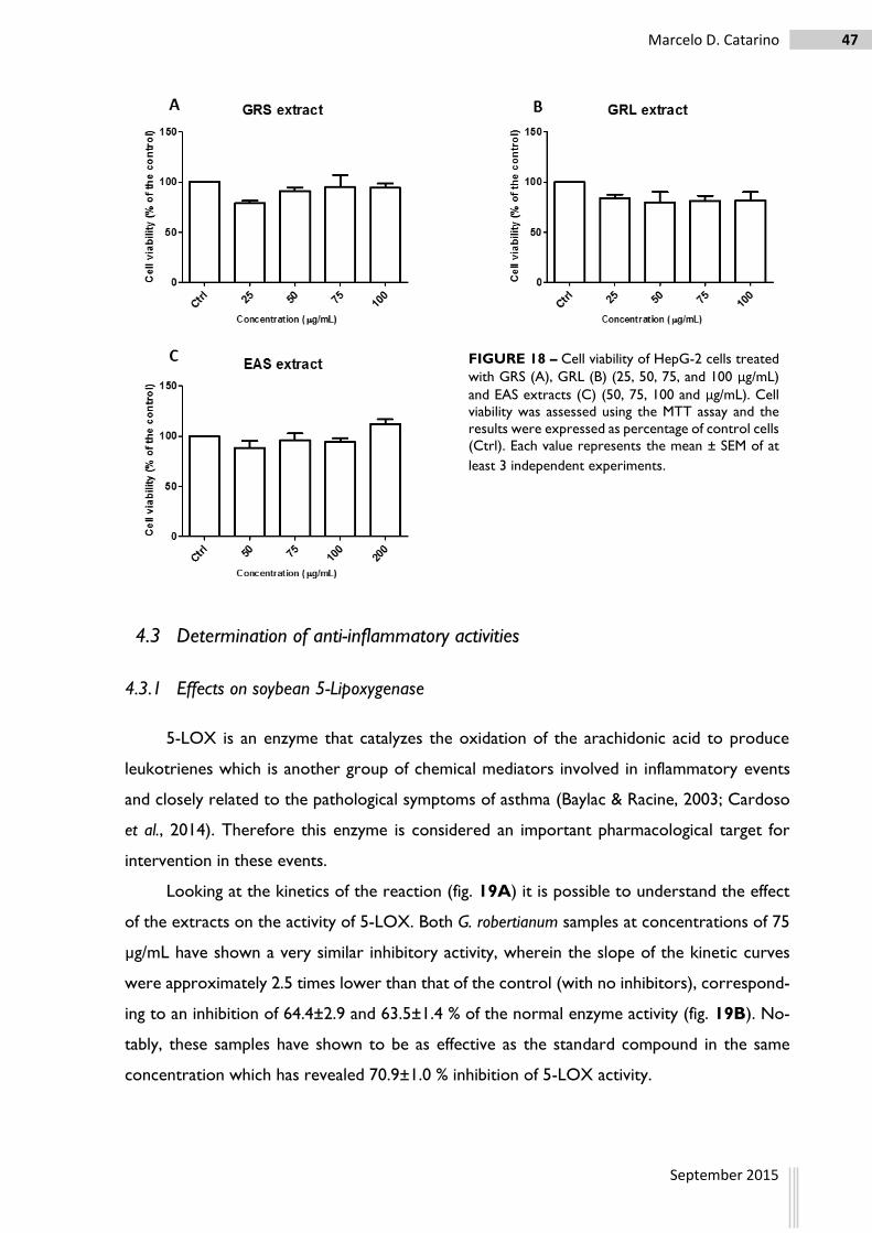
47 Marcelo D. Catarino
September 2015
4.3 Determination of anti-inflammatory activities
4.3.1 Effects on soybean 5-Lipoxygenase
5-LOX is an enzyme that catalyzes the oxidation of the arachidonic acid to produce
leukotrienes which is another group of chemical mediators involved in inflammatory events
and closely related to the pathological symptoms of asthma (Baylac & Racine, 2003; Cardoso
et al., 2014). Therefore this enzyme is considered an important pharmacological target for
intervention in these events.
Looking at the kinetics of the reaction (fig. 19A) it is possible to understand the effect
of the extracts on the activity of 5-LOX. Both G. robertianum samples at concentrations of 75
µg/mL have shown a very similar inhibitory activity, wherein the slope of the kinetic curves
were approximately 2.5 times lower than that of the control (with no inhibitors), correspond-
ing to an inhibition of 64.4±2.9 and 63.5±1.4 % of the normal enzyme activity (fig. 19B). No-
tably, these samples have shown to be as effective as the standard compound in the same
concentration which has revealed 70.9±1.0 % inhibition of 5-LOX activity.
FIGURE 18 – Cell viability of HepG-2 cells treated
with GRS (A), GRL (B) (25, 50, 75, and 100 μg/mL)
and EAS extracts (C) (50, 75, 100 and μg/mL). Cell
viability was assessed using the MTT assay and the
results were expressed as percentage of control cells
(Ctrl). Each value represents the mean ± SEM of at
least 3 independent experiments.

48 Evaluation of biological properties of E. africanus and G. robertianum
September 2015
FIGURE 19 – Kinetic curves of the 5-LOX activity in absence or presence of 75 µg/mL of the samples studied
(A) and their correspondent percentages of inhibition (B). The standard compound (STD) used was ascorbic
acid. Statistical analysis was performed by one-way ANOVA, followed by Tukey’s post-hoc test. ***P<0.001,
compared to the control (Ctrl). Data represent mean ± SEM of 3 independent assays.
The inhibitory activity exhibited by the EAS extract (also at 75 µg/mL) was less pro-
nounced than those mentioned before. This extract was able to inhibit the 5-LOX activity in
29.7±1.1 %, which corresponds to approximately half of the effectiveness of the remaining
samples. Yet, the inhibitory effect of this sample can still be considered relevant.
4.3.2 Effects on NO● production and cell viability on macrophages RAW 264.7
NO● is a signaling molecule resultant from the conversion of arginine into citrulline by
iNOS and responsible for the transduction of a variety of signals closely related with the path-
ogenesis of inflammation (Sharma, Al-Omran & Parvathy, 2007). Therefore, inhibition of this
radical species beneficiates the decrease of inflammatory conditions.
As it is possible to depict from the figure 20, under normal conditions, macrophages
RAW 264.7 produce low levels of nitrites (approximately 0.6±0.2 μM). Upon stimulation of
these cells with LPS for 24h, the production of nitrites increased up to about 24.2±0.5 μM.
Based on the positive effects previously observed regarding the scavenging ability of
chemically-generated NO●, it was expected that the pre-treatment of the cells with the three
most promising samples would attenuate the levels of nitrites production by the LPS-stimu-
lated RAW 264.7 macrophages. Still, this was not observed for the EAS extract, as it did not
influenced the NO● release for any of the tested concentrations (fig. 20D). However, pre-
treatment of non-stimulated cells with 50 and 75 μg/mL of this extract significantly potentiated
cell viability (fig. 20A), indicating an increase of the cell metabolism. Despite not statistically
significant, there is also a tendency for these same concentrations to increase the metabolism
of LPS-stimulated cells when compared to the untreated ones.

49 Marcelo D. Catarino
September 2015
FIGURE 20 – Effects of the pre-treatment with EAS (50, 75, 100 and 200 μg/mL), GRS and GRL (25, 50, 75 and
100 μg/mL) on the cell viability (% of the control) (A, B and C, respectively) and NO● levels (μM) (D, E and F,
respectively) on RAW 264.7 cells after 24h of incubation with () or without () LPS. Statistical analysis was
performed by one-way ANOVA, followed by Tukey’s post-hoc test. **P<0.01; ***P<0.001, compared to the
control without LPS; #P<0.05; ##P<0.01; ###P<0.001, compared to the control with LPS. Data represent mean ±
SEM of 3 independent assays.

50 Evaluation of biological properties of E. africanus and G. robertianum
September 2015
In turn, GRS and GRL extracts revealed significant inhibitory effects for the highest
concentrations tested (100 μg/mL) (fig. 20E and F). However, looking at figure 20C, it is
possible to conclude that the exposure of RAW 264.7 to GRL at this concentration also de-
creased cells viability, comparing to cells treated with LPS alone. Hence, the comparison of
figures 20C and F allowed to conclude that GRL does not exert an effective reduction of
NO● release but instead, it caused cell death of macrophages.
Hence, the only promising results were obtained with GRS which was able to reduce
the nitrites levels without interfering with cell viability (fig. 20B). Therefore, this sample was
subjected to Western blot analysis in order to deeply explore the molecular mechanisms be-
hind its anti-inflammatory effect, specifically whether GRS extract could block the LPS-induced
iNOS and COX-2 protein levels.
4.3.3 Effects on expression of iNOS and COX-2 enzymes
The effect of GRS on the expression of the enzymes iNOS and COX-2 are represented
in figure 21. As expected, in normal conditions, cells do not express neither iNOS nor COX-
2. In turn, when the cells are stimulated with LPS the expression of this enzymes is triggered
indicating that they entered in an inflammatory stage.
iNOS
Ctrl
LPS
GRS
GRS+L
PS
0
50
100
150
*** ***
### ###
iNO
S e
xp
ressio
n
(% o
f co
ntr
ol
wit
h L
PS
)
iNOS
Ctrl
LPS
GRS
GRS+L
PS
0
50
100
150
*** ***
### ###
iNO
S e
xp
ressio
n
(% o
f co
ntr
ol
wit
h L
PS
)
COX-2
Ctrl
LPS
GRS
GRS+L
PS
0
50
100
150
200
### ###
#
***
***
CO
X-2
exp
ressio
n
(% o
f co
ntr
ol
wit
h L
PS
)
FIGURE 21 – Effect of the GRS extract (100µg/L)
pre-treatment in the expression of iNOS and COX-
2 enzymes (% of the LPS) in RAW 264.7 macrophages
after 24h of incubation with () or without () LPS.
An anti-β-Tubulin antibody was used to confirm equal
protein loading and normalize the data. Statistical
analysis was performed by one-way ANOVA, fol-
lowed by Tukey’s post-hoc test. ***P<0.001, com-
pared to the control without LPS; #P<0.05; ###P<0.001, compared to the control with LPS. The
blot is representative of 3 similar blots. iNOS=135
kDa and COX-2=69 kDa

51 Marcelo D. Catarino
September 2015
The results depicted in figure 21 demonstrated that pre-treatment of RAW 264.7 with
100 µg/mL of GRS extract did not decrease the protein levels of the enzymes.
4.4 Identification and quantification of phenolic constituents from E. africanus and
G. robertianum
The hydroethanolic EAS and EAL extracts represented 15 and 13% of the dried plant
material, respectively while the correspondent extracts from G. robertianum accounted for 18
and 15% of dried stems and leaves of the plant, respectively.
The results obtained from Folin – Ciocalteu
method revealed that the total amount of phenol-
ics recovered for E. africanus was higher in stems
(321 mg GAE/g of dry material) than for leaves
(171 mg GAE/g of dry material), while G. roberti-
anum exhibited a reversed panorama with higher
recovery of phenolic compounds in the leaves ex-
tract (462 mg GAE/g of dry material) with regard
to that of stems (194 mg GAE/g of dry material)
(fig. 22). Looking from an overall perspective,
these results indicate that, despite both plants
are rich in phenolic compounds, G. robertianum
as a whole plant clearly stands out when com-
pared to E. africanus.
Overall, the HPLC–DAD–MSn analysis allowed to conclude that, despite the variances
in the peak intensities, both leaves and stems extracts of each plant demonstrated similar
chromatographic profiles between each other, i.e., the composition of the leaves extracs were
similar to that of the stems extracts within the same plant.
E. africanus extracts were particularly enriched in mono- and dicaffeoylquinic acids which
accounted for approximately 90% and 74% of the total quantified phenolics for stems and
leaves, respectively. On the other hand, G. robertianum extracts were more abundant in gallic
and ellagic acid derivatives.
The chromatographic profiles for the two plants can be consulted in figures 23 and 24,
and in the tables 4 and 5, respectively. A more detailed description of their phenolic compo-
sition will be further discussed in below.
FIGURE 22 – Quantification of the total phe-
nolic content on E. africanus leaves and stems
(EAL and EAS, respectively) and G. robertianum
leaves and stems extracts (GRL and GRS, re-
spectively). Statistical analysis was performed
by one-way ANOVA, followed by Tukey’s
post-hoc test. ***P<0.001; **P<0.01, compared
to GRL. Data represent mean ± SEM of 3 in-
dependent assays.
Folin - Ciocalteu
EAL
EAS
GRL
GRS
0
200
400
600
***
**
***
mg
GA
E/g
of
dry
pla
nt
mate
rial

52 Evaluation of biological properties of E. africanus and G. robertianum
September 2015
4.4.1 Phenolic constituents in E. africanus extracts
Seven distinct caffeic acid derivatives were detected in the hydroethanolic extracts of
stems and leaves of E. africanus, constituting the majority of the identified compounds (fig. 23
and table 4). These compounds eluted at 9.7, 13.4, 20.9, 21.9, 22.9, 24.0 and 24.9 min showed
characteristic UV spectra consistent to that described for caffeic and caffeoylquinic acid deriv-
atives (Schütz et al., 2004; Weisz, Kammerer & Carle, 2009). Of those, the major representa-
tive constituents of both extracts were eluted at 13.4 and 24.0 min, respectively.
FIGURE 23 – Chromatographic profile of E. africanus hydroethanolic extracts at 280 nm. Chromatogram corre-
sponding to stems extract is represented in bold lines, while the thin lines represent the chromatogram corre-
sponding to the leaves extract. QA – quinic acid; PCA-hex – protocatechuic acid-hexoside; 1-CQA – 1-
caffeoylquinic acid; FA-der – ferulic acid derivative; ERD-hexa – eriodictyol-hexuronide; CA-der – caffeic acid
derivative; 3,4-diCQA – 3,4-dicaffeoylquinic acid, 3,5-diCQA – 3,5-dicaffeoylquinic acid; 4,5-diCQA – 4,5-
dicaffeoylquinic acid; ERD – eriodictyol.
The compound eluting at 13.4 min was identified as a mono-caffeoylquinic acid and was
assigned to chlorogenic acid i.e., 3-O-caffeoylquinic acid, since its retention time, MS data and
corresponding MS2 fragmentation pattern were in agreement with that of the standard com-
pound. Notably, for both E. africanus extracts, 3-O-caffeoylquinic acid represented approxi-
mately 17% of the quantified phenolics and its total recovery was similar for stems and leaves
(7.3 ± 0.1 and 6.9 ± 0.3 mg/g dried stems and leaves, respectively), thus suggesting that this
phenolic compound is equally distributed in these two plant organs. In turn, the isomeric form
of this compound, i.e. 1-caffeoylquinic acid (with retention time of 9.7 min), was only detected
as a minor constituent in both extracts with slight prevalence in the stems.
The other prevalent caffeoylquinic acid derivative (eluted at 24.0 min) was highly re-
covered from the EAS extract (19.4 ± 0.2 mg/g dried plant material) than from the EAL extract
(16.3 ± 0.1 mg/g dried plant material). The ESI-MS spectrum of this compound revealed a base
peak at m/z 515, with a main MS2 product ion at m/z 353 (−162 Da, loss of caffeoyl moiety),
which in turn showed main fragments on MS3 spectrum at m/z 191 (−162 Da, loss of other
caffeoyl moiety), and at m/z 135. According to this features, this compound was assigned as

53 Marcelo D. Catarino
September 2015
3,5-dicaffeoylquinic acid which together with two other dicaffeoylquinic isomers (MW 516 Da)
that eluted at 22.9 and 24.9 min, accounted for about 71% and 56% of the total quantified
phenolics in the hydroethanolic extracts of stems and leaves, respectively. The UV–vis spec-
trum of these two other compounds, as well as the fragmentation pattern of its molecular ion
at m/z 515 and the corresponding MS2 pattern (which showed a base peak ion at m/z 353),
was similar to that of 3,5-dicaffeoylquinic acid. However, differences were evident on the
HPLC retention times and MS3 fragmentation patterns. Both MS3 spectra of the ion at m/z 353
eluting at 22.9 and 24.9 min showed a base peak ion at m/z 173, which indicates the presence
of a caffeoyl moiety bonded to quinic acid at the position 4 (Clifford et al., 2003). It is also
known that, among the three dicaffeoylquinic acid isomers that exist in nature with this feature
(3,4-diCQA, 1,4-diCQA and 4,5-diCQA), both 1,4-diCQA and 4,5-diCQA elute after 3,5-
diCQA under HPLC reversed-phase conditions (Clifford et al., 2003; Schütz et al., 2004; Weisz,
Kammerer & Carle, 2009). Moreover, 1,4-diCQA is the only one showing the product ions
at m/z 299, m/z 203 and m/z 255 in the MS2 spectrum of the ion at m/z 353 (Clifford, Knight
& Kuhnert, 2005). By these reasons, the compound detected at 24.9 min could be assigned to
1,4-diCQA. On the other hand, the compound eluting at 22.9 min was identified as 3,4-diCQA,
since it eluted before 3,5-diCQA, such as described in the literature for HPLC reversed-phase
conditions (Schütz et al., 2004). The concentrations of the above identified diCQA isomers
(3,4-diCQA and 1,4-diCQA) were similar within the same extract, yet, their overall recovery
was higher in the stems extract (5.6 ± 0.2 and 5.4 ± 0.1 mg/g dried stems, respectively) than
that in the leaves (4.0 ± 0.2 and 3.7 ± 0.1 mg/g dried leaves, respectively).
Notably, E. africanus extracts also contained two caffeoyl-hexuronide derivatives (with
retention times of 20.9 and 21.9 min), which were substantially abundant in the EAL extract
(1.6 ± 0.1 and 1.1 ± 0.0 mg/g dried plant material, respectively), while only vestigial amounts
of these compounds were found in the EAS extract. Both compounds had a molecular ion of
518 Da and possessed equivalent UV–vis and fragmentation patterns on MSn analysis. In par-
ticular, the base peak ion in the MS2 spectrum was observed at m/z 355 (−162 Da, i.e., equiv-
alent to the loss of a caffeoyl moiety or of a hexose). Moreover, the MS3 spectrum of the latter
ion corroborated the hypothesis of a caffeoyl-hexuronide derivative, as characteristic fragment
ions of caffeic acid (at m/z 179 and m/z 135) and of an hexuronide acid (at m/z 175 and m/z 113)
were registered (Bastos, Saldanha & Catharino, 2007; Gu, Zhong & Chen, 1999).

54 Evaluation of biological properties of E. africanus and G. robertianum
September 2015
TA
BL
E 4
– Identifica
tion o
f LC
-DA
D-E
SI/M
Sn d
ata,
and q
uan
tifica
tion o
f th
e m
ost
rele
vant
frac
tions
from
the e
xtr
acts
of E. afr
icanus.
RT
(m
in)
λ max
Co
mp
ou
nd
(MW
) M
ain
fra
gm
en
t E
SI-
MS
n
mg/g
dri
ed
mate
riala
Co
mp
ou
nd
S
tem
s L
eave
s
4.2
269
192
MS2
[191]: 1
73
- -
Quin
ic a
cid
5.3
279
316
MS2
[315]: 1
53; M
S3[1
53]: 1
09
0.4
±0.0
-
Pro
toca
tech
uic
-aci
d-4
-glu
co-
side
9.7
324
354
MS2
[353]: 1
91; M
S3[1
91]: 1
73, 109
- -
1-C
affe
oyl
quin
ic a
cid
13.4
219, 239, 325
354
MS2
[353]: 1
91, 179
7.3
.±0.1
6.9
±0.3
3-C
affe
oyl
quin
ic a
cid
15.6
324
560
MS2
[559]: 1
93; M
S3[1
93]: 1
34
1.5
±0.0
-
Feru
lic a
cid d
eri
vative
19.9
283
464
MS2
[463]: 2
87, 151; M
S3[2
87]: 1
51; M
S4[1
51]: 1
07
1.6
±0.1
7.8
±0.1
Eri
odic
tiol-
O-h
exuro
nid
e
20.9
318
518
MS2
[517]: 3
55, 311, 179; M
S3[3
55]: 3
11, 179, 175, 113,
135; M
S4[3
11]:175, 113, 135
- 1.6
±0.1
C
affe
oyl
-hexuro
nid
e d
eri
va-
tive
21.9
319
518
MS2
[517]: 3
55, 311, 473, 293, 179 ; M
S3[3
55]: 3
11, 179,
175, 113, 135; M
S4[3
11]:175, 113, 135, 251, 293
- 1.1
±0.0
C
affe
oyl
-hexuro
nid
e d
eri
va-
tive
22.9
217, 241, 325
516
MS2
[515]: 3
53; M
S3[3
53]: 1
73, 179, 191, 135; M
S4[1
73]:
111, 155
5.6
±0.2
4.0
±0.2
3,4
- D
icaf
feoyl
quin
ic a
cid
24.0
218, 326
516
MS2
[515]: 3
53; M
S3[3
53]: 1
91, 135; M
S4[1
91]: 1
73, 127
19.4
±0.2
16.3
±0.1
3,5
- D
icaf
feoyl
quin
ic a
cid
610
MS2
[609]: 3
01; M
S3[3
01]: 2
86, 213
- -
Hesp
ere
tin
24.9
218, 242, 327
516
MS2
[515]: 3
53; 203; 299; 255; 173; 179 M
S3[3
53]: 1
73, 179
5.4
±0.1
3.7
±0.1
1,4
- D
icaf
feoyl
quin
ic a
cid
28.4
228, 288
288
MS2
[287]: 1
51
0.2
±0.0
0.2
±0.0
Eri
odic
tyo
l
a D
ata
repre
sent
the m
ean
val
ues
± S
.E.M
.

55 Marcelo D. Catarino
September 2015
Hence, albeit the overall structural information of these two isomers could not be
achieved, the gathered information allow to conclude that these are caffeoyl-hexuronide with
an additional caffeic acid and/or hexose moiety.
Besides the caffeic acid derivatives, other phenolic compounds were detected in low
amounts in the hydroethanolic extracts of E. africanus. These included protocatechuic acid-
hexoside ([M−H]− at m/z 315, eluted at 5.3 min) and a ferulic acid derivative
([M−H]− at m/z 560, eluted at 15.6 min) that overall accounted for 1.9 mg/g of dried stem and
only appeared as vestigial components in leaves.
Flavonoids in E. africanus only comprised flavanones. Of these, hesperetin (co-eluted at
24.0 min) and eriodictyol (eluted at 28.4 min) were detected as minor components in the
extracts of the two plant organs. The most representative flavanone eluted at 19.9 min and
was assigned to eriodictyol-O-hexuronide since its MS data and fragmentation profile were
consistent to those reported in the literature (Manach et al., 2004; Pereira et al., 2013). Inter-
estingly, its abundance was clearly more pronounced in the EAL extract with regard to that of
EAS extract (7.8 ± 0.1 and 1.6 ± 0.1 mg/g dried material, respectively).
4.4.2 Phenolic constituents of G. robertianum extracts
Nine compounds have been identified in G. robertianum extracts, wherein five were as-
signed to ellagic acid and ellagitannin derivatives. These compounds eluted at 10.2 (co-eluted
with chlorogenic acid), 16.5, 17.4, 20.4 and 33.9 min, and together constitute the majority of
the identified compounds in these samples (fig. 24, table 5)
FIGURE 24 – Chromatographic profile of G. robertianum hydroethanolic extracts at 280 nm. Chromato-
gram corresponding to stems extract is represented in bold lines, while the thin lines represent the chromato-
gram corresponding to the leaves extract. DOPAC – 3,4-dihydroxyphenylacetic acid; GA – gallic acid; CQA –
caffeoylquinic acid; ET-der – ellagitannin derivative; G-HHDP-hex – Galloyl-HHDP-hexoside; BCA – brevifolin
carboxylic acid; EA – ellagic acid.

56 Evaluation of biological properties of E. africanus and G. robertianum
September 2015
The compound eluted at 33.9 min was easily identified as ellagic acid since it eluted with
high percentages of acetonitrile and its UV spectra together with the MS fragmentation pat-
terns were in agreement to literature data (Fischer, Carle & Kammerer, 2011; Lee, Johnson
& Talcott, 2005; Meyers, Swiecki & Mitchell, 2006). Galloyl-HHDP-hexoside has also been
detected in G. robertianum extracts. In fact, this compound was detected at two different elut-
ing times, the first co-eluted at 10.2 min and the second at 17.4 min, which indicates that they
are two isomeric forms of the same compound.
The fragmentation of these two compounds (base peak at m/z 633) have released the
fragment at m/z 301 that typically indicates the presence of ellagic acid. Moreover, the frag-
mentation pattern as well as the UV spectra of these compounds are consistent to previous
reports (Fischer, Carle & Kammerer, 2011; Mena et al., 2012).
The two remaining ellagitannin derivatives were eluted at 16.5 and 20.4 min. Although
the exact identification of these compounds was not achieved, the MS2 fragmentation revealed
the presence of the fragment at m/z 301 indicating the presence of ellagic acid. Besides, looking
at the UV spectra of these compounds it is possible to note a similarity to that of the ellagic
acid (Fracassetti et al., 2013; Mullen et al., 2003).
Notably, the compounds eluting at 7.0 and 18.1 min have also shown to be expressive
in the G. robertianum extracts. The former revealed a UV/vis maxima at 172 nm and its full MS
spectrum showed a base peak at m/z 169. Moreover, the product ion at m/z 125 (loss of
carboxylic acid moiety) was obtained in the tandem fragmentation. Comparing this results with
the data in the literature it is possible to clearly identify this compound as gallic acid (Lee,
Johnson & Talcott, 2005). On the other hand, the fragmentation of the compound eluted at
TABLE 5 – Identification of LC-DAD-ESI/MSn data of the most relevant fractions from the extracts
of G. robertianum.
RT (min) λmax Compound
(MW)
Main fragment
ESI- MSn Compound
5.5 285 168 MS2[167]: 123 3,4-Dihydroxyphenylacetic acid
7.0 272 170 MS2[169]: 125 Gallic acid
10.2 220, 325 354 MS2[353]: 191, 179, 135 Caffeoylquinic acid
634 MS2[633]: 301, 463 Galloyl-HHDP- hexoside
16.5 274 485 MS2[484]: 462, 169
MS3[462]: 440, 169, 301, 364, 247 Ellagitanin derivative
17.4 267 634 MS2[633]: 301, 463, 419 Galloyl-HHDP- hexoside
18.1 277, 355 292 MS2[291]: 247 Brevifolin carboxilic acid
20.4 279 463 MS2[462]: 453, 415, 169, 301 Ellagitannin derivative
33.9 279 302 MS2[301]:257, 133, 284, 185 Ellagic acid
57 Marcelo D. Catarino
September 2015
18.1 min (base peak at m/z 291) originated a fragment ion at m/z 247 (again loss of carboxylic
acid moiety) which is characteristic to that of brevifolin carboxylic acid (Antonia Alvarez-
Fernadez et al., 2015). Moreover the UV/vis maxima at 277 and 355 nm is also in agreement
to what has been described for this compound (He & Xia, 2007).
Interestingly, caffeoylquinic acid was also present in the G. robertianum extracts
([M−H]− at m/z 353, eluted at 10.2 min). However this compound, together with the 3,4-dihy-
droxyphenylacetic acid ([M−H]− at m/z 168, eluted at 5.5 min) were only found in minor amounts.


DISCUSSION


61 Marcelo D. Catarino
September 2015
5 DISCUSSION
Because of the health benefits that have been attributed to E. africanus and G. robertianum,
these two plants have been traditionally used in the folk medicine for the treatment of several
ailments. In this study, the cytotoxicity, antioxidant and anti-inflammatory properties of hy-
droethanolic extracts from these two plants were investigated, along with their phenolic com-
position, in order to disclose its potential for therapeutic and/or nutraceutical purposes.
The antioxidant abilities of leaves and stems extracts from both plant species were eval-
uated for their scavenging and reducing capacities, as well as the protective effects towards
oxidative degradation of lipids or fluorescein.
In a general perspective, GRL extract was clearly the most promising sample regarding
to the antioxidant abilities, showing low IC50 values in DPPH●, ABTS●+ and particularly in OH●
scavenging assays (7.6±0.6, 3.9±0.6 and 45.1±2.4 μg/mL, respectively). In fact, in the latter
assay its effect was even better than that of the standard compound (196.2±16.4). The GRS
extract also demonstrated significantly good radical scavenging effects in these three assays,
followed by the EAS extract. Opposing to GRL extract, the EAL extract was undoubtedly the
one with the worst scavenging abilities, registering the highest IC50 values for all the three
assays (30.4±1.7, 17.2±1.6 and 214.2±37.5 μg/mL for DPPH●, ABTS●+and OH●, respectively).
Despite this, it is of noteworthy that the DPPH● scavenging activity herein estimated for the
EAL extract was better than that previously reported by Njenga & Viljoen (2006) for the
acetone extracts of E. africanus (IC50 values ranging from 38 to 50 μg/mL).
According to the HPLC–DAD–MSn analysis, E. africanus extracts were enriched in
caffeoylquinic acids (which accounted for approximately 90% and 74% of the quantified com-
pounds in stems and leaves, respectively), while ellagitannins were the major compounds iden-
tified in G. robertianum extracts. It is worth noting that either chlorogenic acids or ellagitannins
have proven strong antiradical activities (Hagerman et al., 1998; Park, 2013) and hence, it is
probable that these compounds are important contributing players for the overall activity ev-
idenced in the present study. The herein gathered results also corroborate the relevance of
phenolic compounds in the extracts’ antiradical activities since the most promising sample, i.e.,
the GRL extract, was the most enriched in phenolic compounds (462 mg GAE/g of dry mate-
rial), while the least active sample, i.e., EAL extract, was the poorest one (171 mg GAE/g of
dry material).
Notwithstanding, this straight correlation between the phenolic content and antioxidant
activity was not so evidenced in TBARS and ORAC assays for the two plants. E.g. in the first
method, EAL extracts revealed better activity than that of the EAS extract, despite its poverty

62 Evaluation of biological properties of E. africanus and G. robertianum
September 2015
in phenolic compounds with respect to the latter extract (268.6±45.9 against 416.1±237
μg/mL). Also, the EAS extract, which is less enriched in phenolic compounds than GRL, was
undoubtedly the sample that more effectively protected fluorescein from the oxidative dam-
age, revealing an ORAC value that represents approximately twice of the antioxidant potential
of the remaining samples. These results suggest that other compounds that have not been
considered (e.g. presence of non-phenolic compounds), could also contribute for the antioxi-
dant activities of these extracts.
Another possible justification is that the different phenolic compounds present in each
extract could be differently contributing for their antioxidant activities, since significant phe-
nolic differences have been encountered. According to the HPLC-MSn analysis, nine com-
pounds were identified in G. robertianum, from which the major six corresponded to ellagitan-
nins and respective basic units (gallic, ellagic and brevifolin carboxylic acids). The two remaining
minor compounds were identified as caffeoylquinic acid and 3,4-dihydroxyphenylacetic acid.
No flavonoids were detected in the extracts from this plant. On the other hand, twelve peaks
have been identified in the E. africanus extracts, and seven of them matched with caffeoylquinic
acid derivatives. Other phenolic acids, namely protocatechuic and ferulic acids were also pre-
sent in minor quantities, and the only flavonoids identified were hesperetin and eriodictyol
derivatives, both belonging to the flavanones group. The presence of flavonoids were more
expressive in the EAL extract than in the stems, mainly as eriodictiol-O-hexuronide. Consid-
ering that eriodictyol has been reported previously as an effective inhibitor of TBARS (Areias
et al., 2001), it is possible that the EAL extract enrichment in a derivative of this flavanone with
respect to the EAS extract can contribute for its stronger effects on TBARS assay.
Likewise, correlation between high caffeoylquinic acids content and good ORAC values
have already been reported previously (Kayano et al., 2002; Mullen et al., 2011). In fact, strong
ORAC results have also been described for the chlorogenic acids rather than for ellagic acid
and ellagitannins, such as galloyl-HDDP or brevifolin (Ishimoto et al., 2012). Since the EAS
extract is particularly enriched with this type of compounds, this could be a possible explana-
tion for the results obtained in ORAC assay. However, EAL extract that is also abundant in
chlorogenic acids, did not revealed as good results as the stems extract, meaning that these
compounds are not the only explanation for the effects on ORAC assay. In this case, it is
probable that other non-phenolic compounds not identified in the EAS extract may also ac-
count for this result.
Among all antioxidant assays, FRAP was the one for which all the samples revealed the
weakest results. Both GRL and EAS extracts simultaneously demonstrated superior ferrous

63 Marcelo D. Catarino
September 2015
ion reducing capacity compared to the remaining samples. Still these results fall a little short
when compared to BHT, indicating that reducing heavy metals might not be the primary path-
way for their antioxidant properties.
NO● is one of the many chemical mediators produced by the cells of the immune system
upon an inflammatory state, thus, inhibition of this radical counteracts the inflammatory pro-
cess (Cardoso et al., 2014). Therefore, testing samples ability to scavenge chemically-generated
NO● is very useful approach not only for evaluating their antioxidant properties, but also as a
primary screening of their potential anti-inflammatory capacities. The results have shown that
both G. robertianum extracts were potent scavengers of NO●, with IC50 values 14 and 12 times
lower (20.0±0.9 and 24.2±8.0 μg/mL for GRL and GRS, respectively) than that of the standard
compound (285.7±15.4 μg/mL). On the other hand, the results obtained for E. africanus ex-
tracts were not so prominent since both extracts revealed approximately twice the IC50 values
of ascorbic acid (526.4±22.5 and 538.6±16.4 μg/mL for EAL and EAS extracts, respectively).
According to the literature, the scavenging effects of ellagic acid over NO● are stronger than
those of chlorogenic acid (Rodrigues, Toledo Benassi & Bragagnolo, 2014; Srivastava, Jagan
Mohan Rao & Shivanandappa, 2007) and hence this fact can partly explain the results herein
described.
As the overall perspective, antioxidant assays pointed EAL extract as the least promising
sample and because of that, it was then excluded from the subsequent assays.
Due to the functional features of the liver and their role in the metabolic elimination of
xenobiotics, the evaluation of potential hepatotoxicity represents a critical step for attesting
the safety of the samples in study (Gómez-Lechón et al., 2010). In this context, the cell viability
of human hepatic cell line HepG-2 treated with EAL, GRL and GRS extracts was evaluated,
and the results revealed that no significant cytotoxic effects were exhibited for any of the
concentrations tested (25 – 100 μg/mL for G. robertianum extracts and 50 – 200 μg/mL for
EAS extract), thus attesting the safety profile of the extracts studied.
Since scientific data regarding the potential anti-inflammatory activities of E. africanus and
G. robertianum are scarce, this work also investigated the potential benefic effects of these two
plant extracts in inflammatory conditions. In this context, it was assessed whether these three
extracts could interfere with the activity of isolated 5-LOX. Very promising inhibitory effects
were obtained for G. robertianum extracts, which were able to inhibit approximately 65% of
this enzyme activity at 75 μg/mL, while for the same concentration, the EAS extract only re-
tarded the enzyme activity by about 30%.

64 Evaluation of biological properties of E. africanus and G. robertianum
September 2015
Notably, it has been previously demonstrated that chlorogenic acids (at 20 μM) have no
effects over 5-LOX. However, compounds like protocatechuic acid (at 20 μM), ferulic acid (at
50 μM) and eriodictyol (at 40 μM) were shown to be potent inhibitors of this enzyme (Lee et
al., 2010; Maqsood & Benjakul, 2010; Ribeiro et al., 2014). Therefore, despite these three
compounds are only present in minor concentrations, their combined activities could contrib-
ute for the slight inhibitory activity of EAS extracts over 5-LOX. Still, this hypothesis needs to
be confirmed.
On the other hand, the effects of isolated ellagic and gallic acids on lipoxygenase are still
poorly described. Several gallic acid derivatives have been reported as strong inhibitors of this
enzyme (Richard-Forget et al., 1995). Moreover, various extracts from different pomegranate
flowers, which are particularly enriched in gallic and ellagic acid compounds, have also revealed
promising effects on LOX activity (Bekir et al., 2013). Therefore, it is reasonable to assume
that these compounds might be responsible for the G. robertianum extracts effects over 5-
LOX, although with this limited information it is not possible to clearly confirm this hypothesis.
Thus, it would be necessary to test gallic and ellagic acids separately or alternatively, different
extract fractions that correspond to the various compounds identified in G. robertianum ex-
tracts, in order to understand the contribution of the individual compounds and/or possible
synergies on the inhibitory effects of these extracts against 5-LOX activity.
It is worth noting that this is the first time that G. robertianum extracts are described as
inhibitors of 5-LOX activity. In turn, E. africanus has already been described as moderate in-
hibitor of this enzyme, but only essential oils were tested in that study (Njenga & Viljoen,
2006).
Taking this results into account, it is conceivable to suggest that G. robertianum extracts
could be thought as potential pharmacological tools for the treatment of inflammatory disor-
ders in which lipoxygenase plays a crucial role, including asthma, rheumatoid arthritis or in-
flammatory bowel disease.
Both chlorogenic and ellagic acids, as well as some of their derivatives, have been previ-
ously reported for their promising effects as anti-inflammatory agents, namely through inhibi-
tion of iNOS and NO● release, COX-2 and even NF-κB (Umesalma & Sudhandiran, 2010;
Zhang et al., 2010). Moreover, given the results obtained in the chemical scavenging of NO●,
particularly for the G. robertianum extracts, it was expected that these samples would demon-
strate anti-inflammatory potential in biological systems, namely in RAW 264.7 cells. However,
EAS extract showed no evident inhibitory effect on nitrite production by macrophages up to
concentrations of 50 – 200 μg/mL. Despite this, it is interesting to note that macrophages

65 Marcelo D. Catarino
September 2015
treatment with EAS extract potentiated the cell metabolism of non-stimulated cells at the
concentrations of 50 and 75 μg/mL. This could mean an increase on the mitochondrial activity
or increased cell proliferation, which would be interesting to exploit for tissue regeneration
purposes. Either way, further studies would be necessary to exploit this possibility.
On the other hand, G. robertianum extracts showed a relevant inhibitory effect for the
highest concentration (100 μg/mL) on the nitrites levels in LPS-stimulated macrophages. How-
ever, for the GRL extract, this treatment also induced a decrease in cell viability of about 30%
and hence the observed decrement of nitrite levels are probably due to cell death instead of
an anti-inflammatory effect. Still note that as no toxic effect was registered for the treatment
of non-stimulated macrophages with GRL, the results suggest that this extract for itself at 100
μg/mL does not induce toxicity in the cells, but instead it might potentiate the toxicity of LPS.
The GRS extract was the only sample that reduced NO● levels in RAW 264.7 without
decreasing cells viability and therefore, in an attempt to disclose the molecular mechanisms
behind its anti-inflammatory activity, the expression of iNOS and COX-2 enzymes were fur-
ther analyzed by Western blot.
Previous studies have reported that ellagic acid was able to reduce the expression of
both COX-2 and iNOS and even NF-κB in different in vitro and in vivo models (Larrosa et al.,
2010; Mansouri et al., 2015; Umesalma & Sudhandiran, 2010). Therefore, it was expected that
the GRS extract would exhibit similar results on RAW 264.7 cells. However, data obtained in
this study showed that macrophages treatment with GRS extract at 100 μg/mL did not change
the levels of iNOS. A possible justification is that the interactions between the compounds in
the extract might be neutralizing the effects of ellagic acid, or alternatively the concentration
that was added to the medium was insufficient to exert the desirable effects. It is worth noting
that despite not influencing iNOS levels, the GRS extract was still able to decrease NO● levels
in LPS-activated macrophages. This indicates that, at 100 μg/mL, this sample possesses anti-
inflammatory potential through the scavenging of free radicals.
All the gathered data suggest that, given the antioxidant effects demonstrated by both
plants, they could be used as nutraceutical ingredients to enrich the basic nutritional food
dietary with additional nutrients that would provide extra benefits to protect from the nega-
tive effects of the oxidative stress that humans are frequently subject. However, it is important
to point out that chemical models herein used are far to accurately reproduce the effects that
these samples might have in a biological environment. Therefore, the results presented in this
work may only be considered as a primary screening for the samples antioxidant potential. To
further corroborate this data, it would be highly desirable to perform additional biological

66 Evaluation of biological properties of E. africanus and G. robertianum
September 2015
studies such as testing the samples in in vitro systems and even in in vivo animal models that
clearly provide results closer to the reality.
Likewise, the effects on 5-LOX activity indicate that there is a strong potential for the
G. robertianum extracts to be used as anti-inflammatory agents targeting this enzyme. Still, once
again, further studies in in vitro/in vivo models are necessary to validate the herein presented
results.
Regarding to the effects on the NO● production, only G. robertianum extracts exhibited
significant potential to inhibit the levels of this radical, but since GRL extract effects were
correlated to cell death, only the results obtained for the GRS extract were considered rele-
vant. Notwithstanding, after confirming that this extract did not affect the levels of iNOS en-
zyme, it was concluded that its capacity to reduce the nitrite levels was resultant from its
ability to scavenge this radical, which is once again an antioxidant skill. Furthermore, since all
the extracts studied have shown potent antioxidant abilities, evaluation of the intracellular
production of ROS would be another possible approach to explore the beneficial effects that
are attributed to these plants.

CONCLUSIONS AND
FUTURE PERSPECTIVES


69 Marcelo D. Catarino
September 2015
6 CONCLUSIONS AND FUTURE PERSPECTIVES
The main goal of this study was to evaluate the antioxidant, anti-inflammatory and safety
profile of E. africanus and G. robertianum hydroethanolic extracts, as well as to characterize
their phenolic constituents in order to understand if there is potential for exploitation of these
two plants as possible therapeutic and/or nutraceutical agents.
The antioxidant evaluation of the leaves and stems of Eriocephalus africanus (EAL and
EAS, repectively) and Geranium robertianum (GRL and GRS, respectively) was carried out
through DPPH●, ABTS●+, OH●, FRAP, TBARS, NO● and ORAC assays. It was observed that
GRL extract exhibited the best scavenging abilities revealing IC50 values of 7.6±0.6, 3.9±0.6,
45.1±2.4, 63.3±5.4 and 115.8±16.1 μg/mL for DPPH●, ABTS●+, OH●, FRAP and TBARS assays
respectively. The exception was in the ORAC assay where the EAS extract revealed the
strongest antioxidant capacity (4.01±0.3 μM TE). EAL extract was shown to be the less prom-
ising sample amongst the four tested.
When tested for their ability to scavenge the chemical generated NO●, both G. roberti-
anum extracts revealed remarkable results (20.0±0.9 and 24.2±8.0 μg/mL for GRL and GRS,
respectively), indicating that, in a primary approach, these two extracts might possess strong
anti-inflammatory abilities.
Since these plant extracts are intended to be used in human diet, their possible hepa-
totoxicity was also tested. For that, HepG-2, a human hepatocyte cell line, was cultured with
different concentrations of the plant extracts (25 – 100 μg/mL and 50 – 200 μg/mL for G.
robertianum and EAS extracts, respectively) but none revealed cytotoxic effects.
The anti-inflammatory effects of the samples were then evaluated through the inhibi-
tion of the 5-LOX activity as well as of nitrite production by RAW 264.7 macrophages. More-
over, the extract that revealed the most promising effects was also evaluated for its ability to
inhibit intracellular iNOS and COX-2 expression through Western blot analysis.
Both G. robertianum extracts revealed remarkable effect towards 5-LOX, inhibiting ap-
proximately 65% of its activity and hence, these results suggested that the extracts could be
exploited as possible therapeutic agents targeting this enzyme. GRS extract at 100 μg/mL also
caused relevant inhibitory effects on nitrites production by RAW 264.7 cells. The fact that this
extract did not interfered with the levels of intracellular iNOS indicates that the decreased
NO● levels were resultant from the extract ability to scavenge these radicals rather than in-
hibiting the enzyme expression that is responsible for its production. Figure 25 resumes sche-
matically where this extract acts during the inflammatory process.

70 Evaluation of biological properties of E. africanus and G. robertianum
September 2015
FIGURE 25 – Proposed protective mechanisms of GRS in oxidative-stress and inflammatory events. The oxi-
dative stress stimulus trigger the activation of nuclear factor κB (NF-κB) that translocates to the nucleus where
it enhances the transcription of various pro-oxidant and pro-inflammatory mediators genes including those of
the enzymes nicotinamide adenine dinucleotide phosphate oxidase (NOX), xanthine oxidase (XOX), cyclooxy-
genase-2 (COX-2), lipoxygenase (LOX), and inducible nitric oxide synthase (iNOS), as well as several cytokines
and chemokines. The pro-oxidant enzymes together with mitochondrial activity will generate more reactive spe-
cies that will contribute for the increase of the oxidative stress. In the presence of the GRS extract the reactive
species are neutralized attenuating the oxidative stress condition. In turn, the three pro-inflammatory enzymes
produce prostaglandins (PGE2) leukotrientes (LT4) and nitric oxide that are released in the extracellular space
together with various cytokines and chemokines propagating the inflammation by transducing the signals to other
cells. In presence of the GRS extract, the levels of nitric oxide produced by iNOS are reduced due to its ability
to scavenge this radical. Likewise, the GRS extract interferes with the LOX activity reducing the release of
leukotrienes to the extracellular space, contributing for the attenuation of the inflammatory event.

71 Marcelo D. Catarino
September 2015
The total phenolic quantification of the four extracts demonstrated that GRL extract
was the most phenolic enriched sample, while EAL extract was the poorest. Moreover,
through HPLC-MSn analysis it was observed that the E. africanus extracts were more abundant
in chlorogenic acids while G. robertianum had predominance of ellagic acid and ellagitannins.
A close correlation between the antioxidant activities and the total phenolic content
was noted, though there was some exceptions. E.g. in the TBARS assay the EAL extract was
slightly better than the EAS extract (268.6±45.9 against 416.1±237 μg/mL, respectively) which
could be partially related to its higher content in eriodictyol-hexuronide, and the EAS extract
was better than the others in the ORAC assay, which might be explained not only by its
abundance in chlorogenic acids but also by the presence of other non-phenolic compounds
that have not been considered in this study. Moreover, the abudance of G. robertianum in ellagic
acid might contribute to the remarkable effects of these extracts on the chemical generated
NO● scavenging assay. However, against what was expected, the GRL was unable to repro-
duce that effect on RAW 264.7 macrophages, while the GRS extract could induce a significant
decrease of this radical but was ineffective in inhibiting the iNOS and COX-2 expressions. In
order to confirm the in vitro anti-inflammatory activity of the GRS extract other pro-inflam-
matory mediators and signaling pathways should be addressed, namely cytokines, NF-κB tran-
scription factor and mitogen-activated protein kinases (MAPKs).
Overall, these results demonstrate that there is some potential for these extracts to
be exploited as therapeutic agents mainly due to their antioxidant properties. However, it is
of utmost importance to evaluate their properties in biological models in order to better
comprehend their behavior in more physiological environments. Likewise, as very promising
effects were shown for G. robertianum in 5-LOX activity, it would be interesting to investigate
in the future which compounds in the extracts better contribute for these results, as well as
the type of pharmacological inhibition that is exerted over the enzyme in biological systems,
i.e., if it is competitive, non-competitive or mixed inhibition. Moreover, despite E. africanus
extracts did not show relevant effects in nitrites production by RAW 264.7, they demon-
strated to somehow stimulate their metabolism. Therefore, it would be interesting to test
higher concentrations of these samples for their anti-inflammatory properties, or alternatively,
test their effects on other intracellular signaling cascades responsible for cell proliferation and
survival such as the mitogen activated protein kinase signaling cascade.


REFERENCE LIST


75 Marcelo D. Catarino
September 2015
7 REFERENCE LIST
ADOM, Kafui Kwami; IU, Rui Hai - Antioxidant Activity of Grains. Journal of agricultural and food chemistry. 50:21 (2002) 6182–6187. doi: 10.1021/jf0205099.
AMABEOKU, G. J. et al. - Effects of Tarchonanthus camphoratus and Eriocephalus africanus on nociception in mice and pyrexia in rats. Phytomedicine : international journal of phytotherapy and phytopharmacology. . ISSN 0944-7113. 7:6 (2000) 517–522. doi: 10.1016/S0944-7113(00)80038-0.
AMARAL, Sónia et al. - Plant extracts with anti-inflammatory properties-A new approach for characterization of their bioactive compounds and establishment of structure-antioxidant activity relationships. Bioorganic and Medicinal Chemistry. . ISSN 09680896. 17:5 (2009) 1876–1883. doi: 10.1016/j.bmc.2009.01.045.
ANSORENA, D. et al. - Development of nutraceuticals containing marine algae oils. Em DOMÍNGUEZ, HERMINIA B. T. (Ed.) - Functional ingredients from algae for foods and nutraceuticals. [S.l.] : Woodhead Publishing, 2013. ISBN 978-0-85709-512-1. p. 634–657.
ANTONIA ALVAREZ-FERNADEZ, M. et al. - Composition of Nonanthocyanin Polyphenols in Alcoholic- Fermented Strawberry Products Using LC−MS (QTRAP), High- Resolution MS (UHPLC-Orbitrap-MS), LC-DAD, and Antioxidant Activity. Journal of agricultural and food chemistry. 63:7 (2015) 2041–2051. doi: 10.1021/jf506076n.
AREIAS, Filipe Miguel et al. - Antioxidant effect of flavonoids after ascorbate/Fe2+-induced oxidative stress in cultured retinal cells. Biochemical Pharmacology. . ISSN 00062952. 62:1 (2001) 111–118. doi: 10.1016/S0006-2952(01)00621-9.
AYALA, Antonio; MUÑOZ, Mario F.; ARGÜELLES, Sandro - Lipid peroxidation: Production, metabolism, and signaling mechanisms of malondialdehyde and 4-hydroxy-2-nonenal. Oxidative Medicine and Cellular Longevity. . ISSN 19420994. 2014). doi: 10.1155/2014/360438.
BALAMURUGAN, K.; KARTHIKEYAN, J. - Evaluation of luteolin in the prevention of N-nitrosodiethylamine-induced hepatocellular carcinoma using animal model system. Indian Journal of Clinical Biochemistry. . ISSN 09701915. 27:2 (2012) 157–163. doi: 10.1007/s12291-011-0166-7.
BASTOS, DH; SALDANHA, LA; CATHARINO, RR - Phenolic antioxidants identified by ESI-MS from yerba mate (Ilex paraguariensis) and green tea (Camelia sinensis) extracts. Molecules. 12:3 (2007) 423–432. doi: 10.3390/12030423.
BAYLAC, Sabine; RACINE, Philippe - Inhibition of 5-lipoxygenase by essential oils and other natural fragment extracts. International Journal of Aromatherapy. . ISSN 09624562. 13:2-3 (2003) 138–142. doi: 10.1016/S0962-4562(03)00083-3.
BEKIR, Jalila et al. - Chemical composition and antioxidant, anti-inflammatory, and antiproliferation activities of pomegranate (Punica granatum) flowers. Journal of medicinal food. . ISSN 1557-7600. 16:6 (2013) 544–50. doi: 10.1089/jmf.2012.0275.

76 Evaluation of biological properties of E. africanus and G. robertianum
September 2015
BENZIE, I. F.; STRAIN, J. J. - The ferric reducing ability of plasma (FRAP) as a measure of «antioxidant power»: the FRAP assay. Analytical biochemistry. . ISSN 0003-2697. 239:1 (1996) 70–6. doi: 10.1006/abio.1996.0292.
BOHN, Torsten - Dietary factors affecting polyphenol bioavailability. Nutrition Reviews. . ISSN 17534887. 72:7 (2014) 429–452. doi: 10.1111/nure.12114.
BOR, Jung-Yi; CHEN, Hui-Yin; YEN, Gow-Chin - Evaluation of antioxidant activity and inhibitory effect on nitric oxide production of some common vegetables. Journal of agricultural and food chemistry. . ISSN 0021-8561. 54:5 (2006) 1680–1686. doi: 10.1021/jf0527448.
BUFALO, M. C. et al. - Propolis and its constituent caffeic acid suppress LPS-stimulated pro-inflammatory response by blocking NF-κB and MAPK activation in macrophages. Journal of Ethnopharmacology. . ISSN 0378-8741. 149:1 (2013) 84–92. doi: 10.1016/j.jep.2013.06.004.
CAI, Yi-zhong et al. - Structure-radical scavenging activity relationships of phenolic compounds from traditional Chinese medicinal plants. Life sciences. . ISSN 00243205. 78:25 (2006) 2872–2888. doi: 10.1016/j.lfs.2005.11.004.
CARDONA, Fernando et al. - Benefits of polyphenols on gut microbiota and implications in human health. The Journal of nutritional biochemistry. . ISSN 1873-4847. 24:8 (2013) 1415–22. doi: 10.1016/j.jnutbio.2013.05.001.
CARDOSO, Susana M. et al. - Virgin olive oil as a source of anti-inflammatory agents. Em LEONARDIS, ANTONELLA DE (Ed.) - Virgin Olive Oil: Production, Composition, Uses and Benefits for Man. New York : Nova Science Publishers, 2014
CARREAU, Charlotte et al. - Enterodiol and enterolactone, two major diet-derived polyphenol metabolites have different impact on ERα transcriptional activation in human breast cancer cells. Journal of Steroid Biochemistry and Molecular Biology. . ISSN 09600760. 110:1-2 (2008) 176–185. doi: 10.1016/j.jsbmb.2008.03.032.
CATARINO, Marcelo D. et al. - Characterization of phenolic constituents and evaluation of antioxidant properties of leaves and stems of Eriocephalus africanus. Arabian Journal of Chemistry. . ISSN 18785352. 2015). doi: 10.1016/j.arabjc.2015.04.018.
CATARINO, Marcelo D. et al. - Antioxidant Capacities of Flavones and Benefits in Oxidative-Stress Related Diseases. Current Topics in Medicinal Chemistry. . ISSN 1873-4294 (Electronic). 15:2 (2015) 105–119.
CATARINO, Marcelo Dias et al. - Mediaterranean Diet: A Precious Tool for Fighting Inflammatory Diseases. Em COBB, DEAN T. (Ed.) - Polyphenols: Food Sources, Bioactive Properties and Antioxidant Effects. New York : Nova Science Publishers, 2014. ISBN 978-1-63117-858-0. p. 87–112.
CHANDRASEKARA, Anoma; SHAHIDI, Fereidoon - Content of insoluble bound phenolics in millets and their contribution to antioxidant capacity. Journal of Agricultural and Food Chemistry. . ISSN 00218561. 58:11 (2010) 6706–6714. doi: 10.1021/jf100868b.

77 Marcelo D. Catarino
September 2015
CHANG, Yuan-Ching et al. - Structure-activity relationship of C6-C3 phenylpropanoids on xanthine oxidase-inhibiting and free radical-scavenging activities. Biochemical Pharmacology. . ISSN 00062952. 43:11 (2007) 1541 – 1551. doi: 10.1016/j.bcp.2007.11.023.
CHIANG, Yi-ming et al. - Ethyl caffeate suppresses NF-kappaB activation and its downstream inflammatory mediators, iNOS, COX-2, and PGE2 in vitro or in mouse skin. British journal of pharmacology. . ISSN 0007-1188. 146:3 (2005) 352–363. doi: 10.1038/sj.bjp.0706343.
CILLA, Antonio et al. - Foods or Bioactive Constituents of Foods as Chemopreventives in Cell Lines After Simulated Gastrointestinal Digestion: A Review. Em MORALES-GONZÁLEZ, JOSÉ A. (Ed.) - Oxidative Stress and Chronic Degenerative Diseases - A Role for Antioxidants. ISBN 978-953-51-1123-8. p. 131–151.
CLIFFORD, Michael N. et al. - Hierarchical scheme for LC-MSn identification of chlorogenic acids. Journal of Agricultural and Food Chemistry. . ISSN 00218561. 51:1 (2003) 2900–2911. doi: 10.1021/jf026187q.
CLIFFORD, Michael N.; KNIGHT, Susan; KUHNERT, Nikolai - Discriminating between the six isomers of dicaffeoylquinic acid by LC-MS(n). Journal of agricultural and food chemistry. . ISSN 0021-8561. 53:10 (2005) 3821–3832. doi: 10.1021/jf050046h.
COMALADA, M.; XAUS, J.; GÁLVEZ, J. - Bioactive Food as Dietary Interventions for Arthritis and Related Inflammatory Diseases. [S.l.] : Elsevier, 2013. ISBN 9780123971562.
COMALADA, Mònica et al. - Inhibition of pro-inflammatory markers in primary bone marrow-derived mouse macrophages by naturally occurring flavonoids: analysis of the structure-activity relationship. Biochemical pharmacology. . ISSN 0006-2952. 72:8 (2006) 1010–21. doi: 10.1016/j.bcp.2006.07.016.
COS, Paul et al. - Structure-activity relationship and classification of flavonoids as inhibitors of xanthine oxidase and superoxide scavengers. Journal of Natural Products. . ISSN 01633864. 61:1 (1998) 71–76. doi: 10.1021/np970237h.
CRAGG, Gordon M.; NEWMAN, David J. - Natural products: a continuing source of novel drug leads. Biochimica et biophysica acta. . ISSN 0006-3002. 1830:6 (2013) 3670–95. doi: 10.1016/j.bbagen.2013.02.008.
CROZIER, Alan; RIO, Daniele DEL; CLIFFORD, Michael N. - Bioavailability of dietary flavonoids and phenolic compounds. Molecular aspects of medicine. . ISSN 1872-9452. 31:6 (2010) 446–67. doi: 10.1016/j.mam.2010.09.007.
D’ARCHIVIO, Massimo et al. - Polyphenols, dietary sources and bioavailability. Annali dell’Istituto Superiore di Sanita. . ISSN 00212571. 43:4 (2007) 348–361. doi: Vol. 43, no. 4: 348-361.
D’ARCHIVIO, Massimo et al. - Bioavailability of the polyphenols: Status and controversies. International Journal of Molecular Sciences. . ISSN 14220067. 11:4 (2010) 1321–1342. doi: 10.3390/ijms11041321.
DAGLIA, Maria - Polyphenols as antimicrobial agents. Current Opinion in Biotechnology. . ISSN 09581669. 23:2 (2012) 174–181. doi: 10.1016/j.copbio.2011.08.007.

78 Evaluation of biological properties of E. africanus and G. robertianum
September 2015
DIXON, Richard A; PASINETTI, Giulio M. - Flavonoids and isoflavonoids: from plant biology to agriculture and neuroscience. Plant physiology. . ISSN 1532-2548. 154:2 (2010) 453–7. doi: 10.1104/pp.110.161430.
DRÖGE, Wulf - Free radicals in the physiological control of cell function. Physiological reviews. . ISSN 0031-9333. 82:1 (2002) 47–95. doi: 10.1152/physrev.00018.2001.
ERMER, J.; MILLER, J. M.; ERMER JOACHIM, John Miller - Method Validation in Pharmaceutical Analysis: A Guide to Best Practice. Weinheim : Wiley-VCH Verlag GmbH & Co., 2005
ESPÍN, Juan Carlos et al. - Biological Significance of Urolithins, the Gut Microbial Ellagic Acid-Derived Metabolites: The Evidence So Far. Evidence-based complementary and alternative medicine. New York. 2013). doi: 10.1155/2013/270418.
ESPÍN, Juan Carlos; GARCÍA-CONESA, María Teresa; TOMÁS-BARBERÁN, Francisco A. - Nutraceuticals: Facts and fiction. Phytochemistry. . ISSN 00319422. 68:22-24 (2007) 2986–3008. doi: 10.1016/j.phytochem.2007.09.014.
FEDERICO, Alessandro et al. - Chronic inflammation and oxidative stress in human carcinogenesis. International Journal of Cancer. . ISSN 00207136. 121:11 (2007) 2381–2386. doi: 10.1002/ijc.23192.
FERNANDES, Iva et al. - Bioavailability of anthocyanins and derivatives. Journal of Functional Foods. . ISSN 17564646. 7:1 (2014) 54–66. doi: 10.1016/j.jff.2013.05.010.
FERREIRA, Fernanda M. et al. - «MitoTea»: Geranium robertianum L. decoctions decrease blood glucose levels and improve liver mitochondrial oxidative phosphorylation in diabetic Goto-Kakizaki rats. Acta Biochimica Polonica. 57:4 (2010) 399–402.
FERREYRA, María L. Falcone; RIUS, Sebastián P.; CASATI, Paula - Flavonoids: biosynthesis, biological functions, and biotechnological applications. Frontiers in Plant Science. 3:222 (2012) 1–15. doi: 10.3389/fpls.2012.00222.
FISCHER, Ulrike A.; CARLE, Reinhold; KAMMERER, Dietmar R. - Identification and quantification of phenolic compounds from pomegranate (Punica granatum L.) peel, mesocarp, aril and differently produced juices by HPLC-DAD-ESI/MSn. Food Chemistry. . ISSN 03088146. 127:2 (2011) 807–821. doi: 10.1016/j.foodchem.2010.12.156.
FODOREA, Cristina-Stefania et al. - Preliminary HPLC study on some polyphenols of Geranium robertianum L. (Geraniaceae). Revista medico-chirurgicala a Societatii de Medici si Naturalisti din Iasi. Romania. . ISSN 0048-7848. 109:1 (2005) 174–178.
FRACASSETTI, Daniela et al. - Ellagic acid derivatives, ellagitannins, proanthocyanidins and other phenolics, vitamin C and antioxidant capacity of two powder products from camu-camu fruit (Myrciaria dubia). Food Chemistry. . ISSN 03088146. 139:1-4 (2013) 578–588. doi: 10.1016/j.foodchem.2013.01.121.
FREITAS, Pedro Manuel Marques DE - Polyphenols and Neurodegenerative Diseases. [S.l.] : University of Porto, 2012
GARAZD, M. M.; GARAZD, Ya. L.; KHILYA, V. P. - Neoflavones. 1. Natural Distribution and Spectral and Biological Properties. Chemistry of natural compounds. 39:1 (2003) 47–82.

79 Marcelo D. Catarino
September 2015
GERTZ, Melanie et al. - A Molecular Mechanism for Direct Sirtuin Activation by Resveratrol. PLoS ONE. . ISSN 19326203. 7:11 (2012). doi: 10.1371/journal.pone.0049761.
GHARRAS, Hasna El - Polyphenols: Food sources, properties and applications - A review. International Journal of Food Science and Technology. . ISSN 09505423. 44:12 (2009) 2512–2518. doi: 10.1111/j.1365-2621.2009.02077.x.
GÓMEZ-LECHÓN, M. J. et al. - In vitro evaluation of potential hepatotoxicity induced by drugs. Current pharmaceutical design. . ISSN 13816128. 16:17 (2010) 1963–1977. doi: 10.2174/138161210791208910.
GONZÁLEZ, R. et al. - Effects of flavonoids and other polyphenols on inflammation. Critical reviews in food science and nutrition. . ISSN 1549-7852. 51:4 (2011) 331–62. doi: 10.1080/10408390903584094.
GU, JK; ZHONG, DF; CHEN, XY - Analysis of O-glucuronide conjugates in urine by electrospray ion trap mass spectrometry. Fresenius’ journal of analytical chemistry. 365:6 (1999) 553–558. doi: 10.1007/s002160051521.
GU, Liwei et al. - Concentrations of proanthocyanidins in common foods and estimations of normal consumption. The Journal of nutrition. . ISSN 0022-3166. 134:3 (2004) 613–617.
HAGERMAN, Ann E. et al. - High Molecular Weight Plant Polyphenolics (Tannins) as Biological Antioxidants. Journal of Agricultural and Food Chemistry. 46:5 (1998) 1887–1892. doi: 10.1021/jf970975b.
HE, Zhiyong; XIA, Wenshui - Analysis of phenolic compounds in Chinese olive (Canarium album L.) fruit by RPHPLC-DAD-ESI-MS. Food Chemistry. . ISSN 03088146. 105:3 (2007) 1307–1311. doi: 10.1016/j.foodchem.2007.04.049.
HEIM, Kelly E.; TAGLIAFERRO, Anthony R.; BOBILYA, Dennis J. - Flavonoid antioxidants: chemistry, metabolism and structure-activity relationships. The Journal of Nutritional Biochemistry. . ISSN 09552863. 13:10 (2002) 572–584. doi: 10.1016/S0955-2863(02)00208-5.
HIGDON, Jane V; FREI, Balz - Tea Catechins and Polyphenols: Health Effects, Metabolism, and Antioxidant Functions. Critical Reviews in Food Science and Nutrition. 43:1 (2003) 89–143. doi: 10.1080/10408690390826464.
HOOPER, Lee et al. - Flavonoids, flavonoid-rich foods, and cardiovascular risk: a meta-analysis of randomized controlled trials. The American journal of clinical nutrition. United States. . ISSN 1938-3207. 88:1 (2008) 38–50.
ISHIMOTO, Hidekazu et al. - Antioxidative Properties of Functional Polyphenols and Their Metabolites Assessed by an ORAC Assay. Bioscience, Biotechnology, and Biochemistry. . ISSN 0916-8451. 76:2 (2012) 395–399. doi: 10.1271/bbb.110717.
IWASHINA, T. - The structure and distribution of the flavonoids in plants. Journal of Plant Research. 113:3 (2000) 287–299. doi: 10.1007/PL00013940.

80 Evaluation of biological properties of E. africanus and G. robertianum
September 2015
JEMIA, Mariem Ben et al. - Antioxidant activity of Tunisian Geranium robertianum L. (Geraniaceae). Natural product research. . ISSN 1478-6427. 27:22 (2013) 2076–83. doi: 10.1080/14786419.2013.782492.
JURD, Leonard - Recent progress in the chemistry of flavylium salts. [S.l.] : Elsevier, 2013. ISBN 9781483220017.
KAYANO, Shin Ichi et al. - Antioxidant activity of prune (Prunus domestica L.) constituents and a new synergist. Journal of Agricultural and Food Chemistry. . ISSN 00218561. 50:13 (2002) 3708–3712. doi: 10.1021/jf0200164.
KHANSARI, Nemat; SHAKIBA, Yadollah; MAHMOUDI, Mahdi - Chronic inflammation and oxidative stress as a major cause of age-related diseases and cancer. Recent patents on inflammation & allergy drug discovery. 3:1 (2009) 73–80. doi: 10.2174/187221309787158371.
KIM, Hyun Pyo et al. - Anti-inflammatory plant flavonoids and cellular action mechanisms. Journal of pharmacological sciences. . ISSN 1347-8613. 96:3 (2004) 229–245. doi: 10.1254/jphs.CRJ04003X.
KIM, Kyung Hee et al. - Phenolic acid profiles and antioxidant activities of wheat bran extracts and the effect of hydrolysis conditions. Food Chemistry. . ISSN 03088146. 95:3 (2006) 466–473. doi: 10.1016/j.foodchem.2005.01.032.
KNAZE, Viktoria et al. - Intake estimation of total and individual flavan-3-ols, proanthocyanidins and theaflavins, their food sources and determinants in the European Prospective Investigation into Cancer and Nutrition (EPIC) study. British Journal of Nutrition. . ISSN 0007-1145. 108:06 (2012) 1095–1108. doi: 10.1017/S0007114511006386.
KOBAKHIDZE, K. B.; ALANIYA, M. D. - Flavonoids from Geranium robertianum. CHEMISTRY OF NATURAL COMPOUNDS. . ISSN 0009-3130. 40:1 (2004) 89–90. doi: 10.1023/B:CONC.0000025477.18086.8b.
KUMAR, Ramanathan Sambath et al. - Antimicrobial and antioxidant activities of Careya arborea Roxb. stem bark. Iranian Journal of Pharmacology and Therapeutics. . ISSN 17352657. 5:1 (2006) 35–41.
KUMAR, Shashank; PANDEY, Abhay K. - Chemistry and biological activities of flavonoids: an overview. The Scientific World Journal. 2013). doi: 10.1155/2013/162750.
LAMPORT, Daniel J. et al. - The effects of flavonoid and other polyphenol consumption on cognitive performance : A systematic research review of human experimental and epidemiological studies. Nutrition and Aging. 1:2012) 5–25. doi: 10.3233/NUA-2012-0002.
LANDETE, J. M. - Updated knowledge about polyphenols: functions, bioavailability, metabolism, and health. Critical reviews in food science and nutrition. . ISSN 1549-7852. 52:10 (2012) 936–48. doi: 10.1080/10408398.2010.513779.
LARROSA, Mar et al. - Ellagitannins, ellagic acid and vascular health. Molecular aspects of medicine. . ISSN 1872-9452. 31:6 (2010) 513–39. doi: 10.1016/j.mam.2010.09.005.
81 Marcelo D. Catarino
September 2015
LEE, Eun Ju et al. - Phenolic constituents from the flower buds of Lonicera japonica and their 5-lipoxygenase inhibitory activities. Food Chemistry. . ISSN 03088146. 120:1 (2010) 134–139. doi: 10.1016/j.foodchem.2009.09.088.
LEE, Joon Hee; JOHNSON, Jodie V.; TALCOTT, Stephen T. - Identification of ellagic acid conjugates and other polyphenolics in muscadine grapes by HPLC-ESI-MS. Journal of Agricultural and Food Chemistry. . ISSN 00218561. 53:15 (2005) 6003–6010. doi: 10.1021/jf050468r.
LEOPOLDINI, Monica et al. - Antioxidant properties of phenolic compounds: H-atom versus electron transfer mechanism. Journal of Physical Chemistry A. . ISSN 10895639. 108:22 (2004) 4916–4922. doi: 10.1021/jp037247d.
LIU, Rui Hai - Potential synergy of phytochemicals in cancer prevention: mechanism of action. The Journal of nutrition. . ISSN 0022-3166. 134:12 Suppl (2004) 3479S–3485S. doi: 134/12/3479S [pii].
MADE, Sanne M. VAN DER; MENSINK, Ronald P. - Polyphenols for Cholesterol Management. Em GARG, ABHIMANYU (Ed.) - Dyslipidemias SE - 22. [S.l.] : Humana Press, 2015. ISBN 978-1-60761-423-4. p. 371–382.
MANACH, Claudine et al. - Polyphenols: food sources and bioavailability. The American journal of clinical nutrition. . ISSN 0002-9165. 79:5 (2004) 727–747.
MANACH, Claudine et al. - Bioavailability and bioefficacy of polyphenols in humans. I. Review of 93 intervention studies. American Journal of Clinical Nutrition. . ISSN 00029165. 81:1 Suppl (2005) 243S–255S. doi: 81/1/230S [pii].
MANSOURI, Mohammad Taghi et al. - A study of the mechanisms underlying the anti-inflammatory effect of ellagic acid in carrageenan-induced paw edema in rats. Indian Journal of Pharmacology. India. . ISSN 0253-7613. 47:3 (2015) 292–298. doi: 10.4103/0253-7613.157127.
MAQSOOD, Sajid; BENJAKUL, Soottawat - Comparative studies of four different phenolic compounds on in vitro antioxidative activity and the preventive effect on lipid oxidation of fish oil emulsion and fish mince. Food Chemistry. . ISSN 03088146. 119:1 (2010) 123–132. doi: 10.1016/j.foodchem.2009.06.004.
MARAIS, Jannie P. J. et al. - The Stereochemistry of Flavonoids. Em GROTEWOLD, ERICH (Ed.) - The Science of Flavonoids. Ohio : Springer Science+Business Media, Inc., 2006. p. 1–46.
MCCUNE, Letitia M. et al. - Cherries and health: a review. Critical reviews in food science and nutrition. . ISSN 1040-8398. 51:1 (2011) 1–12. doi: 10.1080/10408390903001719.
MEIRELES, Gyselle Cynthia Silva; SANTOS, Adrielle Mangabeira - Mecanismos de ação da laserterapia sobre componentes do processo inflamatório. Revista Eletrônica da Fainor. 3:1 (2010) 30–40.
MENA, Pedro et al. - Rapid and comprehensive evaluation of (Poly)phenolic compounds in pomegranate (Punica granatum L.) Juice by UHPLC-MSn. Molecules. . ISSN 14203049. 17:12 (2012) 14821–14840. doi: 10.3390/molecules171214821.

82 Evaluation of biological properties of E. africanus and G. robertianum
September 2015
MERLE, Hugo et al. - Chemical composition of the essential oils from Eriocephalus africanus L. var. africanus populations growing in Spain. Flavour and Fragrance Journal. . ISSN 0882-5734. 22:6 (2007) 461–464.
MEYERS, Katherine J.; SWIECKI, Tedmund J.; MITCHELL, Alyson E. - Understanding the Native Californian diet: Identification of condensed and hydrolyzable tannins in tanoak acorns (Lithocarpus densiflorus). Journal of Agricultural and Food Chemistry. . ISSN 00218561. 54:20 (2006) 7686–7691. doi: 10.1021/jf061264t.
MULLEN, W. et al. - The antioxidant and chlorogenic acid profiles of whole coffee fruits are influenced by the extraction procedures. Journal of Agricultural and Food Chemistry. . ISSN 00218561. 59:8 (2011) 3754–3762. doi: 10.1021/jf200122m.
MULLEN, William et al. - Analysis of ellagitannins and conjugates of ellagic acid and quercetin in raspberry fruits by LC-MSn. Phytochemistry. . ISSN 00319422. 64:2 (2003) 617–624. doi: 10.1016/S0031-9422(03)00281-4.
NEAGU, Elena et al. - Evaluation of antioxidant capacity of Geranium robertianum extracts. Revue Roumaine de Chimie. . ISSN 00353930. 55:6 (2010) 321–325.
NJENGA, E. W.; VILJOEN, A. M. - In vitro 5-lipoxygenase inhibition and anti-oxidant activity of Eriocephalus L. (Asteraceae) species. South African Journal of Botany. . ISSN 02546299. 72:4 (2006) 637–641.
NJENGA, Elizabeth Wanjiku - The chemotaxonomy, Phylogeny and Biological Activity of the Genus Eriocephalus L. (Asteraceae). [S.l.] : Faculty of Health Sciences, University of the Witwatersrand, 2005
NORTH AMERICAN MENOPAUSE SOCIETY - The role of soy isoflavones in menopausal health: report of The North American Menopause Society/Wulf H. Utian Translational Science Symposium in Chicago, IL (October 2010). Menopause. United States. . ISSN 1530-0374. 18:7 (2011) 732–753. doi: 10.1097/gme.0b013e31821fc8e0.
PANDEY, Kanti Bhooshan; RIZVI, Syed Ibrahim - Plant polyphenols as dietary antioxidants in human health and disease. Oxidative medicine and cellular longevity. . ISSN 1942-0900. 2:5 (2009) 270–278. doi: 10.4161/oxim.2.5.9498.
PARK, Jae B. - Isolation and quantification of major chlorogenic acids in three major instant coffee brands and their potential effects on H2O2-induced mitochondrial membrane depolarization and apoptosis in PC-12 cells. Food & function. England. 4:11 (2013) 1632–1638. doi: 10.1039/c3fo60138b.
PAUN, Gabriela et al. - Evaluation of Geranium spp., Helleborus spp. and Hyssopus spp. polyphenolic extracts inhibitory activity against urease and α-chymotrypsin. Journal of enzyme inhibition and medicinal chemistry. . ISSN 1475-6374. 6366:1 (2013) 1–7. doi: 10.3109/14756366.2012.749399.
PEREIRA, David M. et al. - Phenolics: From chemistry to biology. Molecules. . ISSN 14203049. 14:6 (2009) 2202–2211. doi: 10.3390/molecules14062202.
PEREIRA, Olívia R. et al. - Phenolic constituents of Lamium album: Focus on isoscutellarein derivatives. Food Research International. . ISSN 09639969. 48:1 (2012) 330–335. doi: 10.1016/j.foodres.2012.04.009.

83 Marcelo D. Catarino
September 2015
PEREIRA, Olívia R. et al. - Simultaneous characterization and quantification of phenolic compounds in Thymus x citriodorus using a validated HPLC–UV and ESI–MS combined method. Food Research International. . ISSN 09639969. 54:2 (2013) 1773–1780.
PERRON, Nathan R.; BRUMAGHIM, Julia L. - A review of the antioxidant mechanisms of polyphenol compounds related to iron binding. Cell Biochemistry and Biophysics. . ISSN 10859195. 53:2 (2009) 75–100. doi: 10.1007/s12013-009-9043-x.
PETRUSSA, Elisa et al. - Plant flavonoids-biosynthesis, transport and involvement in stress responses. International Journal of Molecular Sciences. . ISSN 16616596. 14:7 (2013) 14950–14973. doi: 10.3390/ijms140714950.
PIWOWARSKI, Jakub P. et al. - Role of human gut microbiota metabolism in the anti-inflammatory effect of traditionally used ellagitannin-rich plant materials. Journal of Ethnopharmacology. . ISSN 18727573. 155:1 (2014) 801–809. doi: 10.1016/j.jep.2014.06.032.
PORRINI, Marisa; RISO, Patrizia - Factors influencing the bioavailability of antioxidants in foods: A critical appraisal. Nutrition, Metabolism and Cardiovascular Diseases. . ISSN 09394753. 18:10 (2008) 647–650. doi: 10.1016/j.numecd.2008.08.004.
PUUPPONEN-PIMIÄ, R. et al. - Antimicrobial properties of phenolic compounds from berries. Journal of Applied Microbiology. . ISSN 13645072. 90:4 (2001) 494–507. doi: 10.1046/j.1365-2672.2001.01271.x.
RASHIDINEJAD, Ali et al. - Effects of catechin on the phenolic content and antioxidant properties of low-fat cheese. International Journal of Food Science and Technology. . ISSN 09505423. 48:12 (2013) 2448–2455. doi: 10.1111/ijfs.12234.
REUTER, Simone et al. - Oxidative stress, inflammation, and cancer: How are they linked? Free Radical Biology and Medicine. 49:11 (2010) 1603–1616. doi: 10.1016/j.freeradbiomed.2010.09.006.Oxidative.
RIBEIRO, Daniela et al. - Inhibition of LOX by flavonoids: A structure-activity relationship study. European Journal of Medicinal Chemistry. . ISSN 02235234. 72:2014) 137–145. doi: 10.1016/j.ejmech.2013.11.030.
RIBEIRO, Daniela et al. - Inhibition of LOX by flavonoids: a structure-activity relationship study. European journal of medicinal chemistry. . ISSN 1768-3254. 72:2014) 137–45. doi: 10.1016/j.ejmech.2013.11.030.
RICHARD-FORGET, Florence et al. - Inhibition of Horse Bean and Germinated Barley Lipoxygenases by Some Phenolic Compounds. Journal of Food Science. . ISSN 1750-3841. 60:6 (1995) 1325–1329. doi: 10.1111/j.1365-2621.1995.tb04583.x.
RODRIGUES, Naira Poerner; TOLEDO BENASSI, Marta; BRAGAGNOLO, Neura - Scavenging capacity of coffee brews against oxygen and nitrogen reactive species and the correlation with bioactive compounds by multivariate analysis. Food Research International. . ISSN 09639969. 61:2014) 228–235. doi: 10.1016/j.foodres.2013.09.028.
ROMAN, G. Paun et al. - Antioxidant activity of Geranium robertianum concentrated extracts by ultrafiltration process. Planta Medica. . ISSN 0032-0943. 75:09 (2009) 4–5. doi: 10.1055/s-0029-1234557.

84 Evaluation of biological properties of E. africanus and G. robertianum
September 2015
SALIE, F.; EAGLES, P. F.; LENG, H. M. - Preliminary antimicrobial screening of four South African Asteraceae species. Journal of ethnopharmacology. . ISSN 0378-8741. 52:1 (1996) 27–33.
SANDHAR, Harleen Kaur et al. - A review of phytochemistry and pharmacology of flavonoids. Internationale Pharmaceutica Sciencia. 1:1 (2011) 25–41.
SANTANGELO, Carmela et al. - Polyphenols, intracellular signalling and inflammation. Annali dell’Istituto superiore di sanità. . ISSN 0021-2571. 43:4 (2007) 394–405.
SANTHAKUMAR, A. B.; BULMER, A. C.; SINGH, I. - A review of the mechanisms and effectiveness of dietary polyphenols in reducing oxidative stress and thrombotic risk. Journal of Human Nutrition and Dietetics. . ISSN 09523871. 27:1 (2014) 1–21. doi: 10.1111/jhn.12177.
SCALBERT, Augustin; WILLIAMSON, Gary - Dietary Intake and Bioavailability of Polyphenols. Journal of Medicinal Food. . ISSN 1096-620X. 3:2 (2000) 121–125. doi: 10.1089/109662000416311.
SCHÜTZ, Katrin et al. - Identification and quantification of caffeoylquinic acids and flavonoids from artichoke (Cynara scolymus L.) heads, juice, and pomace by HPLC-DAD-ESI/MS(n). Journal of agricultural and food chemistry. . ISSN 0021-8561. 52:13 (2004) 4090–4096.
SEERAM, Navindra P. - Polyphenols Overview. Em Encyclopedia of Dietary Supplements, Second Edition. [S.l.] : CRC Press, 2010. ISBN 978-1-4398-1928-9. p. 632–634.
SHARMA, J. N.; AL-OMRAN, A.; PARVATHY, S. S. - Role of nitric oxide in inflammatory diseases. Inflammopharmacology. Switzerland. . ISSN 0925-4692 (Print). 15:6 (2007) 252–259. doi: 10.1007/s10787-007-0013-x.
SILK, Jennifer J.; SMOLIGA, James M. - Resveratrol: Nutraceutical believed to counteract the detrimental effects of high-fat diet. Lipid Technology. . ISSN 18635377. 26:1 (2014) 15–17. doi: 10.1002/lite.201300315.
SLIMESTAD, Rune; FOSSEN, Torgils; VÅGEN, Ingunn Molund - Onions: A source of unique dietary flavonoids. Journal of Agricultural and Food Chemistry. . ISSN 00218561. 55:25 (2007) 10067–10080. doi: 10.1021/jf0712503.
SMITH, P. K. et al. - Measurement of protein using bicinchoninic acid. Analytical Biochemistry. . ISSN 00032697. 150:1 (1985) 76–85. doi: 10.1016/0003-2697(85)90442-7.
SNYDER, L. R.; KIRKLAND, J. J.; DOLAN, J. W. - Introduction to modern liquid chromatography. 3th. ed. New Jersey : John Wiley & Sons, Inc., 2009. ISBN 0471038229.
SRIVASTAVA, Anup; JAGAN MOHAN RAO, L.; SHIVANANDAPPA, T. - Isolation of ellagic acid from the aqueous extract of the roots of Decalepis hamiltonii: Antioxidant activity and cytoprotective effect. Food Chemistry. . ISSN 03088146. 103:1 (2007) 224–233. doi: 10.1016/j.foodchem.2006.08.010.
SVENDSEN, Mathias T. et al. - Contact sensitization to Geranium robertianum L. in an amateur gardener. Contact Dermatitis. . ISSN 01051873. 72:6 (2015) 420–421. doi: 10.1111/cod.12378.

85 Marcelo D. Catarino
September 2015
TAKANO-ISHIKAWA, Y.; GOTO, M.; YAMAKI, K. - Structure-activity relations of inhibitory effects of various flavonoids on lipopolysaccharide-induced prostaglandin E2 production in rat peritoneal macrophages: comparison between subclasses of flavonoids. Phytomedicine : international journal of phytotherapy and phytopharmacology. . ISSN 0944-7113. 13:5 (2006) 310–7. doi: 10.1016/j.phymed.2005.01.016.
TANWAR, Beenu; MODGIL, Rajni - Flavonoids: Dietary Occurrence and Health Benefits. Spatula DD - Peer Reviewed Journal on Complementary Medicine and Drug Discovery. . ISSN 1309-9914. 2:1 (2012) 59. doi: 10.5455/spatula.20120328100506.
TAPPEL, A L.; LUNDBERG, W. O.; BOYER, P. D. - Effect of temperature and antioxidants upon the lipoxidase-catalyzed oxidation of sodium linoleate. Archives of biochemistry and biophysics. . ISSN 00039861. 42:2 (1953) 293–304. doi: 10.1016/0003-9861(53)90359-2.
THATOI, H. N.; PATRA, J. K.; DAS, S. K. - Free radical scavenging and antioxidant potential of mangrove plants: a review. Acta Physiologiae Plantarum. . ISSN 0137-5881. 36:3 (2013) 561–579. doi: 10.1007/s11738-013-1438-z.
TOFTS, Richard J. - Geranium robertianum L. Journal of Ecology. . ISSN 00220477. 92:3 (2004) 537–555. doi: 10.1111/j.0022-0477.2004.00892.x.
TOMÁS-BARBERÁN, Francisco A.; CLIFFORD, Michael N. - Flavanones, chalcones and dihydrochalcones - Nature, occurrence and dietary burden. Journal of the Science of Food and Agriculture. . ISSN 00225142. 80:7 (2000) 1024–1032. doi: 10.1002/(SICI)1097-0010(20000515)80:7<1024::AID-JSFA567>3.0.CO;2-S.
TSAO, Rong - Chemistry and biochemistry of dietary polyphenols. Nutrients. . ISSN 2072-6643. 2:12 (2010) 1231–46. doi: 10.3390/nu2121231.
UMESALMA, Syed; SUDHANDIRAN, Ganapasam - Differential inhibitory effects of the polyphenol ellagic acid on inflammatory mediators NF-κB, iNOS, COX-2, TNF-α, and IL-6 in 1,2-dimethylhydrazine-induced rat colon carcinogenesis. Basic and Clinical Pharmacology and Toxicology. . ISSN 17427835. 107:2 (2010) 650–655. doi: 10.1111/j.1742-7843.2010.00565.x.
VARDENY, Orly; SOLOMON, Scott D. - Cyclooxygenase-2 inhibitors, nonsteroidal anti-inflammatory drugs, and cardiovascular risk. Cardiology clinics. . ISSN 1558-2264. 26:4 (2008) 589–601. doi: 10.1016/j.ccl.2008.06.004.
VEBERIC, Robert et al. - Anthocyanin composition of different wild and cultivated berry species. LWT - Food Science and Technology. . ISSN 00236438. 60:1 (2015) 509–517. doi: 10.1016/j.lwt.2014.08.033.
WEISZ, Georg M.; KAMMERER, Dietmar R.; CARLE, Reinhold - Identification and quantification of phenolic compounds from sunflower (Helianthus annuus L.) kernels and shells by HPLC-DAD/ESI-MSn. Food Chemistry. . ISSN 03088146. 115:2 (2009) 758–765.
XIAO, Jianbo; MUZASHVILI, Tamar S.; GEORGIEV, Milen I. - Advances in the biotechnological glycosylation of valuable flavonoids. Biotechnology advances. . ISSN 1873-1899. 32:6 (2014) 1145–56. doi: 10.1016/j.biotechadv.2014.04.006.

86 Evaluation of biological properties of E. africanus and G. robertianum
September 2015
YANG, Yong-Jin et al. - Radical scavenging activity and cytotoxicity of active quinic acid derivatives from Scorzonera divaricata roots. Food chemistry. . ISSN 0308-8146. 138:2-3 (2013) 2057–63.
YEN, Gow Chin; DUH, Pin Der - Scavenging Effect of Methanolic Extracts of Peanut Hulls on Free-Radical and Active-Oxygen Species. Journal of Agricultural and Food Chemistry. 42:3 (1994) 629–632.
ZHANG, Xu et al. - Chlorogenic acid protects mice against lipopolysaccharide-induced acute lung injury. Injury. . ISSN 00201383. 41:7 (2010) 746–752. doi: 10.1016/j.injury.2010.02.029.