march, 2019 sayaka nakashima graduate school of
TRANSCRIPT

Dietary flavonoids and their colonic catabolites as
cytoprotective antioxidants against oxidative stress
March, 2019
SAYAKA NAKASHIMA
Graduate School of Environmental and Life Science
(Doctor Course)
OKAYAMA UNIVERSITY, JAPAN

2
CONTENTS
CONTENTS 2
LIST OF TABLES AND FIGURES 6
ABBREVIATION 7
ABSTRACT 9
CHAPTER 1 General Introduction 12
1.1 Functional foods 12
1.2 Flavonoids 12
1.2.1 Chemical structure and classification of flavonoids 13
1.2.2 Dietary sources of flavonoids 13
1.2.3 Metabolism and absorption of flavonoids 14
1.2.4 Biological activities and health benefits of flavonoids 15
1.3 Oxidative stress and disease 16
1.3.1 Reactive oxygen species 16
1.4 Cytoprotective system against xenobiotics and oxidative stress 17
1.4.1 Aryl hydrocarbon receptor signaling pathway 18
1.4.2 Keap1-Nrf2 signaling pathway 18
1.4.3 Interaction between Keap1/Nrf2 pathway and AhR pathway 19
1.5 Click chemistry 22
1.5.1 The Copper (I) -catalyzed azide alkyne cycloaddition 22
1.6 Study outline 23

3
CHAPTER 2 3,4-Dihydroxyphenylacetic acid, a catabolite of dietary quercetin glycosides
produced by gut microbiota have cytoprotective effects against oxidative stress
24
2.1 Introduction 24
2.2 Materials and methods 27
2.2.1 Materials 27
2.2.2 Cell culture and treatment 27
2.2.3 DPPH radical scavenging assay 27
2.2.4 Superoxide scavenging assay in the xanthine/xanthine oxidase
system
28
2.2.5 Determination of hydrogen peroxide formation 28
2.2.6 Reverse transcription-polymerase chain reaction (RT-PCR) 28
2.2.7 Glutathione S-transferase (GST) activity assay 29
2.2.8 Glutathione titration 29
2.2.9 Cell viability determination with MTT assay 30
2.2.10 Statistical analysis 30
2.3 Results 31
2.3.1 DOPAC has radical scavenging and prooxidative activities. 31
2.3.2 DOPAC induces phase 2 drug-metabolizing enzymes in
Hepa1c1c7 cells.
33
2.3.3 DOAPC increases glutathione S-transferase activity in RL34
cells and cellular glutathione content in Hepa1c1c7 cells.
36
2.3.4 DOPAC protects Hepa1c1c7 cells against hydrogen peroxide
induced oxidative stress.
38
2.4 Discussion 40

4
CHAPTER 3 3,4- Dihydroxyphenylacetic acid has cytoprotective effects against oxidative 44
stress through direct modification of Keap1 and aryl hydrocarbon receptor
3.1 Introduction 44
3.2 Materials and methods 47
3.2.1 Materials 47
3.2.2 Instrumental methods of chemical analysis 47
3.2.3 Cell culture and treatment 47
3.2.4 Synthesis of DOPAC propargyl ester 48
3.2.5 CuAAC reaction with DPE and benzyl azide 48
3.2.6 RNA extraction and RT-PCR 49
3.2.7 Competitive inhibition of DPE-modification in cells and in cell
lysate
50
3.2.8 Pulldown assay and western blotting 50
3.2.9 Statistical analysis 50
3.3 Results 51
3.3.1 Design and synthesis of click chemistry probe. 51
3.3.2 Evaluation of chemical and biological properties of DPE. 52
3.3.3 Detection of DPE-modified model proteins in vitro. 54
3.3.4 Detection of the intracellular DPE-modified proteins. 56
3.3.5 Identification of the target proteins of DOPAC. 58
3.4 Discussion 59
CHAPTER 4 Aureusidin, an aurone-type flavonoid with an unique structure, as a potential 61
cytoprotective agent against oxidative stress
4.1 Introduction 61

5
4.2 Materials and methods 63
4.2.1 Materials 63
4.2.2 Cell culture and treatment 63
4.2.3 Synthesis of aureusidin (AU) 63
4.2.4 RNA extraction and RT-PCR 65
4.2.5 Cell viability determination with MTT assay 65
4.2.6 Statistical analysis 65
4.3 Results 66
4.3.1 Synthesis of AU by four-step synthesis. 66
4.3.2 Cytoprotective effects of AU against hydrogen peroxide-induced
oxidative stress.
68
4.3.3 Modulating effects of AU on phase 1 and 2 gene expression. 69
4.4 Discussion 71
CONCLUSION 73
ACKNOWLEDGEMENTS 75
REFERENCE 76

6
LIST OF TABLES AND FIGURES
Figure 1.1 Basic structures of flavonoid subclasses. 14
Figure 1.2 Schematic representation of drug detoxification pathway. 17
Figure 1.3 Schematic representation of Keap1/Nrf2 signaling pathway. 20
Table 1.1 Phase 2 antioxidative enzymes and their functions. 21
Figure 1.4 The Copper (I) -catalyzed azide alkyne cycloaddition. 22
Figure 2.1 Schematic representation of quercetin glycoside metabolism and absorption in the
large intestine.
26
Figure 2.2 Chemical structures of phenolic catabolites of quercetin glycosides. 31
Figure 2.3 Radical scavenging and prooxidative potentials of the phenolic acid catabolites. 32
Figure 2.4 Modulating effects of the phenolic acid catabolites on the gene expression of the
drug metabolizing enzymes.
34
Figure 2.5 Concentration-dependent effects of the DOPAC on the gene expression of the drug
metabolizing enzymes.
35
Figure 2.6 Modulating effects of the DOPAC and quercetin on the total GST activity and the
intracellular GSH level.
37
Figure 2.7 Effect of DOPAC on the hydrogen peroxide-induced cytotoxicity in Hepa1c1c7
cells.
39
Figure 3.1 DOPAC modifies a protein sulfhydryl group in a phenol oxidase-dependent
manner.
45
Figure 3.2 The strategy for detecting DOPAC-targeting proteins using DPE as a click
chemistry probe.
51
Figure 3.3 Esterification of DOPAC with 2-propyn-1-ol to give DPE. 52
Figure 3.4 Chemical and biological properties of DPE and DOPAC. 53
Figure 3.5 CuAAC reaction of DPE with an azide-linked tag molecule. 55
Figure 3.6 Detection of the intracellular DPE-modified proteins. 57
Figure 3.7 Detection of the DPE-modified Keap1 and AhR by pulldown assay. 58
Figure 4.1 Chemical structure of aurone and aureusidin. 62
Figure 4.2 Synthesis of AU in four steps from phloroglucinol. 67
Figure 4.3 Cytoprotective effects of AU on hydrogen peroxide-induced toxicity. 68
Figure 4.4 Modulating effects of AU on HO-1 and NQO1 gene expression. 69
Figure 4.5 Modulating effects of AU on CYP1A1 gene expression. 70

7
ABBREVIATION
AhR, aryl hydrocarbon receptor
ARNT, aryl hydrocarbon receptor nuclear translocator
AU, aureusidin
BNPP, bis(4-nitrophenyl)phosphate
CDNB, 1-chloro-2,4-dinitrobenzene
CuAAC, copper(I)-catalyzed azide alkyne cycloaddition
CYP1A1, cytochrome P450 1A1
DBE, DOPAC butanol ester
DMSO, dimethyl sulfoxide
DOPAC, 3,4-dihydroxyphenylacetic acid
DPE, DOPAC propargyl ester
DPPH, diphenyl-2-picrylhydrazyl
DTNB, 5,5′- dithiobis (2-nitrobenzoic acid)
FOX, ferrous ion oxidation-xylenol orange
GAPDH, glyceraldehyde-3-phosphate dehydrogenase
GCLC, glutamylcysteine ligase, catalytic subunit
GSH, glutathione
GST, glutathione S-transferase
HO-1, heme oxigenase-1
HPA, hippuric acid
HRP, horseradish peroxidase
NBT, nitroblue tetrazolium
NQO1, NAD(P)H:quinone oxidoreductase 1

8
OPAC, 3-hydroxyphenylacetic acid
PBS, phosphate buffered saline
PCA, protocatechuic acid
PTSA, p-toluensulfonic acid monohydrate
Q3G, 3-O-β-glucoside
Q4′G, 4′-O-β-glucoside
ROS, reactive oxygen species
RT-PCR, reverse transcription-polymerase chain reaction
SOD, Superoxide dismutase
TBTA, tris[(1-benzyl-1H-1,2,3-triazol-4-yl)methyl]amine
TLC, thin-layer chromatography
XA, xanthine
xCT, cystine/glutamate antiporter
XOD, XA oxidase
XRE, xenobiotic response element
α-MEM, α-minimum essential medium

9
Abstract
Flavonoids, widely distributed secondary catabolites in plants, have attracted much attention for
their preventive effects against the oxidative stress-related diseases, including cardiovascular
disease, cancer and neurodegenerative disease. Flavonoids have potent free radical scavenging
activities due to their hydroxyl groups attached to rings. However, recent studies have reported that
bioavailability of dietary flavonoids are very low as most flavonoids usually occur in the plant-based
foods and drinks as glycoside forms, and thus they are converted into conjugated metabolites or ring
fission catabolites before or after entering circulation. The circulating concentration of free flavonoid
aglycons is considerably lower than that of other antioxidants such us glutathione, vitamin C and E,
and carotenoids, suggesting that free radical scavenging activities of flavonoids per se do not
completely account for their health benefits. Therefore, it has been considered that the in vivo
biological actions of flavonoids are mainly mediated by their regulation of signaling pathways via
protein modification. Also, the absorption and metabolism of flavonoids should be taken into
account to evaluate their effectiveness. In this study, I investigated the cytoprotective effects of the
microbiota-derived catabolites of quercetin glycosides as well as aurone on the oxidative
stress-induced cytotoxicity and their underlying molecular mechanisms.
In chapter 2, I compared direct and indirect antioxidative activities of major phenolic catabolites
including 3,4-dihydroxyphenylacetic acid (DOPAC), 3-hydroxyphenylacetic acid (OPAC),
3,4-dihydroxybenzoic acid (protocatechuic acid, PCA) and hippuric acid (HPA), which are produced
from quercetin glycosides by gut microbiota. Both catechol moiety containing compounds, DOPAC
and PCA exhibited DPPH radical scavenging and superoxide dismutase-like activities, whereas
OPAC and HPA did not. DOPAC also enhanced the gene expressions of several phase 2
drug-metabolizing enzymes more potently than the other phenolic acid catabolites. DOPAC
significantly inhibited the hydrogen peroxide-induced cytotoxicity in mouse hepatoma Hepa1c1c7

10
cells with the enhancement of the total glutathione S-transferase activity. These results suggested
that DOPAC is partly responsible for the health promoting effects of dietary quercetin.
In chapter 3, target proteins of DOPAC were investigated to elucidate the mechanism where
DOPAC induces the phase 2 drag-metabolizing enzyme expression. I synthesized DOPAC propargyl
ester (DPE) by fisher esterification of DOPAC and 2-propyn-1-ol as a novel tag-free DOPAC probe
which is designed to detect the targeted proteins using the copper(I)-catalyzed azide alkyne
cycloaddition (CuAAC), a representative click reaction. Various cellar proteins labeled with DPE
were detected by the combination of the azide-labeled biotin and horseradish peroxidase
(HRP)-streptavidin. Furthermore, a pull down assay successfully identified Keap1 and aryl
hydrocarbon receptor (AhR) as the target proteins. These findings indicated that DOPAC might
induce the phase 2 enzyme expression via direct modification of Keap1 and AhR.
In chapter 4, I examined the cytoprotective effects of aureusidin (AU) as a representative aurone.
Aurones are a class of flavonoids which confer bright yellow color to ornamental and edible flowers
such as snapdragon and cosmos, and have a unique 5-membered C-ring. I first synthesized AU using
phloroglucinol as a starting material in 4 steps to give AU in 1.3% overall yield. The pretreatment of
AU significantly suppressed the hydrogen peroxide-induced cell death in Hepa1c1c7 cells. RT-PCR
experiments revealed that AU enhanced the gene expressions of phase 2 enzymes, heme
oxygenase-1 and NAD(P)H: quinone oxidoreductase 1. In addition, AU induced increased gene
expression level of CYP1A1, which is regulated by AhR signaling pathway. These results suggested
that AU shows cytoprotective effects against oxidative stress by the enhancement of phase 2 enzyme
expression, possibly through the activation of AhR signaling pathway as well as Keap1/Nrf2
signaling pathway.
In the present study, I provided new insights into flavonoid research as follows: (i) DOPAC, one of
a catabolites of quercetin glycosides produced by colon microbiota might contribute to health

11
promoting effects of dietary quercetin through direct modification of Keap1 and AhR and
consequent induction of phase 2 enzyme expression (chapter 2 and 3) and (ii) AU might protect cells
from oxidative stress through phase 2 enzyme induction via not only activation of Keap1/Nrf2
pathway, but also AhR pathway (chapter 4).
12
CHAPTER 1
General Introduction
1.1 Functional foods
From ancient time, people have empirically recognized that foods provide preventive and
therapeutic benefits for their health even without scientific evidence (Weststrate et al., 2002).
Hippocrates, a Greek physician of the Age of Pericles, who is often called the father of medicine,
coined the phrase “let food be thy medicine and medicine be thy food” 2500 years ago (Hasler,
2012) and ancient Chinese had the concept that "medicine and food share a common origin" (Arai,
2002).
In 1984, a Japanese ad hoc research group launched a systematic and massive national project to
explore the interface between medical and food sciences under the sponsorship of the Ministry of
Education, Science and Culture. As a result of the project, three functions of food were proposed as
follows: The primary is a nutritional function as a source of energy, which is essential for an
individual to survive; the secondary is palatability, which appeal to the sense of taste and smell; the
tertiary is the modulating function of the physiological systems such as endocrine system, immune
system and nervous system (shimizu, 2003). The research defined “functional food” as a food that
has a tertiary function, and in 1993 the term “functional food” first emerged in Nature news with the
headline ‘Japan explores the boundary between food and medicine’ (Swinbanks et al., 1993; Arai, et
al., 2002 ).
1.2 Flavonoids
Flavonoids, which are a class of plant and fungus secondary metabolites, are first discovered from
citrus rind by the Hungarian biochemist Rusznyák and Szent-Györgyi in the 1930s (Rusznyák et al.,
1936). At that time, they were named “vitamin P” because they had abilities to improve capillary

13
fragility and permeability (P for permeability), but later vitamin P was renamed (bio)flavonoids.
From now on, more than 5000 flavonoids have been identified and the number has still been
increasing (Beecher, 2003)
In 1990s, flavonoids attracted greater attention from researchers with the observation of the
phenomenon known as French Paradox: the very low incidence of and mortality rates from
cardio-vascular disease in France despite the fact that saturated fat intakes, serum cholesterol, blood
pressure and prevalence of smoking are no lower there than other countries, which attributed to the
regular drinking of red wine containing variety of flavonoids. (Renaud et al.,1992; Robak et al.,
1996).
1.2.1 Chemical structure and classification of flavonoids
Flavonoids generally have the C6-C3-C6 basic skeleton, in which two C6 units (ring A and ring B)
are connected each other through three carbon bridge that form ring C. Based on the hydroxylation
pattern and variations in the ring C, flavonoids can be classified into different subclasses, such as
flavonols, flavones, flavanones, flavononols, flavan-3-ols, anthocyanidines, isoflavones and aurones.
Only aurones have five-membered ring C instead of six-membered ring C as most flavonoids have.
In most cases, flavonoids except for flavan-3-ols occur in natural as sugar-conjugated form. The
glycosidic linkage is normally located at C-3 and less frequently at the C-5, C-7, and C-3´ and C-4´
positions (Herrmann, 1988).
1.2.2 Dietary sources of flavonoids
The major dietary sources of flavonoids are foods and beverages of plant origin such as vegetables,
fruits, tea, cocoa, coffee, and wine. Each subclass of flavonoids has unique major dietary sources.
For example, flavonols are abundant in onions, broccoli, apples; oranges, lemons, and grapefruits are
rich in flavanones; flavan-3-ols are high in tea leaf; soybeans and products are typical source of
isoflavons (Banjarnahor et al., 2014). As the flavonoids content in foods vary considerably from 10

14
to 104 mg kg-1 of fresh weight (Passamonti et al., 2009), total intakes of flavonoids are different
depending on dietary habits, region, and age. Reportedly, the highest intake of daily flavonoids is in
Iran (1650 mg/day), followed by UK (>1000 mg/day). The high consumption is thought to be
associated with tea drinking habits (Escobar-Cévoli et al., 2017).
1.2.3 Metabolism and absorption of flavonoids
Most flavonoids, except for catechins, present in diet bound to sugar as glycosides, but a growing
number of studies reported that the forms detected in circulation is not flavonoid glycosides, but
conjugated metabolites or catabolites, because they are subjected to deglycosylation and
modification before and/or after absorption. Flavonoid glycosides are considered not to enter cells by
passive diffusion due to their high molecular weight and hydrophilicity. Some flavonoid glycosides
Fig. 1.1. Basic structures of flavonoid subclasses.

15
are deglycosylated into their aglycones by lactase phlorizin hydrolase (LPH), a β-glucosidase which
is located on the outside brush border membrane of the small intestine (Day et al., 2000), and then
pass through the enterocyte membrane. Sodium glucose cotransporter 1 (SGLT1) has a role in
absorption of flavonoid glucosides in the small intestine. Walgren et al., demonstrated that quercetin
4ʹ-glucoside is transported by SGLT1 in human Caco-2 cells and the SGLT1-transfected rodent
G6D3 cells. In enterocytes, cytosolic β-glucosidase also catalyzes deglycosylation of flavonoid
glucosides (Day et al., 1998). After absorbed in small intestine, flavonoid aglycons are transformed
into conjugated metabolites such as methyl, glucuronide and sulfate derivatives by phase 2 enzymes
in the enterocytes and hepatocytes and then finally enter the circulation (Murota et al., 2018). It is
estimated only about 10% of total intake of flavonoids are absorbed in small intestine and remaining
90% of flavonoid glycosides are reach large intestine, where they are subjected to deglicosylation
and ring fission, consequently converted into low molecular weight phenolic compounds by colonic
microbiota and then absorbed by passive diffusion (Cardona et al., 2013).
1.2.4 Biological activities and health benefits of flavonoids
In recent years, researchers have focused on the beneficial effects of flavonoids on human health.
The best-known feature common to every subclass of flavonoids is antioxidative activities.
Flavonoids exert antioxidative effects in vivo as well as in vitro via the following mechanism: (1)
direct free radical scavenging activity, (2) activation of cellular antioxidant enzymes, (3) metal
chelation, (4) inhibition of enzymes associating with free radical generation (Procházková et al.,
2011; Banjarnahor et al., 2014). In addition to antioxidative property, they have broad spectrum of
biological activities, such as anti-inflammatory (Rathee et al., 2009; Serafini et al., 2010),
anti-cancer (So et al., 1997; Batra et al., 2013), and anti-diabetic effects (Vinayagam et al., 2015).
An increasing number of epidemiological studies have shown the association between a high intake

16
of foods rich in flavonoids and lower risk of chronic diseases including cardiovascular disease (Zern
et al., 2005), asthma (Tanaka et al., 2013), and cancer (Batra et al., 2013).
1.3 Oxidative stress and disease
Oxidative stress is a state that the generation of reactive oxygen species (ROS) in cells overwhelms
antioxidant defense systems (Hallwell, 1994; Betterridge, 2000), which caused thorough abnormally
high production of ROS or deficiencies in the antioxidant defenses. Prolonged oxidative stress is
associated with development and progression of many diseases including cardiovascular diseases
(Dhalla et al., 2000; Gracia et al., 2017), neurodegenerative disease (Barnham et al., 2004;
Niedzielska et al., 2014), diabetic (De Cristofaro et al., 2003; Giacco et al., 2010), allergic disorder
(Bowler et al., 2002), and cancer (Khansari et al., 2009; Sosa et al., 2013).
1.3.1 Reactive oxygen species
A free radical can be defined as any chemical species with one or more unpaired electrons in an
atomic orbital. The possession of an unpaired electron endows a free radical with common
characteristics of being more reactive and unstable than the corresponding non-radical although the
actual chemical reactivity varies depending on the type of species (Riley, 1994; Lobo et al., 2010).
Because of their instability, free radicals attempt to donate an electron to or take electrons away from
other molecules to be more stable state. Free radicals that contain oxygen and react with biological
molecules such as proteins, lipids, carbohydrates, and nucleic acids are generally called reactive
oxygen species (ROS) (Bhattacharya, 2015). The general term ROS often includes not only the
radicals •OH, RO2•, NO• and O2• −, but also the non-radicals HOCl, 1O2, ONOO−, O3, and H2O2
(Aruoma, 1994). Free radicals and ROS are generated from normal essential metabolic processes in
the human body or from exogenous sources such as exposure to UV light, ozone, cigarette smoke,
air pollutants, and industrial chemicals. Endogenous free radical formation results from both

17
enzymatic and non-enzymatic reactions. Enzymatic reactions, which are cause of free radicals,
include those involved in the respiratory chain, phagocytosis, prostaglandin synthesis, and the
cytochrome P-450 system. Free radicals can also occur non-enzymatically as a result of reactions of
oxygen with organic compounds as well as those initiated by ionizing reactions (Baguchi et al.,
1998; Liu et al., 1999).
1.4 Cytoprotective system against xenobiotics and oxidative stress
To protect cell from damaging effect of xenobiotics, cells equipped with detoxification mechanism,
which is comprised of phase 1, 2 and 3. In the phase 1 reactions, xenobiotics subjected to oxidation
(catalyzed by cytochrome P450), hydrolysis or reduction to be converted into more polar metabolites
(Oesch et al., 2004). Phase 2 reactions are conjugation reaction coupling metabolites with sulfates,
glucuronides, glutathiones or amino acids, resulting in the formation of less active and toxic
substances (Xu et al., 2005). Finally, in phase 3 the conjugated xenobiotics are exported into urine
and bile through efflux transporters, such as multidrug resistance-associated protein and
P-glycoprotein (Brinkmann et al., 2001; Kerb et al., 2001).
Fig. 1.2. Schematic representation of drug detoxification pathway.

18
1.4.1 Aryl hydrocarbon receptor signaling pathway
Aryl hydrocarbon receptor (AhR) is the ligand-activated transcription factor that regulates gene
expression of drug metabolizing enzyme mainly involved in phase 1 reaction. In its inactivated state,
AhR presents in the cytoplasm forming a complex with heat shock protein 90 homodimer, the
hepatitis B virus X-associated protein and p23 (Petrulis et al., 2002). Once ligand binding, AhR
complex translocates into the nucleus. In the nucleus, AhR is dissociated from its chaperon proteins,
and then forms a heterodimer with aryl hydrocarbon receptor nuclear translocator (ARNT) (Tsuji et
al., 2014). This AhR/ARNT heterodimer binds to xenobiotic response element (XRE) and induces
expressions of XRE-regulated gene including the cytochrome P450 family member CYP1A1, -1A2
and. -1B1, and uridine diphosphate glycosyltransferase 1 family, polypeptide A1 (UGT1A1)
(Ramadoss et al., 2005). AhR is activated by various kinds of exogenous and endogenous ligands.
The best-known AhR ligands are environmental pollutants, including
2,3,7,8-tetrachlorodibenzo-p-dioxin (TCDD) (Fernandez-Salguero et al., 1996) and polychlorinated
biphenyls (PCBs) (Kafafi et al., 1993). Some dietary phytochemicals also reported as AhR ligands,
for example, indole-3-carbinol found in cruciferous vegetables (Bjeldanes et al., 1991) and some
flavonoids, although some of them act as an antagonist (Zhang et al., 2003).
1.4.2 Keap1-Nrf2 signaling pathway
The Keap1-Nrf2 signaling pathway plays a major role in cytoprotective responses to oxidative
stress and electrophiles by regulating phase 2 drug-metabolizing enzyme expressions (Itoh et al.,
1997; Kansanen et al., 2011). Under normal condition, Nrf2 is bound to two molecules of Keap1 in
the cytoplasm and poly-ubiquitinylated by the Cul3 system, thereby degraded by the 26S proteasome
(Kobayashi et al., 2004; Tong et al., 2006). Under oxidative and electrophilic stressed condition,
Keap1 activity is repressed by covalent modification of cysteine residues, Cys151, 273 and 288 are
considered to be critical for the function of Keap1, resulting stabilization of Nrf2 (Zhang et al.,

19
2003). The de novo synthesized Nrf2 escape from Keap1 trapping and translocate into the nucleus
(Kobayashi et al., 2006). Nrf2 then forms heterodimer with small Maf proteins and bind to
antioxidant response element (ARE) for drug-metabolizing (Holtzclaw et al., 2004; Kobayashi et al.,
2005). A series of genes regulated by Keap1-Nrf2-ARE pathway in transcription level are referred to
phases 2 drug-metabolizing enzymes, which contains enzymes protect cells against oxidative and
electrophile damages. Therefore, activators of Keap1-Nrf2 pathway are regarded as “indirect
antioxidants” (Gao et al., 2001; Gao et al., 2004). Representative antioxidative phase 2 enzymes are
as described in table 1.1.
1.4.3 Interaction between Keap1/Nrf2 pathway and AhR pathway
There is a growing number of evidence for cross-talk between Keap1/Nrf2 pathway and AhR
pathway. Miao et al. (2005) found that TCDD, a typical ligand of AhR, induces Nrf2 expression,
demonstrating that the Nrf2 promoter region contains XRE elements. NQO1 gene contains both
ARE and XRE in its promoter region and its induction requires both Nrf2 and AhR (Yeager et al.,
2009). On the other hand, glutathione S-transferase Mu, Pi, and Theta genes do not contain XRE but
ARE, but these genes induced by TCDD in an Nrf2 dependent manner (Yeager et al., 2009). Shin
et al. (2007) found that Nrf2 directly binds ARE in the proximal promoter of AhR and activates AhR
gene expression. The possibility of direct interaction of AhR and Nrf2 is proposed, but evidence to
prove that has not been presented to date.

20
Fig. 1.3. Schematic representation of Keap1/Nrf2 signaling pathway.

21
Enzymes Functions
heme oxigenase-1
(HO-1)
heme catabolism and production of low
molecular antioxidants (bilirubin and CO) Maines, 1988
cystine/glutamate
antiporter (xCT)
Transport of cystine into cells in exchange
for glutamate Shih et al., 2006.
glutamylcysteine
ligase (GCL)
ligation of glutamate and cysteine to form
γ-glutamyl-cysteine Chen et al., 2005
glutathione
peroxidase (GPx) reduction of hydrogen peroxide Cohen et al., 1963
glutathione
reductase (GR) reduction of GSSG to GSH Carlberg et al., 1985
glutathione
S-transferase (GST)
conjugation of electrophiles and reactive
metabolites with GSH Hayes et al., 2005
NAD(P)H:quinone
oxidoreductase 1
(NQO1)
two-electron reduction of quinones to
corresponding hydroquinones
Dinkova-Kostova et al.,
2010
superoxide
dismutase (SOD)
dismutation of superoxide into hydrogen
peroxide and oxygen Fridovich, 1997
Table 1.1. Phase 2 antioxidative enzymes and their functions.
22
1.5 Click chemistry
Since Dr. Barry Sharpless’ group first introduced the concept of “Click chemistry” (Kolb et al.,
2001), the reactions of this category have been used in a variety of fields such as biosciences (Best,
2009), drug discovery (Zeng et al., 2013) and material science (Evans, 2007). In general, the
characteristics of click reactions are described as follows:
giving rapidly desired products with high selectivity, specificity, and yield
coupling two molecular building blocks
generating only inoffensive byproducts that can be removed by nonchromatographic
methods
proceeding with ease under mild condition
1.5.1 The Copper (I)-catalyzed azide alkyne cycloaddition
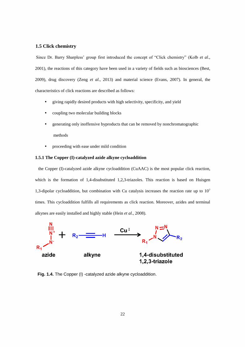
the Copper (I)-catalyzed azide alkyne cycloaddition (CuAAC) is the most popular click reaction,
which is the formation of 1,4-disubstituted 1,2,3-triazoles. This reaction is based on Huisgen
1,3-dipolar cycloaddition, but combination with Cu catalysis increases the reaction rate up to 107
times. This cycloaddition fulfills all requirements as click reaction. Moreover, azides and terminal
alkynes are easily installed and highly stable (Hein et al., 2008).
Fig. 1.4. The Copper (I) -catalyzed azide alkyne cycloaddition.

23
1.6 Study outline
In this study, I evaluated cytoprotective effects of gut microbiota catabolites of quercetin glycosides
and aureusidin (AU) on oxidative stress and investigated the underlying mechanism. Antioxidative
properties of the substances are assessed mainly by determination of effect on hydrogen
peroxide-induced cell death and phase 2 drug-metabolizing enzyme gene expression-inducing
activities in Hepa1c1c7 cells.
I found that DOPAC has the most potent direct and indirect antioxidative abilities in the major
colonic catabolites of quercetin glycosides. Investigation of the target proteins of provides using
click chemistry probe detected Keap1 and AhR as the primary targets. These results suggest that
DOPAC enhances cellular antioxidative activities through activation of phase 2 enzyme expression
by direct modification of Keap1 and AhR.
AU was synthesized in four steps from phloroglucinol and the overall yield was 1.3%. AU
showed significant cytoprotective effect against hydrogen peroxide-induced cytotoxicity and
enhancement of gene expressions of phase 1 and 2 enzymes. These results indicated that AU acts as
cytoprotective antioxidants via induction of phase 2 enzymes through not only Keap1-Nrf2 pathways
but also AhR pathways.
In conclusion, the present study revealed cellular antioxidant mechanism of dietary flavonoids and
colorectal catabolites, providing further scientific evidence supporting health-promoting effects of
flavonoid intake.

24
CHAPTER 2
3,4-Dihydroxyphenylacetic acid, a catabolite of dietary quercetin
glycosides produced by gut microbiota have cytoprotective effects
against oxidative stress
2.1 Introduction
Quercetin is one of the most well studied flavonoids and widely distributed in plants. We consume
quercetin from various plant-based foods and beverages including vegetables, fruits and tea on a
daily basis. The average daily intake of quercetin is estimated 10-50 mg, which varies depending on
the food habits and the region (Formica et al., 1995; Scalbert et al., 2000). Nishimuro et al. reported
that the average daily quercetin consumption of Japanese living in Hokkaido was 16.2 mg and the
major sources were onions and green tea (Nishimuro et al., 2015). On the other hands, for U.S.
adults, the intake of quercetin in approximately 10 mg/day (Sampson et al., 2002).
In recent years, quercetin has attracted much attention for the beneficial effects on our health. The
health promoting effects of quercetin was reported not only by in vitro studies, but also by
epidemiological studies. A prospective cohort study on associations between flavonoids intake and a
risk of cardiovascular disease showed that increased consumption of flavonols including quercetin
and kaempferol reduced cardiovascular disease mortality (Peterson et al., 2012). Another cohort
study reported that the person with higher quercetin intakes had lower risk of mortality
from ischemic heart disease and incidence of asthma and type 2 diabetes (Kenkt et al., 2002).
Quercetin is generally present in our diet as a glycoside form, not an aglycone form. More than 170
different quercetin glycosides have been identified, the major glycoside forms are 3-O-β-glucoside
(Q3G, isoquercetrin) occurring in a variety of fruits and vegetables and 4′-O-β-glucoside (Q4′G)
accounting for considerable portion of total amount of flavonoids in onion (Tsushida et al., 1995;
Yang et al., 2001). It is widely accepted that intact quercetin glycosides are hardly absorbed into the

25
body due to their high hydrophilicity. Only 5-10% of ingested quercetin glycosides are absorbed in
the small colon through sodium glucose cotransporter 1 (SGLT1) or undergo passive absorption
after deglycosylated by lactase phlorizin hydrolase (LPH) which located in the brush border of the
small intestine (Day et al., 1998; Walgren et al.,2000). 90-95% of quercetin glycosides pass through
the small intestine as intact forms or conjugated forms, and reach at the large intestine where an
enormous number of microorganisms inhabit and form a complex community known as the
intestinal microbiome (Duda-Chodak et al., 2015). Food compounds including flavonoids are
subjected to various enzymatic conjugations of the gut microbiota, which is believed to have effect
on availability of them. Quercetin glycosides also can be converted enzymatically into several
phenolic acids. Mullen et al., investigated the fate of ingested Q4′G using rats and identified
DOPAC and OPAC as major catabolites and protocatechuic acid PCA as a minor catabolite formed
in the colon, and detected OPAC and HPA in faces and urin (Mullen et al., 2008). Moreover, studies
reported that Eubacterium ramulus, Bacteroides fragilis and Clostridium perfringens which are
human colorectal bacteria have the capability to degrade quercetin to DOPAC (Schneider et al.,
1999; Peng et al., 2014)
As mentioned above, most of ingested quercetin glycosides are not absorbed into the circulation as
intact forms and converted into several phenolic acids by microbiota in the large intestine which can
enter into enterocytes. Therefore, phenolic catabolites have the potential to account for the health
promoting effects of dietary quercetin. In the present study, I compared the effects of the major
phenolic acid catabolites of dietary quercetin glycosides, including DOPAC, OPAC, PCA and HPA
on DPPH radical, superoxide and detoxification enzyme level in vitro. DOPAC showed most potent
direct and indirect antioxidative properties, suggesting that DOPAC is a predominant antioxidative
catabolite of quercetin glycosides formed by the colonic microbiota in the large colon.

26
Fig. 2.1. Schematic representation of quercetin glycoside metabolism and absorption in
the large intestine.

27
2.2 Materials and methods
2.2.1 Materials
Quercetin, DOPAC, OPAC, PCA and HPA were purchased from Sigma (St. Louis, MO, USA).
1,1-Diphenyl-2-picrylhydrazyl (DPPH) was obtained from Tokyo Chemical Industry (Tokyo,
Japan). α-Minimum essential medium (α-MEM), and Trizol reagent were purchased from Life
Technologies (Carlsbad, CA, USA) Fatal bovine serum was purchased from Nichirei corporation
(Tokyo, Japan). SOD assay kit was purchased from DOJINDO LABORATORIES (Kumamoto,
Japan) Hydrogen peroxide was purchased from nacalai tesque (Kyoto, Japan). All other chemicals
were obtained from Wako Pure Chemicals Industries (Osaka, Japan).
2.2.2 Cell culture and treatment
The mouse hepatoma cell line Hepa1c1c7, obtained from the American Type Culture Collection,
was grown and maintained at 37 °C in an atmosphere of 95% air and 5% CO2 in α-MEM containing
10% fetal bovine serum, 4 mM L-glutamine, 100 U/ml penicillin, and 100 μg/ml streptomycin. For
experiments, cells were seeded in complete medium and treated with each reagent or dimethyl
sulfoxide (DMSO) vehicle (final 0.1%, v/v). RL34 cells were obtained from the Health Science
Research Resources Bank, Osaka, Japan (Yamada et al., 1987). The cells were grown and
maintained at 37 °C in an atmosphere of 95% air and 5% CO2 in Dulbecco's modified Eagle's
medium containing 10% fetal bovine serum, 4 mM L-glutamine, 100 U/ml penicillin, and 100 μg/ml
streptomycin. For experiments, cells were seeded in complete medium and treated with each reagent
or DMSO vehicle (final 0.1%, v/v).
2.2.3 DPPH radical scavenging assay
A test compound (ethanol solution, 2 ml) mixed with a 100mM Tris-HCl buffer (pH 7.4, 2 ml) was

28
added to 0.5 mM DPPH in ethanol (1ml), and the mixture was shaken vigorously and kept for 20
min at room temperature in the dark. The DPPH radical scavenging activity is expressed as the ratio
of the relative decrease in the absorbance of the test sample mixture at 517 nm to that of the 1 mM
Trolox solution. The experiment was done in triplicate. DPPH radical scavenging activity
(%)={(ethanol alone) − (test compound)} / {(ethanol alone) − (Trolox)} × 100.
2.2.4 Superoxide scavenging assay in the xanthine/xanthine oxidase system
SOD-like activity was measured by a xanthine (XA)/XA oxidase (XOD) system using a SOD Test
Wako kit accordingly to the manufacturer's instruction. In this system, SOD-like superoxide
scavenging activity was estimated by measuring the nitroblue tetrazolium (NBT) reduction.
2.2.5 Determination of hydrogen peroxide formation
Hydrogen peroxide amount was determined by the ferrous ion oxidation-xylenol orange (FOX)
method. The FOX reagent-A contained 25 mM H2SO4, 100 mM sorbitol, 250 μM Fe(NH4)2(SO4)2,
and 125 μM xylenol orange. The FOX-B contained 25 mM H2SO4, 100 mM sorbitol, and 250 μM
Fe(NH4)2(SO4)2. This assay method was applicable to determine hydrogen peroxide concentrations
as low as 0.2 μM. Briefly, the collected sample (45 μl) was mixed with 5 μl of methanol and
incubated at room temperature for 30 min. The FOX-A reagent (0.45 ml) or FOX-B reagent for
reference was added and incubated for 30min. The solutions were then centrifuged at 15,000g for
10min at room temperature and the absorbance at 560 nm was measured. The concentration of
hydrogen peroxide was calculated from standard curve.
2.2.6 Reverse transcription-polymerase chain reaction (RT-PCR)
Cells were washed with ice-cold phosphate buffered saline (PBS) (−). Total cellular RNA was

29
isolated using Trizol reagent according to the manufacturer's recommendations. RNA was quantified
by measuring absorbance at 260 nm. Total RNA (5 μg) was reverse transcribed with Oligo dT to
cDNA using ReverTra Ace (TOYOBO, Osaka, Japan). PCR amplification was then performed with
BIOTAQ DNA Polymerase (BIOLINE, Toronto, Canada) and specific primers. Primers used in PCR
amplification are as follows: mNQO1, (F) 5′-TCgAAgAACTTTCAgTATCC-3′ and (R)
5′-TgAAgAgAgTACATggAgCC-3′ (20 cycles, product size 290 bp); mGCLC, (F)
5′-ggCgATgTTCTTgAgACTCTgC-3′ and (R) 5′-TTCCTTCgA TCATgTAACTCCCATA-3′ (26
cycles, product size 99 bp); mxCT, (F) 5′- CCTggCATTTggACgCTACAT-3′ and (R)
5′-TgAgAATTgCTgTgAgCTTgCA- 3′ (25 cycles, product size 182 bp); mHO-1, (F)
5′-gTgATggAgCg TCCACAgC-3′ and (R) 5′-TggTggCCTCCTTCAAgg-3′ (26 cycles, product size
66 bp); mCYP1A1, (F) 5′-CCTCTTTggAgCTgggTTTg-3′ and (R) 5′-
TgCTgTgggggATggTgAAg-3′ (24 cycles, product size 229 bp). The PCR products were separated
on an 3% agarose gel, stained with ethidium bromide, and visualized under UV light using
Luminescent Image Analyzer LAS-3000 (FUJIFILM Corporation, Tokyo, Japan). The relative
density of the bands was analyzes using the Image J Software.
2.2.7 Glutathione S-transferase (GST) activity assay
After treatment, the cells were lysed with RIPA buffer (10 mM Tris- HCl, pH 7.4, 1% Triton
X-100, 0.1% SDS, 150 mM NaCl, 1 mM EDTA, 2.41 μM deoxycholic acid sodium salt, 2mM
phenylmethyl sulfonyl fluoride). Total GST activity was measured using
1-chloro-2,4-dinitrobenzene (CDNB) as a substrate according to the method of Habig and Jakoby
(1981).
2.2.8 Glutathione titration

30
Glutathione (GSH) contents were determined using 5,5′- dithiobis (2-nitrobenzoic acid) (DTNB)
and glutathione reductase according to the method of Baker et al. (1990).
2.2.9 Cell viability determination with MTT assay
Hepa1c1c7 cells were suspended at a density of 5 × 10⁴ cells per well in a 96-well plate. After
overnight preculture, the cells were incubated with DOAPC (50 or 100 μM), quercetin (10 or 50
μM), or hydrogen peroxide (20 or 40 μM) for 30 min or 24 h, followed by additional incubation with
hydrogen peroxide (100 μM) for 6 h. After culturing with hydrogen peroxide, 10 μl of an MTT
solution was added to each well, and the absorbance was measured with an microplate reader
(Benchmarkplus, Bio-Rad laboratories, Hercules, CA, USA) at 570 nm according to the
manufacturer's instructions after incubation at 37 °C for 2–3 h in a humidified CO2 incubator. The
obtained values were compared with each of the controls incubated with vehicle only.
2.2.10 Statistical analysis
All values were expressed as means ± SD. Statistical significance was analyzed by Student's paired
two-tailed t-test or a one-way ANOVA followed by Tukey's HSD using R software. A p-value of
0.05 was regarded to be statistically significant

31
2.3 Results
2.3.1 DOPAC has radical scavenging and prooxidative activities.
I compared radical scavenging activities of quercetin, DOPAC, OPAC, PCA and HPA using a
DPPH method. As shown in Fig. 2.3A, catechol-type phenolic acids including DOPAC and PCA
showed a potent radical scavenging activity, whereas OPAC and HPA did not show any scavenging
activity. The activity of DOPAC is equivalent to that of quercetin and significantly higher than that
of PCA. Since the superoxide anion radical is one of the precursors of ROS and their metabolites,
the SOD-like activity of the phenolic acid metabolites in an XA/XOD system was next examined. As
shown in Fig. 2.3B DOPAC and PCA showed a significant inhibitory effect on the
superoxide-dependent NBT reduction. The superoxide-scavenging activity of DOPAC was
significantly higher than that of PCA, but lower than that of quercetin. Although a representative
catechin from green tea, (−)-epigallocatechin-3-gallate, is an effective antioxidant if free radicals
exist nearby, it can undergo autoxidation and behave as a pro-oxidant (Tang et al., 2014; Wu et al.,
2009). The ROS generating ability of phenolic compounds has also been demonstrated to play
important roles in some biological or chemical systems, oxidative DNA damage and activation of
transcriptional factors (Galati et al., 2003). Hence, I examined whether hydrogen peroxide is
produced during the autoxidation of the quercetin metabolites using a FOX assay. As shown in Fig.
2.3C, DOPAC as well as quercetin produced a significant amount of hydrogen peroxide, whereas the
other metabolites did not. These results suggested that DOPAC possesses a redox-active character in
a manner similar to quercetin.
Fig. 2.2. Chemical structures of phenolic catabolites of quercetin glycosides.

32
Fig. 2.3. Radical scavenging and prooxidative potentials of the phenolic acid catabolites.
(A) Free radical scavenging activity against DPPH radical of quercetin (open circle),
DOPAC (closed square), PCA (closed triangle), OPAC (open triangle) and HPA (open
diamond). A test compound was incubated with 0.5 mM DPPH for 20 min at room
temperature in the dark. The DPPH radical scavenging activity was evaluated by the
absorbance at 517 nm. All values were expressed as means ± SD of three separate
experiments (*p < 0.05 compared with quercetin or DOPAC). (B) SOD-like activity of the
phenolic acid catabolites. The SOD-like superoxide scavenging activity of the text
compounds (50 μM) was estimated by measuring the NBT reduction in an XA/XOD
system. All values were expressed as means ± SD of three separate experiments (*p <
0.05, **p < 0.01 compared between the indicated groups). (C) Prooxidant activity of the
phenolic acid catabolites. The hydrogen peroxide concentration was determined using a
FOX assay. All values were expressed as means ± SD of three separate experiments.

33
2.3.2 DOPAC induces phase 2 drug-metabolizing enzymes in Hepa1c1c7 cells.
I next investigated whether the phenolic acid metabolites of quercetin glycosides have the
ability to induce the gene expression of the drug-metabolizing enzymes, including NQO1,
glutamate-cysteine ligase, catalytic subunit (GCLC), xCT, HO-1 and cytochrome P450 1A1
(CYP1A1). The incubation of Hepa1c1c7 cells with 50 μM DOPAC for 24 h led to a significant
increase in the mRNA levels of GCLC, NQO1 and HO-1 (Fig. 2.4), whereas the other
metabolites at the same concentration showed no significant effect. Although quercetin at 50
μM significantly induced the expression of CYP1A1 as well as all phase 2 enzymes, DOPAC at
this concentration showed no effect on this phase 1 enzyme expression. We next checked the
dose-dependent effect of DOPAC on the expression of the drug-metabolizing enzymes. As
shown in Fig. 2.5, the expressions of NQO1, GCLC and HO-1 were dose-dependently
increased by the treatment of DOPAC at concentrations lower than 50 μM. However, the
mRNA levels of xCT and CYP1A1 were increased only at concentrations higher than 100 μM.
These results suggested that DOPAC induces the expression of the drug-metabolizing enzymes
through different pathways between its lower and higher concentrations.

34
Fig. 2.4. Modulating effects of the phenolic acid catabolites on the gene expression of
the drug metabolizing enzymes. The total RNA was extracted from Hepa1c1c7 cells
treated with quercetin or the phenolic acids at 50 μM for 24 h, then a RT-PCR analysis
for NQO1, GCLC, HO-1, xCT and CYP1A1 was carried out. All values were expressed
as means ± SD of three separate experiments (*p < 0.05 compared with control).

35
Fig. 2.5. Concentration-dependent effects of the DOPAC on the gene expression of the
drug metabolizing enzymes. The total RNA was extracted from Hepa1c1c7 cells treated
with DOPAC at the indicated concentrations for 24 h, then a RT-PCR analysis for NQO1,
GCLC, HO-1, xCT and CYP1A1 was carried out. All values were expressed as means ±
SD of three separate experiments (*p < 0.05 compared with DOPAC 0 μM).

36
2.3.3 DOPAC increases glutathione S-transferase activity in RL34 cells and cellular glutathione
content in Hepa1c1c7 cells.
To gain further evidence for the induction of the drug-metabolizing enzymes by DOPAC, the
effects of DOPAC on the activity of GST, one of the representative phase 2 drug-metabolizing
enzymes, and the intracellular total GSH level were determined. As shown in Fig. 2.6A, the
treatment of normal hepatocyte RL34 cells with 100 μM DOPAC for 24 h resulted in an
enhancement of the total GST activity by approximately 1.4-fold. Quercetin showed equivalent
potency only at concentration of 10 μM, but this concentration induced the strongest GST activity
than higher concentrations (Fig. 2.6 C). The treatment of Hepa1c1c7 cells with higher than 200 μM
of DOPAC for 24 h exhibited a significant alteration in the intracellular GSH level (Fig. 2.6B) These
results indicated that the key step for the DOPAC-stimulated GSH up-regulation might be the
substrate transport by xCT, but not the GSH biosynthesis by GCLC.

37
Fig. 2.6. Modulating effects of the DOPAC and quercetin on the total GST activity and the
intracellular GSH level. RL34 cells were treated with DOPAC (A) or quercetin (C) for 24 h
and the total GST activity was measured by a CDNB assay. (B) Hepa1c1c7 cells were
treated with DOPAC for 24 h and the total GSH level was measured by a DTNB assay. All
values were expressed as means ± SD of three separate experiments (*p < 0.05
compared with DOPAC 0 μM).

38
2.3.4 DOPAC protects Hepa1c1c7 cells against hydrogen peroxide induced oxidative stress.
A number of cellular defenses composed of non-enzymatic and enzymatic components are involved
in the protection against excessive ROS. As already mentioned, DOPAC showed a significant
scavenging effect against the stable free radical DPPH (Fig. 2.3A) as well as an inducing effect on
some antioxidant enzymes such as HO-1 and GST (Figs. 2.5 and 2.6A). These results prompted me
to examine whether DOPAC inhibits the ROS-induced oxidative stress through its direct antioxidant
action or indirect antioxidant action regulated by the transcriptional level. The protective effect of
DOPAC against the hydrogen peroxide-induced cytotoxicity was evaluated by an MTT assay.
Although the incubation of DOPAC even at 100 μM 30 min before the hydrogen peroxide treatment
showed no effect on the cytotoxicity caused by oxidative stress (Fig. 2.7A), pretreatment with
DOPAC for 24 h inhibited it in a concentration-dependent manner (Fig. 2.7B). Thus, the treatment
with DOPAC at the dose required for the induction of the phase 2 drug-metabolizing enzymes, but
not for the GSH up-regulation, actually exhibited an antioxidant effect in Hepa1c1c7 cells. For the
purpose of comparison, I evaluated the effect of quercetin on hydrogen peroxide induced toxicity.
Since 10 μM of quercetin had almost the same GST activity inducing ability as 50 μM of quercetin
(Fig 2.3C), 10 and 50 μM of quercetin was pretreated. As with DOPAC, every concentration of 30
min pretreatment had no effect on the cell viability (Fig. 2.7C), but 24 h pretreatment of 10 μM of
quercetin recovered the cell viability completely (Fig. 2.7D). 50 μM of quercetin itself possessed
toxicity to cells, thus it did not abolished the cytotoxicity of hydrogen peroxide (Fig. 2.7D). 10 μM
of As shown in Fig 2.2C, DOPAC produces ROS by autooxidation reaction. Since transcription of
phase 2 enzymes are induced by ROS accumulation, I checked whether ROS production is involved
in antioxidative cytoprotection of DOPAC. The pretreatment of hydrogen peroxide (20 and 40 μM)
showed no effect on the hydrogen peroxide induced cytotoxicity (Figs. 2.7C and 2.7D), suggesting
that hydrogen peroxide from the autooxidation of DOPAC is not account for cytoprotection.

39
Fig. 2.7. Effect of DOPAC on the hydrogen peroxide-induced cytotoxicity in Hepa1c1c7
cells. Hepa1c1c7 cells were pretreated with DOPAC (50 or 100 μM), quercetin (10 or 50
μM), or hydrogen peroxide (20 or 40 μM) for 30 min (A) , (C) and (E) or 24 h (B), (D) and
(F), then treated with hydrogen peroxide (100 μM) for 6 h. Cell viability was measured
using an MTT assay. All values were expressed as means ± SD of three separate
experiments. Different letters above the bars indicate significant differences among the
treatments for each condition (p < 0.05).

40
2.4 Discussion
In the present study, I identified DOPAC as the most biologically active phenolic acid derived from
the catabolism of quercetin glycosides in terms of antioxidative potentials. DOPAC has been
regarded as one of the major catabolites from quercetin glycosides, such as Q4′G (Mullen et al.,
2008), rutin (quercetin 3-O-rutinoside) (Aura et al., 2002), and hyperoside (quercetin
3-O-galactoside) (Yang et al., 2013). An in vitro catabolic study of quercetin using human fecal
bacteria also demonstrated the formation of DOPAC (Peng et al., 2014), supporting the idea that
DOPAC is a predominant catabolite from quercetin glycosides not only in rodents, but also in
humans. An increased urinary excretion of DOPAC as well as other phenolic acid catabolites has
been observed in human subjects after the consumption of polyphenols from chocolate (Rios et al.,
2003). In addition to its radical scavenging activity, DOPAC inhibits the secretion of the
pro-inflammatory cytokines in lipopolysaccharide-stimulated peripheral blood mononuclear cells
(Monagas et al., 2009) and the expression of P-selectin in resting platelets (Rechner et al., 2005).
Although DOPAC shows relatively weaker biological activities than those of the quercetin aglycone,
DOPAC has some advantages for application as a food chemical because of its lower cytotoxicity.
This idea is supported by the fact that DOPAC is also known as a metabolite of a neurotransmitter
dopamine, suggesting the existence of a metabolic pathway of DOPAC in humans (Gesi et al., 2001).
DOPAC and PCA showed a significant radical scavenging activity against not only the DPPH radical,
but also the superoxide anion radical, whereas OPAC and HPA did not show any scavenging activity
(Figs. 2. 3A and B). The o-diphenol moiety, but not the monophenol, might be essential for the
radical scavenging activity, which is consistent with previous reports (Nakamura et al., 1998; Tsuda
et al., 1996). For example, the number of hydroxyl groups on the phenolic B-ring of anthocyanins
plays an important role in the superoxide-scavenging activity (Tsuda et al., 1996). On the other hand,
the radical scavenging activities of DOPAC were significantly higher than those of PCA (Figs. 2.3A

41
and B). Previous studies regarding the antioxidative mechanism for BHT
(2,6-di-tert-butyl-4-methylphenol) have indicated that an aryl proton at the 4-methyl group of BHT
is easily removed by free radicals (Guyton et al., 1991). It was also reported that dihydroferulic acid
and dihydrosinapic acid with a saturated sidechain showed a more potent radical scavenging activity
than ferulic acid and sinapic acid with an unsaturated side chain, respectively (Shimoji et al., 2002).
These studies would support the idea that the methylene group bound to the benzene ring of DOPAC
plays an enhancing role not only in the radical scavenging effects, but also in the
autoxidation-dependent hydrogen peroxide production (Fig. 2.3C). There is substantial evidence that
the phase 2 drug-metabolizing enzymes including GST, NQO1, HO-1, and glutamate-cysteine ligase,
play important roles in the detoxification of prooxidative or electrophilic toxicants, and their
induction contributes protection against chronic diseases (Nakamura et al., 2010). Among the tested
phenolic acids, only DOAPC at the concentration of 50 μM significantly enhanced the mRNA levels
of GCLC, NQO1 and HO-1 (Figs. 2.4 and 2.5). These 3 genes were transcriptionally up-regulated by
the lower concentrations of DOPAC, whereas the inducible expression of a phase 1
drug-metabolizing enzyme gene, CYP1A1, as well as xCT required higher concentrations (Fig. 2.5).
Lower concentrations of DOPAC also enhanced the total GST activity in normal rat hepatocytic
RL34 cells (Fig. 2.6A),whereas only higher concentrations of DOPAC increased the intracellular
level of GSH (Fig. 2.6B). The former phenomenon is consistent with a previous study showing that
DOPAC significantly up-regulated the gene expression of a GST isozyme, GSTP1, in RL34 cells
(Ishii et al., 2009). However, the concentration dependency of DOPAC for the intracellular GSH
level was correlated with that for the gene expression of xCT, but not GCLC, suggesting that the
up-regulation of the GSH level might be regulated by the substrate uptake and not by the enhanced
biosynthesis. Quercetin has been reported to activate both the Nrf2-dependent pathway (Surh et al.,
2008) and the aryl hydrocarbon receptor (AhR)-dependent pathway (Ciolino et al., 1999). Nrf2 binds

42
to antioxidant response element (ARE) and induces a number of Nrf2-dependent phase 2
drug-metabolizing genes, including NQO1, HO-1, GCLC, etc. (Nakamura et al., 2010). AhR, the
transcription factor binding to XRE, plays a pivotal role in the inducible expression of CYPs
including CYP1A1 (Ciolino et al., 1999). A previous study indicated that cross talk between AhR
and Nrf2 is mediated by the mechanism that the gene promoter of Nrf2 includes at least one
functional XRE (Miao et al., 2005). Both the functional XRE and ARE elements exist in proximity
to the promoters of NQO1 (Favreau et al., 1991). The expression of CYP1A1 was significantly
up-regulated only at concentrations higher than 100 μM DOPAC (Fig. 2.5). Moreover, the inducible
gene expression of NQO1 or HO-1 by DOPAC at concentrations higher than 100 μM was more
remarkable than by its lower concentrations (Fig. 2.5). These results strongly suggested that the
higher concentrations of DOPAC might activate the AhR/XRE pathway and thus synergistically
enhance the Nrf2/ARE pathway. It has recently become clear that the phase 2 gene inducers also act
as indirect antioxidants in a manner different from the radical-scavenging direct antioxidants
(Nakamura et al., 2010). For example, several GSTs have a GSH peroxidase activity towards
hydroperoxides.HO-1 also generates carbon monoxide and biliverdin/bilirubin. A previous study
demonstrated that the overexpression of the HO-1 protein inhibited the hydrogen peroxide-induced
cytotoxicity and carbon monoxide is responsible for this protective effect (Lin et al., 2007). The
results in the present study demonstrate that DOPAC significantly inhibits the hydrogen
peroxide-induced cytotoxicity independent of its radical scavenging potential (Figs. 2.7A and B).
The antioxidant effect in Hepa1c1c7 cells was actually exhibited by the 24-h treatment of DOPAC at
50 μM, which is required for the induction of the phase 2 drug-metabolizing enzymes, but neither for
the GSH up-regulation nor for the CYP1A1 induction, suggesting the significant role of GST and/or
HO-1 in the cellular antioxidant action. The pretreatment of 10 μM quercetin for 24 h, but not that
for 30min, showed the effect similar to that of DOPAC (Figs. 2.7C and D). This concentration of

43
quercetin is also able to enhance the GST activity (Fig. 2.6C). These results suggested that the
cytoprotective activity of quercetin is 5 times more than potent compared to that of DOPAC.
However, the treatment of 50 μM quercetin itself showed a significant toxicity, even though it could
reduce the hydrogen peroxide-induced cytotoxicity (Fig. 2.7D). In addition, the pretreatment of
hydrogen peroxide (20 and 40 μM) showed no effect on the hydrogen peroxide induced cytotoxicity
(Figs. 2.7E and F), suggesting that hydrogen peroxide produced by the autoxidation of DOPAC
could be ruled out in its cytoprotective mechanisms.
In conclusion, I identified DOPAC as a predominant biologically-active catabolite quercetin
glycosides formed by the colonic microbiota in the large intestine. DOPAC may contribute to
preventive effects of dietary quercetin against oxidative stress related diseases via not only direct
radical scavenging activity, but also phase 2 gene expression inducing property.

44
CHAPTER 3
3,4-Dihydroxyphenylacetic acid has cytoprotective effects against
oxidative stress through direct modification of Keap1 and aryl
hydrocarbon receptor
3.1 Introduction
Oxidative stress is defined as imbalance between the production of ROS and antioxidant defense in
the cells and tissues. Although ROS plays important role in regulation of cellular signaling pathways
(Finkel, 2011), because of its high reactivity, it can cause damage to cellular molecules such as
DNA, proteins and lipid, leading various diseases including chronic inflammation, autoimmune
disease, neurological disorders, cardiovascular diseases, and cancer (Birben et al, 2012; Brieger et
al, 2012). As ROS is produced not only by external factors but also by biological processes like
cellular respiration and metabolic processes, cells are equipped with antioxidative systems. Cellular
defenses against oxidative stress are divided into two types: direct and indirect. Direct antioxidative
system is the way to scavenge ROS using low-molecular-weight compounds such as vitamin C and
E, glutathione and β-carotene. Indirect antioxidative system is mediated Keap1/Nrf2/ARE pathway,
which regulates transcription of a series of cytoprotective enzymes (phase 2 enzymes) including
NQO1, HO-1, and GST (Dinkova-Kostova et al., 2008).
Expression of phase 2 enzymes is induced by activation of Keap1/Nrf2/ARE pathway. Under basal
condition, Keap1 binds to Nrf2 in the cytoplasm and Nrf2 is polyubiquinated and consequently
degraded by the proteasome. Under oxidative stress, Keap1 is inactivated by modification of
cysteine residue, leading stabilization and nuclear translocation of Nrf2. After entering the nucleus,
Nrf2 binds to ARE together with small Maf proteins and drive transcription of phase 2 enzyme genes
(Taguchi et al, 2011; Kansanen et al, 2013). Increasing evidence suggests interaction between
Keap1-Nrf2 pathway and AhR pathway. AhR is a transcription factor retained in the cytoplasm.

45
Upon ligand binding on AhR, AhR translocates into the nucleus and forms heterodimer complex
with ARNT, resulting in binding to XRE located in promoter region of AhR responsive genes. Miao
et al. showed nrf2 transcription was regulated by AhR via XRE elements suggesting the presence of
XRE in nrf2 gene promoter (Miao et al., 2005). Additional studies reported that nrf2 and
glutathione-S-transferase contain XRE region (Nioi et al., 2004; Yeager et al., 2009).
In chapter 2, I found that DOPAC had antioxidative properties via inducing phase 2 enzymes, but
an underlying mechanism of this phenomenon have still unclear. DOPAC has catechol structure,
which can be subjected to oxidation by phenol oxidases including tyrosinase and converted into
ortho-quinone in cells (Ishii et al., 2009; Nakamura et al., 2014). O-quinone react with nucleophiles
such as GSH or protein cysteine residue in 1,4-Micheal fashion to form a catechol-nucleophile
conjugate. This characteristic motivated us to seek the target proteins of DOPAC to reveal the
detailed mechanism for its antioxidative effects.
In the present study, I designed a novel DOPAC probe using CuAAC, a representative click
reaction. I introduced an alkyne moiety into DOPAC by esterification with 2-propyn-1-ol to afford
the DOPAC propargyl ester (DPE). I made sure that DPE had the same phase 2 enzyme inducing
efficacy as DOPAC by RT-PCR suggesting DPE behaves in the same fashion as DOPAC in
Hepa1c1c7 cells. Thus I carried out CuAAC reaction with an azide-labeled biotin in lysate of
Fig. 3.1. DOPAC modifies a protein sulfhydryl group in a phenol oxidase-dependent
manner.

46
Hepa1c1c7 cells treated with DPE, visualized western blotting using the horseradish peroxidase
(HRP) conjugated streptavidin, which showed a variety of molecular weight proteins. Finally I
performed pull-down assay using anti-Keap1and anti-AhR antibody and observed direct interaction
between DPE and Keap1 and AhR, indicating that DOPAC induced phase 2 enzyme gene expression
through direct modification of Keap1 and AhR.

47
3.2 Materials and methods
3.2.1 Materials
DOPAC, laccase from Rhus vernicifera, glyceraldehyde-3-phosphate dehydrogenase (GAPDH),
tris[(1-benzyl-1H-1,2,3-triazol-4-yl)methyl]amine (TBTA), bis(4-nitrophenyl)phosphate (BNPP)
and azide-PEG3-biotinconjugate were obtained from Sigma Aldrich (St. Louis, USA).
p-Toluensulfonic acid monohydrate (PTSA), n-butanol, toluene, copper (II) sulfate pentahydrate,
protease inhibitor cocktail and Chemi-Lumi One Super were purchased from nacalai tasque (Kyoto,
Japan). 2-Propyl-1-ol was obtained from Tokyo Chemical Industry (Tokyo, Japan). Streptavidin,
HRP conjugate was purchased from Funakoshi (Tokyo, Japan). Anti-actin antibody, anti-AhR
antibody, anti-Keap1antibody, horseradish peroxidase-linked anti-mouse IgG and horseradish
peroxidase-linked anti-goat IgG were purchased from Santa Cruz Biotechnology (Santa Cruz, USA).
Streptavidin Mag Sepharose was purchased from GE healthcare (Little Chalfont, UK). All other
chemicals such as benzyl azide were purchased from Wako Pure Chemical Industries (Osaka,
Japan).
3.2.2 Instrumental methods of chemical analysis
MS was recorded on ESI-mode by using Bruker MicrOTOF II and MicrOTOF Control 3.0. Data
analysis was carried out using Data Analysis 4.0 SP2. 1H NMR spectra were recorded on Varian
Mercury 300. Chemical shift are described in parts per million (ppm) and coupling constants in Hz.
Multiplicity and qualifier abbreviations are as follows: s=singlet, d=doublet, t=triplet, quint=quintet,
sext=sextet, m=multiplet.
3.2.3 Cell culture and treatment

48
The mouse hepatoma cell line Hepa1c1c7, obtained from the American Type Culture Collection,
was grown and maintained at 37 °C in α-minimum essential medium (MEM-α, Thermo
SCIENTIFIC, Waltham, USA) containing 10% fetal bovine serum, 4mM L-glutamine, 100 U/ml
penicillin, and 100 mg/ml streptomycin. For experiments, cells were seeded in complete medium and
treated with each reagent or DMSO vehicle.
3.2.4 Synthesis of DOPAC propargyl ester
DOPAC (30 mg, 0.18 mmol) and PTSA (6 mg, 0.03 mmol) were dissolved in the solution of
dehydrated toluene (20 ml) and 2-propyl-1-ol (1 ml, 17 mmol), and stirred for 6 h at 40 °C, followed
by cooling to room temperature. The reaction mixture was washed with 5% NaHCO3 solution and
water twice. The aqueous phase was extracted with 20 ml ethyl acetate and combined with the
organic phase. The combined extract was dried over anhydrous magnesium sulfate, filtered, and
evaporated to dryness. The product was purified by preparative thin-layer chromatography (TLC)
(CHCl3:MeOH=9:1) to afford 0.34 mg (0.07 mmol) of DOPAC propargyl ester (DPE). 1H NMR
(300 MHz, CDCl3): 2.48 (1H, t, J=2.5 Hz, H≡C), 3.56 (2H, s, –CH2–Ar), 4.70 (2H, d, J=2.5 Hz, –
O–CH2-C≡), 6.67–6.78 (3H, m, H–Ar–); HR-ESI-MS (negative mode) m/z [M–H]- 205.0508
(205.0506, calcd for C11H9O4). DOPAC butanol ester (DBE) was synthesized in the same method as
DPE, except that n-butanol 1.8 ml (20 mmol) was used instead of 2-propyl-1-ol. 1H NMR (300 MHz,
CDCl3): 0.92 (3H, t, J=7.4 Hz, CH3–CH2–), 1.36 (2H, sext, J=7.4 Hz, CH3–CH2–CH2–), 1.62 (2H,
quin, J=3.5 Hz, –CH2–CH2–CH2–), 3.50 (2H, s, –CH2–Ar), 4.10 (2H, t, J=6.6 Hz, –CH2–CH2–O–),
6.63–6.72 (3H, m, H–Ar–).
3.2.5 CuAAC reaction with DPE and benzyl azide

49
DPE (23 mg, 112 mmol) and benzyl azide (14 μl, 112 mmol) were mixed in 2 ml of 50% t-butanol
in water with copper (II) sulfate pentahydrate (0.28 mg, 1.12 μmol) and ascorbic acid sodium salt
(2.22 mg, 11.2 μmol) at room temperature under the dark condition until the complete consumption
of the reactants checked by TLC. The product was purified by preparative TLC (hexane:ethyl
acetate=3:7), monitored by Dragendorff's reagent. Formation of 1,2,3-triazol was confirmed by
NMR and mass spectrometry: 1H NMR (300 MHz, CDCl3): 3.46 (2H, s, Ar–CH2–C(O)O–), 5.17
(2H, s, –O–CH2–C2N3H–) 5.49 (2H, s, –C2N3H–CH2–Bz), 6.55–6.78 (3H, m, H–Ar–), 7.24–7.38
(5H, m, H–Bz), 7.50 (1H, s, –C2N3H–), HR-ESI-MS (negative mode) m/z [M–H]- 338,1142
(338.1146, calcd for C18H16N3O4).
3.2.6 RNA extraction and RT-PCR
Confluent Hepa1c1c7 cells were treated with DOPAC or DPE at the indicated concentrations for 24
h. The total RNA was extracted from the cells with TRIzol reagent (Thermo Fisher Scientific,
Waltham, MA, USA). The RNA concentration was determined by measuring the absorbance at 260
nm. cDNA was synthesized from total RNA (5 mg) and oligo (dT) primer using PrimeScript Reverse
Transcriptase (Takara-Bio, Kusatsu, Japan) in accordance with manufacturer's instructions. The PCR
reactions were performed using BIOTAQ DNA polymerase (BIOLINE, London, UK) and
genespecific primers: HO-1, (F) 5′-ACATCGACAGCCCCACCAAGTTCAA-3′ and (R)
5′-CTGACGAAGTGACGCCATCTGTGAG-3′; NQO1, (F) 5′-TCGAAGAACTTTCAGTATCC-3′
and (R) 5′-TGAAGAGAGAGTACATGGAGCC-3′; β-actin, (F)
5′-GTCACCCACACTGTGCCCATCTA-3′and (R) 5′-GCAATGCCAGGGTACATGGTGGT-3′.
The PCR products were subjected to agarose gel electrophoresis (3%) stained with ethidium bromide
and and imaged with an LAS3000 image analyzer (Fuji Film, Tokyo, Japan). Densitometric analysis

50
of the bands was carried out using the Image J Software Program (National Institutes of Health,
Bethesda, MD, USA).
3.2.7 Competitive inhibition of DPE-modification in cells and in cell lysate
As for treatment of cells, confluent Hepa1c1c7 cells were preincubated with DOPAC or DBE for
30 min and incubated with 25 μM DPE for 3 h in serum-free MEM-α. As for treatment of cell lysate,
cell lysate prepared from Hepa1c1c7 containing 100 mg proteins were co-incubated with 25 μM
DPE and 0-200 μM DOPAC or DBE in the presence of 30 U laccase. In both experiments, CuAAC
reaction, SDS-PAGE and western blot analysis were subsequently conducted as described before.
3.2.8 Pulldown assay and western blotting
Confluent Hepa1c1c7 cells were treated with 50 μM DPE or 0.1% DMSO in FBS-free MEM-α for
5 h. The treated cells were lysed, and the cell lysate containing 1000 μg of protein were subjected to
ultrafiltration and the CuAAC reaction as mentioned above. The protein solution was incubated with
100 mM Streptavidin Mag Sepharose for 30 min at room temperature with constant shaking, washed
and eluted according to the manufacture's protocol. The eluted proteins were subjected to
SDS-PAGE and transferred to membranes. After blocking, membrane was treated with anti-Keap1
antibody (1:200) or anti-AhR antibody (1:200), followed by incubation with appropriate secondary
antibody and detection using Chemi-Lumi One Super.
3.2.9 Statistical analysis
All values were expressed as means ± SD. Statistical significance was assessed by Student's paired
two-tailed t-test. A p value of 0.05 was regarded to be statistically significant.

51
3.3 Results
3.3.1 Design and synthesis of click chemistry probe.
I designed an alkyne-functionalized clickable DOPAC probe to capture its target proteins. The
strategy for detecting DOPAC-interacting proteins is as follows: 1) DPE is synthesized via fisher
esterification. 2) DPE is conjugated with the target protein. 3) A DPE-protein conjugate is subjected
to click reaction with biotin azide to form a biotin-DPE-protein complex. 4) The complex is detected
with HRP-streptavidin.
To introduce an alkyne functionality into DOPAC, I esterified DOPAC with 2-propyn-1-ol via the
Fischer esterification method, providing DPE in 40% yield (Fig. 3.3). This alkyne functionality
could form a 1,2,3-triazol derivative with azide-functionalized tags such as biotin and fluorescent
dyes, which allow detection in a later step.
Fig. 3.2. The strategy for detecting DOPAC-targeting proteins using DPE as a click
chemistry probe.

52
3.3.2 Evaluation of chemical and biological properties of DPE.
To examine whether the esterification of DOPAC influences the ability to bind to protein
sulfhydryls, I compared the thiol modification ability of DPE with that of DOPAC. I used GAPDH
as a model protein of the thiol modification, because GAPDH has four cysteine residues on each
subunit and is vulnerable to a nucleophilic attack (Nakajima et al., 2007). Laccase was used as a
polyphenol oxidase to oxidize DOPAC and DPE into the o-quinone structures via a two-electron
oxidation as previously reported (Nakamura et al., 2014) . As shown in Fig. 3.4A, the 1-h incubation
of GAPDH with DOPAC or DPE in the presence of laccase resulted in reduction of the free
sulfhydryl amount, whereas no change was observed under the condition without laccase. This result
suggested that DPE had the same ability and phenol oxidase dependency to modify the protein
sulfhydryls as DOPAC. Next, I examined the biological features of DPE such as inducing property
of the phase 2 drug-metabolizing enzyme gene expressions. As shown in Fig. 3.4B, DOPAC
dose-dependently increased the gene expression of HO-1 and NQO1, which is consistent with results
in chapter 2. DPE also enhanced their gene expression in a manner similar to DOPAC in the cultured
cells. The significant biological activity of DPE comparable to the original DOPAC suggested that
the esterification of DOPAC with 2-propyn-1-ol showed no effect on the modifying ability of the
protein sulfhydryls.
Fig. 3.3. Esterification of DOPAC with 2-propyn-1-ol to give DPE.

53
Fig. 3.4. Chemical and biological properties of DPE and DOPAC. (A)
Modification of sulfhydryl groups in GAPDH by DOPAC and DPE. GAPDH (500 μg/ml) was
incubated with DOPAC or DPE in 50 mM sodium phosphate buffer (pH 7.2) for 1 h at
37 °C in the presence or absence of laccase (30 units). The level of residual sulfhydryl
groups in GAPDH was measured by the spectrophotometric method using DTNB. (B)
Induction of the gene expression of HO-1 (black bars) and NQO1 (white bars) by DOPAC
(left) or DPE (right). Hepa1c1c7 cells were treated with DOPAC or DPE for 24 h and total
RNA was extracted. The values represent means ± S.D. of more than three separate
experiments (*p < 0.05 compared with control; Student's t-test.).

54
3.3.3 Detection of DPE-modified model proteins in vitro.
I next confirmed the capability of DPE to undergo a CuAAC reaction with an azide-linked tag
molecule. DPE and benzyl azide were incubated for 24 h at room temperature in the dark with
copper (II) sulfate pentahydrate as the catalyst to afford the proposed product. The purification was
done by preparative TLC according to the fluorescence excited at 254 nm. Also, the reaction of the
isolated product with Dragendorff's reagent afforded an orange colored compound, suggesting that it
contains nitrogen (Rubia et al., 1977). Based on the spectral data, the product was identified as a
1,2,3-triazol compound with the DOPAC ester and benzyl moiety (Fig. 3.5A).
To confirm the efficacy of DPE to tag the target protein, I tried to detect DPE-modified proteins
using GAPDH as an in vitro model protein. DPE was incubated with GAPDH in the presence or
absence of laccase, followed by the CuAAC reaction with the azide-labeled biotin and detection
using the HRP-conjugated streptavidin. DPE-modified GAPDH was detected under the condition
with laccase, affording strong evidence for effective tagging (Fig. 3.5B). The DPE-modified
GAPDH was hardly observed in the absence of laccase, suggesting that DPE covalently binds to
GAPDH through the oxidation-dependent electrophilic attachment to the sulfhydryl group.

55
Fig. 3.5. CuAAC reaction of DPE with an azide-linked tag molecule. (A) Formation of
1,2,3-triazole by CuAAC reaction with DPE and benzyl azide.(B) Detection of GAPDH
tagged by CuAAC reaction with DPE and the azide-labeled biotin. GAPDH (1mg/ml) was
incubated with or without 50 μM DPE in the presence or absence of 30 U laccase in 70
mM sodium phosphate buffer (pH 7.2) for 1 h at 37°C. DPE-tagged GAPDH was
detected by CBB staining (left) and HRP-streptavidin (right). 70 mM sodium phosphate
buffer (pH 7.2) for 1 h at 37°C. DPE-tagged GAPDH was detected by CBB staining (left)
and HRP-streptavidin (right).

56
3.3.4 Detection of the intracellular DPE-modified proteins.
I applied this probe to cultured cells to label the cellular proteins. DPE contains an ester bond,
which would be hydrolyzed by carboxyl esterase present in the cells. Therefore, we used BNPP as an
esterase inhibitor to prevent cleavage of DPE in the cells (Hatfield et al., 2011). After BNPP
pretreatment, the cells were incubated with DPE for the indicated periods, then lysed to extract the
cellular proteins. Free DPE in the cell lysate was removed by ultrafiltration to prevent additional
binding during the CuAAC reaction. Then, CuAAC reaction was carried out to afford the DPE
modified proteins having the biotin tags. After SDS-PAGE, the DPE-modified proteins were
detected by the HRP-conjugated streptavidin. As shown in Fig. 3.6A, the DPE-modified proteins
were observed in the groups not only with but also without the BNPP treatment. These results
suggested that the ester linkage in DPE is resistant to hydrolysis by carboxylesterase.
To verify whether the proteins modified by DPE correspond to the DOPAC targets, I examined the
effect of the pre-incubation with DOPAC for 30 min on the incorporation of DPE into the cellular
proteins. The pretreatment of DOPAC actually inhibited the formation of the DPE-modified proteins,
though this was observed only at the higher concentration of DOPAC (Fig. 3.6C). To investigate the
effect of the esterification, I next synthesized another DOPAC ester using n-butanol instead of
2-propyn-1-ol to afford DBE (Fig. 3.6B). DBE inhibited DPE-modified proteins even at a
concentration lower than that of DOPAC (Fig. 3.6C). These data suggested that the esterification of
DOPAC increases the frequency of competition with DPE possibly by the rise in the intracellular
accumulation resulted from increased membrane permeability. To investigate this possibility, I
compared the inhibitory ability of DOPAC and DPE using cell lysate. As shown in Fig. 3.6D,
co-treatment of DOPAC with DPE in cell lysate exhibited almost the same inhibition against the
formation of the DPE-modified protein as that of DBE. Taken together, these results suggest that
DPE has higher ability to capture intracellular proteins, but targets the same proteins as DOPAC.

57
Fig. 3.6. Detection of the intracellular DPE-modified proteins. (A) Detection of the
DPE-modified proteins in Hepa1c1c7. Confluent Hepa1c1c7 cells were pre-incubated with
0.1% DMSO or 100 μM BNPP and incubated with 100 μM DPE for indicated time periods in
serum-free MEM-α. The DPE-tagged cellular proteins were detected by HRP-streptavidin. (B)
Structural comparison of DBE with DOPAC and DPE. (C) Effect of the pretreatment of DOPAC
or DBE with DPE on the DPE-modified protein formation in Hepa1c1c7 cells. Hepa1c1c7 cells
were preincubated with DOPAC (open circle) or DBE (closed circle) for 30 min and incubated
with 25μM DPE for 3 h in serum-free MEM-α. (D) Effect of the co-treatment of DOPAC or DBE
with DPE on the DPE-modified protein formation in cell lysate. Cell lysate was incubated with
25 μM DPE in combination with DOPAC (open circle) or DBE (closed circle) in the presence
of 30 U laccase for 1 h. The values represent means ± S.D. of three separate experiments.

58
3.3.5 Identification of the target proteins of DOPAC.
I finally attempted to detect the modification of Keap1and AhR in the cells exposed to DPE,
because the expression of a phase 2 enzyme, NQO1, is regulated by the Keap1/Nrf2/ARE pathway
and AhR/ XRE. Human Keap1 contains 27 cysteines, some of which are postulated to be the targets
of electrophiles and oxidants that facilitate the derepression of Nrf2 and give rise to the phase 2
enzyme induction (Taguchi et al., 2011; Jaramillo et al., 2013). AhR is a transcription factor
normally presents in the cytoplasm and upon ligand binding, it translocases into nucleus and bind to
XRE and activates XRE-driven gene expression including some phase 2 enzyme like NQO1 (Hayes
et al., 2009). Electrophilic quinones have reported to have capability to covalently bind to and
activate AhR (Abiko et al., 2015). These led us to assume that direct modification of Keap1and/or
AhR of DOPAC may regulate the gene expression. To confirm this, Hepa1c1c7cells were treated and
Keap1 or AhR was detected by Western blot analysis. The pull-down assay successfully detected the
modification of Keap1 and AhR by DPE (Fig. 3.7). These results indicate that Keap1 and AhR are
potential targets of DOPAC for the up-regulation of phase 2 enzymes in Hepa1c1c7 cells.
Fig. 3.7 Detection of the DPE-modified Keap1 and AhR by pulldown assay. Confluent
Hepa1c1c7 cells were incubated with 50 μM DPE for 5 h in serum-free MEM-α. The cell
lysate was incubated with Streptavidin Mag Sepharose beads for 30 min. The
DPE-modified Keap1 (left) or AhR (right) were detected by immunoblot analysis.

59
3.4 Discussion
In recent years, the idea has been widely accepted that phytochemicals modulate signal
transduction via direct modification of biomolecules, leading to beneficial effects on human health.
Therefore, researchers have made efforts to identify the target molecules interact with food
chemicals. Tachibana et al. found Epigallocatechin-3-Gallate binds with the 67 kDa laminin receptor
and exerts anti-cancer effects (Tachibana et al., 2004) and Hong et al. demonstrated sulforaphane
activates Keap1/Nrf2 pathway by direct modification of Keap1 (Hong et al., 2005). Catechol is
easily oxidized by catechol oxidase to ortho quinone, which covalently modify sulfhydryl groups of
protein cysteine residues (Ishii et al., 2009). Cysteine modification is known to serve as an activity
switch in various proteins such as Keap1 (Dinkova-Kostova et al., 2001) and IkBα kinase (Gupta et
al., 2010). These facts as well as the phase 2 drug-metabolizing enzyme inducing property of
DOPAC motivated me to investigate direct targets of DOPAC.
In this study, I developed a novel tag-free probe for detecting DOPAC-targeting proteins using click
reaction. Click chemistry is a general term for a reactions that are fast, simple to use, easy to purify,
regiospecific, and high yielding. CuAAC is nearly perfect and the most broadly applicable click
reaction which forms 1,4-disubstituted 1,2,3-distributed in the presence of Cu(I) catalysis. Since
being discovered by the groups of Meldal in Denmark (Tornøe et al., 2002) and Sharpless and Folkin
in the U.S. (Rostovtse et al., 2002) in 2002, CuAAC has been used in a wide range of research area
including bioconjugation (Wang et al., 2003), drug discovery (Manetschery et al., 2004), polymers
(Helms et al., 2004) and radiochemistry (Schirrmacher et al., 2007). DOPAC was
alkyne-functionalized via condensation with propargyl alcohol to be labeled with biotin azide via
CuAAC for detection. I found that introduction of terminal alkyne do not affect thiol-modification
(Fig 3.4A) and phase 2 drug-metabolizing enzyme inducing activities (Fig 3.4B), suggesting DPE
captures the same proteins as DOPAC. DPE contains an intramolecular ester bonds, and thus I was

60
concerned about the possibility that DPE is subjected to hydrolysis by intracellular carboxylesterases.
However, DPE without BNPP detected as various proteins as with BNPP, suggesting that DPE is
resistant to cellular esterase, thereby is useful for using in cells (Fig 3.6A). Also advantage of this
probe is compactness, which enable to interact with a variety of proteins. Previous developed
biotin-tagged DOPAC probe detected much less variety of proteins, probably due to sterically bulky
biotin tag (Ishii et al., 2009). It should be noted that coincubation of DPE with quercetin decreased
amount of DPE-modified proteins compared to single incubation of DPE (data is not shown),
indicating that this probe detect not only targets of DOPAC but also polyphenols with catechol
moiety .
In conclusion, this study provides an alternative approach to identify the molecular targets for not
only a colonic microflora produced catabolite of quercetin glycosides, DOPAC, but also the parent
flavonoids. Click reactions afford the single reaction compound of interest quickly and irreversibly
with high yield and specificity. The tag-free probe using click chemistry is extremely useful not only
in the pull down experiments using a biotin-tag and avidin-bound solid phase, but also
histochemistry with the fluorescence tags to quantify or reveal the location. Since the catechol type
polyphenols are potential modifiers of redox-dependent cellular events through sulfhydryl
modification, the present results strongly encourage further investigation into chemical biology of
polyphenols such as the molecular and biochemical mechanisms for biological activities.

61
CHAPTER 4
Aureusidin, an aurone-type flavonoid with an unique structure, as a
potential cytoprotective agent against oxidative stress
4.1 Introduction
Flavonoids, one group of plant secondary metabolites, are widely present in plant origin foods and
beverages, such as vegetables, fruits, tea, wine, and cocoa. Flavonoids commonly share the C6-C3-C6
basic skeleton, where two benzene rings (ring A and ring B) are connected through three carbons
that form ring C. Flavonoids are classified into several different subclasses dependently on variation
of these three rings, in particular the degree of oxidation of ring C, including flavones, flavonols,
flavanones, flavan-3-ols, anthocyanidins, isoflavones, and aurones (Ross et al., 2002).
Aurones [2-benzylidenebenzofuran-3(2H)-ones] are classified as one subclass of flavonoids, even
though their C ring consists of five membered ring instead of six membered ring as other major
flavonoid subclasses have. Aurones serve as bright yellow pigments in several edible and ornament
flowers, such as snapdragon (Antirrhinum majus) and dahlia (Dahlia variabilis) (Ono et al., 2006).
Since aurones are biosynthesized only in limited number of plants, their biological activities have
not well been studied compared to other flavonoids. So far, natural and synthetic aurones have been
reported to show some chemical and biological properties, including P-glycoprotein modulation
(Boumendjel et al., 2002; Hadjeri et al., 2003), free radical scavenging (Venkateswarlu et al., 2004;
Detsi et al., 2009), antimalarial (Carrasco et al., 2014) and anticancer activities (Lawrence et al.,
2003; Cheng et al., 2010)

62
Fig. 4.1 Chemical structure of aurone and aureusidin.
In this study, I tried to evaluate cytoprotective activities of aureusidin (AU). As shown in Fig. 4.1,
AU contains an o-dihydroxyl structure (catechol structure) in the B-ring. Possession of catechol
structure in the B ring of flavonoids is expected to exert a radical scavenging activity, because it can
donate a proton and an electron to hydroxyl, hydroperoxyl, and peroxynitrite radicals, stabilizing
them and resulting in occurring a relatively stable flavonoid radical (Cao et al., 1997). Also, the
catechol type polyphenol can be oxidized into o-quinone, which reacts with cysteine residue on
Keap1 via a Michael addition reaction, resulting in the phase 2 cytoprotective gene expression (Ishii
et al., 2009; Kumar et al., 2014). Based on these facts, I hypothesized that AU has a potential to act
as a potent antioxidant. To test this hypothesis, I first synthesized AU and then examined its
cytoprotective effect against hydrogen peroxide-induced oxidative stress. Furthermore, I investigated
the effect on the gene expression of phase 2 enzymes and CYP1A1, an AhR-regulated gene to reveal
the mechanism underlying AU-induced cellular antioxidative effects.

63
4.2 Materials and methods
4.2.1 Materials
Diethyl ether anhydrous, phloroglucinol anhydrous, and phosphorus (V) oxide were purchased
from Tokyo Chemical Industry Co., Ltd. (Tokyo, Japan). Hydrogen chloride solution 1.0 M in
diethyl ether and chloroacetonitrile and 3,4-dihydroxybenzaldehyde were obtained from
Sigma-Aldrich (St. Louis, MO, USA). TLC silica gel 60 F254 was purchased from Merck (Darmstadt,
Germany). 28% Sodium methoxide methanol solution was purchased from Wako Pure Chemical
Industries, Ltd. (Osaka, Japan). Other chemicals were purchased from Nacalai tesque (Kyoto,
Japan).
4.2.2 Cell culture and treatment
The mouse hepatoma cell line Hepa1c1c7, obtained from the American Type Culture Collection,
was grown and maintained at 37°C in α-MEM containing 10% fetal bovine serum, 4 mM
L-glutamine, 100 U/ml penicillin, and 100 μg/ml streptomycin. For experiments, cells were seeded
in complete medium and treated with each reagent or DMSO vehicle (final 0.1%, v/v).
4.2.3 Synthesis of aureusidin (AU)
AU was synthesized from phloroglucinol in 4 steps as previously reported by David et al. (2005)
and Nguyen et al. (2014) with some modification. Procedures for each steps are as described below.
2-(2-Chloro-1-iminoethyl)-1,3,5-benzenetriol hydrochloride (1).
Phloroglucinol anhydrous (6.0 g, 50 mmol) was dissolved in 25 ml of diethyl ether anhydrous at
0oC. Chloroacetonitrile (3.8 g, 50 mmol), zinc chloride (0.2 g, 1.5 mmol) and 75 ml of hydrogen
chloride solution, 1.0 M in diethyl ether were added sequentially. The solution was stirred at 0oC for

64
3 h and another 21 h at room temperature. The precipitate was collected by filteration, washed with
anhydrous diethyl ether and then completely dried to afford 5.73 g (48%) of 1. 1H NMR
(DMSO-d6): δ5.45 (s, 2H, CH2), 6.07, 6.28 (each d, J=2.0, 1.5 Hz respectively, 2H, H-3 and H-5),
10.91 (s, 2H, CH2), 9.90, 11.54, 12.57 (each s, total 3H, OH).
4,6-Dihydroxy-3(2H)benzofuranone (2).
The compound 1 (2.38 g, 10 mmol) was dissolved in 1 M hydrochloric acid (500 ml). After the
solution was refluxed for one hour, it kept at 0oC overnight. The precipitate in the solution was
collected by filtration, washed with ice-cold water and dried using phosphorous pentoxide in a
vacuum desiccator giving 1.06 g of a mixture compounds of (2) and
2-Chloro-1-(2,4,6-trihydroxyphenyl)ethanone (3). The mixture (1.0 g) and 10 ml of 28% sodium
methoxide methanol solution was refluxed for 2 h. The solvent was evaporated under reduced
pressure and 1 M hydrochloric acid was added until occurring yellow solid. The residue was
collected by filtration and dried to obtain 0.53 g (25%) of 2. 1H NMR (DMSO-d6): δ4.55 (s, 2H,
CH2), 5.91, 5.93 (each d, J=1.5, 1.8 Hz respectively, 2H, H-5/H-7), 10.62 (br. s, total 2H, OH).
Aureusidin (AU) (3).
The compound 2 (0.17 g, 1.0 mmol) and 3,4-dihydroxybenzaldehyde (0.14 g, 1.0 mmol) was
mixed in 200 µl of 36% HCl solution in 14 ml methanol. The reaction mixture was stirred at room
temperature for 24 h and the progress of the reaction is monitored by TLC. After the reaction was
stopped by neutralized with sodium hydroxide, the solution was evaporated under reduced pressure.
The crude products were separated by preparative TLC on silica gel (chloroform: methanol=15:85)
and yellow band was scraped off, diluted with ethyl acetate and methanol and dried to afford yellow
solid. The obtained product was subjected to further purification using solid phase extraction. The

65
yellow solid was dissolved in water/methanol (80:20) and loaded on to a Sep-Pak C18 cartridge
(Waters, Milford, MA, USA) equilibrated with methanol. After the cartridge was washed with water
containing 0.1% acetic acid, the absorbed material was eluted with water/methanol (20:80) and dried
completely in vacuo, giving 24 mg (8.4%) of AU. 1H NMR (MeOH-d6): δ6.00 (d, J=1.5 Hz, H, H-5),
6.18 (d, J=1.5 Hz, H, H-7), 6.56 (s, H, H-10), 6.82.(d, J=8.40 Hz, H, H-5′), 7.19 (dd, J=8.10 and
2.10 Hz, H, H-6′), 7.47 (d, J=2.10 Hz, H, H-2′)
4.2.4 RNA extraction and RT-PCR
Confluent Hepa1c1c7 cells were treated with AU at the indicated concentrations for 6 h or 24 h.
RNA extraction and RT-PCR ware performed as described in chapter 2.
4.2.5 Cell viability determination with MTT assay
Hepa1c1c7 cells were seeded at 5.6 × 10⁴ cells per well in a 96-well plate. After overnight
preculture, the cells were incubated with AU (0, 5, 10, 25, 50 μM) for 16 h, followed by additional
incubation with hydrogen peroxide (100 μM) for 6 h at 37 oC in CO2 incubator. 10 μl of MTT
solution (5 mg/ml) was added to each well and incubated for 2 h at 37 oC in CO2 incubator. After
aspiration of the medium completely, 200 μM of DMSO was added and the plate was shook for 10
min in room temperature. The absorbance was measured with an microplate reader (Benchmarkplus,
Bio-Rad laboratories, Hercules, CA, USA) at 530 nm. The obtained values were compared with each
of the controls incubated with vehicle only.
4.2.6 Statistical analysis
All values were expressed as means ± SD. Statistical significance was analyzed by Student's paired
two-tailed t-test or a one-way ANOVA followed by Tukey's HSD using R software. A p-value of
0.05 was regarded to be statistically significant.

66
4.3 Results
4.3.1 Synthesis of AU by four-step synthesis.
In advance of the cellular experiments, I first prepared AU by four-step synthesis as previously
reported by Bolek et al. in 2005 and Nguyen et al., in 2014 with some modification. Phloroglucinol
was condensed with chroloacetnitrile in the presence of zinc chloride as a catalyst to give
2-(2-chloro-1-iminoethyl)-1,3,5-benzenetriol hydrochloride (1) in 48.3% yield. Next, the iminium
salt (1) was hydrolyzed in 1 M HCl under reflux to afford the mixture of
4,6-dihydroxy-3(2H)benzofuranon (2) and the acetophenone derivatives of
2-chloro-1-(2,4,6-trihydroxyphenyl)ethanone (3) . The mixture was treated with sodium methoxide
in methanol to cyclize (3) to (2). The product (2) was obtained in 25.5% yield. The purified
compound (2) and 3,4-dihydroxybenzaldehyde were dissolved in methanol containing 2.4
equivalents of hydrochloric acid and refluxed to proceed condensation. By monitoring TLC, the
yellow spot was observed, which might be AU. AU was purified using preparative TLC and solid
phase extraction methods to afford AU in 8.4 %. The overall yield from phloroglucinol to AU was
calculated to be 1.3%. The structure of (1), (2) and AU were confirmed by NMR analysis.

67
Fig. 4.2. Synthesis of AU in four steps from phloroglucinol.

68
4.3.2 Cytoprotective effects of AU against hydrogen peroxide-induced oxidative stress.
The protective effects of AU on the hydrogen peroxide-induced cytotoxicity were determined by an
MTT assay. As shown in Fig. 4.2, the 6-h treatment of 100 μM hydrogen peroxide significantly
decrease the cell viability, whereas the treatment with AU alone had no effect on viability. The
treatment of AU prior to the hydrogen peroxide exposure for 16 h significantly mitigated cell
cytotoxicity, suggesting that AU indirectly protects cells from hydrogen peroxide-induced cell
damage.
Fig. 4.3. Cytoprotective effects of AU on hydrogen peroxide-induced toxicity. Hepa1c1c7
cells were treated with various concentration of AU for 16 h, and then exposed to 100 μM
hydrogen peroxide for 6 h. Cell viability was measured using an MTT assay. All values
were expressed as means ± SD of four separate experiments. Different letters above the
bars indicate significant differences among the treatments for each condition (p < 0.05).

69
4.3.3 Modulating effects of AU on phase 1 and 2 gene expression.
Flavonoids containing catechol moiety has a potential to activate Keap1-Nrf2 pathway leading to
the induction of phase 2 enzyme expression. Therefore, I evaluated effects of AU on HO-1 and
NQO1 gene expressions using RT-PCR. The 6-h treatment of AU enhanced the gene expression of
HO-1 and NQO1 in a dose-dependent manner (Fig. 4.4A). The 24-h treatment of AU similarly
increased the NQO1 gene expression, but not so significantly on HO-1 (Fig. 4.4B).
Fig. 4.4. Modulating effects of AU on HO-1 and NQO1 gene expression. Hepa1c1c7
cells were incubated with various concentrations of AU for 6 h (A) or 24 h (B) and then
RT-PCR analysis for HO-1 and NQO1 was performed. All values were expressed as
means ± SD of four three experiments. (*p < 0.05, **p< 0.01 compared with AU 0 μM).

70
Since some types of flavonoids can act as an agonist of AhR (Zhang et al., 2003), I investigated
whether AU induces the CYP1A1 gene expression, which is known as a sensitive indicator of AhR
activation. As shown in Fig. 4.5, CYP1A1 gene transcription is dose-dependently enhanced by the
AU treatment, suggesting that AU has an agonist activity towards AhR.
Fig. 4.5. Modulating effects of AU on CYP1A1 gene expression. Hepa1c1c7 cells were
incubated with various concentrations of AU for 6 h and then RT-PCR analysis for
CYP1A1 was performed. All values were expressed as means ± SD of four three
experiments. (*p < 0.05 compared with AU 0 μM).

71
4.4 Discussion
Flavonoids are classified into several subclasses according to variations of the substitution pattern.
The typical properties shared by almost all flavonoid subclasses are radical scavenging activities and
some biological activities, including anti-inflammatory (Panche et al., 2016) and anticancer (Chahar
et al., 2011), but the degree of effectiveness and the underlying mechanisms are varied due to their
structural differences (Wang et al., 2018). Therefore, systematic understanding of relationship
between chemical structures and biological activities is beneficial for the progress of flavonoid
research and development.
Although AU was synthesized in advance of evaluating its biological activities, the low overall
yield (1.3%) is partly due to difficulties in the AU purification. Although preparative TLC with silica
gel plates was carried out for purification of AU, attached AU was not completely recovered by
eluting owing to its high affinity with silica. Moreover, ethyl acetate and methanol eluted not only
AU but also some kind of substances contained in silica gel plates. Therefore, to remove
contaminants, the eluted products are further subjected to solid phase extraction. Future efforts will
be concerned with the improvement of the AU purification methods to increase the total yield.
This study demonstrates that AU exerts cytoprotective effects against oxidative stress via induction
of phase 2 enzymes. Catechol type polyphenols are known to act as Nrf2 activators since catechol
moiety is oxidized into o-quinone and then reacts with cysteine residues of Keap1, leading to the
activation of Nrf2 (Lee-Hilz et al., 2006). As shown in Fig.4.4, AU significantly enhanced the
expression of phase 2 detoxifying enzyme genes. Lee et al previously reported that several
4,6-dimethoxyurones with different substituted groups showed the potent NQO1 induction and
Nrf2/ARE activating properties (2010). The data suggested that these inducing activities are partly
due to electrophilicity of the aurone compounds.
Phase 2 gene expression is not only regulated by Keap1/Nrf2 signaling pathway, but also

72
AhR/XRE signaling pathway. As shown in Fig. 4.5, AU enhanced CYP1A1 gene expression, a
representative AhR target gene. Jin et al. investigated the relationship between chemical structure
and AhR responsiveness among 14 different flavones and flavonols using Caco2 colon cancer cells
and consequently indicated responsiveness to AhR influenced by the number
(penta- > hexa-> tetra-/tri-hydroxyflavones) and position of the hydroxyl groups (Jin et al., 2018).
Lee et al. reported several functionalizes aurones activate AhR signaling in Hepa1c1c7 cells and
pointed out the possibility that the differences in potency are related to their planarity and size
(2010). Although I found that AU enhances CYP1A1 gene expression, further studies are needed to
determine the structural factors in AU as well as signal transduction mechanisms for AhR/XRE
signaling activation.

73
CONCLUSION
In the present study, I evaluated cytoprotective effects of dietary flavonoids and their colonic
catabolites against oxidative stress, and also investigated underlying molecular mechanisms through
assessment of effects on phase2 drug-metabolizing enzyme expressions and identification of the
target proteins using hepatoma cells.
In the chapter 2, I assessed direct and indirect antioxidative properties of phenolic acids formed by
microbiota from quercetin glycosides in Hepa1c1c7 cells and RL34 cells. The results are
summarized as follows:
1. DOPAC had the strongest radical scavenging activities in all the tested catabolites in vitro.
2. DOPAC exhibited the most potent phase 2 enzyme inducing efficacy in all the tested
catabolites in Hepa1c1c7 cells.
3. DOPAC enhanced GST activity and increased cellular GSH content in RL34 cells and
Hepa1c1c7 cells, respectively.
4. Pretreatment of DOPAC for 24 h, not 30 min significantly attenuated hydrogen
peroxide-induced cytotoxicity.
These results suggested that DOPAC possesses cytoprotective properties against oxidative stress
through induction of phase 2 drug-metabolizing enzymes.
In chapter 3, I synthesized a DPE as a click chemistry probe for detection DOPAC-targeting
proteins and evaluated the efficacy of the probe. Moreover, I used the probe for detection and
identification of DOPAC-interacting proteins in Hepa1c1c7 cells. The results are summarized as
follows:

74
1. DPE exhibit almost the same thiol-modifying and phase 2 drug-metabolizing inducing ability
as DOPAC.
2. Incubation of DPE with GAPDH in the presence of laccase in vitro forms the detectable
DPE-modified GAPDH by HRP-streptavidin.
3. Treatment of Hepa1c1c7 cells with DPE with or without carboxylesterase inhibitor resulted
in detection of various DPE-modified proteins.
4. A pulldown assay detected interaction of DPE with Keap1 and AhR.
These results suggested that DPE is a useful probe for detecting DOPAC-targeting proteins and
DOPAC induce enhanced phase 2 enzyme expression via directly modification with Keap1 and
AhR.
In chapter 4, I synthesized AU and examined its antioxidative effects in Hepa1c1c7 cells. The
results are summarized as follows:
1. AU was synthesized in four steps from phloroglucinol in 1.3% overall yield.
2. AU up-regulated gene expression of HO-1, NQO1 and CYP1A1.
3. AU significantly inhibited hydrogen peroxide induced cytotoxicity.
These results suggested that AU increases phase 2 enzyme expression via activation of not only
Keap1/Nrf2 pathway but also AhR pathway and consequently protects cells from oxidative
stress.
Newly revealed antioxidative actions and their mechanisms of dietary flavonoids and their
catabolites in this study provide additional scientific evidence to support health promoting effects
of flavonoids.

75
ACKNOWLEDGEMENTS
I would first like to express my sincere appreciation and gratitude to my supervisor, Prof.
Yoshimasa Nakamura, for giving me the opportunities to conduct this research, and his patient
guidance, enthusiastic encouragement and useful advice.
I would also like to thank Prof. Yoshiyuki Murata, Assist. Prof. Shintaro Munemasa and Assist.
Prof. Toshiyuki Nakamura for their valuable guidance and thought-provoking suggestions, which
helped me to choose the right direction and successfully complete my dissertation.
My grateful thanks are also extended to Prof. Hiromasa Kiyota for giving valuable technical
advice and Assist. Prof. Teruhiko Nitoda for his help to perform mass spectrometry analysis.
I would like to express my gratitude to all past and present members of Laboratory of Food
Biochemistry and Chemistry of Bio-signaling for their kind guidance and meaningful discussions.
Finally, I would like to offer my special thanks to my family for their continuous and unparalleled
love, help and support throughout my PhD journey. Without their help, this dissertation would not
have been possible.
SAYAKA NAKASHIMA
March, 2019

76
REFERENCE
Abiko Y., Puga A., and Kumagai Y. (2015) Covalent binding of quinones activates the Ahr receptor
in Hepa1c1c7 cells. J Toxicol Sci, 40, 873-886.
Arai S. (2002) Global view on functional foods: Asia perspectives. Brit. J Nutr, 88, S139–S143.
Arai S., Morinaga Y., Ichiishi E., Kiso Y., Yamanazaki M., Morotomi M., Shimizu M., Kuwata T.,
and Kaminogawa S. (2002) Recent Trends in Functional Food Sciences and the Industry in Japan.
Biosci Biotecnol Biochem,66, 2017-2029.
Aruoma O. I. (1994) Nutrition and health aspects of free radicals and antioxidants. Food Chem
Toxicol, 32, 671-683.
Aura A. M., O'Leary K A., Williamson G., Ojala M., Bailey M., Puupponen-Pimiä R., and Poutanen,
K. (2002) Quercetin derivatives are deconjugated and converted to hydroxyphenylacetic acids but
not methylated by human fecal flora in vitro. J Agric Food Chem, 50, 1725-1730.
Bagchi K., and Puri S. (1998) Free radicals and antioxidants in health and disease. East Mediterr
Health, 4, 350-360.
Baker M. A., Cerniglia G. J., and Zaman A. (1990) Microtiter plate assay for the measurement of
glutathione and glutathione disulfide in large numbers of biological samples. Anal Biochem, 190,
360-365.

77
Banjarnahor S. D. S., and Artanti N. (2014) Antioxidant properties of flavonoids, Med J Indones,
23, 239-244.
Barnham K. J., Masters C. L. and Bush A. I. (2004) Neurodegenerative diseases and oxidative stress.
Nat Rev Drug Discov, 3, 205-214.
Batra P., and Sharma A. K. (2013) Anti-cancer potential of flavonoids: recent trends and future
perspectives. 3 Biotech, 3, 439-459.
Beecher G. R. (2003) Overview of dietary flavonoids: nomenclature, occurrence and intake. J Nutr,
133, 3248S-3254S.
Best M. D. (2009) Click Chemistry and Bioorthogonal Reactions: Unprecedented Selectivity in the
Labeling of Biological Molecules. Biochemistry, 48, 6571-6584.
Betterridge D. J. (2000) What is oxidative stress? metabolism, 49, 3-8.
Bjeldanes L. F., Kim J. L., Grose K. R., Bartholomew J. C., Bradfield C. A. (1991) Aromatic
hydrocarbon responsivenessreceptor agonists generated from indole3-carbinol in vitro and in vivo:
comparisons with 2,3,7,8-tetrachlorodibenzop-dioxin. Proc Natl Acad Sci USA ,88, 9543-9547.
Bolek D., and Gütschow M. (2009) Preparation of 4, 6, 3', 4'‐tetrasubstituted aurones via
aluminium oxide-catalyzed condensation. J HETEROCYCLIC CHEM, 42, 1399-1403.
Boumendjel A., Beney C., Deka N., Mariotte A-M., Lawson M. A., Trompier D.,
Baubichon-Cortay H., Pietro A. D. (2002) 4-Hydroxy-6-methoxyaurones with High-Affinity
Binding to Cytosolic Domain of P-Glycoprotein. Chem Pharm Bull, 50, 854-856.

78
Bowler R. P., and Crapo J. D. (2002) Oxidative stress in allergic respiratory disease. J Allergy Clin
Immunol Pract, 110, 349-356.
Brinkmann U., and Eichelbaum (2001) M., Polymorphisms in the ABC drug transporter gene MDRI.
Pharmacogenomics J, 1, 59- 64.
Cao G., Sofic E., and Prior R. L. (1997) Antioxidant and prooxidant behavior of flavonoids:
structure-activity relationships. Free Radic Biol Med, 22, 749–760
Cardona F., Andrés-Lacueva C., Tulipani S., Tinahones F. J., and Queipo-Ortuño M. I. (2013)
Benefits of polyphenols on gut microbiota and implications in human health. J Nutr Biochem, 24,
1415-1422.
Carlberg I., and Mannervik B. (1985) [59] Glutathione reductase, Methods Enzymol, 113, 484-490.
Carrasco M. P., Newton A. S, Gonçalves L., Góis A., Machado M., Gut J., Nogueira F., Hänscheid
T., Guedes R. C., dos Santos D. J. V. A., Rosenthal P. J., and Moreira R. (2014) Probing the aurone
scaffold against Plasmodium falciparum: design, synthesis and antimalarial activity. Eur J Med
Chem, 80, 523-534.
Chahar, M. K., Sharma N., Dobhal M. P., and Joshi Y. C. (2011) Flavonoids: A versatile source of
anticancer drugs. Pharmacogn Rev, 5, 1-12.
Chen Y., Shertzer H. G., Schneider S. N., Nebert D. W., and Dalton T. P. (2005) Glutamate Cysteine
Ligase Catalysis DEPENDENCE ON ATP AND MODIFIER SUBUNIT FOR REGULATION OF
TISSUE GLUTATHIONE LEVELS. J Biol Chem, 280, 33766-33774.

79
Cheng H., Zhang L., Liu Y., Chen S., Cheng H., Lu X., Zheng Z., and Zhou G-C. Design, synthesis
and discovery of 5-hydroxyaurone derivatives as growth inhibitors against HUVEC and some cancer
cell lines. Eur J Med Chem, 45, 5950-5957.
Chun O. K., Chung S. J., and Song W. O. (2007) Estimated dietary flavonoid intake and major food
sources of U.S. adults. J Nutr, 137, 1244-1252.
Ciolino H. P., Daschner P. J., and Yeh G. C. (1999) Dietary flavonols quercetin and kaempferol are
ligands of the aryl hydrocarbon receptor that affect CYP1A1 transcription differentially.
Biochemical J, 340, 715-722.
Cohen G., and Hochstein P. (1963) Glutathione Peroxidase: The Primary Agent for the Elimination
of Hydrogen Peroxide in Erythrocytes. Biochemistry, 2, 1420-1428.
Day A. J., Cañada F. J., Diaz J. C., Kroon P. A., Mclauchlan R., Faulds C. B., Plumb G. W., Morgan
M. R. A., and Williamson G. (2000) Dietary flavonoid and isoflavone glycosides are hydrolysed by
the lactase site of lactase phlorizin hydrolase, FEBS Lett, 468, 166-170.
Day A. J., DuPont M. S., Ridley S., Rhodes M., Rhodes M. J. C., Morgan M. R. A., and Williamson
G. (1998) Deglycosylation of flavonoid and isoflavonoid glycosides by human small intestine and
liver β-glucosidase activity, FEBS Lett, 436, 71-75.
De Cristofaro R., Rocca B., Vitacolonna E., Falco A., Marchesani P., Ciabattoni G., Landolfi R.,
Patrono C., and Davì G. (2003) Lipid and protein oxidation contribute to a prothrombotic state in
patients with type 2 diabetes mellitus. J Thromb Haemost, 1, 250-256.
80
Detsi A., Majdalani M., Kontogiorgis C. A. Hadjipavlou-Litina D., and Kefalas P. (2009) Natural
and synthetic 20-hydroxy-chalcones and aurones: Synthesis, characterization and evaluation of the
antioxidant and soybean lipoxygenase inhibitory activity. Bioorg Med Chem, 17, 8037-8085.
Dhalla N. S., Temsah R. M., and Netticadan T. (2000) Role of oxidative stress in cardiovascular
diseases. J Hypertens, 18, 655-673.
Dinkova-Kostova A. T., and Talalay P. (2007) Direct and indirect antioxidant properties of inducers
of cytoprotective proteins. Mol Nutr Food Res, 52, S128-S138.
Dinkova-Kostova A. T., and Talalay P., (2010) NAD(P)H:quinone acceptor oxidoreductase 1
(NQO1), a multifunctional antioxidant enzyme and exceptionally versatile cytoprotector. Arch
Biochem Biophys, 501, 116-123.
Dinkova-Kostova A. T., Massiah M. A., Bozak R. E., Hicks R. J., and Talalay P. (2001) Potency of
Michael reaction acceptors as inducers of enzymes that protect against carcinogenesis depends on
their reactivity with sulfhydryl groups. Proc Natl Acad Sci. 98, 3404-3409.
Duda-Chodak A., Tarko T., Satora P., Srokal P. (2015) Interaction of dietary compounds, especially
polyphenols, with the intestinal microbiota: a review. Eur J Nutr, 54, 325-341.
Escobar-Cévoli R., Castro-Espín C., Béraud V., Buckl G., and Zamora-Ros R. (2017) An
Overview of Global Flavonoid Intake and its Food Sources, IntechOpen,
https://doi.org/10.5772/67655.

81
Evans R. A. (2007) The Rise of Azide–Alkyne 1,3-Dipolar ‘Click’ Cycloaddition and its Application
to Polymer Science and Surface Modification. AUST J CHEM, 60, 384-395.
Favreau L. V., and Pickett C. B. (1991) Transcriptional regulation of the rat NAD(P)H:quinone
reductase gene: Identification of regulatory elements controlling basal level expression reductase
gene: Identification of regulatory elements controlling basal level expression and inducible
expression by planar aromatic compounds and phenolic antioxidants. J Biol Chem, 266, 4556-4561
Fernandez-Salguero P M., Hilbert D. M., Rudikoff S., Ward J. M., and Gonzalez F. J. (1996)
Aryl-hydrocarbon receptor-deficient mice are resistant to
2,3,7,8-tetrachlorodibenzo-p-dioxin-induced toxicity. Toxicol Appl Pharmacol, 140, 173-179.
Finkel T. (2011) Signal transduction by reactive oxygen species. J Cell Biol, 194, 7-15.
Formica J. V., and Regelson W. (1995) Review of the biology of quercetin and related bioflavonoids.
Food and Chemical Toxicology, 33, 1061-1080.
Fridovich I. (1997) Superoxide anion radical (O2-.), superoxide dismutases, and related matters. J
Biol Chem, 272, 18515-18517.
Galati G., and O'Brien P. J. (2003) Potential toxicity of flavonoids and other dietary phenolics:
Significance for their chemopreventive and anticancer properties. Free Radic Biol Med, 37, 287-303.
Gao X., and Talalya P. (2004) Induction of phase 2 genes by sulforaphane protects retinal pigment
epithelial cells against photooxidative damage. Proc Natl Acad Sci USA, 101, 10446-10451.

82
Gao X., Dinkova-Kostova A. T., and Talalay P. (2001) Powerful and prolonged protection of human
retinal pigment epithelial cells, keratinocytes, and mouse leukemia cells against oxidative damage:
the indirect antioxidant effects of sulforaphane. Proc Natl Acad Sci USA. 98, 15221-15226.
Gesi M., Santinami A., Ruffoli R., Conti G., and Fornai F. (2001) Novel aspects of dopamine
oxidative metabolism (confounding outcomes take place of certainties). Pharmacol Toxicol, 89,
217-224.
Giacco F., and Brownlee M. (2010) Oxidative stress and diabetic complications. Circ Res, 107,
1058-1070.
Gracia K. C., Llanas-Cornejo D., and Husi H. (2017) CVD and Oxidative Stress. J Clin Med Res, 6,
10.3390/jcm6020022.
Gupta S. C., Prasad S., Reuter S., Kannappan R., Yadav V. R., Ravindran J., Hema P. S., Chaturvedi
M. M., Nair M., Aggarwal B.B. (2010) Modification of cysteine 179 of IkappaBalpha kinase by
nimbolide leads to down-regulation of NF-kappaB-regulated cell survival and proliferative proteins
and sensitization of tumor cells to chemotherapeutic agents. J Biol Chem, 285, 35406-35417.
Guyton K. Z., Bhan P., Kuppusamy, P., Zweier J. L., Trush M. A., and Kensler T. W. (1991) Free
radical-derived quinone methide mediates skin tumor promotion by butylated hydroxytoluene
hydroperoxide: Expanded role for electrophiles in multistage carcinogenesisProceedings of the
National Academy of Sciences of the United States of America88. (pp. 946–950).
Habig W. H., and Jakoby W. B. (1981) Assays for differentiation of glutathione S-transferases.
Methods Enzymol, 77, 398-405.

83
Hadjeri M., Barbier M., Ronot X., Mariotte A-M., Boumendjel A., Boutonnat J. (2003)
Modulation of P-Glycoprotein-Mediated Multidrug Resistance by Flavonoid Derivatives and
Analogues, J Med Chem., 46, 2125-2131.
Halliwell B. (1994) Free radicals, antioxidants and human disease: Curiosity, cause or consequence.
Lancet, 344, 721-724.
Hashemzaei M., Delarami Far A., Yari A., Heravi R. E., Tabrizian K., Taghdisi S. M., Sadegh S. E.,
Tsarouhas K., Kouretas D., Tzanakakis G., Nikitovic D., Anisimov N. Y., Spandidos D. A.,
Tsatsakis A. M., and Rezaee R. (2017) Anticancer and apoptosis-inducing effects of quercetin in
vitro and in vivo. Oncol Rep, 38, 819-828.
Hasler M. C. (2002) Foods: Benefits, Concerns and Challenges—A Position Paper from the
American Council on Science and Health. J Nutr, 132, 3772-3781.
Hatfield M. J., and Potter P. M., (2011) Carboxylesterase inhibitors, Expert Opin Ther Pat, 21,
1159-1171.
Hayes D. J., Dinkova-Kostova A. T., and McMahon M., (2009) Cross-talk between Transcription
factors AhR and Nrf2: Lessons for cancer chemoprevention from dioxin. Toxicol Sci, 111, 199-201.
Hayes J. D., Flanagan J. U., and Jowsey I. R. (2004) GLUTATHIONE TRANSFERASES. Annu Rev
Pharmacol Toxicol, 45, 51-88.
Hein C. D., Liu . M., and Dang W. (2008) Click Chemistry, a Powerful Tool for Pharmaceutical
Sciences. Pharm Res, 25, 2216-2230.

84
Helms B., Mynar J. L., Hawker C. J., and Fréchet J. M. (2004) Dendronized linear polymers via
"click chemistry". J Am Chem Soc, 126, 15020-15021.
Herrmann K. (1988) On the occurrence of flavonol and flavone glycosides in vegetables. Z Lebensm
Unters Forsch, 186, 1-5.
Holtzclaw, W. D., Dinkova-Kostova A. T., and Talalay P. (2004) Protection against electrophile and
oxidative stress by induction of phase 2 genes: the quest for the elusive sensor that responds to
inducers. Adv. Enzyme Regul, 44, 335-367.
Hong F., Freeman M. L., and Liebler D. C. (2005) Identification of sensor cysteines in human Keap1
modified by the cancer chemopreventive agent sulforaphane. Chem Res Toxicol, 18, 1917-1926.
Ioku K., Tsushida T., Takei Y., Nakatani N., and Terao J. (1995) Antioxidative activity of quercetin
and quercetin monoglucosides in solution and phospholipid bilayers. Biochim Biophys Acta, 8,
99-104.
Ishii A., Ishikawa M., Miyoshi N., Yasunaga M., Akagawa M., Uchida K., and Nakamura Y. (2009)
Catechol type polyphenol is a potential modifier of protein sulfhydryls: development and application
of a new probe for understanding the dietary polyphenol actions. Chem Res Toxicol, 10, 1689-1698.
Ishii T., Ishikawa M., Miyoshi N., Yasunaga M., Akagawa M., Uchida K., and Nakamura Y. (2009)
Catechol type polyphenol is a potential modifier of protein sulfhydryls: development and application
of a new probe for understanding the dietary polyphenol actions. Chem Res Toxicol, 22, 1689-1698.

85
Itoh K., Chiba T., Takahashi S., Ishii T., Igarashi K., Katoh Y., Oyake T., Hayashi N., Satoh K.,
Hatayama I., Yamamoto M., and Nabeshima Y. (1997) An Nrf2/Small Maf Heterodimer Mediates
the Induction of Phase II Detoxifying Enzyme Genes through Antioxidant Response Elements,
Biochem Biophys Res Commun, 236, 313-322.
Jaramillo C. M., and Zhang D. D. (2013) The emerging role of the Nrf2-Keap 1 signaling pathway in
cancer. Genes Dev, 27, 2179-2191.
Jin U. H., Park H., Li X., Davidson L. A., Allred C., Patil B., Jayaprakasha G., Orr A. A., Mao L.,
Chapkin R. S., Jayaraman A., Tamamis P., and Safe S. (2018) Structure-Dependent Modulation of
Aryl Hydrocarbon Receptor-Mediated Activities by Flavonoids. Toxicol Sci, 64, 205-217.
Kafafi S. A., Afeefy H. Y., Ali A. H., Said H. K., and Kafafi A. G. (1993) Binding of
polychlorinated biphenyls to the aryl hydrocarbon receptor. Environ Health Perspect, 101,
422-428.
Kansanen E., Jyrkkänen H. K., and Levonen A. L. (2011) Activation of stress signaling pathways by
electrophilic oxidized and nitrated lipids. Free Radic Biol Med, 52, 973-982.
Kansanen E., Kuosmanen S. M., Leinonen H., and Levonen A. L. (2013) The Keap1-Nrf2 pathway:
Mechanisms of activation and dysregulation in cancer. Redox Biol, 1, 45-49.
Kerb R., Hoffmeyer S., and Brinkmann U. (2001) ABC drug transporters: hereditary polymorphisms
and pharmacological impact in MDR1, MRP1 and MRP2. Pharmacogenomics, 2, 51-64.

86
Khansari N., Shakiba Y., and Mahmoudi M. (2009) Chronic inflammation and oxidative stress as a
major cause of age-related diseases and cancer. Recent Pat Inflamm Allergy Drug Discov, 3, 73-80.
Knekt P., Kumpulainen J., Järvinen R., Rissanen H., Heliövaara M., Reunanen A., Hakulinen T., and
Aromaa A. (2002) Flavonoid intake and risk of chronic diseases. Am J Clin Nutr, 76, 560-568.
Kobayashi A., Kang M. I., Okawa H., Ohtsuji M, Zenke Y., Chiba T., Igarashi K., and Yamamoto M.
(2004) Oxidative stress sensor Keap1 functions as an adaptor for Cul3-based E3 ligase to regulate
proteasomal degradation of Nrf2. Mol Cell Biol, 24, 7130-7139.
Kobayashi A., Kang M. I., Watai Y., Tong K. I., Shibata T., Uchida K., and Yamamoto M. (2006)
Oxidative and electrophilic stresses activate Nrf2 through inhibition of ubiquitination activity of
Keap1. Mol Cell Biol, 26, 221-229.
Kobayashi, M., and Yamamoto M. (2005) Molecular mechanisms activating the Nrf2 Keap1
pathway of antioxidant gene regulation. Antioxid. Redox Signal, 7, 385-394.
Kolb H. C., Finn M. G., and Sharpless K. B. (2001) Click Chemistry: Diverse Chemical Function
from a Few Good Reactions. Angew Chem Int Ed Engl, 40, 2004-2021.
Kumar H., Kim I. S., More S. V., Kim B. W., and Choi D. K. (2014) Natural product-derived
pharmacological modulators of Nrf2/ARE pathway for chronic diseases. Nat Prod Rep, 31, 109-139.
Lawrence N. J., Rennison D., McGown A. T., and Hadfield J.A. (2003) The total synthesis of an
aurone isolated from Uvaria hamiltonii: aurones and flavones as anticancer agents. Bioorg Med
Chem Lett, 13. 3759-3763.

87
Lee C. Y., Chew E. H., and Go M. L. (2010) Functionalized aurones as inducers of
NAD(P)H:quinone oxidoreductase 1 that activate AhR/XRE and Nrf2/ARE signaling pathways:
synthesis, evaluation and SAR. Eur J Med Chem, 45, 2957-2971.
Lee-Hilz Y. Y., Boerboom A. M., Westphal A. H., Berkel W. J., Aarts J. M., and Rietjens I. M.
(2006) Pro-Oxidant Activity of Flavonoids Induces EpRE-Mediated Gene Expression. Chem Res
Toxicol, 19, 1499-1505.
Lin H. Y., Shen S. C., Lin C.W., Yang L. Y., and Chen, Y. C. (2007) Baicalein inhibition of
hydrogen peroxide-induced apoptosis via ROS-dependent heme oxygenase 1 gene expression.
Biochim Biophys Acta, 1773, 1073-1086.
Liu T., Stern A., and Roberts L. J. (1999) The isoprostanes: Novel prostanglandin-like products of
the free radical catalyzed peroxidation of arachidonic acid. J Biomed Sci, 6, 226-35.
Lobo V., Patil A., Phatak A., and Chandra N. (2010) Free radicals, antioxidants and functional
foods: Impact on human health. Pharmacogn Rev, 4, 118-126.
Maines M. D. (1988) Heme oxygenase: function, multiplicity, regulatory mechanisms, and clinical
applications. FASEB J, 2, 2557-2568.
Manetsch R., Krasiński A., Radić Z., Raushel J., Taylor P., Sharpless K. B., and Kolb H. C. (2004)
In situ click chemistry: enzyme inhibitors made to their own specifications. J Am Chem Soc, 126,
12809-12818.
Miao W., Hu L., Scrivens P. J., and Batist G. (2005) J Biol Chem, 280, 20340-20348.

88
Miao W., Hu L., Scrivens P. J., and Batist G. (2005) Transcriptional regulation of NF-E2 p45-related
factor (NRF2) expression by the aryl hydrocarbon receptor-xenobiotic response element signaling
pathway: direct cross-talk between phase I and II drug-metabolizing enzymes. J Biol Chem, 280,
20340-20348.
Monagas M., Khan N., Andrés-Lacueva C., Urpí-Sardá M., Vázquez-Agell M., Lamuela-Raventós R.
M., and Estruch R. (2009) Dihydroxylated phenolic acids derived from microbialmetabolism reduce
lipopolysaccharide-stimulated cytokine secretion by human peripheral blood mononuclear cells. Br J
Nutr Title, 102, 201-206.
Mullen W., Rouanet J. M., Auger C., Teissèdre P. L., Caldwell S. T., Hartley R. C., Lean M. E.,
Edwards C. A., Crozier A. (2008) Bioavailability of [2-14C]Quercetin-4ʹ-glucoside in Rats. J Agric
Food Chem, 56, 12127-12137.
Murota K., Nakamura Y., and Uehara M. (2018) Flavonoid metabolism: the interaction of
metabolites and gut microbiota. Biosci Biotechnol Biochem, 82, 600-610.
Nakajima H., Amano W., Fujita A., Fukuhara A., Azuma Y. T., Hata F., Inui T., and Takeuchi T.
(2007) The active site cysteine of the proapoptotic protein glyceraldehyde-3-phosphate
dehydrogenase is essential in oxidative stress-induced aggregation and cell death. J Biol Chem, 282,
26562-26574.
Nakamura Y., and Miyoshi N. (2010) Electrophiles in foods: The current status of isothiocyanates
and their chemical biology. Biosci Biotechnol Biochem, 74, 242-255.

89
Nakamura Y., Ishii T., Abe N., and Murata Y. (2014). Thiol modification by bioactivated
polyphenols and its potential role in skin inflammation. Biosci Biotechnol Biochem, 6, 1067-1070.
Nakamura Y., Ohto Y., Murakami A., and Ohigashi H. (1998) Superoxide scavenging activity of
rosmarinic acid from Perilla frutescens Britton var. acuta f. viridis. J Agric Food Chem, 46,
4545-4550.
Nakayama T., Yonekura-Sakakibara K., Sato T., Kikuchi S., Fukui Y., Fukuchi-Mizutani M., Ueda
T., Nakao M., Tanaka Y., Kusumi T., and Nishino T. (2000) Aureusidin Synthase: A Polyphenol
Oxidase Homolog Responsible for Flower Coloration. Science, 290, 1163-1166.
Nguyen Q. V., Lan P. D., Thi H. P., Vu V. C., Le T. N., Chau V. M., and Pham V. C. (2014) Facile,
protection-free, one-pot synthesis of aureusidin. Nat Prod Commun, 9, 1563-1566.
Niedzielska E., Smaga I., Gawlik M., Moniczewski A., Stankowicz P., Pera J., and Filip P. (2016)
Oxidative Stress in Neurodegenerative Diseases. Mol Neurobiol, 53, 4094-4125.
Nioi P., and Hayes J. D. (2004) Contribution of NAD(P)H:quinone oxidoreductase 1 to protection
against carcinogenesis, and regulation of its gene by the Nrf2 basic-region leucine zipper and the
arylhydrocarbon receptor basic helix-loop-helix transcription factors. Mutat Res, 555, 149-171.
Nishimuro H., Ohnishi H., Sato M., Ohnishi-Kameyama M., Matsunaga I., Naito S., Ippoushi K.,
Oike H., Nagata T., Akasaka H., Saitoh S., Shimamoto K., and Kobori M. (2015) Estimated daily
intake and seasonal food sources of quercetin in Japan. Nutrients, 2, 2345-2358.

90
O’Prey J., Brown J., Fleming J., and Harrison P. R. (2003) Effects of dietary flavonoids on major
signal transduction pathways in human epithelial cells. Biochem Pharmacol , 66, 2075-2088.
Oesch F., Hengstler J. G., and Arand M. (2004) Detoxication Strategy of Epoxide Hydrolase—The
Basis for a Novel Threshold for Definable Genotoxic Carcinogens. Nonlinearity Biol Toxicol Med,
2, 21-26.
Ono N., Fukuchi-Mizutan M., Nakamura N., Fukui Y., Yonekura-Sakakibara K., Yamaguchi M.,
Nakayama T., Tanaka T., Kusumi T., and Tanaka T. (2006) Yellow flowers generated by expression
of the aurone biosynthetic pathway. Proc Natl Acad Sci U S A, 103, 111075-11080.
Panche A. N., Diwan A. D., and Chandra S. R. (2016) Flavonoids: an overview, J Nutr Sci, 5,
e47.
Passamonti S., Terdoslavich M.,Franca R., Vanzo A., Tramer F., Braidot E., Petrussa E. and
Vianello A. (2009) Bioavailability of Flavonoids: A Review of Their Membrane Transport and the
Function of Bilitranslocase in Animal and Plant Organisms. Curr Drug Metab, 10, 369-394.
Peng X., Zhang Z., Zhang N., Liu L., Li S., and Wei H. (2014) In vitro catabolism of quercetin by
human fecal bacteria and the antioxidant capacity of its catabolites. Food Nutr Res, 58, 23406.
Peterson J. J., Dwyer J. T., Jacques P. F., and McCullough M. L. (2012) Associations between
flavonoids and cardiovascular disease incidence or mortality in European and US populations. Nutr
Rev,70, 491-508.

91
Petrulis J. R., and Perdew G. H. (2002) The role of chaperone proteins in the aryl hydrocarbon
receptor core complex. Chem Biol Interact, 141, 25-40.
Procházková D., and Wilhelmová N. (2011) Antioxidant and prooxidant properties of flavonoids.
Fitoterapia, 82, 513-523.
Ramadoss P., Marcus C., and Perdew G. H. (2005) Role of the aryl hydrocarbon receptor in drug
metabolism. Expert Opin Drug Metab Toxicol, 1, 9-21.
Rathee P., and Chaudhary H., Rathee S., Rathee D., Kumar V., and Kohli K. (2009) Mechanism of
action of flavonoids as anti-inflammatory agents: a review. Inflamm Allergy Drug Targets, 8,
229-235.
Rechner A. R., and Kroner C. (2005) Anthocyanins and colonic metabolites of dietary polyphenols
inhibit platelet function. Thromb Res, 116, 327-334.
Renaud S., and de Lorgeril M. (1992) Wine, alcohol, platelets, and the French paradox for coronary
heart disease. Lancet, 339, 1523-1526.
Riley P. A. (1994) Free radicals in biology: oxidative stress and effects of ionizing radiation. Int J
Rad Biol, 65, 27-33.
Rios L. Y., Gonthier M. P., Rémésy C., Mila I., Lapierre C., Lazarus S. A., Williamson G., and
Scalbert A. (2003) Chocolate intake increases urinary excretion of polyphenol-derived phenolic
acids in healthy human subjects. Am J Clin Nutr, 77, 912-918.
Robak J., and Gryglewski R. J. (1996) Bioactivity of flavonoids. Pol J Pharmacol, 48, 555-564.

92
Ross J. A., and Kasum C. M. (2002) Dietary flavonoids: bioavailability, metabolic effects, and
safety. Annu Rev Nutr, 22, 19-34.
Rostovtsev V. V., Green L. G., Fokin V. V., and Sharpless K. B. (2002) A stepwise huisgen
cycloaddition process: copper(I)-catalyzed regioselective "ligation" of azides and terminal alkynes.
Angew Chem Int Ed Engl, 41, 2596-2529.
Rubia L. B., and Gomez R. (1977) TLC Sensitivity of Six Modifications of Dragendorff’s Reagent. J
Pharm Sci, 66, 1656-1657.
Rusznyák S.T., and Szent-Györgyi A. (1936) Vitamin P: flavonols as vitamins. Nature, 138, 27.
Sampson L., Rimm E., Hollman P.C., de Vries J.H., Katan M.B. (2002) Flavonol and flavone
intakes in US health professionals. J Am Diet Assoc, 102, 1414-1420.
Scalbert A., and Williamson G. (2000) Dietary intake and bioavailability of polyphenols. J Nutr,
130, 2073S-2085S.
Schirrmacher R., Wangler C., and Schirrmacher E. (2007) Recent developments and trends in
18F-radiochemistry: Syntheses and applications. Mini-Rev Org Chem, 4, 317.
Schneider H., Schwiertz A., Collins M. D. and Blaut M. (1999) Anaerobic transformation of
quercetin-3-glucoside by bacteria from the human intestinal tract. Arch Microbiol, 171, 81-91.
Serafini M., and Peluso I., Raguzzini A. (2010) Flavonoids as anti-inflammatory agents. Proc Nutr
Soc, 69, 273-278.

93
Shih A. Y., Erb H., Sun X., Toda S., Kalivas P. W., and Murphy T. H. (2006) Cystine/glutamate
exchange modulates glutathione supply for neuroprotection from oxidative stress and cell
proliferation. J Neurosci, 26, 10514-10523.
Shimizu T. (2003) Health claims on functional foods: the Japanese regulations and an international
comparison. Nutr Res Res,16, 241-252.
Shimoji Y., Tamura Y., Nakamura Y., Nanda K., Nishidai S., Nishikawa Y., Ishihara N. Uenakai K.
and Ohigashi, H. (2002) Isolation and identification of DPPH radical scavenging compounds in
Kurosu (Japanese unpolished rice vinegar). J Agric Food Chem, 50, 6501-6503.
Shin S., Wakabayashi N., Misra V., Biswal S., Lee G. H., Agoston E. S., Yamamoto M., and
Kensler T. W. (2007) NRF2 modulates aryl hydrocarbon receptor signaling: influence on
adipogenesis. Mol Cell Biol, 27, 7188-7197.
So F. V., and Guthrie N., Chambers A. F., and Carroll K. K. (1997) Inhibition of proliferation of
estrogen receptor positive MCF-7 human breast cancer cells by flavonoids in the presence and
absence of excess estrogen. Cancer Lett, 112, 127-133.
Sosa V., Moliné T., Somoza R., Paciucci R., Kondoh H., and Lleonart M. E. (2013) Oxidative stress
and cancer: an overview. Ageing Res Rev, 12, 376-390.
Surh Y. J., Kundu J. K., and Na H. K. (2008) Nrf2 as amaster redox switch in turning on the cellular
signaling involved in the induction of cytoprotective genes by some chemopreventive
phytochemicals. Planta Med, 74, 1526-1539.

94
Swinbanks D., and O’Brien J. (1993) Japan explore the boundary between food and medicine.
Nature, 364, 180.
Tachibana H., Koga K., Fujimura Y., and Yamada K. (2004) A receptor for green tea polyphenol
EGCG. Nat Struct Mol Biol, 11, 380-381.
Taguchi K., Motohashi H., and Yamamoto M. (2011) Molecular mechanisms of the Keap1-Nrf2
pathway in stress response and cancer evolution. Genes Cells, 16, 1230-1240.
Tanaka T., and Takahashi R. (2013) Flavonoids and Asthma, Nutrients, 5, 2128-2143.
Tang Y., Abe N., Qi H., Zhu B., Murata Y., and Nakamura Y. (2014) Tea catechins inhibit cell
proliferation through hydrogen peroxide-dependent and -independent pathways in human T
lymphocytic leukemia Jurkat cells. Food Sci Technol Res, 20, 1245–1249.
Tong K. I., Kobayashi A., Katsuoka F., and Yamamoto M. Two-site substrate recognition model for
the Keap1-Nrf2 system: a hinge and latch mechanism. Biol Chem, 387, 1311-1320.
Tornøe C. W., Christensen C., and Meldal M. J. (2002) Peptidotriazoles on solid phase:
[1,2,3]-triazoles by regiospecific copper(i)-catalyzed 1,3-dipolar cycloadditions of terminal alkynes
to azides. Org Chem, 67, 3057-3064.
Tsuda T., Shiga K., Ohshima K., Kawakishi S., and Osawa T. (1996) Inhibition of lipid peroxidation
and the active oxygen radical scavenging effect of anthocyanin pigments isolated from Phaseolus
vulgaris L. Biochem Pharmacol, 52, 1033-1039.

95
Tsuji N., Fukuda K., Nagata Y., Okada H., Haga A., Hatakeyama S., Yoshida S., Okamoto T.,
Hosaka M., Sekine, K., Ohtaka K., Yamamoto S., Otaka M., Grave E., and Itoh H. (2014) The
activation mechanism of the aryl hydrocarbon receptor (AhR) by molecular chaperone
HSP90. FEBS Open Bio, 4, 796-803.
Tsushida T., and Suzuki M. (1995) Isolation of flavonoid-glycosides in onion and identification by
chemical synthesis of the glycosides (flavonoids in fruits and vegetables part 1). J Food Sci Technol,
42, 100–108.
Venkateswarlu S., Panchagnula G. K., and Subbaraju G. V. (2004) Synthesis and antioxidative
activity of 3',4',6,7-tetrahydroxyaurone, a metabolite of Bidens frondosa. Biosci Biotechnol Biochem,
68, 2183-2185.
Vinayagam R., and Xu B. (2015) Antidiabetic properties of dietary flavonoids: a cellular mechanism
review, Nutr Metab, 12, 10.1186/s12986-015-0057-7.
Walgren R. A., Lin J. T., Kinne R. K., and Walle T. (2000) Cellular uptake of dietary flavonoid
quercetin 4’-β-glucoside by sodiumdependent glucose transporter SGLT1. J Pharmacol Exp Ther,
294, 837–843.
Wang Q., Chan T. R., Hilgraf R., Fokin V. V., Sharpless K. B., and Finn M. G. (2003)
Bioconjugation by copper(I)-catalyzed azide-alkyne [3 + 2] cycloaddition. Am Chem Soc, 125,
3192-3193.

96
Warren C. A., Paulhill K. J., Davidson L. A., Lupton J. R., Taddeo S. S., Hong M. Y., Carroll R. J.,
Chapkin R. S., and Turner N. D. (2009) Quercetin may suppress rat aberrant crypt foci formation by
suppressing inflammatory mediators that influence proliferation and apoptosis. J Nutr, 139, 101-105.
Weststrate J. A., van Poppel G., and Verschuren P. M. (2002) Functional foods, trends and future.
Brit J Nutr, 88, S233-235.
Wu H., Qi H., Iwasaki D., Zhu B., Shimoishi Y., Murata Y., and Nakamura, Y. (2009) JNK
dependent NFATc1 pathway positively regulates IL-13 gene expression induced by
(−)-epigallocatechin-3-gallate in human basophilic KU812 cells. Free Radic Biol Med, 47,
1028-1038.
Xu C., Li C. Y., and Kong A, T. (2005) Induction of Phase I, II and III Drug Metabolism/Transport
by Xenobiotics. Arch Pharm Res, 28, 249-268.
Yamada M., Okigaki T., and Awai M. (1987) Adhesion and growth of rat liver epithelial cells on an
extracellular matrix with proteins from fibroblast conditioned medium. Cell Struct Funct 12, 53-62.
Yang C. S., Landau J. M., Huang M. T., and Newmark H. L. (2001) Inhibition of carcinogenesis by
dietary polyphenolic compounds. Annu Rev Nutr, 21, 381-406.
Yang J., Qian D., Guo J., Jiang S., Shang E. X., Duan J. A., and Xu J. (2013) Identification of
themajormetabolites of hyperoside produced by the human intestinal bacteria using the ultra
performance liquid chromatography/quadrupole-time-of-flight mass spectrometry. J of
Ethnopharmacol, 147, 174-179.

97
Yeager R. L., Reisman S. A., Aleksunes L. M., and Klaassen C. D. (2009) Introducing the
“TCDD-inducible AhRNrf2 gene battery.” Toxicol Sci, 111, 238-246
Zeng D., Zeglis B. M., Lewis J. S., and Anderson C. J. (2013) The growing impact of bioorthogonal
click chemistry on the development of radiopharmaceuticals. J Nucl Med, 54, 829-832.
Zern T. L. and Fernandez M. L. (2005) Cardioprotective effects of dietary polyphenols. J Nutr, 135,
2291-2294.
Zhang D. D., and Hannink M. (2003) Distinct cysteine residues in Keap1 are required for
Keap1-dependent ubiquitination of Nrf2 and for stabilization of Nrf2 by chemopreventive agents and
oxidative stress. Mol Cell Biol, 23 ,8137-8151.
Zhang S., Qin C., and Safe S. H. (2003) Flavonoids as aryl hydrocarbon receptor
agonists/antagonists: effects of structure and cell context. Environ Health Perspect, 111, 1877-1882.