marine biotoxins in shellfish – okadaic acid and analogues1 · marine biotoxins in shellfish –...
TRANSCRIPT

© European Food Safety Authority, 2008
The EFSA Journal (2008) 589, 1-62
Marine biotoxins in shellfish – okadaic acid and analogues1
Scientific Opinion of the Panel on Contaminants in the Food chain
(Question No EFSA-Q-2006-065A)
Adopted on 27 November 2007
PANEL MEMBERS Jan Alexander, Guðjón Atli Auðunsson, Diane Benford, Andrew Cockburn, Jean-Pierre Cravedi, Eugenia Dogliotti, Alessandro Di Domenico, María Luisa Fernández-Cruz, Johanna Fink-Gremmels, Peter Fürst, Corrado Galli, Philippe Grandjean, Jadwiga Gzyl, Gerhard Heinemeyer, Niklas Johansson, Antonio Mutti, Josef Schlatter, Rolaf van Leeuwen, Carlos Van Peteghem, Philippe Verger.
SUMMARY Okadaic acid (OA) and its analogues, the dinophysis toxins (DTX1, DTX2, and DTX3), together form the group of OA-toxins. These toxins are lipophilic and heat stable, are produced by dinoflagellates and can be found in various species of shellfish, mainly in filter-feeding bivalve molluscs such as oysters, mussels, scallops, and clams. While OA and DTX2 only differ by the position of one methyl group in the molecule, DTX1 has one additional methyl group and DTX3 represents a wide range of derivatives of OA, DTX1 and DTX2 esterified with saturated and unsaturated fatty acids. OA-group toxins cause Diarrhoeic Shellfish Poisoning (DSP), which is characterised by symptoms such as diarrhoea, nausea, vomiting and abdominal pain. These symptoms may occur in humans shortly after consumption of contaminated bivalve molluscs such as mussels, scallops, oysters or clams. Inhibition of serine/threonine phosphoprotein phosphatases is assumed to constitute the mode of action of OA-group toxins. 1 For citation purposes: Opinion of the Scientific Panel on Contaminants in the Food chain on a request
from the European Commission on marine biotoxins in shellfish – okadaic acid and analogues, The EFSA Journal (2008) Journal number, 589, 1-62.

Okadaic acid and analogues
The EFSA Journal (2008) 589, Page 2 of 62
The toxicological database for OA-group toxins is limited and comprises mostly studies on their acute toxicity. Based on LD50 experiments following intraperitoneal injection in mice, the Panel established the following toxic equivalence factors (TEFs): OA = 1, DTX1 = 1, DTX2 = 0.6. For DTX3 the TEF values are equal to those of the corresponding unesterified toxins (OA, DTX1, and DTX2). Pectenotoxins frequently co-occur with OA-group toxins and are currently included in the regulatory limit for OA group toxins but they do not share the same mechanism of action as OA-group toxins. Therefore their toxicity should not be expressed as OA-equivalents and they should not be included in the regulatory limit for the group of OA toxins. No long-term toxicity/carcinogenicity experiments have been reported for OA-group toxins, but OA is identified as a tumour promoter in rodents. OA has shown some evidence for genotoxicity in non-standard in vitro assays. This includes some evidence for unspecific DNA-adduct formation in mammalian cell lines. However, the data are difficult to interpret, and the Panel noted that these effects may be related to the cytotoxicity of OA in these assays. For DTX2 and DTX3 no genotoxicity data are available. The Panel concluded that OA appears to be not mutagenic per se, but induces changes at the chromosome level and is aneugenic in vitro. The Panel noted that these effects may be related to cytotoxicity of OA. The data on the chronic effects of OA in animals or humans were insufficient for a tolerable daily intake (TDI) to be established. In view of the acute toxicity of OA-group toxins, the Panel decided to establish an acute reference dose (ARfD) based on the available human data. Taking into account the uncertainties in the estimated exposure in the various human case reports, the Panel concluded that a lowest-observed-adverse-effect-level (LOAEL) for human illness is in the region of 50 µg OA equivalents/person, this approximates to 0.8 µg OA equivalents/kg bodyweight (b.w.) for adults. An uncertainty factor of three was applied to extrapolate this LOAEL to a no-observed-adverse-effect-level (NOAEL) which resulted in an ARfD of 0.3 µg OA equivalents/kg b.w. The Panel considered it not necessary to apply an additional uncertainty factor for the variation among humans as the data are based on observations in a rather large number of affected shellfish consumers, originating from various countries, and considered to comprise the most sensitive individuals. In order to protect against the acute effects of OA-group toxins, it is important to use a high portion size rather than a long-term average consumption in the health risk assessment of shellfish consumption. Consumption data for shellfish species across the EU, were limited, therefore EFSA requested the Member States to provide information on consumption of relevant shellfish species. Based on data provided by five Member States, the Panel identified 400 g of shellfish meat as the high portion size to be used in the acute risk assessment of marine biotoxins.

Okadaic acid and analogues
The EFSA Journal (2008) 589, Page 3 of 62
It was noted that a 400 g portion of shellfish meat containing OA-group toxins at the current EU limit of 160 µg OA equivalents/kg shellfish meat would result in a dietary exposure of 64 µg toxin. For a 60 kg adult this is equivalent to approximately 1 µg/kg b.w. This figure exceeds the ARfD by approximately 3-fold and is in the region of the LOAEL as derived from the human case studies. Therefore, this intake would be expected to exert effects in susceptible consumers. Based on the consumption and occurrence data, there is an approximately 20% chance of exceeding the ARfD of 0.3 µg OA equivalents/kg b.w. when consuming shellfish currently available on the European market. Thus DSP occurs under the current legislation and the prescribed reference methods for control. The Panel concluded that in order for a 60 kg adult to not exceed the ARfD, a 400 g portion of shellfish should not contain more than 18 µg toxin, i.e. 45 µg OA equivalents/kg shellfish meat. The mouse and the rat bioassay are the officially prescribed reference methods in the EU for the detection of OA-group toxins. The Panel concluded that both methods have shortcomings that make them inappropriate for assessing the current EU limit. The mammalian assays have limited capability to detect OA-group toxins at the current EU regulatory limit of 160 µg OA equivalents/kg shellfish meat, and are not capable of detecting OA-group toxins below this level. In addition, the MBA are not able to detect DTX3. The current EU legislation permits the replacement of the bioassays, provided that the alternative methods have been validated according to an internationally recognised protocol. The phosphoprotein-phosphatase assays and liquid chromatography-mass spectrometry (LC-MS) based methods have the greatest potential to replace the mammalian assays, and to detect levels of OA-group toxins below the current EU regulatory limit. The Panel noted that, while application of single laboratory validation according to recognised international guidelines to demonstrate their fitness-for-purpose can be an impetus for implementation of alternative instrumental analyses of marine biotoxins for regulatory purposes, method performance criteria should be stipulated where possible and validation by interlaboratory trials should be the long-term objective.
Keywords Marine biotoxins, Okadaic acid, DTX1, DTX2, DTX3, shellfish, bivalve molluscs, mammalian biotests, acute reference dose, portion size, methods of analysis, human health, risk assessment.

Okadaic acid and analogues
The EFSA Journal (2008) 589, Page 4 of 62
TABLE OF CONTENTS PANEL MEMBERS ...........................................................................................................1 SUMMARY.......................................................................................................................1 BACKGROUND AS PROVIDED BY THE REQUESTOR ........................................................5 TERMS OF REFERENCE AS PROVIDED BY THE REQUESTOR.........................................10 ACKNOWLEDGEMENT..................................................................................................11 ASSESSMENT ................................................................................................................11 1. Introduction ............................................................................................................11 2. Chemical characteristics.........................................................................................12 3. Regulatory status ....................................................................................................13 4. Methods of analysis................................................................................................14 4. 1 Mammalian bioassays .....................................................................................15 4. 2 Biomolecular methods.....................................................................................19 4. 3 Chemical methods ...........................................................................................20 4.4 Summary of methods .......................................................................................21 5. Occurrence of OA-group toxins.............................................................................24 6. Comparison of LC-MS data with results of mammalian bioassays .......................29 7. Human consumption of shellfish............................................................................30 8. Exposure assessment ..............................................................................................31 9. Toxicokinetics ........................................................................................................33 9.1 Absorption and distribution..............................................................................33 9.2 Metabolism and elimination.............................................................................34 10. Toxicity data...........................................................................................................34 10.1 Mechanistic considerations ............................................................................34 10.2 Effects in laboratory animals..........................................................................36 10.3 Relative potency of analogues........................................................................38 10.4 Chronic toxicity and carcinogenicity .............................................................38 10.5 Genotoxicity ...................................................................................................39 11. Observations in humans ........................................................................................40 12. Hazard characterisation .........................................................................................46 13. Risk characterisation .............................................................................................46 14. Uncertainty analysis ..............................................................................................48 CONCLUSIONS ..............................................................................................................50 RECOMMENDATIONS (INCL. KNOWLEDGE/DATA GAPS) .............................................52 REFERENCES ................................................................................................................54 LIST OF ABBREVIATIONS..............................................................................................61

Okadaic acid and analogues
The EFSA Journal (2008) 589, Page 5 of 62
BACKGROUND AS PROVIDED BY THE REQUESTOR Marine biotoxins, also commonly known as shellfish toxins, are mainly produced by algae or phytoplankton. Based on their chemical structure, the toxins have been classified into eight groups, namely, the azaspiracid (AZA), brevetoxin, cyclic imine, domoic acid (DA), okadaic acid (OA), pectenotoxin (PTX), saxitoxin (STX) and yessotoxin (YTX) groups, as agreed at the Joint FAO/IOC/WHO ad hoc Expert Consultation held in 20042. Two additional groups, palytoxins (PlTX) and ciguatoxins (CTX), may also be considered. STX and its derivatives cause Paralytic Shellfish Poisoning (PSP), and DA causes Amnesic Shellfish Poisoning (ASP). Diarrhoeic Shellfish Poisoning (DSP) is caused by OA-group toxins (OA and dinophysis toxins (DTX)). These toxins can all accumulate in the digestive gland (hepatopancreas) of filter-feeding molluscan shellfish, such as mussels, oysters, cockles, clams and scallops, and pose a health risk to humans if contaminated shellfish are consumed. Marine biotoxin-related illness can range from headaches, vomiting and diarrhoea to neurological problems and in extreme cases can lead to death. To protect public health, monitoring programmes for marine biotoxins have been established in many countries, which often stipulate the use of animal models (for example, the mouse bioassay (MBA) and the rat bioassay (RBA)), for detecting the presence of marine biotoxins in shellfish tissues. In the European Union (EU), bioassays are currently prescribed as the reference methods. Various stakeholders (regulators, animal welfare organisations, scientific organisations) have expressed their concerns about the current legislation in Europe, not only with regard to the use of large numbers of animals, involving procedures which cause significant pain and suffering even though non-animal based methods are available, but also since the scientific community argues that the animal test may not be suitable for all classes of toxins and that the state-of-the-art scientific methodology for the detection and determination of marine biotoxins is not fully reflected in current practices. 1. Legal framework:
In 2004, the purported EU Hygiene Package of regulations, bringing together and replacing the existing hygiene regulations for the food sector previously contained in numerous individual vertical Directives was published. In Annex II Section VII Chapter V (2) to Regulation 853/2004/EC3, are established maximum levels for ASP, PSP and DSP toxins.
2 ftp://ftp.fao.org/es/esn/food/biotoxin_report_en.pdf 3 Regulation (EC) No 853/2004 of the European Parliament and of the Council of 29 April 2004 laying down
specific hygiene rules for food of animal origin. OJ L 139, 30.4.2004, p. 55–205

Okadaic acid and analogues
The EFSA Journal (2008) 589, Page 6 of 62
Annex III of Commission Regulation No 2074/2005/EC4 of 5 December 2005 lays down the recognised testing methods for detecting marine biotoxins. Annex II Chapter II (14) to Regulation (EC) 854/20045, gives the monitoring authorities in the EU Member States the mandate to examine live molluscs for the presence of marine biotoxins. The EU Hygiene Package came into effect on 1 January 2006. 2. The Council Directive 86/609/EEC
Council Directive 86/609/EEC6 makes provision for laws, regulations and administrative provisions for the protection of animals used for experimental and other scientific purposes. This includes the use of live vertebrate animals as part of testing strategies and programmes to detect identify and quantify marine biotoxins. Indeed, the scope of Article 3 of the Directive includes the use of animals for the safety testing of food, and the avoidance of illness and disease. Directive 86/609/EEC sets out the responsibilities that Member States must discharge. As a result of this use of prescriptive language, Member States have no discretion or flexibility, and most of the provisions of the Directive must be applied in all cases. It is clear that Member States have to ensure that: the number of animals used for experimental and other scientific purposes is reduced to the justifiable minimum; that such animals are adequately cared for; and that no unnecessary or avoidable pain, suffering, distress or lasting harm are caused in the course of such animal use. Member States may not (Article 7, 2) permit the use of live animals in procedures that may cause pain, suffering, distress or lasting harm: “if another scientifically satisfactory method of obtaining the result sought and not entailing the use of live animals is reasonably and practicably available”. When animal use can be justified, Directive 86/609/EEC specifies a range of safeguards that Member States must put in place to avoid or minimise any animal suffering that may be caused. All justifiable animal use should be designed and performed to avoid unnecessary pain, suffering, distress and lasting harm (Article 8). Member States must ensure (Article 19, 1) that user establishments undertake experiments as effectively as
4 Commission Regulation (EC) No 2074/2005 of 5 December 2005 laying down implementing measures for
certain products under Regulation (EC) No 853/2004 of the European Parliament and of the Council and for the organisation of official controls under Regulation (EC) No 854/2004 of the European Parliament and of the Council and Regulation (EC) No 882/2004 of the European Parliament and of the Council, derogating from Regulation (EC) No 852/2004 of the European Parliament and of the Council and amending Regulations (EC) No 853/2004 and (EC) No 854/2004 OJ L 338, 22.12.2005, p. 27–59.
5 Regulation (EC) No 854/2004 of the European Parliament and of the Council of 29 April 2004 laying down specific rules for the organisation of official controls on products of animal origin intended for human consumption.OJ L 139, 30.4.2004, p. 206–320.
6 Council Directive 86/609/EEC of 24 November 1986 on the approximation of laws, regulations and administrative provisions of the Member States regarding the protection of animal used for experimental and other scientific purposes. OJ L 358, 18.12.1986, p. 1–28.

Okadaic acid and analogues
The EFSA Journal (2008) 589, Page 7 of 62
possible, with the objective of obtaining consistent results, whilst minimising the number of animals and any suffering caused. This latter requirement necessitates the use of minimum severity protocols, including appropriate observation schedules, and the use of the earliest humane endpoints that prevent further suffering, once it is clear that the scientific objective has been achieved, that the scientific objective cannot be achieved, or that the suffering is more than can be justified as part of the test procedure. The EC and Member States are also required (Article 23, 1) to encourage research into, and the development and validation of, alternative methods that do not require animals, use fewer animals, or further reduce the suffering that may be caused, whilst providing the same level of scientific information.
Recognised testing methods for marine biotoxins and maximum levels
Commission Regulation (EC) No. 2074/20054 specifies a mouse bioassay (MBA) for the determination of paralytic shellfish poisoning toxins (PSP) and a MBA or the rat bioassay (RBA) for lipophilic marine biotoxins. Alternative test methods can be applied if they are validated following an internationally recognised protocol and provide an equivalent level of public health protection. Besides paralytic shellfish poisoning toxins, okadaic acid, dinophysistoxins, pectenotoxins, azaspiracids and yessotoxins, also cyclic imines, (gymnodimine, spirolides and others which are currently not regulated in the EU), all give a positive response in MBAs. The reference method for the domoic acid group (the causative agent of ASP) is based on high-performance liquid chromatography (HPLC). Chapter V (2) (c) and (e) of Section VII of Annex III to Regulation (EC) No 853/20043 establishes that food business operators must ensure that live bivalve molluscs placed on the market for human consumption must not contain marine biotoxins in total quantities (measured in the whole body or any part edible separately) that exceed the following limits:
• 800 micrograms per kilogram for paralytic shellfish poison (PSP), • 20 milligrams of domoic acid per kilogram for amnesic shellfish poison (ASP), • 160 micrograms of okadaic acid equivalents7 per kilogram for okadaic acid,
dinophysistoxins and pectenotoxins in combination, • 1 milligram of yessotoxin equivalents per kilogram for yessotoxins, • 160 micrograms of azaspiracid equivalents per kilogram for azaspiracids.
7 Equivalents: the amount of toxins expressed as the amount of okadaic acid that gives the same toxic response
followed intraperitoneal administration to mice. This applies similarly for the group of yessotoxins and azapiracids, respectively.

Okadaic acid and analogues
The EFSA Journal (2008) 589, Page 8 of 62
3. Joint FAO/IOC/WHO ad hoc Expert Consultation on Biotoxins in Bivalve Molluscs (Oslo, September 26-30 2004)
Based on the available information, the Joint FAO/IOC/WHO ad hoc Expert Consultation suggested provisional acute reference doses (ARfDs)8 for the AZA, OA, STX, DA, and YTX-group toxins, respectively (summarized in the Table 1). The Expert Consultation considered that the database for the cyclic imines, brevetoxins and pectenotoxins was insufficient to establish provisional ARfDs for these three toxin groups. In addition, guidance levels were derived comparing results based on the consumption of 100g, 250g or 380g shellfish meat by adults. However, the Expert Consultation noted that the standard portion of 100 g, which is occasionally used in risk assessment, is not adequate to assess an acute risk, whereas a portion of 250 g would cover 97.5 % of the consumers of most countries for which data were available. Available methods of analysis were reviewed for the 8 toxin groups and recommendations made for choice of a reference method, management of analytical results and development of standards and reference materials. The Joint FAO/IOC/WHO ad hoc Expert Consultation, however, did not have sufficient time to fully evaluate epidemiological data and to assess the effects of cooking or processing for deriving the provisional guidance levels/maximum levels for several toxin groups (especially the AZA and STX groups). The Consultation encouraged Member States to generate additional toxicological data in order to perform more accurate risk assessments and to facilitate validation of toxin detection methods in shellfish. The Joint FAO/IOC/WHO ad hoc Expert Consultation also indicated that there were discrepancies between different risk assessments, especially for determining methods of analysis for certain marine biotoxins and in relation to established maximum limits. Test methods for the eight toxin groups were reviewed and recommendations for Codex purposes made. Mouse bioassays are widely used for shellfish testing but for technical and ethical reasons it is highly desirable to move to new technologies which can meet Codex requirements more adequately. Most currently available methods do not meet fully the strict criteria for Codex type II9 or III10 methods and have therefore not been widely used in routine shellfish monitoring. However, the recommendations made by the Expert Consultation represent the best currently available methods. Liquid chromatography-mass
8 The acute reference dose is the estimate of the amount of substance in food, normally expressed on a body-
weight basis (mg/kg or µg/kg of body weight), that can be ingested in a period of 24 hours or less without appreciable health risk to the consumer on the basis of all known facts at the time of evaluation (JMPR, 2002).
9 A Type II method is the one designated Reference Method where Type I methods do not apply. It should be selected from Type III methods (as defined below). It should be recommended for use in cases of dispute and for calibration purposes.
10 A Type III Method is one which meets the criteria required by the Codex Committee on Methods of Analysis and Sampling for methods that may be used for control, inspection or regulatory purposes.

Okadaic acid and analogues
The EFSA Journal (2008) 589, Page 9 of 62
Table 1: Summary data used in the derivation of the ARfD and current guidance levels. Group toxin
LOAEL(1) NOAEL(2) µg/kg body weight
Safety Factor (Human data (H) Animal data (A))
Provisional Acute RfD8
Derived Guidance Level/ Max Level based on consumption of 100g (1), 250g (2) and 380g (3)
Limit Value currently implemented in EU legislation
AZA
0.4 (1) 10(H) 0.04 µg/kg 2.4 µg/adult a)
0.024 mg/kg SM (1) 0.0096 mg/kg SM (2) 0.0063 mg/kg SM (3)
0.16 mg/kg SM
BTX N/A 0.8 mg/kg SM as Pb Tx-2
Cyclic Imines
N/A
DA
1,000 (1) 10(H) 100 µg/kg 6 mg/adult a)
60 mg/kg SM(1) 24 mg/kg SM(2) 16 mg/kg SM(3)
20 mg/kg SM
OA
1 (1) 3(H) 0.33 µg/kg 20 µg/adult a)
0.2 mg/kg SM (1) 0.08 mg/kg SM (2) 0.05 mg/kg SM(3)
0.16 mg/kg SM
PTX
N/A
STX 2 (1) 3(H) 0.7 µg/kg 42 µg/adult a)
0.42 mg/kg SM(1) 0.17 mg/kg SM(2) 0.11 mg/kg SM(3)
0.8 mg/kg SM
YTX
5,000 (2) 100(A) 50 µg/kg 3 mg/adult a)
30 mg/kg SM(1) 12 mg/kg SM(2) 8 mg/kg SM(3)
1 mg/kg SM
SM = shellfish meat a) Person with 60 kg bodyweight (b.w.) spectrometry (LC-MS) has much potential for multi-toxin analysis and has been recommended for consideration and recommendation by Codex. The Joint FAO/IOC/WHO

Okadaic acid and analogues
The EFSA Journal (2008) 589, Page 10 of 62
ad hoc Expert Consultation is of the opinion that the complexity and chemical diversity of some toxin groups is such that validated quantitative methods to measure all toxins within a group will be extremely difficult. Thus the implementation of a marker compound concept and the use of functional assays should be explored. 4. Working Group Meeting to Assess the Advice from the Joint FAO/IOC/WHO ad
hoc Expert Consultation on Biotoxins in Bivalve Molluscs, Ottawa, Canada, April 10-12, 2006
The working group (WG) discussed available reference methods in particular and concluded that they should be highly specific, highly reproducible, and not prone to false positives or false negatives. The methods are expected to be definitive and may well result in significant rejections of products and must therefore withstand the most robust legal and scientific scrutiny.
In considering their weaknesses and merits, the meeting noted that the various mouse bioassays should be discussed individually since the level of performance and success differs markedly between the official method for PSP by mouse bioassay, the American Public Health Association (APHA) method for brevetoxins and the multiple mouse bioassay “DSP” procedures employed for the other lipophilic toxins such as okadaic acid, azaspiracids and others. Recognizing that the majority of the currently available methods do not meet all Codex criteria for reference methods (Type II), the WG concluded that Codex Committee for Fish and Fishery Products (CCFFP) should consider a variety of biotoxin analytical methods. Wherever possible, reference methods should not be based on animal bioassays. Functional methods, biochemical/immunological and chemical-analytical methods currently in use, and considered to be validated according to Codex standards, should be recommended by CCFFP to the Codex Committee on Methods of Analysis and Sampling (CCMAS) for review and designation as Type II or Type III methods. Because the Expert Consultation has offered 3 different guidance limits associated with three levels of consumption (100g, 250g and 380g) for most toxin groups, it is important to determine which consumption level is appropriate for the protection of consumers. TERMS OF REFERENCE AS PROVIDED BY THE REQUESTOR In accordance with Art. 29 (1) (a) of Regulation (EC) No 178/2002, the Commission asks EFSA to assess the current EU limits with regard to human health and methods of analysis for various marine biotoxins as established in the EU legislation, including new emerging toxins, in particular in the light of

Okadaic acid and analogues
The EFSA Journal (2008) 589, Page 11 of 62
- the report of the Joint FAO/IOC/WHO ad hoc Expert Consultation on Biotoxins in Bivalve Molluscs (Oslo, September 26-30 2004), including the ARfDs and guidance levels proposed by the Expert Consultation,
- the conclusions of the CCFFP working group held in Ottawa in April 2006, - the publication of the report and recommendations of the joint European Centre for the
Validation of Alternative Methods (ECVAM)/DG SANCO Workshop, January 2005, - the report from CRL Working group on Toxicology in Cesenatico October 2005, - any other scientific information of relevance for the assessment of the risk of marine
biotoxins in shellfish for human health. ACKNOWLEDGEMENT EFSA wishes to thank the working group members Jan Alexander, Guðjón Atli Auðunsson, Tore Aune, Diane Benford, Luis Botana, Susan Gallacher, Gerhard Heinemeyer, Philipp Hess, Sophie Krys, Peter Fürst, Angelika Preiss-Weigert, Gian Paolo Rossini, Michael Scotter, Hans van Egmond, Rolaf van Leeuwen, and Philippe Verger. ASSESSMENT 1. Introduction
The EFSA Scientific Panel on Contaminants in the Food Chain (CONTAM) Panel discussed the request of the Commission and decided to provide separate opinions for the different groups of marine biotoxins mentioned in the Background section. The current opinion deals with the okadaic acid (OA)-group toxins, comprising OA and the dinophysistoxin (DTX) analogues. These toxins are usually produced by dinoflagellates (microscopic planktonic algae) that belong to the genera Dinophysis spp. and Prorocentrum spp., and can be found in various species of filter-feeding bivalve molluscs such as oysters, mussels, scallops, and clams. The amount of toxin-producing algae cells can vary considerably over the year. Periods of explosive growth (“algae bloom”) can occur during changes in weather conditions, but other factors such as upwellings, temperature, transparency, turbulence or salinity of the water, and the concentration of dissolved nutrients may also play a role (FAO, 2004). Consequently also the levels of marine biotoxins present in filter-feeding bivalve molluscs will vary over the year. The OA-group toxins are often called DSP-toxins because they cause Diarrhoeic Shellfish Poisoning (DSP), which is characterized by symptoms such as diarrhoea, nausea, vomiting and abdominal pain, and is found in humans shortly after ingestion of contaminated bivalve molluscs. The CONTAM Panel, however, used the classification of the marine biotoxins based on their chemical structures as has been proposed by the Joint FAO/IOC/WHO ad hoc Expert Consultation on Biotoxins in Bivalve Molluscs (2004). Toxins causing DSP were first reported in Japan in 1978. Since then, occurrences of OA-group toxins in shellfish have been
Okadaic acid and analogues
The EFSA Journal (2008) 589, Page 12 of 62
reported from almost all regions of the world, where Europe and Japan appear to be the most affected areas. 2. Chemical characteristics
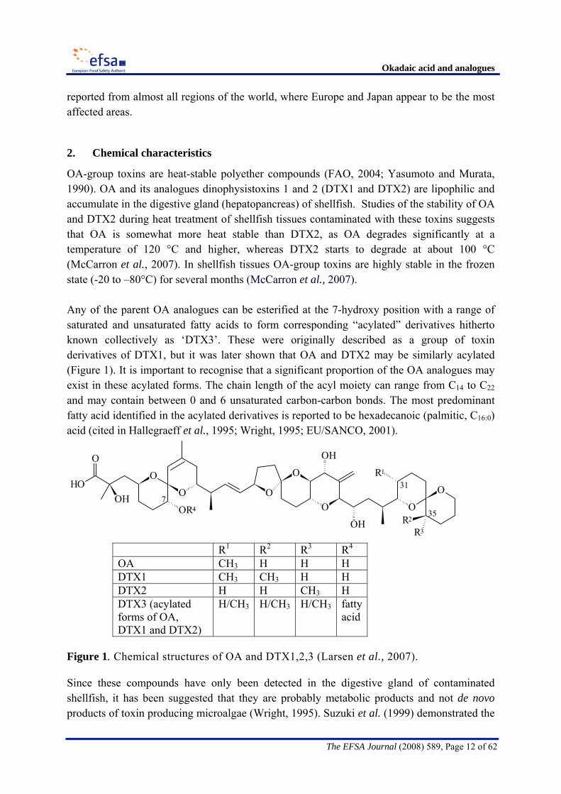
OA-group toxins are heat-stable polyether compounds (FAO, 2004; Yasumoto and Murata, 1990). OA and its analogues dinophysistoxins 1 and 2 (DTX1 and DTX2) are lipophilic and accumulate in the digestive gland (hepatopancreas) of shellfish. Studies of the stability of OA and DTX2 during heat treatment of shellfish tissues contaminated with these toxins suggests that OA is somewhat more heat stable than DTX2, as OA degrades significantly at a temperature of 120 °C and higher, whereas DTX2 starts to degrade at about 100 °C (McCarron et al., 2007). In shellfish tissues OA-group toxins are highly stable in the frozen state (-20 to –80°C) for several months (McCarron et al., 2007). Any of the parent OA analogues can be esterified at the 7-hydroxy position with a range of saturated and unsaturated fatty acids to form corresponding “acylated” derivatives hitherto known collectively as ‘DTX3’. These were originally described as a group of toxin derivatives of DTX1, but it was later shown that OA and DTX2 may be similarly acylated (Figure 1). It is important to recognise that a significant proportion of the OA analogues may exist in these acylated forms. The chain length of the acyl moiety can range from C14 to C22 and may contain between 0 and 6 unsaturated carbon-carbon bonds. The most predominant fatty acid identified in the acylated derivatives is reported to be hexadecanoic (palmitic, C16:0) acid (cited in Hallegraeff et al., 1995; Wright, 1995; EU/SANCO, 2001).
31
7
35
O
O
O
HOOH
OR4
OH
O
OO
O
O
OH
R1
R3R2
R1 R2 R3 R4 OA CH3 H H H DTX1 CH3 CH3 H H DTX2 H H CH3 H DTX3 (acylated forms of OA, DTX1 and DTX2)
H/CH3 H/CH3 H/CH3 fatty acid
Figure 1. Chemical structures of OA and DTX1,2,3 (Larsen et al., 2007).
Since these compounds have only been detected in the digestive gland of contaminated shellfish, it has been suggested that they are probably metabolic products and not de novo products of toxin producing microalgae (Wright, 1995). Suzuki et al. (1999) demonstrated the

Okadaic acid and analogues
The EFSA Journal (2008) 589, Page 13 of 62
transformation of DTX1 to 7-O-acyl-DTX1 in the scallop Patinopecten yessoensis. The ester bond in the acylated compounds can be hydrolyzed to form the parent compounds both chemically by heating in 0.5 M NaOH/90% methanol solution at 75 ºC for 40 minutes or enzymatically using lipase and cholesterol esterase (cited in EU/SANCO, 2001). The acylated derivatives of the OA analogues show an increased liposolubility compared to the parent (unesterified) compounds and possess toxic activity following hydrolysis in the gastroinstinal tract. 3. Regulatory status
For the control of the OA-group toxins in the EU, Council Directive 91/492 EEC11, as amended by Council Directive 97/79/EC12 established that the customary biological method must not give a positive result for the presence of DSP toxins in the edible part of the molluscs, but it did not clarify the interpretation of a positive result and did not specify which biological method should be used. Regulation (EC) No 853/20045 repealing the previous Directives, prescribes in chapter VI: “Health Standards for Live Bivalve Molluscs” that “food business operators must ensure that live bivalve molluscs placed on the market must not contain marine biotoxins in total quantities (measured in the whole body or any part edible separately) that exceed the following limits: for okadaic acid, dinophysistoxins and pectenotoxins, 160 μg of OA equivalents per kg”. The fact that these toxins are grouped together appears to be based on possible co-occurrence of OA-group toxins and pectenotoxins rather than on toxicological considerations, since pectenotoxins do not share the same mechanism of action as OA-group toxins. Commission Regulation (EC) No 2074/20054 provides details about the “Recognised testing methods for detecting marine biotoxins”. Annex III, Chapter III of this regulation deals with lipophilic toxin detection methods. Biological methods are to be used for the detection of OA-group toxins: both a mouse bioassay and a rat bioassay may be used. Commission Regulation (EC) No 2074/20054 also states the following concerning alternative detection methods: “A series of methods, such as high-performance liquid chromatography with fluorescence detection, liquid chromatography, mass spectrometry, immunoassays and functional assays, such as the phosphatase inhibition assay, shall be used as alternative or supplementary to the biological testing methods, provided that either alone or combined they can detect at least the following analogues, that they are not less effective than the biological methods and that their implementation provides an equivalent level of public health protection.
- okadaic acid and dinophysistoxins: a hydrolysis step may be required to detect the presence of DTX3.
- pectenotoxins: PTX1 and PTX2 - yessotoxins: YTX, 45 OH YTX, homo YTX, and 45 OH homo YTX. 11 OJ L 268, 24.9.1991, p. 1-14 12 OJ L 24, 30.1.1998, p. 31-32

Okadaic acid and analogues
The EFSA Journal (2008) 589, Page 14 of 62
- azaspiracids: AZA1, AZA2 and AZA3. If new analogues of public health significance are discovered, they should be included in the analysis. Standards must be available before chemical analysis is possible. Total toxicity shall be calculated using conversion factors based on the toxicity data available for each toxin. The performance characteristics of these methods shall be defined after validation following an internationally agreed protocol”. Currently there is no detailed guidance on how a non-animal-based method can become an accepted alternative method, i.e. which performance criteria should be fulfilled. In addition, conversion factors have not been established. The Commission Regulation (EC) No 2074/20054 (Annex III, Chapter III) also states that “Biological methods shall be replaced by alternative detection methods as soon as reference materials for detecting the toxins prescribed in Chapter V of Section VI of Annex III to Regulation (EC) No 853/2004 are readily available, the methods have been validated and this Chapter has been amended accordingly”. The current legislation permits the replacement of the biological methods, provided that alternative methods have been validated according to an internationally recognised protocol. The application of single laboratory validation (SLV) according to international guidelines to demonstrate their fitness-for-purpose in practice can be an impetus for implementation of instrumental analysis (e.g. liquid chromatography-mass spectrometry (LC-MS)) in regulatory analysis. 4. Methods of analysis
Several published methods exist for the detection of the OA-group toxins in plankton and bivalves. Of these, mammalian bioassays are still applied widely despite growing concern with respect to the use of such methods for reasons of animal welfare, their inherent variability and interference from other biotoxins which may co-exist in a sample. Functional assays and chemical methods are also available, however only one, an LC method with fluorescent detection (LC-FLD) (CEN, 2004) has been formally validated in collaborative studies according to the harmonised protocol of ISO/IUPAC/AOAC (Horwitz, 1995). In attempts to advance, develop and validate non-animal methods, research is being undertaken by a number of groups worldwide. Information on methods that are currently being used or are in the process of being developed and have the potential for use in a regulatory setting is provided below. For a more general overview of other methods, see the Joint FAO/IOC/WHO ad hoc Expert Consultation on Biotoxins in Bivalve Molluscs (2004) and the review paper by Hess et al. (2006).

Okadaic acid and analogues
The EFSA Journal (2008) 589, Page 15 of 62
Supply of appropriate reference material
The main provider in the field of certified reference materials for marine biotoxins has been the National Research Council Canada – Institute for Marine Biosciences, in Halifax, Nova Scotia, Canada (NRCC 57– IMB). To date, only OA is commercially available as a defined reference standard. Further calibrants for DTX1 and DTX2 have been prepared and are scheduled to be certified in 2008. A certified mussel reference material for OA and DTX1 can be obtained from the National Research Council Canada – Institute for Marine Biosciences. A disadvantage of the latter reference material is that the certified toxin levels are 70-fold higher than current European legislative limits. In collaboration with the Marine Institute Ireland, the Norwegian Veterinary Institute, AgResearch (NZ) and the EC-Joint Research Centre-Institute for Reference Materials and Measurement (IRMM), a multitoxin group mussel material (Mytilus edulis) has been prepared and will be certified over the coming years. This material is contaminated at appropriate levels with OA, DTX1 and DTX2. 4. 1 Mammalian bioassays
Commission Regulation (EC) No. 2074/20054 allows for the use of two types of mammalian bioassays for the detection of the OA-group toxins; neither of which have been formally validated. These are described below: Mouse bioassay
Historically, the mouse bioassay (MBA) has been used extensively in biotoxin monitoring and as such is incorporated into EU legislation (Commission Regulation (EC) No 2074/20054 Annex III, Chapter III). The MBA was developed by Yasumoto and colleagues (1978) as an investigative tool for the determination of the causative agents responsible for a food poisoning outbreak associated with the consumption of molluscs in Japan. Essentially, the assay uses acetone extraction of the whole flesh (or the hepatopancreas (HP)) of molluscs followed by evaporation and resuspension of the residue in a 1% solution of Tween 60 surfactant. Mice are then exposed to the extract via intraperitoneal (i.p.) injection and survival monitored over a 24 hour period (see Figure 2).

Okadaic acid and analogues
The EFSA Journal (2008) 589, Page 16 of 62
Figure 2. Sample preparation and extraction methods of hepatopancreas for the MBA (CRL-MB, 2007).

Okadaic acid and analogues
The EFSA Journal (2008) 589, Page 17 of 62
In efforts to improve the specificity of the assay, several modifications to the technique (generally involving an additional partitioning step) were developed (Yasumoto et al., 1984, Lee et al., 1987, Marcaillou-Le Baut et al., 1990, Fernández et al., 2002). Commission Regulation (EC) 2074/20054 allows for the use of different solvents in the liquid/liquid (water) partition step including ethyl acetate, dichloromethane and diethyl ether. A positive result is defined as the death of 2 out of three mice within 24 hours of injection with an extract operationally equivalent to 25 g whole flesh (including HP). The detectability and selectivity depends on the choice of solvents used for extraction and partitioning. Clearly it is not ideal for a regulatory method to allow for such procedural variation, so in an effort to harmonise the methodology used within the EU, the Community Reference Laboratory for marine biotoxins (CRL–MB) has developed a standard operating procedure based on acetone extraction with either diethyl ether or dichloromethane partitioning against water. The Standard Operating Procedure (SOP) for this method has been available at the CRL web page since 2007 (CRL-MB, 2007).
The i.p. LD50 of OA in mice is approximately 200 μg/kg bodyweight (b.w.), i.e. for a 20 g mouse ca. 4 μg (Aune et al., 2007), which is equal to the amount injected in the MBA if the shellfish contains OA-group toxins at the regulatory limit of 160 µg OA equivalents/kg shellfish. The LD50 is the dose that kills 50% of the exposed animals (if a sufficiently large number of animals are used). Using the MBA according to the SOP (see above), the probability of detecting OA-group toxins at the current EU legal limit is 40 to 50%, the probability of detecting OA-group toxins at 1.25 times the current EU legal limit (i.e. 200 µg OA-equivalents/kg shellfish flesh) using the MBA is ca. 90 % (see Aune et al., 2007, and calculations in the footnote13), this estimate is based only on a single laboratory and a single mouse strain. In the event of no death, the mice may develop specific symptoms of OA-group toxins that constitute a significative indicator of contamination and potential risk. The minimum amount of toxin (4 µg OA) administered i.p. needed to kill a 20 g mouse within 24 hr has been described as one mouse unit (MU) (Yasumoto et al., 1978).
13The current regulatory limit of 160 µg/kg shellfish flesh applies to the whole flesh (WF). As the MBA
determines the equivalent value actually in hepatopancreas (HP), the concentration in HP equivalent to 160 µg/kg WF is ca. 800 µg/kg HP. The current MBA protocol contains a 5-fold concentration factor (20 g HP into 4 mL Tween 60), and the equivalent of 5 g HP suspended in 1 mL is injected into each mouse. Therefore, at the current limit (equivalent to 800 µg/kg HP), 4 µg OA equivalents are injected into each of 3 mice. This dose of 4 µg equates to a dose of ca. 200 µg/kg bodyweight. As determined by Aune et al. (2007), the prevalence of death in mice injected at 206 µg/kg is 50%. Due to the steepness of the dose-response curve, the prevalence of death at 200 µg/kg bodyweight is 43 %. This means that each mouse has a 43% probability of dying when injected with 4 µg OA equivalents. The summation of all the probabilities for each of the eight scenarios with 3 mice shows that the total probability of detecting a positive is only 40% at the regulatory limit. The calculations show that due to the steepness of the dose-response curve, the probability of detecting a positive at 200 µg/kg OA equivalents in WF, i.e. a dose of 5 µg OA equivalents is already 90% (as the probability for the individual mouse is ca. 80% to die).

Okadaic acid and analogues
The EFSA Journal (2008) 589, Page 18 of 62
The advantages of the MBA include: • the provision of a measure of total toxicity based on the biological response of the
animal to the toxin(s); • it does not require complex analytical equipment;
The major disadvantages of the MBA include:
• the outcome depends on the choice of solvents used; • it is labour intensive and cannot be readily automated; • it requires specialised animal facilities and expertise; • the high variability in results between laboratories due to e.g. specific animal
characteristics (strain, sex, age, weight, general state of health, diet, stress); • the potential for false positive results due to interferences (e.g. free fatty acids); • the potential for false negative results; • it is not selective for solely the OA-group toxins; • it is not quantitative; • the i.p. route is not appropriate for the complete detection of some relevant toxins of
the OA group requiring hydrolysis in the gastrointestinal tract (DTX3). • the injection volume of one mL exceeds good practice guidelines (less < 0.5 mL)
intended to minimise stress to mice; • in many countries the use of the MBA is considered unacceptable for ethical reasons.
Rat bioassay
In the original procedure (Kat, 1983) shellfish hepatopancreas mixed with normal rat feed is fed to pre-starved white female rats. In the procedure currently applied in the Netherlands (Van der Hoeven, 2007) 10 g of shellfish hepatopancreas (if possible and desired) or 10 g of shellfish meat (e.g. for cockles) is collected and fed to female rats that have been starved for 24 hours. After a 16 h-period the consistency of the faeces (softening) is observed along with the quantity of food eaten. The test results are expressed as -, +/-, +, ++ or +++, where a response of + (++) in the rat is considered to correspond with severe complaints with diarrhoea and nausea in man. An exact limit of detection of the rat bioassay cannot be given, but it is near the current legal limit of 160 μg of OA equivalents/kg. The advantages of the rat bioassay include:
• it does not involve extraction of toxin and therefore it avoids any toxin loss due to methodology;
• it does not require complex analytical equipment. The disadvantages of rat bioassay include:
• lack of specificity, since it will also detect other diarrhoeic agents in the sample, e.g. azaspiracids;
• it requires specialised animal facilities and expertise; • variation in sensitivity and symptomology amongst rats.

Okadaic acid and analogues
The EFSA Journal (2008) 589, Page 19 of 62
4. 2 Biomolecular methods
EU regulation 2074/2005 allows for the use of alternative methods for the detection of the OA-group of toxins; none of which have been formally validated. The two major assay methods are described below: Protein phosphatase inhibition assay OA and DTXs are specific inhibitors of protein phosphatase-1 (PP1) and -2A (PP2A), (Bialojan and Takai, 1988; Cohen, 1989). Para-nitrophenylphosphate (pNPP) is an especially suitable artificial substrate for PP2A (Takai and Mieskes, 1991) and can be used for quantitative analysis of the OA-group of toxins, with a colorimetric phosphatase-inhibition assay (Simon and Vernoux, 1994). It will also detect microcystins, produced by cyanobacteria ((Fontal et al., 1999), however to date these are not considered to be common in marine shellfish. A colorimetric phosphatase assay has been developed by Tubaro et al. (1996) using a commercial PP2A preparation. The procedure is capable of detecting 10 µg OA/kg hepatopancreas. A PP2A method with fluorimetric detection (Vieytes et al., 1997) is capable of detecting 2 µg OA/kg hepatopancreas. The fluorimetric assay shows a good correlation with both HPLC and the bioassay (González et al., 2002). DTX3 can only be detected in the protein phosphatase inhibition assay if an alkaline hydrolysis step is included. A further variant is a method using PP2A enzyme in a competitive displacement assay for OA and the DTXs. (Døskeland et al., 2000, Serres et al., 2000). The main advantages of the phosphatase inhibition assay include:
• it is very sensitive; • it is highly specific to those compounds which are protein phosphatase inhibitors; • it provides a measure of total OA equivalents provided that hydrolysis of DTX3 is
applied; • it requires only OA as calibrant.
The main disadvantages of the phosphatase inhibition assay include: • it requires a good quality enzyme to be readily available; • it does not provide any information on the toxin profile.
Immunoassays
There are a number of immunodiagnostic methods for the OA-group toxins which incorporate antibodies raised against OA. None of these methods have been fully validated. Several commercial enzyme-linked immunosorbent assay (ELISA) kits and optical biosensor

Okadaic acid and analogues
The EFSA Journal (2008) 589, Page 20 of 62
antibody-based methods are available. Immunological methods for toxin detection exploit the affinity of antibodies for their antigens. Antibody-based methods detect only the chemicals possessing the specific structure recognised by the antibody used in the assay, without providing information about the activity of the analogues being detected. The efficacy of immunological methods in the detection of different analogues is a function of the affinity of the antibody used in the assay for that analogue. The relative abundance of the analogues detected in an antibody-based procedure does not unequivocally mirror the relative abundance of those analogues in the mixture subjected to analysis. The simplification of antibody-based methods (use of a single antibody to detect a set of analogues) is accompanied by the loss of chemical discrimination and quantification of the different analogues (as in chemical methods), without providing information about the overall activity of the mixture (as in functional assays). The main advantages of an antibody-based method are:
• it is very sensitive; • it is fast, easy to use, and can be applied to screen many samples at any one time for
further confirmatory analysis.
The main disadvantages of antibody-based methods are: • the accuracy is questionable when mixtures of analogues are being analyzed which is
most often the case; • it does not provide any information on the toxin profile.
4. 3 Chemical methods
Physico-chemical methods (mainly liquid chromatography (LC)) combined with fluorescence detection (FLD) or mass spectrometry (MS) are useful for identification and quantification of the OA group of toxins. At the time of preparation of this opinion, the only inter-laboratory validated method for the OA toxins is a liquid chromatography (LC)-fluorescence method (Lee et al., 1987) for OA in mussel digestive gland with a LOQ of 100 µg OA per kg hepatopancreas. The method has been standardised by CEN (2004). However, although the method has been used for DTX1 and DTX2 validation data for these analogues are lacking. Liquid chromatography-mass spectrometry (LC-MS) as well as liquid chromatography-mass spectrometry/mass spectrometry (LC-MS/MS), which together in the following text are termed LC-MS/(MS), methods are increasingly being used in monitoring programs. One method (McNabb et al., 2005), with a LOQ of 40 µg OA/kg shellfish tissue, has undergone an intensive single-laboratory validation and a limited inter-laboratory study, although this study did not include real samples. Some EU Member States are currently using LC-MS/(MS) data to supplement information generated by the MBA by parallel testing. The development of LC-MS/(MS) methodology is promising. In a recent proficiency test organised by the CRL

Okadaic acid and analogues
The EFSA Journal (2008) 589, Page 21 of 62
(2006) with samples of shellfish with high toxin levels, eight laboratories reported results obtained by LC-MS/(MS) with the data indicating low interlaboratory variability (HORRAT14 < 1, after removal of outliers). In the proficiency-testing scheme QUASIMEME (Quality Assurance in Marine Environmental Matrices in Europe) development exercises for OA group compounds (e.g. round 49, exercise 760, DE10, report issue 1, 14-09-07), 13 laboratories reported data using LC-MS(/MS), and achieved a between-laboratory coefficient of variation (CV) of 16 - 21 % for the total hydrolysed OA-equivalents in a mussel and a clam tissue respectively. For both matrices, 11 out of 13 laboratories achieved satisfactory z-scores for the total OA-equivalents. Some of the methods in use have been developed to allow multi-toxin group detection (Stobo et al. 2005, McNabb et al. 2005, Fux et al. 2007). The available data from in-house and interlaboratory studies suggested that between-laboratory variability was lower when laboratories used their own in-house validated method, than when they adhered to a strictly standardized protocol. The major advantages of LC-MS/(MS) methods include:
• it is highly specific and sensitive; • it can screen and measure the OA-group toxins individually provided hydrolysis is
applied; • it gives information on the OA-group toxin profiles in samples; • it can be automated;
The major disadvantages LC-MS/(MS) methods include: • it requires costly equipment and highly trained personnel; • it requires a wide range of reference standards for identification and quantification.
4.4 Summary of methods
From the above brief summary of methods it can be seen that although currently prescribed by EU legislation, the mammalian bioassays have not been fully validated. Recent information has confirmed that the mouse bioassay only has a 40 to 50% chance of detecting a positive response for a sample containing OA at the current regulatory limit of 160 µg/kg. Very limited quantitative data on the rat bioassay are available. Additionally, Council Directive 86/609/EEC6 states that Member States may not permit the use of live animals in procedures that may cause pain, suffering distress or lasting harm if another scientific satisfactory method of obtaining the result sought and not entailing the use of live animals is reasonably and practicably available. At this time however, none of the methods for the detection of toxins from the OA group have been validated by interlaboratory studies for all the analogues (OA, DTX1, DTX2 and esters
14 The Horwitz ratio (HORRAT) is a normalised performance parameter that indicates the acceptability of
analytical methods with respect to reproducibility. It is the ratio of the actual observed relative standard deviation among laboratories to the corresponding predicted relative standard deviation calculated from the Horrwitz equation.

Okadaic acid and analogues
The EFSA Journal (2008) 589, Page 22 of 62
thereof). It is particularly important therefore, that the various methods are evaluated for their fitness for purpose. The most objective way of comparing methods is by comparison of the corresponding analytical performance characteristics. Table 2 summarises the performance characteristics for the three main groups of tests (mammalian assays, biomolecular methods and chemical methods). The evidence available at this time suggests that the phosphoprotein-phosphatase assays and LC-MS/(MS) based methods have the greatest potential to replace the mammalian assays. Moreover, they are able to detect OA-group toxins at concentrations below the current regulatory limit of 160 µg/kg. In principle, the potential of LC-MS/(MS) analysis for the detection of OA-group toxins in shellfish is enhanced by the improving availability of reference standards and materials, and is realised by the satisfactory performance in single-laboratory validation of LC-MS/(MS) methods using internationally-agreed protocols. The LC-MS/(MS) based methods also have the possibility for multi-toxin group detection/quantification. However, before these methods can be used there are a number of obstacles to overcome such as validation results that support their use.

Okadaic acid and analogues
The EFSA Journal (2008) 589, Page 23 of 62
Table 2. Overview of the performance characteristics of the three main groups of methods of detection of OA-group toxins (OA, DTX1, DTX2, and esters of these).
Method Type Bioassays Protein phosphatase 2a assays LC-based analyses Subtype Subtype Subtype
Mouse Rat Fluorime-tric
Colorime-tric
Receptor-based
FLD MS
Performance Characteristic according to
(SOP of the CRL-MB 2007)
van der Hoeven, 2007
(Vieytes et al., 1997 González et al., 2002)
Tubaro, 1996)
Kleivdal, 2004
(CEN 2004 validated for HP)
various
Qual./Quant. Qual. Semi-Quan.
Quant. Quant. Quant. Quant. Quant.
Reported LOD in shellfish
approx. 160 μg OA equ./kg
approx. 160 μg OA equ./kg
26 µg OA equ./kg
10 µg OA equ./kg
unknown approx. 15 µg OA/kg
1-10 µg OA/kg
Reported LOQ in shellfish
N/a N/a 41 µg OA equ./kg
32 µg OA equ./kg
unknown approx. 40 µg OA/kg
30-50 µg OA/kg
Specificity None (any lipophilic bioactive)
Little (any lipophilic bioactive with diarrheic effect on oral exposure)
Highf) High High (interference unknown)
High (interference unknown)
High (interference unknown)
Selectivity N/a N/a High for OA group
High for OA group
High for OA group
High for individual toxins
High for individual toxins
Duration ( min for 1 sample) a)
48 h 17 h 3h 3h 3h 24h 24h
Repeatability (as within-batch CV)
N/a N/a 10-20% 10% unknown approx. 10%
5-10%
UCM at legal limit (quan.) c)
b) N/a 10-30% 18% unknown unknown 25-30%d)
Status of standardisation.
CRL N/a Pending validation
Pending validation
Pending validation
CEN Pending validation
Status of interlab. valid.
N/a N/a Under way Under way in-house valid.
OA done Under way
a) The duration of the test is given as the minimal time for 1 sample including time for preparation and extraction (to allow comparison). It was decided to avoid any estimation of time duration for several samples analysed simultaneously, this factor depending on the laboratory for a significant part.
b) Using the MBA according to the SOP (CRL-MB, 2007) the probability of detecting OA-group toxins at the current EU legal limit is less than 50%, the probability of detecting OA-group toxins at 1.25 times the current EU legal limit (i.e. 200 µg OA-equivalent/kg shellfish flesh) using the MBA is ca. 90 % (see Aune et al., 2007, and calculations in the annex 1), estimate only based on single lab and mouse strain.
c) Uncertainty of Measurement (UCM) as 95 % confidence interval of the long-term (between-batch) coefficient of variation (CV)).
d) Marine Institute LC-MS method (Ireland), unpublished information. e) Not applicable. f) Only interference from microcystins and nodularin. Equ. = equivalents

Okadaic acid and analogues
The EFSA Journal (2008) 589, Page 24 of 62
5. Occurrence of OA-group toxins
Data description and reporting of results
Following a request by EFSA, a number of Member States provided data on the occurrence of OA and analogues in shellfish. With a few exceptions, the data submissions covered samples collected and tested during 2001 to 2006 with most samples from the last two years. Overall, 6072 sample results were considered by the Panel for this assessment. Table 3 shows a summary of the number of samples submitted dependent on the providing country, type of sampling, and type of analytical methods applied. Where available, the limits of detection (LOD) and the limit of quantification (LOQ) of the methods used are also indicated. Table 3: Data on OA-group marine biotoxin submissions obtained from Member States up to March 2007.
Country Year(s) of harvesting
Number of samples
Purpose of testinge)
Method of testing
LOD (µg/kg
shellfish)
LOQ (µg/kg
shellfish)
Number tested
by MBA
Denmark 1999-2003 161 PreMC and PostMC LC-MS 5 No data NA
France 2001-2006 40 PostMC LC-MS No data No data 39 27 ELISA Pos/Nega) No data 0 60 LC-FLD < 50b) No data 0 Germany 2004-2006 550
PreMC and PostMC
LC-MS < 1-10c) No data 0 Ireland 2004/2005 758 PreMC LC-MS 10 30 758
The Netherlands 2001-2006 165 (14 pre
and 151 post MC)
PreMC and PostMC LC-MS 8-17 No data 8f)
Norway 2004-06 1849 PreMC LC-MS 20d) 60 NA Portugal 2005/06 1074 PreMC LC-MS No data No data 0 Spain 2005 3 PreMC LC-MS No data 49 0 Sweden 2005/06 928 PreMC LC-MS 4d) 10 0 United Kingdom 2003-2006 457 PreMC LC-MS 10 No data 405g)
a) ELISA data mainly given as positive/negative and not quantified b) The limit of detection (LOD) varies between 20-50 µg/kg c) The limit of detection (LOD) varies between 1 and 10 µg/kg d) Data extrapolated from hepatopancreas to whole meat e) PreMC and PostMC cover samples taken before products are sent to the market and product sampled at the
market, respectively f) The samples from the Netherlands were tested in rat not mouse bioassay g) The MBA was conducted with a 5 hour observation period only. A result was recorded as positive if 2 out of 3
mice exhibited a combination of clinical signs within the observation period. There are considerable differences in the number of analyses per year and country. Moreover, several different analytical methods have been used (LC-MS, LC-FLD, ELISA) and the matrix analysed consisted either of whole shellfish meat (including the hepatopancreas) or of hepatopancreas, in which the OA-group toxins accumulate and thus provides better sensitivity for their detection. In the latter case, the results must be calculated in terms of the whole tissue sample in order to check for compliance with legal limits.

Okadaic acid and analogues
The EFSA Journal (2008) 589, Page 25 of 62
As prescribed in the respective EU legislation, most of the data were related to pre-marketing control (PreMC) measurements, i.e. before samples are harvested for further processing or direct consumption. Germany, Denmark and The Netherlands submitted both data on PreMC and post-marketing control (PostMC). With the exception of Germany, the other countries have indicated the origin of the shellfish samples. The post-marketing data from Germany revealed that samples that were collected at stores and supermarkets had multiple (mostly unknown) origins. OA concentration in shellfish
There are some differences in the way results on occurrence of OA-group toxins were reported. As mentioned earlier, the OA group includes the three toxins OA, DTX1 and DTX2, as well as a number of acylated analogues, collectively termed DTX3, which can only be quantified after hydrolysis. Not all Member States submitted results for all individual OA analogues, and in some cases only the sum for the combined OA group-toxins was reported without information on which analogues were detected. Basic statistics of concentration data of OA-group toxins were calculated for the results submitted by each country as shown in Table 4. Depending upon whether screening for compliance with legal limits or analogue specific determination was the objective, the selectivity and sensitivity of the analytical methods applied in the Member States differ widely. Because this may have a considerable influence on the result, for further statistical analyses only the 5,980 samples that were analysed by LC-MS techniques were considered. Table 4: Statistics of LC-MS data of OA-group toxins in shellfish (pre- and post market samples) provided by Member States up to March 2007.
Median Mean P95 Maximum ≤LOD >160 µg/kg
Country
Number
of
samples µg/kg shellfish %
Denmark 161 130 270 1148 2516 0% 30% France 40 35 77 263 526 3% 4% Germany 550 10 28 135 380 66% 3% Ireland 758 120 268 1190 5370 35% 41% The Netherlands 163 10 11 10 48 96% 0% Norway 1846 38 130 525 6550 16% 19% Portugal 1074 35 148 676 5158 30% 21% Spain 3 332, 259, 335 Sweden 928 64 158 584 2412 14% 31% UK 457 10 92 439 5388 46% 18% All 5980 33 138 571 6550 30% 22%
For most of the data no information is available on measurement uncertainty.
Okadaic acid and analogues
The EFSA Journal (2008) 589, Page 26 of 62
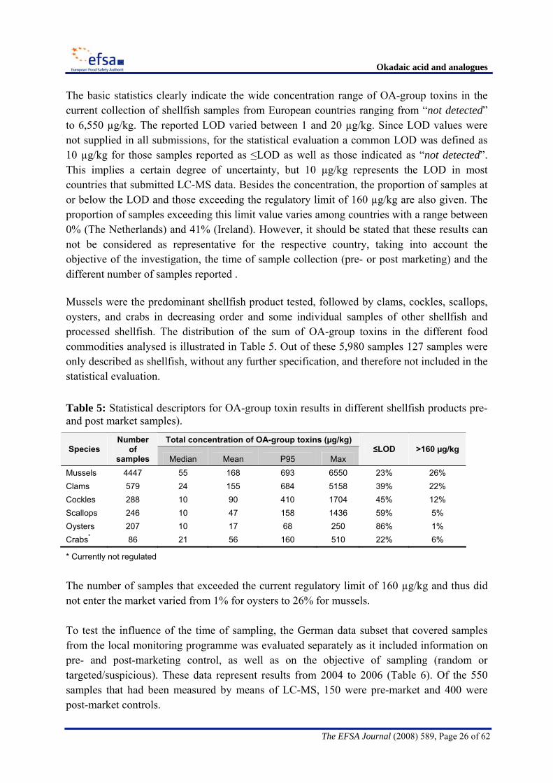
The basic statistics clearly indicate the wide concentration range of OA-group toxins in the current collection of shellfish samples from European countries ranging from “not detected” to 6,550 µg/kg. The reported LOD varied between 1 and 20 µg/kg. Since LOD values were not supplied in all submissions, for the statistical evaluation a common LOD was defined as 10 µg/kg for those samples reported as ≤LOD as well as those indicated as “not detected”. This implies a certain degree of uncertainty, but 10 µg/kg represents the LOD in most countries that submitted LC-MS data. Besides the concentration, the proportion of samples at or below the LOD and those exceeding the regulatory limit of 160 µg/kg are also given. The proportion of samples exceeding this limit value varies among countries with a range between 0% (The Netherlands) and 41% (Ireland). However, it should be stated that these results can not be considered as representative for the respective country, taking into account the objective of the investigation, the time of sample collection (pre- or post marketing) and the different number of samples reported . Mussels were the predominant shellfish product tested, followed by clams, cockles, scallops, oysters, and crabs in decreasing order and some individual samples of other shellfish and processed shellfish. The distribution of the sum of OA-group toxins in the different food commodities analysed is illustrated in Table 5. Out of these 5,980 samples 127 samples were only described as shellfish, without any further specification, and therefore not included in the statistical evaluation. Table 5: Statistical descriptors for OA-group toxin results in different shellfish products pre- and post market samples).
Total concentration of OA-group toxins (µg/kg) Species
Number of
samples Median Mean P95 Max ≤LOD >160 µg/kg
Mussels 4447 55 168 693 6550 23% 26% Clams 579 24 155 684 5158 39% 22% Cockles 288 10 90 410 1704 45% 12% Scallops 246 10 47 158 1436 59% 5% Oysters 207 10 17 68 250 86% 1% Crabs* 86 21 56 160 510 22% 6%
* Currently not regulated
The number of samples that exceeded the current regulatory limit of 160 µg/kg and thus did not enter the market varied from 1% for oysters to 26% for mussels. To test the influence of the time of sampling, the German data subset that covered samples from the local monitoring programme was evaluated separately as it included information on pre- and post-marketing control, as well as on the objective of sampling (random or targeted/suspicious). These data represent results from 2004 to 2006 (Table 6). Of the 550 samples that had been measured by means of LC-MS, 150 were pre-market and 400 were post-market controls.

Okadaic acid and analogues
The EFSA Journal (2008) 589, Page 27 of 62
Table 6: Overview of LC-MS data of OA-group toxins obtained from the German official surveillance programme of food control.
Type of sampling
Monitoring (pre-MC)
random (post-MC)
targeted (post-MC)
N 150 334 66 Median µg/kg 10 10 106 Mean µg/kg 10 16 130 95th percentile µg/kg 10 51 345
Max µg/kg 46 233 380 ≤LOD 90% 65% 13.8 % >160 µg/kg 0% 0.3% 32 %
Considerably higher values were reported for targeted sampling and in situations where contamination was suspected. In contrast, the data from the post-marketing investigations of samples originating from different countries in general only revealed relatively low levels which might be an indication that the pre-marketing control to a great extent prevents lots with high concentrations from reaching the market. It was noted that all German samples were cooked before analysis. Concentrations of individual analogues of OA-group toxins
For a total of 2,419 samples complete individual data were reported for the concentration of the sum of the OA-group toxins as well as for OA, DTX1, DTX2 and DTX3 individually. Statistical descriptions of these results are presented in Tables 7 and 8. For the statistical evaluation of the whole data set a value of 10 µg/kg was assigned to those analogues that were reported as “non-detected”. Table 7: Number of samples for which numerical data on OA-group toxins have been reported presented in different concentration ranges.
Sum of OA and analogues OA DTX1 DTX2 DTX3 Concentration range
Number of samples <LOD 610 1328 1914 2051 775 ≥LOD up to 160 μg toxin/kg shellfish meat
1309 988 416 302 1495
>160 μg toxin /kg shellfish meat 500 103 89 66 149

Okadaic acid and analogues
The EFSA Journal (2008) 589, Page 28 of 62
Table 8. Statistical descriptors of the concentrations of OA and its analogues for samples for which numerical data on OA and all analogues have been reported (n = 2419).
Parameter
Sum of OA and analogues
(μg toxin /kg shellfish meat)
OA (μg toxin/kg shellfish meat)
DTX1 (μg toxin /kg shellfish
meat)
DTX2 (μg toxin /kg shellfish
meat)
DTX3 (μg toxin /kg shellfish
meat)
median 80 < LOD < LOD < LOD 20 95th
percentile 521 142 104 94 197
Of all the samples, about 25% were below the LOD for the sum of OA and analogues and consequently 75% had measurable levels of OA-group toxins. Regarding the individual analogues, DTX3 had the highest and DTX2 the lowest number of positive results. On average, OA contributed 27% to the concentration of OA-group toxins, DTX1 24%, DTX2 16% and DTX3 34%. This is consistent with published results, in which acylated analogues were reported to contribute considerably to the concentration of total OA-group toxins (Vale and Sampayo, 2002a). The considerable contribution of DTX3 to the total concentration of OA-group toxins may have implications for the discussion on the toxicity since esterified compounds must be hydrolysed in vivo before exerting their toxic effect (see later chapters). Moreover, the importance of DTX3 has to be taken into account when interpreting data from mouse bioassays which are performed by i.p.injection thereby avoiding hydrolysis in the gastrointestinal tract as occurs following oral ingestion. Variation of the concentrations of OA group toxins in individual mussels
OA group toxins may not be homogenously distributed among lots of shellfish. To date there are no generally accepted procedures readily available to make adequate sampling of shellfish possible. This may lead to non-representative samples, which do not accurately reflect the mean toxin concentration in shellfish from one batch. Several 5 kg packages of mussels (deriving from one commercial batch) were taken from a shellfish processing establishment which cooks and freezes mussels before delivering them to the customers in packages from 250 g to 10 kg. In order to determine the homogeneity of OA group toxins within one package 20 individual mussels were selected from the same package and were analysed separately by the German NRL. The results are shown in Figure 3.

Okadaic acid and analogues
The EFSA Journal (2008) 589, Page 29 of 62
0
20
40
60
80
100
120
1 2 3 4 5 6 7 8 9 10 11 12 13 14 15 16 17 18 19 200
200
400
600
800
1000
1200
1400OA
DTX 2DTX 1
µg/k
g (O
A &
DTX
2)
µg/k
g (D
TX1)
Figure 3: Concentrations (µg toxin/kg shellfish) of OA, DTX1 and DTX2 in individual mussels taken from the same commercial batch. Note that DTX1 concentrations are related to the right axis (0-1400 µg toxin/kg shellfish). The presented data of the investigated package have a wide variation of the concentrations for each toxin within the 20 mussels, as shown by the means and standard deviations (OA: 11 ± 15.6, DTX1: 230 ± 321, DTX2: 28 ± 27 µg/kg). Moreover, the toxin profiles differ considerably. For the chemical–analytical methods it is usually recommended to start the analysis with an initial weight of 150 g shellfish (without shells), corresponding to 20–30 mussels. Figure 3 shows that mean results can be strongly influenced by the sampling procedure. This demonstrates the need for a representative sampling procedure, as is usual for other contaminants in food, such as aflatoxins in nuts that show a similarly heterogeneous distribution. 6. Comparison of LC-MS data with results of mammalian bioassays
An issue raised frequently in scientific discussions on marine biotoxins is the comparability of the mammalian bioassay data with results obtained using LC-MS. In an attempt to address this issue the Panel has evaluated a total of 1,210 samples (shown in Table 3) that were tested both with mammalian bioassays and LC-MS. The Panel identified the number of samples that exceeded the maximum limit of 160 μg/kg based on LC-MS analysis but were tested negative in the mammalian bioassays. The results are shown in Table 9.

Okadaic acid and analogues
The EFSA Journal (2008) 589, Page 30 of 62
Table 9: Concentration of OA-group toxins measured by LC-MS in samples comparatively tested by mammalian bioassays.
Concentration (µg/kg) determined by LC-MS Mouse
Bioassay
Number of
samples Median Mean P95 Max ≤LOD >160 µg/kg
Negative 755 22 66 240 2240 44% 100 (13%) Positive 455 240 486 1810 8864 11% 325 (71%)
a) In this evaluation the data from Ireland, UK, F and NL were considered. About 80% of the above samples were identified as “mussels”, mostly M. edulis. Of the samples tested negative in the MBA, 13% exceeded the regulatory limit of 160 µg/kg when analysed by LC-MS, whereas 29% of the MBA positive samples did not exceed this level using LC-MS. It can be assumed that all bivalve molluscs showing a negative response in mammalian bioassays will reach the market and will thus be consumed. From this perspective, it is not unrealistic to estimate the dietary intake of OA-group toxins based on the LC-MS data for those samples that tested negative in the mammalian bioassays. 7. Human consumption of shellfish
Limited consumption data were available for individual shellfish species across the EU. The EFSA concise database does not yet provide sufficient information since there is no differentiation between meal sizes for fish and other seafood. Therefore, EFSA requested the Member States to provide information on shellfish consumption. Data have been submitted by France, Germany, Italy, The Netherlands and the UK. A compilation of the data received is presented in Table 10. The mean portion sizes for consumers only ranged between 10 g (France, bivalve molluscs) and 136 g (The Netherlands). The data from Germany, Italy and the UK are within this range. The German national food consumption survey performed by a weighing protocol in the late 1980s indicates a minimum meal size of mussels of 2 g (mainly as an ingredient in dishes), a median of 63 g, a mean of 107 g and a 95th percentile of 400g among mussel consumers. The maximum portion size reported in this study was 1,500 g. The French Calipso study differentiated mussels and bivalve molluscs. The maximum portions for mussels (245 g) and all bivalve molluscs (415 g) varied, whereas the mean portions were similar. A survey reported by the United Kingdom indicates a mean shellfish meal size of 114 g and a maximum of 239 g. A Dutch study reported a mean portion size of 136 g of shellfish and a maximum of 480 g. These data are for consumers only. The surveys show a large variation in the percentage of the populations consuming shellfish and it is unclear whether the data are related to cooked or uncooked shellfish.

Okadaic acid and analogues
The EFSA Journal (2008) 589, Page 31 of 62
Table 10: Shellfish eating habits in France, Italy, The Netherlands, the UK, and Germany, based on national food consumption surveys.
Country
Study
Number of consumers N (%)
Number of eating occasions for consumers/year
Mean portion weight (g)
95th percentile
Maximum portion weight (g)
Maximum frequency
France (7 days) INCA 1999 (11%) NA 10 NA
France (FFQ)
CALIPSO (bivalve molluscs)
962/997 (96%) NA 32 94 415 NA
France (FFQ)
CALIPSO (mussels)
862/997 (86%) NA 22 70 245 NA
Italy (7 days)
INN-CA 1994-96
212/1,981 (11%) 47 83 1,000 4/week
Germany (7 days)
NVS 1985-88
150/23,239 (0.6%) 171 107 400 1,500 3/week
UK (7 days) NDNS 2000-01
212/1,631 (13%) 51 114 239 4/week
The Netherlands (2 days)
DNFCS 1997-98
47/4,285 (1.1%) 39 136 465 480 NA
Because OA-group toxins have acute toxic effects, it is important to identify a high portion size rather than a long term average consumption in order to protect the health of the consumer. In the studies presented in the table above, the maximum reported sizes are in the range of 239 to 1,500 g. The Panel noted the highest portion sizes of 1,000 g and 1,500 g, and considered it likely that the shells were included in these weight estimates. Therefore, the Panel considered the 95th percentile as a more realistic estimate of the portion size for high consumers. As shown in Table 10 the 95th percentile values range from 70-465 g and the Panel chose the figure of 400 g to be used as a high portion size in acute exposure assessments. This is in good agreement with the report of the Joint FAO/IOC/WHO ad hoc expert consultation on marine biotoxins (FAO/IOC/WHO, 2004) where 380 g was reported as the highest 97.5th percentile portion size for consumers only.
8. Exposure assessment
Deterministic estimate of dietary exposure to OA-group toxins
Consumption of a 400 g portion containing the 95th percentile of the OA concentration, 240 µg/kg, as presented in Table 9, would result in an exposure of 96 µg OA equivalents per person. For concentrations at the current regulatory limit of 160 µg/kg the equivalent exposure would be 64 µg OA equivalents per person.
These results are conservative but not unrealistic estimates of OA dietary exposure in the EU.

Okadaic acid and analogues
The EFSA Journal (2008) 589, Page 32 of 62
Probabilistic estimate of dietary exposure to OA-group toxins
A probabilistic estimate of dietary exposure to OA-group toxins has been performed by a Monte Carlo simulation using the distributions of both the occurrence data and the data on the consumption of shellfish. Compared to the deterministic estimate the probabilistic exposure estimate provides information on the chance to exceed a specific exposure level. Because a person eating shellfish will not eat the same portion size containing the same level of toxins each time, the probabilistic calculation includes all the combinations of all different occurrence and consumption data. For the probabilistic estimate the same concentration data obtained by the LC-MS measurements of the samples tested negative in the mammalian bioassays (Table 9) were used15. Because insufficient information is available on the distribution of portion sizes, the Panel decided to use a triangular distribution as a simple and pragmatic approach. A triangular distribution is characterised by three values, the minimum, the most probable and the maximum. In the case of shellfish consumption a value of 0 was used as a minimum. From the range of 10 to 136 g reported as mean consumption figures in Table 10 the Panel chose a value of 100 g to be used as “most probable” value, although there is no evidence that it is the most frequently consumed portion. The better-documented large portion size of 400 g (see chapter 7) was used to represent the maximum. The resulting probabilistic dietary exposure distribution has a median value of approximately 6 µg/person, a mean of approximately 14 µg/person, and a 95th percentile of approximately 54 µg/person. The probabilistic exposure estimate is presented in Figure 4 illustrating the chance to exceed a specific level of exposure to OA equivalents when consuming a single portion of shellfish.
15 All samples with quantifiable levels of OA-group toxins (48% of the total number) were characterized by a
lognormal distribution, which has been derived by the best fit analysis of the @RISK tool. This distribution function was truncated at the LOQ of 30 µg/kg [RiskLognorm(133.81; 142.77; RiskShift (7.9); RiskTruncate(30;)]. The samples reported at or below the LOQ (52% of the total number) were randomly assigned a numerical value by using a discrete distribution [RiskDiscrete({0;1};0.52; 0.48)] reflecting the ratio of non-quantifiable/ quantifiable samples (52%/48%). This implies that 52% of the samples reported at or below the LOQ were assigned a “0” (zero). The remaining 48% of these samples were assigned a value between 0 and 30 µg/kg by using a uniform distribution function [RiskUniform(0;30)].

Okadaic acid and analogues
The EFSA Journal (2008) 589, Page 33 of 62
0%
10%
20%
30%
40%
50%
60%
70%
80%
90%
100%
0 20 40 60 80 100 120 140 160 180 200 220 240µg of OA equivalents per person
Prob
abili
tyof
exc
eedi
ngO
A d
ose
Figure 4: Probability of dietary exposure to OA-group toxins resulting from consumption of a single portion of shellfish. The dotted lines in figure 4 illustrate the chance to exceed a dietary intake of OA toxins as derived in the deterministic estimate. The chance to exceed an intake of 64 µg, corresponding with consumption of a portion of e.g. 400 g containing OA at the level of the current EU limit value, is about 4 %. The chance to exceed an exposure of 96 µg, corresponding to a consumption of a portion of e.g. 400 g containing the 95th percentile of the OA concentration, is about 2 %. As shown in the dotted line at the left side there is approximately 20% chance of exceeding the intake corresponding with the ARfD established in chapter 12. 9. Toxicokinetics 9.1 Absorption and distribution Experiments conducted in adult Swiss mice given single oral doses by gavage of 50 or 90 μg [3H]OA/kg b.w. show that OA is well absorbed by the gastrointestinal tract. Of the respective doses, 49% were found in intestinal tissue plus contents, and 12 % of the dose was found in 24-h urine samples (Matias et al., 1999). OA was distributed to all internal organs within 5 minutes, and disappeared from the stomach within 2 days, the heart, lung, liver, kidney and coecum within 4 weeks, and small and large intestine between 4 and 8 weeks. The relative distribution was: intestinal content > urine > intestinal tissue > lung > liver > stomach > kidney > blood. The high concentrations in the intestinal tissue and contents after 24 hours are indicative of slow elimination of OA. The

Okadaic acid and analogues
The EFSA Journal (2008) 589, Page 34 of 62
facts that OA was present in the liver and bile and all organs and fluids, and that concentrations in intestinal content were approximately 2-7 fold higher than in the faeces after 24 hours, indicate that enterohepatic circulation occurs (Matias et al., 1999). In studies in mice using anti-OA antibody, OA was detected in lung, liver, heart, kidney and small and large intestines 5 min. after oral administration. OA was detected in liver and blood vessels for 2 weeks after dosing and in the intestines for 4 weeks (EU/SANCO, 2001). Studies in pregnant mice demonstrated that OA may pass the placental barrier. Foetal tissue contained more OA than maternal liver or kidney: 5.60 percent of the administered label was found in foetal tissue compared to 1.90 and 2.55 percent respectively as measured by scintillation counting and LC with fluorescent detection after derivatization with 9-anthryldiazomethane (Matias and Creppy, 1996a). 9.2 Metabolism and elimination
According to Matias and Creppy (1996b), OA was detected in bile and intestinal contents 1 h after intramuscular injection. The elimination pattern showed biliary excretion and enterohepatic circulation. Administration of cholestyramine, preventing the enterohepatic circulation, decreased the cyclic elimination profile of OA. It was concluded that OA is poorly metabolized in mice. There are no quantitative data on OA toxicokinetics in humans, but there are indications that DTX3 is converted to DTX1 in the human gastro-intestinal tract. Following the occurrence of OA-related symptoms in humans after consumption of MBA-negative blue mussels, Gracia et al., (2005) detected DTX3 in extracts from these mussels by HPLC-MS. Following hydrolysis DTX1 was identified in these extracts, and DTX1, but not DTX3, was found in faeces of the affected persons. 10. Toxicity data
10.1 Mechanistic considerations
The proximal molecular targets of OA comprise the type 1 and type 2A serine/threonine phosphoprotein phosphatases, as binding of OA to these enzymes determines their inhibition (Bialojan and Takai, 1988). Subsequent studies have shown that other OA analogues also bind to these phosphatases (Nishiwaki et al., 1990). The extensive inhibition of protein dephosphorylation in biological systems caused by OA and related compounds then leads to increased intracellular levels of phosphoproteins containing phosphoserine/thereonine (Haystead et al., 1989). Protein phosphorylation is one of the major mechanisms by which cell functioning and proliferation are controlled. Severe alteration of phosphorylation states of many cellular

Okadaic acid and analogues
The EFSA Journal (2008) 589, Page 35 of 62
proteins eventually leads to the collapse of regulatory processes, and a variety of molecular alterations have been shown in systems that have been challenged with OA (Rossini, 2000). The molecular mechanism responsible for the diarrhoetic symptoms observed in both animals and humans after ingestion of OA was originally proposed to involve hyperphosphorylation of proteins that control sodium secretion by intestinal cells (Cohen et al., 1990). Subsequent studies aiming at ascertaining the details of intestinal responses to OA, however, have shown a different picture. A study analysing the effect of OA on ion fluxes in intestinal cell monolayers revealed that the toxin does not significantly affect ion currents but rather attenuates the cellular response to secretagogues, such as forskolin and carbachol (Tripuraneni et al., 1997). This type of response was confirmed by the use of stripped rabbit colonic mucosa, leading to the conclusion that OA does not act as a secretagogue in the intestine (Tripuraneni et al., 1997). The analysis of OA effects on transepithelial electrical resistance showed that the toxin significantly decreased the resistance of cell monolayers in the absence of detectable cytotoxicity, lending support to the notion that OA disrupts the barrier function of intestinal cells and increases paracellular permeability (Tripuraneni et al., 1997). This effect was also confirmed by measuring the permeation of mannitol through the cell monolayers, as OA caused a three-fold increase in mannitol flux across cell monolayers. Those observations led to the conclusion that OA does not directly stimulate intestinal secretion, but increases the paracellular permeability of intestinal epithelial cells, indicating that this alteration is the most likely cause of diarrhoea in animals and humans that have ingested OA (Tripuraneni et al., 1997). This latter hypothesis has been tested using an endoscopic system, and it was found that in vivo sprinkling of rat colonic mucosa with OA causes a significant decrease in transepithelial electrical resistance without any measurable effect on ion currents of the tissue (Hosokawa et al., 1998). Decreased transepithelial electrical resistance in colonic mucosa from OA-sprinkled intestine was detected in the absence of cell lysis, as judged by measurements of lactate dehydrogenase release from tissues (Hosokawa et al., 1998), confirming the results obtained with cell monolayers (Tripuraneni et al., 1997). Because E-cadherin is the protein responsible for cell-cell adhesion of epithelial cells (Nollet et al., 2000), the destruction of the cell pool of E-cadherin induced by OA intestinal and other epithelial cells (Malaguti and Rossini, 2002; Ronzitti et al., 2007) is probably associated with the increased paracellular permeability of intestinal epithelium. Injection of OA and other diarrhoeic shellfish toxins into mice causes vessel congestion and extravasation into the lamina propria (Hamano et al., 1986; Terao et al., 1986), and instillation of DTX1 in ligated loops of the rat intestine leads to rapid fluid accumulation
Okadaic acid and analogues
The EFSA Journal (2008) 589, Page 36 of 62
(Edebo et al., 1988). In line with these observations sprinkling of rat colon with OA was found to cause mucosal oedema and submucosal fluid accumulation (Hosokawa et al., 1998). Based on the available evidence the Panel concluded that the mechanism by which OA induces diarrhoea in animals and humans includes submucosal fluid accumulation in the intestine wall, the fluid then crosses the epithelial barrier by paracellular pathway and is eventually secreted into the intestinal lumen. 10.2 Effects in laboratory animals
Toxicological information was recently reviewed by Joint FAO/IOC/WHO ad hoc Expert Consultation (2004) and major parts of the information below is taken from this document and from a draft report of the subgroup on OA-group toxins by Ito et al. (2004) unless stated otherwise. Acute Toxicity The most important acute effects of OA-group toxins in mice and rats are lethality, intestinal injury including diarrhoea and liver injury. Essentially the same toxicological endpoints have been reported following i.p. and oral exposure to the toxins. Toxicity following intraperitoneal administration
With respect to lethality following i.p. administration, only two studies reported LD50 values (Tubaro et al., 2003; Aune et al. 2006), whereas other studies only reported whether the toxin dose was lethal or not. According to Aune et al. (2006) LD50 values for OA and DTX2 in mice were 204 and 350 μg/kg b.w., respectively. An LD50 for OA of 225 µg/kg was reported by Tubaro et al., (2003). Other studies reported lethality in mice at doses of 200 μg/ kg b.w. for OA, 160 μg/kg b.w. for DTX1 and 200 to 500 μg/kg b.w. for DTX3. Intestinal injury occurred in mice at 200 µg/kg b.w. for OA, at 50-500 µg/kg b.w. for DTX1 and at 375 µg/kg b.w. for DTX3. Intestinal injury in rats occurred at 375 µg/kg b.w. Liver injury was observed in mice and rats at 375 μg/kg b.w. for both OA, DTX1 and DTX3. Toxicity following oral administration
Reports on acute oral toxicity of OA vary considerably. The lethal oral dose of OA-group toxins in mice may be 2-10 times higher than the i.p. lethal dose. A lethal oral dose of OA at 400 µg/kg b.w. was reported by Ito et al. (2002). Le Hégarat et al. (2006) reported a lethal dose of approximately 600 µg/kg b.w., whereas Tubaro et al. (2003) reported lethal oral doses for OA between 1,000 and 2,000 µg/kg b.w in mice (0/5 dead at 1 mg/kg b.w., 4/5 dead at 2

Okadaic acid and analogues
The EFSA Journal (2008) 589, Page 37 of 62
mg/kg b.w.). According to Ogino et al. (1997), the lethal oral dose of DTX1 is less than that of OA, at approximately 300 µg/kg b.w.. Liver injury was observed at 1,000-2,000 μg OA/kg b.w. in mice. For DTX1 liver injury did not occur in mice and rats up to an oral dose of 750 μg/kg b.w. whereas for DTX3 the same dose caused liver injury in mice and rats. The liver damage was characterised by degeneration of the sinusoidal endothelial cell lining, dissociation of ribosomes from the rough endoplasmic reticulum, swelling and lysis of hepatocytes, single cell necrosis, and fatty changes in the central lobular areas (Ito et al., 2004). In mice, oral doses of OA (75, 150 and 250 µg/kg b.w.) induced hypersecretion in the small intestine. The amount of fluid became prominent after 15 minutes, and reached a maximum after 60 minutes at the lower dose. At the highest dose it reached a maximum after 24 hours. After about 1 hour, severe mucosal injury in the small intestine was seen; extravasion of serum into lamina propria of villi, degeneration of absorptive epithelium of iliac villi, and desquamation of the degenerated epithelium from the lamina propria. At 150 µg/kg b.w. OA, erosion was seen in the small intestine, stomach and large intestine after 45 min, 60 min and 2 hours respectively. Recovery was noted after 6 hours, 24 hours and 7 days, respectively. (Ito et al., 2004). Mice receiving a single oral dose of OA by gavage of 50 µg/kg b.w. did not show any health effect, although the toxin was absorbed and distributed throughout the body, but at an OA dose of 90 µg/kg b.w. diarrhoea did occur (Mathias et al., 1999). Mice administered DTX3 orally at 600 and 700 µg/kg showed light diarrhoea within 3 hours, and slightly reduced body weight at 24 hours, but recovered after 48 hours. After 24 hours, the stomach had light erosions, the small intestine displayed signs of the last phase of recovery, and after 48 hours these changes became unclear. The intestinal changes associated with these levels of DTX3, both injury and recovery, were almost the same as those described for OA, but less prominent. The injuries attributed to DTX3 were restricted to the gastrointestinal organs. Via the i.p. route, DTX3 was not absorbed, but stayed in the abdomen and caused bleeding there. In the rat, the small intestine is the most sensitive organ. When OA was injected in ligated loops of the mid duodenum of male rats (200 g b.w.), enterocytes at the top of the villi became swollen and subsequently detached from the basal membrane (EC, 2004). According to Ito (2006) rats are more tolerant to OA than mice, and a reported LOAEL for intestinal fluid accumulation in rats following oral administration of OA is between 200 and 400 µg/kg b.w..

Okadaic acid and analogues
The EFSA Journal (2008) 589, Page 38 of 62
Following oral administration the critical effect for OA was diarrhoea in mice. The NOAEL for this effect was 50 μg/kg b.w.. 10.3 Relative potency of analogues
The toxicological basis for using toxicity equivalence factors (TEFs) in the evaluation of the combined acute toxicity of toxins of the OA group is that these toxins share a common biochemical mechanism of action. This is supported by the fact that the relative potency of acute toxicity (LD50) in mice following i.p administration of OA and DTX2 is similar to their relative inhibitory effect on PP2A (Aune et al., 2007). It is presumed that the combined exposure to two or more toxins will be additive with respect to dose (dose-addition), although data to support this are currently lacking. It is assumed that the relative acute oral toxicities mirror the relative acute toxicity following i.p administration. This assumption is not valid for the acylated toxins (DTX3). By i.p. dosing the acylated DTX3 is about 2 times less toxic than OA. However, information on oral toxicity of DTX3 indicates that relative i.p. toxicity may underestimate oral toxicity in comparison with OA, possibly due to poor availability of DTX3 from the peritoneal cavity. According to Yanagi et al. (1989) DTX3 is slightly less active than OA concerning fluid accumulation in the mouse intestinal loop assay. The biological activity (following i.p. administration) of DTX3 toxins increases with the degree of unsaturation of the fatty acid chain. Human data on DTX3 toxicity following consumption of crabs indicate a slightly lower toxicity for DTX3 than for OA (Aune et al., 2006). Since the DTX3 is a very weak inhibitor of PP2A (Takai, et al., 1992), hydrolysis to free the corresponding unesterified parent toxins (OA, DTX1 and DTX2) will most likely be a rate limiting step for exerting the toxic effects. Information on the i.p. toxicity of the OA-group toxins (see chapter 10.2) did not support a conclusion that the acute toxicity of DTX1 is different from that of OA, whereas DTX2 is clearly less toxic than OA. The following TEFs relative to OA were proposed: OA 1 DTX1 1 DTX2 0.6 Since DTX3 can be hydrolysed in the gut and, depending on the rate of the free toxin release, are apparently only slightly less toxic than their non-acylated homologues, TEF values for DTX3 can be considered to be equal to those of the corresponding unesterified toxins (OA, DTX1, DTX2). 10.4 Chronic toxicity and carcinogenicity
Both OA and DTX1 are tumour promoters in two-stage experiments following topical administration on mouse skin (Fujiki et al., 1988; Suganuma et al., 1988).

Okadaic acid and analogues
The EFSA Journal (2008) 589, Page 39 of 62
OA enhanced neoplastic changes (adenomatous hyperplasia and adenocarcinomas) in the rat glandular stomach after initiation with N-methyl-N-nitro-N-nitrosoguanidine (100 mg/L in the drinking water during the first 8 weeks, equivalent to about 2 mg/kg b.w./day), followed by oral exposure to OA (0.25 mg/L in drinking water, equivalent to about 15 µg/kg b.w./day) for 46 weeks, and then 0.5 mg/L in drinking water (equivalent to about 30 µg/kg b.w./day) for 17 weeks to follow (Suganuma et al. , 1992; Fujiki and Suganuma, 1993). The percentages of neoplastic change-bearing rats in the groups treated with MNNG plus OA, MNNG alone, or OA alone, were 75, 46.4 and 0 %, respectively. When only adenocarcinomas were recorded, the results were 18.8, 14.3 and 0%, respectively. In this study, no data were reported on organs other than the stomach. Except for this study no chronic toxicity/carcinogenicity studies have been reported for OA and DTX1. No data are available for DTX2. 10.5 Genotoxicity
OA did not induce mutations in Salmonella typhimurium TA 98 or TA 100 with or without a metabolic activation system, but was mutagenic in Chinese hamster lung cells without metabolic activation using diphtheria toxin resistance as a marker (Aonuma et al. 1991). Induction of OA DNA adducts using the 32P-postlabelling technique was shown in baby hamster kidney (BHK) 21 C13 fibroblasts, human (HESV) keratinocytes and human bronchial epithelial cells (Fessard et al., 1996). The DNA adduct formation did not show a clear dose-response relationship in BHK cells and HESV keratinocytes, whereas a dose-dependent response was observed at low and intermediate (non-toxic) concentrations (0.1-1 nM) in the bronchial cells (Huynh et al., 1998). OA was negative in the Chinese hamster ovary cell HPRT-mutation assay (with and without metabolic activation) and in an in vitro unscheduled DNA synthesis (UDS) assay in rat hepatocytes (Le Hégarat et al., 2004). OA did not interfere with repair process induced by 2-AAF DNA damage. OA has been shown to cause changes at the chromosome level in cells, and to induce sister chromatid exchange in human lymphoblastoid cells and Chinese hamster ovary cells (Tohda et al., 1993). It induces aneuploidy in CHO-K1 cells as assayed by the cytokinesis-block micronucleus assay coupled to fluorescence in situ hybridization (FISH), both in the presence and absence of rat liver S9 (Le Hégarat et al., 2004, 2005, 2006.) At concentrations sufficient to induce apoptosis OA induced micronuclei containing whole chromosomes in Caco-2 cells (Carvalho et al., 2006). In colonocytes of mice gavaged with single doses of OA (115-1341 µg/kg b.w.) results for induction of micronuclei were inconclusive (Le Hégarat, et al., 2003, 2006).

Okadaic acid and analogues
The EFSA Journal (2008) 589, Page 40 of 62
Overall, the data show some evidence for genotoxicity in vitro in non-standard assays, including evidence for unspecific DNA-adduct formation in mammalian cell lines which is difficult to interpret, and thus it is noted some effects may be related to the cytotoxicity of OA in the in vitro assays. There is evidence for aneugenicity in vitro in a mammalian cell line which is unlikely to be related to a direct effect of OA on DNA. Standard bacterial reversion, mammalian gene mutation assays and an unscheduled DNA-synthesis (USD) assay in rat hepatocytes were negative. The in vivo relevance of the positive in vitro findings is unclear and has not been investigated. No genotoxicity data are available for DTX1, DTX2, and DTX3. In summary, it appears that OA is not mutagenic per se, but induces changes at the chromosome level and is aneugenic at least in vitro; these effects may be related to toxicity. 11. Observations in humans
DSP incidents have been reported in many countries around the world, including Japan (Yasumoto et al., 1978), the Netherlands (Kat et al., 1979 and 1983), Norway (Underdal et al., 1985; Torgersen et al., 2005), Sweden (Krogh et al., 1985), Belgium (De Schriver et al., 2002), Portugal (Vale and Sampayo, 1999 and 2002a), the UK (Scoging and Bahl, 1998; COT 2006), Canada (Quilliam et al., 1993), Chile (Lembeye et al., 1993; Garcia et al., 2005) and New Zealand (Fernandez and Cembella, 1995). This chapter focuses on the reports that provide quantitative information on the toxins consumed. Further details of these reports are given in COT (2006). The predominant symptoms induced by OA are nausea, vomiting, diarrhoea and abdominal pain, beginning from 30 min to a few hours after consumption of contaminated shellfish. Fever, chill and headache have also been reported in some incidents. Symptoms usually resolve within 2-3 days of consumption. No information is available relating to possible longer term effects or repeated exposure. Information on the doses and profiles of OA-related toxins provided in the majority of reports of DSP outbreaks is very limited. The toxin concentrations are particularly uncertain if the tested shellfish were harvested at a different time from those actually consumed. Many reports do not provide information on the amount of contaminated shellfish consumed by the affected individuals, and where exposure assessments are reported, little information is given on how these estimates have been derived. In addition, these reports provided no information on the effects of cooking on levels of OA toxins in shellfish. However, it is generally accepted that the toxins are chemically stable and therefore not readily degraded by heat, whereas loss of fluid during cooking can result in a 25-80% increase in the concentration of the lipophilic toxins in cooked shellfish flesh, compared to the uncooked shellfish (Hess and Jorgensen, 2007).

Okadaic acid and analogues
The EFSA Journal (2008) 589, Page 41 of 62
In several older reports on human illness, toxin levels in contaminated shellfish have been determined by MBA, and hence no information is available on the profile of the OA-group analogues. MBA results were reported as either mouse units (MU) or OA equivalents, calculated on the basis of toxicity in mice following i.p. administration, and with different protocols (e.g. observation times, extraction solvents). This results in additional uncertainty in the estimation of dietary intakes associated with human illness. The first reports relate to incidents in June and July of 1976 and 1977, in which a total of 164 individuals in Japan were reported to have developed diarrhoea (92%), nausea (80%), vomiting (79%) and abdominal pain (53%) following consumption of mussels or scallops (Yasumoto et al., 1978). Symptoms occurred between 30 minutes and a few hours following shellfish consumption, with time to onset rarely exceeding 12 hours. The major toxin involved in this incident was identified as DTX1 (Murata et al., 1982). Analyses of leftover mussel specimens from meals eaten by eight of the individuals who became ill in 1977 indicated that dietary intakes are likely to have corresponded to 48 µg OA equivalents/person for mild symptoms or 80-280 µg OA equivalents/person b.w. for severe symptoms. Since this original report, reports from Sweden, Norway, Portugal and the UK, have indicated that several hundreds of cases of human illness have been associated with LOAELs generally in the region of 50 µg OA equivalents/person (see Table 11). In one incident involving consumption of razor clams mild symptoms were reported by a single individual with an estimated dietary intake 25 µg/person OA equivalents, however there were considerable uncertainties relating to the amount of OA ingested, because the measured concentrations were derived from environmental samples collected the next day rather than from meal left overs (Vale and Sampayo, 2002b). The most recent incident occurred in the UK in June 2006, in which illness was reported for 159 individuals who ate mussels at a chain of restaurants in London (COT, 2006). One of the affected restaurants indicated a reported response rate of 1-10%. Three samples obtained from the supplier that had been harvested on 14, 15 and 19 June and served in the restaurants were analysed by LC-MS following hydrolysis, showing concentrations of 258-302 µg OA equivalents/kg shellfish meat (Table 11). Two of these samples were positive in the MBA, whereas one was negative. Analyses for norovirus were negative, eliminating this as a possible cause of the illness. These samples were also found to contain various PTX toxins (PTX2, PTX2 seco acid and 7-epi PTX2 seco acid) at concentrations up to 513 μg/kg shellfish meat (COT, 2006), but the toxicological significance of this observation is unclear. In view of the tumour promoting effects of OA and DTX1 in animal studies, there have been attempts to assess whether there may be a link between cancer risk and exposure to OA-group toxins in humans. Based on the possibility that residual levels of toxins may be present in shellfish harvested from beds recently re-opened, Cordier et al. (2000) assessed mortality rates in French coastal areas with differing numbers of closures. The authors considered their

Okadaic acid and analogues
The EFSA Journal (2008) 589, Page 42 of 62
findings may suggest a possible association between living in areas with a high rate of closures and some digestive cancers, but acknowledged the large number of assumptions that had been made in the study. Another study found a statistically significant correlation between consumption of molluscs and incidence of total and colorectal cancer in different regions of Spain (Lopez-Rodas et al., 2006). A 7-fold increase in bivalve molluscs consumption was associated with a two-fold increase in colorectal cancer. It is not possible to determine whether OA toxin exposure played a role in these observations.

Okadaic acid and analogues
The EFSA Journal (2008) 589, Page 43 of 62
Table 11. Summary of OA-group toxin epidemiology data.
Cases Reported OA-group toxin contamination of shellfish
Shellfish implicated in outbreak(s)
Source country contaminated shellfish
Method used to determine toxin concentration
Toxins detected
Source of sample tested to provide epidemiology data
sample storage prior to analysis
Assumptions and comments Dietary intake of OA-group toxins
8 cases, reported in some detail (6 M + 2F) (Yasumoto et al., 1978; Murata et al., 1982)
4-8.5 MU/g hepatopancreas
Mytilus.edulis (mussel) Patinopecten yessoensis (scallop) Chlamys nipponensis akazara (scallop)
Japan MBA with acetone extraction
DTX1 Correlation of human illness and mouse toxicity investigated with 3 lots of mussel specimens causing poisoning.
No storage information of samples prior to analysis given
The 3 lots tested were representative of the shellfish consumed. Assumed storage did not cause degradation of the toxin Assumed the reported number of mussels consumed was accurate Assumed all of the toxin was in the hepatopancreas and the average weight was the 0.8 g
Upon eating M. edulis Mild symptoms: 12 MU/person (48 µg OA equivalents/person) Severe symptoms: 19-70 MU/person, 80-280 µg OA equivalents/person
300-400 individuals (Underdal et al., 1985)
1.5-2 MU/g hepatopancreas
M.edulis (mussel)
Norway and Sweden
MBA acetone +diethyl ether
Not available
Not stated Not stated Value of 10-15 MU estimated by authors without the basis for this reported Samples from an unspecified source contained 1.5-2 MU/g hepatopancreas. Amount of mussel meat consumed by affected individuals reported as 30-200 g but this data is not linked to toxicity data.
10-15 MU/person caused symptoms, severity not stated but existence of distinct individual variation noted. 40-60 µg OA equivalents/person
2 cases, 1M + 1F (Scoging and Bahl, 1998)
20,300 μg OA per kg shellfish
M. edulis Imported MBA HPLC
OA (DTX2 and 3 not tested for)
Left over mussels
Both reported to have consumed 10 mussels weighing 200 g. Estimated dietary intake/person calculated from an assumption of 25% edible tissue yield from mussels.
49 patients (Scoging and Bahl, 1998)
253-367 μg/kg shellfish flesh
M. edulis (mussels)
UK MBA HPLC
OA Mussel samples from restaurant.
Not stated No details available on amount of shellfish consumed by affected individuals.
18 cases (Vale and Sampayo, 1999)
1300 μg OA equivalents/kg shellfish flesh
Donax trunculus (donax clams)
Portugal HPLC (Lee et al., 1987)
OA but mainly OA ester (DTX3)
Left over crabs Not stated Effect of any storage not known. TEF used to calculate OA equivalents not stated
Severe symptoms: 117-130 μg OA equivalents/person Mild symptoms: ‘ate little shellfish’ Individuals who ate 500 g clams reported to have most severe symptoms. Those with mild symptoms reported to have eaten ‘little’. Authors estimated edible tissue proportion of Donax clams as being 18-20% of whole shellfish), suggesting consumption of 90-100 g edible shellfish in those who ate a 500 g portion.

Okadaic acid and analogues
The EFSA Journal (2008) 589, Page 44 of 62
Table 11. Summary of OA-group toxin epidemiology data (continued).
Cases Reported OA-group toxin contamination of shellfish
Shellfish implicated in outbreak(s)
Source country contaminated shellfish
Method used to determine toxin concentration
Toxins detected
Source of sample tested to provide epidemiology data
sample storage prior to analysis
Assumptions and comments Dietary intake of OA-group toxins
6 cases following consumption of razor clams; one case following consumption of crabs (Vale and Sampayo, 2002b)
Razor clams: 500 μg OA equivalents /kg flesh Crabs: 322 μg OA equivalents /kg edible crab parts
Solen marginatus (razor clams) Carcinus maenas (green crabs)
Portugal LC-MS 80% methanol extraction
OA but mainly OA ester (DTX3) DA (low levels)
Leftover cooked crabs from a meal. Left over razor clams were not available
Stored frozen for 1.5 months prior to analysis
There is considerable uncertainty in the estimated dietary intakes from razor clams as concentrations were from samples harvested 1 day later and do not necessarily represent what was eaten Authors note that DTX3 are unstable. Therefore the effect of sample storage on toxin stability and hence concentrations measured is unknown. The authors note that the toxin dietary intake for crabs may have been underestimated TEF used to calculate OA equivalents.not stated.
Razor clams: Symptom severity of +++++: 175 μg OA equivalents/person Symptom severity of +++: 75 μg OA equivalents/person Symptom severity of +: 25 μg OA equivalents/person Crabs: 45 μg OA equivalents/person Authors estimated individual may have eaten around 30 crabs containing around 140 g edible parts
Approx 200 individuals Typical DSP symptoms but of delayed onset (Torgersen et al., 2005) (Aune et al. 2006)
Leftovers from crab meals causing DSP indicated DTX3 levels at 1,050 to 1,500 µg OA equivalents/kg brown meat
Cancer pagurus (brown crab)
Norway LC-MS/MS DTX3 None none Risk assessment suggesting individuals became ill following a dietary intake of 75-150 μg OA esters as OA equivalents/person. Risk assessment in press and currently unavailable (Aune et al., 2006)
75-150 µg OA equivalents/person 1.25-2.5 µg/kg b.w.
39 cases from 72 individuals eating mussels (Aune, 2001) Aune pers comm..
550-650 μg OA equivalents/kg shellfish flesh
M. edulis (mussels)
Norway MBA HPLC
Not stated (DTX3 not analysed for)
Residue from blue mussel dishes
Not stated Toxin concentration may have been underestimated as DTX3 was not analysed. . No precise information about size of consumption among the individuals. Therefore a crude estimate of dietary intake was based on general information about consumption amongst Norwegians.
.

Okadaic acid and analogues
The EFSA Journal (2008) 589, Page 45 of 62
Table 11. Summary of OA-group toxin epidemiology data (continued).
Cases Reported OA-group toxin contamination of shellfish
Shellfish implicated in outbreak(s)
Source country contaminated shellfish
Method used to determine toxin concentration
Toxins detected
Source of sample tested to provide epidemiology data
sample storage prior to analysis
Assumptions and comments Dietary intake of OA-group toxins
159 individuals (COT, 2006)
258-302 μg OA equivalents/kg shellfish flesh
M. edulis (mussels)
UK MBA LC-MS
Sample 1: OA, DTX1 and OA/DTX1 esters, PTXs Sample 2 and 3: OA and OA esters, PTXs
Samples obtained from the supplier
Not stated Amount of mussels consumed unknown although it is known that restaurant served 500g and 1 kg portions. Yield of edible tissue reported as 28-30% of the affected batches, 29% used in estimation. Estimated dose calculated assuming a 60 kg b.w., and a toxin concentration of 27.5 μg/100 g shellfish meat, based on the average of the toxin concentration determined in 3 samples implicated in the incident.
40-80 µg OA equivalents/person

Okadaic acid and analogues
The EFSA Journal (2008) 589, Page 46 of 62
12. Hazard characterisation
Because of insufficient data on the toxic effects of OA-group toxins in animals or humans, in particular on chronic toxicity and carcinogenicity, no tolerable daily intake (TDI) could be established. In view of the acute toxicity of OA-group toxins, the Panel decided to establish an acute reference dose (ARfD). Considering the available human data and taking into account the uncertainties in exposure estimates, the Panel concluded that a lowest-observed-adverse-effect-level (LOAEL) for human illness is in the region of 50 µg OA equivalents/person. This is based on information originating from studies comprising the largest numbers of individuals and approximates to 0.8 µg OA equivalents/kg b.w. for adults based on 60 kg b.w.. Usually an uncertainty factor between 3 and 10 is applied to convert a LOAEL into a no-observed-adverse-effect-level (NOAEL). Because the symptoms considered here are relatively mild and reversible, the Panel applied a factor of 3 to the LOAEL which results in a NOAEL of 0.3 µg OA equivalent/kg b.w.. The Panel considered it not necessary to apply an additional uncertainty factor for the variation among humans as the data are based on observations in a rather large number of affected shellfish consumers, originating from various countries, and considered to comprise the most sensitive individuals. Therefore, the Panel derived an acute reference dose of 0.3 µg OA equivalents/kg b.w.. In support of this ARfD based on data from humans, a NOAEL of 50 µg/kg b.w. for a single oral dose of OA inducing diarrhoea in mice was identified. Using the default uncertainty factor of 100 to allow for intra- and inter-species variation would indicate an ARfD for humans of 0.5 µg/kg b.w. which is in the same region as the established ARfD. The Panel noted that long-term administration of oral doses of OA (15 µg OA/kg b.w. per day for 46 weeks, followed by 30 µg OA/kg b.w. per day for 17 weeks) resulted in tumour promotion in rats indicating that the ARfD might not be sufficiently protective for exposure resulting from frequent high level consumption of shellfish. However, the Panel recognised that such an exposure scenario is unlikely to occur. 13. Risk characterisation
Because OA-group toxins have acute toxic effects, the Panel concluded that the identification of a high portion size rather than a long term average consumption is of importance to assess the health risk of the consumers. It considered the 95th percentile as a realistic estimate of the portion size for high consumers, and chose the figure of 400 g to be used in acute exposure assessments.

Okadaic acid and analogues
The EFSA Journal (2008) 589, Page 47 of 62
A 400 g portion of shellfish meat containing OA-group toxins at the current EU limit of 160 µg OA equivalents/kg shellfish meat would result in an intake of 64 µg toxin (equivalent to 1 µg/kg b.w. in a 60 kg adult). This intake exceeds the ARfD by about 3-fold and is in the region of the LOAEL in the human studies. Therefore this intake would be expected to exert effects in susceptible consumers. As indicated in chapter 6 the Panel assumed that all shellfish samples showing a negative response in mammalian bioassays will reach the market and will thus be consumed. Therefore, the concentration data derived by LC-MS for these samples (Table 9) could be used to estimate the dietary intake of OA-group toxins. Consumption of a 400 g portion of shellfish meat containing OA-group toxins at 240 µg OA equivalents/kg shellfish meat (corresponding to the 95th percentile of the concentration, see Table 9) would result in an intake of 96 µg toxin (equivalent to 1.6 µg/kg b.w. in a 60 kg adult). This intake exceeds the ARfD of 0.3 µg OA equivalents/kg b.w. by about 5-fold and it is therefore likely that such levels will result in diarrhoeic shellfish poisoning. Using the distribution of the concentration data the Panel estimated that a 60 kg person consuming a portion of 400 g of shellfish meat has a chance of approximately 40% to exceed the ARfD of 0.3 µg OA equivalents/kg b.w., corresponding to 18 µg OA equivalents/person for a 60 kg adult. . In order for a 60 kg adult to avoid exceeding the ARfD, a 400 portion of shellfish should not contain more than 18 µg toxin, i.e. 45 µg OA equivalents/kg shellfish meat. Table 12: Deterministic intake estimate of OA and its DTX analogues. Contamination data from Table 9 and consumption data as derived in chapter 7.
Concentration of toxin (µg/kg shellfish)
Portion size (kg)
Intake (µg OA
equivalents per portion)
160 (EU limit value)
0.4 64
240 (95th percentile concentration)
0.4 96
45 (based on ARfD)
0.4 18

Okadaic acid and analogues
The EFSA Journal (2008) 589, Page 48 of 62
The risk of illness is not confined to people who eat large portions of shellfish. From the probabilistic exposure estimate as presented in Figure 4 (chapter 8) based on the distributions of both the concentration and the consumption data, it can be delineated that there is a chance of approximately 20% to exceed the ARfD of 0.3 µg OA equivalents/kg b.w., corresponding to 18 µg OA equivalents/person for a 60 kg adult, when consuming shellfish containing levels of OA-group toxins that could be present in shellfish currently available on the European market.
14. Uncertainty analysis The evaluation of the inherent uncertainties in the assessment of exposure to OA-group toxins has been performed following the guidance of the Opinion of the Scientific Committee related to Uncertainties in Dietary Exposure Assessment (EFSA, 2006). In addition, the draft report on “Characterizing and Communicating Uncertainty in Exposure Assessment” which is in preparation to be published as WHO/IPCS monograph, has been considered (WHO/IPCS, 2007). According to the guidance provided by the EFSA opinion (2006) the following sources of uncertainties have been considered: Assessment objectives, exposure scenario, exposure model, and model input (parameters). Assessment objectives
The objectives of the assessment were clearly specified in the terms of reference and the Panel prepared a risk assessment including the derivation of an ARfD, description of the different detection methods, and an exposure assessment for the current situation. The uncertainty of the assessment objectives is considered to be negligible. Exposure scenario / exposure model
It is uncertain whether the available occurrence data are representative for the contamination of shellfish in the European Union. Data are only available from a limited number of Member States, and there is only limited information on regions, and shellfish species and no information on seasons, water depth, and temperature related to sample collection. In addition, most monitoring data refer to (non-representative) pre-market control samples. The possible impact of the processing of shellfish (e.g. cooking) could not be considered due to lack of data. A major uncertainty is the use of data collected for pre-market control purposes for risk assessment considerations. Regarding shellfish consumption surveys from only a few countries are available, and these surveys differ in study design, hampering a direct comparison. The overall uncertainty in the model estimations is considered to be medium to high.

Okadaic acid and analogues
The EFSA Journal (2008) 589, Page 49 of 62
Model input (parameters)
An assessment of the quality of the occurrence data was carried out, including a decision on how to deal with samples reported as “non detected” (below the limit of detection), and the consideration that some of the occurrence data were produced while developing and validating the analytical methods. Uncertainty in the analytical results is caused by the lack of information on the use of a hydrolysis step when data are reported as “total OA”. An additional uncertainty is the lack of information on the use of TEF factors in the reporting of levels of the OA-congeners. However, if these TEFs have not been used the resulting exposure estimate is slightly higher than in the case of application of TEFs. So, assuming “non use” of TEFs can be considered to be conservative. Although analytical methodology is assumed to deliver comparable results, is questioned whether appropriate calibration standards for OA were always available. Uncertainties regarding the analytical methodology for the OA congeners have not been considered in this evaluation. An acceptable set of occurrence data, to be used in the exposure assessment, was only available from two Member States. The exposure estimate can therefore not be considered to be representative, and thus the uncertainty is high. Regarding the human case studies used for the derivation of the ARfD there is uncertainty with respect to the ingested amount of OA-toxins. On the other hand, these studies cover a wide range of shellfish consumers. Therefore, the overall uncertainty in the derivation of the ARfD is considered to be low.
In Table 13 a summary of the uncertainty evaluation is presented, highlighting the main sources of uncertainty and indicating an estimate of whether the respective source of uncertainty might have led to an over- or underestimation of the exposure or the resulting risk.

Okadaic acid and analogues
The EFSA Journal (2008) 589, Page 50 of 62
Table 13. Summary of qualitative evaluation of the impact of uncertainties on the risk assessment of the dietary exposure of OA-group toxins.
Sources of uncertainty
Direction/ Magnitude
Uncertainty in analytical results: different outcome of bioassays and alternative detection methods, treatment of “non detects”, whole flesh vs. hepatopancreas, reporting of total OA-equivalents
++/--a)
Uncertainty about origin of the occurrence data (pre- or post market samples, season, processed vs. fresh etc)
++/-
Occurrence data used in the assessment only from a few Member States
++/--
Incomplete database for shellfish consumption in Europe; data only from limited number of Member States
++/--
Methodological differences in consumption surveys ++/- Application of monitoring data for risk assessment purposes ++/- Probabilistic dietary exposure estimates extrapolated from a small number of countries to European situation
++/--
Uncertainty whether TEFs have been applied in monitoring results ++/- Limitations in the database for establishing the ARfD +/- No data on chronic toxicity available +/-- a) +, ++, +++ = uncertainty with potential to cause small, medium or large over-estimation of exposure/risk
-, --, --- = uncertainty with potential to cause small, medium or large under-estimation of exposure/risk (EFSA, 2006).
The Panel considered the impact of the uncertainties on the risk assessment of exposure to OA-group toxins from shellfish consumption and concluded that its assessment of the acute risk is likely to be conservative- i.e. more likely to over- than to underestimate the risk. CONCLUSIONS Hazard identification
• Okadaic acid (OA) and dinophysis toxins (DTX1, DTX2, and DTX3) are together termed OA-group toxins. These lipophilic toxins are heat stable, are produced by dinoflagellates and can be found in various species of shellfish, mainly bivalve molluscs. While OA and DTX2 only differ by the position of one methyl group in the molecule, DTX1 has one additional methyl group and DTX3 represents a wide range of derivatives of OA, DTX1 and DTX2 esterified with saturated and unsaturated fatty acids.

Okadaic acid and analogues
The EFSA Journal (2008) 589, Page 51 of 62
• OA and analogues cause acute diarrhoea in experimental animals and humans. DTX3 is hydrolysed in the gastro-intestinal tract to OA, DTX1 or DTX2. OA, DTX1 and DTX2 inhibit phosphoprotein phosphatase 2A (PP2A) in cells. This is believed to constitute the mode of action for these compounds.
• The content of toxins is expressed as the sum of OA equivalents. Based on LD50
experiments following i.p. injection in mice, the following OA toxic equivalence factors (TEFs) were established by the Panel: OA = 1, DTX1 = 1 and DTX2 = 0.6. For DTX3 the TEF values are those of the corresponding unesterified toxins (OA, DTX1, DTX2).
• The data on the chronic effects of OA in animals or humans were insufficient for a
tolerable daily intake (TDI) to be established. However, there was evidence that OA is a tumour promoter in rodents.
• In view of the acute toxicity the Panel decided to establish an acute reference dose
(ARfD). Taking into account the uncertainties in exposure estimates in the human case studies, the Panel concluded that a LOAEL for human illness is in the region of 0.8 µg OA equivalents/kg bodyweight (b.w.). A factor of three was applied to extrapolate the LOAEL to a NOAEL which resulted in an ARfD of 0.3 µg OA equivalents/kg b.w.. The Panel considered it not necessary to apply an additional uncertainty factor for the variation among humans as the data are based on observations in a rather large number of affected shellfish consumers, originating from various countries, and considered to comprise the most sensitive individuals.
Occurrence/Exposure
• There is a lack of representative data regarding the contamination of shellfish in the different Member States particularly regarding the geographical location, seasons of sampling, water depth, temperature, and species of shellfish.
• The available occurrence data for OA group toxins in shellfish are primarily from (non-
representative) premarket controls. Levels determined by liquid chromatography-mass spectrometry (LC-MS) in samples that tested negative in the mouse bioassay (MBA) have been used for exposure assessment.
• Consumption data for shellfish are only available for a few Member States. These data
seldomly distinguish between neither shellfish species nor the type of processing. In addition, different study designs were used in the collection of the consumption data.
• From the available data, the Panel chose the figure of 400 g to be used as a high portion
size in acute exposure assessments.
Okadaic acid and analogues
The EFSA Journal (2008) 589, Page 52 of 62
Risk characterisation
• Consumption of a 400 g portion of shellfish containing OA-group toxins at the current EU limit of 160 µg OA equivalents/kg shellfish meat would result in a dietary exposure of 64 µg OA equivalents/person. This is approximately three times higher than the ARfD and in the range of the LOAEL for diarrhoeic shellfish poisoning (DSP) in humans.
• Based on the consumption and occurrence data there is a chance of approximately 20%
to exceed the ARfD of 0.3 µg OA equivalents/kg b.w. when consuming shellfish currently available on the European market. Thus DSP occurs under the current legislation and the prescribed reference methods for control.
Methods of analysis
• The mouse and the rat bioassay are the officially prescribed reference methods in the EU for the determination of OA-group toxins. The Panel concluded that both methods have shortcomings that make them inappropriate for assessing the current EU limits. The mammalian assays have limited capability to detect OA-group toxins at the current EU regulatory level of 160 µg OA equivalents/kg shellfish meat. Performance characteristics have not been established.
• The phosphoprotein-phosphatase-based assays and the LC-MS/(MS) based methods
have the greatest potential to replace the mammalian assays, and to detect levels of OA-group toxins below the current regulatory level.
• Neither the mammalian assays, nor the (bio)chemical alternative methods have been
formally validated in interlaboratory studies, following internationally recognised protocols. An exception is an HPLC method with fluorescence detection (CEN standard 14524), which has been validated for OA only.
RECOMMENDATIONS (INCL. KNOWLEDGE/DATA GAPS) Hazard identification
• Reporting systems for outbreaks of diarrhoeic shellfish poisoning in Member States should be improved to better reflect the true incidence and to allow efficient follow up of causative shellfish species.
• Detailed reports on shellfish consumption and reliable data on toxin content in the event
of outbreaks of diarrhoeic shellfish poisoning should be provided in order to reduce uncertainty in the ARfD for OA-group toxins.

Okadaic acid and analogues
The EFSA Journal (2008) 589, Page 53 of 62
• Further toxicological data should be generated in particular to address the assumption of dose additivity, also at the molecular target PP2A, following exposure to combinations of OA-group toxins. Milligram to gram amounts of purified OA-group toxins should be produced for this purpose.
• Toxic equivalence factors values should be verified for the oral route of administration. • Information is needed on the toxicity of OA-group toxins when combined with other
lipophilic toxins that often co-occur in contaminated shellfish, such as azaspiracids, yessotoxins and pectenotoxins.
• Because pectenotoxins do not share the same mechanism of action as OA-group toxins
they must not be included in the regulatory limit for OA-group toxins. Occurrence/exposure
• As OA-group toxins are not homogenously distributed over a batch requirements for harmonised sampling procedures should be established.
• Effects of shellfish processing (e.g. storage, cooking, and freezing) on toxin levels
should be investigated.
• The database on shellfish consumption should be extended including data on portion size, frequency and individual shellfish species.
Methods of analysis
• Reference calibrants for all analogues and certified tissue reference materials with relevant compositions and levels of OA-group toxins should be produced.
• It should be investigated to what extent reference and screening methods can be based
on performance criteria, thereby allowing selection from several methods rather than one officially prescribed single method. The feasibility of the Single Laboratory Validation concept should be further explored.
• Rapid and cost effective screening methods should be developed and validated to
reliably detect OA-group toxins at the level of interest.

Okadaic acid and analogues
The EFSA Journal (2008) 589, Page 54 of 62
REFERENCES Aune T, Larsen, S, Aasen, J.A.B, Rehmann, N., Satake, M., Hess, P., 2007. Relative toxicity
of dinophysistoxin-2 (DTX2) compared with okadaic acid, based on acute intraperitoneal toxicity in mice. Toxicon 49, 1-7.
Aune, T., Torgersen, T., Aasen, J., Castberg, T., Naustvoll, L.-J. and Woll, A., 2006. Risk assessment of DSP toxins in brown crabs (Cancer pagurus). In: Molluscan Shellfish Safety. Proceedings of the 5th International Conference on Molluscan Shellfish Safety, Galway, Ireland, June 14th-18th, 2004 ( Eds.: K. Henshilwood, B. Deegan, T. McMahon, C. Cusack, S. Keaveney, J. Silke, M. O´Cinneide, D. Lyons and P. Hess), pp. 464-468.
Aune. T., 2001. Risk assessment of toxins associated with DSP, PSP and ASP in seafood. In: Mycotoxins and Phycotoxins in perspective at the turn of the Millennium. Proceedings of the Xth International IUPAC Symposium on Mycotoxins and Phycotoxins., Brasil 2000 (Eds. W.J.de Koe, R.A.Samson, H.P.van Egmond and M.Sabino)., pp. 515-526.
Aonuma, S., Ushijima, T., Nakayasu, M., Shima, H., Sugimura, T., Nagao, M., 1991. Mutation induction by okadaic acid, a protein phosphatase inhibitor, in CHL cells, but not in S. typhimurium. Mutation Research Fundamental and Molecular Mechanisms of Mutagenesis 250, 375-381.
Bialojan, C. and Takai, A., 1988. Inhibitory effect of a marine sponge toxin, okadaic acid, on protein phosphatases. Biochem. J. 256, 283-290.
Carvalho, P.-S., Catia, R., Moukha, S., Matias, W.G. and Creppy, E.E., 2006. Comparative Study of Domoic Acid and Okadaic Acid Induced - Chromosomal Abnormalities in the CACO-2 Cell Line. Int. J. Environ. Res. Publich Health. 3(1), 4-10.
CEN (European Standardization Committee), 2004. Foodstuffs-Determination of okadaic acid and dinophysis toxin in mussels-HPLC method with solid phase extraction clean-up after derivatization and fluorimetric detection. CEN standard 14524, European Standardization Committee, Brussels, Belgium.
CRL-MB (Community Reference Laboratory for Marine Biotoxins), 2007. Standard Operating Procedure for detection of Okadaic acid, Dinophysistoxins and Pectenotoxins by Mouse Bioassay. Working group CRLMB/NRL. Version 4.0 (April 2007). Available (after registration) at URL: https://www.aesa.msc.es/crlmb/web/CRLMB.jsp.
Cohen, P., 1989. The structure and regulation of protein phosphatases. Annu. Rev. Biochem. 58, 453-508.
Cohen, P., Charles, F., Holmes, B. and Tsukitani, Y., 1990. Okadaic acid: a new probe for the study of cellular regulation. TIBS 15, 98-102.
Cordier, S., Monfort, C., Miossec, L., Richardson, S. and Belin, C., 2000. Ecological analysis of digestive cancer mortality related to contamination by diarrhetic shellfish poisoning toxins along the coasts of France. Environ Res 84, 145-50.
COT (The Committee on Toxicity of Chemicals in Food, Consumer Products and the Environment), 2006. Statement on risk assessment of marine biotoxins of the okadaic acid, pectenotoxin, azaspiracid and yessotoxin groups in support of human health. Available at URL:
http://www.food.gov.uk/multimedia/pdfs/cotstatementlipophilic200616.pdf De Schrijver, K., Maes, I., De Man, L. and Michelet, J., 2002. An outbreak of diarrhoeic
shellfish poisoning in Antwerp, Belgium. Eurosurveillance 7, 139-141.

Okadaic acid and analogues
The EFSA Journal (2008) 589, Page 55 of 62
Døskeland S.O., Serres M.H., Fladmark K.E.; 2000. Assay for phosphatase-targeting toxins. PCT Gazette, 18 May 2000, Patent Application PCT/BG99/03756, International Publication Number: WO 00/28325. Available at URL: http://v3.espacenet.com/textdoc?DB=EPODOC&IDX=WO0028325&F=1&RPN=EP1129352&DOC=cca34af1985008c1813bf7b07931d8a84f&QPN=EP1129352.
Edebo, L., Lange, S., Li, X.P. and Allenmark, S., 1988. Toxic mussels and okadaic acid induce rapid hypersecretion in the rat small intestine. APMIS 96, 1029-1035.
EFSA (European Food Safety Authority), 2006. Guidance of the Scientific Committee on a request from EFSA related to Uncertainties in Dietary Exposure Assessment. EFSA Journal 438, 1-54.
EU/SANCO (European Union/Directorate General for Health and Consumer Affairs), 2001. Report of the meeting of the working group on toxicology of DSP and AZP 21 to 23rd May 2001, Brussels.
FAO (Food and Agriculture Organization of the United Nations), 2004. Marine biotoxins. FAO Food and Nutrition Paper 80. Food and Agriculture Organization, Rome, Italy, p. 53.
FAO/IOC/WHO (Food and Agriculture Organization of the United Nations/ Intergovernmental Oceanographic Commission of UNESCO/ World Health Organization), 2004. Report of the Joint FAO/IOC/WHO Ad hoc Expert Consultation on Biotoxins in Bivalve Molluscs, Oslo, Norway, September 26-30, 2004. Advance Pre-Publication Copy. Available at URL: ftp://ftp.fao.org/es/esn/food/biotoxin_report_en.pdf.
Fernandez, M. L. and Cembella, A. D., 1995. Mammalian Bioassays. Hallegraeff, G. M. and et.al. Manual on Harmful Marine Microalgae. [10, Part B], 213-228. UNESCO.
Fernández, M. L., Míguez, A., Cacho, E., Martínez, A., Diógene, J. and Yasumoto, T., 2002. Bioensayos con mamíferos y ensayos bioquímicos y celulares para la detección de ficotoxinas, In Floraciones algales nocivas en el cono sur americano. (Sar, E. A., Ferrario, M. E. and Reguera, B., Eds.) pp 77-120, Instituto Español de Oceanografía, Pontevedra, Spain.
Fessard, V., Grosse, Y., Pfohl, L.A. and Puiseux, D.S., 1996. Okadaic acid treatment induces DNA adduct formation in BHK21 C13 fibroblasts and HESV keratinocytes. Mutation Research 361, 133-141.
Fontal, O.I., Vieytes, M.R., Baptista de Sousa, J.M., Louzao, M.C. and Botana, L.M., 1999. A fluorescent microplate assay for microcystin-LR. Anal Biochem 269(2), 289–296.
Fujiki, H. and Suganuma, M., 1993. Tumor promotion by inhibitors of protein phosphatases 1 and 2A: The okadaic acid class of compounds. Advances in Cancer Research, 61, 143-194.
Fujuki, H., Suganuma, M., Suguri, H., Yoshizawa, S., Tagaki, K., Uda, N., Wakamatsu, K., Yamada, K. and Murata, M., 1988. Diarrheic shellfish toxin, dinophysistoxin-1, is a tumor promoter in mouse skin. Jpn. J. Cancer Res. 79, 1089-1093.
Fux E., McMillan D., Bire R. and Hess P., 2007. Development of an Ultra Performance Liquid Chromatography – Mass Spectrometry method for the detection of lipophilic marine toxins. J. Chromatogr. 1157, 273-280.
Garcia, C., Truan, D., Lagos, M., Santelices, J.P., Diaz, J.C. and Lagos, N., 2005. Metabolic transformation of Dinophysistoxin-3 into Dinophysistoxin-1 causes human intoxication by consumption of O-Acyl-Derivatives dinophysistoxins contaminated shellfish. J. Toxicol. Sci. 30, 287 – 296.

Okadaic acid and analogues
The EFSA Journal (2008) 589, Page 56 of 62
González, J.C., Leira, F., Fontal, O.I, Vieytes, M.R., Arévalo, F.F., Vieites, J.M., Bermúdez-Puente, M., Muñizd, S., Salgado, C., Yasumoto, T. and Botana, L.M., 2002. Inter-laboratory validation of the fluorescent protein phosphatase inhibition assay to determine diarrhetic shellfish toxins: intercomparison with liquid chromatography and mouse bioassay. Analytica Chimica Acta 466, 233–246.
Hallegraeff, G.M., Anderson, D.M. and Cembella, A.D. (eds.), 1995. Manual on Harmful Marine Microalgae. IOC Manuals and Guides 33. UNESCO, Paris.
Hamano, Y., Kinoshita, Y. and Yasumoto, T., 1986. Enteropathogenicity of diarrhetic shellfish toxins in intestinal models. J. Food Hyg. Soc. Japan 27, 375-379.
Haystead, T.A., Sim, A.T.R., Carling, D., Honnor, R.C., Tsukitani, Y., Cohen, P. and Hardie, D.G., 1989. Effects of the tumor promoter okadaic acid on intracellular protein phosphorylation and metabolism. Nature 337, 78-81.
Hess, P., Grune, B., Anderson, D.B., Aune, T., Botana, L.M., Caricato, P., van Egmond, H.P., Halder, M., Hall, S., Lawrence, J.F., Moffat, C., Poletti, R., Richmond, J., Rossini, G.P., Seamer, C. and Serratosa Vilageliu, J., 2006. Three Rs Approaches in Marine Biotoxin Testing, The Report and Recommendations of a joint ECVAM/DG SANCO Workshop (ECVAM Workshop 55). Altern. Labor. Anim. (ATLA) 34, 193-224. Available at URL: http://altweb.jhsph.edu/publications/journals/atla/34_2/ECVAM%20Workshop%2055%2034.2.pdf.
Hess P. and Jorgensen K., 2007. Personal communication. Horwitz, W., 1995. Protocol for the design, conduct and interpretation of method-
performance studies (Technical Report), Pure & Applied Chem. 67, 331-343. Hosokawa, M., Tsukada, H., Saitou, T., Kodama, M., Onomura, M., Nakamura, H., Fukuda,
K. and Seino, Y., 1998. Effects of okadaic acid on rat colon. Dig. Dis. Sci. 43, 2526-2535. Huynh, C., Pinelli, E., Puiseux-Dao, S., Boulekbache, H., Pfohl-Leszkowicz, A., 1998.
Okadaic iacid and DNA adduct formation. In: Reguera, B., Blanco, J., Fernandez, M.L., Wyatt, T. (eds.) harmful Algae. Proceedings of the VIII International Conference on Harmful Algae, 1997, Vigo, Spain, pp. 581-583.
Ito, 2006, 12th International Conference on Harmful Algae, 2006, Abstract PO 08-01, page 198.
Ito, Speijers, Fessard, Hégarat and Yasumoto 2004, Draft to the Joint FAO/IOC/WHO (Food and Agriculture Organization of the United Nations/ Intergovernmental Oceanographic Commission of UNESCO/ World Health Organization) ad hoc Expert Consultation on Biotoxins in Bivalve Molluscs, Oslo, Norway.
Ito, E., Yasumoto, T., Takai, A., Imanishi, S. and Harada, K., 2002. Investigation of the distribution and excreation of okadaic acid in mice using immunostaining method. Toxicon 40, 159-165.
JMPR (The Joint Food and Agriculture Organization of the United Nations/ World Health Organization Meeting on Pesticide Residues), 2002. Further guidance on derivation of the ARfD. Pesticide residues in food – 2002. Report of the JMPR 2002, FAO Plant Production and Protection Paper, 172, FAO, Rome, pp. 4–8.
Kat, M., 1979. The occurrence of Prorocentrum species and coincidental gastrointestinal illness of mussel consumers. Anderson, D. M., White, A. W., and Baden, D. G. Toxic Dinoflagellates. 73-77. Amsterdam, Elsevier.
Kat, M., 1983. Diarrhetic mussel poisoning in the Netherlands related to the dinoflagellate Dinophysis acuminata. Antonie Van Leeuwenhoek 49, 417-27.

Okadaic acid and analogues
The EFSA Journal (2008) 589, Page 57 of 62
Kleivdal, H., 2004. PP2A competitive displacement assay for determination of the diarrhetic shellfish poisoning (DSP) toxins okadaic acid and dinophysis toxins. Document submitted for FAO/IOC/WHO call for data, through E-mail to Dr. Hajime Toyofuku, 11 June 2004.
Krogh, P., Edler, L., Granelli, E., and Nyman, U., 1985. Outbreak of diarrhetic shellfish poisoning on the west coast of Sweden. Anderson, D. M., White, A. W., and Baden, D. G. Toxic Dinoflagellates, 501-504. Amsterdam, Elsevier.
Larsen, K., Petersen, D., Wilkins, A.L., Samdal,I.A. Sandvik M., Rundberget, T., Goldstone,D., Arcus, V., Hovgaard,P., Rise, F., Rehmann, N., Hess, P. and Miles., C.O., 2007. Clarification of the C-35 Stereochemistries of Dinophysistoxin-1 and Dinophysistoxin-2 and Its Consequences for Binding to Protein Phosphatase. Chem Res. Toxicol. In press.
Lee, J.S., Yanagi, T., Kenna, R. and Yasumoto, T., 1987. Fluorimetric determination of diarrhetic shellfish toxins by high-performance liquid chromatography. Agric. Biol. Chem. 51, 877-881.
Le Hégarat, L., Jacquin, A.G., Bazin, E. and Fessard, V., 2006. Genotoxicity of the marine toxin okadaic acid in human Caco-2 cells and in mice gut cells. Environ. Toxicol. 21(1), 55-64.
Le Hégarat, L., Orsiere, T., Botta, A. and Fessard, V., 2005. Okadaic acid: Chromosomal non-disjunction analysis in human lymphocytes and study of aneugenic pathway in CHO-K1 cells. Mutat. Res., 578, 53-63.
Le Hégarat, L., Peuch, L., Fessard, V., Poul, J.M. and Dragacci, S., 2003. Aneugenic potential of okadaic acid revealed by the micronucleus assay combined with the FISH technique in CHO-K1 cells. Mutagenesis 18, 293-298.
Le Hégarat, L., Nesslany, F., Mourot, A., Marzin, D. and Fessard, V., 2004. Lack of DNA damage induction by okadaic acid, a marine toxin, in the CHO-Hprt and the in vitro UDS assays. Mutat Res 564, 139-147.
Lembeye, G., Yasumoto, T., Zhao, J. and Fernadez, R., 1993. DSP Outbreak in Chilean Fjords. Smayda, T. J. and Shimizu, K. Toxic Pytoplankton Blooms in the Sea. 525-529. Amsterdam, Elsevier.
Lopez-Rodas, V., Maneiro, E., Martinez, J., Navarro, M. and Costas, E., 2006. Harmful algal blooms, red tide and human health: diarretic shellfish poisoning and colorectal cancer. An. R. Acad. Nac. Farm. 72, 391-408.
Malaguti, C. and Rossini, G.P., 2002. Recovery of cellular E-cadherin precedes replenishment of estrogen receptor and estrogen-dependent proliferation of breast cancer cells rescued from a death stimulus. J. Cell. Physiol. 192, 171-181.
Marcaillou-Le Baut, C., Bardin, B., Bardouil, M., Bohec, M., Maselin, P. and Truquet, P., 1990. Étude de la decontamination de moules toxiques. Rapport IFREMER DERO-90-02 MR, p.21.
Matias, W.G. and Creppy, E.E., 1996a. Transplacental passage of [3~]-okadaic acid in pregnant mice measured by radioactivity and high-performance liquid chromatography. Human Exp. Toxicol. 15(3), 226-230.
Matias, W.G. and Creppy, E.E., 1996b. Evidence for enterohepatic circulation of okadaic acid in mice. Toxic Substance Mechanism 15, 405-414.
Matias, W.G., Traore, A. and Creppy, E.E., 1999. Variations in the distribution of okadaic acid in organs and biological fluids of mice related to diarrhoeic syndrome. Human Exp. Toxicol. 18, 345-350.

Okadaic acid and analogues
The EFSA Journal (2008) 589, Page 58 of 62
McCarron, P. Emteborg, H. and Hess, P., 2007. Freeze-drying for the stabilisation of shellfish toxins in mussel tissue (Mytilus edulis) reference materials. Anal. Bioanal. Chem. 387(7), 2475-2486.
McNabb, P., Selwood, A.I., Holland, P.T., Aasen, J., Aune, T., Eaglesham, G., Hess, P., Igarishi, M., Quilliam, M., Slattery, D., Van de Riet, J., Van Egmond, H., Van den Top, H. and Yasumoto, T., 2005. A Multiresidue LC-MS method for algal toxins in shellfish: validation and inter-laboratorystudies. J. AOAC Int. 88, 761–72.
Murata, M., Shimatani, M., Sugitani, H., Oshima, Y., and Yasumoto, T., 1982. Isolation and structural elucidation of the causative toxin of the diarrhoetic shellfish poisoning. Bull. Jpn. Soc. Sci. Fish 48, 549-552.
Nishiwaki, S., Fujiki, H., Suganuma, M., Furuya-Suguri, H., Matsushima, R., Iida, Y., Ojika, M., Yamada, K., Uemura, D., Yasumoto, T., Schmitz, F.J. and Sugimura, T., 1990. Structure-activity relationship within a series of okadaic acid derivatives. Carcinogenesis 11, 1837-1841.
Nollet, F., Kools, P. and van Roy, F., 2000. Phylogenetic analysis of the cadherin superfamily allows identification of six major subfamilies besides several solitary members. J. Mol. Biol. 299, 551-572.
Ogino, H., Kumagai, M., and Yasumoto, T., 1997. Toxicologic evaluation of yessotoxin. Nat Toxins 5(6), 255-259.
Quilliam, M., Gilgan, M., Pleasance, S., DeFrietas, A. and Douglas, D., 1993. Confirmation of an incident of diarrhetic shellfish poisoning in Eastern Canada. Smadya, T. and Shimizu, Y. 547-552. Amsterdam, Elsevier.
Ronzitti, G., Hess, P., Rehmann, N. and Rossini, G.P., 2007. Azaspiracid-1 alters the E-cadherin pool in epithelial cells. Toxicol. Sci., 95, 427-435.
Rossini, G.P., 2000. Neoplastic activity of DSP toxins: The effects of okadaic acid and related compounds on cell proliferation: Tumor promotion or induction of apoptosis? In: Botana, L.M. (Ed.). Seafood and Freshwater Toxins Marcel Dekker, New York, U.S.A, pp. 257-288.
Scoging, A. and Bahl, M., 1998. Diarrhetic shellfish poisoning in the UK. Lancet 352, 117- Serres, M.H., Fladmark, K.E. and Doskeland, S.O., 2000. An ultrasensitive competitive
binding assay for the detection of toxins affecting protein phosphatases. Toxicon 38, 347-360.
Simon, J. F. and Vernoux, J. P., 1994. "Highly sensitive assay of okadaic acid using protein phosphatase and paranitrophenyl phosphate." Natural Toxins 2, 293-301.
Stobo, L., Lacaze, J-P., Scott, A., Gallacher, S.,Smith, E. & Quilliam, M.A., 2005. Liquid chromatography-mass spectrometry for analysis of lipophilic shellfish toxins described in EU Commission Decision 2002/225/EC. J. AOAC Int. 88, 1371–1382.
Suganuma, M., Fujuki, H., Suguri, H., Yoshizawa, S., Hiroti, M., Nakayusa, M., Ijika, M., Wakamatsu, K., Yamada, K. and Sugimura, T., 1988. Okadaic acid: an additional non-phorbol-12-tetradecanoate-13-acetate-type tumor promoter. Proc. Natl. Acad. Sci. 85, 1768 – 1771.
Suginuma, M., Tatematsu, M., Yatsunami, J., Yoshizawa, S., Okabe, S., Uemura, D. and Fujiki, H., 1992. An alternative theory of tissue specificity by tumour promotion of okadaic acid in glandular stomach of SD rats. Carcinogenesis, 13, 1841-1845.

Okadaic acid and analogues
The EFSA Journal (2008) 589, Page 59 of 62
Suzuki, T., Ota, H. and Yamasaki, M., 1999. Direct evidence of transformation of dinophysistoxin-1 to 7-O-acyl-dinophysistoxin-1 (dinophysistoxin-3) in the scallop Patinopecten yessoensis. Toxicon, 37, 187-198.
Takai, A. and G. Mieskes, 1991. Inhibitory effect of okadaic acid on the p-nitrophenyl phospate phosphatase activity of protein phosphatases. Biochem. J. 275, 233-239.
Takai, A., Murata, M., Torigoe, K., Isobe, M., Mieskes, G. and Yasumoto, T., 1992. Inhibitory effect of okadaic acid derivatives on protein phosphatases. Biochem. J. 284, 539-544.
Terao, K., Ito, E., Yanagi, T. and Yasumoto, T., 1986. Histopathological studies on experimental marine toxin poisoning. 1. Ultrastructural changes in the small intestine and liver of suckling mice induced by dinophysistoxin-1 and pectenotoxin-1. Toxicon 24, 1141-1151.
Tohda, H., Nagao, M., Sugimura, T. and Oikawa, A., 1993 Okadaic acid, a protein phosphatase inhibitor, induces sister-chromatid exchanges depending on the presence of bromodeoxyuridine. Mutat. Res. 289(2), 275–280.
Torgersen, T., Aasen, J., and Aune, T., 2005. Diarrhetic shellfish poisoning by okadaic acid esters from Brown crabs (Cancer pagurus) in Norway. Toxicon 46(5), 572-578.
Tripuraneni, J., Koutsoris, A., Pestic, L., De Lanerolle, P. and Hect, G., 1997. The toxin of diarrhetic shellfish poisoning, okadaic acid, increases intestinal epithelial paracellular permeability. Gastroenterology 112, 100-108.
Tubaro, A., Sosa, S., Carbonatto, M., Altinier, G., Vita, F., Melato, M., Satake, M., and Yasumoto, T., 2003. Oral and intraperitoneal acute toxicity studies of yessotoxin and homoyessotoxins in mice. Toxicon 41, 783–792
Tubaro, A., Florio, C., Luxich, E., Sosa, S., Della Loggia, R. and Yasumoto, T., 1996. A protein phosphatase 2A inhibition assay for a fast and sensitive assessment of okadaic acid contamination in mussels. Toxicon 34, 743-752.
Underdal, B., Yndestad, M, and Aune, T., 1985. DSP intoxication in Norway and Sweden, Autumn 1984- Spring 1984. Anderson, D. M., White, A. W., and Baden, D. G. Toxic Dinoflagellates, 489-494. Amsterdam, Elsevier.
Vale, P. and Sampayo, M.A., 2002a. Esterification of DSP toxins by Portuguese bivalves from the Northwest coast determined by LC-MS--a widespread phenomenon. Toxicon 40, 33-42.
Vale, P. and Sampayo, M., 2002b. First confirmation of human diarrhoeic poisonings by okadaic acid esters after ingestion of razor clams (Solen marginatus) and green crabs (Carcinus maenas) in Aveiro lagoon, Portugal and detection of okadaic acid esters in phytoplankton. Toxicon 40(7), 989-996.
Vale, P. and Sampayo, M.A., 1999. Esters of okadaic acid and dinophysistoxin-2 in Portuguese bivalves related to human poisonings. Toxicon 37, 1109-1121.
Van der Hoeven, F.A., 2007. Het bepalen van de toxicititeitsgraad van diarrhetic shellfish poison (DSP) met behulp van de rattentest. Draft CKP protocol 1.22, Centrum Kleine Proefdieren, Wageningen, the Netherlands.
Vieytes, M.R., Fontal, O.I., Leira, F., Baptista de Sousa, J.M. and Botana, L.M., 1997. A Fluorescent Microplate Assay for Diarrheic Shellfish Toxins. Anal. Biochem. 248, 258-264.
Okadaic acid and analogues
The EFSA Journal (2008) 589, Page 60 of 62
WHO/IPCS (World Health Organization/International Programme on Chemical Safety), 2007. Guidance Document on Characterizing and Communicating Uncertainty in Exposure Assessment. In press.
Wright, J.L.C., 1995. Dealing with seafood toxins: present approaches and future options. Food Res. Int. 28, 347-358.
Yanagi, T., Murata, M., Torigoe, K. and Yasumoto, T., 1989. Biological activities of semisynthetic analogs of dinophysistoxin-3, thje major diarrhetic shellfish toxin. Agric. Biol. Chem. 53, 525-529.
Yasumoto, T. 2001. Personal communication. Yasumoto, T. and Murata, M., 1990. Polyether toxin involved in seafood poisoning. Marine
toxins. S. Hall and G. Stritchartz. Washington D.C., American Chemical Society: 120-132.
Yasumoto, T., Murata, M., Oshima, Y., Sano, M., Matsumoto, G. K., and Clardy, J., 1984. Diarrhetic Shellfish Poisoning. Ragelis, Edward. ACS Symposium Series (no. 262): Seafood Toxins. American Chemical Society.
Yasumoto, T., Oshima, Y. and Yamaguchi, M., 1978. Occurrence of a new type of toxic shellfish poisoning in the Tohuku District. Bull. Jap. Soc. Sci. Fish. 44(11), 1249-1255.

Okadaic acid and analogues
The EFSA Journal (2008) 589, Page 61 of 62
LIST OF ABBREVIATIONS ARfD Acute reference dose ASP Amnesic Shellfish Poisoning AZA Azaspiracid CEN European Committee for Standardization CONTAM Panel on Contaminants in the Food chain CRL-MB Community Reference Laboratory for marine biotoxins CTX Ciguatoxins DA Domoic acid DSP Diarrhoeic Shellfish Poisoning DTX Dinophysis toxins DTX1 Dinophysis toxin 1 DTX2 Dinophysis toxin 2 DTX3 Dinophysis toxin 3 EFSA European Food Safety Authority ELISA Enzyme-linked immunosorbent assay EU European Union HPLC High-performance liquid chromatography i.p. Intraperitoneal LC-FDL Liquid chromatography-fluorescent detection LC-MS Liquid chromatography-mass spectrometry LC-MS/(MS) Liquid chromatography-mass spectrometry/(mass spectrometry) LD50 Lethal dose – 50% of the animals die LOAEL Lowest-observed-adverse-effect level LOD Limit of detection LOQ Limit of quantification MBA Mouse bioassay MU Mouse unit NOAEL No-observed-adverse-effect level NRL National Reference Laboratory OA Okadaic acid PlTX Palytoxins pNPP Para-nitrophenylphosphate Post-MC Post-marketing control PP1 protein phosphatase-1 PP2A protein phosphatase-PP2A PreMC Pre-marketing control PSP Paralytic shellfish poisoning PTX Pectenotoxin RBA Rat bioassay SM Shellfish meat SOP Standard operating procedure STX Saxitoxin TDI Tolerable daily intake TEFs Toxic equivalence factors
Okadaic acid and analogues
The EFSA Journal (2008) 589, Page 62 of 62
WHO World Health Organization YTX Yessotoxin