mary capper mt(ascp)sh - webedcafe.com · sysmex xn automated hematology analyzer* ... •...
TRANSCRIPT

1
Immature Granulocyte Enumeration –Our Journey from Manual to
Automated Reporting
Mary Capper MT(ASCP)SHSupervisor, Hematology/Hemostasis
University of Iowa Hospitals and Clinics
1
Objectives
• Describe the steps necessary to implement the automated IG parameter
• Describe the benefits of the automated IG over the manual differential
• Discuss the methods used to educate the clinical staff on the value of the automated IG count.
• Review the performance of the automated IG count on the new Sysmex XN Automated Hematology Analyzer*
*Pending FDA Clearance. For Investigational Use OnlyNot Available for Sale in the United States
Performance Characteristics have not been Established.
2
2
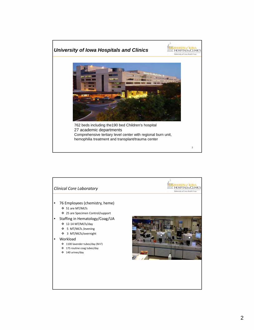
University of Iowa Hospitals and Clinics
762 beds including the190 bed Children’s hospital27 academic departments Comprehensive tertiary level center with regional burn unit, hemophilia treatment and transplant/trauma center
Clinical Core Laboratory
• 76 Employees (chemistry, heme) 51 are MT/MLTs
25 are Specimen Control/support
• Staffing in Hematology/Coag/UA 12‐14 MT/MLTs/day
5 MT/MLTs /evening
3 MT/MLTs/overnight
• Workload 1100 lavender tubes/day (M‐F)
175 routine coag tubes/day
140 urines/day
3
Instrumentation in Hematology
What we currently have – HST (2 instruments/1 slidemaker/stainer) and DM96 Cellavision
Alpha line (1 instrument/1 slidemaker/stainer) and Cellavision
WAM (Work Area Manager – Middleware)
Standardization of processing and autovalidation rules
Future Wish List: WAM for Body Fluids Module
Body Fluids on Cellavision
Combined Line for all instruments
Why use the automated IG count?
Detection of a Left Shift may be clinically important but...
We do not have a good definition for left shift
?Band Absolute # or Band %
?Presence of Metas, Myelos or Pros
?Ratio immature/total granulocyte count (neonatology)

4
Classification of Immature GranulocytesMetamyelocytes, Myelocytes, Promyelocytes
• Manual Differential: The Gold Standard?
100 cells vs 32,000
• Consistency in classifying cells
Chemical identification vs. visual
• Rumke confidence limits for low‐incidence cells– At a value of 5%, 95% confidence interval is 1.6‐11.3%
Classification of Immature Granulocytes
Metamyelocyte
Promyelocyte Myelocyte
Metamyelocyte

5
Metamyelocyte vs. Band
Meta
Band
Meta?
XE IG Parameter Goals
• Efficiency– Reduce smear reviews and manual differentials
• Improve patient care– Accuracy
– Precision
• Reduce TAT
• Employee Satisfaction
6
Define the IG parameter
• What it includes:
Metamyelocytes, Myelocytes, Promyelocytes
• How the analyzer generates the results………
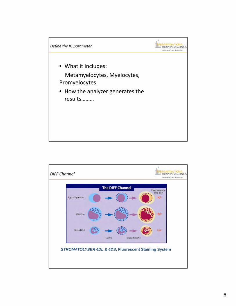
STROMATOLYSER 4DL & 4DS, Fluorescent Staining System
DIFF Channel

7
Differential Technology
• STROMATOLYSER‐4DL™ & ‐4DS™– Maintains integrity of cells
– Fluorescent stain binds to nucleic acid
– Enhanced resolution
– Better separation of cell clusters
complexity of nucleus
– More sensitivity & specific flagging
+ +
8
Project Outline – How we approached the project
• Software Installation ‐ standard for XE‐5000• Correlation Studies (vs. Manual diff)• Establish Reference Ranges or Review Cutoff• Reproducibility• Procedure Changes
– Hematology – rebuild CBC and Adif reporting– LIS‐ include the new parameters of IG and NRBC
• Training• Communication with Clinical Staff
Other Considerations
• Modify reports to included IG parameter
(absolute only reported, generated from % cutoff)
• QC material
• Proficiency Testing– CAP Survey FH9

9
Correlation Study vs. Manual Differentials
• Performed 100 normal, 100 abnormal/ 200‐cell diffs – Adhering to lab criteria for classification of cells (bands vs. metas)
– Selection Criteria• Normal vs. Abnormals
– IG’s are a “Rare Cell Event”
• Statistics generated using EP‐evaluator
• Sensitivity Specificity Report Generated
Reference Range
Criteria for what is an “abnormal” differential at UIHC?
Bands ≥5%
Metas, Myelos or Pro ≥1%
Blast ≥1%
Abnormal lymphoid cells – various criteria

10
Setting Review Criteria
• IG Present: – If IG% ≤ 2.0% and no other IP Messages
• Report Automated Differential
– If IG% ≥2.0%, Review Smear
• Confidence in IG number not in question, but at ≥2% (user defined), slide reviewed for other abnormalities and manual differential reported
– Toxic changes associated with leukemoid reactions/sepsis
– Hematologic abnormalities, such as MPD, CML
Note: All patients whose smears contained promyelocytes also had other flags, and thus slides were reviewed. Eased concern for missing blasts.
Clinical Staff Notification
Power Point on All In House Computer Screens
Identifying the IG parameter initial use
Identifying the RDW‐SD parameter addition
Noting the addition of NRBC reports on all CBCs
Opportunity to call or email with any questions

11
Outcome After IG Implementation
• Reduction in Slide Review Rate?
• Overall rate remained 23‐28% of diffs to manual review
• Confidence of accurate reporting
• Efficiency of processing samples
• Standardization of process
Looking to the Future
What about 5% for a cutoff value?
What is the clinical significance if this is the onlyflag?
How might this decrease the number of slides that have manual diffs for review?
12
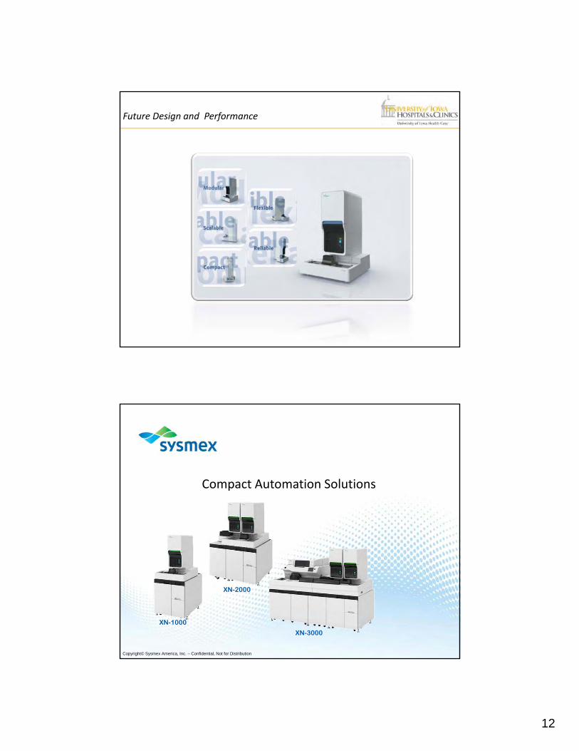
Future Design and Performance
Copyright© Sysmex America, Inc. – Confidential, Not for Distribution
Compact Automation Solutions
XN-2000
XN-1000
XN-3000

13
Copyright© Sysmex America, Inc. – Confidential, Not for Distribution
Scalable Automation Solutions
XN-9000
Modular Clinical Utility
• Basic Channels
– NRBC Standard With Every XN‐CBC
– IG Standard With Every XN‐Diff
– Body Fluids With Diff
• Advanced Channels
– Thrombopoiesis (IPF)
– Erythropoiesis (RET‐He)
– Leukopoiesis (WPC) (XN‐20 only)
14
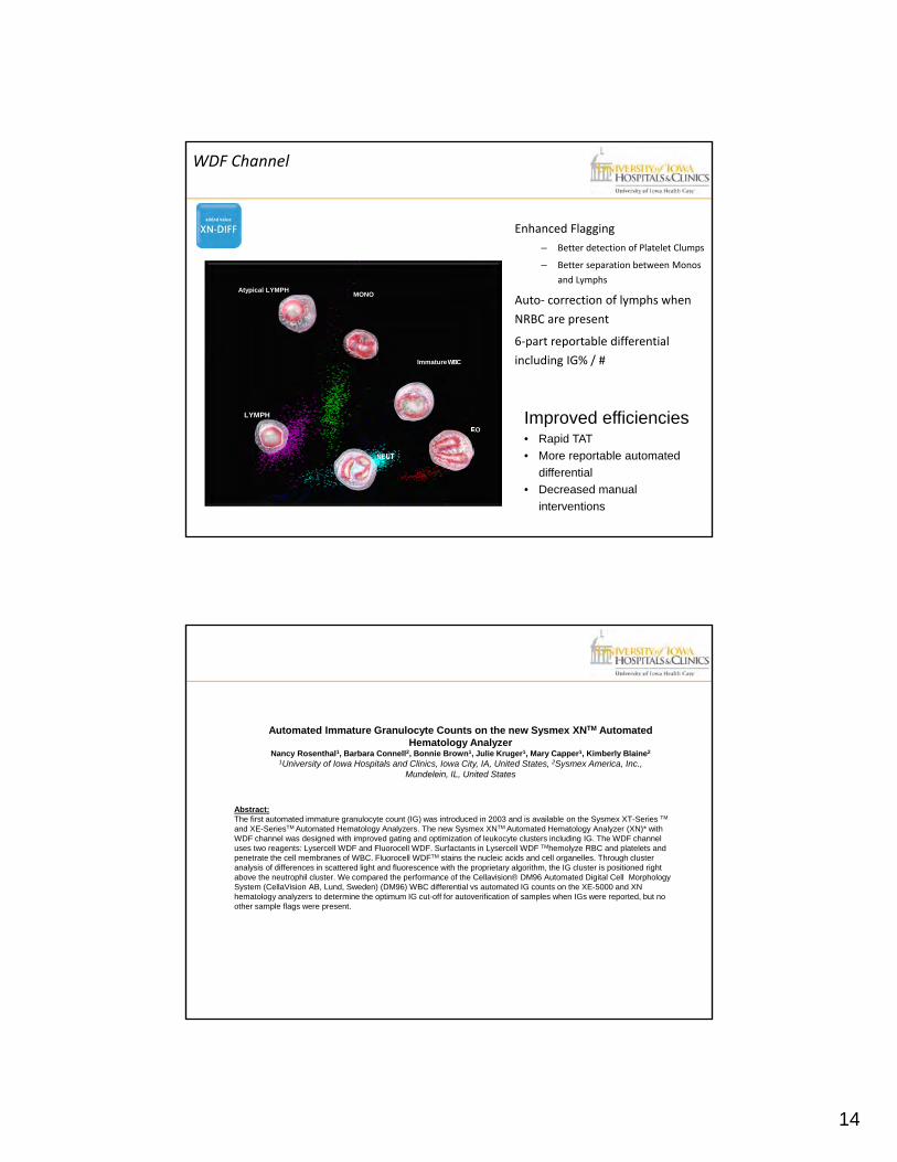
WDF Channel
Enhanced Flagging
– Better detection of Platelet Clumps
– Better separation between Monos
and Lymphs
Auto‐ correction of lymphs when
NRBC are present
6‐part reportable differential
including IG% / #
LYMPH
MONO
NEUT
EO
Atypical LYMPH
ImmatureWBC
Improved efficiencies• Rapid TAT
• More reportable automated
differential
• Decreased manual
interventions
Automated Immature Granulocyte Counts on the new Sysmex XNTM Automated Hematology Analyzer
Nancy Rosenthal1, Barbara Connell2, Bonnie Brown1, Julie Kruger1, Mary Capper1, Kimberly Blaine2
1University of Iowa Hospitals and Clinics, Iowa City, IA, United States, 2Sysmex America, Inc.,Mundelein, IL, United States
Abstract:The first automated immature granulocyte count (IG) was introduced in 2003 and is available on the Sysmex XT-Series TM
and XE-SeriesTM Automated Hematology Analyzers. The new Sysmex XNTM Automated Hematology Analyzer (XN)* with WDF channel was designed with improved gating and optimization of leukocyte clusters including IG. The WDF channel uses two reagents: Lysercell WDF and Fluorocell WDF. Surfactants in Lysercell WDF TMhemolyze RBC and platelets and penetrate the cell membranes of WBC. Fluorocell WDFTM stains the nucleic acids and cell organelles. Through cluster analysis of differences in scattered light and fluorescence with the proprietary algorithm, the IG cluster is positioned right above the neutrophil cluster. We compared the performance of the Cellavision® DM96 Automated Digital Cell Morphology System (CellaVision AB, Lund, Sweden) (DM96) WBC differential vs automated IG counts on the XE-5000 and XN hematology analyzers to determine the optimum IG cut-off for autoverification of samples when IGs were reported, but no other sample flags were present.

15
Methods: 163 samples received in the University of Iowa s Hospitals and Clinics hematologylaboratory, Iowa City, IA, were analyzed on the Sysmex XE-5000 and XN-1000 analyzer for a complete blood count and differential.Following CLSI guidelines (H20-A2), two slides were prepared on each sample and 200-cell differentials were performed by two technologists utilizing the DM96, and were compared to the XE-5000 and XN automated IG counts. WBC morphology was also noted on each sample. Statistical analysis using the EP Evaluator version 9 was performed on all samples. The data was also evaluated at varying levels of IG to determine the optimum IG cut-off to use for autoverification.
Immature granulocytes
WDF Channel Reaction WDF Scattergram
Results: In 163 samples, IG% ranged from 0.0 -30.9% and correlation coefficients between the DM96 IG enumeration, XE-5000 and the XN were r= 0.8587, r= 0.8608, and r= 0.9671 respectively. This represents exceptional correlation, considering limitations of the manual differential in enumerating rare cell events. If the level of review for the IG is set at <=2.0 IG%, then 52 and 49 samples of 114 could be auto-verified on the XE-5000 and XN respectively. If the level for review is set a <= 5.0%, then 72 and 83 samples could be auto-verified on the XE-5000 and XN respectively. This represents an increase in auto-verified results of 17% and 30% for the XE-5000 and XN respectively.
N=163DM96 Manual IG vs XE-5000 IG%Correlation Coefficient (R) = 0.8587
N=163DM96 Manual IG vs XN-10 IG%
Correlation Coefficient (R) = 0.8608N=163
XE-5000 vs XN-10 IG%Correlation Coefficient (R) = 0.9671
Manual vs Automated Immature Granulocyte Correlation

16
The WDF Channel provides accurate automated IG counts as confirmed by a respectable correlationbetween the DM96, XE-5000 and the XN. These results were excellent considering the low levels ofIGs observed and the well-known limitations of manual differentials and rare cell events. Reporting theautomated IG count using a cut-off of <=5% would increase the number of auto-verified results by 30%on the XN analyzer and would improve productivity and efficiency in the laboratory.
* The XN is for investigational use only in the US pending 510(k) FDA review
Conclusions
Questions??