massive pph
TRANSCRIPT
Dr.Sudesh HapughawatteBase HospitalKarawanella.
Massive obstetric haemorrhage is variably defined as
blood loss from the uterus or genital tract >1500ml,
a decrease in haemaglobin of > 4 g/dl
acute transfusion of > 4 units blood.
Any blood loss seriously compromising life of patient
Massive Hemorrhage in Pregnancy, Oxford Journals Medicine BJA: CEACCP Volume 5, Issue 6 Pp. 195-198.
Restore circulating volume Contact key personnel Arrest bleeding Request laboratory investigations Request suitable red cells Request platelets Request FFP Request Cryoprecipitate Suspect DIC
Management of massive blood loss: A template guideline British Journal of Anaesthesia, 2000; 85: 487–91
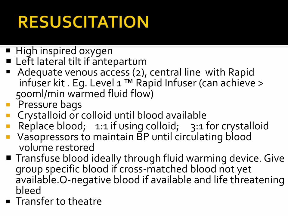
High inspired oxygen Left lateral tilt if antepartum Adequate venous access (2), central line with Rapid
infuser kit . Eg. Level 1 ™ Rapid Infuser (can achieve > 500ml/min warmed fluid flow)
Pressure bags Crystalloid or colloid until blood available Replace blood; 1:1 if using colloid; 3:1 for crystalloid Vasopressors to maintain BP until circulating blood
volume restored Transfuse blood ideally through fluid warming device. Give
group specific blood if cross-matched blood not yet available.O-negative blood if available and life threatening bleed
Transfer to theatre
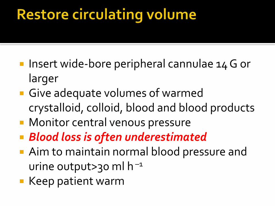
Insert wide‐bore peripheral cannulae 14 G or larger
Give adequate volumes of warmed crystalloid, colloid, blood and blood products
Monitor central venous pressure Blood loss is often underestimated Aim to maintain normal blood pressure and
urine output>30 ml h –1
Keep patient warm
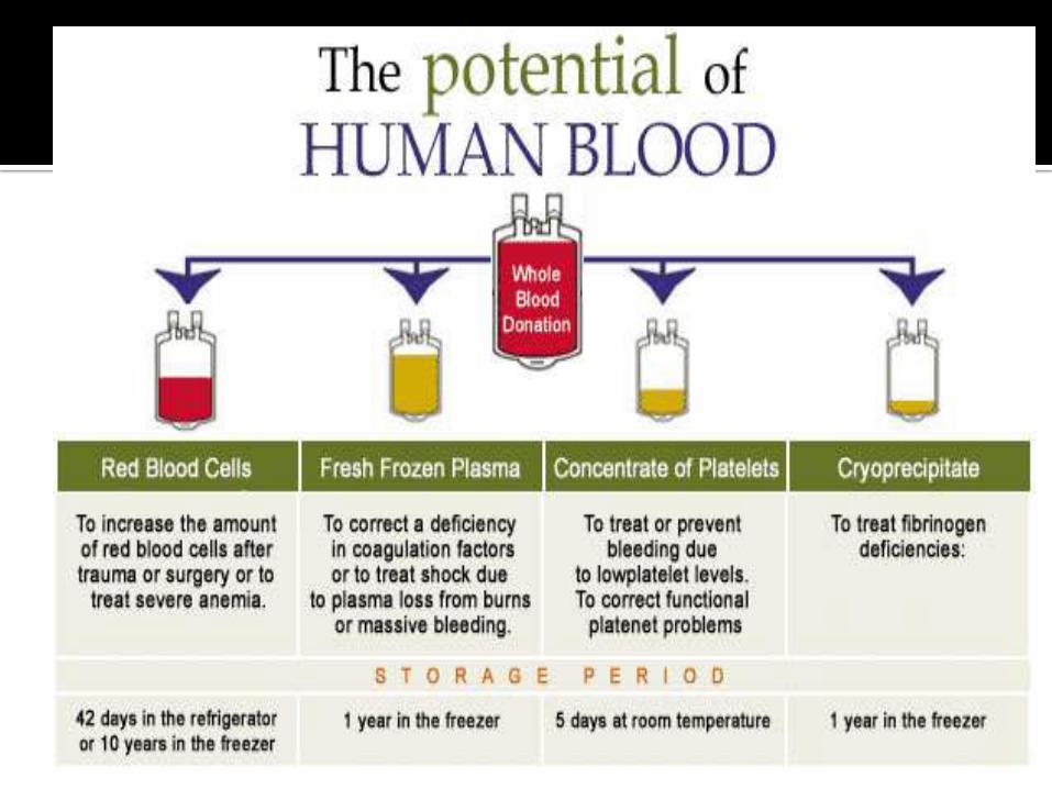
1:1:1:1 RedCell:FFP:Platelet:Fibrinogen
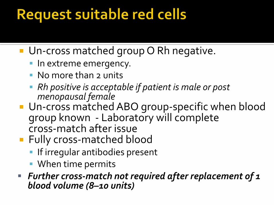
Un‐cross matched group O Rh negative. In extreme emergency. No more than 2 units Rh positive is acceptable if patient is male or post
menopausal female Un‐cross matched ABO group‐specific when blood
group known - Laboratory will complete cross‐match after issue
Fully cross‐matched blood If irregular antibodies present When time permits
Further cross‐match not required after replacement of 1 blood volume (8–10 units)
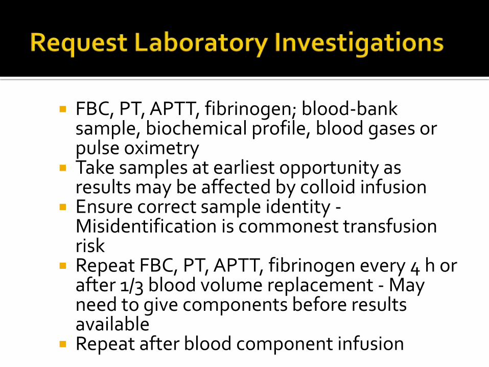
FBC, PT, APTT, fibrinogen; blood‐bank sample, biochemical profile, blood gases or pulse oximetry
Take samples at earliest opportunity as results may be affected by colloid infusion
Ensure correct sample identity -Misidentification is commonest transfusion risk
Repeat FBC, PT, APTT, fibrinogen every 4 h or after 1/3 blood volume replacement - May need to give components before results available
Repeat after blood component infusion
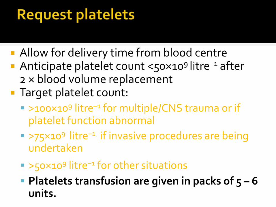
Allow for delivery time from blood centre Anticipate platelet count <50×109 litre–1 after
2 × blood volume replacement Target platelet count:
>100×109 litre–1 for multiple/CNS trauma or if platelet function abnormal
>75×109 litre–1 if invasive procedures are being undertaken
>50×109 litre–1 for other situations
Platelets transfusion are given in packs of 5 – 6 units.
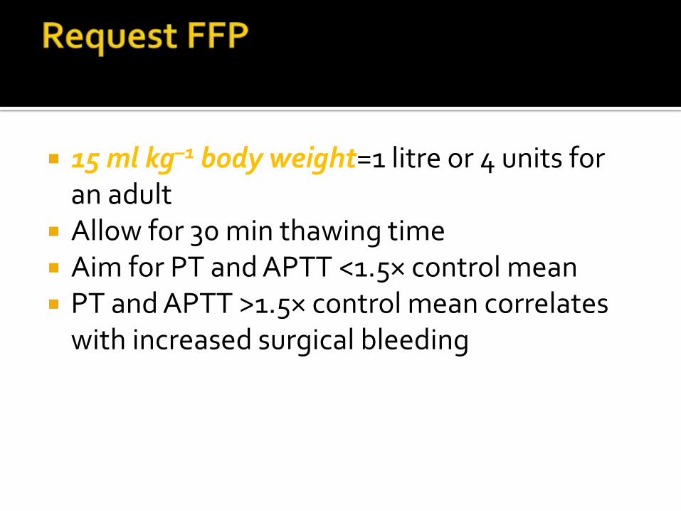
15 ml kg–1 body weight=1 litre or 4 units for an adult
Allow for 30 min thawing time Aim for PT and APTT <1.5× control mean PT and APTT >1.5× control mean correlates
with increased surgical bleeding
10-20 Single Donor Units Replaces fibrinogen and factor VIII Aim for fibrinogen >1.0 g litre–1
Allow for delivery time plus 30 min thawing time Fibrinogen <0.5 strongly associated with micro
vascular bleeding Fibrinogen deficiency develops early when
plasma‐poor red blood cells used for replacement
Uterine massage along with• Use of oxytocins• Use of ergometrine• Misoprostol• Prostoglandin• Combinations of above• Fluid and blood transfusion
Antibirinolytic agent Inhibits plasmin RCT- administer tranexamic acid in bleeding
trauma patients to avoid trauma induced coagulopathy at an early stage.
Tranexamic acid – LD 1g over 10 min then infusion 1g over 8 hours
CRASH 2 Trial - 2010
With the advice of hematologist/transfusion consultants
In uncontrollable bleeds 90ug/kg dose IV initially Early before the patient goes into DIC As factor VII needs all other clotting factors in
adequate doses to act.
General anaesthetic with rapid sequence induction is generally advocated if actively bleeding or coagulopathic. Reduce dose of induction agent if severe on-going bleeding
Regional anaesthesia is relative contraindication but may be maintained if patient has an epidural in situ and bleeding is controlled
Consider arterial line, central venous catheter and urinary catheter but only after definitive treatment has commenced. Their insertion must not delay resuscitation and fluid management
Use fluid warmer and aim to keep patient normothermic Regular monitoring of haemoglobin level and coagulation using near
patient devices if available (e.g. HemoCue). Fresh frozen plasma, platelet transfusion and cryoprecipitate may be necessary if coagulopathy develops. Liaise early with the haematology department for optimal and timely product replacement.
Consider systemic haemostatic agents Vitamin K Tranexamic acid Recombinant Factor VIIa (NovoSeven®)
Transfer to a high dependency unit or ICU Oxygenate the patient Anticipate coagulopathy and treat clinically until
coagulation results available Monitoring of all vital parameters – HR, BP, CVP,
UOP, SpO2 Correction of fluids and electrolyte imbalances Look for lung injury/pulmonary edema and treat
need of intubation and mechanical ventilation Correct hypothermia Correct acidosis Maintain a good UOP
Coagulopathy DIC Hypothermia Acidosis Transfusion related acute lung injury (TRALI) Transfusion associated circulatory overload
(TACO) Transfusion associated dyspnoea (TAD) Tissue hypoxia Multi organ failure
Hypothermia Coagulopathy Acid-Base disturbances Electrolyte abnormality Citrate toxicity System failure Inappropriate, unnecessary and
under/delayed transfusion
Continuing Education in Anaesthesia Critical Care and Pain June 2014: 14 (3)

THANK YOU!