master planning of the implant case - prosites, …c1-preview.prosites.com/36100/wy/docs/chap 5 -...
TRANSCRIPT

60
C H A P T E R 5
MASTER PLANNING OF THE IMPLANT CASE
Over the past three decades implant dentistry has become the leading and most dynamic discipline in the dental fi eld. Oral reconstruction with dental implants has gone from just single-tooth replacements and over-dentures to encompass sophisti-cated surgical and prosthetic techniques and principles. Every specialty within dentistry plays an important part in the suc-cessful outcomes of these very rewarding cases. This chapter describes the interdisciplinary approach to comprehensive treatment planning and the many facets involved in quality, long-standing aesthetic and functional treatment. 1 , 2
Initial Consultation The initial consultation, or at least an appointment to expose the patient to implant or other oral reconstruction, can be initiated by a variety of dental practitioners. An orthodontist may evaluate a patient with congenitally missing teeth. An endodontist may determine that a tooth is fractured and is not suitable for endodontics. A periodontist may feel that progres-sive, uncontrolled or refractory periodontal disease may not benefi t from further traditional treatment. An oral surgeon might prepare teeth being extracted for ridge preservation or determine that ridge augmentation will provide optimal support for dental implants. Most often, though, the general dentist, or prosthodontist, sees a patient with reconstructive needs and makes the appropriate initial consultation for treatment.
In the initial consultation the patient ’ s medical and dental status can be identifi ed and evaluated. If implant therapy is an
appropriate option, then a preliminary treatment plan can be developed.
The patient ’ s health status should be evaluated in a way similar to the screening admissions procedure conducted with patients entering the hospital. 3-5 The main components to be considered are: 1. The chief complaint 2. The history of the present illness 3. The medical history 4. The dental status
Chief Complaint The chief complaint may range from “ I don ’ t like how I look ” to “ I have worn dentures for 37 years, and I can no longer function with them. ” The focus in evaluation of the patient ’ s chief complaint is whatever factors prompted the person to seek rehabilitation at this time. Sometimes the discussion will reveal concerns beyond those the patient fi rst mentions. For example, patients may say that their dentures no longer func-tion well, but subsequently, they may describe pain during mastication. This additional information can be an important diagnostic aid. If patients cite cosmetic concerns, these must be placed in context. Implant dentistry often cannot match the needs, wants, or desires of the person whose primary goal is to look fundamentally different. However, if functional concerns are the primary goals and cosmetic concerns are secondary, implant dentistry usually can give such patients what they want.
Charles A. Babbush
Joel L. Rosenlicht

Chapter 5 Master Planning of the Implant Case 61
History of Present Illness The next component of interest is the history of the present illness. The practitioner must identify what in the patient ’ s history produced the present situation, especially in cases in which atrophy in the maxilla or mandible is severely advanced. Did the patient have poor quality care? Did the patient decline to seek any care at all? Did the patient lose teeth prematurely and not have the appropriate dietary intake to sustain good levels of bone support? Has the patient been edentulous for several decades, and did this extended time lead to severe atrophy? Was the patient involved in a traumatic injury: Did a baseball bat, a thrown ball, a fi st or some other object trau-matize one of more teeth and cause their demise? Was any pathological lesion or tumor involved in the cause of tooth loss and subsequent bone loss?
Medical History In gathering the patient ’ s medical history, special attention should be given to whether the patient has the ability to physi-cally and emotionally sustain all the procedures that may be required in implant therapy, including surgery, a variety of anesthetics and pain-control drugs, and prosthetic rehabilita-tion. 6-8 The American Dental Association provides a long-form health questionnaire on their website that is an excellent tool for gathering this information, available at https://siebel.ada.org/ecustomer_enu/start.swe?SWECmd=Start . 9 Figure 5-1 shows an example of a typical health history questionnaire.
In addition to obtaining the patient ’ s health history, the doctor must assess vital signs (blood pressure, pulse, and res-piration) and record these assessments in the patient ’ s chart. When a patient has not had a comprehensive medical work-up for several years or when fi ndings are positive on the health questionnaire, additional laboratory testing may be advisable. These tests may include complete blood count, urinalysis, or sequential multiple analysis of the blood chemistry (SMAC).
TABLE 5-1 Complete metabolic panel
Test procedure Units Reference range
Sodium mmol/L 135-146 Potassium mmol/L 3.5-5.3 Chloride mmol/L 98-110 Carbon dioxide mmol/L 21-33 Calcium mg/dL 8.6-10.2 Alkaline phosphate Units/L 33-130 AST Units/L 10-35 ALT Units/L 6-40 Bilirubin, total mg/dL 0.2-1.2 Glucose mg/dL 65-99 Urea nitrogen mg/dL 7-25 Creatinine mg/dL 0.60-1.18 BUN/creatinine ratio 6-22 Protein, total g/dL 6.2-8.3 Albumin g/dL 3.6-5.1 Globulin, calculated g/dL 2.2-3.9 A/G ratio 1.0-2.1 Egfr non-African American mL/min/ 1.73 m 2 > or = 60 Egfr African American mL/min/ 1/73 m 2 > or = 60
Patients who manifest systemic disease that interferes with their normal daily living pattern (e.g., inhibits their employ-ment, restricts their social activity, or otherwise does not al-low them to function physically and mentally in a normal or almost normal manner) should not be considered as candi-dates for an elective procedure such as oral implant recon-struction (R,R). Classifying patients according to the follow-ing numerical ratings as established by the American Society of Anesthesiology is helpful in the selection process (R):
Class I: A patient who has no organic disease or in whom the disease is localized and causes no systemic disturbances.
Class II: A patient exhibiting slight to moderate systemic disturbance which may or may not be associated with the surgical complaint and which interferes only moderately with the patient ’ s normal activities and general physiologic equilibrium.
Class III: A patient exhibiting severe systemic disturbance which may or may not be associated with the surgical complaint and which seriously interferes with the patient ’ s normal activity.
Class IV: A patient exhibiting extreme systemic disturbance which may or may not be associated with the surgical complaint, which interferes seriously with the patient ’ s normal activities, and which has already become a threat to life.
Class V: The rare person who is moribund before operating, whose preoperative condition is such that the patient is expected to die within 24 hours even if not subjected to the additional strain of surgery.
Class VI: A patient who is considered brain dead and is a potential organ donor.
BOX 5-1 The American Society of Anesthesiologists ’ classifi cation of presurgical risk
The results can contribute to the patient ’ s medical profi le ( Table 5-1 ). 2 , 3
Combining the information from the health questionnaire, the vital signs, and the laboratory test results will enable the doctor to categorize each patient into one of the fi ve classifi ca-tions of presurgical risk formulated by the American Society of Anesthesiology ( Box 5-1 ). 8 According to this scheme, a Class I category includes the patient who is physiologically normal, has no medical diseases, and lives a normal daily life-style. The Class II category includes the patient who has some type of medical disease, but the disorder is controlled with

62 Chapter 5 Master Planning of the Implant Case
Figure 5-1. Health history questionnaire.
HEALTH QUESTIONNAIRE
Patient’s Name:_______________________________________
I. In the following questions, circle yes or no, whichever applies. Your answers are for our records onlyand will be considered confidential.
II. DO YOU HAVE OR HAVE YOU HAD ANY OF THE FOLLOWING DISEASES OR PROBLEMS:
III. ARE YOU TAKING ANY OF THE FOLLOWING:
IV. ARE YOU ALLERGIC OR HAVE YOU REACTED ADVERSELY TO:
V. WOMEN:
Date:___________________________
7.8.9.
10.11.12.13.14.15.
16.17.
YesYesYes
YesYesYesYesYesYesYes YesYesYesYes
NoNoNo
NoNoNoNoNoNoNo NoNoNoNo
YesYesYesYesYesYes
NoNoNoNoNoNo
Has there been any change in your general health within in past year?My last physical examination was on ______________________Are you under the care of a physician? _____________________If so, what is the condition being treated? _________________________Name and address of physicianHave you had any serious illness or operations?If so, what was it? ____________________________________________Have you been hospitalized or had a serious illness within the past five (5) years?If so, what was the problem? ____________________________________
1.2.3.
4.5.
6.
YesYesYes
YesYes
Yes
NoNoNo
NoNo
No
Rheumatic fever or rheumatic heart diseaseCongenital heart lesions, mitral valve prolapseCardiovascular disease (heart trouble,heart attack, coronary insufficiency,coronary occlusion, high blood pressure,arteriosclerosis, strokeAllergiesSinus troubleAsthma or hay feverHives or skin rashFainting spells or seizuresDiabetesDo you urinate (pass water) more than six timesa day?Are you thirsty much of the time?Does your mouth frequently become dry?Hepatitis, jaundice, or liver diseaseArthritis
18.19.20.21.22.23.24.25.26.
27.28.
29.
YesYesYesYesYesYesYesYesYes
YesYes
YesYes
Yes
NoNoNoNoNoNoNoNoNo
NoNo
NoNo
No
Inflammatory rheumatism (painful swollen joints)Stomach ulcersKidney troubleTuberculosisDo you have a persistent cough or cough up blood?Low blood pressureVenereal disease/herpes/AIDSOtherHave you had abnormal bleeding associated withprevious extractions, surgery, trauma?Do you bruise easily?Have you ever had a blood transfusion?If so, explain_______________________________Do you have any blood disorders, such as anemia?Have you had surgery or x-ray treatment for tumor,growth, or other conditions of your mouth or lips?Are you taking any drying medicines?If so, what _________________________________
30.31.32.33.34.35.
Antibiotics or sulfa drugsAnticoagulants (blood thinners)Medicine for high blood pressureCortisone (steroids)TranquilizersAntihistamines
YesYesYesYesYes
NoNoNoNoNo
36.37.38.39.40.
AspirinInsulin, tolbutamide (Orinase) or similar drugDigitalis, or drugs for heart troubleNitroglycerinOther
YesYesYesYesYesYesYesYesYes
NoNoNoNoNoNoNoNoNo
41.42.43.44.45.46.47.48.49.
Local anestheticsPenicillin or other antibioticsSulfa drugsBarbiturates, sedatives or sleeping pillsAspirinIodineCodeine or other narcoticsOtherHave you had any serious trouble associatedwith any previous dental treatment?If so, explain ________________________________
Yes No55. Are you pregnant?
Signature of Patient ___________________________________________
Doctor’s Signature ____________________________________________
Yes No56. Do you have any problems with your menstrual period?
Yes
Yes
YesYes Yes
No
No
NoNo No
50.
51.
52.53.
54.
Do you have any disease, condition, or problem notlisted that you think I should know about?Are you employed in any situation that exposesyou regularly to x-rays or other ionizing radiation?Are you wearing contact lenses?Do you smoke cigarettes, cigars, pipe, or chew tobacco?How many each day?________Do you use recreational drugs?
medications. The patient can thus engage in normal daily activity. An example of this category of patient is one with hypertension who has been placed on antihypertensive medica-tion and, as a result, has normal blood pressure and no other impairments. The Class III category includes the patient who has multiple medical problems, such as advanced-stage hyper-tensive cardiovascular disease or insulin-dependent diabetes, with impaired normal activity. Patients in the Class IV and V categories have advanced states of disease. Class VI is a patient who is considered brain dead and is a potential organ donor. For example, a patient in the Class IV category has a serious medical condition requiring immediate attention, such as the
person with acute gallbladder disease who needs immediate treatment. The patient in the Class V category is usually mori-bund and will not survive the next 24 hours. Most patients who seek implant reconstruction fall into the Class I or II categories and sometimes Class III. For obvious reasons, patients in Classes IV and V are not appropriate candidates for implant procedures. However, consideration of whether a patient falls into Class I, II, or III will enable the implant practitioner to more effectively decide what kinds of proce-dures should be undertaken, where the surgery should be per-formed, and what kind of anesthesia is appropriate. Furthermore, cases with patients categorized as Class III may

Chapter 5 Master Planning of the Implant Case 63
Figure 5-2. I-CAT cone beam CT scanner installed in a dental offi ce environment.
require preparatory measures such as stabilizing or controlling a diabetic patient before implant surgery can be considered.
Dental Status It is essential to obtain a comprehensive understanding of the patient ’ s dental, as well as medical, status. In addition to ques-tioning patients about their dental history, a thorough exami-nation should be conducted. An evaluation of the hard and soft tissues of the entire maxillofacial skeleton should be included and appropriate radiographic studies must be obtained.
Today ’ s modern dental offi ces can provide a host of radio-graphic information through digital and computer analog equipment that allows unprecedented detail and data applica-tions never available before. Digital panoramic and offi ce-based cone beam CT scanners ( Figure 5-2 ) are now readily available. These devices can give studies that accurately defi ne the full scope of the maxilla and mandible, as well as the accompanying vital structures (i.e., sinus, fl oor of the nose, position of the mandibular canal, mental foramen) ( Figure 5-3 ). In addition, information about the thickness of cortical plates, bone densities, and soft tissue contours is easily obtained. Chapter 8 , Contemporary Radiographic Evaluation of the Implant Candidate, and Chapter 18 , An Introduction to Guided Surgery, expand on this technology. There still is a place for conventional fi lm-based radiographs because much valuable information can be learned from them. These might include occlusal fi lms, lateral cephalometric images, and periapical or panoramic images ( Figure 5-4 ). 10 However, with the advent of digital referenced planning software our ability to diagnose and plan procedures virtually takes radiographic diagnosis and treatment planning to a new level ( Figures 5-5 and 5-6 ).
In addition to gathering the dental history, a thorough clinical exam should include the patient ’ s teeth, soft tissue, and hard tissue. Mounted casts also should be obtained, and become an important component of the patient ’ s treatment plan ( Figure 5-7 ).
The patient ’ s facial appearance also should be documented with preoperative extraoral and intraoral photographs ( Figure
Figure 5-3. A, Panoramic radiograph demonstrating severe advanced maxillary and mandibular atrophy. B, Panoramic radiograph demonstrating the maxillary sinus cavities, nasal anatomy, defi ned inferior alveolar canals, and mental foramen.
A B
5-8 ). In addition to acting as risk management tools, these preoperative documents usually serve as references for all members of the implant team during detailed case planning. Nontangible considerations also deserve attention. The patient ’ s needs, wants, desires, and psychosocial conditions should be ascertained and recorded. Issues of self-confi dence and self-esteem should also be reviewed ( Figure 5-9 ).

64 Chapter 5 Master Planning of the Implant Case
Figure 5-4. A, Conventional occlusal radiograph. B, Conventional lateral cephalometric radiograph. C, Conventional periapical radiograph. D, Conventional panoramic radiograph.
A
B
C
D
Figure 5-5. CT scan 3-D SimPlant surgical planning software program demonstrates a well-planned implant reconstruction in both the maxilla (A) and mandible (B).
A
B
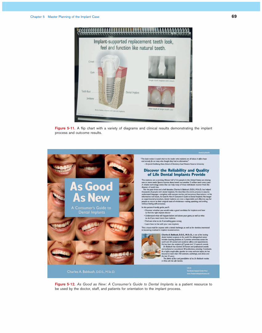
Patient Education In addition to providing the practitioner with crucial informa-tion concerning the patient ’ s needs and wants, the initial con-sultation also should serve to educate and orient the patient. Various visual aids can assist with this task, including models representing completed forms of single-tooth, multiple-tooth, and full-arch reconstruction ( Figure 5-10 ). Photographs also can communicate to the patient the potential appearance of the fi nal reconstruction in the oral cavity ( Figure 5-11 ). Vid-eotapes and DVDs, available from most commercial compa-nies that sell implants, can demonstrate various implant procedures and provide a general overview. All of these pre-sentation aids should be noted in the patient ’ s chart as risk-management tools.
Printed literature can serve multiple purposes. Brochures that introduce implants and explain how they work can be sent to patients who inquire about implant reconstruction. Patients going through an implant consultation should be given a port-folio of literature to take home. This information will enable them to better communicate with friends and relatives about the process of implant reconstruction. Printed literature also can serve as an educational tool if public education lectures are part of the doctor ’ s practice domain ( Figure 5-12 ).

Figure 5-6. A cone beam CT scan demonstrating a panoramic view (top) and cross-sectional views (bottom) of an intended implant placement.
Figure 5-7. A, A study cast mounted in a semiadjustable articulator for the replacement of two bicuspid maxillary teeth. B, A study cast mounted in a semiadjustable articulator for the reconstruction of an endentulous maxilla.
A B

66 Chapter 5 Master Planning of the Implant Case
AAA B
CDC
Figure 5-8. This series of facial photographs demonstrates the need to obtain pretreatment facial documentation ( A and B; E and F ) so that a valid comparison can be made with the fi nal postsurgi-cal/prosthetic results ( C and D; G and H ). (From Babbush CA: As good as new: a consumer ’ s guide to dental implants, Lyndhurst, OH, The Dental Implant Center Press, 2004.)

Chapter 5 Master Planning of the Implant Case 67
E
H
F
G
Figure 5-8, cont’d.

68 Chapter 5 Master Planning of the Implant Case
Figure 5-9. The loss of self-esteem and self-confi dence is evident in the patient ’ s facial features. (From Babbush CA: As good as new: a consumer ’ s guide to dental implants, Lyndhurst, OH, 2004, The Dental Implant Center Press.)
Figure 5-10. A, A giant model of an implant with an abutment and removable crown prosthesis that can be used to demonstrate the details of the parts and pieces to a patient. B, A Lucite model that can be used to demonstrate the actual size in a single-tooth implant reconstruction to a patient. C, A Lucite model which can be used to demonstrate a full-arch All-on-4 reconstruction to a patient. ( B, From Babbush CA: As good as new: a consumer ’ s guide to dental implants, Lyndhurst, OH, 2004, The Dental Implant Center Press.)
A
B
C
Joint Treatment Planning The next phase in the treatment planning process involves the entire implant team. This phase typically begins with a confer-ence between the surgeon and restorative dentist. Other spe-cialties (e.g., periodontology, endodontics, orthodontics) may participate in this initial discussion and the hygienist or labora-tory technician may also be included. The planning confer-ences, which often bring key individuals together physically but may also be conducted via telephone or email, provide opportunities for the team to review the patient ’ s chief com-plaints, expectations, history, and current medical and dental status. Based on all this information, team members can for-mulate a detailed treatment plan ( Figure 5-13 ).
Some patients must undergo one or more preliminary pro-cedures before the treatment plan can be completed. If the patient ’ s oral hygiene is poor or marginal the patient may need to make improvements and be reevaluated over a 6- to 12-month period ( Figure 5-14 ). In more complex cases, orth-odontic ( Figure 5-15 ) or orthognathic procedures ( Figure 5-16 ) may be necessary to correct abnormal jaw relationships before the patient is treated with implants. Periodontic, end-odontic, prosthetic restorative, and oral surgical procedures may need to be performed with extractions ( Figure 5-17 ).
In the course of this preparatory phase, some patients may be found to be inappropriate candidates for implant recon-
Chapter 5 Master Planning of the Implant Case 69
Figure 5-11. A fl ip chart with a variety of diagrams and clinical results demonstrating the implant process and outcome results.
Figure 5-12. As Good as New: A Consumer ’ s Guide to Dental Implants is a patient resource to be used by the doctor, staff, and patients for orientation to the implant process.

70 Chapter 5 Master Planning of the Implant Case
Figure 5-13. The implant team (surgeon, prosthodontist, and laboratory technician) formulate a detailed treatment plan.
Figure 5-14. A, A patient with poor oral hygiene and an existing pathology returned for additional implant placement. The disease process and oral hygiene maintenance must be revised and imple-mented prior to further treatment. B, A patient who required extraction of all remaining teeth and reconstruction of implants. All the acute pathological process must be eradicated prior to initiation of defi nitive treatment. ( B, From Babbush CA: As good as new: a consumer ’ s guide to dental implants, Lyndhurst, OH, 2004, The Dental Implant Center Press.)
A B
Figure 5-15. A, The spaces left by congenitally missing lateral maxillary incisor teeth were not adequate for implant placement. B, Orthodontic treatment was required to widen these spaces.
A B
struction. Alternative methods such as a fi xed prosthesis, a removable partial denture, or full dentures may be indicated.
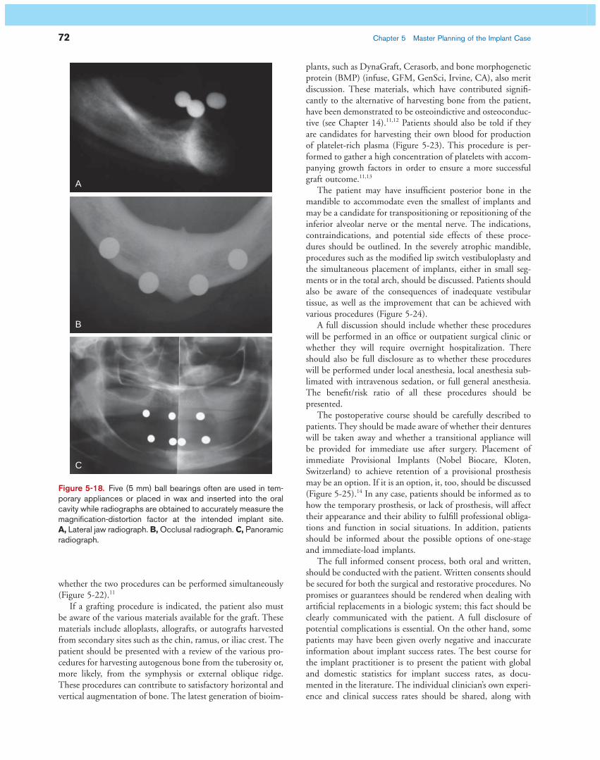
A defi nitive treatment plan eventually will be formulated, usually with collaboration by the surgeon and restorative dentist. After the treatment plan is fi nalized, fabricated tem-plates or surgical guides from diagnostic set ups are made to use during surgery. In routine cases in which there is adequate bone and there are no anatomical anomalies, this may not be needed. When traditional radiographs (nondigital fi lms) are used, a common reference to determine fi lm distribution is the use of a fi xed-size reference object such as 5-mm ball bearings ( Figure 5-18 ). These markers can be placed within a diagnostic set up or surgical stent to assist in accuracy of transferring information to a diagnostic cast ( Figure 5-19 ). Some of the advantages of cone beam CT scans is that the reconstruction can be made virtually on the screen or the digital information

Chapter 5 Master Planning of the Implant Case 71
Figure 5-16. Orthognathic mandibular surgery and chin advancement were required prior to the implant reconstruction in this case.
Figure 5-17. A, Panoramic radiograph demonstrating bone loss involving the maxillary dentition and the mandibular molars and incisors. B, Panoramic radiograph showing the 4-month postextraction healed sites. Panoramic radiograph (C) and facial photo (D) of the patient demonstrating the 20-year posttreatment results. ( D , From Babbush CA: As good as new: a consumer ’ s guide to dental implants, Lyndhurst, OH, The Dental Implant Center Press, 2004.)
A B
C
D
can be sent out for rapid prototyping of stereolithic models ( Figures 5-20 and 5-21 ).
Final Treatment Considerations Various treatment options can be presented to the patient for approval. Every aspect of this discussion should be docu-mented in the patient ’ s chart as a risk management tool.
The patient should be informed of the anticipated number of implants and whether an ancillary procedure such as sinus grafting is necessary. If maxillary anthroplasty with augmenta-tion bone grafting (a sinus lift) is indicated, the patient should be aware of the amount of bone remaining between the resid-ual crest of the ridge and the sinus fl oor. The amount of residual bone will determine whether the sinus graft can be carried out as a staged procedure before implant placement or
72 Chapter 5 Master Planning of the Implant Case
Figure 5-18. Five (5 mm) ball bearings often are used in tem-porary appliances or placed in wax and inserted into the oral cavity while radiographs are obtained to accurately measure the magnifi cation-distortion factor at the intended implant site. A, Lateral jaw radiograph. B, Occlusal radiograph. C, Panoramic radiograph.
A
B
C
whether the two procedures can be performed simultaneously ( Figure 5-22 ). 11
If a grafting procedure is indicated, the patient also must be aware of the various materials available for the graft. These materials include alloplasts, allografts, or autografts harvested from secondary sites such as the chin, ramus, or iliac crest. The patient should be presented with a review of the various pro-cedures for harvesting autogenous bone from the tuberosity or, more likely, from the symphysis or external oblique ridge. These procedures can contribute to satisfactory horizontal and vertical augmentation of bone. The latest generation of bioim-
plants, such as DynaGraft, Cerasorb, and bone morphogenetic protein (BMP) (infuse, GFM, GenSci, Irvine, CA), also merit discussion. These materials, which have contributed signifi -cantly to the alternative of harvesting bone from the patient, have been demonstrated to be osteoindictive and osteoconduc-tive (see Chapter 14 ). 11 , 12 Patients should also be told if they are candidates for harvesting their own blood for production of platelet-rich plasma ( Figure 5-23 ). This procedure is per-formed to gather a high concentration of platelets with accom-panying growth factors in order to ensure a more successful graft outcome. 11 , 13
The patient may have insuffi cient posterior bone in the mandible to accommodate even the smallest of implants and may be a candidate for transpositioning or repositioning of the inferior alveolar nerve or the mental nerve. The indications, contraindications, and potential side effects of these proce-dures should be outlined. In the severely atrophic mandible, procedures such as the modifi ed lip switch vestibuloplasty and the simultaneous placement of implants, either in small seg-ments or in the total arch, should be discussed. Patients should also be aware of the consequences of inadequate vestibular tissue, as well as the improvement that can be achieved with various procedures ( Figure 5-24 ).
A full discussion should include whether these procedures will be performed in an offi ce or outpatient surgical clinic or whether they will require overnight hospitalization. There should also be full disclosure as to whether these procedures will be performed under local anesthesia, local anesthesia sub-limated with intravenous sedation, or full general anesthesia. The benefi t/risk ratio of all these procedures should be presented.
The postoperative course should be carefully described to patients. They should be made aware of whether their dentures will be taken away and whether a transitional appliance will be provided for immediate use after surgery. Placement of immediate Provisional Implants (Nobel Biocare, Kloten, Switzerland) to achieve retention of a provisional prosthesis may be an option. If it is an option, it, too, should be discussed ( Figure 5-25 ). 14 In any case, patients should be informed as to how the temporary prosthesis, or lack of prosthesis, will affect their appearance and their ability to fulfi ll professional obliga-tions and function in social situations. In addition, patients should be informed about the possible options of one-stage and immediate-load implants.
The full informed consent process, both oral and written, should be conducted with the patient. Written consents should be secured for both the surgical and restorative procedures. No promises or guarantees should be rendered when dealing with artifi cial replacements in a biologic system; this fact should be clearly communicated with the patient. A full disclosure of potential complications is essential. On the other hand, some patients may have been given overly negative and inaccurate information about implant success rates. The best course for the implant practitioner is to present the patient with global and domestic statistics for implant success rates, as docu-mented in the literature. The individual clinician ’ s own experi-ence and clinical success rates should be shared, along with

Chapter 5 Master Planning of the Implant Case 73
Figure 5-20. A and B, A series of radiographic guides are used to obtain the proper relationships in radiographic information for treatment plan development. C and D, Radiographic guides in place in the mandible (C) and maxilla (D). E and F, SimPlant software images that are used to develop the fi nal treatment recommendations.
A
C
E
B
D
F
Figure 5-19. A, A surgical guide is used to establish proper alignment for a mandibular All-on-4 reconstruction. B, Mounted study cast with a surgical template with 5-mm ball bearings mounted in place.
A B

74 Chapter 5 Master Planning of the Implant Case
A
C
B
D
Figure 5-22. A, Panoramic radiograph demonstrating severe advanced maxillary atrophy requiring a staged procedure to graft the maxillary sinuses. B, Panoramic radiograph demonstrating adequate residual bone inferior to the sinus cavity making it possible to carry out simultaneous grafting and implant placement.
A B
Figure 5-21. A, Data transferred to planning software, example of 3-D movable image with bone, implant, and prosthesis icons active. B, Example of the plan from 3-D software without prosthesis or bone icon active. C, 2-D cross-sectional view. The implant is within the triangle of best available bone with excellent placement from anatomical surgical and prosthesis requirements. D, Surgical guide ready for try-in for fl apless surgical procedure.

Chapter 5 Master Planning of the Implant Case 75
Figure 5-23. The production of platelet-rich plasma is an in-offi ce, at the time of the surgery proce-dure. A, The patient ’ s intravenous blood is drawn (22.5 mL) at the initiation of the surgical procedure. B, The anticoagulated blood sample is inserted into a disposable processing unit in the centrifuge for a two-level spin over 14 minutes. C, The platelet concentrate is reconstituted and removed from the processing unit. D, It is loaded into an application syringe with calcium chloride and topical thrombin in order to produce the fi nal platelet concentrate gel.
A
C
B
D
Figure 5-24. Preoperative (A) and postoperative (B) clinical photos demonstrate a completed ves-tibuloplasty case after 3 years.
A B

76 Chapter 5 Master Planning of the Implant Case
Figure 5-25. Immediate provisional implants (IPIs) mini-implants that can be utilized to stabilize a provisional prosthesis. A, The maxilla at the completion of maxillary reconstruction. B, Transfer copings are incorporated into the provisional prosthesis. C, The provisional prosthesis temporarily cemented in the maxilla. D, Panoramic radiograph demonstrating the IPI in place in maxilla.
A
C
B
D
some discussion of what options will likely be available in the event of an implant failure. Once again, this discussion should be noted in the patient ’ s chart. 15
Full discussion of fees and methods of payment should ensue, along with a discussion of potential reimbursement by third parties and managed-care groups and the impact of such reimbursement on the patient ’ s fi nancial obligation. The patient should understand that implant dentistry involves reconstructive procedures. The plates, cylinders, and screws used are orthopedic devices, comparable with the implants used in the tibia, fi bula, and other skeletal areas of the body. An understanding of this fact may increase the likelihood of insurance compensation.
Patients should walk away from the fi nal consultation with a clear understanding of their postsurgical obligations such as
ongoing home care. They should be given an overview of the armamentarium they will be using in this endeavor, including different types of manual and mechanical brushes, dental fl oss, super fl oss, and chemotherapeutic agents such as oral chlorhexidine antibacterial rinses. Finally, they should know what to expect as a schedule for periodic evaluations (see Chapter 30 ). 16 , 17
An open attitude, combined with a comprehensive and systematic approach to diagnosis and treatment planning, will predictably lay the foundation for successful results. Figures 5-26 through 5-34 present a variety of cases demonstrating successful outcomes. The following chapters of this text con-centrate on the variety of procedures currently available to assist the implant practitioner in achieving routine success for the implant patient.
Text continued on p. 85.

Figure 5-26. A, Presurgical panoramic radiograph showing the failed dentition and nonrestored previously placed implants in the mandible approximately 4 years prior to the patient ’ s initial consultation. B and C, Preoperative clinical photos of the maxilla and mandible showing a lack of any crowns in the mandible and severe advanced caries and periodontal disease in the maxilla. D and E, The mandibular over-denture prosthesis that the patient altered himself over many years of wear. F and G, The 5-year postsurgical follow-up photo and panoramic radiograph of the patient ’ s maxillary and mandibular reconstruction bar over-denture prosthesis with internal locking mechanisms. ( F, From Babbush CA: As good as new: a consumer ’ s guide to dental implants, Lyndhurst, OH, 2004, The Dental Implant Center Press.)
A
C
E
B
D
F
G

78 Chapter 5 Master Planning of the Implant Case
Figure 5-27. A and B, Preoperative photo and panoramic radiograph showing the retained decidu-ous cuspid in the right maxilla. C and D, The 4-year follow-up photo and panoramic radiograph showing the results of tooth extraction, immediate implant placement, immediate provisional restora-tion, and fi nally, permanent restoration.
A
C
B
D
Figure 5-28. A, A 36-year-old patient with congenitally missing second bicuspids. The initial treat-ment plan had been to make four three-unit bridges for this patient, but when she heard about dental implants she decided to come for a consultation. B, The 4-year postsurgical follow-up panoramic radiograph showing four individual self-standing implant reconstructions.
A B

Chapter 5 Master Planning of the Implant Case 79
Figure 5-29. A, The presurgical radiograph of the patient ’ s maxilla and mandible showing the terminal dentition in the maxilla. The patient had an over 20-year history of implant reconstruction starting in the left mandible with IMZ press-fi t cylinders followed by Replace Root Form implants in the right mandible. B , Panoramic radiograph showing the immediate postextraction implants in the maxilla. C , The 5-year follow-up panoramic radiograph of the maxillary reconstruction, a connector bar over-denture with internal fi xation mechanism with no palatal coverage.
B
A
C

80 Chapter 5 Master Planning of the Implant Case
Figure 5-30. A, A female patient with a multiyear history of a failed maxillary subperiosteal implant (with cast metal Ceramco prosthesis cemented to the subperiosteal implant) that had invaded and settled into the maxillary sinus and nasal cavity. B, The mandibular reconstruction showed implants that were malposed, undersized, and under-engineered to hold the prosthesis. The failure of the prosthesis was total. C and D, The 5-year postsurgical follow-up clinical photo and panoramic radiograph show the patient restored with connector bars, over-denture, and internal locking mechanism.
A
C
B
D

Chapter 5 Master Planning of the Implant Case 81
Figure 5-31. A and B, Presurgical panoramic radiograph and clinical photo of terminal dentition in the mandible. All the teeth were extracted and, simultaneously, six endosteal Replace implants were positioned. The healing abutments were put into place immediately and held in place by a provisional prosthesis. C and D, Four months later the patient was reconstructed with a cast milled bar with fi xa-tion devices and restored with over-dentures in the mandible and a regular maxillary removable full prosthesis in the maxilla. E, The mandibular prosthesis was retained with Lew-passive attachments.
A
C
B
D
E

82 Chapter 5 Master Planning of the Implant Case
Figure 5-32. A, Panoramic radiograph showing the patient ’ s presurgical condition, which consisted of a fractured left mandibular prosthesis and edentulous area in the right maxilla. This patient (age 87) sought treatment for a reconstruction. B, The 1-year follow-up panoramic radiograph shows the patient reconstructed with a fi xed prosthesis over a series of IMZ press-fi t implants. C, The patient ’ s follow-up panoramic radiograph (taken on her 105th birthday in 2008) demonstrates the long-range follow-up for this individual.
A
B
C

Chapter 5 Master Planning of the Implant Case 83
Figure 5-33. A, The presurgical panoramic radiograph of the patient ’ s mixed dentition, which had selected failures and was ultimately treatment-planned for removal. B and C, Preoperative views of the maxilla and mandible. D, Panoramic radiograph showing the patient ’ s fi nal reconstruction with endosteal Replace implants in each quadrant. These were ultimately restored with porcelain-fused-to-metal fi xed appliances.
A
C
B
D

84 Chapter 5 Master Planning of the Implant Case
Figure 5-34. A, The initial panoramic radiograph of the patient taken 10 years ago when the patient fi rst came in for consultation. B to D, The panoramic radiograph and clinical photos of the patient 10 years later, when he had fi nally reached the demise of his total dentition. All of the remaining teeth were removed and immediately replaced with implants in both the maxilla and mandible. They were then reconstructed with locator attachments. E to I, The patient ’ s clinical photos and a photo of the prosthesis ( E to G ) and 4-year clinical photo and panoramic radiograph ( H and I ) showing the follow-up of the reconstruction.
A
C
E
B
D
F

Chapter 5 Master Planning of the Implant Case 85
I
G H
REFERENCES
1. American Association of Oral and Maxillofacial Surgeons : Surgical update: The team approach in dental implants , Rosemont, IL , 1991 , AAOMS .
2. Babbush CA : Dental implants: the art and science, Chapter 1 , Philadel-phia , 2001 , WB Saunders , pp 3-18 .
3. Rose LF , Kaye D : Internal medicine for dentistry , ed 2 , St Louis , 1990 , Mosby .
4. Babbush CA : Surgical atlas of dental implant techniques , Philadelphia , 1980 , WB Saunders .
5. Babbush CA : Dental implants: principles and practice , Philadelphia , 1991 , WB Saunders .
6. Babbush CA : Evaluation and selection of the endosteal implant patient . In McKinney Jr RV : Endosteal dental implants , St Louis , 1991 , Mosby .
7. Dripps RD , Eckenhkoff JE , Vandam LD : Introduction to anesthesia , ed 5 , Philadelphia , 1980 , WB Saunders .
8. Babbush CA : Master planning the implant case: a sequential analysis . In Babbush CA : Dental implants: The art and science , Philadelphia , 2001 , WB Saunders , pp 3 - 18 .
9. American Dental Association : ADA update, August 2006. Chicago . www.ada.org/prof/resources/pubs/epubs/update/update_0608.pdf , 2006 .
10. Kraut RA , Babbush CA : Radiographic evaluation of the implant candi-date . In Babbush CA : Dental implants: the arts and science , Philadelphia , 2001 , WB Saunders , pp 35 - 58 .
11. Babbush CA : Maxillary antroplasty with augmentation bone grafting . In Babbush CA : Dental implants: the art and science , Philadelphia , 2001 , WB Saunders , pp 151 - 180 .
12. Clokie CML , Sandor GKB : Bone: present and future . In Babbush CA : Dental implants: the art and science , Philadelphia , 2001 , WB Saunders , pp 59 - 84 .
13. Babbush CA , et al : An in vitro and in vivo evaluation of autologous platelet concentrate in oral reconstruction , J Impl Dent 12 ( 1 ): 24 - 34 , 2003 .
14. Petrungaro PS : Transitional phase: patient management with transitional implants . In Babbush CA : Dental implants: the art and science , Philadel-phia , 2001 , WB Saunders , pp 403 - 422 .
15. Rymond RT : Dental risk management . In Babbush CA : Dental implants: the art and science , Philadelphia , 2001 , WB Saunders , pp 461 - 480 .
16. Mortilla LDT : Hygiene and soft tissue management: the hygienist ’ s per-spective . In Babbush CA : Dental implants: the art and science , Philadel-phia , 2001 , WB Saunders , pp 423 - 444 .
17. Meffert RM : Hygiene and soft tissue management: the doctor ’ s perspec-tive . In Babbush CA : Dental implants: the art and science , Philadelphia , 2001 , WB Saunders , pp 445 - 460 .
Figure 5-34, cont’d.