mastitis
TRANSCRIPT

Mastitis in Dairy Cattle
AVS 346Dairy Cattle Technology

Mastitis
An inflammation of the milk secreting tissues of the udder, caused by microbial infections in one or more quarters.
Disease of Humans Affects 25 to 30 percent of all quarters The most costly disease of dairy cattle
$200 /cow/year $2 Billion annually
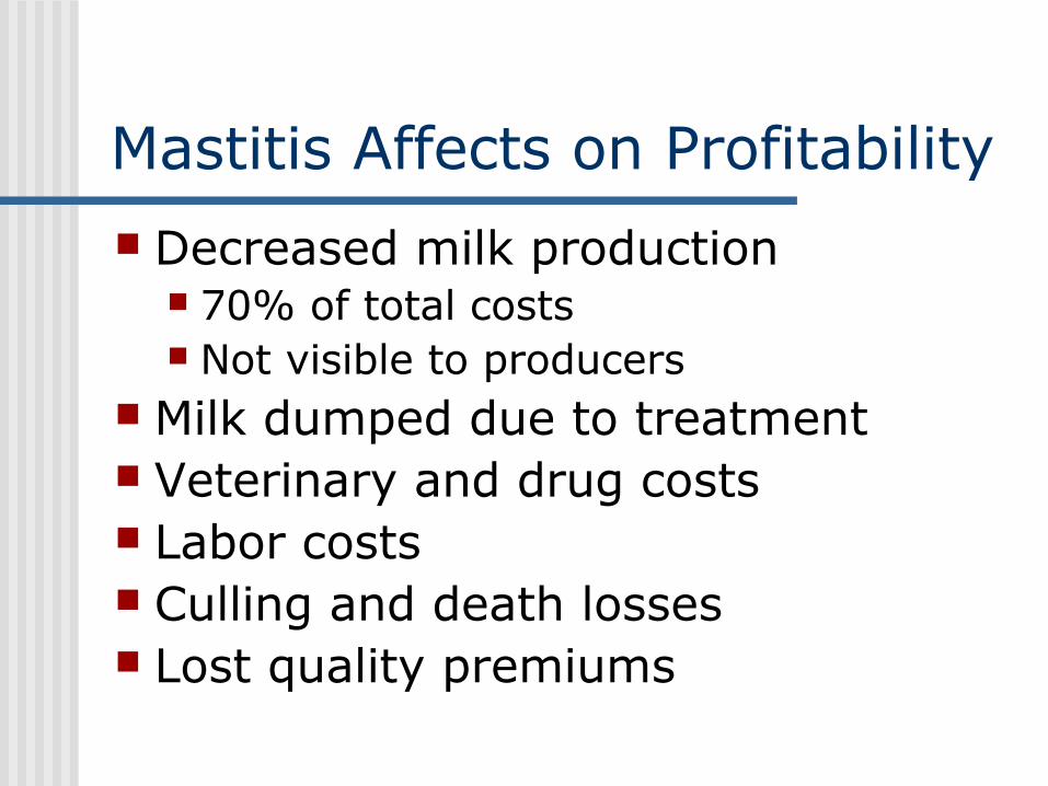
Mastitis Affects on Profitability Decreased milk production
70% of total costs Not visible to producers
Milk dumped due to treatment Veterinary and drug costs Labor costs Culling and death losses Lost quality premiums
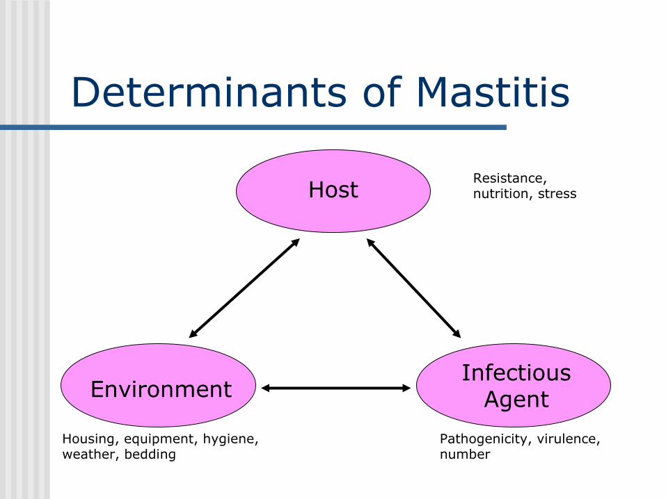
Determinants of Mastitis
Host
Infectious AgentEnvironment
Pathogenicity, virulence, number
Resistance, nutrition, stress
Housing, equipment, hygiene, weather, bedding
Mastitis Infection Almost always caused by bacteria
that generally enter through the teat canal.
Four ways for cow to get mastitis!! The environment inside the udder is
warm and moist with plenty of available nutrients, so bacteria multiply rapidly.
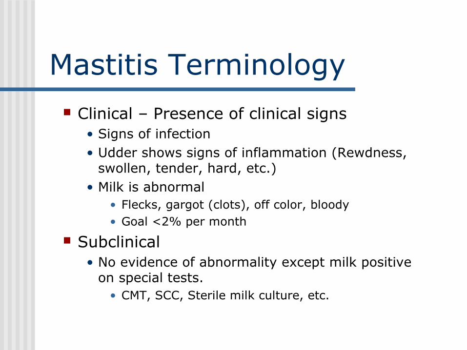
Mastitis Terminology Clinical – Presence of clinical signs
• Signs of infection• Udder shows signs of inflammation (Rewdness,
swollen, tender, hard, etc.)• Milk is abnormal
• Flecks, gargot (clots), off color, bloody• Goal <2% per month
Subclinical• No evidence of abnormality except milk positive
on special tests.• CMT, SCC, Sterile milk culture, etc.

Mastitis in a Herd
Clinical
Subclinical
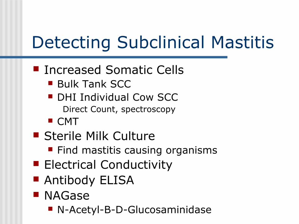
Detecting Subclinical Mastitis Increased Somatic Cells
Bulk Tank SCC DHI Individual Cow SCC
Direct Count, spectroscopy CMT
Sterile Milk Culture Find mastitis causing organisms
Electrical Conductivity Antibody ELISA NAGase
N-Acetyl-B-D-Glucosaminidase
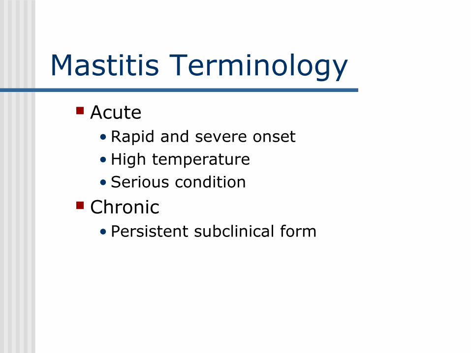
Mastitis Terminology Acute
• Rapid and severe onset• High temperature• Serious condition
Chronic• Persistent subclinical form

Mastitis Affects on Milk Composition
Milk Production: Decreases milk production by causing tissue damage, reduced lactose production and scar tissue formation in the udder.
Milk Quality and Composition: Increasing somatic cell count
• Polymorphonuclear neutrophils Decreasing lactose, casein, and fat production, Increasing blood components such as Na, K, Cl,
bicarbonate, IgG and serum albumin. • Electrical potential disrupted
Bacteria, blood cells and enzymes• Proteolysis• Lipolysis and globule breakdown• Off flavors
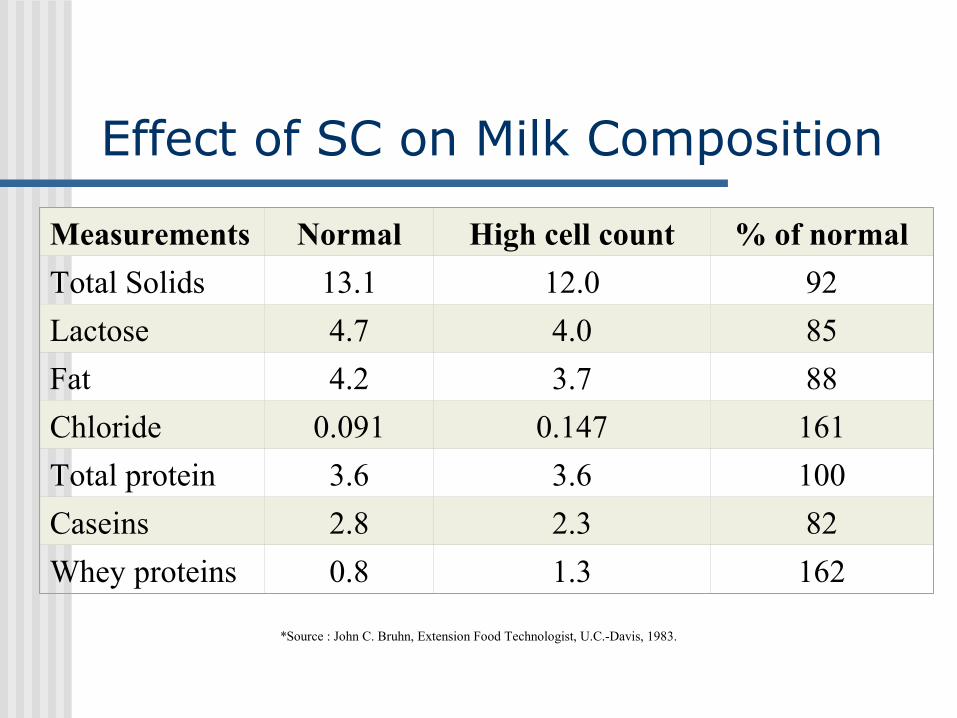
Effect of SC on Milk Composition
Measurements Normal High cell count % of normal
Total Solids 13.1 12.0 92
Lactose 4.7 4.0 85
Fat 4.2 3.7 88
Chloride 0.091 0.147 161
Total protein 3.6 3.6 100
Caseins 2.8 2.3 82
Whey proteins 0.8 1.3 162
*Source : John C. Bruhn, Extension Food Technologist, U.C.-Davis, 1983.
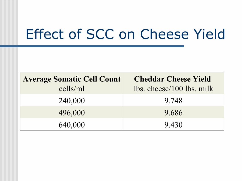
Effect of SCC on Cheese Yield
Average Somatic Cell Count cells/ml
Cheddar Cheese Yield lbs. cheese/100 lbs. milk
240,000 9.748
496,000 9.686
640,000 9.430

Types of Mastitis Contagious Environmental

Contagious Mastitis Primary habitat bacteria live on/in the udder
and teat lesions Poor survival of bacteria in the environment Is spread from cow to cow, primarily during
milking by milk-contaminated fomites at milking, sponge, milker's hands, milking machine
Staphylococcus aureus, Streptococcus agalactia, Mycoplasma bovis and sometimes streptococcus uberis are contagious mastitis causing organisms.
Usually chronic, subclinical mastitis
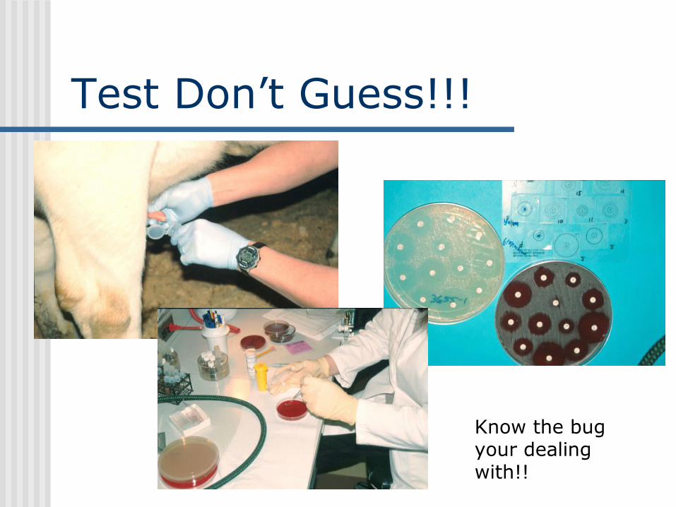
Test Don’t Guess!!!
Know the bug your dealing with!!
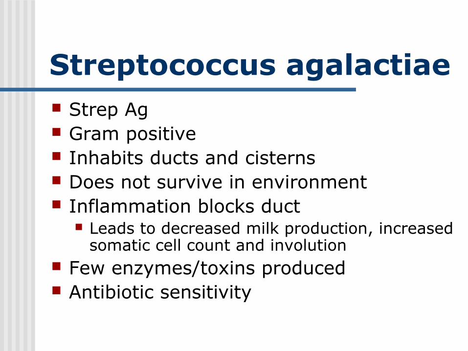
Streptococcus agalactiae Strep Ag Gram positive Inhabits ducts and cisterns Does not survive in environment Inflammation blocks duct
Leads to decreased milk production, increased somatic cell count and involution
Few enzymes/toxins produced Antibiotic sensitivity
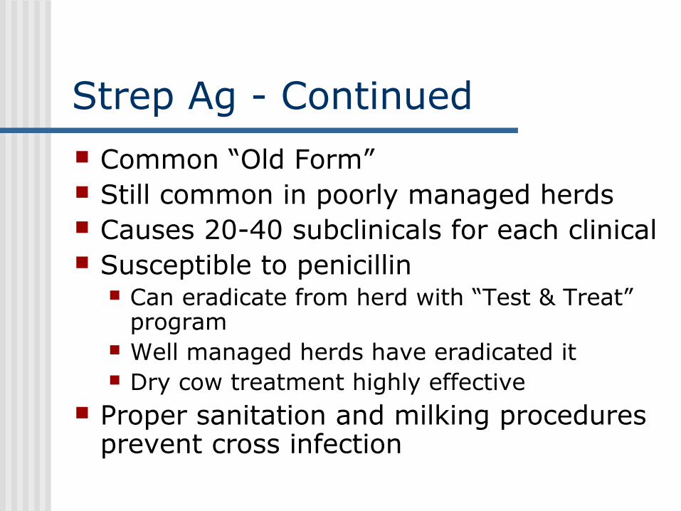
Strep Ag - Continued Common “Old Form” Still common in poorly managed herds Causes 20-40 subclinicals for each clinical Susceptible to penicillin
Can eradicate from herd with “Test & Treat” program
Well managed herds have eradicated it Dry cow treatment highly effective
Proper sanitation and milking procedures prevent cross infection

Sources of Strep Ag Major source is the infected cow.
Injected into udder during milking Squawking
Contaminated floors and stalls
Newly purchased cows
Heifer calves suckling penmates.
Milking personnel as carriers

Staphylococcus aureus #1 cause of mastitis in US Many forms
acute, chronic, subclinical (chronic, subclinical predominates)
Produces many enzymes/toxins (catalase, coagulase)
Invasive-hyaluronidase Resists phagocytosis & immune system Forms abscesses; may result in fibrosis Facultative intracellular pathogen Decreased milk production and increased
somatic cell count
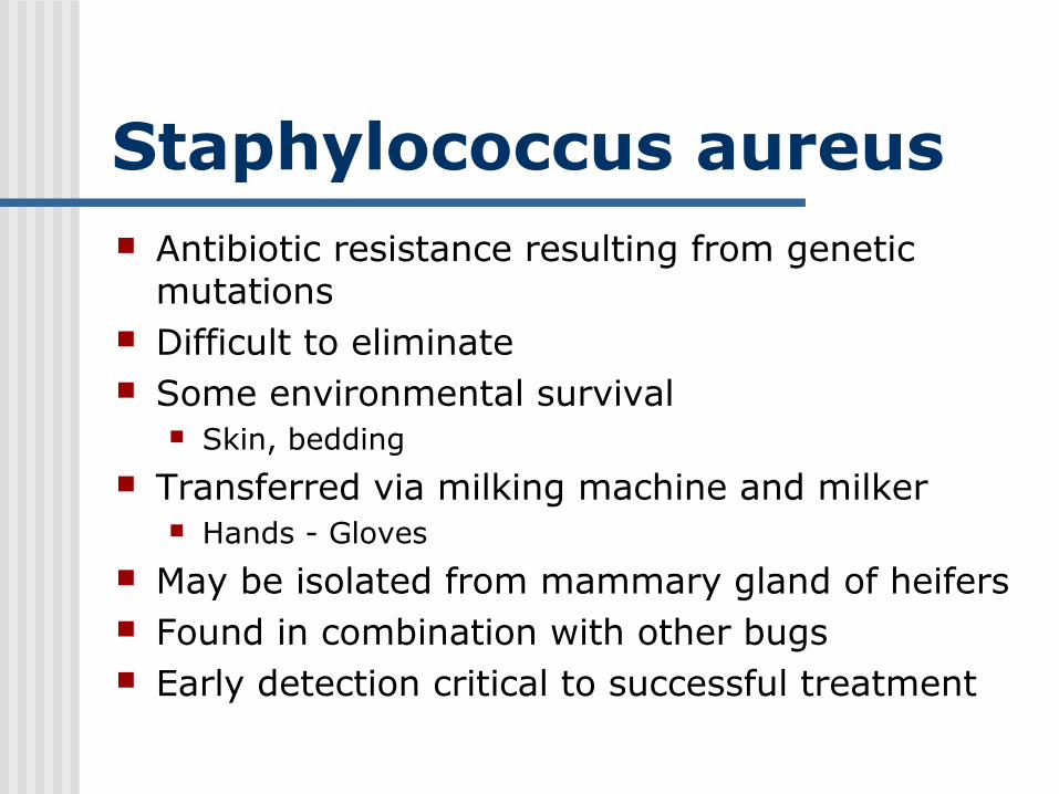
Staphylococcus aureus Antibiotic resistance resulting from genetic
mutations Difficult to eliminate Some environmental survival
Skin, bedding Transferred via milking machine and milker
Hands - Gloves May be isolated from mammary gland of heifers Found in combination with other bugs Early detection critical to successful treatment

Mycoplasma Between a bacteria and a virus No cell wall so antibiotics are ineffective Control by biosecurity Spread through contaminated antibiotics,
syringes milking units, common cloths, etc. Teat dipping is a good preventative Isolation and culling Usually in well-managed herds NYS Study – 10% of herds have infected cow Maine BT Study 2002 – 3% of herds

Control of Contagious Mastitis Dip teats in germicide after pre and post milking Treat quarters with dry cow antibiotics at end of
lactation Milking order or separate claw for infected cows Flush milk claws (hot water or germicide) after
milking infected cows (backflushing) Individual cloth/paper towels to wash/dry teats Clean hands, latex gloves Culture new cow additions Cull persistently infected cows Minimize teat end lesions Dry treat heifers before calving

Environment Mastitis Environment to cow Incidence increases as the incidence
of contagious mastitis decreases Primary habitat of bacterial is in the
environment (feces, soil, bedding, water)
Environmental contact at milking time or between milkings
Environmental Mastitis Organisms from the bedding, stalls,
corrals, etc. gain entrance through fatigued teat canals after or during milking to cause infection.
Streptococcus dysgalactia, Streptococcus uberus, and Coliform (E. coli, Klebsiella) are a few the organisms that live in the environment.
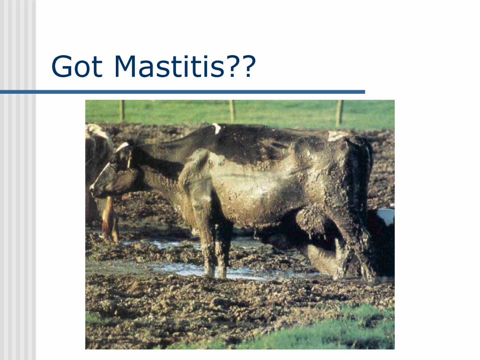
Got Mastitis??

Coliforms E. coli, Klebsiella spp., Enterobacter spp.,
Citrobacter spp. Gram negative Feces, bedding, wet dirty udders Transient peracute/acute mastitis Endotoxin
Very sick - death More prevalent in warm, wet weather

Sources Infection occurs when contaminated material
contacts and enters teat canal Infections occur at any stage
Sediment from unclean surroundings, flank, and udder
Manure Polluted water (barnyard) Unclean equipment Wet bedding, especially green sawdust Infected quarter of other cows
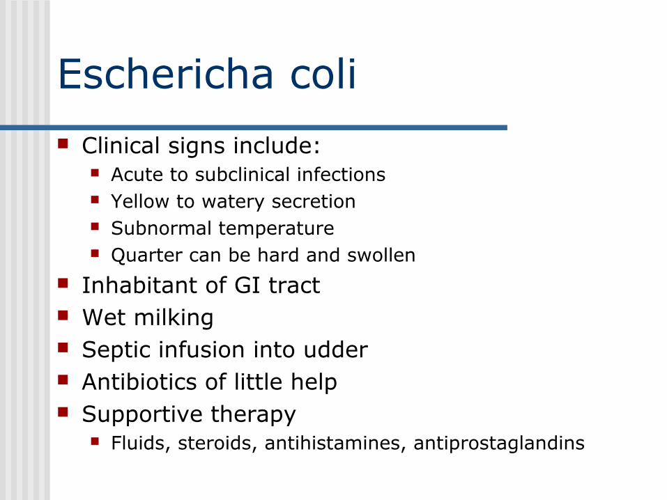
Eschericha coli Clinical signs include:
Acute to subclinical infections Yellow to watery secretion Subnormal temperature Quarter can be hard and swollen
Inhabitant of GI tract Wet milking Septic infusion into udder Antibiotics of little help Supportive therapy
Fluids, steroids, antihistamines, antiprostaglandins

Klebsiella Clinical signs similar to E. coli
Acute to subclinical infections Yellow to watery secretion Subnormal temperature Quarter can be hard and swollen
Associated with soil contamination Grows well in wood products Switch bedding Maintain high pH in bedding
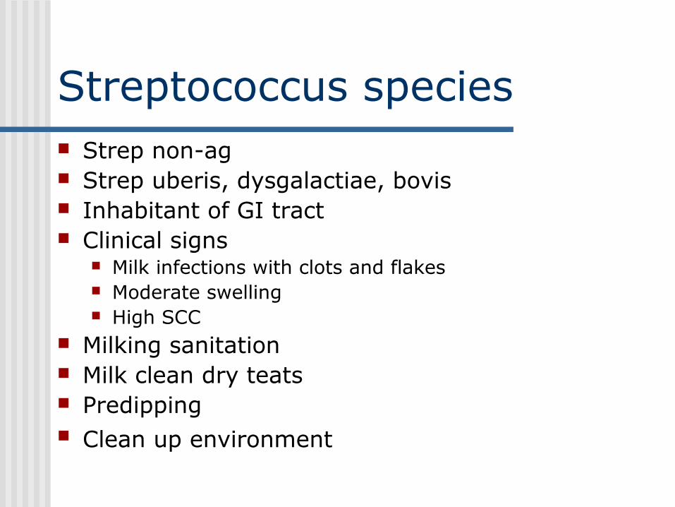
Streptococcus species Strep non-ag Strep uberis, dysgalactiae, bovis Inhabitant of GI tract Clinical signs
Milk infections with clots and flakes Moderate swelling High SCC
Milking sanitation Milk clean dry teats Predipping Clean up environment
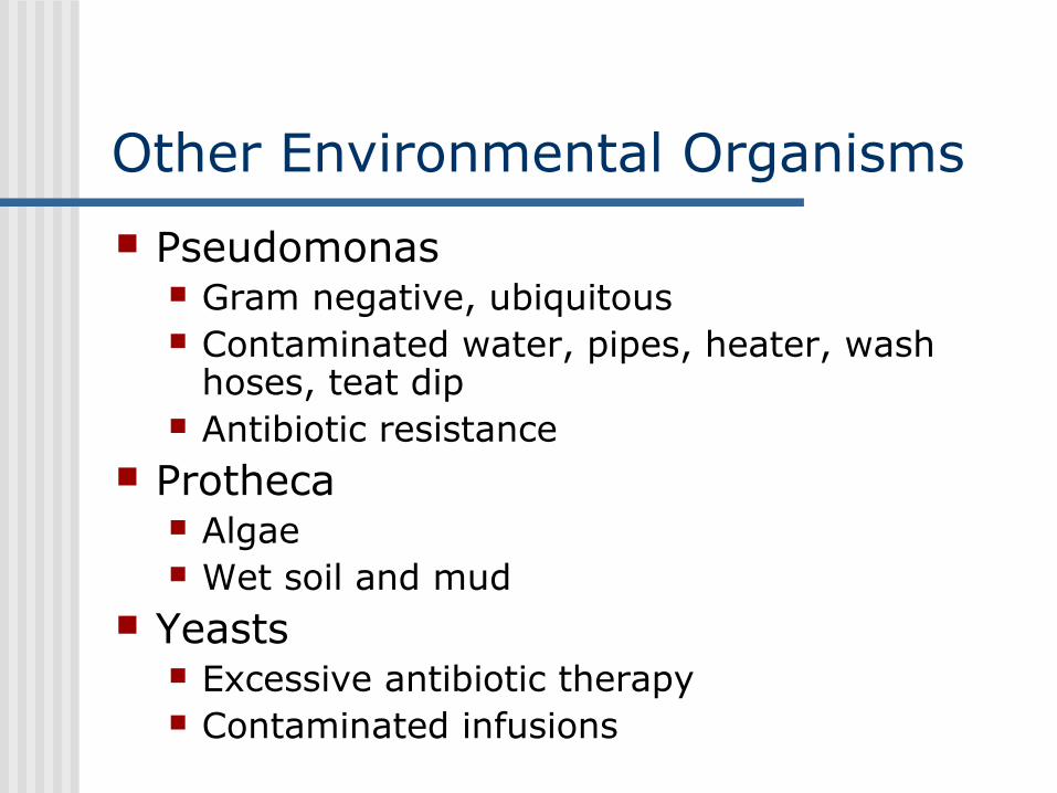
Other Environmental Organisms
Pseudomonas Gram negative, ubiquitous Contaminated water, pipes, heater, wash
hoses, teat dip Antibiotic resistance
Protheca Algae Wet soil and mud
Yeasts Excessive antibiotic therapy Contaminated infusions

Control of Environmental Mastitis More difficult to control than the contagious pathogens Most are resistant to germicides in teat dip and antibiotics
in dry cow therapy Key is to ID source and remove (bedding, ponds, mud) Clip or flame udders Milk only clean dry teats Clean parlor, stalls, bedding Barrier dips Predip teats with germicide before milking – No water Keep cows standing after milking - feeding Sterile single-dose infusion products Sterile infusion techniques (alcohol swab)
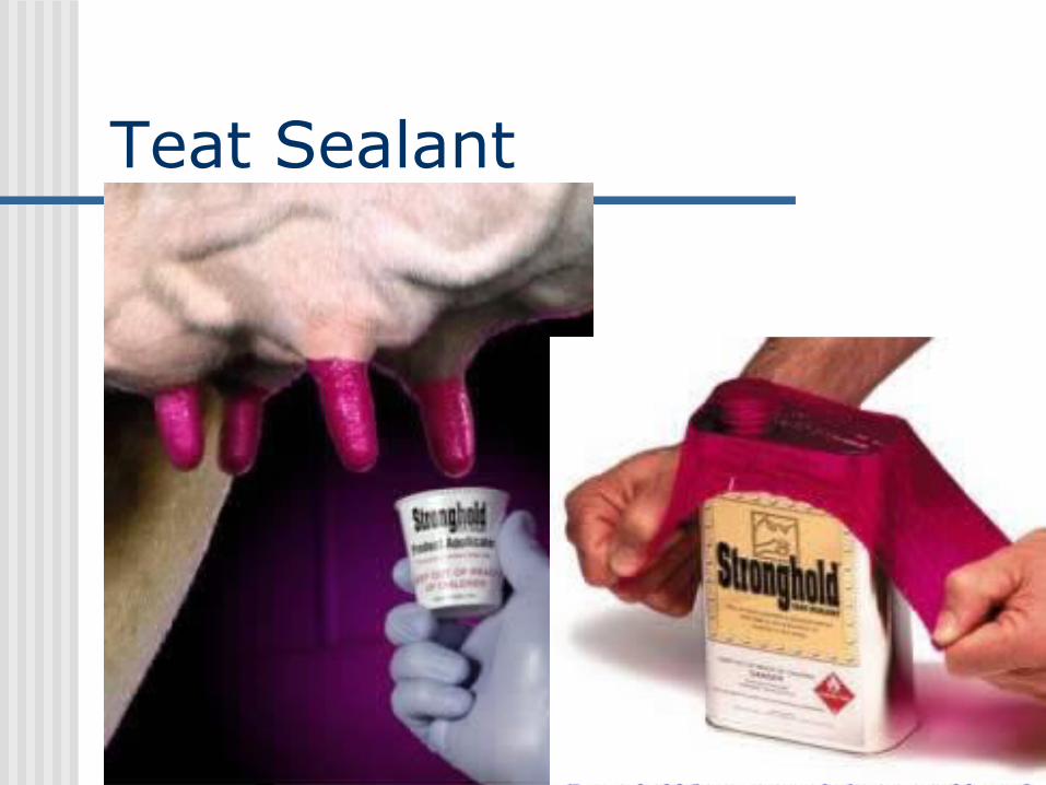
Teat Sealant

Orbeseal

Orbeseal data

Timing of Infection

Somatic Cell Counts - SCC Epithelial cells and white blood cells Changes with milk production, infection, age, stage. Measures the level of udder stress/damage/irritation
Under 240,000 /ml uninfected Over 240,000 /ml infected Legal limit 750,000/ml not very stringent Not a measure of actual mastitis infection Do not treat based solely on SCC!
Easy way to assess the mastitis level in a herd Excellent mastitis management tool Highest correlation with milk production of any DHIA
measure SCC probably can't be too low
Not the SCC but response to infection which is important
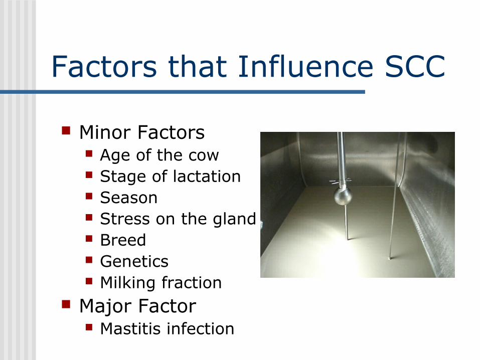
Factors that Influence SCC
Minor Factors Age of the cow Stage of lactation Season Stress on the gland Breed Genetics Milking fraction
Major Factor Mastitis infection
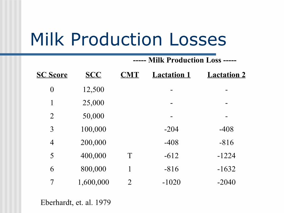
Milk Production Losses----- Milk Production Loss -----
SC Score SCC CMT Lactation 1 Lactation 2
0 12,500 - -
1 25,000 - -
2 50,000 - -
3 100,000 -204 -408
4 200,000 -408 -816
5 400,000 T -612 -1224
6 800,000 1 -816 -1632
7 1,600,000 2 -1020 -2040
Eberhardt, et. al. 1979
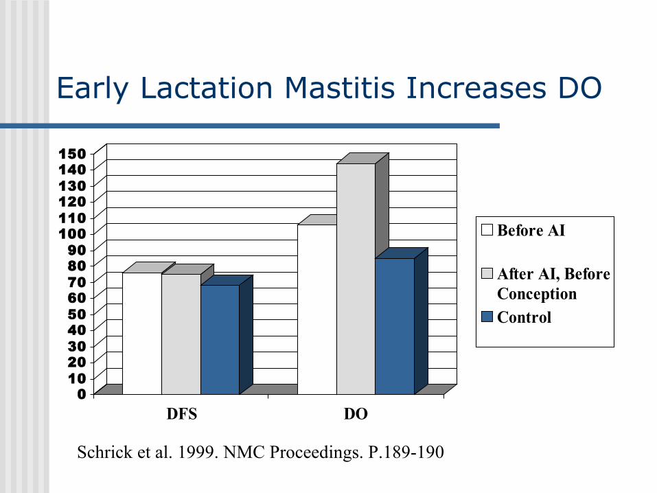
Early Lactation Mastitis Increases DO
0102030405060708090100110120130140150
DFS DO
Before AI
After AI, BeforeConceptionControl
Schrick et al. 1999. NMC Proceedings. P.189-190
Mastitis Treatment IMM Therapy
Injection of antibiotics into udder Systemic Therapy
Antibiotics IV or IM Supportive Therapy
Remove toxins – Frequent milkout Treat dehydration, swelling and pain Know bug
Lactational Therapy Likelihood of success?
Dry Cow Therapy Larger dose, longer acting product
Mastitis Treatment
IMM Therapy Use an approved product Use proper technique Have culture reports and sensitivities Make best guess on first drug Cow history, treatments and results Don’t give up on a certain antibiotic, often
response is seen with longer course of therapy Have a standard protocol

Mastitis Treatment ProtocolsGrade Clinical Signs Treatment
1 Milk abnormalUdder not swollenCow normal
Take sterile milk sample and culture.Decide to treat based on results.Possible supportive therapy
2 Milk abnormalUdder swollenCow normal
Take sterile milk sample and culture.Treat in udder with antibiotic. Possible systemic/supportive therapy
3 Milk abnormalUdder swollenCow sick
Take sterile milk sample and culture.Treat systemically and in udder with antibiotics and supportive therapy

Supportive Treatment Reduced risk of antibiotic residues Organic herds Oxytocin / Stripping
Eliminates toxins and bacteria food Not effective against contagious bugs
Aspirin, Antihistamines, Anti-inflammatory
Fluids – dehydration
Mastitis Prevention Proper Milking Techniques
Procedures, training, monitoring Keep cows clean!
Proper Bedding• Sand is the best bedding• Organic bedding (sawdust, etc.) must be dry• Stall sized to fit cows• Udder flaming, tail docking
Nutrition Vitamins and minerals
Milk contagious cows last Maintain milking equipment
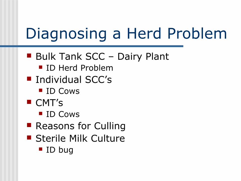
Diagnosing a Herd Problem Bulk Tank SCC – Dairy Plant
ID Herd Problem Individual SCC’s
ID Cows CMT’s
ID Cows Reasons for Culling Sterile Milk Culture
ID bug

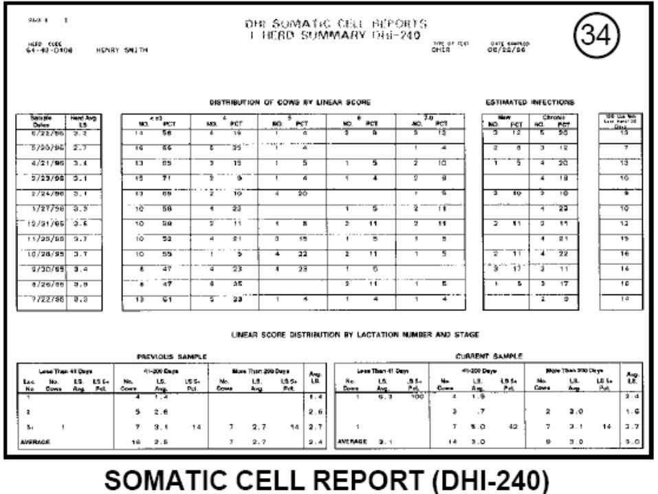
Flow of DHIA Data
DART, Raleigh, NC
LOOP - Ithaca, NY
Mail or Download
Supervisor Upload
Farm
ComponentsLab
Data & Milk Samples
USDA-AIPL
Associations
AI Studs

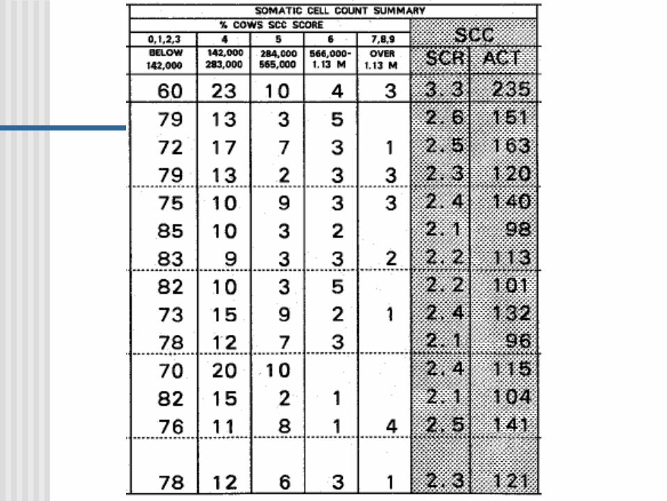
DHIA Individual Cow SCC Level of new infections
• Low (<4) last month - high (>4) this month Level of chronic infections
• High last month - high this month First Lactation animals affected When are infections happening?


Culling


Graph of Previous SCS vs Current SCS

Average SCS by Lactation

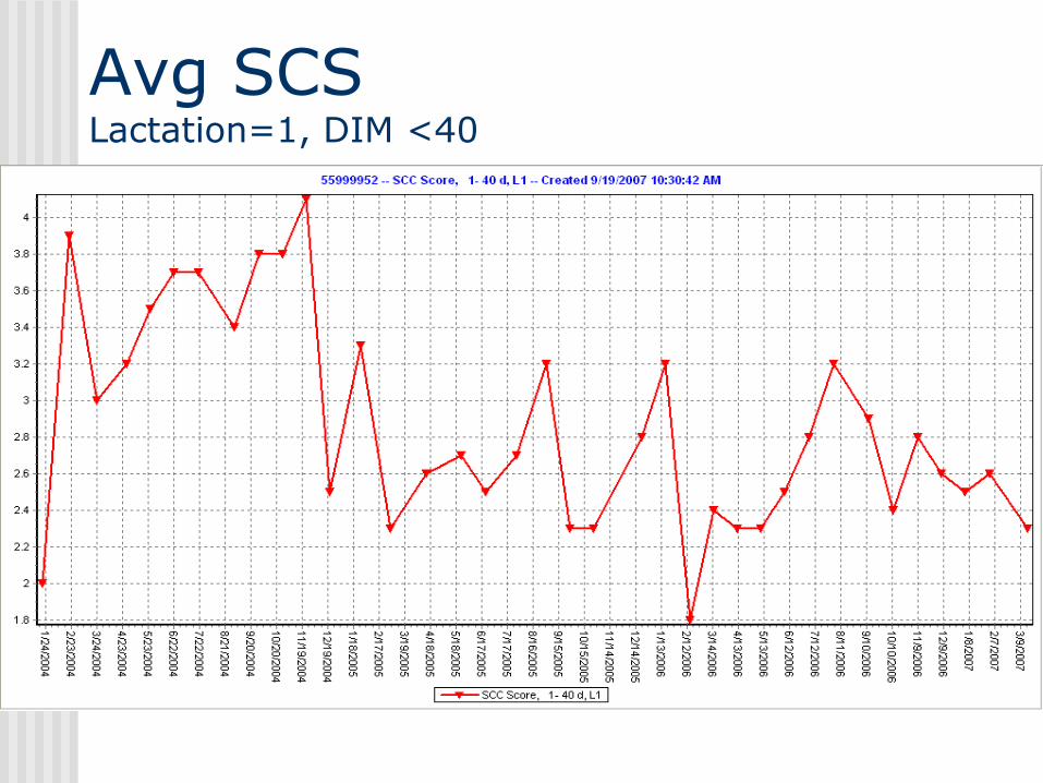
Avg SCS Lactation=1, DIM <40

SCS Throughout Lactation