mastr directs myod-dependent satellite cell differentiation during
TRANSCRIPT

MASTR directs MyoD-dependent satellitecell differentiation during skeletalmuscle regeneration
Mayssa H. Mokalled, Aaron N. Johnson, Esther E. Creemers,1 and Eric N. Olson2
Department of Molecular Biology, University of Texas Southwestern Medical Center at Dallas, Dallas, Texas 75390, USA
In response to skeletal muscle injury, satellite cells, which function as a myogenic stem cell population, becomeactivated, expand through proliferation, and ultimately fuse with each other and with damaged myofibers to promotemuscle regeneration. Here, we show that members of the Myocardin family of transcriptional coactivators, MASTRand MRTF-A, are up-regulated in satellite cells in response to skeletal muscle injury and muscular dystrophy. Globaland satellite cell-specific deletion of MASTR in mice impairs skeletal muscle regeneration. This impairment issubstantially greater when MRTF-A is also deleted and is due to aberrant differentiation and excessive proliferationof satellite cells. These abnormalities mimic those associated with genetic deletion of MyoD, a master regulator ofmyogenesis, which is down-regulated in the absence of MASTR and MRTF-A. Consistent with an essential role ofMASTR in transcriptional regulation of MyoD expression, MASTR activates a muscle-specific postnatal MyoDenhancer through associations with MEF2 and members of the Myocardin family. Our results provide new insightsinto the genetic circuitry of muscle regeneration and identify MASTR as a central regulator of this process.
[Keywords: muscle regeneration; satellite cells; MASTR; MRTF-A; MEF2; MyoD]
Supplemental material is available for this article.
Received September 21, 2011; revised version accepted December 15, 2011.
Mammalian skeletal muscle possesses the potential toundergo self-repair in response to injury or disease. Theadvent of stem cell and regenerative medicine has sparkedrenewed interest in understanding the molecular and cel-lular mechanisms of muscle regeneration (Price et al. 2007;Goyenvalle et al. 2011; Tuan 2011). By enhancing muscleregeneration, stem cell-based therapies have potential ap-plications in multiple conditions associated with musclewasting, including muscular dystrophy, chronic diseasesassociated with physical inactivity and loss of musclemass, and aging. Deciphering the molecular mechanismsof muscle regeneration is essential for the development ofsuch therapies.
Muscle repair is a multistep process that includesmyofiber degeneration, regeneration, and remodeling (LeGrand and Rudnicki 2007; Ten Broek et al. 2010; Tsivitse2010). Muscle degeneration is characterized by sarcomeredisruption, cell permeabilization, and induction of an acuteinflammatory response. The regeneration process is markedby the activation of a myogenic stem cell population, re-ferred to as satellite cells (SCs), which give rise to prolifer-
ating myoblasts, followed by myoblast differentiation andfusion into regenerated myofibers. During the final stagesof muscle repair, myofibers remodel to produce maturemuscle fibers and recover the contractile capacity of theinjured muscle.
SCs are the primary population of adult skeletal musclestem cells, comprising ;1%–4% of muscle nuclei in nor-mal skeletal muscle (Whalen et al. 1990). Quiescent SCsare characterized by three criteria: a unique localizationbetween the sarcolemma and the basal lamina; a distinctcellular morphology that includes a small cytoplasmicvolume, an enlarged nucleus, and condensed chromatin;and expression of the Pax7 transcription factor (Buckinghamand Relaix 2007; Biressi and Rando 2010; Kang and Krauss2010). In response to chemical or mechanical muscledamage, exercise, or overuse, quiescent SCs become ac-tivated and migrate to the injury site, where they proliferateand up-regulate MyoD and Myf5 expression. Upon activa-tion, the majority of SCs down-regulate Pax7 expressionand undergo terminal muscle cell differentiation, whereasa small proportion of cells down-regulate MyoD expres-sion, maintain Pax7 expression, and revert back to quies-cence to sustain the SC pool (Le Grand and Rudnicki 2007;Olguin et al. 2007; Biressi and Rando 2010; Wu et al. 2010).
Expression of the myogenic regulatory factor MyoDspecifies the identity of embryonic and adult muscle pro-genitor cells (Rudnicki et al. 1993; Buckingham 1994;
1Present address: Academic Medical Center, Meibergdreef 15, 1105 AZAmsterdam, The Netherlands.2Corresponding author.E-mail [email protected] is online at http://www.genesdev.org/cgi/doi/10.1101/gad.179663.111.
190 GENES & DEVELOPMENT 26:190–202 � 2012 by Cold Spring Harbor Laboratory Press ISSN 0890-9369/12; www.genesdev.org
Cold Spring Harbor Laboratory Press on April 3, 2019 - Published by genesdev.cshlp.orgDownloaded from

Olson and Klein 1994; Tapscott 2005). MyoD expression,which marks SC activation and differentiation, is requiredfor normal muscle regeneration (Megeney et al. 1996).MyoD knockout mice show severely deficient skeletalmuscle regeneration following injury, due to excessive pro-liferation and defective differentiation of SCs (Sabourinet al. 1999; Yablonka-Reuveni et al. 1999; Cornelisonet al. 2000; White et al. 2000; Schuierer et al. 2005).Reminiscent of its expression in activated SCs, differen-tiating myoblasts up-regulate MyoD and activate transcrip-tion of MyoD target genes, resulting in irreversible cellcycle arrest and myogenic differentiation. On the otherhand, down-regulation of MyoD in activated SCs causesreturn to quiescence (Yoshida et al. 1998). Relatively littleis known of the upstream activators of MyoD expression.Deciphering the mechanisms that control MyoD expres-sion and function in SCs should provide important in-sights into the molecular basis of muscle regeneration.
Members of the Myocardin family of transcription fac-tors (Myocardin, MRTF-A, MRTF-B, and MASTR) playimportant roles in differentiation and remodeling of cardiac,smooth, and skeletal muscle cells (Creemers et al. 2006a;Pipes et al. 2006; Posern and Treisman 2006; Olson andNordheim 2010). MASTR is a muscle-enriched MEF2 co-activator (Creemers et al. 2006a), whereas Myocardin andMRTF-A/-B are SRF coactivators (Olson and Nordheim2010). Expression of dominant-negative MRTF-A in mouseskeletal muscle causes muscle hypoplasia (S Li et al. 2005);however, MRTF-A and MRTF-B knockout mice do notshow obvious skeletal muscle defects (J Li et al. 2005; Ohet al. 2005; Li et al. 2006; Sun et al. 2006). MRTF-A/-Bdouble-knockout mice revealed that MRTF-A/-B are redun-dant in the brain and heart, and possibly in other tissueswhere they are coexpressed (Mokalled et al. 2010; EN Olsonand MH Mokalled, unpubl.). The in vivo functions ofMASTR and MRTF-A/-B in mammalian skeletal muscle de-velopment, maintenance, or regeneration remain unknown.
Here, we show that MASTR and MRTF-A are up-regulated in SCs in response to injury. Global and SC-specific deletion of MASTR in mice causes impaired muscleregeneration in response to chemical injury and musculardystrophy. This regeneration defect is exacerbated byMRTF-A deletion and is due to aberrant differentiation andexcessive proliferation of SCs. These abnormalities resem-ble those of mice lacking MyoD, which is down-regulatedin the absence of MASTR and MRTF-A. Consistent witha regulatory role of MASTR in the control of MyoD ex-pression, MASTR activates a muscle-specific MyoD en-hancer together with MEF2 and MRTF-A. These findingsreveal a previously unrecognized regulatory network withinmyogenic stem cells in which MASTR functions as a pri-mary upstream activator of skeletal muscle regeneration.
Results
Up-regulation of MASTR in response to muscle injury
Members of the Myocardin family of transcription factorshave been implicated in pathological remodeling of car-diac and vascular smooth muscle (Parmacek 2007; Olson
and Nordheim 2010; Small et al. 2010), but little is knownof their potential roles in skeletal muscle disease. To in-vestigate the involvement of this family of coactivatorsin the response of skeletal muscle to injury, we surveyedthe expression of MASTR, MRTF-A, and MRTF-B in re-sponse to a series of skeletal muscle injuries that induceregeneration.
Injection of skeletal muscle with cardiotoxin (Ctx)causes myofiber degeneration followed by regenerationand repair, as activated SCs proliferate, differentiate, andfuse with injured myofibers (Yan et al. 2003). As shown inFigure 1A, MASTR was up-regulated in the adult tibialisanterior (TA) muscle at days 3 and 7 following Ctx injury.MRTF-A was also up-regulated in response to Ctx injury,whereas MRTF-B showed relatively little change in ex-pression. The pattern of MASTR and MRTF-A expressionduring muscle regeneration resembled that of MyoD. In-jection of the TA muscle with barium chloride (BaCl2)also causes muscle injury followed by regeneration and wasaccompanied by similar changes in expression of MASTR,MRTF-A, and MyoD (Fig. 1A). In addition, MASTR andMRTF-A were up-regulated in parallel with MyoD in theTA muscle of mdx mice, concomitant with muscle de-generation and regeneration (Fig. 1A).
Expression of MASTR and MRTF-A in SCs
To determine whether MASTR and MRTF-A are ex-pressed in SCs in response to muscle injury, SCs wereisolated from adult skeletal muscle and sorted using pos-itive selection for CD34 and negative selection of Sca1,CD31, and CD45 (Supplemental Fig. S1A,B; Beauchampet al. 2000; Sherwood et al. 2004; Montarras et al. 2005;Sacco et al. 2008). MASTR expression was enriched insorted SCs compared with either unsorted cells (UCs) orwhole quadriceps (Quad), whereas MRTF-A was expressedat comparable levels in SCs and UCs (Fig. 1B). Pax7 andMyoD expression was also enriched in sorted SCs (Fig. 1B;Supplemental Fig. S1C), confirming the identity of thiscell population. MASTR and MRTF-A expression wasunchanged when SCs were activated to proliferate by ex-posure to growth medium and was up-regulated when SCswere induced to differentiate into myotubes in the pres-ence of differentiation medium (Fig. 1C; Musaro andBarberi 2010). Similarly, MASTR and MRTF-A expressionwas up-regulated during differentiation of C2C12 myo-blasts into myotubes (Fig. 1C). These findings suggestedthe potential involvement of MASTR and MRTF-A in SC-mediated responses to muscle injury and differentiation.
MASTR is required for skeletal muscle regeneration
To investigate the function of MASTR in vivo, we gen-erated a conditional MASTR knockout (MKO) allele byinserting loxP sites for Cre-mediated deletion in introns 1and 7 of the protein-coding region of the gene using ho-mologous recombination. Expression of Cre recombinaseresulted in deletion of exons 2–7 of the MASTR locus,thereby eliminating the MEF2-binding and SAP do-mains and generating what is expected to be a null al-lele (Supplemental Fig. S2A). Correct targeting and germline
MRTFs and MEF2 regulate muscle regeneration
GENES & DEVELOPMENT 191
Cold Spring Harbor Laboratory Press on April 3, 2019 - Published by genesdev.cshlp.orgDownloaded from

transmission of the MASTR allele was confirmed bySouthern blot and PCR of genomic DNA (SupplementalFig. S2B). By breeding MASTRFloxed mice to CAG-Cretransgenic mice, we obtained global MKO mice (Supple-mental Fig. S2C), which were viable, fertile, and withoutobvious phenotypic defects.
We subjected MKO mice to Ctx injury and evaluatedtheir regeneration potential. Newly regenerated myofi-bers are readily identifiable by the presence of centralizednuclei, which distinguish them from pre-existing myo-fibers, in which nuclei are positioned at the cell periphery.By H&E staining, wild-type TA muscles were mostly com-posed of regenerating myofibers 7 d post-injury (Fig. 2A).MKO TA muscles at 7 d post-injury showed no central-ized nuclei and instead were composed of degeneratingnecrotic fibers, fibrotic tissue, and inflammatory cells(Fig. 2A). By day 14 post-injury, regenerated wild-typemyofibers remained centrally nucleated and became ho-mogeneous in size, indicative of their maturation (Fig.2A). In contrast, MKO myofibers at day 14 post-injury re-mained heterogeneous in size and were fewer in numbercompared with wild-type muscle (Fig. 2A).
Wild-type muscle injured with Ctx initiates expressionof Desmin, a marker of skeletal muscle differentiation,7 d post-injury and shows robust Desmin expression inmature myofibers by day 14 post-injury (Fig. 2A). Desmin-positive myofibers were absent in MKO TA muscle 7 dpost-injury and were still fewer in number and immature
at day 14 post-injury (Fig. 2A). In addition, the number ofcentralized nuclei in MKO mice at days 7 and 14 post-injury was significantly decreased compared with wild-type muscle (Fig. 2B). Muscle regeneration in response toinjection of BaCl2 was also compromised in MKO mice(Fig. 2A). Thus, MASTR is required for efficient regener-ation in two independent models of muscle injury.
To further investigate the requirement of MASTR forskeletal muscle regeneration, we crossed the MKO mice tomdx mice, which harbor a null mutation in the Dystro-phin gene. Mdx mice undergo extensive muscle degener-ation and regeneration at ;3 wk of age, until they exhausttheir regeneration capacity by ;7 wk of age (McGeachieet al. 1993; Banks and Chamberlain 2008). Trichrome andwheat germ agglutinin (WGA) stainings revealed severemuscle damage and increased fibrosis in MKO;mdx TAmuscle compared with mdx muscle (Fig. 2C). MKO;mdxmice also showed a 20% reduction in body weight andmuscle weight at 4 and 6 wk of age compared with mdxmice (Fig. 2D). We conclude that MASTR regulates skel-etal muscle regeneration after chemical injury and in amuscular dystrophy disease model.
Aberrant muscle regeneration resultingfrom SC-specific deletion of MASTR
MASTR expression in proliferating and differentiatingSCs and the requirement for MASTR in skeletal muscle
Figure 1. Up-regulation of MASTR and MRTF-Aduring muscle regeneration. (A) Shown is the expres-sion of MASTR, MRTF-A, and MRTF-B in the TAmuscle following Ctx and barium chloride (BaCl2)injuries and in mdx mice. qPCR shows MASTR andMRTF-A up-regulation 3 d after Ctx and BaCl2 in-jection. MASTR and MRTF-A are also up-regulated inthe TA muscle of 2-, 4-, and 6-wk-old mdx micecompared with wild-type (WT) mice. For Ctx andBaCl2 injury, relative expression at days 3 and 7 post-injury is normalized to GAPDH levels and to baselinelevel at day 0. For mdx mice, relative expression isnormalized to GAPDH levels and to wild-type ex-pression for each time point. MyoD expression isused as control for muscle regeneration and parallelsMASTR and MRTF-A expression patterns. (B) Expres-sion of MASTR and MRTF-A in sorted SCs. SortedSCs (SC), mononuclear unsorted cells (UCs), andquadriceps mRNA samples (Quad) were used forqPCR. Relative expression is normalized to the levelsof ribosomal 18S RNA. The levels of Pax7 and MyoDare shown as controls. (C) Up-regulation of MASTRand MRTF-A expression during SC and C2C12 dif-ferentiation. Sorted SCs, activated SCs maintainedunder growth conditions, and differentiating SCs atdays 1, 2, or 3 of differentiation were used for qPCR.MASTR and MRTF-A are expressed in sorted andactivated SCs, and are up-regulated at days 1 and 2 ofdifferentiation. Similarly, qPCR on differentiatingC2C12 cultures shows approximately sixfold to sev-enfold up-regulation of MASTR and MRTF-A by day2 of differentiation.
Mokalled et al.
192 GENES & DEVELOPMENT
Cold Spring Harbor Laboratory Press on April 3, 2019 - Published by genesdev.cshlp.orgDownloaded from

regeneration suggest that MASTR regulates SC-mediatedskeletal muscle regeneration. However, because MASTRis expressed in mature muscle fibers in addition to SCs,global gene deletion could not definitely distinguish itsfunction in one population versus the other. Therefore,to directly test the role of MASTR in SCs, we crossed theMASTRFloxed mice to Pax7-Cre-ERT2 mice, in which a Crerecombinase-estrogen receptor expression cassette wasknocked into the Pax7 locus, allowing for tamoxifen-inducible, SC-specific expression of Cre (Lepper et al.2009). We subjected the MASTRFloxed/Floxed;Pax7-Cre-ERT2
mice, hereafter referred to as conditional MKO (cMKO)mice, to tamoxifen treatment followed by Ctx injury.At days 3 and 7 post-injury, control TA muscle showedsignificant recovery, with total replacement of damagedmyofibers with newly regenerated, Desmin-positive, cen-trally nucleated myofibers (Fig. 2E). However, as observedwith the global deletion of MASTR, muscle regenerationwas defective in cMKO mice, with a significant decreasein the number of regenerating myofibers, persistence ofnecrotic fibers, and greatly reduced Desmin expression
(Fig. 2F). These findings demonstrate that MASTR per-forms a SC-autonomous role in regulating skeletal mus-cle regeneration after injury.
MASTR and MRTF-A cooperatively regulate musclemass and regeneration
To determine whether MRTF-A and MASTR might act ina common pathway during skeletal muscle regeneration,we generated MASTR/MRTF-A double-knockout (dKO)mice by crossing MKO mice to the previously generatedMRTF-A knockout (AKO) mouse line (Li et al. 2006).AKO mice showed no abnormalities in muscle regener-ation compared with wild-type mice (Supplemental Fig.S3A). Approximately 40% of the dKO mice showed peri-natal lethality (Fig. 3A), which may reflect an essentialfunction for MASTR and MRTF-A during neuronal de-velopment. dKO mice that survived beyond postnatal day1 (P1) were viable and fertile, but showed a significantdecrease in body mass and muscle mass, as shown bynuclear magnetic resonance (NMR) analysis of lean mass
Figure 2. Aberrant muscle regeneration ofMASTR-null mice. (A) Regeneration of MKOmuscle following chemical injury. TA musclefrom wild-type (WT) and MKO mice wasinjected with Ctx and BaCl2 and assayed forregeneration by H&E and by Desmin immuno-histochemistry at days 7 and 14 post-injury.Regenerating and degenerating fibers are indi-cated by white and black arrowheads, respec-tively. At day 7 post-Ctx or post-BaCl2 injury,wild-type muscle contains regenerating myofi-bers that are centrally nucleated and hetero-geneous in size. MKO muscle shows fewerregenerating fibers and is mostly composed ofnecrotic fibers and regions of fibrotic tissue andinflammatory cells (black arrows). By day 14post-Ctx injury, regenerating wild-type fibers aremature, centrally nucleated, and homogeneousin size, whereas regenerating MKO fibers areheterogeneous in size and decreased in number.Bar, 65 mm. (B) Quantitation of regenerationshows a significant decrease in the number ofcentralized nuclei in MKO muscle comparedwith wild-type muscle at days 7 and 14 post-injury. Analysis was performed on five animalsfor each genotype and time point and on fivesections from each animal; (*) P < 0.01. (C)Enhanced mdx regeneration defect by MASTRdeletion. Trichrome staining and immunohisto-chemistry for Desmin and WGA show enhancedmuscle damage and fibrosis in 4-wk-oldMKO;mdx mice compared with mdx mice.Bar, 125 mm. (D) MKO;mdx mice have decreasedbody mass and muscle mass compared withlittermate mdx mice; (*) P < 0.01. (E) Impairedregeneration of cMKO mice following Ctx in-jury. Staining for Desmin and WGA shows de-fective TA muscle regeneration of cMKO miceat days 3 and 7 post-Ctx injury. Bar, 45 mm.
MRTFs and MEF2 regulate muscle regeneration
GENES & DEVELOPMENT 193
Cold Spring Harbor Laboratory Press on April 3, 2019 - Published by genesdev.cshlp.orgDownloaded from

content (Fig. 3B,C). Muscle mass was not significantlychanged in MKO or AKO mice (Li et al. 2006; Sun et al.2006). By 44 wk of age, dKO mice showed dramatic mus-cle wasting (Fig. 3B) but normal TA muscle histology,with only a mild decrease in myofiber size comparedwith either wild-type or MKO TA muscle (SupplementalFig. S4A). In addition, the regeneration defect of MKOmice was significantly enhanced in dKO mice (Fig. 3D).For example, dKO TA muscle completely lacked regener-ating myofibers 7 d post-injury and showed more severemyofiber dropout following injury than MKO mice.
Regulation of MyoD expression by MASTRand MRTF-A
The regenerative defects in MKO and dKO mice are re-markably similar to those of MyoD-null mice (Megeneyet al. 1996). To determine whether MyoD down-regulationmight account for the observed abnormalities, we ana-lyzed MyoD expression in wild-type and mutant muscle.As shown in Figure 3E, MyoD expression was comparablebetween wild-type and dKO mice at birth, but was strik-ingly undetectable in dKO mice at P14. MyoD expression
was also diminished in MKO mice, but unchanged in AKOmice (Fig. 3E; Supplemental Fig. S3B). By immunohisto-chemistry, MyoD protein was undetectable in the TA mus-cle of dKO mice 7 d post-injury (Fig. 3F).
MyoD expression was also significantly down-regu-lated in sorted MKO SCs compared with wild-type SCs(Fig. 3G). Upon activation in culture, wild-type SCs in-duce MyoD expression, whereas activated MKO SCsshowed blunted MyoD induction (Fig. 3G). In addition,MyoD levels were undetectable in sorted or activated dKOSCs. MyoD expression is therefore sensitive to MASTRlevels under multiple contexts, and MASTR plus MRTF-Aappear essential for normal postnatal expression ofMyoD.
To determine whether the block to MyoD expressionwas sufficient to account for the failure in differentiationof dKO SCs, we performed an in vitro MyoD rescueexperiment (Fig. 3H). Forced expression of MyoD by ret-roviral delivery rescued the differentiation defect of dKOSCs, as shown by increased myotube formation in MyoD-infected dKO SCs compared with control-infected dKOSCs, suggesting that the dKO phenotype is due to MyoDdown-regulation (Fig. 3I).
Figure 3. Cooperative regulation of MyoD expres-sion, muscle mass, and muscle regeneration byMASTR and MRTF-A. (A) Partial lethality of dKOmice at birth. Survival curve shows ;40% perinatallethality of dKO mice compared with wild-type(WT) or MKO mice. Mice that survived the firstweek of postnatal life were viable and fertile. (B)Loss of muscle mass and kyphosis of dKO mice by44 wk of age. (C) Decreased muscle mass in dKOmice. Percent lean mass was measured by NMRfrom wild-type, MKO, and dKO mice. dKO mice aresignificantly leaner than wild-type or MKO litter-mates. Lean mass content decreased with age andreached ;70% in 44-wk-old dKO mice. MKO micedo not show a significant decrease in muscle mass atbaseline; (*) P < 0.01. (D) Defective regeneration ofdKO muscle following Ctx injury. Regeneration ofthe TA muscle was examined by Desmin immuno-histochemistry at days 7 and 14 after Ctx adminis-tration. Regenerating and degenerating fibers areindicated by white and black arrowheads, respec-tively. Bar, 65 mm. (E) Down-regulation of MyoDmRNA levels in MKO and dKO muscle. Hindlimbneonatal muscle and TA muscle from 2-, 3-, and 44-wk-old mice were used for qPCR. MyoD levels werenormalized to GAPDH and wild-type MyoD levelsfor each time point. MyoD expression was signifi-cantly diminished in MKO muscle and was un-detectable in dKO muscle, starting from 2 wk ofage; (*) P < 0.01. (F) Down-regulation of MyoDprotein levels in dKO mice. MyoD immunohisto-
chemistry on injured TA muscle from wild-type and dKO mice reveals a dramatic decrease in the number of MyoD-positive cells indKO muscle at day 7 post-injury. (G) Down-regulation of MyoD mRNA levels in MKO and dKO SCs. Sorted and activated SCs fromwild-type, MKO, and dKO mice were used for qPCR. Relative expression was normalized to MyoD levels in wild-type sorted SCs. MyoDlevels were down-regulated in MKO SCs and were undetectable in dKO SCs. (H) Rescue of the dKO SC differentiation defect by MyoDexpression. dKO SCs were infected with control or MyoD-expressing retrovirus, allowed to differentiate for 4 d, and stained with anti-myosin antibody to assay differentiation. MyoD overexpression rescues the dKO differentiation defect. Bar, 90 mm. (I) Quantification ofthe percent myotube fusion confirms that MyoD overexpression rescues the dKO SC differentiation defect; (*) P < 0.01.
Mokalled et al.
194 GENES & DEVELOPMENT
Cold Spring Harbor Laboratory Press on April 3, 2019 - Published by genesdev.cshlp.orgDownloaded from

MASTR restricts proliferation and promotesdifferentiation of SCs
To understand the temporal requirement for MASTR andMRTF-A during muscle regeneration, we monitored thegrowth and differentiation of MKO and dKO SCs undergrowth and differentiation culture conditions. Under dif-ferentiation conditions, MKO SCs formed fewer myotubescompared with wild-type cultures, and dKO SCs showedeven fewer myotubes than MKO SCs (Fig. 4A,C). Whenequal numbers of wild-type and MKO cells were plated atsubconfluency under growth conditions, MKO and dKOSCs achieved confluency faster than wild-type SCs, sug-gesting a hyperproliferative phenotype (data not shown).To confirm that MKO and dKO SCs are hyperproliferativeunder growth conditions, we assayed cell proliferation witha 1-h BrdU pulse and found that mutant SCs showed asignificant increase in BrdU incorporation compared withwild-type cells (Fig. 4B,D). Moreover, consistent with ourin vitro proliferation assays, the number of proliferating
PCNA-positive cells was increased in injured MKO mus-cle at days 3 and 7 post-injury (Fig. 4G). These resultsconfirm that the MKO phenotype was enhanced by dele-tion of MRTF-A and suggest that MASTR and MRTF-Aregulate muscle regeneration by restricting proliferationand promoting differentiation of SCs.
To further explore the molecular basis of the MKO anddKO regeneration phenotype, we performed microarray anal-ysis on activated wild-type and MKO SCs. Gene ontologycluster analysis of down-regulated transcripts uncovereda significant enrichment of genes involved in cell cycle regu-lation, mRNA processing, and actin cytoskeletal dynamics(Supplemental Fig. S5; Supplemental Table S1). Microarrayanalysis also revealed down-regulation of MyoD expressionin MKO SCs. Strikingly, the set of down-regulated tran-scripts in MKO SCs parallels that of MyoD-null cells (Sup-plemental Fig. S5; Seale et al. 2004; Ishibashi et al. 2005;Asakura et al. 2007), suggesting that MASTR and MyoD actin a common molecular pathway to control SC functions.
Figure 4. Increased proliferation and defective dif-ferentiation of MKO and dKO SCs. (A) Defectivedifferentiation of MKO and dKO SCs in vitro.Bright-field microscope images show impaired dif-ferentiation of MKO SCs and enhanced differentia-tion defect of dKO SCs, compared with wild-type(WT) cells. Desmin immunohistochemistry con-firms aberrant differentiation of MKO and dKOSCs. Extensive multinucleated myotube networksare observed in wild-type but not mutant SC cul-tures. MyoD immunohistochemistry shows that thenumber of MyoD-positive cells is decreased in MKOSC cultures compared with wild-type cells. Bars: forDIC, 200 mm; for Desmin, 65 mm; for MyoD, 90 mm.(B) Increased proliferation of MKO and dKO cells.Wild-type, MKO, and dKO SCs were maintainedunder growth conditions and pulsed with BrdU for 1h. Anti-BrdU immunocytochemistry shows that thenumber of BrdU-positive cells (white arrowheads) isincreased in MKO and dKO SCs at days 2, 3, and 4 ofculture. Bar, 70 mm. (C) Quantitation of MKO anddKO SC differentiation. Percent myotube fusion,calculated from A, confirms the differentiation de-fect of MKO and dKO SCs; (*) P < 0.01. (D)Quantitation of MKO and dKO SC proliferation.Percent BrdU-positive cells, calculated from B, con-firms the increase in proliferation of MKO SCscompared with wild-type cells; (*) P < 0.01. (E,F)Down-regulation of genes involved in cell cyclearrest following MASTR deletion. Wild-type,MKO, and dKO TA muscle (E) and SC cultures (F)were used for qPCR. Expression of CyclinG1, Reti-noblastoma (Rb), protein phosphatase 2 (Pp2a),growth arrest-specific 2 (Gas2), and growth arrestand DNA damage-inducible 45a (Gadd45a) mRNAis significantly diminished following MASTR/MRTF-A deletion; (*) P < 0.01. (G) Increased cellproliferation in the cMKO muscle following Ctxinjury. PCNA immunohistochemistry shows an in-crease in the number of PCNA-positive cells (arrow-heads) in the cMKO TA muscle at days 3 and 7 post-injury.
MRTFs and MEF2 regulate muscle regeneration
GENES & DEVELOPMENT 195
Cold Spring Harbor Laboratory Press on April 3, 2019 - Published by genesdev.cshlp.orgDownloaded from

Since mutant SCs showed hyperproliferation, we val-idated the expression of cell cycle arrest genes down-regulated in both our MKO SC microarray and the pub-lished MyoD microarray by quantitative PCR (qPCR)(Hosoyama et al. 2011; Seale et al. 2004; Tomczak et al.2004; Ishibashi et al. 2005; Asakura et al. 2007). Indeed,expression of CyclinG1, Retinoblastoma (Rb), proteinphosphatase 2 (Pp2a), growth arrest-specific 2 (Gas2), andgrowth arrest and DNA damage-inducible 45a (Gadd45a)was significantly reduced in MKO and dKO whole muscleand SCs compared with wild-type controls (Fig. 4E,F).The similarities between the MKO and MyoD knock-out mouse phenotypes and gene expression profiles, andthe down-regulation of MyoD in our mutant mice, suggestthat MASTR and MRTF-A regulate MyoD expression toinitiate cell cycle arrest and promote differentiation of SCs.
MEF2A and MEF2C are expressed in SCsand are required for SC differentiation
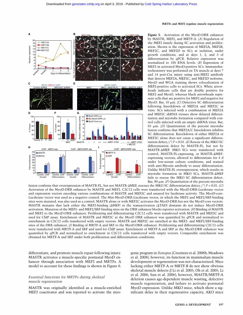
MASTR is a potent coactivator of MEF2 transcriptionalactivity (Creemers et al. 2006b); however, the expressionand potential function of MEF2 transcription factors inSCs remain uncharacterized. To investigate the mecha-nism by which MASTR regulates MyoD expression, wefirst assayed the expression of MEF2 family members insorted, proliferating, and differentiating SCs by qPCR.MEF2A expression was detected at low levels in sortedand proliferating SCs and peaked at day 2 of differentia-tion; enriched MEF2C expression was detected in sorted,proliferating, and differentiating SCs (Fig. 5A). Theseresults confirmed previous microarray results (data notshown) and showed that MEF2A and MEF2C, but notMEF2B or MEF2D, are enriched and up-regulated duringSC activation and differentiation in vitro (Fig. 5A). MEF2A/Care therefore expressed in a manner similar to MASTR inproliferating and differentiating SCs. We then colabeledMEF2 and MyoD in the mouse TA muscle at days 7 and14 post-Ctx injury using a MEF2 antibody that detectsMEF2A, MEF2C, and MEF2D (Haberland et al. 2007). In-terestingly, MEF2 was expressed in almost all MyoD-expressing SCs at days 7 and 14 post-injury (Fig. 5B). Byday 14 post-injury, MEF2 expression was maintained ina subset of interstitial cells that were negative for MyoDand in centrally nucleated, regenerating myofibers (Fig. 5B).These expression studies suggest that MEF2A/C couldcooperate with MASTR to regulate muscle regeneration.
To examine the function of MEF2 in SCs, we knockeddown MEF2A and MEF2C in cultured SCs using shRNAlentivirus and assessed SC differentiation by myosin (MF20)immunohistochemistry. Differentiation and fusion weredefective in SCs infected with MEF2A/C-shRNA com-pared with control shRNA-infected cells (Fig. 5C,D).However, knockdown of MEF2A or MEF2C alone didnot impair SC differentiation, suggesting a redundant rolefor the MEF2 family members in differentiating SCs (Fig.5D). These results demonstrate that MEF2A and MEF2Care expressed in SCs and are required for SC differentiationand suggest that MASTR and MEF2A/C cooperatively reg-ulate MyoD expression and muscle regeneration.
To investigate the requirement for MASTR/MEF2 in-teraction in SC differentiation, we attempted to rescuethe MKO phenotype by overexpressing full-length MASTR(FL) and a MASTR mutant lacking the MEF2-bindingdomain (DMEF) in MKO SCs. MASTR-FL restored MKOSC differentiation, whereas MASTR-DMEF failed to rescuethe MKO differentiation defect (Fig. 5E,F). These findingssuggest that the MKO phenotype is mediated by the as-sociation of MASTR with MEF2.
MASTR and MEF2 bind and activatethe MyoD DRR enhancer
MyoD transcription is controlled by an embryonic coreenhancer and an adult DRR enhancer. Activation of theDRR enhancer at P14 is concomitant with inactivation ofthe core enhancer and correlates with the time at whichMyoD expression decreases in MKO and dKO muscle(Fig. 3E; Kucharczuk et al. 1999; Chen et al. 2001, 2002).Interestingly, the DRR enhancer harbors one canonicalMEF2-binding site and a MEF2/SRF hybrid site (L’Honoreet al. 2007), suggesting that MASTR and MEF2 coregulatethe DRR enhancer to induce MyoD expression. To testthis hypothesis, we performed luciferase assays in C2C12myoblasts using MyoD-DRR and MyoD-core reporter con-structs. MASTR alone or with MEF2 activated the DRRbut not the core enhancer in a dose-dependent fashion(Fig. 5G). MASTR mutants lacking either the MEF2-bindingdomain or the transactivation domain failed to activatethe MyoD-DRR reporter, suggesting that the partnershipbetween MASTR and MEF2 is required for MyoD activa-tion. Moreover, mutating the MEF2-binding site and theMEF2/SRF hybrid sites blocked reporter activation, sug-gesting that MASTR and MEF2 directly regulate DRR ac-tivation (Fig. 5G). Chromatin immunoprecipitation (ChIP)in proliferating C2C12 myoblasts confirmed that MASTRand MEF2 bind the MEF2 and MEF2/SRF hybrid sites of thenative MyoD DRR enhancer (Fig. 5H). Strikingly, binding ofMASTR and MEF2 to the MyoD DRR enhancer significantlyincreased in differentiating C2C12 cells (Fig. 5H).
Since MASTR and MRTF-A exert their functions throughassociation with MEF2 and SRF, respectively, these find-ings suggested that the MASTR/MEF2 and MRTF/SRFpathways converge to direct skeletal muscle regenerationby restricting proliferation and promoting differentiationof SCs. Indeed, ChIP assays showed binding of MRTF-Aand SRF to the MEF2/SRF hybrid site of the DRR enhancer(Fig. 5I). We conclude that the MASTR/MEF2 complex isrecruited to the MyoD DRR enhancer with MRTF-A andSRF and activates MyoD expression in differentiating SCs.
Discussion
This study identifies members of the Myocardin and MEF2transcription factor families as key components of thegene regulatory network that drives MyoD expression andmuscle regeneration in adult mice. MASTR and MRTF-Aare up-regulated in SCs in response to injury and musculardystrophy. Global and SC-specific loss-of-function studiesin mice demonstrate that MASTR and MRTF-A regulateMyoD expression, direct SCs to exit the cell cycle and
Mokalled et al.
196 GENES & DEVELOPMENT
Cold Spring Harbor Laboratory Press on April 3, 2019 - Published by genesdev.cshlp.orgDownloaded from
differentiate, and promote muscle repair following injury.MASTR activates a muscle-specific postnatal MyoD en-hancer through association with MEF2 and MRTFs. Amodel to account for these findings is shown in Figure 6.
Essential functions for MRTFs during skeletalmuscle regeneration
MASTR was originally identified as a muscle-enrichedMEF2 coactivator and was reported to activate the myo-
genic program in Xenopus (Creemers et al. 2006b; Meadowset al. 2008); however, its function in mammalian muscledevelopment or regeneration was not characterized. Micelacking either MRTF-A or MRTF-B do not show obviousskeletal muscle defects (J Li et al. 2005; Oh et al. 2005; Liet al. 2006; Sun et al. 2006); however, MASTR/MRTF-Adeletion causes age-dependent muscle wasting, defectivemuscle regeneration, and failure to activate postnatalMyoD expression. Unlike MKO mice, which show a sig-nificant delay in their regenerative capacity, AKO mice
Figure 5. Activation of the MyoD-DRR enhancerby MASTR, MEF2, and MRTF-A. (A) Regulation ofthe MEF2 family during SC activation and prolifer-ation. Shown is the expression of MEF2A, MEF2B,MEF2C, and MEF2D in SCs at isolation, undergrowth conditions, and at days 1, 2, and 3 ofdifferentiation by qPCR. Relative expression wasnormalized to 18S RNA levels. (B) Expression ofMEF2 in activated MyoD-positive SCs. Immunohis-tochemistry was performed on TA muscle at days 7and 14 post-Ctx injury using anti-MEF2 antibodythat detects MEF2A, MEF2C, and MEF2D isoforms.MyoD and WGA staining shows colocalization ofMEF2-positive cells to activated SCs. White arrow-heads indicate cells that are double positive forMEF2 and MyoD, whereas black arrowheads repre-sent cells that are positive for MEF2 and negative forMyoD. Bar, 10 mm. (C) Defective SC differentiationfollowing knockdown of MEF2A and MEF2C invitro. SCs infected with a combination of MEF2Aand MEF2C shRNA viruses show delayed differen-tiation and myotube formation compared with con-trol cells infected with an empty shRNA virus. Bar,65 mm. (D) Quantitation of the percent myotubefusion confirms that MEF2A/C knockdown inhibitsSC differentiation. Knockdown of either MEF2A orMEF2C alone does not cause a significant differen-tiation defect; (*) P < 0.01. (E) Rescue of the MKO SCdifferentiation defect by MASTR-FL, but not byMASTR-DMEF. MKO SCs were transfected withcontrol, MASTR-FL-expressing, or MASTR-DMEF-expressing vectors; allowed to differentiate for 4 dunder low-serum culture conditions; and stainedwith anti-Myosin antibody to assay differentiation.Unlike MASTR-FL overexpression, which results inmyotube formation in MKO SCs, MASTR-DMEFfails to rescue the MKO SC differentiation defect.Bar, 90 mm. (F) Quantitation of the percent myotube
fusion confirms that overexpression of MASTR-FL, but not MASTR-DMEF, rescues the MKO SC differentiation defect; (*) P < 0.01. (G)Activation of the MyoD-DRR enhancer by MASTR and MEF2. C2C12 cells were transfected with the MyoD-DRR-Luciferase vectorand expression vectors encoding various combinations of MASTR and MEF2C and assayed for luciferase activity. The MyoD-core-Luciferase vector was used as a negative control. The Mut-MyoD-DRR-Luciferase vector, in which the MEF2 and MEF2/SRF-bindingsites were mutated, was also used as a control. MASTR alone or with MEF2C activates the MyoD-DRR but not the MyoD-core vectors.MASTR mutants that lack either the MEF2-binding (DMEF) or the transactivation (DTAD) domains do not induce MyoD-DRRactivation. Mutation of the MEF2- and MEF2/SRF-binding sites on the DRR enhancer blocks reporter activation. (H) Binding of MASTRand MEF2 to the MyoD-DRR enhancer. Proliferating and differentiating C2C12 cells were transfected with MASTR and MEF2C andused for ChIP assay. Enrichment of MASTR and MEF2C at the MyoD DRR enhancer was quantified by qPCR and normalized toenrichment in C2C12 cells transfected with empty vectors. MASTR and MEF2C are enriched at the MEF2- and MEF2/SRF-bindingsites of the DRR enhancer. (I) Binding of MRTF-A and SRF to the MyoD-DRR enhancer. Proliferating and differentiating C2C12 cellswere transfected with MRTF-A and SRF and used for ChIP assay. Enrichment of MRTF-A and SRF at the MyoD-DRR enhancer wasquantified by qPCR and normalized to enrichment in C2C12 cells transfected with empty vectors. Comparable enrichment wasobtained for MRTF-A and SRF under both proliferation and differentiation conditions.
MRTFs and MEF2 regulate muscle regeneration
GENES & DEVELOPMENT 197
Cold Spring Harbor Laboratory Press on April 3, 2019 - Published by genesdev.cshlp.orgDownloaded from

regenerate normally after injury, suggesting that MASTRand MRTF-A perform cooperative and nonredundant func-tions during skeletal muscle regeneration. The lack of aregeneration defect in AKO mice could be explained byredundancy of MRTF-A and MRTF-B in SCs. Indeed,MRTF-A and MRTF-B share the highest homology amongMRTF family members and perform interchangeable func-tions in multiple cell and tissue contexts (Cen et al. 2003;Mokalled et al. 2010). In this regard, brain- and heart-specific deletions of both MRTFs result in multiple devel-opmental neuronal and cardiac defects and lethality be-fore adulthood, while the brain and heart develop normallyin MRTF-A or MRTF-B single-knockout mice (Mokalledet al. 2010; EN Olson and MH Mokalled, unpubl.).Although MRTF-B is expressed at low levels in regener-ating and dystrophic muscle, this level of expression maybe sufficient to compensate for the absence of MRTF-A.This study is the first to demonstrate a function for theMyocardin family of transcription factors in SC differen-tiation and muscle regeneration.
Regulation of the MyoD DRR enhancerby MRTFs and MEF2
Our study adds the Myocardin and MEF2 families of trans-cription factors to the multiple factors that drive postnatalMyoD expression in SCs to regulate adult muscle regener-ation. Similar to our mutant mice, MyoD-null SCs prolifer-ate excessively, fail to differentiate, and exhibit a molecularsignature reflective of a proliferative state (Megeney et al.1996; Sabourin et al. 1999; White et al. 2000). In mice as wellas in humans, myogenic expression of MyoD is linked to theactivity of two muscle-specific enhancers: a 300-base-pair(bp) core and a 750-bp DRR enhancer (Goldhamer et al.1992, 1995; Tapscott et al. 1992; Asakura et al. 1995; Chenand Goldhamer 1999; Chen et al. 2001, 2002). The coreenhancer controls MyoD expression during developmentbut is inactive after birth (Faerman et al. 1995; Goldhameret al. 1995; Chen and Goldhamer 1999; Chen et al. 2001),
whereas the DRR enhancer is dispensable during develop-ment but is essential in mature muscle and activated SCs(Chen et al. 2002; L’Honore et al. 2003). Interestingly, thelevel of MyoD expression is comparable between wild-type and dKO muscle at birth, but undetectable at 2 wk ofage, suggesting that MASTR and MRTF-A control post-natal MyoD expression in SCs via the DRR enhancer. Thestriking temporal regulation of MyoD in MKO and dKOmice parallels that of MyoD activation in postnatal mus-cle and supports our finding that MRTFs regulate MyoDDRR activation in SCs. Our findings demonstrate thatMASTR binds and activates the MyoD DRR enhancerthrough association with MEF2 and MRTF-A.
Novel functions for MEF2 transcriptionfactors in muscle regeneration
MEF2 has been implicated in multiple aspects of mus-cle development (Potthoff and Olson 2007; Olson andNordheim 2010), yet the functions of MEF2 in either SCdifferentiation or muscle regeneration are unknown. Al-though Drosophila MEF2 was shown to activate the MyoDortholog nautilus, these findings were not extended tomammalian skeletal muscle (Sandmann et al. 2006). Here,we demonstrate that MASTR coactivates MEF2 to driveSC differentiation by showing that (1) MEF2A/C are up-regulated with MASTR in differentiating SCs and arerequired for SC differentiation, (2) the MASTR/MEF2Ccomplex binds to and activates the adult MyoD DRR en-hancer in myoblasts, and (3) the MASTR-DMEF mutantfails to rescue the MKO SC phenotype. Interestingly,MEF2A and MEF2C are dysregulated in patients withmyotonic dystrophy (Bachinski et al. 2010), suggesting theirinvolvement in skeletal muscle regeneration in humans.
Unlike MASTR, which coactivates MEF2, MRTF-A isan SRF activator. SRF binds to the MEF2/SRF hybrid siteof the MyoD DRR enhancer to regulate MyoD expressionduring muscle regeneration (L’Honore et al. 2003). In contrastto transgenic mice overexpressing a DRR-b-galactosidasereporter, which activate b-galactosidase expression fol-lowing muscle injury, mutation of the MEF2/SRF sitefails to activate DRR reporter expression in injured mice(L’Honore et al. 2003). Our findings demonstrate that MEF2and SRF bind to the MEF2/SRF hybrid site and confirmthe relevance of this site for activating MyoD expressionduring skeletal muscle regeneration. In addition, SRF selec-tively regulates the levels of MyoD in myoblasts withoutaffecting the levels of Myf5 (Gauthier-Rouviere et al. 1996;Soulez et al. 1996; Carnac et al. 1998). Interestingly, Myf5expression was also unchanged in our dKO mice (Supple-mental Fig. S4B), suggesting that the regulatory functionsof MASTR and MRTF-A are specific to MyoD, but not toall muscle regulatory factors.
By unveiling novel functions for the Myocardin familyof transcription factors in SC differentiation and muscleregeneration, this study suggests that MRTFs could act asdirect sensors of the SC niche and may present a potentialtherapeutic target for muscle regenerative therapy. Dy-namic signaling from the SC niche dictates SC activationin settings of homeostasis or injury, respectively (Gopinath
Figure 6. Schematic model for MASTR, MEF2, and MRTF-Afunction during SC differentiation. MASTR coactivates MEF2and cooperates with MRTF-A to regulate MyoD expression, SCdifferentiation, and skeletal muscle regeneration in response toinjury.
Mokalled et al.
198 GENES & DEVELOPMENT
Cold Spring Harbor Laboratory Press on April 3, 2019 - Published by genesdev.cshlp.orgDownloaded from

and Rando 2008; Kuang et al. 2008). MRTFs couple ex-tracellular signaling to gene transcription and could po-tentially sense extracellular alterations of the SC micro-environment. For example, MRTF-A and MRTF-B aresensitive to extracellular Rho signaling, which drives theirnuclear translocation and consequent transcriptional acti-vation (Miralles et al. 2003; Posern and Treisman 2006;Olson and Nordheim 2010). Interestingly, inhibition ofRhoA in myoblast cultures, whether by dominant-negativeexpression or antagonist treatment, specifically alters MyoDbut not Myf5 expression (Carnac et al. 1998). This effecton MyoD expression is specific to RhoA, but not to Rac orCdc42, and requires functional SRF (Carnac et al. 1998).Moreover, concomitant with age-dependent changes withinthe SC systemic microenvironment (Hall et al. 2010;Conboy et al. 2005; Conboy and Rando 2005), MRTF ex-pression levels were reported to decrease with aging(Sakuma et al. 2008), suggesting dynamic cross-talk be-tween MRTFs and the SC niche. Because MRTFs linkextracellular signaling to changes in gene regulation andare required for MyoD expression and SC differentiation,it will be of interest to examine whether perturbation ofthe SC niche affects MRTF signaling and whether manip-ulation of MRTF signaling affects muscle regeneration.
Materials and methods
Mouse lines
The MKO allele was generated using homologous recombinationin embryonic stem (ES) cells. The pGKNEO-F2L2DTA vector,which contains a neomycin resistance gene flanked by FRT andloxP sites and a diphtheria toxin gene cassette, was used forMASTR targeting. The 59, knockout, and 39 arms of the targetingconstruct were generated by high-fidelity PCR amplification (RocheExpand High-Fidelity Long Template). The targeting vector waslinearized with PvuI and electroporated into 129SvEv-derived EScells. Isolated ES cell clones were analyzed for incorporation ofthe 59 and 39 loxP sites by Southern blotting using 59 and 39 probes.Clones with the targeted MASTR allele were injected into 3.5-dC57BL/6 blastocysts, and the resulting chimeras were crossed toC57BL/6 females to achieve germline transmission of the targeted(MASTRneo-loxP) allele. The MASTRneo-loxP mice were crossed toFlpe transgenic mice and then to CAG-Cre transgenic mice to ob-tain the MKO allele. MKO mice were then crossed to AKO mice(Li et al. 2006) to generate dKO mice. MKO mice were also crossedto mdx mice to generate MKO;mdx mice. MASTRFloxed micewere crossed to Pax7-Cre-ERT2 mice (Lepper et al. 2009) to gen-erate the SC-specific cMKO allele.
SC isolation
Mononuclear cells were isolated from adult mouse hindlimbmuscles following muscle digestion and myofiber dissociation(Sherwood et al. 2004). Eight-week-old to 12-wk-old mice wereused for all experiments. CD34+/Sca1�/CD31�/CD45� SCs werethen sorted from mononuclear cells with a yield of ;4%. Gatingof the SC population was determined using a negative controlsample stained with phycoerythrin (PE)-conjugated antibodiesfor Sca1/CD31/CD45, and a positive control sample stained withAlexa-488-conjugated anti-CD34 antibody. SCs were then main-tained under growth conditions (Ham F20 with 20% fetal calfserum, supplemented with 5 ng/mL bFGF) or differentiation con-ditions (DMEM1 with 2% horse serum).
BrdU labeling
SC cultures were maintained under growth conditions. Wild-type,MKO, and dKO SCs were labeled with BrdU at a final concentra-tion of 10 mM for 1 h at days 2, 3, and 4 of culture. BrdU im-munohistochemistry was then used to assay BrdU incorporation.
Chemical muscle injury
Chemical injury experiments were performed using Ctx and BaCl2(Yan et al. 2003; Cornelison et al. 2004). Briefly, mice wereanesthetized with 5% Avertin. Left TA muscles were injectedwith 100 mL of 10 mM Ctx or 50 mL of 1.2% BaCl2 in sterilesaline. Mice were allowed to recover for 3, 7, or 14 d post-injury.Injected and contralateral TA muscles were harvested for sec-tioning and staining.
Histology and immunohistochemistry
Mice were anesthetized and transcardially perfused with PBS,followed by 4% paraformaldehyde prior to muscle dissection. TAmuscles were then post-fixed in 4% paraformaldehyde for 48 h,embedded in paraffin, and sectioned. Sections were stained withH&E or WGA using standard procedures. Immunohistochemis-try using monoclonal anti-BrdU (Roche), monoclonal anti-MyoD(BD Biosciences), monoclonal anti-Desmin (DAKO), monoclonalanti-MF20 (Hybridoma Bank), polyclonal anti-PCNA (SantaCruz Biotechnology), and polyclonal anti-MEF2-C21 (SantaCruz Biotechnology) antibodies was performed using standardprotocols.
Quantitative real-time PCR
Total RNA was purified from the indicated tissues or cells usingTRIzol reagent according to the manufacturer’s instructions(Invitrogen). For RT–PCR, total RNA was used as a templatefor RT using random hexamer primers. qPCR was performedusing TaqMan probes (ABI).
Luciferase assays
The MyoD-Core and MyoD-DRR luciferase constructs werecloned (Tapscott et al. 1992). C2C12 cells, plated in 24-wellplates (5 3 104 cells per well), were transfected with 150 ng ofluciferase construct, 20 ng of pCMV-LacZ, and a total of 250 ng ofexpression plasmids encoding MASTR, MEF2C, or an empty con-trol vector. FuGENE 6 reagent (1.4 mL) (Roche) was complexedwith the DNA and added to freshly plated C2C12 cells for 12 h.Cells were harvested 48 h after transfection in 150 mL of passivelysate buffer (Promega). Twenty microliters of cell lysate wasused for luciferase or b-galactosidase assays. Luciferase activitywas normalized to b-galactosidase levels and to luciferase ac-tivity in C2C12 cells transfected with a mix of reporter genes andempty vector.
ChIP
ChIP assays were performed using EZ ChIP (Millipore) followingthe manufacturer’s instructions. Briefly, C2C12 cells were trans-fected with either a negative control empty vector or with Flag-MASTR, myc-MEF2C, myc-MRTF-A, or Flag-SRF vectors aspreviously described. Medium was changed 12 h after transfectioninto either growth or differentiation medium and collected 24 hlater for ChIP. For each immunoprecipitation, chromatin from 3 3
106 cells was cross-linked with 1% formaldehyde and sonicatedinto 400- to 1000-bp fragments. Ten percent of the chromatin
MRTFs and MEF2 regulate muscle regeneration
GENES & DEVELOPMENT 199
Cold Spring Harbor Laboratory Press on April 3, 2019 - Published by genesdev.cshlp.orgDownloaded from

from the control, MASTR, MEF2C, MRTF-A, and SRF sampleswas used to purify the input DNA. The other 90% of the chro-matin was immunoprecipitated with anti-Flag or anti-myc aga-rose affinity beads (Sigma). DNA was purified from the ChIP andinput samples and was analyzed by qPCR using primers spanningthe MEF2-binding or MEF2/SRF hybrid site of the MyoD-DRRenhancer. Fold enrichment for each of the control, MASTR,MEF2C, and MRTF-A ChIP samples was normalized first to theenrichment in the corresponding input sample and then to theenrichment in the control sample transfected with empty vector.
Statistical analysis
Results are expressed as means 6 SEM. Unpaired two-tailedStudent’s t-test with Welch correction was performed to deter-mine statistical significance between groups. P-values of <0.01were considered significant.
Acknowledgments
We are grateful to Chen-Ming Fan (Carnegie Institute, Maryland)for providing the Pax7-Cre-ERT2 mice, Jose Cabrera for graphics,and Sasha Qi and Lil Sutherland for technical help. This workwas supported by grants from the NIH and the Robert A. Welchfoundation to E.N.O.
References
Asakura A, Lyons GE, Tapscott SJ. 1995. The regulation ofMyoD gene expression: Conserved elements mediate expres-sion in embryonic axial muscle. Dev Biol 171: 386–398.
Asakura A, Hirai H, Kablar B, Morita S, Ishibashi J, Piras BA,Christ AJ, Verma M, Vineretsky KA, Rudnicki MA. 2007.Increased survival of muscle stem cells lacking the MyoDgene after transplantation into regenerating skeletal muscle.Proc Natl Acad Sci 104: 16552–16557.
Bachinski LL, Sirito M, Bohme M, Baggerly KA, Udd B, Krahe R.2010. Altered MEF2 isoforms in myotonic dystrophy andother neuromuscular disorders. Muscle Nerve 42: 856–863.
Banks GB, Chamberlain JS. 2008. The value of mammalianmodels for duchenne muscular dystrophy in developingtherapeutic strategies. Curr Top Dev Biol 84: 431–453.
Beauchamp JR, Heslop L, Yu DS, Tajbakhsh S, Kelly RG, WernigA, Buckingham ME, Partridge TA, Zammit PS. 2000. Expres-sion of CD34 and Myf5 defines the majority of quiescent adultskeletal muscle satellite cells. J Cell Biol 151: 1221–1234.
Biressi S, Rando TA. 2010. Heterogeneity in the muscle satellitecell population. Semin Cell Dev Biol 21: 845–854.
Buckingham, M. 1994. Muscle differentiation. Which myogenicfactors make muscle? Curr Biol 4: 61–63.
Buckingham M, Relaix F. 2007. The role of Pax genes in thedevelopment of tissues and organs: Pax3 and Pax7 regulatemuscle progenitor cell functions. Annu Rev Cell Dev Biol
23: 645–673.Carnac G, Primig M, Kitzmann M, Chafey P, Tuil D, Lamb N,
Fernandez A. 1998. RhoA GTPase and serum response factorcontrol selectively the expression of MyoD without affectingMyf5 in mouse myoblasts. Mol Biol Cell 9: 1891–1902.
Cen B, Selvaraj A, Burgess RC, Hitzler JK, Ma Z, Morris SW,Prywes R. 2003. Megakaryoblastic leukemia 1, a potenttranscriptional coactivator for serum response factor (SRF),is required for serum induction of SRF target genes. Mol CellBiol 23: 6597–6608.
Chen JC, Goldhamer DJ. 1999. Transcriptional mechanismsregulating MyoD expression in the mouse. Cell Tissue Res
296: 213–219.
Chen JC, Love CM, Goldhamer DJ. 2001. Two upstreamenhancers collaborate to regulate the spatial patterning andtiming of MyoD transcription during mouse development.Dev Dyn 221: 274–288.
Chen JC, Ramachandran R, Goldhamer DJ. 2002. Essential andredundant functions of the MyoD distal regulatory regionrevealed by targeted mutagenesis. Dev Biol 245: 213–223.
Conboy IM, Rando TA. 2005. Aging, stem cells and tissueregeneration: Lessons from muscle. Cell Cycle 4: 407–410.
Conboy IM, Conboy MJ, Wagers AJ, Girma ER, Weissman IL,Rando TA. 2005. Rejuvenation of aged progenitor cells byexposure to a young systemic environment. Nature 433:760–764.
Cornelison DD, Olwin BB, Rudnicki MA, Wold BJ. 2000.MyoD�/� satellite cells in single-fiber culture are differenti-ation defective and MRF4 deficient. Dev Biol 224: 122–137.
Cornelison DD, Wilcox-Adelman SA, Goetinck PF, Rauvala H,Rapraeger AC, Olwin BB. 2004. Essential and separable rolesfor Syndecan-3 and Syndecan-4 in skeletal muscle develop-ment and regeneration. Genes Dev 18: 2231–2236.
Creemers EE, Sutherland LB, McAnally J, Richardson JA, OlsonEN. 2006a. Myocardin is a direct transcriptional target ofMef2, Tead and Foxo proteins during cardiovascular devel-opment. Development 133: 4245–4256.
Creemers EE, Sutherland LB, Oh J, Barbosa AC, Olson EN.2006b. Coactivation of MEF2 by the SAP domain proteinsmyocardin and MASTR. Mol Cell 23: 83–96.
Faerman A, Goldhamer DJ, Puzis R, Emerson CP Jr, Shani M.1995. The distal human myoD enhancer sequences directunique muscle-specific patterns of lacZ expression duringmouse development. Dev Biol 171: 27–38.
Gauthier-Rouviere C, Vandromme M, Tuil D, Lautredou N,Morris M, Soulez M, Kahn A, Fernandez A, Lamb N. 1996.Expression and activity of serum response factor is requiredfor expression of the muscle-determining factor MyoD inboth dividing and differentiating mouse C2C12 myoblasts.Mol Biol Cell 7: 719–729.
Goldhamer DJ, Faerman A, Shani M, Emerson CP Jr. 1992.Regulatory elements that control the lineage-specific expres-sion of myoD. Science 256: 538–542.
Goldhamer DJ, Brunk BP, Faerman A, King A, Shani M, EmersonCP Jr. 1995. Embryonic activation of the myoD gene isregulated by a highly conserved distal control element.Development 121: 637–649.
Gopinath SD, Rando TA. 2008. Stem cell review series: Aging ofthe skeletal muscle stem cell niche. Aging Cell 7: 590–598.
Goyenvalle A, Seto JT, Davies KE, Chamberlain J. 2011. Ther-apeutic approaches to muscular dystrophy. Hum Mol Genet
20: R69–R78. doi: 10.1093/hmg/ddr105.Haberland M, Arnold MA, McAnally J, Phan D, Kim Y, Olson
EN. 2007. Regulation of HDAC9 gene expression by MEF2establishes a negative-feedback loop in the transcriptionalcircuitry of muscle differentiation. Mol Cell Biol 27: 518–525.
Hall JK, Banks GB, Chamberlain JS, Olwin BB. 2010. Preven-tion of muscle aging by myofiber-associated satellite celltransplantation. Sci Transl Med 2: 57ra83. doi: 10.1126/scitranslmed.3001081.
Hosoyama T, Nishijo K, Prajapati SI, Li G, Keller C. 2011. Rb1gene inactivation expands satellite cell and postnatal myo-blast pools. J Biol Chem 286: 19556–19564.
Ishibashi J, Perry RL, Asakura A, Rudnicki MA. 2005. MyoDinduces myogenic differentiation through cooperation of itsNH2- and COOH-terminal regions. J Cell Biol 171: 471–482.
Kang JS, Krauss RS. 2010. Muscle stem cells in developmentaland regenerative myogenesis. Curr Opin Clin Nutr MetabCare 13: 243–248.
Mokalled et al.
200 GENES & DEVELOPMENT
Cold Spring Harbor Laboratory Press on April 3, 2019 - Published by genesdev.cshlp.orgDownloaded from

Kuang S, Gillespie MA, Rudnicki MA. 2008. Niche regulation ofmuscle satellite cell self-renewal and differentiation. CellStem Cell 2: 22–31.
Kucharczuk KL, Love CM, Dougherty NM, Goldhamer DJ.1999. Fine-scale transgenic mapping of the MyoD coreenhancer: MyoD is regulated by distinct but overlappingmechanisms in myotomal and non-myotomal muscle line-ages. Development 126: 1957–1965.
Le Grand F, Rudnicki M. 2007. Satellite and stem cells inmuscle growth and repair. Development 134: 3953–3957.
Lepper C, Conway SJ, Fan CM. 2009. Adult satellite cells andembryonic muscle progenitors have distinct genetic require-ments. Nature 460: 627–631.
L’Honore A, Lamb NJ, Vandromme M, Turowski P, Carnac G,Fernandez A. 2003. MyoD distal regulatory region containsan SRF binding CArG element required for MyoD expressionin skeletal myoblasts and during muscle regeneration. Mol
Biol Cell 14: 2151–2162.L’Honore A, Rana V, Arsic N, Franckhauser C, Lamb NJ,
Fernandez A. 2007. Identification of a new hybrid serumresponse factor and myocyte enhancer factor 2-bindingelement in MyoD enhancer required for MyoD expressionduring myogenesis. Mol Biol Cell 18: 1992–2001.
Li J, Zhu X, Chen M, Cheng L, Zhou D, Lu MM, Du K, EpsteinJA, Parmacek MS. 2005. Myocardin-related transcriptionfactor B is required in cardiac neural crest for smooth muscledifferentiation and cardiovascular development. Proc Natl
Acad Sci 102: 8916–8921.Li S, Czubryt MP, McAnally J, Bassel-Duby R, Richardson JA,
Wiebel FF, Nordheim A, Olson EN. 2005. Requirement forserum response factor for skeletal muscle growth and mat-uration revealed by tissue-specific gene deletion in mice.Proc Natl Acad Sci 102: 1082–1087.
Li S, Chang S, Qi X, Richardson JA, Olson EN. 2006. Re-quirement of a myocardin-related transcription factor fordevelopment of mammary myoepithelial cells. Mol Cell Biol
26: 5797–5808.McGeachie JK, Grounds MD, Partridge TA, Morgan JE. 1993.
Age-related changes in replication of myogenic cells in mdxmice: Quantitative autoradiographic studies. J Neurol Sci
119: 169–179.Meadows SM, Warkman AS, Salanga MC, Small EM, Krieg PA.
2008. The myocardin-related transcription factor, MASTR,cooperates with MyoD to activate skeletal muscle geneexpression. Proc Natl Acad Sci 105: 1545–1550.
Megeney LA, Kablar B, Garrett K, Anderson JE, Rudnicki MA.1996. MyoD is required for myogenic stem cell function inadult skeletal muscle. Genes Dev 10: 1173–1183.
Miralles F, Posern G, Zaromytidou AI, Treisman R. 2003. Actindynamics control SRF activity by regulation of its coactiva-tor MAL. Cell 113: 329–342.
Mokalled MH, Johnson A, Kim Y, Oh J, Olson EN. 2010.Myocardin-related transcription factors regulate the Cdk5/Pctaire1 kinase cascade to control neurite outgrowth, neu-ronal migration and brain development. Development 137:2365–2374.
Montarras D, Morgan J, Collins C, Relaix F, Zaffran S, CumanoA, Partridge T, Buckingham M. 2005. Direct isolation ofsatellite cells for skeletal muscle regeneration. Science 309:2064–2067.
Musaro A, Barberi L. 2010. Isolation and culture of mousesatellite cells. Methods Mol Biol 633: 101–111.
Oh J, Richardson JA, Olson EN. 2005. Requirement of myocar-din-related transcription factor-B for remodeling of branchialarch arteries and smooth muscle differentiation. Proc NatlAcad Sci 102: 15122–15127.
Olguin HC, Yang Z, Tapscott SJ, Olwin BB. 2007. Reciprocalinhibition between Pax7 and muscle regulatory factorsmodulates myogenic cell fate determination. J Cell Biol
177: 769–779.Olson EN, Klein WH. 1994. bHLH factors in muscle develop-
ment: Dead lines and commitments, what to leave in andwhat to leave out. Genes Dev 8: 1–8.
Olson EN, Nordheim A. 2010. Linking actin dynamics and genetranscription to drive cellular motile functions. Nat Rev MolCell Biol 11: 353–365.
Parmacek MS. 2007. Myocardin-related transcription factors:Critical coactivators regulating cardiovascular developmentand adaptation. Circ Res 100: 633–644.
Pipes GC, Creemers EE, Olson EN. 2006. The myocardin familyof transcriptional coactivators: Versatile regulators of cellgrowth, migration, and myogenesis. Genes Dev 20: 1545–1556.
Posern G, Treisman R. 2006. Actin’ together: Serum responsefactor, its cofactors and the link to signal transduction.Trends Cell Biol 16: 588–596.
Potthoff MJ, Olson EN. 2007. MEF2: A central regulator ofdiverse developmental programs. Development 134: 4131–4140.
Price FD, Kuroda K, Rudnicki MA. 2007. Stem cell basedtherapies to treat muscular dystrophy. Biochim Biophys
Acta 1772: 272–283.Rudnicki MA, Schnegelsberg PN, Stead RH, Braun T, Arnold
HH, Jaenisch R. 1993. MyoD or Myf-5 is required for theformation of skeletal muscle. Cell 75: 1351–1359.
Sabourin LA, Girgis-Gabardo A, Seale P, Asakura A, RudnickiMA. 1999. Reduced differentiation potential of primaryMyoD�/�myogenic cells derived from adult skeletal muscle.J Cell Biol 144: 631–643.
Sacco A, Doyonnas R, Kraft P, Vitorovic S, Blau HM. 2008. Self-renewal and expansion of single transplanted muscle stemcells. Nature 456: 502–506.
Sakuma K, Akiho M, Nakashima H, Akima H, Yasuhara M.2008. Age-related reductions in expression of serum responsefactor and myocardin-related transcription factor A in mouseskeletal muscles. Biochim Biophys Acta 1782: 453–461.
Sandmann T, Jensen LJ, Jakobsen JS, Karzynski MM, EichenlaubMP, Bork P, Furlong EE. 2006. A temporal map of transcrip-tion factor activity: mef2 directly regulates target genes at allstages of muscle development. Dev Cell 10: 797–807.
Schuierer MM, Mann CJ, Bildsoe H, Huxley C, Hughes SM.2005. Analyses of the differentiation potential of satellitecells from myoD�/�, mdx, and PMP22 C22 mice. Bmc
Musculoskel Dis 6: 15. doi: 10.1186/1471-2474-6-15.Seale P, Ishibashi J, Holterman C, Rudnicki MA. 2004. Muscle
satellite cell-specific genes identified by genetic profiling ofMyoD-deficient myogenic cell. Dev Biol 275: 287–300.
Sherwood RI, Christensen JL, Conboy IM, Conboy MJ, RandoTA, Weissman IL, Wagers AJ. 2004. Isolation of adult mousemyogenic progenitors: Functional heterogeneity of cellswithin and engrafting skeletal muscle. Cell 119: 543–554.
Small EM, Thatcher JE, Sutherland LB, Kinoshita H, Gerard RD,Richardson JA, Dimaio JM, Sadek H, Kuwahara K, Olson EN.2010. Myocardin-related transcription factor-a controls myo-fibroblast activation and fibrosis in response to myocardialinfarction. Circ Res 107: 294–304.
Soulez M, Rouviere CG, Chafey P, Hentzen D, Vandromme M,Lautredou N, Lamb N, Kahn A, Tuil D. 1996. Growth anddifferentiation of C2 myogenic cells are dependent on serumresponse factor. Mol Cell Biol 16: 6065–6074.
Sun Y, Boyd K, Xu W, Ma J, Jackson CW, Fu A, Shillingford JM,Robinson GW, Hennighausen L, Hitzler JK, et al. 2006. Acute
MRTFs and MEF2 regulate muscle regeneration
GENES & DEVELOPMENT 201
Cold Spring Harbor Laboratory Press on April 3, 2019 - Published by genesdev.cshlp.orgDownloaded from

myeloid leukemia-associated Mkl1 (Mrtf-a) is a key regulatorof mammary gland function. Mol Cell Biol 26: 5809–5826.
Tapscott SJ. 2005. The circuitry of a master switch: Myod andthe regulation of skeletal muscle gene transcription. De-
velopment 132: 2685–2695.Tapscott SJ, Lassar AB, Weintraub H. 1992. A novel myoblast
enhancer element mediates MyoD transcription. Mol Cell
Biol 12: 4994–5003.Ten Broek RW, Grefte S, Von den Hoff JW. 2010. Regulatory
factors and cell populations involved in skeletal muscleregeneration. J Cell Physiol 224: 7–16.
Tomczak KK, Marinescu VD, Ramoni MF, Sanoudou D,Montanaro F, Han M, Kunkel LM, Kohane IS, Beggs AH.2004. Expression profiling and identification of novel genesinvolved in myogenic differentiation. FASEB J 18: 403–405.
Tsivitse S. 2010. Notch and Wnt signaling, physiological stimuliand postnatal myogenesis. Int J Biol Sci 6: 268–281.
Tuan RS. 2011. Role of adult stem/progenitor cells in osseointe-gration and implant loosening. Int J Oral Maxillofac Im-
plants 26: 50–62.Whalen RG, Harris JB, Butler-Browne GS, Sesodia S. 1990.
Expression of myosin isoforms during notexin-induced re-generation of rat soleus muscles. Dev Biol 141: 24–40.
White JD, Scaffidi A, Davies M, McGeachie J, Rudnicki MA,Grounds MD. 2000. Myotube formation is delayed but notprevented in MyoD-deficient skeletal muscle: Studies inregenerating whole muscle grafts of adult mice. J Histochem
Cytochem 48: 1531–1544.Wu X, Wang S, Chen B, An X. 2010. Muscle-derived stem cells:
Isolation, characterization, differentiation, and application incell and gene therapy. Cell Tissue Res 340: 549–567.
Yablonka-Reuveni Z, Rudnicki MA, Rivera AJ, Primig M,Anderson JE, Natanson P. 1999. The transition from pro-liferation to differentiation is delayed in satellite cells frommice lacking MyoD. Dev Biol 210: 440–455.
Yan Z, Choi S, Liu X, Zhang M, Schageman JJ, Lee SY, Hart R,Lin L, Thurmond FA, Williams RS. 2003. Highly coordinatedgene regulation in mouse skeletal muscle regeneration. J Biol
Chem 278: 8826–8836.Yoshida N, Yoshida S, Koishi K, Masuda K, Nabeshima Y. 1998.
Cell heterogeneity upon myogenic differentiation: Down-regulation of MyoD and Myf-5 generates ‘reserve cells.’ J CellSci 111: 769–779.
Mokalled et al.
202 GENES & DEVELOPMENT
Cold Spring Harbor Laboratory Press on April 3, 2019 - Published by genesdev.cshlp.orgDownloaded from

10.1101/gad.179663.111Access the most recent version at doi: 26:2012, Genes Dev.
Mayssa H. Mokalled, Aaron N. Johnson, Esther E. Creemers, et al. skeletal muscle regenerationMASTR directs MyoD-dependent satellite cell differentiation during
Material
Supplemental
http://genesdev.cshlp.org/content/suppl/2012/01/25/26.2.190.DC1
References
http://genesdev.cshlp.org/content/26/2/190.full.html#ref-list-1
This article cites 78 articles, 38 of which can be accessed free at:
License
ServiceEmail Alerting
click here.right corner of the article or
Receive free email alerts when new articles cite this article - sign up in the box at the top
Copyright © 2012 by Cold Spring Harbor Laboratory Press
Cold Spring Harbor Laboratory Press on April 3, 2019 - Published by genesdev.cshlp.orgDownloaded from