matrix metalloproteinases 2 and 9 are differentially ... · matrix metalloproteinases 2 and 9 are...
TRANSCRIPT
Matrix Metalloproteinases 2 and 9 Are Differentially Expressedin Patients with Indeterminate and Cardiac Clinical Forms ofChagas Disease
Rafaelle Christine Gomes Fares,a Juliana de Assis Silva Gomes,b Luciana Ribeiro Garzoni,c Mariana Caldas Waghabi,d
Roberto Magalhães Saraiva,e Nayara Ingrid Medeiros,a Roberta Oliveira-Prado,a Luiz Henrique Conde Sangenis,e
Mayara da Costa Chambela,e Fernanda Fortes de Araújo,a Andréa Teixeira-Carvalho,a Marcos Paulo Damásio,b
Vanessa Azevedo Valente,b Karine Silvestre Ferreira,b Giovane Rodrigo Sousa,f Manoel Otávio da Costa Rocha,f
Rodrigo Correa-Oliveiraa,g
Centro de Pesquisas René Rachou-FIOCRUZ, Belo Horizonte/MG, Brazila; Departamento de Morfologia, Instituto de Ciências Biológicas, UFMG, Belo Horizonte/MG, Brazilb;Laboratório de Ultra-Estrutura Celular, Instituto Oswaldo Cruz-FIOCRUZ, Rio de Janeiro/RJ, Brazilc; Laboratório de Genômica Funcional e Bioinformática, Instituto OswaldoCruz-FIOCRUZ, Rio de Janeiro/RJ, Brazild; Instituto de Pesquisa Evandro Chagas, FIOCRUZ, Rio de Janeiro/RJ, Brazile; Faculdade de Medicina, Programa de Pós-Graduaçãoem Ciências da Saúde, Infectologia e Medicina Tropical, UFMG, Belo Horizonte/MG, Brazilf; Instituto Nacional de Ciência e Tecnologia em Doenças Tropicais INCT-DT, BeloHorizonte/MG, Brazilg
Dilated chronic cardiomyopathy (DCC) from Chagas disease is associated with myocardial remodeling and interstitial fibrosis,resulting in extracellular matrix (ECM) changes. In this study, we characterized for the first time the serum matrix metallopro-teinase 2 (MMP-2) and MMP-9 levels, as well as their main cell sources in peripheral blood mononuclear cells from patients pre-senting with the indeterminate (IND) or cardiac (CARD) clinical form of Chagas disease. Our results showed that serum levels ofMMP-9 are associated with the severity of Chagas disease. The analysis of MMP production by T lymphocytes showed that CD8�
T cells are the main mononuclear leukocyte source of both MMP-2 and MMP-9 molecules. Using a new 3-dimensional model offibrosis, we observed that sera from patients with Chagas disease induced an increase in the extracellular matrix components incardiac spheroids. Furthermore, MMP-2 and MMP-9 showed different correlations with matrix proteins and inflammatory cyto-kines in patients with Chagas disease. Our results suggest that MMP-2 and MMP-9 show distinct activities in Chagas diseasepathogenesis. While MMP-9 seems to be involved in the inflammation and cardiac remodeling of Chagas disease, MMP-2 doesnot correlate with inflammatory molecules.
Chagas disease, a neglected disease caused by Trypanosomacruzi, remains a serious public health problem and affects
about 10 million people in Latin America (1). During the chronicphase, most patients remain in the asymptomatic clinical form(indeterminate), which can last their whole lifetimes. However,about 20 to 30% of the patients develop the cardiac form of thedisease, which can be complicated by cardiac arrhythmias, heartfailure, stroke, and sudden death (2, 3). Chagas heart disease ischaracterized by intense myocarditis of an inflammatory naturewith a progressive fibrotic process affecting the myocardium ofboth ventricles, which leads to myocardial remodeling, interstitialfibrosis, and changes in the extracellular matrix (ECM) (4–6). TheECM remodeling is regulated by proteolytic enzymes, such as ma-trix metalloproteinases (MMPs) (7).
MMPs are important enzymes that participate in many physi-ological and pathological conditions by degrading ECM mole-cules (e.g., collagen, laminin, and fibronectin) and by releasingcryptic epitopes from the ECM (7–11). However, these enzymeshave a spectrum of biological functions and also act on severalbiomolecules, including cytokines, hormones, and chemokines(7, 8).
Among the MMPs, MMP-2 and MMP-9 are well known fortheir involvement in the pathogenesis of a wide spectrum of car-diovascular disorders (9, 10, 12–16) and are able to degrade allcomponents of the heart matrix, contributing to the ECM remod-eling that occurs with progressive ventricular dilatation (12–14).
The participation of MMP-2 and MMP-9 has been studied inexperimental T. cruzi infection. Gutierrez et al. (17) showed in
experimental T. cruzi infection that the inhibition of MMP-2 andMMP-9 decreased inflammation and improved the prognosis ofthe infected animals. However, the involvement of MMPs in hu-man Chagas disease has not yet been explored. In this study, weshowed for the first time that MMP-2 and MMP-9, as well asinterleukin 1� (IL-1�) and tumor necrosis factor alpha (TNF-�),may be differentially involved in cardiac remodeling in patientswith the indeterminate and cardiac forms of Chagas disease.
MATERIALS AND METHODSStudy population. The patients with Chagas disease who agreed to par-ticipate in this study were identified and selected at the Referral Outpa-tient Center for Chagas Disease at the Clinical Hospital of the FederalUniversity of Minas Gerais (UFMG), Minas Gerais, Brazil, and at EvandroChagas Clinical Research Institute (IPEC) at Fundação Oswaldo Cruz-Riode Janeiro (FIOCRUZ-RJ), Rio de Janeiro, Brazil. Patients who consentedto participate in this study were enrolled in a prospective cohort studyinitiated 15 years ago as previously described (18). The patients weregrouped as indeterminate (IND) and cardiac (CARD) patients, as previ-
Received 1 February 2013 Returned for modification 2 March 2013Accepted 26 March 2013
Published ahead of print 15 July 2013
Editor: J. F. Urban, Jr.
Address correspondence to Rodrigo Correa-Oliveira, [email protected].
Copyright © 2013, American Society for Microbiology. All Rights Reserved.
doi:10.1128/IAI.00153-13
3600 iai.asm.org Infection and Immunity p. 3600 –3608 October 2013 Volume 81 Number 10
on Septem
ber 22, 2015 by guesthttp://iai.asm
.org/D
ownloaded from
on S
eptember 22, 2015 by guest
http://iai.asm.org/
Dow
nloaded from
on Septem
ber 22, 2015 by guesthttp://iai.asm
.org/D
ownloaded from
on S
eptember 22, 2015 by guest
http://iai.asm.org/
Dow
nloaded from

ously reported (5, 6). The IND group included individuals ranging in agefrom 36 to 65 years (average age, 53 � 17 years), with no significantalterations in their electrocardiograms, chest X-rays, echocardiograms,esophagograms, and barium enemas. The CARD patients included indi-viduals ranging in age from 35 to 69 years (average age, 52 � 17 years). Allpatients in the CARD group presented with dilated cardiomyopathy andheart failure, and their left ventricle ejection fractions averaged 42% �14%. Healthy individuals, ranging in age from 30 to 49 years (average age,39 � 9 years), with a normal physical examination, electrocardiogram,and echocardiogram and negative serological tests for Chagas disease,were included as a control group (noninfected [NI]).
Ethics statement. Written informed consent was obtained from allindividuals prior to their inclusion in the study. This study was carried outin full accordance with all International and Brazilian accepted guidelinesand was approved by the Ethics Committee at Centro de Pesquisas RenéRachou-FIOCRUZ (15/2011 CEPSH-IRR), UFMG (ETIC 001/97), andIPEC (CAAE 0058.0.009.000-09).
Serum MMP-2 and MMP-9 measurement. Ten milliliters of periph-eral blood was collected without anticoagulant for serum separation.MMP-2 and MMP-9 concentrations were determined using a MilliplexKit (Lifescience) with Luminex xMAP technology. The assay was per-formed according to the manufacturer’s instructions at a serum dilutionof 1:20. Briefly, the methodology uses digital signal processing capable ofclassifying polystyrene beads (microspheres) dyed with distinct propor-tions of two fluorophores. A fluorescent Streptavidin-phycoerythrin(PE)-conjugated antibody is added for the identification of MMP concen-trations by their mean fluorescence intensity (MFI). The results are ex-pressed in nanograms per milliliter.
Gelatin zymography. MMP-2 and MMP-9 were examined by gelatinzymography of serum samples. Forty micrograms of protein from serumwere electrophoresed through a 12% polyacrylamide gel copolymerizedwith gelatin (1 mg/ml; type A from porcine skin; Sigma). After electro-phoresis, the gels were washed with 2.5% Triton X-100 diluted in 50 mMTris, pH 7.5, and incubated overnight at 37°C in 50 mM Tris and 10 mMCaCl2 solution. Staining was performed using Coomassie brilliant blue(R-250; Bio-Rad), and the gels were destained using specific solution (250ml ethanol, 80 ml acetic acid). Clear gelatinolytic bands on a uniform bluebackground were measured by densitometry with the image analyzer GS-800 Calibrated Densitometer (Bio-Rad). Band intensities were quantifiedusing Quantity One software.
Flow cytometric analysis of peripheral blood. Whole blood was col-lected in Vacutainer tubes containing EDTA (Becton, Dickinson, USA),and 100-�l samples were mixed in tubes with 2 �l of undiluted monoclo-nal antibodies conjugated with fluorescein isothiocyanate (FITC), PE,peridinin chlorophyll protein complex (PerCP), or allophycocyanin(APC) for the following molecules: CD14 (M�P9), CD8 (HIT8a), CD8(SK1), and CD4 (RPA-T4) (BD Pharmingen USA). Antibodies wereadded, and the cells were incubated and washed after erythrocyte lysis.The cells were then permeabilized and incubated with monoclonal anti-bodies against MMP-2 (1A10) or MMP-9 (56129) (R&D Systems, USA)conjugated with PE or FITC, respectively. After incubation, the cells werefixed, and phenotypic analyses were performed by flow cytometry using aBecton, Dickinson FACSCalibur cytometer. The analyses were performedusing FlowJo software (Treestar, USA), gating 7 � 104 cells according totheir forward and side scatter properties. Specific gating strategies wereused to select the lymphocyte and monocyte populations, as previouslydescribed (19).
Detection of cytokine and chemokine levels in sera with a CBA. TheCytometric Bead Array (CBA) immunoassay kit (BD Biosciences, USA)was used for TNF-� and IL-1� measurements as recommended by themanufacturer and as previously described (18, 20). Data were acquired ina FACSCalibur flow cytometer (Becton, Dickinson), and the analyseswere performed using CBA software (Becton, Dickinson). The resultswere expressed in picograms per milliliter.
Primary cardiac cell cultures and cardiac spheroid serum treatment.Swiss Webster mice were obtained from CECAL-FIOCRUZ. They werebred and manipulated according to approved recommendations of theFIOCRUZ Ethics Committee for Animal Use (CEUA-FIOCRUZ protocol232/04). The hearts of 18-day-old mouse embryos were subjected to en-zymatic dissociation using 0.05% trypsin and 0.01% collagenase in phos-phate-buffered saline (PBS) (pH 7.2) at 37°C, as previously described(21). To allow formation of cardiac spheroids (three-dimensional [3D]microtissues), 2.5 � 104 cardiomyocytes were plated in agarose-coated96-U-well plastic plates as described previously (22). After 1 week of cul-ture, the 3D microtissues were incubated in Dulbecco’s modified Eagle’smedium (DMEM) containing 5% sera from patients with Chagas diseaseor noninfected individuals in a 24-well plate. The cultures were main-tained at 37°C, 5% CO2 for 72 h.
Immunoblotting. The immunoblotting technique was performed aspreviously described (23, 24). Briefly, after serum treatment, cardiacspheroids were washed, lysed in radioimmunoprecipitation assay buffer[50 mM Tris-HCl, 150 mM NaCl, 1% Triton X-100, 1 mM ethylene gly-col-bis-(�-amino-ethyl ether)N,N,N=,N=-tetra-acetic acid, pH 8.0, andprotease inhibitor cocktail (Sigma)], and sonicated, and the protein con-centration was measured using the RC-DC protein quantification kit(Bio-Rad). Proteins in the lysates (10 �g/lane) were separated by sodiumdodecyl sulfate-polyacrylamide gel electrophoresis (12%), followed bytransfer and incubation with specific antibodies. The antibodies used wereanti-MMP-2 (1:1,000), anti-MMP-9 (1:1,000), anti-laminin (1:1,000),anti-fibronectin (1:5,000), and anti-connexin 43 (1:10,000), all fromSigma, and rabbit polyclonal anti-collagen type 1 (1:5,000) from Novatec.After incubation with a secondary goat anti-rabbit-horseradish peroxi-dase (HRP) IgG and secondary goat anti-mouse-HRP IgG antibody(Pierce Thermo Scientific), the membranes were washed and incubatedwith a Super Signal West Pro chemiluminescence kit (Pierce Biotechnol-ogy) and exposed to X-ray film. The films were scanned, and proteinquantification was performed with Quantity One software. Western blotresults were normalized by values of GAPDH (glyceraldehyde-3-phos-phate dehydrogenase) for each sample in the same gel.
Statistical analysis. Statistical analyses were performed using Graph-Pad Prism version 5.0 software (San Diego, CA, USA). The followingnonparametric tests were performed: the Mann-Whitney test when com-paring two groups and the Kruskal-Wallis test followed by Dunn’s post hocanalysis test when comparing three or more groups. Correlation analyseswere done using Pearson’s correlation coefficient (JMP software; SAS). Inall cases, significance was considered to be a P value of �0.05.
RESULTSSerum levels of MMP-9 are associated with the severity of Cha-gas disease. Several studies have shown that extracellular matrixdegradation by MMPs, specifically MMP-2 and MMP-9, is in-volved in the pathogenesis of a wide spectrum of cardiovasculardisorders (9, 12–15, 25). In order to investigate the involvement ofthese MMPs in Chagas disease, we measured the levels of MMP-2and MMP-9 in sera from patients with Chagas disease and non-infected individuals by Milliplex assay and zymography. Milliplexis based on immunoreactivity and may also measure the MMP-tissue inhibitor of metalloproteinase (TIMP) complex (indistin-guishable from free MMP) and degradation products. On theother hand, zymography visualizes only enzyme forms, not theMMP-TIMP complex, and this type of analysis might give the falseimpression that the samples contain activity (16, 26). The resultsof the Milliplex assay showed that patients from the CARD grouppresented higher levels of MMP-2 in serum than the IND (P �0.006) and NI (P � 0.002) groups (Fig. 1A). The serum MMP-9levels from the CARD group were also higher than those from theNI group (P � 0.04) (Fig. 1A). Aiming to compare the MMP-9and MMP-2 levels, we calculated the relative index, considering
Metalloproteinases 2 and 9 in Chagas Disease
October 2013 Volume 81 Number 10 iai.asm.org 3601
on Septem
ber 22, 2015 by guesthttp://iai.asm
.org/D
ownloaded from

the MMP-9/MMP-2 ratio. Although the serum MMP-9 levelswere higher than those of MMP-2, the MMP-9/MMP-2 ratio didnot show any significant differences between the groups (Fig. 1A).
Gelatin zymography was carried out on sera from the samepatients in order to confirm the above-described results. MMP-2levels were undetectable in serum samples from NI individuals,and no statistical difference was observed between the IND andCARD groups (Fig. 1B). The results still showed that MMP-9 lev-els were higher in serum samples from the CARD group than inthose from the IND group (P � 0.003). Moreover, MMP-9 levelswere lower in serum samples from the IND group than in thosefrom NI individuals (P � 0.002) (Fig. 1B). The MMP-9/MMP-2level ratios were similar between the IND and CARD groups (Fig.1B). A correlation analysis between the Milliplex and zymographydata was done, but significant correlations were not found (datanot shown).
CD8� T cells are the main mononuclear leukocyte source ofMMP-2 and MMP-9 molecules. The increased levels of MMP-9in serum samples from CARD patients suggested a role of thisMMP in the pathogenesis of cardiomyopathy. Some previousstudies showed that monocytes/macrophages and lymphocytesare able to secrete MMPs (27, 28). Considering that monocytes/macrophages and lymphocytes are important during T. cruzi in-fection and play a major role in driving distinct patterns of theimmune response in the IND and CARD groups (29, 30), we in-vestigated the frequencies and cellular sources of MMP-2- and
MMP-9-producing cells within the mononuclear leukocyte pop-ulation. Our findings demonstrated that both CD4� and CD8� Tcells from IND and CARD patients showed higher intracellularexpression of MMP-2 than CD4� and CD8� T cells from NI in-dividuals (P 0.005) (Fig. 2A). On the other hand, only theCARD group showed a percentage of CD4� and CD8� T cellscoexpressing MMP-9 higher than that of the NI group (P 0.005)(Fig. 2B). However, no difference was observed in the MMP-2 orMMP-9 expression by monocytes (CD14� cells) in all groups (Fig.2A and B).
In order to identify the most relevant source of MMP-2 andMMP-9 within the mononuclear cell subsets, additional analysiswas carried out to provide evidence regarding the number ofMMP-2� and MMP-9� mononuclear cells (Fig. 3). Our resultsshowed that, in all groups, CD8� T cells are the main cell sourcefor MMP-2 production among the cells evaluated (Fig. 3A). Fur-thermore, in patients with Chagas disease, the majority of MMP-9production was also derived from CD8� T cells (Fig. 3B).
Sera from patients with Chagas disease induced an increasein the extracellular matrix components in cardiac spheroids.We performed three-dimensional cardiomyocyte culture experi-ments to test whether the sera from patients with Chagas diseaseare capable of inducing the expression of extracellular matrix pro-teins by cardiomyocytes. Our results showed a higher expressionof collagen type I when spheroids were incubated in contact withsera from the IND and CARD groups than with sera from the NI
FIG 1 Analysis of serum levels of MMP-2 and MMP-9. The analysis of serum levels of MMP-2 and MMP-9 by Milliplex assay (A) and by zymography (B) wasperformed as described in Materials and Methods. The groups evaluated were IND (n � 23; gray bars), CARD (n � 27; dark-gray bars), and NI (n � 25; whitebars). The serum level data are expressed as medians with interquartile ranges. The results of zymography were obtained by subtracting the values of the area(mm2) of the selected band and the volume (optical density [OD]) and are expressed as medians with interquartile ranges. The proteolytic band that indicatesMMPs is represented in panel B. Significant differences (P 0.05) in the charts are identified by connecting lines and asterisks for comparisons between thegroups.
Fares et al.
3602 iai.asm.org Infection and Immunity
on Septem
ber 22, 2015 by guesthttp://iai.asm
.org/D
ownloaded from

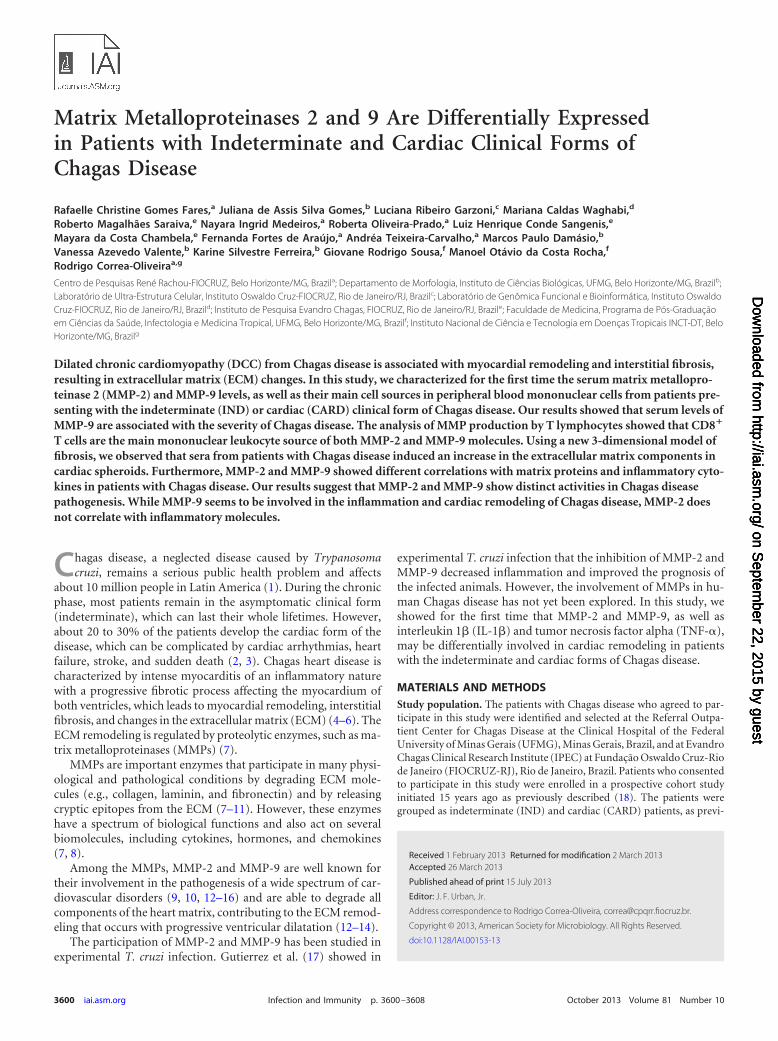
group (P � 0.003 and P � 0.001, respectively) (Fig. 4). The fi-bronectin and MMP-2 expression levels were higher in spheroidsstimulated with sera from the IND group than in those stimulatedwith sera from the NI group (P � 0.01 and P � 0.03, respectively)(Fig. 4). On the other hand, laminin and MMP-9 expression levelswere not different between the groups. However, we observed atendency toward higher expression of MMP-9 in spheroids incontact with sera from CARD patients (Fig. 4).
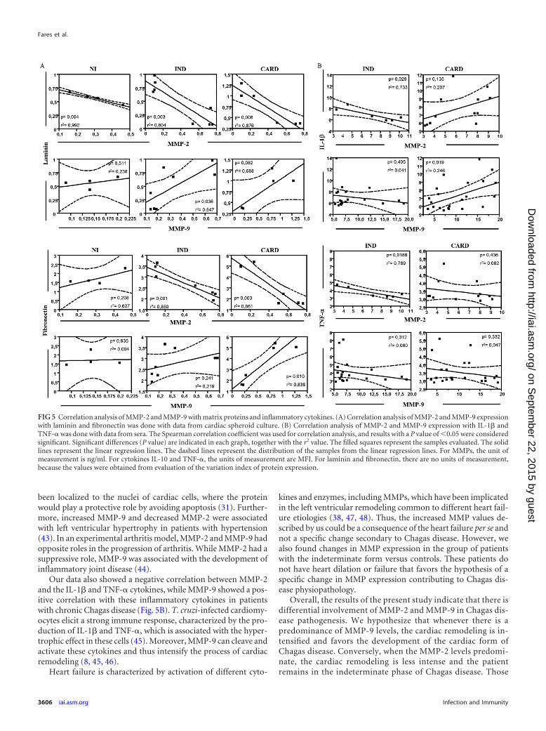
MMP-2 and MMP-9 are correlated with the cardiac spheroidremodeling induced by sera of patients with Chagas disease. TheMMPs are involved in the remodeling of cardiac tissue by degrad-ing ECM molecules (7, 31). In order to confirm if the presence ofMMPs correlates with ECM proteins and a possible role of thesemolecules in the cardiac remodeling of Chagas disease, we per-formed a correlative analysis. A significant negative correlationwas seen between MMP-2 expression and laminin expression inall groups (Fig. 5A). However, a significant positive correlationwas observed between MMP-9 expression and laminin expressionin the IND group (Fig. 5A). Regarding fibronectin, a significantnegative correlation was seen between MMP-2 expression andfibronectin expression in the IND and CARD groups (Fig. 5A).However, a significant positive correlation was observed betweenMMP-9 expression and fibronectin expression in the CARDgroup (Fig. 5A). There was no significant correlation betweenMMPs and collagen expression in the studied groups (data notshown).
MMP-9 seems to be involved with the inflammation and car-diac remodeling of Chagas disease, whereas MMP-2 does notcorrelate with inflammatory molecules. Our next step was toanalyze the correlation between serum TNF-� and IL-1� levels
and MMP levels measured by zymography, since TNF-� andIL-1� are the main cytokines involved in MMP activation (10, 32,33). A significant negative correlation was seen between MMP-2levels and serum IL-1� levels in the IND group (Fig. 5B). How-ever, a positive correlation was observed between MMP-2 levelsand serum IL-1� levels in the CARD group (Fig. 5B). A negativecorrelation was seen between MMP-9 levels and serum IL-1� lev-els in the IND group (Fig. 5B). On the other hand, a significantpositive correlation was observed between MMP-9 levels and se-rum IL-1� levels in the CARD group (Fig. 5B). Regarding TNF-�levels, a significant negative correlation was seen between MMP-2levels and TNF-� levels in the IND group (Fig. 5B). However, nosignificant correlation was observed either between MMP-2 levelsand TNF-� levels in the CARD group (Fig. 5B) or betweenMMP-9 levels and TNF-� levels in both the IND and CARDgroups (Fig. 5B).
Overall, our results suggest that MMP-2 and MMP-9, as well asIL-1� and TNF-�, may be differentially involved in cardiac re-modeling in the IND and CARD groups.
DISCUSSION
Chronic Chagas disease has a great variety of clinical presentationseven within patients with the cardiac form. Although the classifi-cation of the cardiac form into stages helps to distinguish patientswith worse prognosis (34), sudden death and other major clinicalevents still occur among patients in the initial stages of Chagasheart disease. Therefore, one of the most important goals of study-ing physiopathological mechanisms in Chagas disease is the iden-tification of potential risk factors for the occurrence of major clin-ical events (sudden death, heart failure, malignant arrhythmias,
FIG 2 Ex vivo analysis of intracytoplasmic levels of MMP-2 (A) and MMP-9 (B) in lymphocytes and monocytes. The groups evaluated were IND (n � 7; graybars), CARD (n � 10; dark-gray bars), and NI (n � 6; white bars). The data are expressed as the median percentages of T lymphocytes and monocytes withinterquartile ranges. Significant differences (P 0.05) in the charts are identified by connecting lines and asterisks for comparisons between the groups. Flowcytometric analyses are shown below.
Metalloproteinases 2 and 9 in Chagas Disease
October 2013 Volume 81 Number 10 iai.asm.org 3603
on Septem
ber 22, 2015 by guesthttp://iai.asm
.org/D
ownloaded from

and stroke) or disease progression among patients still in the in-determinate form or in initial stages of the cardiac form (5, 6).Chagas heart disease is characterized by an intense and progressivefibrotic process (4), and the study of biomarkers involved in theestablishment and development of fibrosis is essential to identifysuch potential risk factors (35).
In this study, we characterized for the first time the serum levelsof MMP-2 and -9, their potential proteolytic activity, as well astheir main sources within mononuclear cells in human Chagasdisease. Although there are no reports about the putative role ofMMPs in human Chagas disease, the involvement of MMPs inother cardiac diseases, particularly in the cardiac matrix remodel-ing that occurs in heart failure and after acute myocardial infarc-tion, has been widely studied (12–14, 36, 37, 38, 48).
Our results showed that patients with Chagas heart diseasecomplicated by heart failure present higher serum levels of
MMP-9 (Fig. 1). Moreover, experimental studies pointed out thatMMP serum levels may be associated with cardiovascular compli-cations of hypertension (49). Polyakova et al. (14) showed thatelevated expression of MMP-2 and MMP-9 is associated with col-lagen maturation in heart failure, demonstrating an importantrole of these enzymes in fibrosis through collagen configuration,activation, and deposition. In experimental models of Chagas dis-ease, high MMP-2 and -9 proteolytic activity had already beendescribed, which suggested an important role for MMPs in T.cruzi-induced acute myocarditis (17). Here, we showed elevatedlevels of these enzymes in chronic Chagas disease. Furthermore,the ratio analysis showed that MMP-9 levels were higher thanthose of MMP-2 (Fig. 1A), which is corroborated by others (12)who reported that MMP-2 is produced at lower levels thanMMP-9 by several cell types.
Our group previously showed the essential role of immune
FIG 3 Evaluation of MMP-2- and MMP-9-producing peripheral blood mononuclear cells and their subpopulations. Shown is analysis of the most relevantsources of MMP-2 (A) and MMP-9 (B) within the mononuclear cell subsets. The cells evaluated were monocytes and CD4� and CD8� T lymphocytes. The dataare expressed as the median numbers of MMP-producing cells with interquartile ranges. The data are also shown as pie charts, which represent the proportionsof CD4� and CD8� T cells and CD14 monocytes within MMP-2- and MMP-9-producing mononuclear cells. Significant differences (P 0.05) in the charts areidentified by connecting lines and asterisks for comparisons between the groups.
Fares et al.
3604 iai.asm.org Infection and Immunity
on Septem
ber 22, 2015 by guesthttp://iai.asm
.org/D
ownloaded from
cells, such as monocytes (39) and lymphocytes (18, 40), in Chagasdisease pathogenesis. In the present study, we observed that CD4�
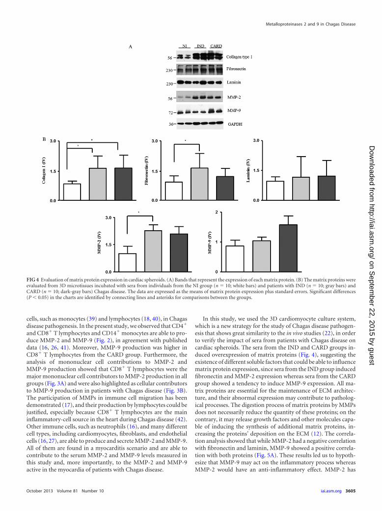
and CD8� T lymphocytes and CD14� monocytes are able to pro-duce MMP-2 and MMP-9 (Fig. 2), in agreement with publisheddata (16, 26, 41). Moreover, MMP-9 production was higher inCD8� T lymphocytes from the CARD group. Furthermore, theanalysis of mononuclear cell contributions to MMP-2 andMMP-9 production showed that CD8� T lymphocytes were themajor mononuclear cell contributors to MMP-2 production in allgroups (Fig. 3A) and were also highlighted as cellular contributorsto MMP-9 production in patients with Chagas disease (Fig. 3B).The participation of MMPs in immune cell migration has beendemonstrated (17), and their production by lymphocytes could bejustified, especially because CD8� T lymphocytes are the maininflammatory-cell source in the heart during Chagas disease (42).Other immune cells, such as neutrophils (16), and many differentcell types, including cardiomyocytes, fibroblasts, and endothelialcells (16, 27), are able to produce and secrete MMP-2 and MMP-9.All of them are found in a myocarditis scenario and are able tocontribute to the serum MMP-2 and MMP-9 levels measured inthis study and, more importantly, to the MMP-2 and MMP-9active in the myocardia of patients with Chagas disease.
In this study, we used the 3D cardiomyocyte culture system,which is a new strategy for the study of Chagas disease pathogen-esis that shows great similarity to the in vivo studies (22), in orderto verify the impact of sera from patients with Chagas disease oncardiac spheroids. The sera from the IND and CARD groups in-duced overexpression of matrix proteins (Fig. 4), suggesting theexistence of different soluble factors that could be able to influencematrix protein expression, since sera from the IND group inducedfibronectin and MMP-2 expression whereas sera from the CARDgroup showed a tendency to induce MMP-9 expression. All ma-trix proteins are essential for the maintenance of ECM architec-ture, and their abnormal expression may contribute to patholog-ical processes. The digestion process of matrix proteins by MMPsdoes not necessarily reduce the quantity of these proteins; on thecontrary, it may release growth factors and other molecules capa-ble of inducing the synthesis of additional matrix proteins, in-creasing the proteins’ deposition on the ECM (12). The correla-tion analysis showed that while MMP-2 had a negative correlationwith fibronectin and laminin, MMP-9 showed a positive correla-tion with both proteins (Fig. 5A). These results led us to hypoth-esize that MMP-9 may act on the inflammatory process whereasMMP-2 would have an anti-inflammatory effect. MMP-2 has
FIG 4 Evaluation of matrix protein expression in cardiac spheroids. (A) Bands that represent the expression of each matrix protein. (B) The matrix proteins wereevaluated from 3D microtissues incubated with sera from individuals from the NI group (n � 10; white bars) and patients with IND (n � 10; gray bars) andCARD (n � 10; dark-gray bars) Chagas disease. The data are expressed as the means of matrix protein expression plus standard errors. Significant differences(P 0.05) in the charts are identified by connecting lines and asterisks for comparisons between the groups.
Metalloproteinases 2 and 9 in Chagas Disease
October 2013 Volume 81 Number 10 iai.asm.org 3605
on Septem
ber 22, 2015 by guesthttp://iai.asm
.org/D
ownloaded from
been localized to the nuclei of cardiac cells, where the proteinwould play a protective role by avoiding apoptosis (31). Further-more, increased MMP-9 and decreased MMP-2 were associatedwith left ventricular hypertrophy in patients with hypertension(43). In an experimental arthritis model, MMP-2 and MMP-9 hadopposite roles in the progression of arthritis. While MMP-2 had asuppressive role, MMP-9 was associated with the development ofinflammatory joint disease (44).
Our data also showed a negative correlation between MMP-2and the IL-1� and TNF-� cytokines, while MMP-9 showed a pos-itive correlation with these inflammatory cytokines in patientswith chronic Chagas disease (Fig. 5B). T. cruzi-infected cardiomy-ocytes elicit a strong immune response, characterized by the pro-duction of IL-1� and TNF-�, which is associated with the hyper-trophic effect in these cells (45). Moreover, MMP-9 can cleave andactivate these cytokines and thus intensify the process of cardiacremodeling (8, 45, 46).
Heart failure is characterized by activation of different cyto-
kines and enzymes, including MMPs, which have been implicatedin the left ventricular remodeling common to different heart fail-ure etiologies (38, 47, 48). Thus, the increased MMP values de-scribed by us could be a consequence of the heart failure per se andnot a specific change secondary to Chagas disease. However, wealso found changes in MMP expression in the group of patientswith the indeterminate form versus controls. These patients donot have heart dilation or failure that favors the hypothesis of aspecific change in MMP expression contributing to Chagas dis-ease physiopathology.
Overall, the results of the present study indicate that there isdifferential involvement of MMP-2 and MMP-9 in Chagas dis-ease pathogenesis. We hypothesize that whenever there is apredominance of MMP-9 levels, the cardiac remodeling is in-tensified and favors the development of the cardiac form ofChagas disease. Conversely, when the MMP-2 levels predomi-nate, the cardiac remodeling is less intense and the patientremains in the indeterminate phase of Chagas disease. Those
FIG 5 Correlation analysis of MMP-2 and MMP-9 with matrix proteins and inflammatory cytokines. (A) Correlation analysis of MMP-2 and MMP-9 expressionwith laminin and fibronectin was done with data from cardiac spheroid culture. (B) Correlation analysis of MMP-2 and MMP-9 expression with IL-1� andTNF-� was done with data from sera. The Spearman correlation coefficient was used for correlation analysis, and results with a P value of 0.05 were consideredsignificant. Significant differences (P value) are indicated in each graph, together with the r2 value. The filled squares represent the samples evaluated. The solidlines represent the linear regression lines. The dashed lines represent the distribution of the samples from the linear regression lines. For MMPs, the unit ofmeasurement is ng/ml. For cytokines IL-10 and TNF-�, the units of measurement are MFI. For laminin and fibronectin, there are no units of measurement,because the values were obtained from evaluation of the variation index of protein expression.
Fares et al.
3606 iai.asm.org Infection and Immunity
on Septem
ber 22, 2015 by guesthttp://iai.asm
.org/D
ownloaded from

processes may be IL-1� and TNF-� dependent. These data areinnovative and represent an advance in the knowledge of themechanisms involved in the establishment/maintenance ofChagas heart disease pathology.
ACKNOWLEDGMENTS
This work was supported by the Conselho Nacional de DesenvolvimentoCientífico e Tecnológico (CNPq) (grant 478846/2009-6), the Fundação deAmparo Pesquisa do Estado de Minas Gerais (FAPEMIG) (grant APQ-02601-10), the Fundação Oswaldo Cruz (FIOCRUZ), the Instituto Nacio-nal de Ciência e Tecnologia em Doenças Tropicais (INCT-DT), and theNational Institutes of Health.
We thank the staff at the Laboratório de Imunologia Celular e Molec-ular, FIOCRUZ, for technical assistance. We also thank the Program forTechnological Development in Tools for Health (PDTIS), FIOCRUZ, forthe use of its facilities. R.C.-O., M.O.C.R., A.T.-C., and J.A.S.G. thankCNPq for fellowships (Bolsa de produtividade em Pesquisa).
We declare that no competing interests exist.
REFERENCES1. World Health Organization. August 2012. Fact sheet 340. World Health
Organization, Geneva, Switzerland. http://www.who.int. Accessed 29 Oc-tober 2012.
2. Moncayo A, Silveira AC. 2009. Current epidemiological trends for Cha-gas disease in Latin America and future challenges in epidemiology, sur-veillance and health policy. Mem. Inst. Oswaldo Cruz 104(Suppl I):17–30.
3. Tanowitz HB, Machado FS, Jelicks LA, Shirani J, de Carvalho AC,Spray DC, Factor SM, Kirchhoff LV, Weiss LM. 2009. Perspectives onTrypanosoma cruzi-induced heart disease (Chagas disease). Prog. Cardio-vasc. Dis. 51:524 –539.
4. Nunes MP, Colosimo EA, Reis RC, Barbosa MM, da Silva JL, BarbosaF, Botoni FA, Ribeiro AL, Rocha MO. 2012. Different prognostic impactof the tissue Doppler-derived E/e� ratio on mortality in Chagas cardio-myopathy patients with heart failure. J. Heart Lung Transplant. 31:634 –641.
5. Rocha MO, Ribeiro AL, Teixeira MM. 2003. Clinical management ofchronic Chagas cardiomyopathy. Front. Biosci. 1:44 –54.
6. Rocha MO, Teixeira MM, Ribeiro AL. 2007. An update on the manage-ment of Chagas cardiomyopathy. Expert Rev. Anti Infect. Ther. 5:727–743.
7. Geurts N, Opdenakker G, Van den Steen PE. 2012. Matrix metallopro-teinases as therapeutic targets in protozoan parasitic infections. Pharma-col. Ther. 133:257–279.
8. Parks WC, Wilson CL, Lopez-Boado YS. 2004. Matrix metalloprotei-nases as modulators of inflammation and innate immunity. Nat. Rev.Immunol. 4:617– 629.
9. Nagase H, Visse R, Murphy G. 2006. Structure and function of matrixmetalloproteinases and TIMPs. Cardiovasc. Res. 69:562–573.
10. Hu J, Van den Steen PE, Sang QX, Opdenakker G. 2007. Matrixmetalloproteinase inhibitors as therapy for inflammatory and vasculardiseases. Nat. Rev. Drug Discov. 6:480 – 498.
11. Kupai K, Szucs G, Cseh S, Hajdu I, Csonka C, Csont T, Ferdinandy P.2010. Matrix metalloproteinase activity assays: importance of zymogra-phy. J. Pharmacol. Toxicol. Methods 61:205–209.
12. Li YY, McTiernan CF, Feldman AM. 2000. Interplay of matrix metallo-proteinases, tissue inhibitors of metalloproteinases and their regulators incardiac matrix remodeling. Cardiovasc. Res. 46:214 –224.
13. Vanhoutte D, Schellings M, Pinto Y, Heymans S. 2006. Relevance ofmatrix metalloproteinases and their inhibitors after myocardial infarc-tion: a temporal and spatial window. Cardiovasc. Res. 69:604 – 613.
14. Polyakova V, Loeffler I, Hein S, Miyagawa S, Piotrowska I, Dammer S,Risteli J, Schaper J, Kostin S. 2010. Fibrosis in endstage human heartfailure: severe changes in collagens metabolism and MMP/TIMP profiles.Int. J. Cardiol. 151:18 –33.
15. Gao CQ, Sawicki G, Suarez-Pinzon WL, Csont T, Wozniak M, Ferdi-nandy P, Schulz R. 2003. Matrix metalloproteinase-2 mediates cytokine-induced myocardial contractile dysfunction. Cardiovasc. Res. 57:426 –433.
16. Opdenakker G, Van den Steen PE, Van Damme J. 2001. Gelatinase B: atuner and amplifier of immune functions. Trends Immunol. 22:571–579.
17. Gutierrez FR, Lalu MM, Mariano FS, Milanezi CM, Cena J, Gerlach RF,Santos JE, Torres-Dueñas D, Cunha FQ, Schulz R, Silva JS. 2008.Increased activities of cardiac matrix metalloproteinases matrix metallo-proteinase (MMP)-2 and MMP-9 are associated with mortality during theacute phase of experimental Trypanosoma cruzi infection. J. Infect. Dis.197:1468 –1476.
18. de Araújo FF, Corrêa-Oliveira R, Rocha MO, Chaves AT, Fiuza JA,Fares RC, Ferreira KS, Nunes MC, Keesen TS, Damasio MP, Teixeira-Carvalho A, Gomes JA. 2012. Foxp3�CD25(high) CD4� regulatory T cellsfrom indeterminate patients with Chagas disease can suppress the effectorcells and cytokines and reveal altered correlations with disease severity.Immunobiology 217:768 –777.
19. Gomes JA, Bahia-Oliveira LMG, Rocha MOC, Correa-Oliveira R. 2003.Evidence that development of severe cardiomyopathy in human Chagasdisease is due to a non-balanced Th1 specific immune response. Infect.Immun. 71:1185–1193.
20. Chen QJ, Lu L, Peng WH, Hu J, Yan XX, Wang LJ, Zhang Q, Zhang RY,Shen WF. 2009. Polymorphisms of MMP-3 and TIMP-4 genes affectangiographic coronary plaque progression in non-diabetic and type 2 di-abetic patients. Clin. Chim. Acta 405:97–103.
21. Meirelles MN, de Araujo Jorge TC, Miranda CF, de Souza W, BarbosaHS. 1986. Interaction of Trypanosoma cruzi with heart muscle cells: ultra-structural and cytochemical analysis of endocytic vacuole formation andeffect upon myogenesis in vitro. Eur. J. Cell Biol. 41:198 –206.
22. Garzoni LR, Adesse D, Soares MJ, Rossi MI, Borojevic R, de MeirellesMN. 2008. Fibrosis and hypertrophy induced by Trypanosoma cruzi inthree-dimensional cardiomyocyte-culture system. J. Infect. Dis. 197:906 –915.
23. Adesse D, Lisanti MP, Spray DC, Machado FS, Meirelles MN, TanowitzHB, Garzoni LR. 2010. Trypanosoma cruzi infection results in the reducedexpression of caveolin-3 in the heart. Cell Cycle 9:1639 –1646.
24. de Oliveira FL, Araújo-Jorge TC, de Souza EM, de Oliveira GM,Degrave WM, Feige JJ, Bailly S, Waghabi MC. 2012. Oral administrationof GW788388, an inhibitor of transforming growth factor beta signaling,prevents heart fibrosis in Chagas disease. PLoS Negl. Trop. Dis. 6:e1696.doi:10.1371/journal.pntd.0001696.
25. Opdenakker G. 2001. New insights in the regulation of leukocytosis andthe role played by leukocytes in septic shock. Verh. K. Acad. Geneeskd.Belg. 63:531–538.
26. Vandooren J, Geurts N, Martens E, Van den Steen PE, Opdenakker G.2013. Zymography methods for visualizing hydrolytic enzymes. Nat.Methods 10:211–220.
27. Brunner S, Kima JO, Methe H. 2010. Relation of matrix metalloprotei-nase-9/tissue inhibitor of metalloproteinase-1 ratio in peripheral circulat-ing CD14� monocytes to progression of coronary artery disease. Am. J.Cardiol. 105:429 – 434.
28. Edsparr K, Basse PH, Goldfarb RH, Albertsson P. 2011. Matrix metal-loproteinases in cytotoxic lymphocytes impact on tumour infiltration andimmunomodulation. Cancer Microenviron. 4:351–360.
29. Gomes JA, Bahia-Oliveira LM, Rocha MO, Busek SC, Teixeira MM,Silva JS, Correa-Oliveira R. 2005. Type 1 chemokine receptor expressionin Chagas’ disease correlates with morbidity in cardiac patients. Infect.Immun. 73:7960 –7966.
30. Souza PE, Rocha MO, Menezes CA, Coelho JS, Chaves AC, Gollob KJ,Dutra WO. 2007. Trypanosoma cruzi infection induces differential mod-ulation of costimulatory molecules and cytokines by monocytes and Tcells from patients with indeterminate and cardiac Chagas’ disease. Infect.Immun. 75:1886 –1894.
31. Mannello F, Medda V. 2012. Nuclear localization of matrix metallopro-teinases. Prog. Histochem. Cytochem. 47:27–58.
32. Lockwood CJ, Oner C, Uz YH, Kayisli UA, Huang SJ, Buchwalder LF,Murk W, Funai EF, Schatz F. 2008. Matrix metalloproteinase 9 (MMP9)expression in preeclamptic decidua and MMP9 induction by tumor ne-crosis factor alpha and interleukin 1 beta in human first trimester decidualcells. Biol. Reprod. 78:1064 –1072.
33. Deschamps AM, Spinale FG. 2006. Pathways of matrix metalloproteinaseinduction in heart failure: bioactive molecules and transcriptional regula-tion. Cardiovasc. Res. 69:666 – 676.
34. Andrade JP, Marin-Neto JA, Paola AA, Vilas-Boas F, Oliveira GM,Bacal F, Bocchi EA, Almeida DR, Fragata Filho AA, Moreira MC,Xavier SS, Oliveira Junior WA, Dias JC. 2011. Sociedade Brasileira deCardiologia. Latin American guidelines for the diagnosis and treatment of
Metalloproteinases 2 and 9 in Chagas Disease
October 2013 Volume 81 Number 10 iai.asm.org 3607
on Septem
ber 22, 2015 by guesthttp://iai.asm
.org/D
ownloaded from

Chagas cardiomyopathy. Chagásica Arq. Bras. Cardiol. 97(Suppl. 3):1– 48.(In Portuguese.)
35. Menezes C, Costa GC, Gollob KJ, Dutra WO. 2011. Clinical aspects ofChagas disease and implications for novel therapies. Drug Dev. Res. Sep.72:471– 479.
36. Schulz R. 2007. Intracellular targets of matrix metalloproteinase-2 incardiac disease: rationale and therapeutic approaches. Annu. Rev. Phar-macol. Toxicol. 47:211–242.
37. Adair-Kirk TL, Senior RM. 2008. Fragments of extracellular matrix asmediators of inflammation. Int. J. Biochem. Cell Biol. 40:1101–1110.
38. Kelly D, Cockerill G, Ng LL, Thompson M, Khan S, Samani NJ, SquireIB. 2007. Plasma matrix metalloproteinase-9 and left ventricular remod-elling after acute myocardial infarction in man: a prospective cohortstudy. Eur. Heart J. 28:711–718.
39. Gomes JA, Campi-Azevedo AC, Teixeira-Carvalho A, Silveira-Lemos D,Vitelli-Avelar D, Sathler-Avelar R, Peruhype-Magalhães V, Silvestre KF,Batista MA, Schachnik NC, Correa-Oliveira R, Eloi-Santos S, Martins-Filho OA. 2012. Impaired phagocytic capacity driven by downregulationof major phagocytosis-related cell surface molecules elicits an overallmodulatory cytokine profile in neutrophils and monocytes from the in-determinate clinical form of Chagas disease. Immunobiology 217:1005–1016.
40. Keesen TS, Gomes JA, Fares RC, de Araújo FF, Ferreira KS, Chaves AT,Rocha MO, Correa-Oliveira R. 2012. Characterization of CD4(�) cyto-toxic lymphocytes and apoptosis markers induced by Trypanossoma cruziinfection. Scand. J. Immunol. 76:311–319.
41. Clark RT, Nance JP, Noor S, Wilson EH. 2011. T-cell production ofmatrix metalloproteinases and inhibition of parasite clearance by TIMP-1during chronic Toxoplasma infection in the brain. ASN Neuro. 3:1–12.
42. Reis DD, Jones EM, Chapadeiro E, Tostes S, Gazzinelli G, Colley DG,McCurley T. 1993. Characterization of inflammatory infiltrates in
chronic chagasic myocardial lesions: presence of TNF-alpha and domi-nance of granzyme A�, CD8� lymphocytes. Am. J. Trop. Med. Hyg. 48:637– 644.
43. Fontana V, Silva PS, Gerlach RF, Tanus-Santos JE. 2012. Circulatingmatrix metalloproteinases and their inhibitors in hypertension. Clin.Chim. Acta 413:656 – 662.
44. Itoh T, Matsuda H, Tanioka M, Kuwabara K, Itohara S, Suzuki R. 2002.The role of matrix metalloproteinase-2 and matrix metalloproteinase-9 inantibody-induced arthritis. J. Immunol. 169:2643–2647.
45. Manque PA, Probst CM, Pereira MC, Rampazzo RC, Ozaki LS, PavoniDP, Silva Neto DT, Carvalho MR, Xu P, Serrano MG, Alves JM,Meirelles MN, Goldenberg S, Krieger MA, Buck GA. 2011. Trypanosomacruzi infection induces a global host cell response in cardiomyocytes. In-fect. Immun. 79:1855–1862.
46. Aoki MP, Carrera-Silva EA, Cuervo H, Fresno M, Gironès N, Gea S.2012. Nonimmune cells contribute to crosstalk between immune cells andinflammatory mediators in the innate response to Trypanosoma cruzi in-fection. J. Parasitol. Res. 2012:737324. doi:10.1155/2012/737324.
47. Spinale FG, Janicki JS, Zile MR. 2013. Membrane-associated matrixproteolysis and heart failure. Circ. Res. 112:195–208.
48. Matsunaga T, Abe N, Kameda K, Hagii J, Fujita N, Onodera H, KamataT, Ishizaka H, Hanada H, Osanai T, Okumura K. 2005. Circulating levelof gelatinase activity predicts ventricular remodeling in patients with acutemyocardial infarction. Int. J. Cardiol. 105:203–208.
49. Ahmed SH, Clark LL, Pennington WR, Webb CS, Bonnema DD,Leonardi AH, McClure CD, Spinale FG, Zile MR. 2006. Matrix metal-loproteinases/tissue inhibitors of metalloproteinases: relationship be-tween changes in proteolytic determinants of matrix composition andstructural, functional, and clinical manifestations of hypertensive heartdisease. Circulation 113:2089 –2096.
Fares et al.
3608 iai.asm.org Infection and Immunity
on Septem
ber 22, 2015 by guesthttp://iai.asm
.org/D
ownloaded from

Correction for Fares et al., Matrix Metalloproteinases 2 and 9 AreDifferentially Expressed in Patients with Indeterminate and CardiacClinical Forms of Chagas Disease
Rafaelle Christine Gomes Fares,a Juliana de Assis Silva Gomes,b Luciana Ribeiro Garzoni,c Mariana Caldas Waghabi,d
Roberto Magalhães Saraiva,e Nayara Ingrid Medeiros,a Roberta Oliveira-Prado,a Luiz Henrique Conde Sangenis,e
Mayara da Costa Chambela,e Fernanda Fortes de Araújo,a Andréa Teixeira-Carvalho,a Marcos Paulo Damásio,b
Vanessa Azevedo Valente,b Karine Silvestre Ferreira,b Giovane Rodrigo Sousa,f Manoel Otávio da Costa Rocha,f
Rodrigo Correa-Oliveiraa,g
Centro de Pesquisas René Rachou-FIOCRUZ, Belo Horizonte/MG, Brazila; Departamento de Morfologia, Instituto de Ciências Biológicas, UFMG, Belo Horizonte/MG, Brazilb;Laboratório de Ultra-Estrutura Celular, Instituto Oswaldo Cruz-FIOCRUZ, Rio de Janeiro/RJ, Brazilc; Laboratório de Genômica Funcional e Bioinformática, Instituto OswaldoCruz-FIOCRUZ, Rio de Janeiro/RJ, Brazild; Instituto de Pesquisa Evandro Chagas, FIOCRUZ, Rio de Janeiro/RJ, Brazile; Faculdade de Medicina, Programa de Pós-Graduaçãoem Ciências da Saúde, Infectologia e Medicina Tropical, UFMG, Belo Horizonte/MG, Brazilf; Instituto Nacional de Ciência e Tecnologia em Doenças Tropicais INCT-DT, BeloHorizonte/MG, Brazilg
Volume 81, no. 10, p. 3600 –3608, 2013. Page 3605: Figure 4 should appear as shown below.
Citation Fares RCG, Gomes JDAS, Garzoni LR, Waghabi MC, Saraiva RM, MedeirosNI, Oliveira-Prado R, Sangenis LHC, Chambela MDC, de Araújo FF, Teixeira-Carvalho A, Damásio MP, Valente VA, Ferreira KS, Sousa GR, Rocha MODC, Correa-Oliveira R. 2015. Correction for Fares et al., Matrix metalloproteinases 2 and 9 aredifferentially expressed in patients with indeterminate and cardiac clinical formsof Chagas disease. Infect Immun 83:847–848. doi:10.1128/IAI.02799-14.
Copyright © 2015, American Society for Microbiology. All Rights Reserved.
doi:10.1128/IAI.02799-14
AUTHOR CORRECTION
February 2015 Volume 83 Number 2 iai.asm.org 847Infection and Immunity
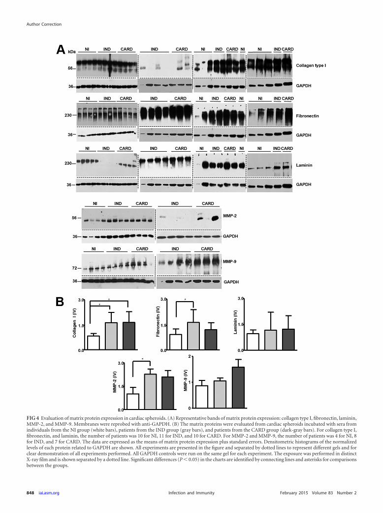
FIG 4 Evaluation of matrix protein expression in cardiac spheroids. (A) Representative bands of matrix protein expression: collagen type I, fibronectin, laminin,MMP-2, and MMP-9. Membranes were reprobed with anti-GAPDH. (B) The matrix proteins were evaluated from cardiac spheroids incubated with sera fromindividuals from the NI group (white bars), patients from the IND group (gray bars), and patients from the CARD group (dark-gray bars). For collagen type I,fibronectin, and laminin, the number of patients was 10 for NI, 11 for IND, and 10 for CARD. For MMP-2 and MMP-9, the number of patients was 4 for NI, 8for IND, and 7 for CARD. The data are expressed as the means of matrix protein expression plus standard errors. Densitometric histograms of the normalizedlevels of each protein related to GAPDH are shown. All experiments are presented in the figure and separated by dotted lines to represent different gels and forclear demonstration of all experiments performed. All GAPDH controls were run on the same gel for each experiment. The exposure was performed in distinctX-ray film and is shown separated by a dotted line. Significant differences (P � 0.05) in the charts are identified by connecting lines and asterisks for comparisonsbetween the groups.
Author Correction
848 iai.asm.org February 2015 Volume 83 Number 2Infection and Immunity