mbuihg ygfuyil
DESCRIPTION
mhjbh mvbfxrvjk kn,mjchrlTRANSCRIPT
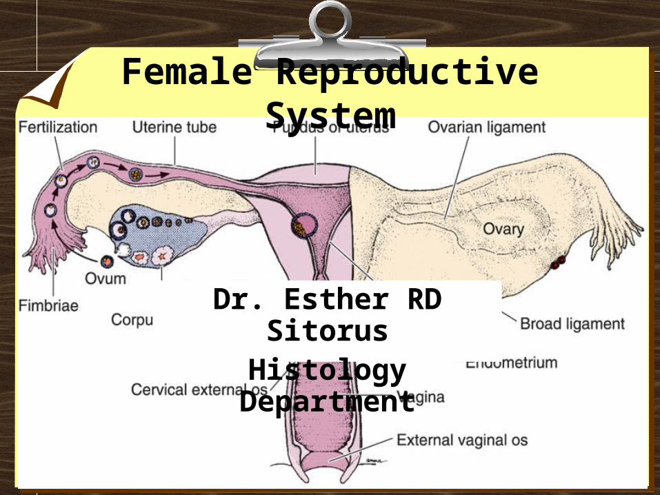
Dr. Esther RD SitorusHistology Department
Female Reproductive System

Introduction• Six major functions :
1. Production of female gametes, the ova
2. Reception of male gametes, the spermatozoa
3. Provision of a suitable environment for fetilization of ova by spermatozoa
4. Provision of an environment for development of the fetus
5. A means for expulsion of developed fetus to the external environment
6. Nutrition of the newborn

Introduction
• Three structural units on the basis of function:
1. The ovaries
2. The genital tract
3. The breasts

Introduction• INTERNAL PARTS :
• OVARIES• OVIDUCT• UTERUS• VAGINA
• EXTERNAL PARTS :• OPENING OF THE VAGINA• LABIA (MAJORA & MINORA)• VESTIBULE • CLITORIS• NOTE : ALTHOUGH NOT GENITAL ORGANS, THE MAMMARY
GLANDS ARE IMPORTANT ACCESSORY ORGANS OF THE FEMALE REPRODUCTIVE TRACT.
• ANATOMICAL INTEGRATION FOR REPRODUCTION
FERTILIZATION AND DEVELOPMENT
DELIVER AND EXIT

ovary

Ovary Ovary • GROSS ANATOMY
• CLOSE APROXIMATION TO OVIDUCT• ARE PAIRED OVAL BODIES THAT LIE ON EACH SIDE
OF THE UTERUS• HELD IN POTITION TO UTERUS BY LIGAMENTS
• 2 DISTINCT ANATOMICAL REGIONS• COVERED BY A MESOTHELIUM THAT IS CONTINUOUS
WITH THAT OF THE MESOVARIUM , THE SQUAMOUS CELLS BECOME CUBOIDAL AND FORM THE SURFACE EPITHELIUM OF THE OVARY = GERMINAL EPITHELIUM (OLD TERM)
• MEDULLARY-HIGHLY VASCULAR, CT, LYMPHATICS AND NERVES
• CORTEX-FOLLICLES, CT, AND SOME SMOOTH MUSCLE
• TUNICA ALBUGINEA TO SEPARATE EPITHELIUM FROM CORTEX

ovaryovary

Ovarian follicle
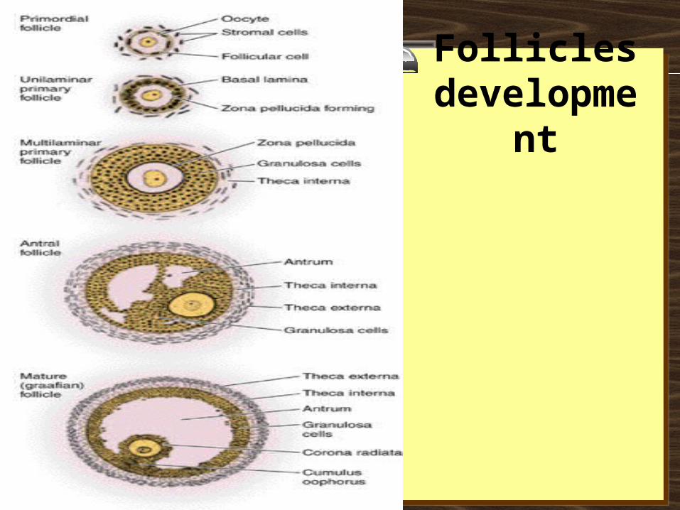
Follicles development

Primordial follicles • Located in the cortex just beneath tunica albuginea. • One layer of flattened follicular cells surround the
oocyte (about 30 µm in diameter). • The nucleus of the oocyte is positioned eccentric in
the cell. • It appears very light and contains a prominent nucleolus.• Most organelles of the oocyte aggregate in the centre of
the cell, where they form the vitelline body (probably not visible in any of the available preparations).
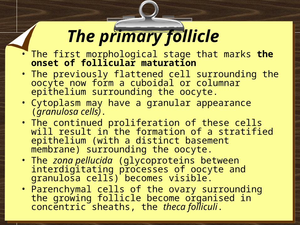
The primary follicle • The first morphological stage that marks the onset of
follicular maturation• The previously flattened cell surrounding the oocyte now
form a cuboidal or columnar epithelium surrounding the oocyte.
• Cytoplasm may have a granular appearance (granulosa cells).
• The continued proliferation of these cells will result in the formation of a stratified epithelium (with a distinct basement membrane) surrounding the oocyte.
• The zona pellucida (glycoproteins between interdigitating processes of oocyte and granulosa cells) becomes visible.
• Parenchymal cells of the ovary surrounding the growing follicle become organised in concentric sheaths, the theca folliculi.
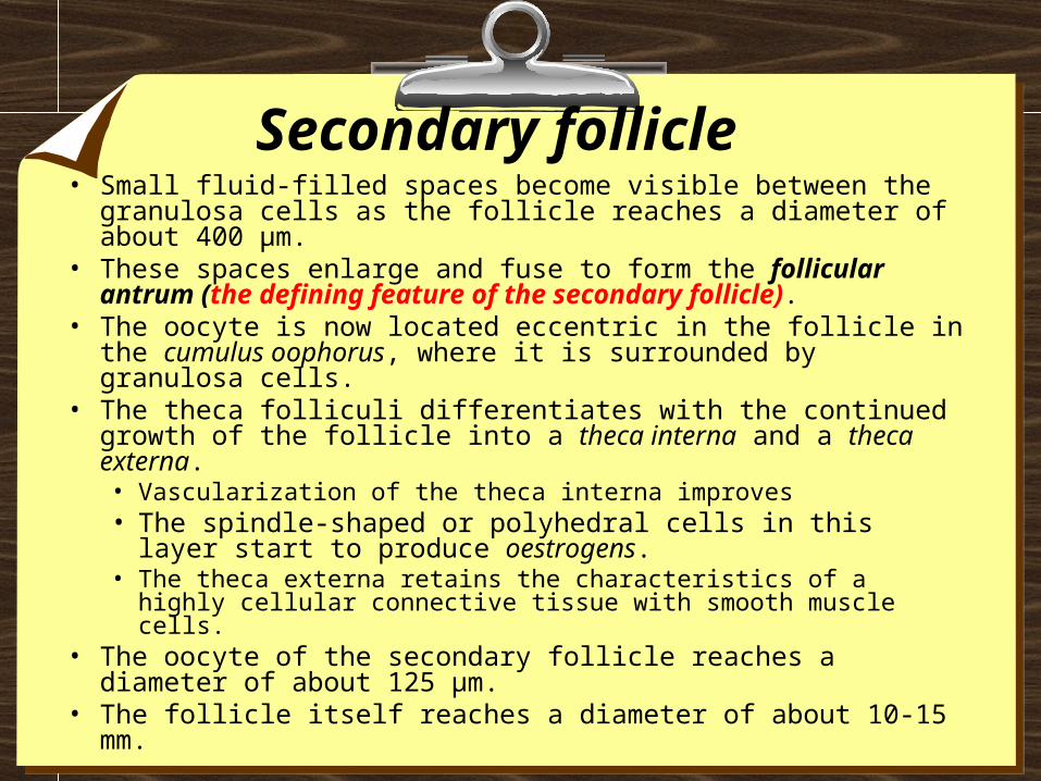
Secondary follicle • Small fluid-filled spaces become visible between the granulosa
cells as the follicle reaches a diameter of about 400 µm. • These spaces enlarge and fuse to form the follicular antrum (the
defining feature of the secondary follicle). • The oocyte is now located eccentric in the follicle in the cumulus
oophorus, where it is surrounded by granulosa cells. • The theca folliculi differentiates with the continued growth of the
follicle into a theca interna and a theca externa. • Vascularization of the theca interna improves • The spindle-shaped or polyhedral cells in this layer start to
produce oestrogens. • The theca externa retains the characteristics of a highly cellular
connective tissue with smooth muscle cells. • The oocyte of the secondary follicle reaches a diameter of about
125 µm.• The follicle itself reaches a diameter of about 10-15 mm.

Mature or tertiary or preovulatory or Graafian follicleMature or tertiary or preovulatory or Graafian follicle
• Increases further in size (in particular in the last 12h before ovulation).
• The Graafian follicle forms a small "bump" on the surface of the ovary, the stigma (or macula pellucida). • The stigma is characterised by a thinning of the capsule and a
progressive restriction of the blood flow to it.
• Prior to ovulation the cumulus oophorus separates from the follicular wall.
• The oocyte : floating freely in the follicular antrum. • It is still surrounded by granulosa cells which form the corona
radiata.
• The follicle finally ruptures at the stigma and the oocyte is released from the ovary
Atresia • Atresia is the name for the degenerative process by which oocytes (and
follicles) perish without having been expelled by ovulation. • Only about 400 oocytes ovulate - about 99.9 % of the oocytes that where
present at the time of puberty undergo atresia. • Atresia may effect oocytes at all stages of their "life" - both prenatally
and postnatally. • By the sixth month of gestation about 7 million oocytes and oogonia are
present in the ovaries. • By the time of birth this number is reduced to about 2 million. Of these
only about 400.000 survive until puberty. • Atresia is also the mode of destruction of follicles whose maturation is
initiated during the cyclus (10-15) but which do not ovulate. • Atresia is operating before puberty to remove follicles which begin to
mature during this period (none of which are ovulated). • Given that atresia affects follicles at various stages of their development
it is obvious that the process may take on quite a variety of histological appearances

Corpus luteum

The Corpus luteum• The wall of the follicle collapses into a folded structure
(characteristic for the corpus luteum). • Vascularization increases • Connective tissue network is formed. • Theca interna cells and granulosa cells triple in size and start
accumulating lutein within a few hours after ovulation (granulosa lutein cells and theca lutein cells and produce progesterone and oestrogens)
• Hormone secretion in the corpus luteum ceases within 14 days after ovulation if the oocyte is not fertilised (the corpus luteum degenerates into a corpus albicans - whitish scar tissue within the ovaries).
• Hormone secretion continues for 2-3 month after ovulation if fertilisation occurs.

Corpus albicans
Oviduct

Oviduct
• Functions : as a conduit for the oocyte, from the ovaries to the uterus.
• Histologically :• the oviduct consists of :
• a mucosa and a muscularis.
• The peritoneal surface of the oviduct is lined by a serosa and subjacent connective tissue.
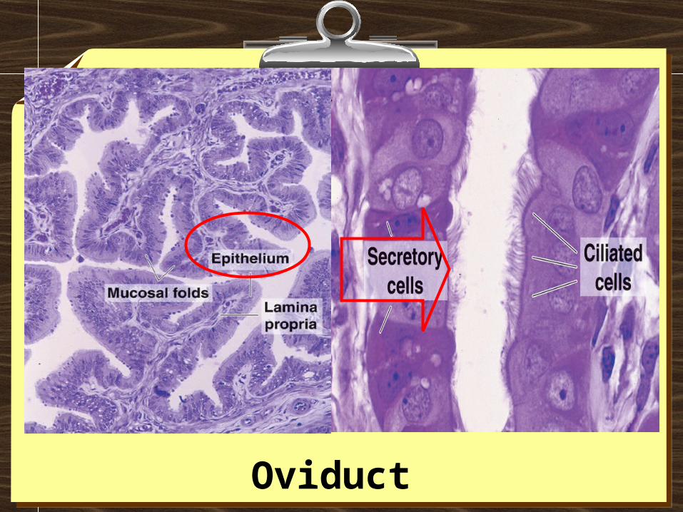
Oviduct • The mucosa
• Is formed by a ciliated and secretory epithelium resting on a very cellular lamina propria.
• The number of ciliated cells and secretory cells varies along the oviduct (see below).
• Secretory activity varies during the menstrual cycle, and resting secretory cells are also referred to as peg-cells.
• Some of the secreted substances are thought to nourish the oocyte and the very early embryo.
• The muscularis • Consists of an inner circular muscle layer and an outer
longitudinal layer. • An inner longitudinal layer is present in the isthmus and the
intramural part (see below) of the oviduct. • Peristaltic muscle action seems to be more important for the
transport of sperm and oocyte than the action of the cilia.

Oviduct Oviduct • Four subdivisions of the oviduct :
1. The infundibulum : funnel-shaped (up to 10 mm in diameter) end of the oviduct. • Finger-like extensions of its margins, the fimbriae, are closely applied
to the ovary. • Ciliated cells are frequent. Their cilia beat in the direction of
2. the ampulla of the oviduct. • Mucosal folds, or plicae, and secondary folds which arise from the
plicae divide the lumen of the ampulla into a very complex shape. • Fertilization usually takes place in the ampulla.
3. The isthmus is the narrowest portion (2-3 mm in diameter) of the parts of the oviduct located in the peritoneal cavity. • Mucosal folds are less complex and the muscularis is thick. An inner,
longitudinal layer of muscle is present in the isthmus and the
4. Intramural part of the oviduct, which penetrates the wall of the uterus. • The mucosa is smooth, and the inner diameter of the duct is very small.

Oviduct Oviduct

The Uterus The Uterus
• The uterus is divided into 1. Body (upper two-thirds) and 2. Cervix
• The walls of the uterus are composed of a• Mucosal layer (the endometrium)• A fibromuscular layer (the myometrium). • The peritoneal surface of the uterus is covered
by a serosa
• Myometrium • The muscle fibres of the uterus form layers with
preferred orientations of fibres (actually 4), but this is very difficult to see in most preparations.
• The muscular tissue hypertrophies during pregnancy, and GAP-junctions between cells become more frequent.
The UterusThe Uterus
• Endometrium • Consists of a simple columnar epithelium (ciliated cells
and secretory cells) and an underlying thick connective tissue stroma.
• The mucosa is invaginated to form many simple tubular uterine glands.
• The glands extend through the entire thickness of the stroma.
• The stromal cells of the endometrium are embedded in a network of reticular fibres.
• The endometrium is subject to cyclic changes that result in menstruation. Only the mucosa of the body of the uterus takes part in the menstrual cycle
The UterusThe Uterus
The UterusThe Uterus • Endometrium
• The endometrium can be divided into two zones based on their involvement in the changes during the menstrual cycle: the the basalisbasalis and the and the functionalisfunctionalis.
• The basalis is not sloughed off during menstruation but functions as a regenerative zone for the functionalis after its rejection.
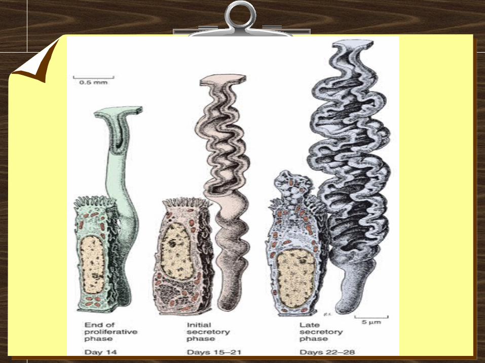
• The functionalis is the luminal part of the endometrium. It is sloughed off during every menstruation and it is the site of cyclic changes in the endometrium. These cyclic changes are divided into a number of phases: proliferative (or follicular), secretory (or luteal), and menstrual.


Pregnancy

Placenta
Placenta
PlacentaPlacenta

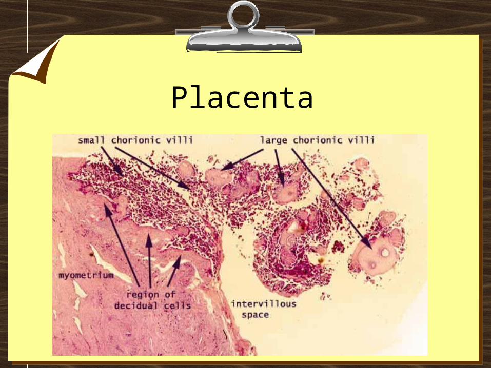
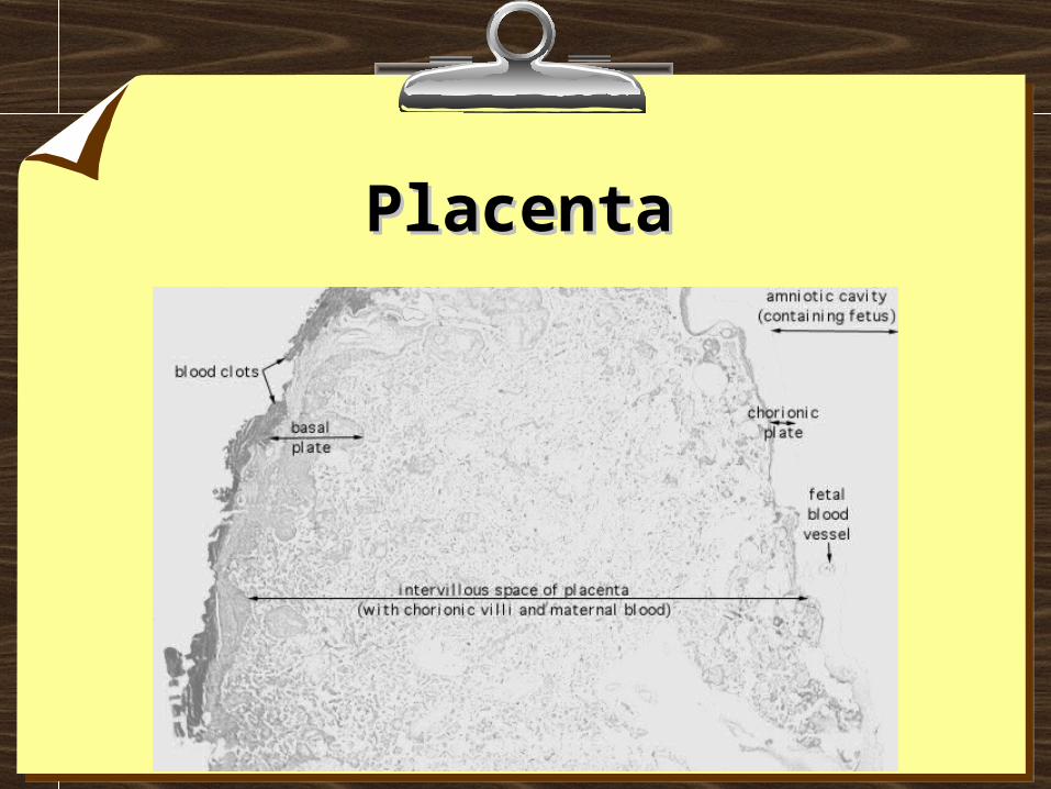
Placenta• The placenta may be usefully understood as a "parasite"
feeding on blood from the endometrium (Imagine scooping out a portion of the endometrium).
• The resulting bowl will fill with blood from broken vessels in the endometrial stroma.
• Now lay a cover over the bowl, and imagine many "roots" extending down from the cover into the blood-filled hollow (the roots can absorb oxygen and nutrients from the blood in which they are bathed).

Placenta Placenta
• The cover is the chorionic plate of the placenta. • The "roots" are the chorionic villi.
• Both the placenta and the chorionic villi are entirely fetal tissue (orange in the diagram above).
• "Anchoring villi" attach the placenta to the endometrium. • Smaller branching villi extend out into the intervillous space. • Fetal circulation passes down the umbilical cord, though vessels in the villi, and back
up the umbilical cord. • Maternal blood "spills" from open endometrial arteries (the spiral arteries) into the
intervillous space (pink in the diagram above), and returns into endometrial veins. • The chorionic villi are surrounded and bathed by "lakes" of maternal blood. Within the
intervillous space, maternal blood is not contained by blood vessels• The surface of the chorionic villi is an epithelial layer, the fetal syncytiotrophoblast,
which has the ability to grow invasively into the maternal endometrium. The syncytiotrophoblast also has microvilli on the surface for absorbing nutrients from maternal blood.
• Beneath the syncytiotrophoblast (i.e., toward the core of the villus), is the cytotrophoblast, a layer of cuboidal cells which eventually disappear. (The cytotrophoblast also forms trophoblast columns, masses of cells filling the ends of anchoring villi.)
• Maternal endometrial stromal tissue adjacent to the placenta differentiates into large decidual cells (so named because the outer layer of the endometrium is shed at birth along with the placenta). Decidual cells may intermix with fetal cells in the cytotrophoblast. The boundary between maternal and fetal tissue is immunologically interesting.

Umbilical CordUmbilical Cord
• The umbilical cord is simply a conduit carrying fetal blood between the fetus and the placenta. It normally contains two arteries and one vein, surrounded by extensive mesenchymal tissue ("Wharton's jelly").
• Consists of so-called "mucous" or mesenchymal connective tissue, also called Wharton's jelly (widely scattered mesenchymal fibroblasts within soft, jelly-like ground substance of hyaluronic acid and chondroitin sulfate)
• Surrounded by a thin stratified squamous epithelium and including typically two arteries and one veintwo arteries and one vein. [The second vein in this image presumably represents one portion of a double U-shaped bend in this single vein.] The arteries lack internal and external elastic layers.

Umbilical CordUmbilical Cord

Cervix Cervix • TRANTITIONAL EPITHELIUM (T zone) GOES FROM
SQUAMOUS (ectocervix) TO SECRETING (UTERINE GLANDS) COLUMNAR EPITHELIUM (endocervix).
• VISCOUS OF MUCUS GLANDS CHANGES WITH MENSTRUAL CYCLE
• CERVIX TO VESTIBULE
• MULTILAYERED
• MUCOSAL
• FOLDS OF STRATIFIED EPITH
• NOT KERATINIZED BUT KERATOHYALIN GRANULES MAY BE VISIBLE
• NO GLANDS BUT CELLS ARE HIGH IN GLYCOGEN
• MUCUS COMES FROM CERVICAL GLANDS
• MUSCULARIS-SMOOTH MUSCLE
• ADVENTITIAL

Vagina Vagina • The vagina is a fibromuscular tube with a wall consisting of
three layers: • Mucosa
• The stratified squamous epithelium (deep stratum basalis, intermediate stratum spinosum, superficial layers of flat eosinophilic cells which do contain keratin but which do not normally form a true horny layer) rests on a very cellular lamina propria (many leukocytes). Towards the muscularis some vascular cavernous spaces may be seen (typical erectile tissue).
• Muscularis • Inner circular and outer longitudinal layers of smooth muscle are present.
Inferiorly, the striated, voluntary bulbospongiosus muscle forms a sphincter around the vagina.
• Adventitia • The part of the adventitia bordering the muscularis is fairly dense and
contains many elastic fibres. Loose connective tissue with a prominent venous plexus forms the outer part of the adventitia.

Vagina

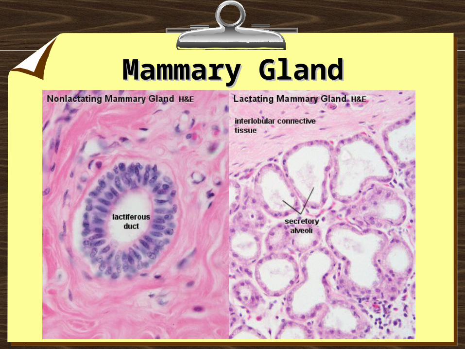
Female Accessory Reproductive Glands - Mammary Glands
• The mammary glands are modified glands of the skin (resembles that of sweat glands).
• Compound branched alveolar glands, which consist of 15-25 lobes separated by dense interlobar connective tissue and fat (Each lobe contains an individual gland)
• The excretory duct of each lobe, also called lactiferous duct, has its own opening on the nipple.
• The lactiferous duct has a two layered epithelium - basal cells are cuboidal whereas the superficial cells are columnar.
• Beneath the nipple, the dilated lactiferous duct forms a lactiferous sinus , which functions as a reservoir for the milk.
• Branches of the lactiferous duct are lined with a simple cuboidal epithelium.
• The secretory units are alveoli, which are lined by a cuboidal or columnar epithelium.
• A layer of myoepithelial cells is always present between the epithelium and the basement membrane of the branches of the lactiferous duct and the alveoli.

Breast
Mammary GlandMammary Gland