measurement of immune function:. detect antigens and / or antibodies. immunological tests rely upon:...
TRANSCRIPT

Measurement of Immune function:
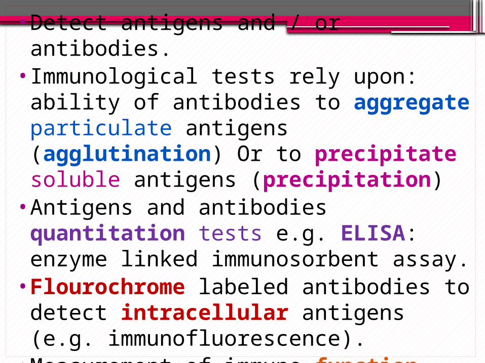
•Detect antigens and / or antibodies.•Immunological tests rely upon: ability of
antibodies to aggregate particulate antigens (agglutination) Or to precipitate soluble antigens (precipitation)
•Antigens and antibodies quantitation tests e.g. ELISA: enzyme linked immunosorbent assay.
•Flourochrome labeled antibodies to detect intracellular antigens (e.g. immunofluorescence).
•Measurement of immune function (e.g., complement fixation and cytotoxic T lymphocyte assay)
•Clinical setting (assessment of hypersensitivity).

Antigen detection:
Detect the specific reaction between the antigens & antibodies. Agglutination: to detect particulate
antigens: o Direct agglutination.o Indirect (passive) agglutination.o Agglutination inhibition. precipitation: to detect soluble antigens: oRadial immunodiffusion. oDouble-diffusion

Agglutination tests:
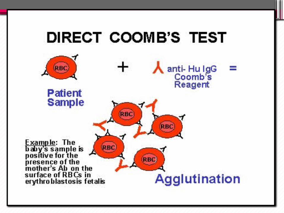
o Direct agglutination:Agglutination of particulate or cell-bounded antigen by antibodies (mainly IgM, multivalent Ab).oAgglutination of RBCs: haemagglutination.Example:• Blood grouping: group A RBCs + anti- A
antibodies = agglutination.• Coombs test*: diagnosis of alloimmune
hemolytic anemia of newborn.
Agglutination of RBC is called haemagglutination
•Insert a picture

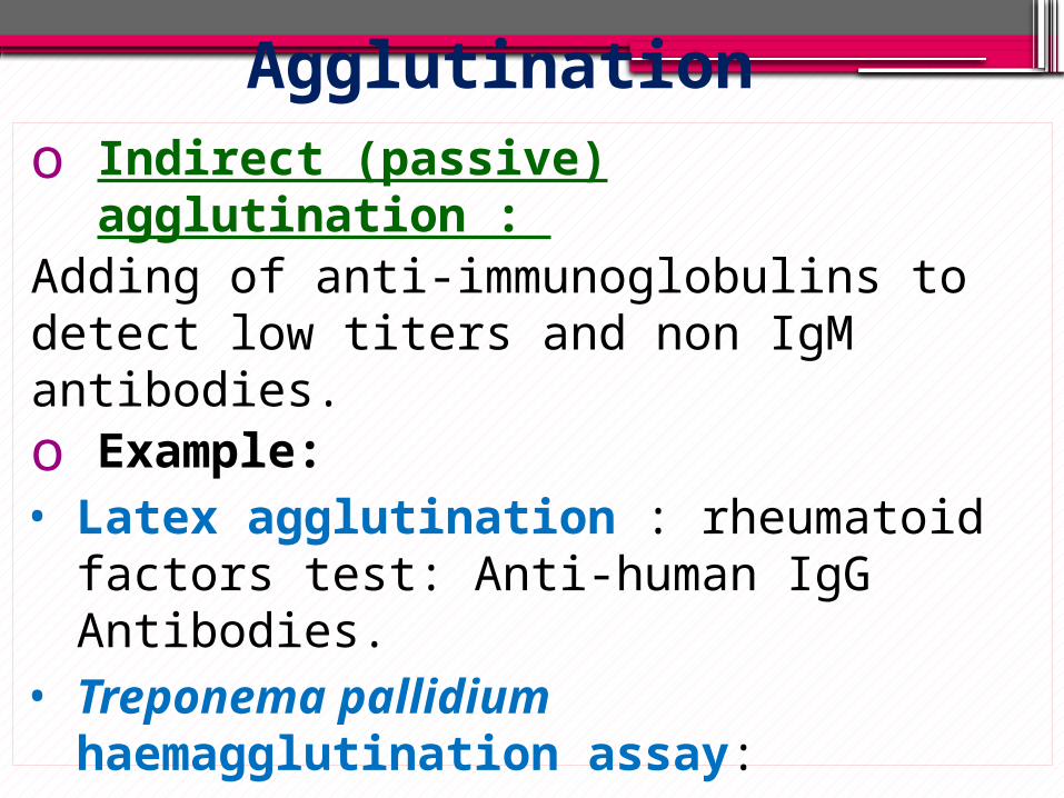
Agglutinationo Indirect (passive) agglutination : Adding of anti-immunoglobulins to detect low titers and non IgM antibodies.o Example:• Latex agglutination : rheumatoid factors test:
Anti-human IgG Antibodies.• Treponema pallidium haemagglutination assay:
(TPHA): Diagnosis of syphilis.


RBCs +treponemal antigens
TPHA

Agglutinationo Agglutination inhibition• Haemadsorption: is a direct agglutination of
erythrocytes by certain viruses (spontaneous agglutination).•Inhibition of erythrocytes agglutination by
anti-virus antibodies in patient’s serum.

Haemadsorption and agglutination inhibition:
• N

N
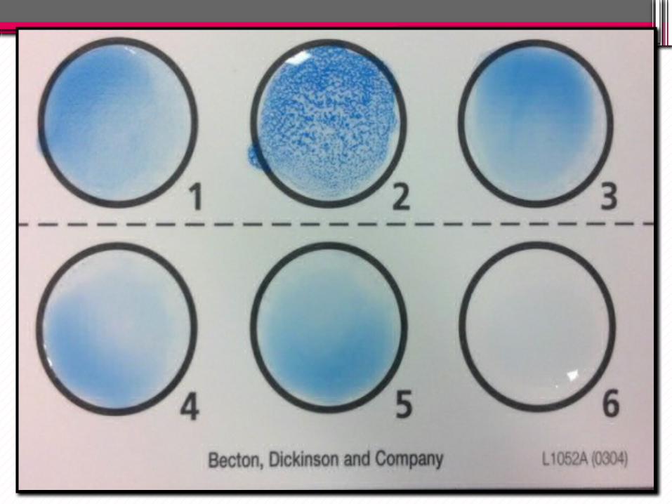
•Antibody titer: the lowest concentration of antibody that causes agglutination in vitro. •Serial doubling dilution of patient’s
serum creates zone of equivalence; and so positive reaction. •Prozone phenomena: False-negative agglutination due to excess antibodies or antigen concentration.
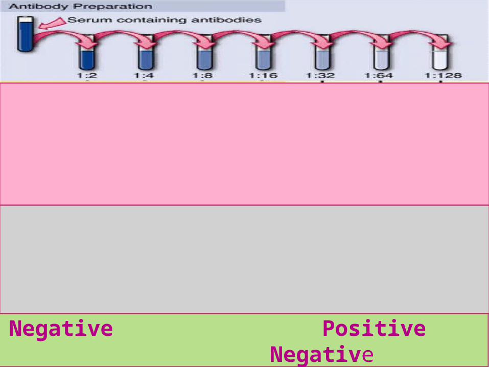
Negative Positive Negative

Precipitation: soluble antigens
oRadial immunodiffusion: Mancini technique:• Soluble antigen reacts with soluble
antibodies in semisolid medium. • Formation of immuno-precipitin lines.• Quantitative assay.• Clinical applications:•Diagnosis of complement deficiency•Calculation of Hb F for diagnosis of Beta-
thalassemia.


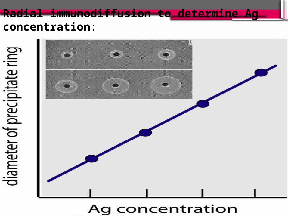
Radial immunodiffusion to determine Ag concentration:
•
•
•
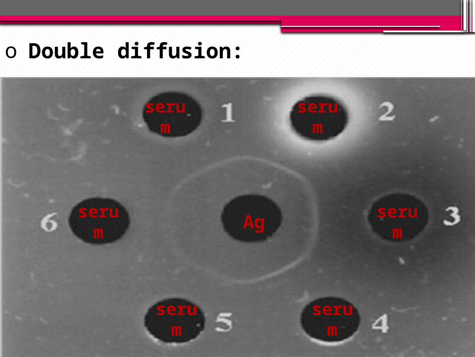
o Double diffusion:
Ag
serum
serum
serum
serum
serum
serum


Other widely used techniques
•Immunofluorescent microscopy.•Flow cytometery.•Enzyme linked immunosorbent assay
(ELISA).
N
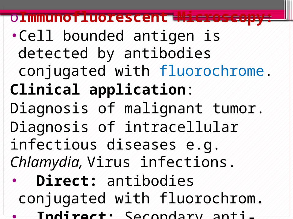
oImmunofluorescent Microscopy:•Cell bounded antigen is detected by
antibodies conjugated with fluorochrome.Clinical application: Diagnosis of malignant tumor.Diagnosis of intracellular infectious diseases e.g. Chlamydia, Virus infections.• Direct: antibodies conjugated with
fluorochrom.• Indirect: Secondary anti-human globulin
conjugated with fluorochrome.

N
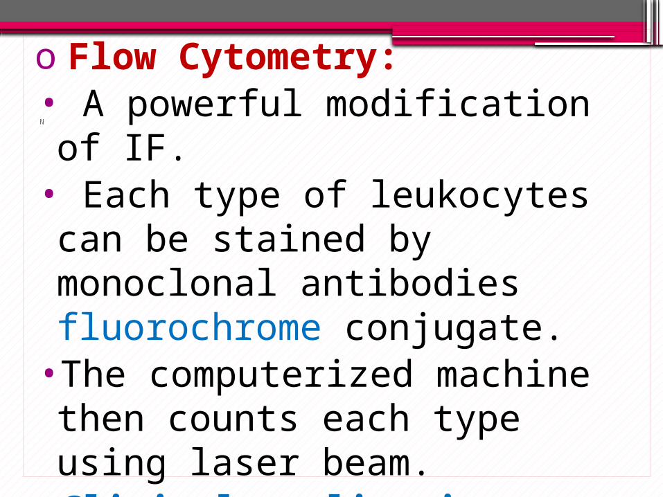
o Flow Cytometry:• A powerful modification of IF. • Each type of leukocytes can be stained
by monoclonal antibodies fluorochrome conjugate.•The computerized machine then counts
each type using laser beam. •Clinical application:Calculation of CD4/CD8 Ratio.

N
o Enzyme Linked Immunosorbent Assay (ELISA):
- Quantitative assay. - Soluble antigens or antibodies fixed on micro-titer plate wells. - Secondary antibodies linked with enzyme reacts with the complex. -Substrate (colorless) converted into colored end product which is measured by spectrophotometry.

ELISA for antigen detection• Add the patient’s serum then wash• add specific antibodies to the antigen• wash• add 2° antibody linked with
the enzyme then wash• add specific substrate.• read the reaction.
P N

ELISA for Antibody detection
Antigen coating
1° Antibody in the patient's
serum
2° antibody (enzyme-
linked)
Chromogenic substrate
Indirect ELISA

ELISA •Clinical application:Diagnosis of infectious diseases: HIV, HBV, HCV …..
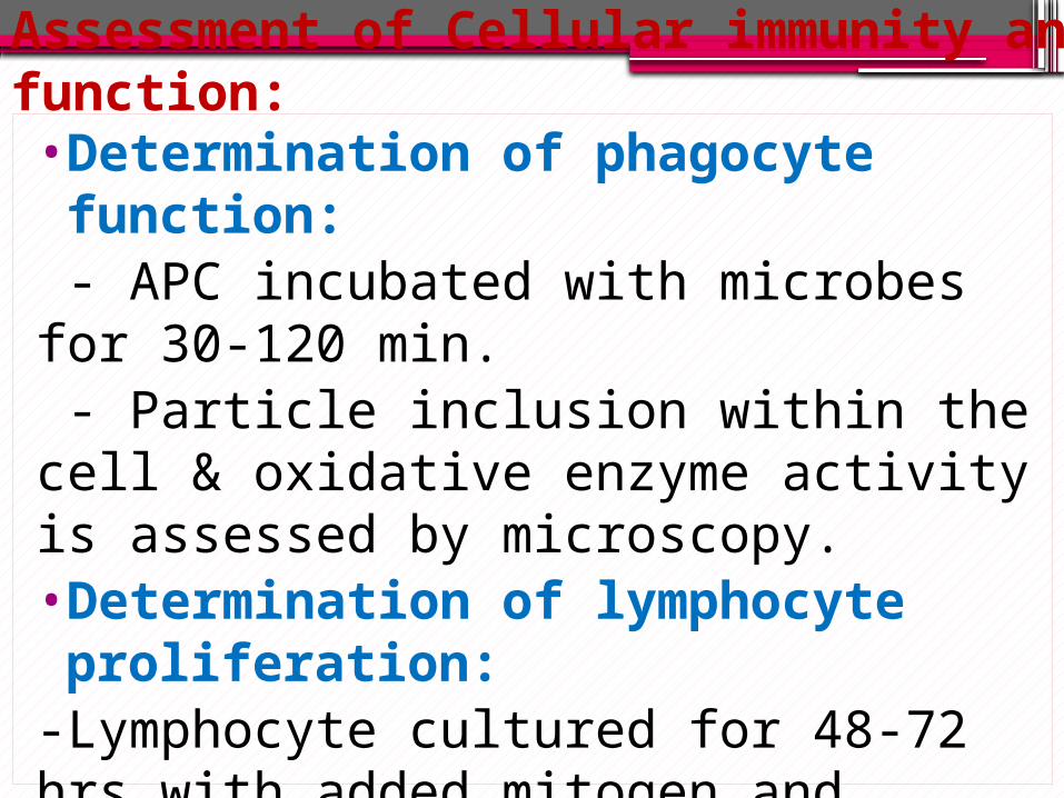
Assessment of Cellular immunity and function:
•Determination of phagocyte function: - APC incubated with microbes for 30-120 min. - Particle inclusion within the cell & oxidative enzyme activity is assessed by microscopy.•Determination of lymphocyte proliferation:-Lymphocyte cultured for 48-72 hrs with added mitogen and radioactive material.- incorporation of radioactive material into the new formed DNA is measured by radioimmunoassay.

Assessment of hyper sensitivity•Allergy skin testing (type I hypersensitivity):
scratch or intradermal injection of a small amount of diluted allergen. Sensitive (atopic) individuals develop a wheal-and-flare (redness & swelling) reaction within 20 minutes.•Complement fixation (types II and III):•Contact dermatitis and delayed hypersensitivity
(type IV): Application of antigen to the surface of the skin (contact dermatitis) or injected intradermally. Wheal-and-flare reactions are evident only 24 to 72 hours after challenge.

Type I hypersensitivity reaction

Type II (DTH) hypersensitivity reaction