measurements of foetal growth via transabdominal ultrasonography … · 493 measurements of foetal...
TRANSCRIPT

493
MEASUREMENTS OF FOETAL GROWTH VIA TRANSABDOMINAL ULTRASONOGRAPHY DURING FIRST HALF OF PREGNANCY AT EWES FROM SYNTHETIC POPULATION BULGARIAN MILK
N. Metodiev¹*, d. diMov², i. Ralchev² and e. Raicheva¹ ¹ Institute of Animal Science, Department Sheep Breeding, BG - 2230 Kostinbrod, Bulgaria² University of Forestry, Faculty of Veterinary Medicine, BG - 1756 Sofia, Bulgaria
Abstract
Metodiev, N., d. diMov, i. Ralchev and e. Raicheva, 2012. Measurements of foetal growth via transab-dominal ultrasonography during first half of pregnancy at ewes from synthetic population Bulgarian milk. Bulg. J. Agric. Sci., 18: 493-500
The aim of present work was to determine values of some measurements of foetal growth via transabdominal ultrasonog-raphy during first half of pregnancy at ewes from Synthetic Population Bulgarian Milk. Two parallel experiments were carried out with ewes, which were in different gestational phases. The first experiment was carried out with 19 ewes, conceived in three consecutive days. Three consecutive ultrasound observations in 10 days intervals were done, on days 40-42, 51-53 и 61-63 after conceiving. The following measurements were taken: head diameters: biparieatal (BPD) and occipito-nasal (ONL), Placentome size: width and length. The second experiment was carried out with 17 ewes, conceived in the same day. The examination days were 25, 36 and 46 after artificial insemination. It was measured: Day 25: diameter of embryonic vesicle; length of embryo Day 36: crown-rump length; Day 46: crown-rump length; head diameters: biparieatal (BPD) and occipito-na-sal (ONL); placentome size: width and length. All measurements were in millimeters (mm). The effect of gestational phase on foetal growth is analysed by one way ANOVA. The coefficients of correlation (r) and determination (R²) between gestational phase and embryofetal measurements were established by standard regression. The gestational phase influence significantly ((P<0.001) all taken measurements. The mean values for BPD were11.16 mm on 40-42 gestational days and reached 21,08 mm on 61-63 days, and for ONL -15.17 mm on 40-42 days and reached 32.94 mm on 61-63 days. The size of palcentomes varied in wide range for all taken measurements. On days 40-42 the mean values were: length- 13.67 mm and width – 10.06 mm, and reached on 61-63 days mean length – 33.02 mm and mean width – 21.33 mm. It was established high, positive and significant correlation between head diameters and gestational age (r=0.93*** и r=0.93***). It was obtained moderate values of the coefficient of determination for the size of placentomes and gestational age (R²= R²= 0.46 и R²= 0.47). The embroyfetal length was 11.08 mm on day 25, 26.03 mm on day 36 and 51.79 mm on day 46. It was established high, positive and significant correlation between CRL and gestational age (r= 0.84***).
Key words: ultrasound, ewe, foetal measurements, gestational age
Bulgarian Journal of Agricultural Science, 18 (No 4) 2012, 493-500Agricultural Academy
* Corresponding author: [email protected]
Introduction
The real-time B-mode ultrasonography is the earli-est, most accurate, safest, fastest and most economical
method of pregnancy diagnosis in sheep at farm level (Ganie et al., 2009). There are two approaches, that is use to study the female reproductive tract of small ru-minants – transrectal and transabodminal, and the ap-
494 N. Metodiev, D. Dimov, I. Ralchev and E. Raicheva
proach, that would be choose depends on diagnosis, that is needed, probes and size of flocks (Kähn, 2004). The author, Kähn, (2004), recommends for early preg-nancy diagnosis transrectal approach as more suitable until 35-th gestational day, the two approaches between 35-th and 70-th day of pregnancy, and during the sec-ond half of pregnancy, the transabodminal approach is more appropriate to use because of the better visualiza-tion of uterus.
When the date of mating is unknown, monitoring fetal development allows estimation of gestational age (Gonzáles de Bulnes et al., 1998). The most practical measurements of foetal structures are (by Karen et al., 2001): embryonic vesicle; crown-rump length; fetal head diameters; thoracic diameters; foetal heart rate; placentome size. Ideally, intrauterine fetal growth rate should be defined in the same population as that evalu-ated (Ali and Hayder, 2007). There are no foetal meas-urements and growth standards for Synthetic Popula-tion Bulgarian Milk (SPBM) sheep breed.
The aim of present work was to determine values of some measurements of foetal growth via transabdomi-nal ultrasonography during the first half of pregnancy at ewes from Synthetic Population Bulgarian Milk.
Materials and Methods
Two parallel experiments were carried out with ewes from SPBM, which were in different gestational phas-es. The dates of artificial inseminations were known, as the day of first insemination was Day 1 of pregnancy. All ewes were between 2.5 to 6.5 years old, clinically healthy during the experiments. All ewes were insemi-nated artificially with non-diluted semen from the first signs of estrus (detecting by teasers) to its end, in inter-vals of 12 hours. It was used Eickemeyer Medic 2000 device and sector probe with frequency 5 MHz. It was used transabodminal approach in right abdominal wall, in the area between pecten ossis pubis and last rib. An ultrasonic jelly was put on the probe, before attaching it to the skin.
Experiment 1 The experiment was carried out with 19 ewes, con-
ceived in three consecutive days. All ewes were con-
ceived as that was established previously by non-return method. Three consecutive ultrasound observations in 10 days intervals were done, on days 40-42, 51-53 и 61-63 after conceiving. All measurements were taken after freezing the images with built-in electronic calli-pers. Where two fetuses were predicted and measured, but after the lambing, the prognosis was untrue, we made mean measurements. During the second observa-tion from the series, one ewe was missed to observe.
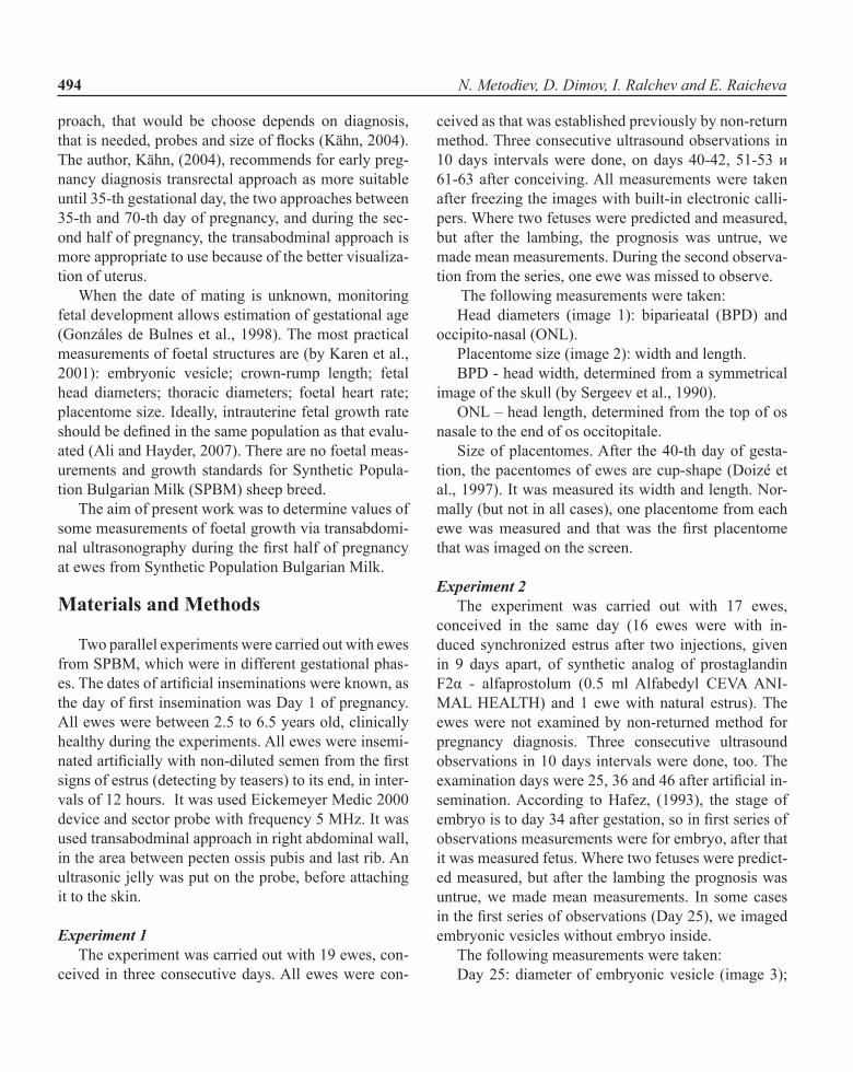
The following measurements were taken: Head diameters (image 1): biparieatal (BPD) and
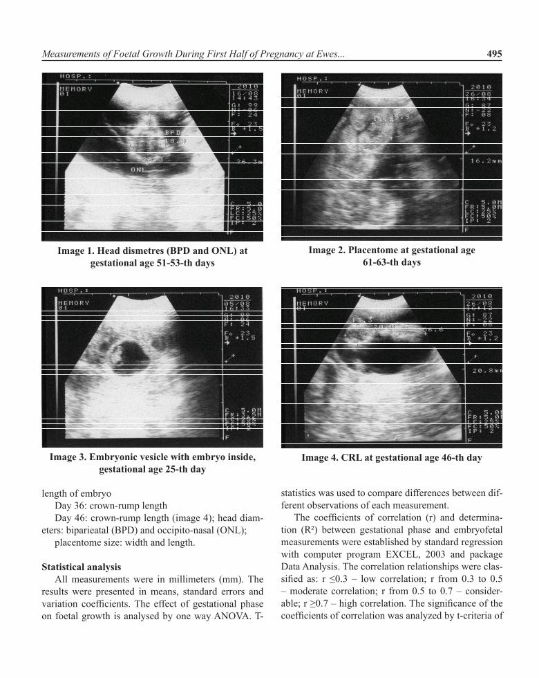
occipito-nasal (ONL). Placentome size (image 2): width and length.BPD - head width, determined from a symmetrical
image of the skull (by Sergeev et al., 1990).ONL – head length, determined from the top of os
nasale to the end of os occitopitale.Size of placentomes. After the 40-th day of gesta-
tion, the pacentomes of ewes are cup-shape (Doizé et al., 1997). It was measured its width and length. Nor-mally (but not in all cases), one placentome from each ewe was measured and that was the first placentome that was imaged on the screen.
Experiment 2The experiment was carried out with 17 ewes,
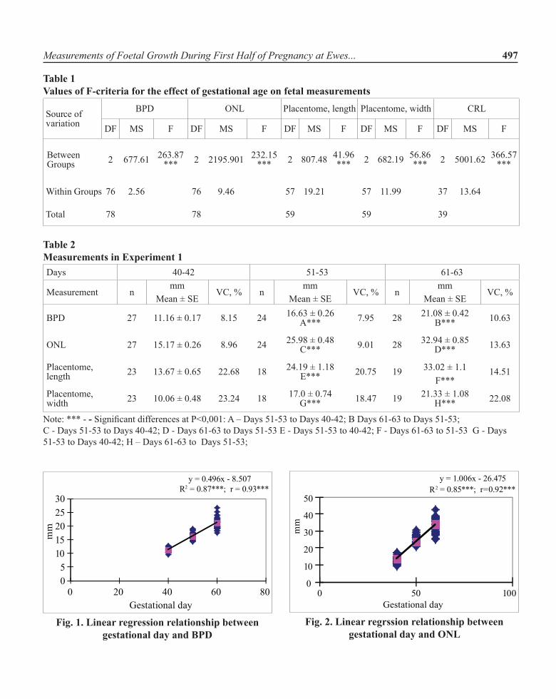
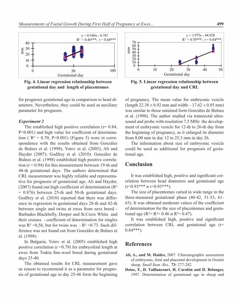
conceived in the same day (16 ewes were with in-duced synchronized estrus after two injections, given in 9 days apart, of synthetic analog of prostaglandin F2α - alfaprostolum (0.5 ml Alfabedyl CEVA ANI-MAL HEALTH) and 1 ewe with natural estrus). The ewes were not examined by non-returned method for pregnancy diagnosis. Three consecutive ultrasound observations in 10 days intervals were done, too. the examination days were 25, 36 and 46 after artificial in-semination. According to Hafez, (1993), the stage of embryo is to day 34 after gestation, so in first series of observations measurements were for embryo, after that it was measured fetus. Where two fetuses were predict-ed measured, but after the lambing the prognosis was untrue, we made mean measurements. In some cases in the first series of observations (Day 25), we imaged embryonic vesicles without embryo inside.
The following measurements were taken: Day 25: diameter of embryonic vesicle (image 3);
Measurements of Foetal Growth During First Half of Pregnancy at Ewes... 495
length of embryoDay 36: crown-rump lengthDay 46: crown-rump length (image 4); head diam-
eters: biparieatal (BPD) and occipito-nasal (ONL);placentome size: width and length.
Statistical analysisAll measurements were in millimeters (mm). The
results were presented in means, standard errors and variation coefficients. The effect of gestational phase on foetal growth is analysed by one way aNova. t-
statistics was used to compare differences between dif-ferent observations of each measurement.
The coefficients of correlation (r) and determina-tion (R²) between gestational phase and embryofetal measurements were established by standard regression with computer program EXCEL, 2003 and package Data Analysis. The correlation relationships were clas-sified as: r ≤0.3 – low correlation; r from 0.3 to 0.5 – moderate correlation; r from 0.5 to 0.7 – consider-able; r ≥0.7 – high correlation. The significance of the coefficients of correlation was analyzed by t-criteria of
Image 1. Head dismetres (BPD and ONL) at gestational age 51-53-th days
Image 2. Placentome at gestational age 61-63-th days
Image 3. Embryonic vesicle with embryo inside, gestational age 25-th day
Image 4. CRL at gestational age 46-th day

496 N. Metodiev, D. Dimov, I. Ralchev and E. Raicheva
Student. The effectiveness of regression equations was determined by values of R² and the significances of the regressions by the values of F-criteria.
Results
Experiment 1The obtained values of F–criteria showed, that
the gestational phase had highly significant effect on all measurements (P<0.001) (Table 1). The biparietal diameter had mean value 11.16 mm on 40-42 gesta-tional days and reached 21.08 mm on 61-63 days, as the differences between 40-42 days and 51-53 days and between 51-53 and 61-63 were highly significant (P<0.001) (Table 2). The occipitonasal diameter had mean value15.17 mm on 40-42 gestational days and reached 32,94 mm on 61-63 days, as the differences between 40-42 days and 51-53 days and between 51-53 and 61-63 were highly significant (P<0.001) (Table 2). There were established positive, highly by values and significant coefficients of correlation (r= 0.93*** и r= 0.92***) (Figures 1 and 2) about head diameters – BPD and ONL respectively. The size of placentomes varied in wide range for all taken measurements (Table 2). On days, 40-42 the mean values were length- 13.67 mm and width – 10.06 mm, and reached on 61-63 days mean length – 33.02 mm and mean width – 21.33 mm. For all measurements the differences between 40-42 days and 51-53 days and between 51-53 and 61-63 were highly significant (P<0.001) (Table 2).
Experiment 2The gestational phase influence significantly
((P<0,001) on embroyfetal length (or CRL), as the F-criteria was presented on table 1. The embroyfetal length was 11.08 mm on day 25, 26.03 mm on day 36 and 51.79 mm on day 46, as the differences between days 25 and 36, and 36 and 46 were highly significant (Table 3.) There was established highly significant pos-itive correlation (r= 0.84, P<0.001) between CRL and gestational age (Figure 5.) The mean value of the size of embryonic vesicle was - length 22.38 ± 0.92 mm and width – 17.62 ± 0.85 mm. Because of the small size of the screen, we could not study the following develop-ment of the vesicle. Nevertheless, the size of the vesicle
gave the additional information about development of the early pregnancy.
Discussion
Experiment 1 The obtained high values of coefficients of corre-
lation and determination for head diameters (BPD - r= 0.93, P<0.001, R² = 0.87, P<0.001; ONL r= 0.92, P<0.001, R² = 0.85, P<0.001) (Figures 1 and 2) were in conformity with those that were obtained from other researchers. Haibel and Perkins (1989) established co-efficient of determination R² = 98.63% to Suffolk ewes that were studied in period 43-96 gestational days. Sergeev et al. (1990), reported high positive correla-tion (r = 0.957) for BPD measured at ewes crosses be-tween Booroola x South Australian Merino. Gonzáles de Bulnes et al. (1998), in study with Manchega ewes obtained high coefficient of correlation for BPD (r = 0.96), measured between days 32-90 and high coeffi-cient of correlation for ONL, measured between days 38-91. High coefficient of determination (R²= 0.94) between gestational days 60-120 obtained Greenwood et al. (2002) to ewes crosses Suffolk x (Finn Landrace x Dorset). Gunduz et al. (2010) established high coef-ficient of determination for BPD at Kivircik ewes, mea-sured in the period of 8-th to 20-th gestational week - (R²= 0.955) for singles and (R²= 0.937) for twins.
the authors gave advantages to one or another di-ameter as more accurate for prediction of gestational age. Kelly and Nehman (1989), recommended ONL as more accurate, but Haibel and Perkins (1989), Sergeev et al. (1990) and Gonzáles de Bulnes et al. (1998) said that the BPD was the most representative parameter for prediction during second third of pregnancy. In ad-dition, Sergeev et al. (1990) said that the head length proved more difficult to measure.
the obtained results in our study showed, that in-dependently using of both diameters was suitable for prognosis of gestational age. in both measurements the correlation coefficients were high and variation was low (VC were between 7.95% и 13.96%).
Our observations for size of placentomes coincided to those of на Gonzáles de Bulnes et al., (1998). The authors reported that it was existed a great variation of
Measurements of Foetal Growth During First Half of Pregnancy at Ewes... 497
Table 2 Measurements in Experiment 1Days 40-42 51-53 61-63
Measurement nmm
Mean ± SEVC, % n
mmMean ± SE
VC, % nmm
Mean ± SEVC, %
BPD 27 11.16 ± 0.17 8.15 24 16.63 ± 0.26 A*** 7.95 28 21.08 ± 0.42
B*** 10.63
ONL 27 15.17 ± 0.26 8.96 24 25.98 ± 0.48 C*** 9.01 28 32.94 ± 0.85
D*** 13.63
Placentome, length 23 13.67 ± 0.65 22.68 18 24.19 ± 1.18
E*** 20.75 19 33.02 ± 1.1 14.51F***
Placentome, width 23 10.06 ± 0.48 23.24 18 17.0 ± 0.74
G*** 18.47 19 21.33 ± 1.08 H*** 22.08
Note: *** - - Significant differences at P<0,001: A – Days 51-53 to Days 40-42; B Days 61-63 to Days 51-53;C - Days 51-53 to Days 40-42; D - Days 61-63 to Days 51-53 E - Days 51-53 to 40-42; F - Days 61-63 to 51-53 G - Days 51-53 to Days 40-42; H – Days 61-63 to Days 51-53;
Table 1 Values of F-criteria for the effect of gestational age on fetal measurements
Source of v ariation
BPD ONL Placentome, length Placentome, width CRL
DF MS F DF MS F DF MS F DF MS F DF MS F
Between Groups 2 677.61 263.87
*** 2 2195.901 232.15 *** 2 807.48 41.96
*** 2 682.19 56.86 *** 2 5001.62 366.57
***
Within Groups 76 2.56 76 9.46 57 19.21 57 11.99 37 13.64
Total 78 78 59 59 39
y = 0.496x - 8.507 R2 = 0.87***; r = 0.93***
05
1015202530
0 20 40 60 80
mm
Gestational day
Fig. 1. Linear regression relationship between gestational day and BPD
y = 1.006x - 26.475 R2 = 0.85***; r=0.92***
0
10
20
30
40
50
0 50 100
mm
Gestational day
Fig. 2. Linear regrssion relationship between gestational day and ONL

498 N. Metodiev, D. Dimov, I. Ralchev and E. Raicheva
y = 0.360x - 3.413 R2 = 0.47***; r = 0.68***
05
10152025
0 20 40 60 80
mm
Gestational day
Fig. 3. Linear regression relationship between gestational day and width of palcentomes
Table 3 Measurements in Experiment 2day 25 36 46
Measurement n Mean ± SE,mm VC, % Min.,
mmMax., mm n Mean ± SE,
mm VC, % Min., mm
Max., mm n Mean ± SE,
mm VC, % Min., mm
Max., mm
Embryonic vesicle, length
17 22.38 ± 0.92 16.98 16.1 27.7
Embryonic vesicle, widht
17 17.62 ± 0.85 19.86 10.4 22.9
embryofetal length (CRL)
13 11.08 ± 0.59 19.22 7.3 14.6 16
26.03 ± 0.71 a***
10.95 21.4 31.1 1151.79 ±
1.73B***
11.06 42.7 63.1
BPD 13 13.88 ± 0.45 11.6 11.9 17
oNl 13 20.31 ± 0.66 11.77 16.7 24.9
Placentome, length 11 19.13 ±
1.03 17.88 14.2 24.4
Placentome, width 11 12.44 ±
1.11 29.66 7.4 19.7
Note: *** - Significant differences at P<0,001 : A – Day 36 to Day 25 ден; B Day 46 to Day 36
size of placentomes in the same foetus. It was known, that the number and weight of placentomes varied be-tween two uterine horns and between females and breed of ewes (Alexander, 1964). The palcentomes, that we measured, were chosen accidentally, as we took those that first appeared on the screen.
The placentomes of ewe became visible by ultra-sound on day 32 as small nodules and after day 39 they had cup-shaped form and reached maximal size on day 74 (Doizé et al., 1997). The same authors observed sig-nificant effect of the gestational age on the placentomes size of ewes (F= 63.74, P<0.001), but with low coef-ficient of determination (R²= 15.59). The authors ob-tained high coefficient of determination (R²= 70.34) for placentomes size and gestationa age to Alpine goats.
In our study, the size of placentomes varied in wide range in all gestational phases, and the coefficient of determination for width and length had moderate val-ues (R²= 0.46) and (R²= 0.47 (Figures 3 and 4). Our
results were similar to those, obtained from ali and Hayder (2007) at Ossimi ewes. The authors established moderate value for the coefficient of determination of the size of placentomes (R² = 0.38).
The obtained results for placentomes size deter-mined that these measurements were poorer reliable
Measurements of Foetal Growth During First Half of Pregnancy at Ewes... 499
y = 0.546x - 6.747 R2 = 0.46***; r = 0.68***
010203040
0 50 100
mm
Gestational dayFig. 4. Linear regression relationship between
gestational day and length of placentomes
y = 1.975x - 44.928 R2 = 0.70***; r = 0.84***;
010203040506070
0 10 20 30 40 50
mm
Gestational day
Fig. 5. Linear regression relationship between gestational day and CRL
for prognosis gestational age in comparison to head di-ameters. Nevertheless, they could be used as auxiliary parameter for prognosis.
Experiment 2 The established high positive correlation (r= 0.84,
P<0.001) and high value for coefficient of determina-tion ( R² = 0.70, P<0.001) (Figure 5) were in corre-spondence with the results obtained from Gonzáles de Bulnes et al. (1998), Yotov et al. (2005); Ali and Hayder (2007); Godfrey et al. (2010). Gonzáles de Bulnes et al. (1998) established high positive correla-tion (r = 0.94) for this measurement between 19-th and 48-th gestational days. the authors determined that cRl measurement was highly reliable and representa-tive for prognosis of gestational age. ali and hayder, (2007) found out high coefficient of determination (R² = 0.876) between 25-th and 50-th gestational days. Godfrey et al. (2010) reported that there was differ-ence in regression in gestational days 28-th and 42-th between single and twins at ewes from next breed - Barbados Blackbelly, Dorper and St.Croix White and their crosses - coefficient of determination for singles was R² =0,56, but for twins was - R² =0.73. Such dif-ference was not found out from Gonzáles de Bulnes et al. (1998).
In Bulgaria, Yotov et al. (2005) established high positive correlation (r =0.79) for embryofetal length at ewes from Trakia fine-wool breed during gestational days 25-40.
the obtained results for cRl measurement gave us reason to recommend it as a parameter for progno-sis of gestational age in day 25-46 form the beginning
of pregnancy. The mean value for embryonic vesicle (length 22.38 ± 0.92 mm and width – 17.62 ± 0.85 mm) was similar to those ontained form Gonzáles de Bulnes et al. (1998). The author studied via transrectal ultra-sound and probe with resolution 7,5 MHz the develop-ment of embryonic vesicle for 12-th to 26-th day from the beginning of pregnancy, as it enlarged its diameter from 8,00 mm in day 12 to 25,3 mm in day 26.
The information about size of embryonic vesicle could be used as additional for prognosis of gesta-tional age.
Conclusion
It was established high, positive and significant cor-relation between head diameters and gestational age (r=0.93*** и r=0.93***).
The size of placentomes varied in wide range in the three-measured gestational phase (40-42, 51-53, 61-63). It was obtained moderate values of the coefficient of determination for the size of placentomes and gesta-tional age (R²= R²= 0.46 и R²= 0.47).
It was established high, positive and significant correlation between CRL and gestational age (r= 0.84***).
References
Ali, A., and M. Haider, 2007. Ultaronographic assessment of embryonic, fetal and placental development in Ossimi sheep. Small Rum. Res., 73: 277-282.
Doize, F., D. Vaillancourt, H. Carabin and D. Belanger, 1997. determination of gestational age in sheep and
500 N. Metodiev, D. Dimov, I. Ralchev and E. Raicheva
goats using transrectal ultrasonographic measurements of placentomes. Theriogenology, 48: 449-460.
Ganie, B. A., M. Z. Khan, R. Islam, D. M. Makhdoomi, S. Qureshi and G. M. Wani, 2009. evaluation of different techniques for pregnancy diagnosis in sheep. Small. Rum. Res, 85:135-141.
Godfrey, R. W., L. Larson, AJ. Weis and S. T. Willard, 2010. evaluation of ultrasonography to measure fetal size and heart rate as predictors of fetal age in hair sheep. Sheep and Goat Research Journal, 25: 60-65.
Gonzáles de Bulnes, A., J. S. Moreno and A. L.Sebastián, 1998. Estimation of fetal development in Manchega dairy ewes by transrectal ultrasonographic measurements. Small Ruminant Research, 27: 243-250.
Gunduz, M. C., O. Turna, M. Ucmak, S. Apaydin, G. Kasikci, B. Ekiz and N. I. Gezer, 2010. Prediction of gestational week in Kivircik ewes using fetal ultrasound measurements. Agricultural Journal, 5 (2): 110-115.
Haibel, G. K. and N. R. Perkins, 1989. Real-time ultra-sonic biparietal diameter of secong trimester Suffolk and
Finn sheep fetuses and prediction of gestational age. The-riogenoly, 32: 863-869.
Hafez E. S. E., 1993. Reproduction in farm animals/edited by E.S.E. Hafez – 6th ed. (USA).
Kelly, R.W., and J. P. Newnham, 1989. estimation of gesa-tional age in Merino ewes by ultrasound measurement of fetal head size. Austr. J. Agric. Res., 40: 1293-1299.
Kähn, W., 2004. Veterinary reproductive ultrasonography. Printed in Germany. ISBN 3-89993-005-3.
Karen, A., K. Uhlich, S. Brabant, K. Blume and O. Szen-ci, 2001. Pregnancy diagnosis in sheep. Review of the most practical methods. Acta Vet. Brno, 70: 115-126.
Sergeev, I., D. O. Kleemann, S. K. Walker, D. H. Smith, T. I. Grosser, T. Mann and R. F. Seamark, 1990. Real-time ultrasound imaging for predicting ovine fetal age. Theriogenology, 34: 593-601.
Yotov, S., M. Dimitrov and N. Vassilev, 2005. early pregnancy diagnosis and examination of embryofetal growth in the local ovine breed. Bul. J. Agric. Sci., 11: 119-124.
Received November, 23, 2011; accepted for printing June, 2, 2012.