mechanism of polymerase collision release from sliding...
TRANSCRIPT

Mechanism of polymerase collision releasefrom sliding clamps on the lagging strand
Roxana E Georgescu1, Isabel Kurth1,Nina Y Yao, Jelena Stewart, Olga Yurievaand Mike O’Donnell*
Howard Hughes Medical Institute, The Rockefeller University,New York, NY, USA
Replicative polymerases are tethered to DNA by sliding
clamps for processive DNA synthesis. Despite attachment
to a sliding clamp, the polymerase on the lagging strand
must cycle on and off DNA for each Okazaki fragment.
In the ‘collision release’ model, the lagging strand poly-
merase collides with the 50 terminus of an earlier com-
pleted fragment, which triggers it to release from DNA
and from the clamp. This report examines the mechanism
of collision release by the Escherichia coli Pol III poly-
merase. We find that collision with a 50 terminus does not
trigger polymerase release. Instead, the loss of ssDNA on
filling in a fragment triggers polymerase to release from
the clamp and DNA. Two ssDNA-binding elements are
involved, the s subunit of the clamp loader complex and
an OB domain within the DNA polymerase itself. The ssubunit acts as a switch to enhance polymerase binding at
a primed site but not at a nick. The OB domain acts as a
sensor that regulates the affinity of Pol III to the clamp in
the presence of ssDNA.
The EMBO Journal (2009) 28, 2981–2991. doi:10.1038/
emboj.2009.233; Published online 20 August 2009
Subject Categories: genome stability & dynamics
Keywords: lagging strand; Okazaki fragment; replication
fork; replisome; sliding clamp
Introduction
Chromosomal replicases are distinguished from repair DNA
polymerases by their multi-subunit composition, rapid syn-
thetic rate and high processivity (Benkovic et al, 2001;
McHenry, 2003; O’Donnell, 2006). High processivity derives
from a ring-shaped sliding clamp that is assembled onto DNA
by a clamp loader machine. A DNA polymerase held to DNA
by a mobile sliding clamp is nicely suited to highly processive
synthesis on the leading strand of the replication fork but
stands in contrast to the actions required on the lagging
strand, which is synthesized as a series of Okazaki fragments
in the direction opposite fork progression. Therefore,
the lagging strand polymerase must rapidly dissociate
after extension of each Okazaki fragment and recycle to a
new upstream RNA primer to start the next fragment
(Kornberg and Baker, 1992).
Biochemical studies in diverse systems show that highly
processive replicases rapidly recycle from one Okazaki frag-
ment to the next, including Escherichia coli Pol III, eukaryotic
Pol d and bacteriophages T4 and T7 (O’Donnell, 1987;
Hacker and Alberts, 1994; Stukenberg et al, 1994; Lee et al,
2006; Yang et al, 2006; Langston and O’Donnell, 2008).
However, the mechanism that triggers the replicase to dis-
sociate on reaching the end of an Okazaki fragment is poorly
understood.
In this report, we examine the detailed mechanism of
collision release in the E. coli system. The E. coli replicase,
DNA polymerase III holoenzyme (Pol III HE), consists of two
molecules of the heterotrimeric Pol III core (a, DNA poly-
merase; e, 30–50 exonuclease; and y) that attach to the two
t subunits within the clamp loader (g1t2d1d01w1 c1) to form
Pol III*; association of Pol III* with the b clamp forms the
HE (McHenry, 2003; Johnson and O’Donnell, 2005). Studies
in the E. coli system show that Pol III HE undergoes two types
of recycling. In one polymerase-recycling process, Pol III HE
remains tightly bound to DNA by the b clamp during proces-
sive synthesis, but Pol III* quickly releases from b on
completing a DNA fragment, thereby releasing Pol III* to
recycle to a new primed site (O’Donnell, 1987; Stukenberg
et al, 1994). This recycling process is referred to as ‘collision
release’ as Pol III HE collides with the 50 terminus of
a downstream Okazaki fragment. Once released, Pol III*
rapidly reassociates with a new b clamp that has been
assembled onto an RNA primer synthesized by primase.
The second mechanism of polymerase recycling involves
the release of Pol III* from b prematurely, before the Okazaki
fragment is complete (Li and Marians, 2000; McInerney and
O’Donnell, 2004). This mechanism is also observed in the
T4 and T7 systems, in which it appears to be signalled
by priming or clamp assembly on new RNA primers
and is therefore referred to as ‘premature release’ or ‘signal-
ling release’ (Li and Marians, 2000; Lee et al, 2006; Yang
et al, 2006; Hamdan et al, 2009). The existence of two
polymerase-recycling mechanisms on the lagging strand
may reflect the importance of keeping lagging strand synth-
esis coupled with leading strand synthesis during chromo-
some duplication.
Mechanistic studies of collision release in the E. coli system
have shown that the t subunit helps disengage Pol III from
DNA and b on completing replication to a nick (Leu et al,
2003; Lopez de Saro et al, 2003). Although the exact mechan-
ism by which t functions is not known, it is suggested that
t may be located near the active site in the Pol III a subunit
in which it can recognize DNA structural changes on com-
pleting an Okazaki fragment (Leu et al, 2003). In fact, the
t subunit binds the C-terminal region of a (Kim and McHenry,
1996), and structural studies show that this region is close to
the polymerase active site (Bailey et al, 2006; Wing et al,Received: 26 March 2009; accepted: 22 July 2009; published online:20 August 2009
*Corresponding author. DNA Replication, Howard Hughes MedicalInstitute, The Rockefeller University, 1230 York Avenue, Box 228, NewYork, NY 10021, USA. Tel.: þ 1 212 327 7255; Fax: þ 1 212 327 7253;E-mail: [email protected] authors contributed equally to this work
The EMBO Journal (2009) 28, 2981–2991 | & 2009 European Molecular Biology Organization | All Rights Reserved 0261-4189/09
www.embojournal.org
&2009 European Molecular Biology Organization The EMBO Journal VOL 28 | NO 19 | 2009
EMBO
THE
EMBOJOURNAL
THE
EMBOJOURNAL
2981
2008). The C-terminal region of E. coli Pol III a subunit
contains two b-binding sites; one at the C-terminus and
another 240 residues internal to the C-terminus (Lopez de
Saro et al, 2003; Dohrmann and McHenry, 2005). The internal
site is essential for processive function with b and is nicely
positioned to bind the clamp in the a structure (Bailey et al,
2006; Wing et al, 2008). The extreme C-terminal b-binding
site in a helps it to function with b and is required for
function with t (Dohrmann and McHenry, 2005; Lamers
et al, 2006).
This study focuses on the mechanism of collision release in
the E. coli system. We first examine whether Pol III* recog-
nizes the 50 terminus to trigger collision release. The results
show that 50 terminal recognition is not involved in collision
release. Hence, we shifted our focus to the role of ssDNA as
a possible trigger for the release process, as ssDNA is lost
on complete conversion to dsDNA. There exist two ssDNA-
binding elements within Pol III*. One is an OB domain within
a that binds template ssDNA and is positioned near the active
site (Bailey et al, 2006; Wing et al, 2008). It is suggested that
on completing an Okazaki fragment, the OB domain triggers
a conformational change in Pol III that disconnects it from
b (Wing et al, 2008). The other ssDNA-binding element in
Pol III* is the t subunit. Our earlier studies on t show that
it binds to ssDNA and has a function in collision release
(Leu et al, 2003).
Collision release requires Pol III to disconnect from two
substrates, DNA and the b clamp. This study shows that the
OB domain and t subunit have distinct functions in collision
release. The OB domain is a sensor that modulates the affinity
of Pol III to the b clamp in response to ssDNA. Furthermore,
we show that the t subunit is a DNA switch that strengthens
Pol III* binding to primed DNA, but not completed DNA
(i.e. dsDNA or DNA with a nick). Thus, on completing
an Okazaki fragment, both the t switch and OB sensor
are activated, and this relaxes the grip of Pol III* to DNA
(t subunit) and to the b clamp (OB domain), resulting in the
release of Pol III* from b and DNA.
Results
Pol III HE does not recognize the 5 0 terminus as a signal
to disengage from bTo address whether Pol III HE recognizes a 50 terminal duplex
as a signal to release from the b clamp and DNA, we blocked
the 50 terminus using a site-specific DNA-binding protein,
Epstein–Barr virus origin binding protein 1 (EBNA1), which
fully occludes its recognition sequence (Bochkarev et al,
1996). We placed the EBNA1 site at the extreme 50 terminus
of a DNA primer annealed to a 7.2-kb primed circular
M13mp18 ssDNA (illustrated in Figure 1A). Pol III HE extends
the primer full circle, resulting in a nick on reaching the
50 terminus of the same primer. If Pol III HE must recognize
the 50 terminus to release from b, EBNA1 should prevent the
recognition process and stop Pol III* from dissociating from band DNA.
To monitor Pol III* release from b and DNA on completing
replication, we performed a challenge assay using circular
primed DNAs of different sizes. The 7.2-kb primed circular
donor ssDNA contains an EBNA1 site. The challenge DNA is
an 8.6-kb primed M13Gori ssDNA circle (O’Donnell, 1987;
Stukenberg et al, 1994; Turner and O’Donnell, 1995). Pol III
HE is first assembled onto the donor DNA in the presence or
absence of EBNA1 along with only two dNTPs to prevent
chain elongation. In a separate reaction, the b clamp is
assembled onto a five-fold excess of 8.6 kb primed challenge
DNA using the g-complex clamp loader. The two reactions are
then mixed and replication is initiated by adding the remain-
ing dNTPs and a32P-dTTP.
Pol III HE is processive and rapidly converts the 7.2-kb
DNA into a circular duplex (i.e. RFII). If Pol III HE must
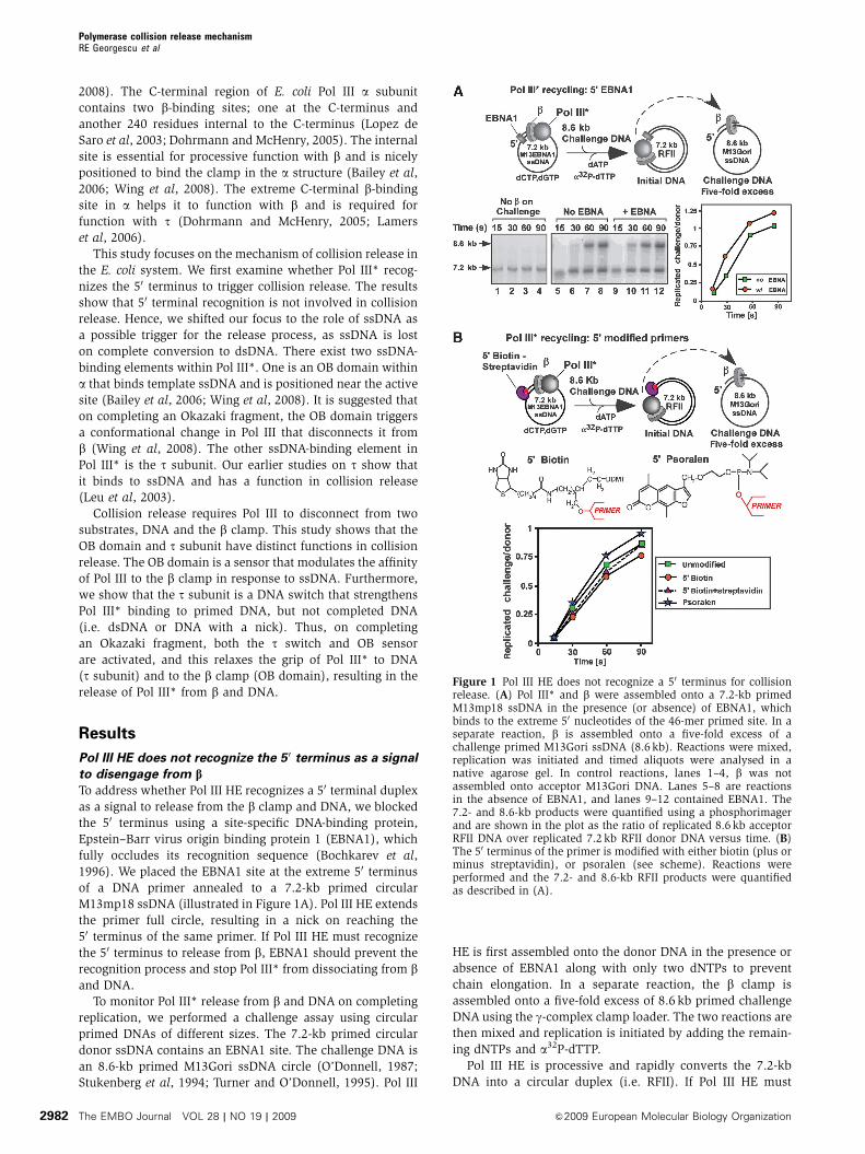
Figure 1 Pol III HE does not recognize a 50 terminus for collisionrelease. (A) Pol III* and b were assembled onto a 7.2-kb primedM13mp18 ssDNA in the presence (or absence) of EBNA1, whichbinds to the extreme 50 nucleotides of the 46-mer primed site. In aseparate reaction, b is assembled onto a five-fold excess of achallenge primed M13Gori ssDNA (8.6 kb). Reactions were mixed,replication was initiated and timed aliquots were analysed in anative agarose gel. In control reactions, lanes 1–4, b was notassembled onto acceptor M13Gori DNA. Lanes 5–8 are reactionsin the absence of EBNA1, and lanes 9–12 contained EBNA1. The7.2- and 8.6-kb products were quantified using a phosphorimagerand are shown in the plot as the ratio of replicated 8.6 kb acceptorRFII DNA over replicated 7.2 kb RFII donor DNA versus time. (B)The 50 terminus of the primer is modified with either biotin (plus orminus streptavidin), or psoralen (see scheme). Reactions wereperformed and the 7.2- and 8.6-kb RFII products were quantifiedas described in (A).
Polymerase collision release mechanismRE Georgescu et al
The EMBO Journal VOL 28 | NO 19 | 2009 &2009 European Molecular Biology Organization2982
recognize the 50 terminus for collision release, then EBNA1
should prevent this recognition and Pol III* will not release
and transfer to the 8.6-kb challenge DNA. On the other
hand, if Pol III HE does not need to recognize the 50
terminus it should still undergo collision release and cycle
to the challenge DNA to produce an 8.6-kb RFII product.
A control reaction confirms that the challenge DNA is only
replicated when it is pre-loaded with a b clamp (Figure 1A,
lanes 1–4).
In the absence of EBNA1, Pol III* dissociates on complet-
ing the 7.2-kb DNA and readily cycles to the 8.6-kb challenge
DNA (Figure 1A, lanes 5–8). In the presence of EBNA1, the
rate of 8.6 kb RFII product formation is the same as in the
absence of EBNA1 (Figure 1A, lanes 9–12 and quantitation to
the right), indicating that occluding the 50 terminus does not
prevent the release of Pol III*. To exclude the possibility that
Pol III HE binds the 50 terminus by displacing EBNA1, we
used 32P-labelled EBNA1 and analysed whether Pol III HE
displaces it on replicating the circular DNA (Supplementary
Figure S1). The results show that Pol III HE does not displace32P-EBNA1 from DNA on completing replication, confirming
that Pol III HE does not have access to the 50 terminus in the
challenge experiments.
It is possible that Pol III* dissociates from b through a
conformational change that occurs as a consequence of
bumping into a protein block, rather than by triggering the
collision release mechanism. Two lines of evidence indicate
that a block to forward progression does not destabilize
Pol III HE. First, Pol III* remains stably attached to b on
DNA after encountering a template lesion, indicating that
a sudden block to forward motion does not induce the
polymerase to dissociate (McInerney and O’Donnell, 2007).
Second, when an E. coli Pol III HE collides with an in-line
RNA polymerase, the replisome remains bound to DNA,
whereas it displaces the RNA polymerase and takes over
the mRNA primer to continue DNA synthesis (Pomerantz and
O’Donnell, 2008). This result indicates that encounter of a
protein block per se does not induce the polymerase to
release from DNA. Nevertheless, we cannot rigorously
exclude that a conformational change occurs when the
polymerase hits the EBNA1 block, inducing polymerase
dissociation.
To further support that 50 end recognition is not required
for collision release, we repeated the cycling experiments
but used an oligonucleotide that was biotinylated at the
50 terminus in the presence or absence of streptavidin (see
scheme in Figure 1B). The result shows that after Pol III HE
collides with the streptavidin bound to the 50 terminus, it
cycles to the challenge 8.6 kb DNA at the same rate as when it
collides with an unmodified primer (Figure 1B). We also used
a primer containing a bulky 50 psoralen moiety and again
observe transfer of polymerase to the challenge DNA
(Figure 1B). These results support the conclusion that colli-
sion release by Pol III HE does not require recognition of a 50
terminus.
Pol III* can dissociate from the 3 0 terminus before it
dissociates from the b clamp
If recognition of the 50 terminus does not trigger collision
release, it is possible that Pol III* detaches from the
30 terminus before it dissociates from the b clamp. If so,
Pol III*-b may remain together and slide along duplex DNA,
as suggested by an earlier study (O’Donnell and Kornberg,
1985). To test whether Pol III* dissociates from b and DNA on
colliding with a 50 terminus, we observed its behaviour on
encountering a short duplex in its path, during primer exten-
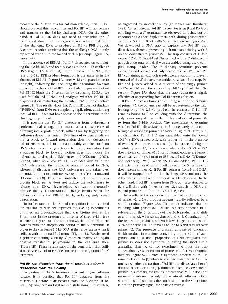
sion of a 5.4-kb fX174 ssDNA (see scheme in Figure 2A).
We developed a DNA trap to capture any Pol III* that
dissociates, thereby preventing it from reassociating with bon the downstream primer #2. The trap consists of 11-fold
excess 7.2 kb M13mp18 ssDNA primed with a 30 dideoxyoli-
gonucleotide onto which b was assembled using the g-com-
plex clamp loader. The 30 dideoxy terminus prevents
extension and subsequent polymerase release. We used Pol
III* containing an exonuclease-deficient e subunit to prevent
removal of the 30 dideoxynucleotide. As a test of the trap, Pol
III* and b were added to a mixture of the singly primed
fX174 ssDNA and the excess trap M13mp18 ssDNA. The
results (Figure 2A) show that the trap substrate is highly
effective at sequestering free Pol III* from solution.
If Pol III* releases from b on colliding with the 50 terminus
of primer #2, the polymerase will be sequestered by the trap,
leaving only the 2.3-kb product. In contrast, if Pol III*
remains bound to b on colliding with the 50 terminus, the
polymerase may slide over the duplex and extend primer #2
to form the 3.4-kb product. The experiment to examine
whether Pol III* dissociates from b immediately on encoun-
tering a downstream primer is shown in Figure 2B. First, sub-
stoichiometric Pol III HE was assembled onto the 5.4-kb
fX174 ssDNA primed only with primer #1 (in the presence
of two dNTPs to prevent extension). Then a second oligonu-
cleotide (primer #2) is rapidly annealed to the fX174 ssDNA
downstream of primer #1. Short oligonucleotides are known
to anneal rapidly (o1 min) to SSB-coated ssDNA (O’Donnell
and Kornberg, 1985). When dNTPs are added, Pol III HE
will extend primer #1 until it collides with the 50 terminus of
downstream primer #2. If Pol III* releases from b and DNA,
it will be trapped by b on the challenge DNA and only the
2-kb extension product of primer #1 will be observed. On the
other hand, if Pol III* releases from DNA but stays attached to
b, it will slide with b over primer #2, reattach to DNA and
extend primer #2 to form the 3.4-kb segment.
The results of the experiment show that in the presence
of primer #2, a 2-kb product appears, rapidly followed by a
3.4-kb product (Figure 2B). This result indicates that on
colliding with primer #2, Pol III* can stay attached to b,
release from the 30 terminus of the 2-kb product, and slide
over primer #2, whereas staying bound to b. Quantitation of
the replication products, shown below the gel, indicates that
35% of the time Pol III* remains bound to b and diffuses over
primer #2. The presence of a small amount of full-length
5.4 kb product in reactions containing primer #2 is a back-
ground due to a small proportion of DNA templates that
primer #2 does not hybridize to during the short 1-min
annealing time. A control experiment without the trap
shows about 75% extension of primer #2 after 60 s (Supple-
mentary Figure S2). Hence, a significant amount of Pol III*
remains bound to b, whereas it slides over primer #2. It is
unclear whether the portion of Pol III* that dissociates from bdoes so before, or during b diffusion over the downstream
primer. In summary, the results indicate that Pol III* does not
necessarily release precisely at the site of collision with a
50 terminus and supports the conclusion that the 50 terminus
is not the primary signal for collision release.
Polymerase collision release mechanismRE Georgescu et al
&2009 European Molecular Biology Organization The EMBO Journal VOL 28 | NO 19 | 2009 2983
The OB domain of Pol III a subunit is needed for
processive function with the b clamp
The experiments so far indicate that another trigger besides
50 duplex recognition underlies collision release of Pol III*
from b and DNA. On completing an Okazaki fragment, the
ssDNA template strand is completely converted into duplex
DNA, so the loss of contacts between the polymerase and
ssDNA could bring about collision release. An invariant
property of C-family DNA polymerases is the presence of an
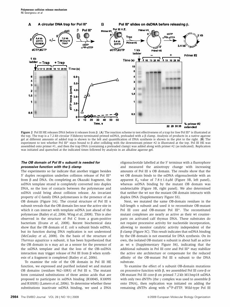
OB domain (Figure 3A). The crystal structure of Pol III asubunit reveals that the OB domain lies near the active site in
which it can interact with template ssDNA just ahead of the
polymerase (Bailey et al, 2006; Wing et al, 2008). This is also
observed in the structure of Pol C from a gram-positive
bacterium (Evans et al, 2008). Recent biochemical data
show that the OB domain of E. coli a subunit binds ssDNA,
but its function during DNA replication is not understood
(McCauley et al, 2008). On the basis of the structure of
Thermus aquaticus a subunit, it has been hypothesized that
the OB domain in a may act as a sensor for the presence of
the ssDNA template and that the loss of the OB–ssDNA
interaction may trigger release of Pol III from b when synth-
esis of a fragment is completed (Bailey et al, 2006).
To examine the role of the OB domain in Pol III HE
function, we expressed and purified isolated wt and mutant
OB domains (residues 962–1083) of Pol III a. The mutant
form contained substitutions of three amino acids that are
proposed to participate in ssDNA binding (R1004S, K1009S
and R1010S) (Lamers et al, 2006). To determine whether these
substitutions inactivate ssDNA binding, we used a DNA
oligonucleotide labelled at the 50 terminus with a fluorophore
and measured the anisotropy change with increasing
amounts of Pol III a OB domain. The results show that the
wt OB domain binds to the ssDNA oligonucleotide with an
apparent Kd value of 7.8±1.6 mM (Figure 3B, left panel),
whereas ssDNA binding by the mutant OB domain was
undetectable (Figure 3B, right panel). We also determined
that neither the wt nor the mutant OB domain interacts with
duplex DNA (Supplementary Figure S3).
Next, we mutated the same OB-domain residues in the
full-length a subunit and used it to reconstitute OB-mutant
Pol III core and OB-mutant Pol III*. The reconstituted
mutant complexes are nearly as active as their wt counter-
parts on activated calf thymus DNA. These substrates do
not require processive activity for nucleotide incorporation,
allowing to monitor catalytic activity independent of the
b clamp (Figure 3C). This result indicates that ssDNA binding
by the OB domain is not essential for DNA synthesis. On its
own, the isolated OB-mutant a subunit is about half as active
as wt a (Supplementary Figure S4), indicating that the
additional subunits in Pol III core and Pol III* may stabilize
the active site architecture or compensate for the reduced
affinity of the OB-mutant Pol III a subunit to the DNA
substrate.
To examine the effect of a subunit OB-domain mutations
on processive function with b, we assembled Pol III core-b or
OB-mutant Pol III core-b on primed 7.2 kb M13mp18 ssDNA
with only two dNTPs (the g complex was used to assemble bonto DNA), then replication was initiated on adding the
remaining dNTPs along with a32P-dTTP. Wild-type Pol III
Figure 2 Pol III HE releases DNA before it releases from b. (A) The reaction scheme to test effectiveness of a trap for free Pol III* is illustrated atthe top. The trap is a 7.2-kb circular 30dideoxy-terminated primed ssDNA, preloaded with a b clamp. Analysis of products in a native agarosegel at different amounts of added trap is shown to the left and quantification of DNA synthesis is shown in the plot to the right. (B) Theexperiment to test whether Pol III* stays bound to b after colliding with the downstream primer #2 is illustrated at the top. Pol III HE wasassembled onto primer #1, and then the trap DNA (containing a preloaded clamp) was added along with primer #2 (as indicated). Replicationwas initiated and quenched at the indicated times followed by analysis in an alkaline agarose gel.
Polymerase collision release mechanismRE Georgescu et al
The EMBO Journal VOL 28 | NO 19 | 2009 &2009 European Molecular Biology Organization2984
core-b rapidly converts the 7.2-kb substrate to a RFII circular
duplex (Figure 3D, lanes 1–4). In contrast, the OB-mutant Pol
III core-b yields only short products, indicating that the
OB-mutant Pol III core is not processive with b (lanes 5–8,
quantitation is in Supplementary Figure 5A). Hence, ssDNA
binding by the OB domain of a is required for processive
function of Pol III core with b.
In summary, these results show that the OB domain of Pol
III a binds ssDNA (Figure 3B) and that ssDNA binding is not
required for intrinsic DNA polymerase activity (Figure 3C)
but is required for processive function with the b clamp
(Figure 3D). We show later in this report that ssDNA
binding by the OB domain is required for optimal binding
of a to b specifically at a primer/terminus, and that this
accounts for the deficiency of the OB-mutant Pol III in
function with b.
The s subunit partially rescues the OB-domain mutant
The defect in processivity of the OB-mutant Pol III core may
be explained by an altered affinity of Pol III core for DNA or
the b clamp (or both). The t subunit of the clamp loader is
known to increase the binding of Pol III core to the b clamp
on DNA (Stukenberg and O’Donnell, 1995), and therefore one
may predict that the t subunit will partially compensate for
ssDNA-binding mutations in the OB domain of Pol III core.
To examine this possibility, we tested the a OB mutant
reconstituted into Pol III* (which contains t) in b-dependent
assays using primed M13mp18 ssDNA. The results, in
Figure 3E lanes 7–12, show that the OB-mutant Pol III*-bproduces full-length RFII duplex products, although it is less
efficient and generates many short products compared with
wt Pol III*-b (Figure 3E, lanes 1–6; see Supplementary Figure
5B for quantitation). Hence, the t subunit partially rescues
the defective processivity of the OB-mutant Pol III core in
b-dependent synthesis.
OB-mutant Pol III* is more deficient on the lagging
strand than the leading strand
The results obtained thus far indicate that the OB–ssDNA
interaction is needed for processive synthesis of Pol III core
with b, and thus one may expect that the OB mutant will not
function properly on either the leading or the lagging strand.
However, there may be no ssDNA on the leading strand, as
leading strand synthesis does not require SSB (Mok and
Marians, 1987). In this case, leading strand synthesis should
not be hindered by OB mutations that eliminate ssDNA bind-
ing. Furthermore, the leading strand polymerase connects to
DnaB helicase through the t subunit (Dallmann et al, 2000),
and this polymerase-helicase connection may confer proces-
sivity to the leading strand polymerase as shown earlier in the
T4 and T7 systems (Xi et al, 2005; Johnson et al, 2007).
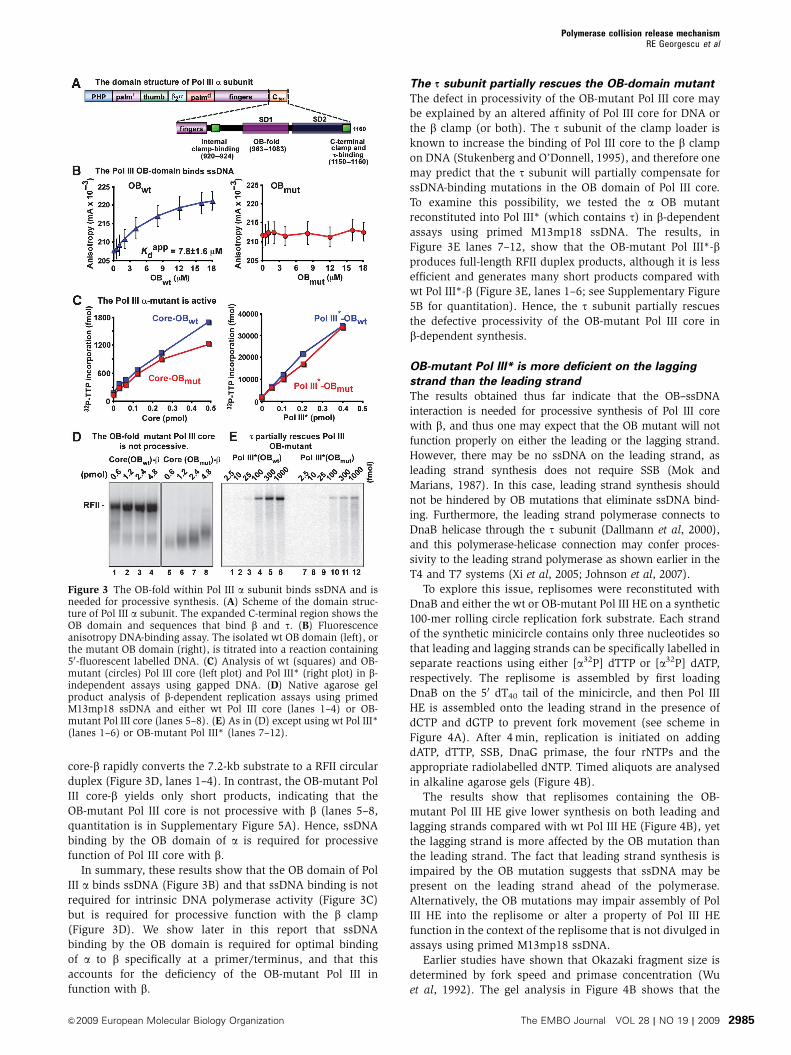
To explore this issue, replisomes were reconstituted with
DnaB and either the wt or OB-mutant Pol III HE on a synthetic
100-mer rolling circle replication fork substrate. Each strand
of the synthetic minicircle contains only three nucleotides so
that leading and lagging strands can be specifically labelled in
separate reactions using either [a32P] dTTP or [a32P] dATP,
respectively. The replisome is assembled by first loading
DnaB on the 50 dT40 tail of the minicircle, and then Pol III
HE is assembled onto the leading strand in the presence of
dCTP and dGTP to prevent fork movement (see scheme in
Figure 4A). After 4 min, replication is initiated on adding
dATP, dTTP, SSB, DnaG primase, the four rNTPs and the
appropriate radiolabelled dNTP. Timed aliquots are analysed
in alkaline agarose gels (Figure 4B).
The results show that replisomes containing the OB-
mutant Pol III HE give lower synthesis on both leading and
lagging strands compared with wt Pol III HE (Figure 4B), yet
the lagging strand is more affected by the OB mutation than
the leading strand. The fact that leading strand synthesis is
impaired by the OB mutation suggests that ssDNA may be
present on the leading strand ahead of the polymerase.
Alternatively, the OB mutations may impair assembly of Pol
III HE into the replisome or alter a property of Pol III HE
function in the context of the replisome that is not divulged in
assays using primed M13mp18 ssDNA.
Earlier studies have shown that Okazaki fragment size is
determined by fork speed and primase concentration (Wu
et al, 1992). The gel analysis in Figure 4B shows that the
Figure 3 The OB-fold within Pol III a subunit binds ssDNA and isneeded for processive synthesis. (A) Scheme of the domain struc-ture of Pol III a subunit. The expanded C-terminal region shows theOB domain and sequences that bind b and t. (B) Fluorescenceanisotropy DNA-binding assay. The isolated wt OB domain (left), orthe mutant OB domain (right), is titrated into a reaction containing50-fluorescent labelled DNA. (C) Analysis of wt (squares) and OB-mutant (circles) Pol III core (left plot) and Pol III* (right plot) in b-independent assays using gapped DNA. (D) Native agarose gelproduct analysis of b-dependent replication assays using primedM13mp18 ssDNA and either wt Pol III core (lanes 1–4) or OB-mutant Pol III core (lanes 5–8). (E) As in (D) except using wt Pol III*(lanes 1–6) or OB-mutant Pol III* (lanes 7–12).
Polymerase collision release mechanismRE Georgescu et al
&2009 European Molecular Biology Organization The EMBO Journal VOL 28 | NO 19 | 2009 2985

speed of the leading strand OB-mutant replisome is the same
as the wt replisome. Therefore, as primase concentration is
held constant in these experiments, the lagging strand should
be primed at the same frequency and should produce similar
sized Okazaki fragments. However, the results clearly show
a decrease in Okazaki fragment size from 1 to 0.5 kb for the
OB-mutant replisome (Figure 4B, right). Furthermore, quan-
titation of the products shows a much greater reduction of
lagging strand synthesis by the OB-mutant replisome than
of leading strand synthesis (23% of wt versus 53% of wt,
respectively, Figure 4B, bottom). Therefore, the lagging
strand OB-mutant Pol III probably releases from most
Okazaki fragments well before they are complete.
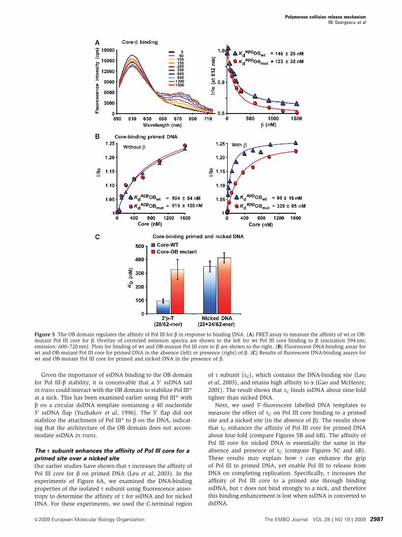
Binding of the OB domain to ssDNA regulates the
affinity of Pol III for bTo understand why the OB-mutant Pol III core is less proces-
sive with b, we developed fluorescence-based assays to
measure the affinity of wt and OB-mutant Pol III for DNA
and the b clamp. To measure the affinity of Pol III core for the
b clamp, Pol III core was reconstituted using a y subunit
labelled with AlexaFlor594 (FRET donor) and the b clamp
was labelled using AlexaFlor647 (FRET acceptor). FRET
assays were performed by titrating b into a constant amount
of Pol III core, and reactions were excited at 594 nm.
Representative fluorescence emission spectra during a titra-
tion of b into wt Pol III core are shown in the left panel of
Figure 5A. Binding of b to Pol III core gives a FRET signal,
manifested by a decrease in fluorescence intensity at 612 nm
and a corresponding fluorescence intensity increase at
680 nm (due to emission from the acceptor that is excited
by resonance energy transfer from the donor). Figure 5A
(right panel) shows the fluorescence intensity change of wt or
OB-mutant Pol III core at increasing b concentration.
Quantitation of the results shows that wt and OB-mutant
Pol III core bind to b with similar affinity. Hence, the OB
mutations do not affect binding of Pol III core to the b clamp
in the absence of DNA.
To measure the affinity of wt and OB-mutant Pol III core for
primer/template DNA, we used a 34/62-mer primed site
containing a fluorescent moiety on the template strand, 21
residues from the 50 terminus. This substrate registers a
change in the fluorescence intensity on binding Pol III core.
Titrations of wt and OB-mutant Pol III core into reactions
containing the primed template are shown in the left panel of
Figure 5B. Although the OB domain interacts with ssDNA
(Figure 3B), we observed no significant difference in affinity
between wt and OB-mutant Pol III core for the primed site,
suggesting that ssDNA interaction by the OB domain does not
directly stabilize Pol III on DNA in the absence of b.
As the OB-mutant Pol III core is clearly defective in
function with b (Figure 3D) yet binds to b in the absence
of DNA with similar affinity to wt Pol III core (Figure 5A),
it seemed possible that the OB mutant may be defective in
binding to b when the clamp is on DNA. To test this we
measured the affinity of wt and OB-mutant Pol III core for a
primed site in the presence of b (Figure 5B, right panel). As bcan slide off the short linear DNA, the effect of b will be
underestimated in this assay. Nonetheless, we observed a
five-fold enhancement in affinity of wt Pol III core for the
b–DNA complex relative to DNA alone, compared with only
two-fold enhancement for OB-mutant Pol III core. These
results indicate that when the OB domain binds template
ssDNA, it increases the affinity of Pol III core for b. To confirm
that this effect is specific for a primer/template, we measured
the affinity of wt and OB-mutant Pol III core for a fluores-
cently labelled nicked substrate in the presence of the bclamp; as expected there was no significant difference be-
tween wt and OB-mutant Pol III binding to nicked DNA
(Figure 5C). The measured Kd values of wt and mutant OB
Pol III core binding to a nick, are comparable to the Kd value
of OB-mutant Pol III core binding to a primed substrate
(Figure 5B, left panel).
To determine whether Pol III begins to lose its affinity for band the DNA template before reaching the terminal nucleo-
tide, we measured the affinity of wt Pol III core to a synthetic
62-mer DNA substrate designed to contain ssDNA gaps of
defined length (Supplementary Figure S6). We observe that
the affinity of Pol III core for b and DNA decreases gradually
as the ssDNA gap is brought close to a nick. This result is
consistent with the position of the OB domain in the Taq
a-subunit structure, which indicates that the OB domain
interacts with ssDNA several residues ahead of the polymer-
ase active site (Wing et al, 2008) We presume that the koff is
slow relative to the rate of synthesis, accounting for the
observed synthesis of Pol III* HE to the last nucleotide of a
primed M13mp18 circular substrate (Stukenberg et al, 1994).
Figure 4 The OB-mutant Pol III HE is deficient in lagging strandsynthesis. (A) Scheme of replisome assembly on a minicircle replica-tion fork substrate using DnaB helicase and wt or OB-mutant Pol IIIHE (B) Leading (left panel) and lagging (right panel) strands can beselectively labelled depending on whether a-32P dATP or a-32P dTTP ispresent during synthesis. Aliquots were removed at the indicatedtimes and analysed in a 1.2% alkaline agarose gel followed byautoradiography. The relative level of DNA synthesis at 320 s isshown at the bottom of the gel.
Polymerase collision release mechanismRE Georgescu et al
The EMBO Journal VOL 28 | NO 19 | 2009 &2009 European Molecular Biology Organization2986
Given the importance of ssDNA binding to the OB domain
for Pol III-b stability, it is conceivable that a 50 ssDNA tail
in trans could interact with the OB domain to stabilize Pol III*
at a nick. This has been examined earlier using Pol III* with
b on a circular dsDNA template containing a 40 nucleotide
50 ssDNA flap (Yuzhakov et al, 1996). The 50 flap did not
stabilize the attachment of Pol III* to b on the DNA, indicat-
ing that the architecture of the OB domain does not accom-
modate ssDNA in trans.
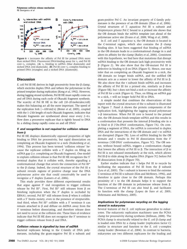
The s subunit enhances the affinity of Pol III core for a
primed site over a nicked site
Our earlier studies have shown that t increases the affinity of
Pol III core for b on primed DNA (Leu et al, 2003). In the
experiments of Figure 6A, we examined the DNA-binding
properties of the isolated t subunit using fluorescence aniso-
tropy to determine the affinity of t for ssDNA and for nicked
DNA. For these experiments, we used the C-terminal region
of t subunit (tC), which contains the DNA-binding site (Leu
et al, 2003), and retains high affinity to a (Gao and McHenry,
2001). The result shows that tC binds ssDNA about nine-fold
tighter than nicked DNA.
Next, we used 50-fluorescent labelled DNA templates to
measure the effect of tC on Pol III core binding to a primed
site and a nicked site (in the absence of b). The results show
that tC enhances the affinity of Pol III core for primed DNA
about four-fold (compare Figures 5B and 6B). The affinity of
Pol III core for nicked DNA is essentially the same in the
absence and presence of tC (compare Figures 5C and 6B).
These results may explain how t can enhance the grip
of Pol III to primed DNA, yet enable Pol III to release from
DNA on completing replication. Specifically, t increases the
affinity of Pol III core to a primed site through binding
ssDNA, but t does not bind strongly to a nick, and therefore
this binding enhancement is lost when ssDNA is converted to
dsDNA.
Figure 5 The OB domain regulates the affinity of Pol III for b in response to binding DNA. (A) FRETassay to measure the affinity of wt or OB-mutant Pol III core for b. Overlay of corrected emission spectra are shown to the left for wt Pol III core binding to b (excitation 594 nm;emission: 600–720 nm). Plots for binding of wt and OB-mutant Pol III core to b are shown to the right. (B) Fluorescent DNA-binding assay forwt and OB-mutant Pol III core for primed DNA in the absence (left) or presence (right) of b. (C) Results of fluorescent DNA-binding assays forwt and OB-mutant Pol III core for primed and nicked DNA in the presence of b.
Polymerase collision release mechanismRE Georgescu et al
&2009 European Molecular Biology Organization The EMBO Journal VOL 28 | NO 19 | 2009 2987
Discussion
E. coli Pol III HE derives its high processivity from the b clamp,
which encircles duplex DNA and tethers the polymerase to the
primed template during replication (Kong et al, 1992). However,
during lagging strand synthesis, Pol III HE must rapidly come on
and off DNA during each cycle of Okazaki fragment synthesis.
The scarcity of Pol III HE in the cell (10–20 molecules/cell)
makes this balancing act all the more important. The speed of
the replication fork (B650 ntd/s) (Breier et al, 2005), coupled
with the 1–2 kb length of each Okazaki fragment, indicates that
Okazaki fragments are synthesized about once every 2–4 s.
How does a processive replicase that is tightly bound to DNA
by a sliding clamp rapidly come on and off DNA?
50 end recognition is not required for collision release
from bPol III HE displays diametrically opposed properties of tight
binding to DNA for processivity, but rapid dissociation on
completing an Okazaki fragment to a nick (Stukenberg et al,
1994). This process has been termed ‘collision release’ be-
cause the replicase collides with a 50 duplex on filling an
ssDNA gap (i.e. completing an Okazaki fragment). One model
to explain collision release is that Pol III HE recognizes the 50
terminal duplex that it collides with, thereby signalling a
conformational change that severs the connection of Pol III*
to the b clamp. Indeed, the crystal structure of the Pol III asubunit reveals regions of positive charge near the DNA
polymerase active site that could conceivably be used to
recognize a 50 duplex (Lamers et al, 2006).
This study provides three different experimental results
that argue against 50 end recognition to trigger collision
release by Pol III*. First, Pol III* still releases from b on
finishing replication when the 50 duplex is occluded by
EBNA1; second, Pol III* still releases from b when it collides
with a 50 biotin moiety, even in the presence of streptavidin.
And third, when Pol III* collides with a 50 terminus it can
remain attached to b and diffuse on dsDNA away from the
50 terminus before dissociating, and therefore release does
not need to occur at the collision site. These lines of evidence
indicate that Pol III HE does not recognize the 50 terminus to
trigger collision release from b and DNA.
Collision release is signalled by loss of ssDNA
Bacterial replicases belong to the C-family of DNA poly-
merases, including the gram-negative Pol III a subunit and
gram-positive Pol C. An invariant property of C-family poly-
merases is the presence of an OB domain (Zhao et al, 2006).
Crystal structures of T. aquaticus Pol III a subunit and
Streptomyces griseus Pol C bound to a primed site shows that
the OB domain binds the ssDNA template just ahead of the
polymerase active site (Evans et al, 2008; Wing et al, 2008).
In E. coli and T. aquaticus a, the OB domain is located in
the C-terminal region, which also contains the b clamp
binding sites. It has been suggested that binding of ssDNA
to the OB domain leads to a conformational change in a and
alters its affinity for the clamp (Bailey et al, 2006). Consistent
with this hypothesis, we find here that mutations that disrupt
ssDNA binding to the OB domain lack high processivity with
b (Figure 2). We also show that the OB-mutant Pol III is
defective in binding to b on DNA (Figure 5). Hence, one may
infer that on completing an Okazaki fragment to a nick, the
OB domain no longer binds ssDNA, and the unfilled OB
domain acts as a sensor to lower the affinity of Pol III to b.
We also show that the t subunit binds ssDNA and increases
the affinity of Pol III for a primed site, similarly as b does
(Figure 5B); but t does not bind a nick or increase the affinity
of Pol III for a nick (Figure 6). Thus, on filling an ssDNA gap
to a nick, t will no longer hold Pol III to DNA.
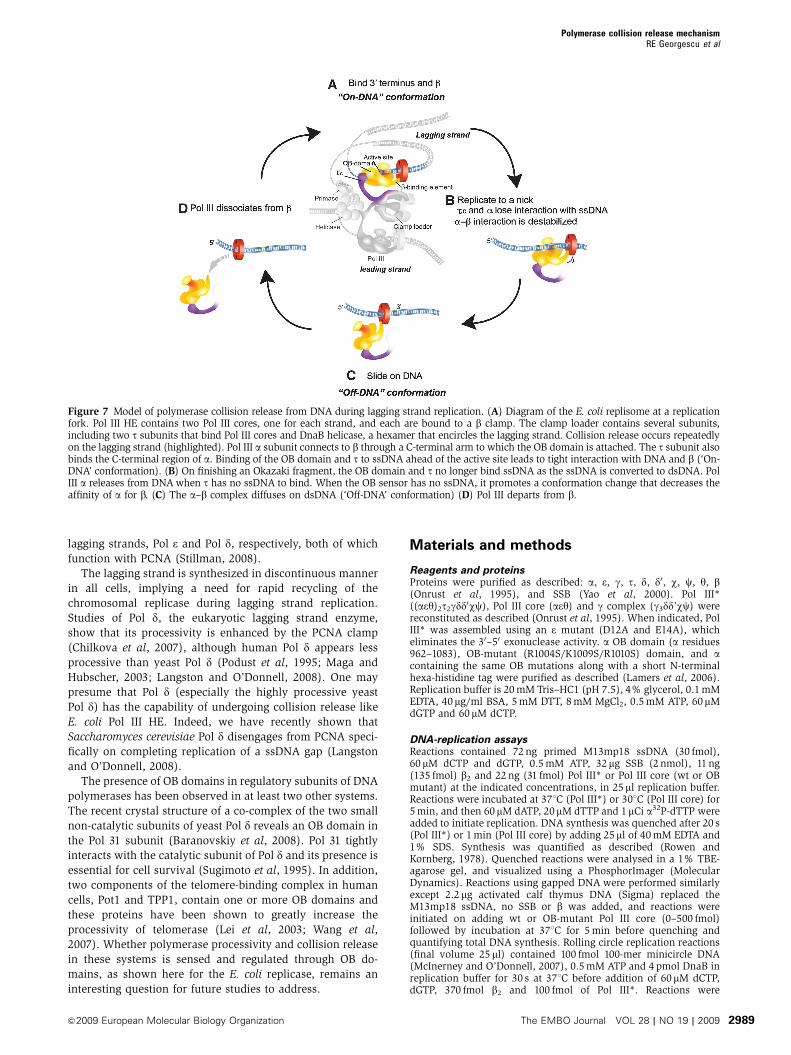
A simple model that combines the observations of this
report with the crystal structure of the a subunit is illustrated
in Figure 7. Panel A shows the protein components of the
replication fork, highlighting the lagging strand polymerase
t–b interactions. When Pol III core is bound to the primed
site, the OB domain binds template ssDNA and this results in
a conformation that presents the internal b-binding site in ato bind to b (‘On-DNA’ conformation, Figure 7A). On com-
pleting a segment of DNA, the ssDNA is converted to duplex
DNA and the interactions of the OB domain and t to ssDNA
are disrupted (Figure 7B). Loss of ssDNA binding by the OB
domain and t results in Pol III dissociation from the 30
terminus (‘Off-DNA’ conformation, Figure 7C). The OB sen-
sor, without bound ssDNA, triggers a conformation change
that lowers the affinity of Pol III to b. The interaction of b to
Pol III is not released immediately at the nick, which allows
Pol III-b to slide along the duplex DNA (Figure 7C) before Pol
III dissociation from b (Figure 7D).
Earlier studies indicate that t helps Pol III to recycle by
facilitating the separation of Pol III from the b subunit
(Leu et al, 2003). The t subunit is known to bind the extreme
C-terminus of Pol III a subunit (Kim and McHenry, 1996), and
therefore is quite close to the OB domain. Perhaps this
proximity of t to the OB domain helps it to support the
function of the OB domain in polymerase release from b.
The C-terminus of Pol III can also bind b, and facilitates
its function with the clamp (Lopez de Saro et al, 2003;
Dohrmann and McHenry, 2005).
Implications for polymerase recycling on the lagging
strand in eukaryotes
Several features of the E. coli replicase generalize to eukar-
yotes. The eukaryotic replication machinery uses the PCNA
clamp for processivity during synthesis (Stillman, 2008). The
PCNA clamp is structurally related to the E. coli b clamp and
is loaded onto DNA by a clamp loader (replication factor C)
similar in structure and function to the E. coli g-complex
clamp loader (Bowman et al, 2004). In contrast to bacteria,
eukaryotes use two different replicases for the leading and
Figure 6 t increases the affinity of Pol III for primed DNA morethan nicked DNA. Fluorescent DNA-binding assay for tC and Pol IIIcore–tC complex. (A) tC binding to ssDNA (blue diamonds) andnicked DNA (red diamonds). (B) Pol III core–tC complex binding toprimed DNA (triangles) and a nicked DNA (diamonds).
Polymerase collision release mechanismRE Georgescu et al
The EMBO Journal VOL 28 | NO 19 | 2009 &2009 European Molecular Biology Organization2988
lagging strands, Pol e and Pol d, respectively, both of which
function with PCNA (Stillman, 2008).
The lagging strand is synthesized in discontinuous manner
in all cells, implying a need for rapid recycling of the
chromosomal replicase during lagging strand replication.
Studies of Pol d, the eukaryotic lagging strand enzyme,
show that its processivity is enhanced by the PCNA clamp
(Chilkova et al, 2007), although human Pol d appears less
processive than yeast Pol d (Podust et al, 1995; Maga and
Hubscher, 2003; Langston and O’Donnell, 2008). One may
presume that Pol d (especially the highly processive yeast
Pol d) has the capability of undergoing collision release like
E. coli Pol III HE. Indeed, we have recently shown that
Saccharomyces cerevisiae Pol d disengages from PCNA speci-
fically on completing replication of a ssDNA gap (Langston
and O’Donnell, 2008).
The presence of OB domains in regulatory subunits of DNA
polymerases has been observed in at least two other systems.
The recent crystal structure of a co-complex of the two small
non-catalytic subunits of yeast Pol d reveals an OB domain in
the Pol 31 subunit (Baranovskiy et al, 2008). Pol 31 tightly
interacts with the catalytic subunit of Pol d and its presence is
essential for cell survival (Sugimoto et al, 1995). In addition,
two components of the telomere-binding complex in human
cells, Pot1 and TPP1, contain one or more OB domains and
these proteins have been shown to greatly increase the
processivity of telomerase (Lei et al, 2003; Wang et al,
2007). Whether polymerase processivity and collision release
in these systems is sensed and regulated through OB do-
mains, as shown here for the E. coli replicase, remains an
interesting question for future studies to address.
Materials and methods
Reagents and proteinsProteins were purified as described: a, e, g, t, d, d0, w, c, y, b(Onrust et al, 1995), and SSB (Yao et al, 2000). Pol III*((aey)2t2gdd0wc), Pol III core (aey) and g complex (g3dd’wc) werereconstituted as described (Onrust et al, 1995). When indicated, PolIII* was assembled using an e mutant (D12A and E14A), whicheliminates the 30–50 exonuclease activity. a OB domain (a residues962–1083), OB-mutant (R1004S/K1009S/R1010S) domain, and acontaining the same OB mutations along with a short N-terminalhexa-histidine tag were purified as described (Lamers et al, 2006).Replication buffer is 20 mM Tris–HC1 (pH 7.5), 4% glycerol, 0.1 mMEDTA, 40mg/ml BSA, 5 mM DTT, 8 mM MgCl2, 0.5 mM ATP, 60mMdGTP and 60 mM dCTP.
DNA-replication assaysReactions contained 72 ng primed M13mp18 ssDNA (30 fmol),60 mM dCTP and dGTP, 0.5 mM ATP, 32mg SSB (2 nmol), 11 ng(135 fmol) b2 and 22 ng (31 fmol) Pol III* or Pol III core (wt or OBmutant) at the indicated concentrations, in 25ml replication buffer.Reactions were incubated at 371C (Pol III*) or 301C (Pol III core) for5 min, and then 60 mM dATP, 20mM dTTP and 1mCi a32P-dTTP wereadded to initiate replication. DNA synthesis was quenched after 20 s(Pol III*) or 1 min (Pol III core) by adding 25ml of 40 mM EDTA and1% SDS. Synthesis was quantified as described (Rowen andKornberg, 1978). Quenched reactions were analysed in a 1% TBE-agarose gel, and visualized using a PhosphorImager (MolecularDynamics). Reactions using gapped DNA were performed similarlyexcept 2.2 mg activated calf thymus DNA (Sigma) replaced theM13mp18 ssDNA, no SSB or b was added, and reactions wereinitiated on adding wt or OB-mutant Pol III core (0–500 fmol)followed by incubation at 371C for 5 min before quenching andquantifying total DNA synthesis. Rolling circle replication reactions(final volume 25 ml) contained 100 fmol 100-mer minicircle DNA(McInerney and O’Donnell, 2007), 0.5 mM ATP and 4 pmol DnaB inreplication buffer for 30 s at 371C before addition of 60mM dCTP,dGTP, 370 fmol b2 and 100 fmol of Pol III*. Reactions were
Figure 7 Model of polymerase collision release from DNA during lagging strand replication. (A) Diagram of the E. coli replisome at a replicationfork. Pol III HE contains two Pol III cores, one for each strand, and each are bound to a b clamp. The clamp loader contains several subunits,including two t subunits that bind Pol III cores and DnaB helicase, a hexamer that encircles the lagging strand. Collision release occurs repeatedlyon the lagging strand (highlighted). Pol III a subunit connects to b through a C-terminal arm to which the OB domain is attached. The t subunit alsobinds the C-terminal region of a. Binding of the OB domain and t to ssDNA ahead of the active site leads to tight interaction with DNA and b (‘On-DNA’ conformation). (B) On finishing an Okazaki fragment, the OB domain and t no longer bind ssDNA as the ssDNA is converted to dsDNA. PolIII a releases from DNA when t has no ssDNA to bind. When the OB sensor has no ssDNA, it promotes a conformation change that decreases theaffinity of a for b. (C) The a–b complex diffuses on dsDNA (‘Off-DNA’ conformation) (D) Pol III departs from b.
Polymerase collision release mechanismRE Georgescu et al
&2009 European Molecular Biology Organization The EMBO Journal VOL 28 | NO 19 | 2009 2989
incubated at 371C for 4 min before addition of 13 pmol SSB, 50 mMeach of CTP, GTP, UTP, 60 mM dATP, 10mM dTTP, 1mCi a32P-dTTPand 150 pmol DnaG to initiate replication. Reactions were processedas described above.
Polymerase-cycling assaysInitial (donor) primed 7.2 kb M13mp18 ssDNA (containing anEBNA1 site) was prepared by annealing a DNA 30-mer (unmodifiedor 50 modification with biotin or psoralen), or a 46-mer containingthe 18-bp EBNA1 site at the extreme 50 terminus. The challenge8.6 kb M13Gori ssDNA was primed with a DNA 30-mer. Donorreactions were assembled in 47 ml of replication buffer containing144 ng (60 fmol) primed 7.2 kb M13mp18 ssDNA, 1.6 mg (84 pmol)SSB, 22 ng (270 fmol) b2, and 44 ng (62 fmol) Pol III*, followed by a3-min incubation at 371C. When present, EBNA1 (7.5 pmol) wasadded to primed M13mp ssDNA and incubated 1 min before addingother proteions. Challenge DNA reactions were assembled in 47mlof replication buffer containing 720 ng 8.6 kb challenge primedM13Gori ssDNA, 4 mg (53 pmol) SSB, 66 ng (815 fmol) b, and 10 ng(52 fmol) g complex followed by 3 min at 371C. Replication wasinitiated by mixing the donor with challenge reactions and adding6ml of 1.5 mM dATP, 0.5 mM [a-32P]dTTP. Aliquots were removedand processed as described above.
Fluorescent OB domain DNA-binding assaysIncreasing amounts of wt or mutant OB domains were titrated intoreactions containing 1mM ssDNA (Cy5-28-mer) or dsDNA (Cy5-28/28-mer) in 20 mM Tris–HCl pH 7.5, 10% glycerol, 75 mM NaCl,0.5 mM EDTA, 20 mM arginine, 30 mM glutamate, at 231C. Cy5-28-mer was (50-Cy5-CCCATCGTATACGAAGGGAGTCGACTGG-30). Cy5-dsDNA was prepared by annealing the above oligonucleotide withits complementary 28-mer. Anisotropy was measured in a PTIspectrofluorimeter configured in the T-format and using the time-trace function to generate 35 data points, which were averaged.Anisotropy is defined by anisotropy¼ (IVV�IVH)/(IVVþ 2IVH), whereIVV and IVH are the intensities of the vertical and horizontalcomponents of the emitted light using vertical polarized excitation.Differences in the response of the detector to vertical and horizontalpolarized light were corrected (G factor) automatically by thespectrofluorimeter.
Fluorescent Pol III core DNA-binding assaysAnalysis of Pol III core plus and minus t were as follows. Increasingamounts of Pol III core or Pol III core–tC complex (0–1.5 mM finalconcentration) were titrated into reactions containing 10 nM ofeither fluorescent primed template [34/62(50F)], or singly nickeddsDNA [34/28/62(50F)] in 20 mM Tris–HCl pH 7.5, 10% glycerol,25 mM NaCl, 0.5 mM EDTA, 1 mM DTT. After 30 min at 231C,anisotropy measurements were performed as described above.Control titrations were performed using only tC. Pol III core-DNA-bbinding reactions used an internal fluorescein label on the 62-mer[62(iF): 50-CCCATCGTATACGAAGGGAGTCGACTGAGCTAGCTAGC/iF-T/CTATAGCTAGCTAGCTAGCT] annealed to a complementary34-mer to form a 28-mer 50overhang. Pol III core/tC-DNA-bindingreactions used the same 62-mer sequence, but 50-end labelled withfluorescein, annealed either to the 34-mer, to form a 28-mer50overhang, or with the 34-mer and a complementary 28-mer, toform a nicked duplex [34/28/62(50F)].
Analysis of Pol III core binding to b was as follows. Increasingamounts of wt or OB-mutant Pol III core (0–1.5mM final
concentration) were titrated into reactions containing 10 nMfluorescent primed template [34/62(iF)] in 20 mM Tris–HCl pH7.5, 10% glycerol, 75 mM NaCl, 1 mM DTT, 0.5 mM EDTA(performed in the absence or presence of 50 nM b). After 20 minat 231C, samples were excited at 490 nm and emission (500–600 nm) was recorded using a PTI spectrofluorometer. For dataanalysis, binding isotherms were constructed under the simplifyingassumption that the DNA concentration is adequately below the Kd
such that total added protein is equivalent to free protein. Theequilibrium titration data were fit to a single-site binding model.
FRET Pol III core b-binding assayPol III core was reconstituted using a y subunit labelled withAlexaFlor594 (FRET donor) and the b clamp was labelled usingAlexaFlor647 (FRET acceptor) using maleimide chemistry (Invitro-gen, Molecular Probes). Reaction contained 100 nM wt or OB-mutant Pol III core and variable amounts of b2 (0–1800 nM) in 60 mlof 20 mM Tris–HCl (pH 7.5), 1 mM DTT, 0.5 mM EDTA. Reactionswere incubated for 15 min at 221C and then analysed in a PTIspectrofluorimeter. Fluorescence resulting from direct (i.e. non-sensitized) b2-AlexaFlor647 excitation was eliminated by subtract-ing the spectra of b2-AlexaFlor647 in the absence of Pol III core.
Analysis of Pol III HE sliding over an intramolecular duplexReactions contained 141 fmol of fX174 ssDNA pre-annealed withprimer #1, 75 fmol Pol III*, 6.2 pmol b2, 132 pmol SSB and were pre-incubated at 301C for 2 min in replication buffer containing 60 mMdCTP and dGTP. The trap was prepared by loading 32 pmol b clamponto 840 fmol M13mp18: Pr3BddC using 2.5 pmol g complex inreplication buffer containing 60mM dATP, 20mM dTTP and 1mCi[a-32P]dTTP. Primer #2 was annealed for 1 min as described(O’Donnell and Kornberg, 1985) before initiating replication onadding the M13mp18 trap (final volume 25ml). Reactions wereincubated at 301C for the indicated times and DNA products wereanalysed in a 1.2% alkaline agarose gel. Effectiveness of the trapwas assessed by mixing reactions before initiating synthesis onadding Pol III*. Primers for fX174 ssDNA were primer #1 (30-mer)that anneals to map position 2794–2823, and primer #2 (15-mer)that anneals to map position 791–805. The trap used a 30dd 30-merannealed to M13mp18 ssDNA: (50-GTTAAAGGCCGCTTTTGCGGGATCGTCACddC-30).
Supplementary dataSupplementary data are available at The EMBO Journal Online(http://www.embojournal.org).
Acknowledgements
We are grateful to Lance Langston for helpful suggestions andcritical reading of the manuscript. We also thank Maija Skangalisand other members of the laboratory for help in purifying some ofthe proteins used in this study. This work was supported by a grantfrom the NIH (GM38839).
Conflict of interest
The authors declare that they have no conflict of interest.
References
Bailey S, Wing RA, Steitz TA (2006) The structure of T. aquaticusDNA polymerase III is distinct from eukaryotic replicative DNApolymerases. Cell 126: 893–904
Baranovskiy AG, Babayeva ND, Liston VG, Rogozin IB, Koonin EV,Pavlov YI, Vassylyev DG, Tahirov TH (2008) X-ray structure of thecomplex of regulatory subunits of human DNA polymerase delta.Cell Cycle (Georgetown, TX) 7: 3026–3036
Benkovic SJ, Valentine AM, Salinas F (2001) Replisome-mediatedDNA replication. Ann Rev Biochem 70: 181–208
Bochkarev A, Barwell JA, Pfuetzner RA, Bochkareva E, Frappier L,Edwards AM (1996) Crystal structure of the DNA-binding domain
of the Epstein-Barr virus origin-binding protein, EBNA1, bound toDNA. Cell 84: 791–800
Bowman GD, O’Donnell M, Kuriyan J (2004) Structural analysis of aeukaryotic sliding DNA clamp-clamp loader complex. Nature 429:724–730
Breier AM, Weier HU, Cozzarelli NR (2005) Independence ofreplisomes in Escherichia coli chromosomal replication. ProcNatl Acad Sci USA 102: 3942–3947
Chilkova O, Stenlund P, Isoz I, Stith CM, Grabowski P, LundstromEB, Burgers PM, Johansson E (2007) The eukaryotic leading andlagging strand DNA polymerases are loaded onto primer-ends via
Polymerase collision release mechanismRE Georgescu et al
The EMBO Journal VOL 28 | NO 19 | 2009 &2009 European Molecular Biology Organization2990
separate mechanisms but have comparable processivity in thepresence of PCNA. Nucleic Acids Res 35: 6588–6597
Dallmann HG, Kim S, Pritchard AE, Marians KJ, McHenry CS (2000)Characterization of the unique C terminus of the Escherichia colitau DnaX protein. Monomeric C-tau binds alpha AND DnaB andcan partially replace tau in reconstituted replication forks. J BiolChem 275: 15512–15519
Dohrmann PR, McHenry CS (2005) A bipartite polymerase-proces-sivity factor interaction: only the internal beta binding site of thealpha subunit is required for processive replication by the DNApolymerase III holoenzyme. J Mol Biol 350: 228–239
Evans RJ, Davies DR, Bullard JM, Christensen J, Green LS, GuilesJW, Pata JD, Ribble WK, Janjic N, Jarvis TC (2008) Structure ofPolC reveals unique DNA binding and fidelity determinants. ProcNatl Acad Sci USA 105: 20695–20700
Gao D, McHenry CS (2001) tau binds and organizes Escherichiacoli replication through distinct domains. Partial proteolysis of term-inally tagged tau to determine candidate domains and to assigndomain V as the alpha binding domain. J Biol Chem 276: 4433–4440
Hacker KJ, Alberts BM (1994) The rapid dissociation of the T4 DNApolymerase holoenzyme when stopped by a DNA hairpin helix. Amodel for polymerase release following the termination of eachOkazaki fragment. J Biol Chem 269: 24221–24228
Hamdan SM, Loparo JJ, Takahashi M, Richardson CC, van Oijen AM(2009) Dynamics of DNA replication loops reveal temporal con-trol of lagging-strand synthesis. Nature 457: 336–339
Johnson A, O’Donnell M (2005) Cellular DNA replicases: compo-nents and dynamics at the replication fork. Ann Rev Biochem 74:283–315
Johnson DE, Takahashi M, Hamdan SM, Lee SJ, Richardson CC(2007) Exchange of DNA polymerases at the replication fork ofbacteriophage T7. Proc Natl Acad Sci USA 104: 5312–5317
Kim DR, McHenry CS (1996) Biotin tagging deletion analysis ofdomain limits involved in protein-macromolecular interactions.Mapping the tau binding domain of the DNA polymerase III alphasubunit. J Biol Chem 271: 20690–20698
Kong XP, Onrust R, O’Donnell M, Kuriyan J (1992) Three-dimen-sional structure of the beta subunit of E. coli DNA polymerase IIIholoenzyme: a sliding DNA clamp. Cell 69: 425–437
Kornberg A, Baker TA (1992) DNA Replication, 2nd edn., New York:W.H. Freeman
Lamers MH, Georgescu RE, Lee SG, O’Donnell M, Kuriyan J (2006)Crystal structure of the catalytic alpha subunit of E. coli replica-tive DNA polymerase III. Cell 126: 881–892
Langston L, O’Donnell M (2008) DNA Polymerase delta is highlyprocessive with PCNA and undergoes collision release uponcompleting DNA. J Biol Chem 283: 29522–29531
Lee JB, Hite RK, Hamdan SM, Xie XS, Richardson CC, van Oijen AM(2006) DNA primase acts as a molecular brake in DNA replica-tion. Nature 439: 621–624
Lei M, Podell ER, Baumann P, Cech TR (2003) DNA self-recognitionin the structure of Pot1 bound to telomeric single-stranded DNA.Nature 426: 198–203
Leu FP, Georgescu R, O’Donnell M (2003) Mechanism of the E. colitau processivity switch during lagging-strand synthesis. Mol Cell11: 315–327
Li X, Marians KJ (2000) Two distinct triggers for cycling of thelagging strand polymerase at the replication fork. J Biol Chem275: 34757–34765
Lopez de Saro FJ, Georgescu RE, O’Donnell M (2003) A peptideswitch regulates DNA polymerase processivity. Proc Natl Acad SciUSA 100: 14689–14694
Maga G, Hubscher U (2003) Proliferating cell nuclear antigen (PCNA):a dancer with many partners. J Cell Sci 116 (Pt 15): 3051–3060
McCauley MJ, Shokri L, Sefcikova J, Venclovas C, Beuning PJ,Williams MC (2008) Distinct double- and single-stranded DNAbinding of E. coli replicative DNA polymerase III alpha subunit.ACS Chem Biol 3: 577–587
McHenry CS (2003) Chromosomal replicases as asymmetric dimers:studies of subunit arrangement and functional consequences. MolMicrobiol 49: 1157–1165
McInerney P, O’Donnell M (2004) Functional uncoupling of twinpolymerases: mechanism of polymerase dissociation from a lag-ging-strand block. J Biol Chem 279: 21543–21551
McInerney P, O’Donnell M (2007) Replisome fate upon encounter-ing a leading strand block and clearance from DNA by recombi-nation proteins. J Biol Chem 282: 25903–25916
Mok M, Marians KJ (1987) The Escherichia coli preprimosome andDNA B helicase can form replication forks that move at the samerate. J Biol Chem 262: 16644–16654
O’Donnell M (2006) Replisome architecture and dynamics inEscherichia coli. J Biol Chem 281: 10653–10656
O’Donnell ME (1987) Accessory proteins bind a primedtemplate and mediate rapid cycling of DNA polymerase IIIholoenzyme from Escherichia coli. J Biol Chem 262:16558–16565
O’Donnell ME, Kornberg A (1985) Dynamics of DNA polymerase IIIholoenzyme of Escherichia coli in replication of a multiprimedtemplate. J Biol Chem 260: 12875–12883
Onrust R, Finkelstein J, Turner J, Naktinis V, O’Donnell M (1995)Assembly of a chromosomal replication machine: two DNApolymerases, a clamp loader, and sliding clamps in one holo-enzyme particle. III. Interface between two polymerases and theclamp loader. J Biol Chem 270: 13366–13377
Podust VN, Podust LM, Muller F, Hubscher U (1995) DNA poly-merase delta holoenzyme: action on single-stranded DNA and ondouble-stranded DNA in the presence of replicative DNA heli-cases. Biochemistry 34: 5003–5010
Pomerantz RT, O’Donnell M (2008) The replisome uses mRNAas a primer after colliding with RNA polymerase. Nature 456:762–766
Rowen L, Kornberg A (1978) Primase, the dnaG protein ofEscherichia coli. An enzyme which starts DNA chains. J BiolChem 253: 758–764
Stillman B (2008) DNA polymerases at the replication fork ineukaryotes. Mol Cell 30: 259–260
Stukenberg PT, O’Donnell M (1995) Assembly of a chromosomalreplication machine: two DNA polymerases, a clamp loader,and sliding clamps in one holoenzyme particle. V. Fourdifferent polymerase-clamp complexes on DNA. J Biol Chem270: 13384–13391
Stukenberg PT, Turner J, O’Donnell M (1994) An explanation forlagging strand replication: polymerase hopping among DNAsliding clamps. Cell 78: 877–887
Sugimoto K, Sakamoto Y, Takahashi O, Matsumoto K (1995) HYS2,an essential gene required for DNA replication in Saccharomycescerevisiae. Nucleic Acids Res 23: 3493–3500
Turner J, O’Donnell M (1995) Cycling of Escherichia coli DNApolymerase III from one sliding clamp to another: model forlagging strand. Methods Enzymol 262: 442–449
Wang F, Podell ER, Zaug AJ, Yang Y, Baciu P, Cech TR, Lei M (2007)The POT1-TPP1 telomere complex is a telomerase processivityfactor. Nature 445: 506–510
Wing RA, Bailey S, Steitz TA (2008) Insights into the replisome fromthe structure of a ternary complex of the DNA polymerase IIIalpha-subunit. J Mol Biol 382: 859–869
Wu CA, Zechner EL, Reems JA, McHenry CS, Marians KJ(1992) Coordinated leading- and lagging-strand synthesis at theEscherichia coli DNA replication fork. V. Primase action regulatesthe cycle of Okazaki fragment synthesis. J Biol Chem 267:4074–4083
Xi J, Zhang Z, Zhuang Z, Yang J, Spiering MM, Hammes GG,Benkovic SJ (2005) Interaction between the T4 helicase loadingprotein (gp59) and the DNA polymerase (gp43): unlocking of thegp59-gp43-DNA complex to initiate assembly of a fully functionalreplisome. Biochemistry 44: 7747–7756
Yang J, Nelson SW, Benkovic SJ (2006) The control mechanism forlagging strand polymerase recycling during bacteriophage T4DNA replication. Mol Cell 21: 153–164
Yao N, Hurwitz J, O’Donnell M (2000) Dynamics of beta andproliferating cell nuclear antigen sliding clamps in traversingDNA secondary structure. J Biol Chem 275: 1421–1432
Yuzhakov A, Turner J, O’Donnell M (1996) Replisome assemblyreveals the basis for asymmetric function in leading and laggingstrand replication. Cell 86: 877–886
Zhao XQ, Hu JF, Yu J (2006) Comparative analysis of eubacterialDNA polymerase III alpha subunits. Genomics ProteomicsBioinformatics 4: 203–211
Polymerase collision release mechanismRE Georgescu et al
&2009 European Molecular Biology Organization The EMBO Journal VOL 28 | NO 19 | 2009 2991