mechanism of transcriptional activation by pseudomonas...
TRANSCRIPT

JOURNAL OF BACTERIOLOGY, Nov. 2009, p. 6654–6664 Vol. 191, No. 210021-9193/09/$12.00 doi:10.1128/JB.00902-09Copyright © 2009, American Society for Microbiology. All Rights Reserved.
Mechanism of Transcriptional Activation by Pseudomonas aeruginosa ExsA�†Christopher A. Vakulskas, Keith M. Brady,‡ and Timothy L. Yahr*
Department of Microbiology, University of Iowa, Iowa City, Iowa
Received 10 July 2009/Accepted 17 August 2009
ExsA is a transcriptional activator of the Pseudomonas aeruginosa type III secretion system (T3SS). The T3SSconsists of >40 genes organized within 10 transcriptional units, each of which is controlled by the transcrip-tional activator ExsA. ExsA-dependent promoters contain two adjacent ExsA binding sites that when occupiedprotect the �30 to �70 region from DNase I cleavage. The promoters also possess regions bearing strongresemblance to the consensus �10 and �35 regions of �70-dependent promoters. The spacing distance betweenthe putative �10 and �35 regions of ExsA-dependent promoters, however, is increased by 4 to 5 bp comparedto that in typical �70-dependent promoters. In the present study, we demonstrate that ExsA-dependenttranscriptional activation requires a 21- or 22-bp spacer length between the �10 and �35 regions. Despite theatypical spacing in this region, in vitro transcription assays using �70-saturated RNA polymerase holoenzyme(RNAP-�70) confirm that ExsA-dependent promoters are indeed �70 dependent. Potassium permanganatefootprinting experiments indicate that ExsA facilitates an early step in transcriptional initiation. AlthoughRNAP-�70 binds to the promoters with low affinity in the absence of ExsA, the activator stimulates transcrip-tion by enhancing recruitment of RNAP-�70 to the PexsC and PexsD promoters. Abortive initiation assaysconfirm that ExsA enhances the equilibrium binding constant for RNAP while having only a modest effect onthe isomerization rate constant.
Pseudomonas aeruginosa is an opportunistic pathogen of hu-mans, causing acute and chronic infections in immunocompro-mised individuals (43, 44). A primary determinant of P. aerugi-nosa virulence is a type III secretion system (T3SS) (16, 55).The T3SS promotes tissue destruction and phagocytic avoid-ance through the action of several toxins that are translocatedinto eukaryotic host cells (4, 45). Mutants lacking a functionalT3SS are attenuated for virulence in both tissue culture andanimal infection models (3, 30).
The primary regulator of T3SS gene expression is ExsA (17,53, 54). ExsA controls T3SS gene expression by directly bind-ing to and activating transcription from all T3SS promoters (6,31). ExsA is a member of the large family of AraC/XylS tran-scriptional regulators (17). The domain structure of these pro-teins generally consists of a conserved 100-amino-acid helix-turn-helix DNA binding domain located at the carboxyterminus and an amino-terminal dimerization and/or ligandbinding domain (15). One way in which AraC/XylS familymembers can be distinguished from one another is by the typeof bound ligand; ligands include sugars, small metabolites,urea, aromatic compounds, and proteins (20, 42). Family mem-bers responsive to protein ligands currently constitute a smallgroup of AraC/XylS activators that regulate T3SS gene expres-sion (42). Representatives of this subfamily are found in P.aeruginosa (ExsA), Salmonella enterica (InvF), and Shigellaflexneri (MxiE) (8, 11, 12). Transcriptional activation by P.
aeruginosa ExsA is antagonized by ExsD through a direct bind-ing interaction (38). ExsD functions as an antiactivator byinhibiting the DNA binding activity of ExsA (48). Similarly,transcriptional activation by MxiE is antagonized by theOspD1 antiactivator through a direct binding interaction (40).In contrast, transcriptional activation by S. enterica InvF and S.flexneri MxiE is dependent upon protein coactivators that di-rectly bind to their respective activators (11, 41).
Transcriptional start sites for several ExsA-dependentpromoters have been mapped by primer extension (53, 54).The transcriptional start sites for the PexsD, PexoS, and Porf1
promoters are favorably positioned downstream from near-consensus �10 (TATAAT) and �35 (TTGACA) recogni-tion hexamers typical of �70-dependent promoters in bothEscherichia coli and P. aeruginosa (14, 26). Atypical, how-ever, is the apparent increase in spacing (21 to 22 bp) be-tween the �10 and �35 elements of ExsA-dependent pro-moters compared to the optimal spacing of 17 bp for typical�70-dependent promoters (6). Whether these near-consen-sus promoter sequences of ExsA-dependent promoters trulyserve as recognition hexamers for �70-saturated RNA poly-merase holoenzyme (RNAP-�70) is not known.
The DNA binding properties of ExsA have been charac-terized through genetic and biochemical studies (6, 7, 31).Purified ExsA is monomeric in solution and specificallybinds to T3SS promoters with apparent equilibrium con-stants in the low nanomolar range (1 to 5 nM). Two distinctExsA-promoter probe complexes are detected by electro-phoretic mobility shift assays. Whereas the higher-mobilitycomplex represents one ExsA monomer bound to the pro-moter probe, the lower-mobility complex represents twobound ExsA molecules (6). An alignment of all 10 ExsA-dependent promoters identified an ExsA consensus bindingsite that is centered around highly conserved guanine andcytosine nucleotides at the �47 and �45 positions, respec-
* Corresponding author. Mailing address: University of Iowa, 540BEckstein Medical Research Building, Iowa City, IA 52242-1101.Phone: (319) 335-9688. Fax: (319) 335-8228. E-mail: [email protected].
† Supplemental material for this article may be found at http://jb.asm.org/.
‡ Present address: JMI Laboratories, 345 Beaver Kreek Centre,Suite A, North Liberty, IA 52317.
� Published ahead of print on 28 August 2009.
6654
on May 1, 2018 by guest
http://jb.asm.org/
Dow
nloaded from

tively (6). Further determinants for ExsA binding include aconserved adenine-rich region centered at the �51 positionand several highly conserved nucleotides within the �35region. The available data suggest that there are two distinctExsA binding sites, one overlapping the putative �35 region(site 1) and a second consisting of the adenine-rich region(site 2) (6). In support of this, DNase I footprinting assaysreveal an ExsA-dependent region of protection that beginsat the �35 region and extends upstream to the �70 positionrelative to the transcriptional start site (6, 31). This regionof protection is similar to that of other AraC family tran-scriptional activators such as RhaR and RhaS, both of whichbind to two adjacent promoter sites (51). Furthermore, nu-cleotide substitutions within the adenine-rich, �47G/�45C,and �35 regions result in a significant decrease in ExsA-dependent transcriptional activation (6). The same substi-tutions, however, have variable effects on DNA binding.Whereas substitutions in the �47G/�45C or �35-like re-gion significantly impair ExsA binding to sites 1 and 2,substitutions in the A-rich region inhibit binding only to site2. These data suggest that two adjacent ExsA binding sitesare required for full transcriptional activation and that bind-ing of monomeric ExsA to site 1 is required for binding ofthe second ExsA molecule to site 2. Occupation of site 2 isdependent upon the amino terminus of ExsA (7), whichlikely includes a multimerization domain, typical of mostAraC family members (15). This sequential monomer bind-ing route of promoter complex assembly is a common pro-tein-DNA interaction scheme that permits kinetic discrimi-nation of specific and nonspecific DNA sequences (35).
Transcriptional activators generally function by recruitingRNAP to nonstandard promoters and/or facilitating isomer-ization to an open complex. In this study we characterized themechanism of transcriptional activation by ExsA. Genetic datademonstrate that the extended spacing between the �10 and�35 regions of T3SS promoters is an essential determinant forExsA-dependent activation. Whereas RNAP-�70 binds toT3SS promoters inefficiently in the absence of ExsA, our datademonstrate that ExsA facilitates transcription by recruitingRNAP-�70 to the PexsC and PexsD promoters, where it thenforms an active open complex.
MATERIALS AND METHODS
Bacterial strains and culture conditions. The bacterial strains and plasmidsused in this study are summarized in Table S1 in the supplemental material.Escherichia coli strains were maintained on L-agar plates containing the follow-ing antibiotics as necessary: gentamicin, 15 �g/ml; ampicillin, 50 or 100 �g/ml;tetracycline, 10 �g/ml; kanamycin, 50 �g/ml; and spectinomycin, 50 �g/ml. P.aeruginosa strains were maintained on Vogel Bonner minimal medium (49) withthe following antibiotics as indicated: gentamicin, 100 �g/ml; carbenicillin, 300�g/ml; and tetracycline, 50 �g/ml. To assay for T3SS gene expression P. aerugi-nosa strains were grown with vigorous aeration at 30°C in tryptic soy brothsupplemented with 100 mM monosodium glutamate, 1% glycerol, and 2 mMEGTA as indicated. �-Galactosidase assays were performed as previously de-scribed, and the reported values are the averages of those from at least threeindependent experiments (13).
Plasmid construction and promoter mutagenesis. The primer sequences usedto generate PCR products and the vectors into which each product was clonedare provided in Tables S2 and S3 in the supplemental material, respectively.Addition or deletion mutations in the PexoT and PexsD promoters were con-structed using a two-step PCR. In the first step, megaprimers were generated byPCR using a forward primer incorporating the mutation and a common reverseprimer. The megaprimers were gel purified (IBI Scientific, Peosta, IA) and used
in a subsequent PCR with a common forward primer. The final PCR productswere cloned as HindIII/EcoRI restriction fragments into mini-CTX-lacZ andintegrated onto the PA103 chromosome as previously described (27). The PtacI
constructs were generated by cloning annealed complementary oligonucleotideswith KpnI/HindIII ends into mini-CTX-lacZ. Point mutations in the PtacI pro-moter were introduced by QuikChange site-directed mutagenesis (Stratagene).To limit �-galactosidase toxicity, E. coli subcloning strains were transformed withthe LacIq-overexpressing plasmid pMS421 (21).
Purification of ExsAHis and RNAP. ExsAHis was purified by metal affinitychromatography and shown to possess DNA binding activity by electrophoreticmobility shift assay as previously described (6). RNAP was purified from P.aeruginosa strain AK1012 (lacking expression of lipopolysaccharide O antigen)(33) as previously described (9) with the following modifications. Overnightcultures were diluted to an optical density at 600 nm (OD600) of 0.1 into 5 litersof tryptic soy broth and grown with shaking at 37°C. At an OD600 of 1.0, cellswere harvested by centrifugation (10 min, 6,000 � g, 4°C), washed with 500 ml of0.85% NaCl, collected by centrifugation, and resuspended in 60 ml purificationbuffer (20 mM Tris-HCl [pH 8.0], 0.05 mM EDTA, 1.7 mM phenylmethylsulfonylfluoride, 0.3 mM dithiothreitol [DTT], and 5% glycerol) containing 0.1 M NaCl.Cells were lysed via passage through a French pressure cell, and unbroken cellswere removed by centrifugation (30 min, 35,000 � g, 4°C). Polyethyleneimine(0.5% [wt/vol] final concentration; Sigma) was added to the soluble fraction andincubated at 4°C for 30 min with constant stirring. The precipitate was collectedby centrifugation (30 min, 35,000 � g, 4°C) and washed with purification buffercontaining 0.25 M NaCl using a Dounce homogenizer. Following centrifugation,RNAP was extracted with purification buffer containing 0.8 M NaCl. Insolublematerial was removed by centrifugation, and solid ammonium sulfate (30% finalconcentration) was added to the soluble fraction and incubated at 4°C for 2 hwith constant stirring. Insoluble material was removed by centrifugation, andammonium sulfate (60% final concentration) was added to the soluble fractionand allowed to precipitate as described above. The precipitate was collected bycentrifugation, homogenized in 11 ml of purification buffer containing 0.1 MNaCl, and dialyzed for 18 h at 4°C against 2 liters of purification buffer containing0.1 M NaCl. Prior to heparin column chromatography, the material was sub-jected to ultracentrifugation (100,000 � g for 30 min at 4°C) to ensure solubility.Soluble material was loaded onto a 5-ml heparin column and developed with alinear elution gradient (0.1 to 1 M NaCl), and peak fractions (based on poly-merase activity) were pooled. The heparin column procedure was repeated asecond time, followed by a final purification using Superdex-300 gel filtrationchromatography. Purified polymerase was stored at �20°C in purification buffercontaining 0.1 M NaCl and 50% glycerol. The specific activity of RNAP wasdetermined as described previously (2). One unit of RNAP activity is defined asthe amount of enzyme required to incorporate 1 pmol of UMP into acid-precipitable material in 20 min. Protein samples were denatured in sodiumdodecyl sulfate-polyacrylamide gel electrophoresis sample buffer and electropho-resed on 15% acrylamide denaturing gels. Gels were analyzed by Coomassie blueand silver staining methods.
Transcription templates. Supercoiled plasmid templates containing the PexsD
promoter fused to the rpoC terminator were generated and purified as describedpreviously (7). Supercoiled minicircle templates were created by cloning PCR-generated PexsC and PexsD promoters (nucleotides [nt] �238 to �192 relative tothe transcriptional start site) as SacI/KpnI fragments into pSA508 (10). Theresulting plasmids add 29 additional bases upstream of the rpoC transcriptionalterminator to generate 221-base transcripts from each promoter. Minicirclepurification was as described previously (10) with the following modifications.T3SS promoter-containing pSA508 derivatives were introduced by transforma-tion into E. coli strain SA1751 [�int� xis439 cI857 (cro-chlA)�H1]. Transformantswere grown in 800 ml of LB containing ampicillin (50 �g/ml) to an OD600 of 0.8at 30°C, heat shocked at 42°C for 15 min, returned to 30°C in an ice-water bath,and grown at 30°C for an additional 30 min. Cells were harvested by centrifu-gation, and plasmid DNA was isolated with the Fast-Ion plasmid maxi kit (IBIScientific, Peosta, IA). Transcription templates were subjected to agarose gelelectrophoresis and visualized by methylene blue staining. Supercoiled DNA wasexcised and gel purified, and analytical samples were examined by agarose gelelectrophoresis to confirm that the preparations were largely free of nickedtemplate.
In vitro transcription assays. Single-round transcription assays (20-�l finalvolume) were performed by incubating ExsAHis (35 nM) with supercoiled tran-scription templates (2 nM) at 25°C in 1� transcription buffer (40 nM Tris-HCl[pH 7.5], 50 mM KCl, 10 mM MgCl2, 0.01% Tween 20, and 1 mM DTT)containing the initiating nucleotides ATP and GTP (0.75 mM) for the PexsD andPexsC promoters, respectively. After 10 min, 25 nM E. coli RNAP holoenzyme(Epicentre, Madison, WI) or P. aeruginosa RNAP holoenzyme was added, and
VOL. 191, 2009 ExsA-DEPENDENT TRANSCRIPTIONAL ACTIVATION 6655
on May 1, 2018 by guest
http://jb.asm.org/
Dow
nloaded from

open complexes were allowed to form for 5 min at 30°C. Elongation was allowedto proceed by the addition of the remaining unlabeled nucleotides (0.75 mMeach, including 5 �Ci [-32P]CTP) in 1� transcription buffer containing heparin(50-�g/ml final concentration). Reactions were stopped after 10 min at 30°C bythe addition of stop buffer (20 �l) (98% formamide, 20 mM EDTA, 0.05%bromophenol blue, and 0.05% xylene cyanol). Samples were heated to 95°C for5 min and electrophoresed immediately on 5% denaturing urea-polyacrylamidegels.
Transcriptional start site identification. RNA was isolated from mid-log-phase (OD600 of 0.8) bacterial cells using RNAprotect reagent and an RNA miniisolation kit (Qiagen, Valencia, CA). Purified mRNA (200 ng) and gene-specific,antisense primers (positioned 500 bases downstream of the translational startsites) were used in reverse transcription reactions to generate cDNA for the PexsD
and PexsC promoters with the SuperScript III first-strand synthesis system (In-vitrogen). Reverse transcriptase (RT) reactions were allowed to proceed at 55°Cfor 30 min and were terminated by incubation at 70°C for 15 min. cDNA waspurified using the MinElute PCR purification kit (Qiagen). The 5� ends of theresulting cDNAs were identified using the PCR-based method of 5� rapid am-plification of cDNA ends (RACE) (46); 10 independent clones for each pro-moter were sequenced to confirm the start site.
Abortive initiation assays. The steady-state properties of the abortive initia-tion assay have been described previously (39). Abortive initiation assays wereperformed with PexsD or PexsC supercoiled minicircle templates (2 nM) in thepresence and absence of ExsAHis (35 nM) in 1� transcription buffer. The sub-strates for the abortive initiation reactions were as follows: for the PexsD pro-moter, 1 mM ATP, 1 mM UTP, and 0.33 �Ci [-32P]UTP to form pppApApApUpU and pppApApUpU; for the PexsC promoter, 1 mM GTP, 1 mM CTP, 1 mMUTP, and 0.33 �Ci [-32P]UTP to form pppGpCpUpUpU and pppCpUpUpU.Reaction mixtures including ExsAHis were incubated in 1� transcription bufferwith template DNA for 10 min at 25°C prior to nucleotide/RNAP addition. Tomeasure the lag time to open complex formation (�), two separate reactions wereperformed for each of the seven RNAP concentrations tested (25, 28.6, 33.3, 40,50, 66.7, and 100 nM). The first set of reaction mixtures contained template,ExsAHis (35 nM as indicated), and substrate nucleotides in 1� transcriptionbuffer, and the reaction was initiated by the addition of RNAP. The second setof reaction mixtures contained template, ExsAHis (35 nM), and RNAP in 1�transcription buffer and were preincubated for 60 min at 30°C. Transcription wasinitiated by the addition of nucleotides. Both sets of reactions were allowed toproceed at 30°C. Samples were taken at various time points (1 to 120 min), thereactions were terminated with stop buffer, and the products were electropho-resed on denaturing 25% polyacrylamide gels. Gels were subjected to phosphor-imaging and densitometry. The rate of abortive synthesis was calculated forreactions initiated with nucleotides by linear regression analysis (least squares).Curves plotted for reactions initiated by RNAP addition were analyzed by draw-ing a line through the curve but parallel to the reaction initiated by nucleotideaddition. �obs was also obtained for these curves by linear regression analysis bysolely using values 3 times greater than the initial estimate for �obs, as describedpreviously (39), yielding results comparable to those with the first method.GraphPad Prism (GraphPad Software, Inc) was used to plot abortive initiationdata and evaluate �obs.
Potassium permanganate footprinting. Supercoiled minicircles carrying PexsC
or PexsD were used as templates for the potassium permanganate footprintingreactions (57). Reaction mixtures containing ExsAHis were incubated for 10 minat 25°C to allow DNA binding in 1� potassium permanganate reaction buffer (40mM Tris-HCl pH [7.5], 25 mM KCl, 10 mM MgCl2, 1 mM DTT, 0.1% Tween 20,and 100 ng/ml bovine serum albumin). RNAP was added to the indicated con-centrations, and open complex formation was allowed to proceed for 3 min at25°C. Potassium permanganate (Sigma-Aldrich, St. Louis, MO) was immediatelyadded (10 mM final concentration) and allowed to modify DNA for 1 min at25°C. Reactions were stopped with termination buffer (0.5 M potassium acetate[pH 7.0], 1.5 M 2-mercaptoethanol, 5 mM EDTA), and products were purifiedwith a PCR column purification kit (IBI Scientific) and eluted into 30 �l elutionbuffer (10 mM Tris-HCl [pH 8.5]). Modification by potassium permanganate wasdetected by primer extension. Primers (50 pmol) were end labeled with 50 �Ci[ -32P]ATP (Perkin-Elmer) and 10 U polynucleotide kinase (New England Bio-labs, Ipswich, MA) as instructed by the manufacturer. Primer extension reactions(10 �l) were performed on the potassium permanganate-modified plasmids with1 mM deoxynucleoside triphosphates, 0.5 �l end-labeled primer, and 1.25 unitssequencing-grade Taq DNA polymerase (Promega, Madison, WI) in 1� se-quencing buffer (50 mM Tris-HCl [pH 9.0], 2 mM MgCl2) under the followingconditions: 1 cycle of 2 min at 94°C and 30 cycles of 0.5 min at 94°C, 0.5 min at55°C, and 1 min at 72°C. Stop buffer (10 �l) was immediately added to eachreaction mixture to terminate DNA synthesis. Dideoxy sequencing reactions for
A and T were generated using the same thermocycling program and the follow-ing reaction components: 5 fmol PexsC/PexsD minicircle, 0.5 pmol labeled primer,1.25 units sequencing-grade Taq DNA polymerase, 500 �M termination nucle-otide (ddATP or ddTTP), and 20 �M elongation nucleotides (dCTP, 7-deaza-dGTP, dATP, and dTTP). Sequencing reactions were terminated with an equalvolume of stop buffer and run alongside primer extension reactions on denatur-ing 6% polyacrylamide gels. Gels were dried and visualized by phosphorimaging,and analysis was performed with MultiGauge v3.0 software (Fujifilm).
RESULTS
The spacing between the �10 and �35 regions is critical forExsA-dependent activation. Each of the ExsA-dependentpromoters used in this study (PexoT, PexsD, and PexsC) con-tains hexamers that match the consensus �10 and �35 re-gions of �70-dependent promoters at 4/6 or 5/6 of the nu-cleotide positions (Fig. 1A; see Fig. 2A and 4). Nevertheless,it is not known whether the �10 and/or �35 region is trulyrecognized by RNAP-�70. One reason for this uncertainty isthat the �10 and �35 regions of ExsA-dependent promot-ers are separated by 21 or 22 bp, whereas optimal spacingfor �70-dependent promoters is 17 bp (2, 24, 26, 50). Oneinterpretation of these data is that the increased spacingbetween the �10 and �35 regions prevents transcription inthe absence of ExsA and that ExsA functions by overcomingthe increased spacing. We predicted that if this was true,reducing the spacing of ExsA-dependent promoters to 17 bpwould reconstitute a functional �70-dependent promoterand eliminate the requirement for ExsA. To test this hy-pothesis, a series of lacZ transcriptional reporters was con-structed in which the spacing between the �10 and �35regions of PexoT was incrementally increased (22 to 24 bp) ordecreased (16 to 20 bp) from the native spacing of 21 bp byinserting or deleting nucleotides at the �23 position relativeto the start of transcription (Fig. 1A). The resulting report-ers were integrated at a neutral chromosomal attachmentsite in wild-type PA103 and an exsA mutant (18). Strainswere cultured under permissive (Ca2�-limiting) conditionsfor T3SS gene expression and assayed for �-galactosidaseactivity. Whereas expression of the native PexoT-lacZ reporteris exsA dependent, reporter derivatives in which the spacingwas increased to 23 to 24 bp or decreased to 16 to 20 bp werelargely inactive in either the absence or presence of exsA(Fig. 1B). The reporter having a spacing of 22 bp (PexoT-22-lacZ),however, retained exsA dependence, although activity wasreduced 3-fold compared to the native PexoT-lacZ pro-moter. This finding is consistent with the fact that all ExsA-dependent promoters have a spacing of 21 or 22 bp (6) andsuggests that the spacing distance between the putative �10and �35 elements is critical for ExsA-dependent activationof T3SS promoters.
The lack of activity for the PexoT-17-lacZ reporter was some-what surprising, as we expected that reducing the spacing tothe �70-dependent consensus of 17 bp might result in consti-tutive, ExsA-independent expression. We considered the pos-sibility that the nucleotide sequence surrounding the �23 po-sition is important for promoter activity. To test this possibility,we changed the nucleotides flanking the �23 position of thenative PexoT-lacZ reporter to its complementary sequence whileat the same time maintaining the spacing and G�C ratio of thenative promoter (Fig. 1A). The resulting reporter (PexoT-C-lacZ)
6656 VAKULSKAS ET AL. J. BACTERIOL.
on May 1, 2018 by guest
http://jb.asm.org/
Dow
nloaded from

retained significant ExsA-dependent activity compared to thenative PexoT reporter (Fig. 1C). This finding suggests that nu-cleotides adjacent to the �23 position are not essential forExsA-dependent transcription and is consistent with the ideathat the spacing distance between the putative �10 and �35regions is a primary determinant of ExsA-dependent activa-tion.
Another trivial explanation for the lack of constitutive PexoT-17-lacZ
reporter activity is that the putative �10 and �35 hexamers ofthe PexoT promoter are inadequate for �70-dependent activa-tion. To test this possibility we used the PexsD promoter, sinceboth the �10 and �35 regions match the �70-dependent con-sensus sites at five of six positions. Deletions (4 bp) wereseparately introduced into the PexsD-lacZ reporter at three dif-ferent locations, in each case reducing the spacing between the�10 and �35 regions from 21 bp to 17 bp (Fig. 2A). Comparedto the native PexsD-lacZ construct, however, none of the modi-fied reporters (PexsD-17A-lacZ, PexsD-17B-lacZ, and PexsD-17C-lacZ)had significant activity in either the absence or presence ofexsA (Fig. 2B). To examine this further, the 21-bp spacer re-gion from the PexsD-lacZ reporter was replaced with the 16-bpspacer region from the constitutive PtacI promoter (Fig. 2A).The PtacI promoter is active in P. aeruginosa (Fig. 2D) and haspreviously been studied both in vivo and in vitro (19). The
PexsD-PtacI hybrid reporter (PexsD-tacI16), however, had very lit-tle activity in either the absence or presence of exsA (Fig. 2C).Finally, hybrid reporters were constructed by replacing the�10 and/or �35 region of the PtacI-lacZ reporter with the cor-responding elements from the PexsD promoter (Fig. 2A).Whereas the native PtacI-lacZ reporter had significant activity inthe absence of exsA, the hybrid PtacI-lacZ reporters containingthe �10 and/or �35 region from the PexsD promoter demon-strated a 7- to 14-fold reduction in activity (Fig. 2D). Thesecombined data indicate that even when properly spaced, theputative �10 and �35 hexamers of PexsD are suboptimal for�70-dependent transcription and suggest that ExsA may func-tion by recruiting RNAP to the promoter.
ExsA activates transcription from T3SS promoters in vitro.To examine the mechanism of ExsA-dependent transcriptionalactivation, we developed a single-round in vitro transcriptionassay using ExsAHis purified from E. coli (Fig. 3A, lane 1) andRNAP-�70 holoenzyme purified from P. aeruginosa (lane 2) orRNAP-�70 holoenzyme from E. coli (obtained commercially).The specific activity of RNAP-�70 isolated from P. aeruginosawas three- to fourfold lower than that of E. coli RNAP-�70
(data not shown). The initial transcription template consistedof supercoiled plasmid DNA carrying the PexsD promoter fusedto a strong transcriptional terminator (rpoCter). The plasmid
FIG. 1. The spacing distance between the �10 and �35 regions is critical for ExsA-dependent activation of the PexoT promoter. (A) Diagramof the native and mutant PexoT-lacZ reporters. The PexoT16-lacZ-PexoT24-lacZ reporter constructs carry incremental 1-bp insertions (lowercase) ordeletions (dash) between the putative �10 and �35 elements of the native PexoT promoter as indicated. A 14-bp segment of the native PexoTreporter was replaced with the complementary sequence (underlined), resulting in the PexoT-C-lacZ reporter. The �10 and �35 regions are indicatedin bold. ExsA binding sites 1 and 2 are indicated by solid arrows. (B and C) Wild-type (wt) PA103 and the exsA::� mutant carrying the indicatedreporters were cultured under inducing conditions for T3SS gene expression and assayed for �-galactosidase activity. The reported values (Millerunits) are the averages from three independent experiments, and error bars represent the standard errors of the means.
VOL. 191, 2009 ExsA-DEPENDENT TRANSCRIPTIONAL ACTIVATION 6657
on May 1, 2018 by guest
http://jb.asm.org/
Dow
nloaded from

template was preincubated with ExsAHis, RNAP-�70, and theinitiating nucleotide for transcription (ATP, as determinedbelow) for 15 min. Ribonucleotides (including [-32P]CTP)were then added in the presence of heparin (to prevent RNAP-�70 from reinitiating), and transcripts were allowed to elongatefor 10 min. RNAP-�70 from either E. coli or P. aeruginosagenerated the expected terminated transcript of 261 nt in anExsA-dependent manner (Fig. 3B). Since the E. coli RNAPholoenzyme used for these studies is �70 saturated and the P.aeruginosa RNAP holoenzyme isolated from log-phase cells ispresumed to be largely �70 saturated (Fig. 3A, lane 2), weconclude that ExsA-dependent promoters are �70 dependent.
ExsA-independent transcription was not observed at highRNAP concentrations (200 nM) and at incubation times of as
long as 2 h (data not shown). Because the transcription tem-plates used in these experiments are self-replicating, super-coiled plasmids (5 kb in length), we hypothesized that in theabsence of ExsA, strong constitutive plasmid promoters mightoutcompete the PexsD or PexsC promoter for RNAP occupancy.A similar result was previously observed for the gal promotersin E. coli and was addressed by constructing small supercoiledplasmid templates called minicircles (10). To generate PexsC
and PexsD minicircles, the promoters were cloned upstream ofthe rpoC transcriptional terminator in the parental vectorpSA508 (3.4 kb). The minicircles excise in vivo from the pa-rental plasmid as supercoiled plasmids through a temperature-dependent recombination event. The resulting PexsC and PexsD
minicircles (0.83 kb) consist solely of the cloned promoter
FIG. 2. Spacing requirements of the PexsD promoter and the role of the �10 and �35 regions. (A) Diagram of the native PexsD promoter andmutant derivatives, PtacI, and hybrid promoters derived from both PexsD and PtacI. The PexsD-17A-lacZ, PexsD-17B-lacZ, and PexsD-17C-lacZ reporters containthe indicated 4-bp deletions (dashes) between the �10 and �35 regions. To construct PexsD-tacI16-lacZ, the native 21-bp spacer region from PexsD wasreplaced with the 16-bp spacer from PtacI. The PtacI-PexsD(�35)-lacZ, PtacI-PexsD(�10)-lacZ, and PtacI-PexsD(�35/�10)-lacZ reporters were constructed byreplacing the �10 and/or �35 regions of PtacI with the corresponding regions of PexsD. The putative �10 and �35 regions are indicated in bold.(B to D) Wild-type PA103 and the exsA::� mutant carrying the indicated reporters were cultured under T3SS-inducing conditions and assayed for�-galactosidase activity. The reported values represent the averages from three independent experiments, and error bars indicate the standarderrors of the means.
6658 VAKULSKAS ET AL. J. BACTERIOL.
on May 1, 2018 by guest
http://jb.asm.org/
Dow
nloaded from

fragments (0.43 kb), the rpoC transcriptional terminator, andresidual plasmid sequences. Similar to the findings presentedin Fig. 3B, the larger parental PexsC or PexsD plasmids werepermissive for ExsA-dependent transcription, while ExsA-in-dependent transcripts were undetectable (Fig. 3C). Minicircletemplates derived from the parental plasmids, however, sup-ported both ExsA-dependent and -independent transcription,although the amount of terminated transcript in the absence ofExsA was significantly reduced for both the PexsC and PexsD
minicircle templates. Detection of ExsA-independent tran-scription from the minicircle templates is consistent with thepossibility that strong promoters on the parental plasmid out-compete the PexsC and PexsD promoters for RNAP-�70 occu-pancy and suggests that RNAP-�70 binds to T3SS promoterspoorly in the absence of ExsA.
Transcription from the PexsC and PexsD promoters initiates 8to 9 nt downstream of near-consensus �10 TATA boxes. Tran-scriptional start sites for several ExsA-dependent promoterswere previously mapped by primer extension (53, 54). As ex-pected of �70-dependent promoters, the PexsD, PexoS, and Porf1
promoters initiate transcription 7 to 9 bp downstream of the�10 TATA box (Fig. 4). The PexsC start site, however, mapped
to two adjacent nucleotides located 50 bases upstream of theputative �10 region. To resolve this apparent discrepancy, 5�RACE and abortive initiation assays were used to reexaminethe transcriptional start sites for the PexsC and PexsD promoters.
For the RACE assays, mRNA was isolated from wild-typePA103 and an exsA mutant grown under inducing conditionsfor T3SS gene expression (i.e., with EGTA). The mRNA wasreverse transcribed into cDNA using gene-specific primers(exsC or exsD) and cloned into a plasmid vector for sequencing.At least 10 clones were sequenced for each promoter/RNAsample. Consistent with the previous primer extension data,the 5� end of the exsD mRNA mapped to nucleotides located7 to 9 bases downstream of the �10 region in both the wildtype and the exsA mutant (Fig. 4). The 5� end of the exsCmRNA, however, mapped to two distinct regions located 50 bpupstream and 8 to 9 bp downstream of the �10 region.Whereas the position of the former site is consistent with theprevious primer extension data, the location of the latter site isnearly identical to the PexsD, PexoS, and Porf1 promoters withrespect to the putative �10/�35 regions (Fig. 4). Subsequentstudies (described below) indicate that the PexsC promoter ini-tiates transcription at the second site.
FIG. 3. Purified ExsAHis activates transcription in vitro. (A) Silver-stained gel of ExsAHis purified from E. coli (lane 1) and �70-RNAP purifiedfrom P. aeruginosa (lane 2). Molecular mass standards (in kDa) are indicated on the left. (B) Single-round in vitro transcription assays. ExsAHis(35 nM) was incubated with 2 nM supercoiled PexsD promoter template (pOM90-PexsD) at 25°C in the presence of rATP. After 10 min, RNAP fromE. coli or P. aeruginosa (25 nM of each, normalized to specific activity of E. coli RNAP) was added, and the reaction mixture was incubated for5 min at 30°C. Heparin and the remaining ribonucleotides (including 5 �Ci [32P]CTP) were immediately added, and the reaction mixture wasincubated for 10 min at 30°C. Reactions were terminated, and the resulting products were electrophoresed on a 5% polyacrylamide-urea gel andsubjected to phosphorimaging. The ExsA-dependent terminated transcript (261 nt) from the PexsD promoter is indicated. A transcript originatingfrom an undetermined plasmid promoter is indicated with an asterisk. (C) Single-round in vitro transcription assays were performed as describedabove using the parental pSA508-PexsC/pSA508-PexsD plasmid templates or minicircles derived from the parental plasmids. The terminatedtranscript (221 nt) from these templates is indicated. A transcript originating from a weak plasmid promoter is indicated with an asterisk.
VOL. 191, 2009 ExsA-DEPENDENT TRANSCRIPTIONAL ACTIVATION 6659
on May 1, 2018 by guest
http://jb.asm.org/
Dow
nloaded from

An inherent limitation of 5� RACE analysis is that the exactstarting nucleotide cannot always be determined. To moreprecisely map the start sites, we analyzed abortive transcriptionproducts. Abortive RNA synthesis is thought to occur at allpromoters and results from RNAP that initiates transcriptionbut fails to clear the promoter and randomly aborts transcrip-tion within 15 to 20 nt (32). By starving an in vitro transcriptionreaction for one or more nucleotides and incorporating specificradiolabeled nucleotides, the length of the abortive transcriptscan reveal the exact transcriptional start site. The abortiveinitiation assay mixtures for the PexsC promoter containedGTP, CTP, and radiolabeled UTP. Under these conditions, theputative start site located 50 bp upstream of the �10 regionwould generate a 10- to 12-nt transcript before terminatingat an adenine, while the start site located 8 to 9 bp down-stream of the �10 region would generate a 4- to 5-nt prod-uct. In the presence of ExsAHis, two abortive transcriptswere generated (Fig. 5A, lane 4). The shorter product wasnearly identical in size to the abortive transcript (4 nt, pp-pAAUU) from the well-characterized Pl8UV5 promoter whenprovided only ATP and radiolabeled UTP (data not shown).These data are consistent with the aborted PexsC transcriptsrepresenting pppCUUU (4 nt) and pppGCUUU (5 nt) andindicate that PexsC transcription initiates from the G and Cnucleotides located 8 and 9 nt downstream of the �10 re-gion (Fig. 4). The fact that the same products were notdetected when the transcription reaction mixtures lackedUTP further supports this conclusion (Fig. 5A, lanes 1 and2). The pppCUUU and pppGCUUU abortive transcriptswere also were generated in the absence of ExsAHis, albeitto a much lesser extent and only after an extended incuba-tion period (Fig. 5A, lane 6). The failure to detect the 10- to12-nt product from the site located 50 bp upstream of the�10 region suggests that this start site mapped by primerextension and RACE may be an artifact.
Abortive initiation assays for the PexsD promoter that werelimited to ATP and radiolabeled UTP generated two ExsA-dependent products (pppAAUU and pppAAAUU) (Fig. 5B,lanes 1 and 2), the shorter of which is identical in size to theaborted transcript generated by the Pl8UV5 promoter (data notshown). This finding is consistent with transcription initiatingat the adenine nucleotides located 7 to 8 bp downstream of the�10 region (Fig. 4). To confirm this finding, the assays wererepeated in the presence of ATP, UTP, and radiolabeled GTP,where only the terminal nucleotide would be labeled. As ex-
pected, the resulting abortive products (pppAAUUG and pppAAAUUG) were 1 nt longer (Fig. 5B, lane 2 versus lane 4) andExsA dependent (lane 3 versus lane 4).
The ability to measure ExsA-independent abortive productscan provide information regarding the mechanism of transcrip-tional activation by ExsA (discussed below). Unfortunately,extended incubation of RNAP-�70 with the PexsD promoter inthe absence of ExsA resulted in the appearance of backgroundbands, which raised questions as to whether the abortive tran-scripts were truly arising from the PexsD promoter. To deter-mine whether the aborted products were indeed derived fromPexsD, a mutant promoter (PexsD�GG) in which two additionalguanine nucleotides were added between nt �6 and �7 wasgenerated. Compared to the wild-type PexsD promoter, theproducts generated from the PexsD�GG promoter (pppAAUUGGG and pppAAAUUGGG) were 2 bases greater inlength (Fig. 5C, lane 2 versus lane 3) and, importantly, wereclearly detected in the absence of ExsAHis if the incubationtime was extended to 240 min.
ExsA promotes an early step in transcriptional initiation.The most common rate-limiting steps during transcriptionalinitiation are closed and open complex formation (32). Closedcomplexes result from the binding of RNAP to the promoter;transition of the closed complex to an open complex involvesunwinding of the �10 region of the promoter to single-stranded DNA (ssDNA). Open complex formation providesRNAP access to the template strand and is required for sub-sequent elongation of the transcript (32). The open complexcan be detected with the DNA modification reagent potassiumpermanganate, which preferentially oxidizes pyrimidine basesin ssDNA (1). To determine whether ExsA is required for theinitial steps in initiation of PexsC and PexsD transcription, theminicircles carrying these promoters were incubated withExsAHis and/or RNAP-�70 and then subjected to potassiumpermanganate modification. Modified minicircles were thenused as templates in primer extension reactions with DNApolymerase and radiolabeled primers. DNA polymerase termi-nates transcription at bases oxidized by potassium permanga-nate, and the resulting enrichment of the terminated fragmentsindicates the regions of ssDNA. Incubation of the PexsC andPexsD minicircles with both ExsAHis and a low concentration ofRNAP-�70 (1.5 nM) resulted in strong permanganate modifi-cation of the �10 regions within each promoter (Fig. 6A andB). Weaker modification of the same regions could also beseen with RNAP-�70 alone. These findings demonstrate that
FIG. 4. Diagram of the Porf1, PexoS, PexsD, and PexsC promoters. The putative �10 and �35 regions are boxed. Transcription start sites previouslymapped by primer extension (53, 54) are indicated with an asterisk. The start sites mapped in this study by RACE analysis and by abortive initiationassays are in bold or underlined, respectively. A putative transcriptional terminator from a transcript located upstream of the PexsC promoter isindicated by the double underline.
6660 VAKULSKAS ET AL. J. BACTERIOL.
on May 1, 2018 by guest
http://jb.asm.org/
Dow
nloaded from

RNAP-�70 can bind to the PexsD and PexsC promoters indepen-dent of ExsA and that ExsA facilitates transcriptional initiationby enhancing recruitment of RNAP-�70 to the promoterand/or promoting isomerization to an open complex.
ExsA facilitates a rate-limiting step prior to open complexformation at the PexsC and PexsD promoters. Potassium per-manganate footprints indicate that ExsA functions at the levelof transcription initiation, but this method cannot discernwhether ExsA recruits RNAP and/or promotes isomerizationto the open complex. The abortive initiation assay, however,can be used to estimate the isomerization rate constant andoverall reaction rate for a given promoter by measuring the lagtime to steady-state synthesis of abortive transcripts. To ana-lyze the kinetics of transcription initiation at the PexsC andPexsD promoters, abortive transcripts were generated at variousconcentrations of RNAP-�70 in the absence or presence ofsaturating concentrations of ExsAHis, and the lag time (�obs) toopen complex formation was recorded for each RNAP-�70
concentration on a � plot. The resulting � plots for both thePexsC and PexsD promoters show an inverse linear relationshipbetween the lag time to open complex formation and theRNAP-�70 concentration (Fig. 7A and B). The overall reactionrates for the PexsC and PexsD promoters increased 13- and11-fold in the presence of ExsAHis, respectively (Table 1). Inboth cases, the stimulatory effect of ExsAHis resulted primarilyfrom an increase in the equilibrium binding constant for
FIG. 5. Abortive initiation assays for the PexsC and PexsD promoters.Reactions using the PexsC (A) or PexsD (B and C) minicircle templateswere allowed to proceed for 40 min or 240 min, as indicated, withRNAP and substrate nucleotide sets (asterisks indicate labeled nucle-otides) in the absence or presence of ExsAHis. Reactions were termi-nated, and the products were electrophoresed through a 25% dena-turing polyacrylamide gel and visualized by phosphorimaging. Controlreactions using a supercoiled minicircle template lacking T3SS pro-moters were performed with each substrate nucleotide set in the pres-ence of ExsAHis. (C) Abortive initiation assays with the PexsD andmodified PexsD�GG promoters in the absence or presence of ExsAHis.Abortive initiation reactions were allowed to proceed for 240 min inthe presence of RNAP, unlabeled ATP/UTP, and labeled GTP. Con-trol reactions in the presence of ExsAHis were incubated for 40 min.
FIG. 6. ExsA stimulates formation of open complexes as measuredby potassium permanganate footprinting. (A and B) Supercoiled mini-circles carrying the PexsC (A) or PexsD (B) promoter (1.6 nM) wereincubated in the absence (�) or presence (�) of ExsAHis (30 nM) for10 min. RNAP was added to the indicated concentrations and incu-bated for 3 min. Reaction mixtures were then treated with potassiumpermanganate (except for the control [KMnO4]), and the modifiedminicircles were used as templates in primer extension reactions witha radiolabeled coding-strand primer. Primer extension productswere subjected to denaturing electrophoresis and phosphorimaging.Dideoxy sequencing reactions for A and T are indicated. The diagramsto the left show the transcriptional start sites (bold), the �10 regions(boxed), and the nucleotides modified by potassium permanganate(indicated with asterisks).
VOL. 191, 2009 ExsA-DEPENDENT TRANSCRIPTIONAL ACTIVATION 6661
on May 1, 2018 by guest
http://jb.asm.org/
Dow
nloaded from
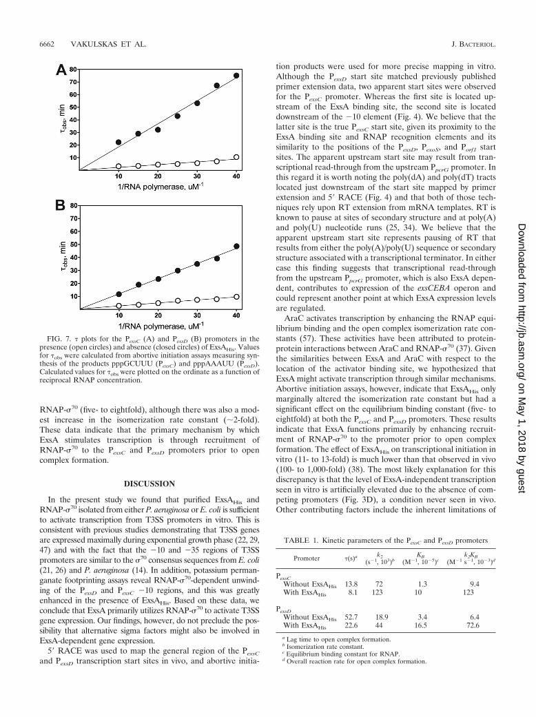
RNAP-�70 (five- to eightfold), although there was also a mod-est increase in the isomerization rate constant (2-fold).These data indicate that the primary mechanism by whichExsA stimulates transcription is through recruitment ofRNAP-�70 to the PexsC and PexsD promoters prior to opencomplex formation.
DISCUSSION
In the present study we found that purified ExsAHis andRNAP-�70 isolated from either P. aeruginosa or E. coli is sufficientto activate transcription from T3SS promoters in vitro. This isconsistent with previous studies demonstrating that T3SS genesare expressed maximally during exponential growth phase (22, 29,47) and with the fact that the �10 and �35 regions of T3SSpromoters are similar to the �70 consensus sequences from E. coli(21, 26) and P. aeruginosa (14). In addition, potassium perman-ganate footprinting assays reveal RNAP-�70-dependent unwind-ing of the PexsD and PexsC �10 regions, and this was greatlyenhanced in the presence of ExsAHis. Based on these data, weconclude that ExsA primarily utilizes RNAP-�70 to activate T3SSgene expression. Our findings, however, do not preclude the pos-sibility that alternative sigma factors might also be involved inExsA-dependent gene expression.
5� RACE was used to map the general region of the PexsC
and PexsD transcription start sites in vivo, and abortive initia-
tion products were used for more precise mapping in vitro.Although the PexsD start site matched previously publishedprimer extension data, two apparent start sites were observedfor the PexsC promoter. Whereas the first site is located up-stream of the ExsA binding site, the second site is locateddownstream of the �10 element (Fig. 4). We believe that thelatter site is the true PexsC start site, given its proximity to theExsA binding site and RNAP recognition elements and itssimilarity to the positions of the PexsD, PexoS, and Porf1 startsites. The apparent upstream start site may result from tran-scriptional read-through from the upstream PpcrG promoter. Inthis regard it is worth noting the poly(dA) and poly(dT) tractslocated just downstream of the start site mapped by primerextension and 5� RACE (Fig. 4) and that both of those tech-niques rely upon RT extension from mRNA templates. RT isknown to pause at sites of secondary structure and at poly(A)and poly(U) nucleotide runs (25, 34). We believe that theapparent upstream start site represents pausing of RT thatresults from either the poly(A)/poly(U) sequence or secondarystructure associated with a transcriptional terminator. In eithercase this finding suggests that transcriptional read-throughfrom the upstream PpcrG promoter, which is also ExsA depen-dent, contributes to expression of the exsCEBA operon andcould represent another point at which ExsA expression levelsare regulated.
AraC activates transcription by enhancing the RNAP equi-librium binding and the open complex isomerization rate con-stants (57). These activities have been attributed to protein-protein interactions between AraC and RNAP-�70 (37). Giventhe similarities between ExsA and AraC with respect to thelocation of the activator binding site, we hypothesized thatExsA might activate transcription through similar mechanisms.Abortive initiation assays, however, indicate that ExsAHis onlymarginally altered the isomerization rate constant but had asignificant effect on the equilibrium binding constant (five- toeightfold) at both the PexsC and PexsD promoters. These resultsindicate that ExsA functions primarily by enhancing recruit-ment of RNAP-�70 to the promoter prior to open complexformation. The effect of ExsAHis on transcriptional initiation invitro (11- to 13-fold) is much lower than that observed in vivo(100- to 1,000-fold) (38). The most likely explanation for thisdiscrepancy is that the level of ExsA-independent transcriptionseen in vitro is artificially elevated due to the absence of com-peting promoters (Fig. 3D), a condition never seen in vivo.Other contributing factors include the inherent limitations of
FIG. 7. � plots for the PexsC (A) and PexsD (B) promoters in thepresence (open circles) and absence (closed circles) of ExsAHis. Valuesfor �obs were calculated from abortive initiation assays measuring syn-thesis of the products pppGCUUU (PexsC) and pppAAAUU (PexsD).Calculated values for �obs were plotted on the ordinate as a function ofreciprocal RNAP concentration.
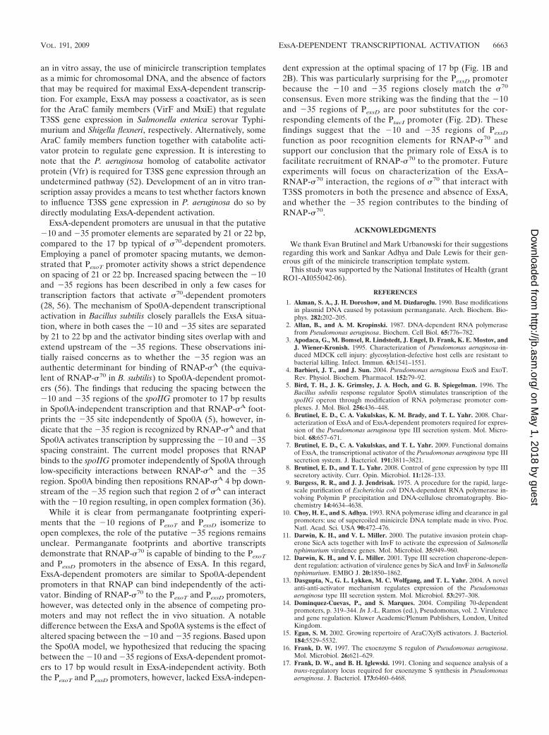
TABLE 1. Kinetic parameters of the PexsC and PexsD promoters
Promoter �(s)a k2(s�1, 103)b
KB(M�1, 10�5)c
k2KB(M�1 s�1, 10�3)d
PexsCWithout ExsAHis 13.8 72 1.3 9.4With ExsAHis 8.1 123 10 123
PexsDWithout ExsAHis 52.7 18.9 3.4 6.4With ExsAHis 22.6 44 16.5 72.6
a Lag time to open complex formation.b Isomerization rate constant.c Equilibrium binding constant for RNAP.d Overall reaction rate for open complex formation.
6662 VAKULSKAS ET AL. J. BACTERIOL.
on May 1, 2018 by guest
http://jb.asm.org/
Dow
nloaded from
an in vitro assay, the use of minicircle transcription templatesas a mimic for chromosomal DNA, and the absence of factorsthat may be required for maximal ExsA-dependent transcrip-tion. For example, ExsA may possess a coactivator, as is seenfor the AraC family members (VirF and MxiE) that regulateT3SS gene expression in Salmonella enterica serovar Typhi-murium and Shigella flexneri, respectively. Alternatively, someAraC family members function together with catabolite acti-vator protein to regulate gene expression. It is interesting tonote that the P. aeruginosa homolog of catabolite activatorprotein (Vfr) is required for T3SS gene expression through anundetermined pathway (52). Development of an in vitro tran-scription assay provides a means to test whether factors knownto influence T3SS gene expression in P. aeruginosa do so bydirectly modulating ExsA-dependent activation.
ExsA-dependent promoters are unusual in that the putative�10 and �35 promoter elements are separated by 21 or 22 bp,compared to the 17 bp typical of �70-dependent promoters.Employing a panel of promoter spacing mutants, we demon-strated that PexoT promoter activity shows a strict dependenceon spacing of 21 or 22 bp. Increased spacing between the �10and �35 regions has been described in only a few cases fortranscription factors that activate �70-dependent promoters(28, 56). The mechanism of Spo0A-dependent transcriptionalactivation in Bacillus subtilis closely parallels the ExsA situa-tion, where in both cases the �10 and �35 sites are separatedby 21 to 22 bp and the activator binding sites overlap with andextend upstream of the �35 regions. These observations ini-tially raised concerns as to whether the �35 region was anauthentic determinant for binding of RNAP-�A (the equiva-lent of RNAP-�70 in B. subtilis) to Spo0A-dependent promot-ers (56). The findings that reducing the spacing between the�10 and �35 regions of the spoIIG promoter to 17 bp resultsin Spo0A-independent transcription and that RNAP-�A foot-prints the �35 site independently of Spo0A (5), however, in-dicate that the �35 region is recognized by RNAP-�A and thatSpo0A activates transcription by suppressing the �10 and �35spacing constraint. The current model proposes that RNAPbinds to the spoIIG promoter independently of Spo0A throughlow-specificity interactions between RNAP-�A and the �35region. Spo0A binding then repositions RNAP-�A 4 bp down-stream of the �35 region such that region 2 of �A can interactwith the �10 region resulting, in open complex formation (36).
While it is clear from permanganate footprinting experi-ments that the �10 regions of PexoT and PexsD isomerize toopen complexes, the role of the putative �35 regions remainsunclear. Permanganate footprints and abortive transcriptsdemonstrate that RNAP-�70 is capable of binding to the PexoT
and PexsD promoters in the absence of ExsA. In this regard,ExsA-dependent promoters are similar to Spo0A-dependentpromoters in that RNAP can bind independently of the acti-vator. Binding of RNAP-�70 to the PexoT and PexsD promoters,however, was detected only in the absence of competing pro-moters and may not reflect the in vivo situation. A notabledifference between the ExsA and Spo0A systems is the effect ofaltered spacing between the �10 and �35 regions. Based uponthe Spo0A model, we hypothesized that reducing the spacingbetween the �10 and �35 regions of ExsA-dependent promot-ers to 17 bp would result in ExsA-independent activity. Boththe PexoT and PexsD promoters, however, lacked ExsA-indepen-
dent expression at the optimal spacing of 17 bp (Fig. 1B and2B). This was particularly surprising for the PexsD promoterbecause the �10 and �35 regions closely match the �70
consensus. Even more striking was the finding that the �10and �35 regions of PexsD are poor substitutes for the cor-responding elements of the PtacI promoter (Fig. 2D). Thesefindings suggest that the �10 and �35 regions of PexsD
function as poor recognition elements for RNAP-�70 andsupport our conclusion that the primary role of ExsA is tofacilitate recruitment of RNAP-�70 to the promoter. Futureexperiments will focus on characterization of the ExsA–RNAP-�70 interaction, the regions of �70 that interact withT3SS promoters in both the presence and absence of ExsA,and whether the �35 region contributes to the binding ofRNAP-�70.
ACKNOWLEDGMENTS
We thank Evan Brutinel and Mark Urbanowski for their suggestionsregarding this work and Sankar Adhya and Dale Lewis for their gen-erous gift of the minicircle transcription template system.
This study was supported by the National Institutes of Health (grantRO1-AI055042-06).
REFERENCES
1. Akman, S. A., J. H. Doroshow, and M. Dizdaroglu. 1990. Base modificationsin plasmid DNA caused by potassium permanganate. Arch. Biochem. Bio-phys. 282:202–205.
2. Allan, B., and A. M. Kropinski. 1987. DNA-dependent RNA polymerasefrom Pseudomonas aeruginosa. Biochem. Cell Biol. 65:776–782.
3. Apodaca, G., M. Bomsel, R. Lindstedt, J. Engel, D. Frank, K. E. Mostov, andJ. Wiener-Kronish. 1995. Characterization of Pseudomonas aeruginosa-in-duced MDCK cell injury: glycosylation-defective host cells are resistant tobacterial killing. Infect. Immun. 63:1541–1551.
4. Barbieri, J. T., and J. Sun. 2004. Pseudomonas aeruginosa ExoS and ExoT.Rev. Physiol. Biochem. Pharmacol. 152:79–92.
5. Bird, T. H., J. K. Grimsley, J. A. Hoch, and G. B. Spiegelman. 1996. TheBacillus subtilis response regulator Spo0A stimulates transcription of thespoIIG operon through modification of RNA polymerase promoter com-plexes. J. Mol. Biol. 256:436–448.
6. Brutinel, E. D., C. A. Vakulskas, K. M. Brady, and T. L. Yahr. 2008. Char-acterization of ExsA and of ExsA-dependent promoters required for expres-sion of the Pseudomonas aeruginosa type III secretion system. Mol. Micro-biol. 68:657–671.
7. Brutinel, E. D., C. A. Vakulskas, and T. L. Yahr. 2009. Functional domainsof ExsA, the transcriptional activator of the Pseudomonas aeruginosa type IIIsecretion system. J. Bacteriol. 191:3811–3821.
8. Brutinel, E. D., and T. L. Yahr. 2008. Control of gene expression by type IIIsecretory activity. Curr. Opin. Microbiol. 11:128–133.
9. Burgess, R. R., and J. J. Jendrisak. 1975. A procedure for the rapid, large-scale purification of Escherichia coli DNA-dependent RNA polymerase in-volving Polymin P precipitation and DNA-cellulose chromatography. Bio-chemistry 14:4634–4638.
10. Choy, H. E., and S. Adhya. 1993. RNA polymerase idling and clearance in galpromoters: use of supercoiled minicircle DNA template made in vivo. Proc.Natl. Acad. Sci. USA 90:472–476.
11. Darwin, K. H., and V. L. Miller. 2000. The putative invasion protein chap-erone SicA acts together with InvF to activate the expression of Salmonellatyphimurium virulence genes. Mol. Microbiol. 35:949–960.
12. Darwin, K. H., and V. L. Miller. 2001. Type III secretion chaperone-depen-dent regulation: activation of virulence genes by SicA and InvF in Salmonellatyphimurium. EMBO J. 20:1850–1862.
13. Dasgupta, N., G. L. Lykken, M. C. Wolfgang, and T. L. Yahr. 2004. A novelanti-anti-activator mechanism regulates expression of the Pseudomonasaeruginosa type III secretion system. Mol. Microbiol. 53:297–308.
14. Dominquez-Cuevas, P., and S. Marques. 2004. Compiling 70-dependentpromoters, p. 319–344. In J.-L. Ramos (ed.), Pseudomonas, vol. 2. Virulenceand gene regulation. Kluwer Academic/Plenum Publishers, London, UnitedKingdom.
15. Egan, S. M. 2002. Growing repertoire of AraC/XylS activators. J. Bacteriol.184:5529–5532.
16. Frank, D. W. 1997. The exoenzyme S regulon of Pseudomonas aeruginosa.Mol. Microbiol. 26:621–629.
17. Frank, D. W., and B. H. Iglewski. 1991. Cloning and sequence analysis of atrans-regulatory locus required for exoenzyme S synthesis in Pseudomonasaeruginosa. J. Bacteriol. 173:6460–6468.
VOL. 191, 2009 ExsA-DEPENDENT TRANSCRIPTIONAL ACTIVATION 6663
on May 1, 2018 by guest
http://jb.asm.org/
Dow
nloaded from

18. Frank, D. W., G. Nair, and H. P. Schweizer. 1994. Construction and char-acterization of chromosomal insertional mutations of the Pseudomonasaeruginosa exoenzyme S trans-regulatory locus. Infect. Immun. 62:554–563.
19. Fujita, M., and A. Amemura. 1992. In vitro interactions of PseudomonasRNA polymerases with tac and RNA I promoters. Biosci. Biotechnol. Bio-chem. 56:1644–1648.
20. Gallegos, M. T., R. Schleif, A. Bairoch, K. Hofmann, and J. L. Ramos. 1997.Arac/XylS family of transcriptional regulators. Microbiol. Mol. Biol. Rev.61:393–410.
21. Grana, D., T. Gardella, and M. M. Susskind. 1988. The effects of mutationsin the ant promoter of phage P22 depend on context. Genetics 120:319–327.
22. Ha, U., and S. Jin. 2001. Growth phase-dependent invasion of Pseudomonasaeruginosa and its survival within HeLa cells. Infect. Immun. 69:4398–4406.
23. Reference deleted.24. Harley, C. B., and R. P. Reynolds. 1987. Analysis of E. coli promoter se-
quences. Nucleic Acids Res. 15:2343–2361.25. Harrison, G. P., M. S. Mayo, E. Hunter, and A. M. Lever. 1998. Pausing of
reverse transcriptase on retroviral RNA templates is influenced by secondarystructures both 5� and 3� of the catalytic site. Nucleic Acids Res. 26:3433–3442.
26. Hawley, D. K., and W. R. McClure. 1983. Compilation and analysis ofEscherichia coli promoter DNA sequences. Nucleic Acids Res. 11:2237–2255.
27. Hoang, T. T., A. J. Kutchma, A. Becher, and H. P. Schweizer. 2000. Integra-tion-proficient plasmids for Pseudomonas aeruginosa: site-specific integrationand use for engineering of reporter and expression strains. Plasmid 43:59–72.
28. Hobman, J. L. 2007. MerR family transcription activators: similar designs,different specificities. Mol. Microbiol. 63:1275–1278.
29. Hogardt, M., M. Roeder, A. M. Schreff, L. Eberl, and J. Heesemann. 2004.Expression of Pseudomonas aeruginosa exoS is controlled by quorum sensingand RpoS. Microbiology 150:843–851.
30. Holder, I. A., A. N. Neely, and D. W. Frank. 2001. Type III secretion/intoxication system important in virulence of Pseudomonas aeruginosa infec-tions in burns. Burns 27:129–130.
31. Hovey, A. K., and D. W. Frank. 1995. Analyses of the DNA-binding andtranscriptional activation properties of ExsA, the transcriptional activator ofthe Pseudomonas aeruginosa exoenzyme S regulon. J. Bacteriol. 177:4427–4436.
32. Hsu, L. M. 2009. Monitoring abortive initiation. Methods 47:25–36.33. Jarrell, K., and A. M. Kropinski. 1977. The chemical composition of the
lipopolysaccharide from Pseudomonas aeruginosa strain PAO and a sponta-neously derived rough mutant. Microbios 19:103–116.
34. Klarmann, G. J., C. A. Schauber, and B. D. Preston. 1993. Template-di-rected pausing of DNA synthesis by HIV-1 reverse transcriptase duringpolymerization of HIV-1 sequences in vitro. J. Biol. Chem. 268:9793–9802.
35. Kohler, J. J., S. J. Metallo, T. L. Schneider, and A. Schepartz. 1999. DNAspecificity enhanced by sequential binding of protein monomers. Proc. Natl.Acad. Sci. USA 96:11735–11739.
36. Kumar, A., and C. P. Moran, Jr. 2008. Promoter activation by repositioningof RNA polymerase. J. Bacteriol. 190:3110–3117.
37. Martin, R. G., and J. L. Rosner. 2001. The AraC transcriptional activators.Curr. Opin. Microbiol. 4:132–137.
38. McCaw, M. L., G. L. Lykken, P. K. Singh, and T. L. Yahr. 2002. ExsD is anegative regulator of the Pseudomonas aeruginosa type III secretion regulon.Mol. Microbiol. 46:1123–1133.
39. McClure, W. R. 1980. Rate-limiting steps in RNA chain initiation. Proc.Natl. Acad. Sci. USA 77:5634–5638.
40. Parsot, C., E. Ageron, C. Penno, M. Mavris, K. Jamoussi, H. d’Hauteville, P.Sansonetti, and B. Demers. 2005. A secreted anti-activator, OspD1, and itschaperone, Spa15, are involved in the control of transcription by the type IIIsecretion apparatus activity in Shigella flexneri. Mol. Microbiol. 56:1627–1635.
41. Pilonieta, M. C., and G. P. Munson. 2008. The chaperone IpgC copurifieswith the virulence regulator MxiE. J. Bacteriol. 190:2249–2251.
42. Plano, G. V. 2004. Modulation of AraC family member activity by proteinligands. Mol. Microbiol. 54:287–290.
43. Richards, M. J., J. R. Edwards, D. H. Culver, and R. P. Gaynes. 2000.Nosocomial infections in combined medical-surgical intensive care units inthe United States. Infect. Control Hosp. Epidemiol. 21:510–515.
44. Richards, M. J., J. R. Edwards, D. H. Culver, R. P. Gaynes, et al. 1999.Nosocomial infections in medical intensive care units in the United States.Crit. Care Med. 27:887–892.
45. Sato, H., and D. W. Frank. 2004. ExoU is a potent intracellular phospho-lipase. Mol. Microbiol. 53:1279–1290.
46. Scotto-Lavino, E., G. Du, and M. A. Frohman. 2006. Amplification of 5� endcDNA with ‘new RACE.’ Nat. Protoc. 1:3056–3061.
47. Shen, D. K., D. Filopon, H. Chaker, S. Boullanger, M. Derouazi, B. Polack,and B. Toussaint. 2008. High-cell-density regulation of the Pseudomonasaeruginosa type III secretion system: implications for tryptophan catabolites.Microbiology 154:2195–2208.
48. Thibault, J., E. Faudry, C. Ebel, I. Attree, and S. Elsen. 2009. The anti-activator ExsD forms a 1:1 complex with ExsA to inhibit transcription of typeIII secretion operons. J. Biol. Chem. 284:15762–15770.
49. Vogel, H. J., and D. M. Bonner. 1956. Acetylornithinase of Escherichia coli:partial purification and some properties. J. Biol. Chem. 218:97–106.
50. Warne, S. E., and P. L. deHaseth. 1993. Promoter recognition by Escherichiacoli RNA polymerase. Effects of single base pair deletions and insertions inthe spacer DNA separating the �10 and �35 regions are dependent onspacer DNA sequence. Biochemistry 32:6134–6140.
51. Wickstrum, J. R., J. M. Skredenske, A. Kolin, D. J. Jin, J. Fang, and S. M.Egan. 2007. Transcription activation by the DNA-binding domain of theAraC family protein RhaS in the absence of its effector-binding domain. J.Bacteriol. 189:4984–4993.
52. Wolfgang, M. C., V. T. Lee, M. E. Gilmore, and S. Lory. 2003. Coordinateregulation of bacterial virulence genes by a novel adenylate cyclase-depen-dent signaling pathway. Dev. Cell 4:253–263.
53. Yahr, T. L., and D. W. Frank. 1994. Transcriptional organization of thetrans-regulatory locus which controls exoenzyme S synthesis in Pseudomonasaeruginosa. J. Bacteriol. 176:3832–3838.
54. Yahr, T. L., A. K. Hovey, S. M. Kulich, and D. W. Frank. 1995. Transcrip-tional analysis of the Pseudomonas aeruginosa exoenzyme S structural gene.J. Bacteriol. 177:1169–1178.
55. Yahr, T. L., and M. C. Wolfgang. 2006. Transcriptional regulation of thePseudomonas aeruginosa type III secretion system. Mol. Microbiol. 62:631–640.
56. York, K., T. J. Kenney, S. Satola, C. P. Moran, Jr., H. Poth, and P. Young-man. 1992. Spo0A controls the sigma A-dependent activation of Bacillussubtilis sporulation-specific transcription unit SpoIIE. J. Bacteriol. 174:2648–2658.
57. Zhang, X., T. Reeder, and R. Schleif. 1996. Transcription activation param-eters at ara pBAD. J. Mol. Biol. 258:14–24.
6664 VAKULSKAS ET AL. J. BACTERIOL.
on May 1, 2018 by guest
http://jb.asm.org/
Dow
nloaded from