mechanismofhiv-1rnadimerizationinthecentralregion ... · near the central polypurine tract (cppt),...
TRANSCRIPT

Mechanism of HIV-1 RNA Dimerization in the Central Regionof the Genome and Significance for Viral Evolution*
Received for publication, April 15, 2013, and in revised form, July 1, 2013 Published, JBC Papers in Press, July 9, 2013, DOI 10.1074/jbc.M113.477265
Dorota Piekna-Przybylska‡, Gaurav Sharma§, and Robert A. Bambara‡1
From the ‡Department of Microbiology and Immunology, School of Medicine and Dentistry, University of Rochester, Rochester,New York 14642 and §Department of Electrical and Computer Engineering, University of Rochester, Rochester, New York 14627
Background: HIV-1 genome dimerization is important for viral survival.Results: The central region of HIV-1 genome contains a G-rich sequence promoting recombination and capable of forming adimeric G-quartet.Conclusion: Parallel alignment and proximity of two HIV-1 genomes associated at the 5� end can be maintained throughdimeric G-quartet formed in the central region of viral genome.Significance: Determining mechanism and factors involved in HIV-1 genome dimerization.
The genome ofHIV-1 consists of two identical or nearly iden-tical RNA molecules. The RNA genomes are held in the same,parallel orientation by interactions at the dimer initiation site(DIS). Previous studies showed that in addition to interactionsat DIS, sequences located 100 nucleotides downstream from the5� splice site can dimerize in vitro through an intermolecularG-quartet structure. Here we report that the highly conservedG-rich sequence in the middle portion of the HIV-1 genomenear the central polypurine tract (cPPT) dimerizes spontane-ously under high ionic strength in the absence of protein. Theantisense RNA does not dimerize, strongly indicating that RNAdimerization does not exclusively involve A:U and G:C basepairing. The cation-dependent reverse transcriptase pausingprofile, CD spectra profile, and cation-dependent associationand thermal dissociation characteristics indicate G-quartetstructures. Different forms of G-quartets are formed includ-ing monomers and, significantly, intermolecular dimers. Ourresults indicate that RNA genome dimerization and parallelalignment initiated through interactions at DIS may be greatlyexpanded and stabilized by formation of an intermolecularG-quartet at a distant site near the cPPT. It is likely that forma-tion of G-quartet structure near the cPPT in vivo keeps the RNAgenomes in proximity over a long range, promoting geneticrecombination in numerous hot spots.
HIV-1 efficiently diversifies its population within an infectedperson, allowing it to evade host immune response and thera-peutic treatments. The error-prone HIV-1 reverse transcrip-tase (RT) contributes significantly to viral diversity. Viral evo-lution is further accelerated through the process of geneticrecombination. The presence of two often non-identical copies
of viral genome in a single virion is a fundamental requirementfor genetically significant recombination. One copy of genomicRNA is sufficient for viral replication. The role of the secondcopy is to allow the RT to switch templates during reverse tran-scription. This mixes mutations from two genetically differentviral genomes, allowing the virus to evolve into more robustforms.The two genomic RNAs adhere within the cytoplasm; thus,
the viral genomes are packaged into the virion as a dimer ratherthan two RNA monomers (1, 2). The sequences involved ininitial viral genome dimerization and encapsidation partiallyoverlap and are localized close to the 5� end (3–5). One of thesesequences forms the stem-loop 1 secondary structure with ahighly conserved palindromic sequence called the dimer initi-ation site (DIS).2 The first stable interaction is a contact withinthe loops of two stem-loop 1 hairpins called RNA kissing (6, 7).The interaction is then thought to spread to the stem regions,with their palindromic sequences promoting base pairing; how-ever, this contact is not sufficient by itself to produce dimers invivo that resist extraction and gel electrophoresis (8). Otherinteractions along the genome seem to be desirable to keep aconsistent proximity that would facilitate a generally uniformprobability of template switching of the RT throughout reversetranscription, desirable for efficient recombination.Studies in vitro show that in HIV-1 and other retroviruses,
certain G-rich genomic sequences can form G-quartet struc-tures (9–13). Four guanine bases can associate through Hoog-steen hydrogen bonding and form a G-tetrad, and two or moreguanine tetrads can stack on top of each other to form aG-quartet. TheG-tetrads can formby repeated folding of eithera single nucleic acid molecule or by association of two or fourmolecules. The structure has been intensively analyzed intelomeres, and in recent years G-quartets have been shown toregulate activity of promoters, formation ofDNA flaps, stabilityof mRNA, and alternative splicing and translation (14–21).Very little is known about G-quartets in viruses, including
* This work was supported, in whole or in part, by National Institutes of HealthGrants GM049573 (to R. A. B.) and GM097334 (to G. S.) and University ofRochester Developmental Center for AIDS Research Grant P30 AI078498(NIAID). This work was also supported by the University of RochesterSchool of Medicine and Dentistry.
1 To whom correspondence should be addressed: Dept. of Microbiology andImmunology, School of Medicine and Dentistry, University of Rochester,601 Elmwood Ave., Rochester, NY 14642. Tel.: 585-275-2764; Fax: 585-275-6007; E-mail: [email protected].
2 The abbreviations used are: DIS, dimer initiation site; cPPT, central polypu-rine tract; NC, nucleocapsid protein; TP, transfer product; DE, donor exten-sion product; nt, nucleotide(s).
THE JOURNAL OF BIOLOGICAL CHEMISTRY VOL. 288, NO. 33, pp. 24140 –24150, August 16, 2013© 2013 by The American Society for Biochemistry and Molecular Biology, Inc. Published in the U.S.A.
24140 JOURNAL OF BIOLOGICAL CHEMISTRY VOLUME 288 • NUMBER 33 • AUGUST 16, 2013
by guest on February 28, 2020http://w
ww
.jbc.org/D
ownloaded from

HIV-1 and their possible function in the viral life cycle. G-quar-tet structure formation was found to be essential to maintainthe Epstein-Barr virus genome during latency in proliferatingcells (22). In HIV-1 and Moloney murine sarcoma virus(MuSV), G-rich sequences in the gag region near DIS wereshown to form an intermolecular G-quartet (9–13). This struc-ture adheres the two RNA genomes; thus, it represents a differ-ent mode of genome dimerization in retroviruses from that ofthe DIS hairpin interaction. Formation of dimeric G-quartetstructure correlateswith hot spots of recombination, exhibitingan increased rate of template switching (23, 24).Here we show that short RNA templates from the central
region of the HIV-1 genome, containing the G-rich sequencesnear the central polypurine tract (cPPT), are able to form bothmonomer and dimer G-quartet structures. The cPPT is locatedin the integrase gene and is a region where one of two primersfor synthesis of (�) strand DNA is produced during reversetranscription. The G tract in the cPPT and two other G runslocated downstream from the cPPT are highly conservedamong different HIV-1 isolates and several closely related sim-ian immunodeficiency virus (SIV) species. By application ofassays in vitro and affinity selectionmethods, we found that theRNA templates dimerize through theG-rich regions, indicatingthat in addition to contacts near the 5� end, the central regionsof the viral RNA genomes are likely to be maintained in prox-imity through dimer G-quartet formation. In reconstitutedsystems of reverse transcription, the formation of G-quartetsfacilitates RT in switching templates during synthesis ofminus strand DNA, suggesting that the structure supportsan increased recombination rate.
EXPERIMENTAL PROCEDURES
Materials—DNA oligonucleotides and the HPLC-purifiedRNA strand used for CD spectra analyses were purchased fromIntegrated DNA Technologies, Inc. (Coralville, IA). HIV-1nucleocapsid protein (NC; 55 amino acids) was generously pro-vided by Dr. Robert J. Gorelick. HIV-1 reverse transcriptase(p66/p51 heterodimer) was purified as described previously(25). The [�-32P]ATP was purchased from PerkinElmer LifeSciences. The sequences of viruses were analyzed with the HIVdatabase (www.hiv.lanl.gov).Preparation of RNA Templates—RNA molecules were tran-
scribed in vitro (Ambion T7-MEGAshortscript kit; AppliedBiosystems) fromDNA templates amplified by PCR usingVentDNA polymerase (New England Biolabs) and two overlappingoligomers with the sequence of the desired region. The follow-ing RNA strands were used in our studies. (a) For the reversetranscription assay, the wild-type and mutant RNA templatestrands with the cPPT region (4309–4396 in the RNA genome)sequences from the NL4-3 HIV-1 were made from DNA tem-plates synthesized with a pair of oligomers, 1/2 and 3/4, respec-tively. (b) For affinity selection analysis, the non-tagged RNAsand poly(A)-tagged RNAs were made from DNA generatedusing oligomers 1/2 and 1/5 for the cPPT region (4309–4396)of NL4-3 HIV-1, 6/7 and 6/8 for the gag region (290–403) ofNL4-3 HIV-1, 9/10 and 9/11 for the gag region (303–415) ofMALHIV-1, and 12/13 and 14/15 for 5�-UTR with DIS (1–520and 183–520) of NL4-3 HIV-1. (c) For analysis of dimerization
in a native gel, the RNA template of the cPPT region (4309–4396) in NL4-3 HIV-1 was made from DNA generated usingoligomers 1/2, and for the antisense sequence of the cPPTregion, oligomers 16/17 were used. (d) For transfer assays, thedonor and acceptor templates for cPPT region (4309–4396) ofNL4-3HIV-1 weremade fromDNA generated using oligomers1/5 and 18/2, respectively. After transcription in vitro, the RNAtemplateswere purified by polyacrylamide/urea gel electropho-resis and resuspended in water. RNAs were quantitated by UVabsorption using a GeneQuant II fromAmersham Biosciences.DNA Oligonucleotides—The forward oligonucleotides (1, 3,
6, 9, 12, 14, 16, 18) for all DNA templates used to generate RNAswere designed to contain the phage T7 RNA polymerase pro-moter sequence (GGA TCC TAA TAC GAC TCA CTA TAG)immediately upstream of the desired template sequence. Themutations and additions are underlined: 1, 5�-GGA TCC TAATAC GAC TCA CTA TAG GGC AGT ATT CAT CCA CAATTT TAA AAG AAA AGG GGG GAT TGG GGG GTA CAGTG-3�; 2, 5�-GTC TGT TGC TAT TAT GTC TAC TAT TCTTTC CCC TGC ACT GTA CCC CCC AAT CCC CCC-3�; 3,5�-GGA TCC TAA TAC GAC TCA CTA TAG GGC AGTATT CAT CCA CAA TTT TAA AAG AAA AGG GGG GATTGT GTGGTA CAG TG-3�; 4, 5�-GTC TGT TGC TAT TATGTC TAC TAT TCT TTT CTC TGC ACT GTA CCA CACAAT CCC CCC-3�; 5, 5�-TTT TTT TTT TTT TTT TTT TTTTTT TTT TTT GTC TGT TGC TAT TAT GTC TAC TATTCTTTCCCCTGCACTGTACCCCCCAATCCCCCC-3�;6, 5�-GGA TCC TAA TAC GAC TCA CTA TAG GGT GAGTAC GCC AAA AAT TTT GAC TAG CGG AGG CTA GAAGGA GAG AGA TGG GTG CGA GAG CGT CGG TAT TAAGC-3�; 7, 5�-GCC TTA ACC GAA TTT TTT CCC ATT TATCTA ATT CTC CCC CGC TTA ATA CCG ACG CTC TCGCAC CCA T-3�; 8, 5�-TTT TTT TTT TTT TTT TTT TTTTTT TTT TTT GCC TTA ACC GAA TTT TTT CCC ATTTAT CTA ATT CTC CCC CGC TTA ATA CCG ACG CTCTCG CAC CCA T-3�; 9, 5�-GGA TCC TAA TAC GAC TCACTA TAG GGT GAG TAC GCC AAT TTT TGA CTA GCGGAGGCTAGAAGGAGAGAGATGGGTGCGAGAGCGTCA GTA TTA AGC-3�; 10, 5�-CCC TTA ACC GAA TTTTCT CCC ATG CAT CTA ATT TTC CCC CGC TTA ATACTG ACG CTC TCG CAC CCA T-3�; 11, 5�-TTT TTT TTTTTT TTT TTT TTT TTT TTT TTT CCC TTA ACC GAATTT TCT CCC ATG CAT CTA ATT TTC CCC CGC TTAATA CTG ACG CTC TCG CAC CCA T-3�; 12, 5�-GGA TCCTAA TAC GAC TCA CTA TAG GGT CTC TCT GGT TAGACCAGATC-3�; 13, 5�-CCCAGTATTTGTCTACAGCCTTCT GAT GTC-3�; 14, 5�-GGA TCC TAA TAC GAC TCACTA TAG GCG CCC GAA CAG GGA CTT GAA AG-3�; 15,5�-TTT TTT TTT TTT TTT TTT TTT TTT TCC CAG TATTTG TCT ACA GCC TTC TGA TGT C-3�; 16, 5�-GGA TCCTAA TAC GAC TCA CTA TAG GGT CAT AAG TAG GTGTTA AAA TTT TCT TTT CCC CCC TAA CCC CCC-3�; 17,5�-CAG ACA ACG ATA ATA CAG ATG ATA AGA AAGGGGACGTGACATGGGGGGTTAGGGGGGAAAAGAAAATT-3�; 18, 5�-GGATCCTAATACGACTCACTATAGGGA AAA AAA AAA CAG TAT TCA TCC ACA ATT TTAAAA GAA AAG GGG GGA TTG GGG GGT ACA GTG-3�;19, 5�-GTC TGT TGC TAT TAT GTC-3�; 20, 5�-TTT TTT
RNA Dimerization in the Central Region of the HIV-1 Genome
AUGUST 16, 2013 • VOLUME 288 • NUMBER 33 JOURNAL OF BIOLOGICAL CHEMISTRY 24141
by guest on February 28, 2020http://w
ww
.jbc.org/D
ownloaded from

TTT TTT TTT TGT CT-3�; a, 5�-AAT CCC CCC TTT TCTT-3�; b, 5�-TAC CCC CCA ATC CCC C-3�; c, 5�-CTG CACTGT ACC CCC C-3�; d, 5�-TCT TTC CCC TGC ACTG-3�; e,5�-TCT ACT ATT CTT TCC C-3�; f, 5�-CTA TTA TGT CTACTA T-3�; g, 5�-GTC TGT TGC TAT TAT G-3�.RNA Oligonucleotide—The RNA oligonucleotide was 5�-GGG
GGGAUUGGGGGGUACAGUGCAGGGG-3�.Preparation of the 5�-Radiolabeled RNA Template and the
DNAPrimer—DNAprimerswere labeled at the 5� end usingT4polynucleotide kinase (New England Biolabs) and [�-32P]ATP(6000 Ci/mmol). Preparation of 5�-radiolabeled RNA templatewas performed as follows. The gel-cleaned RNA template wastreated with shrimp alkaline phosphatase (Fermentas) at 37 °Cfor 60 min and then incubated at 65 °C for 25 min to inactivatethe enzyme. After cooling on ice, the reaction mixture wastreated with [�-32P]ATP (6000 Ci/mmol), 10� PNK T4 Poly-nucleotide Kinase Reaction buffer and T4 polynucleotidekinase (New England Biolabs). After incubation for 1 h at 37 °C,the radiolabeled RNA and DNA primers were separated fromunincorporated radionucleotides using a Micro Bio-Spin col-umn (Bio-Rad).Reverse Transcription Assay—RNA (200 fmol) and the 5�
end-labeledDNAprimer 19 (300 fmol) were heated to 95 °C for2 min and annealed by slow cooling to 37 °C in 9 �l. HIV-1 RTwas added to the substrate and incubated for 4 min before thereaction was initiated with MgCl2 and dNTPs. Reactions werecarried out in 12.5 �l at a final concentration of 50 mM Tris-HCl, pH 8.0, 50mMKCl or LiCl, 1mMDTT, 1mMEDTA, 32 nMHIV-1 RT, 6 mM MgCl2, and 50 �M dNTPs. After 30 min ofincubation at 37 °C, reverse transcriptions were stopped with 1volume of termination buffer (10mMEDTA, pH 8.0, 90% form-amide (v/v), and 0.1% each xylene cyanol and bromphenolblue). Extension products were resolved on a 6% polyacryl-amide, 8 M urea gel and analyzed using phosphorimaging (GEHealthcare). Sizes of DNA products were estimated by using a5�-radiolabeled 10-bp DNA ladder (Invitrogen).Circular Dichroism—CD spectra were obtained at 25 °C over
a wavelength range of 220–340 nm using an AVIV circulardichroism spectrometer, model 202. The RNAwas at a concen-tration of 4�M in 10mMTris HCl, pH 7.5, 0.3mM EDTA and inthe presence of KCl, NaCl, or LiCl at 10 and 50 mM concentra-tions. Before analysis, the samples were heated to 90 °C for 10min, gently cooled at a rate of 1 °C/5min, and incubated at 4 °Covernight. Spectra were recorded using a quartz cell of 1-mmoptical path length, with data collected every nanometer at abandwidth of 1 nm. Each spectrum was recorded three timesand base-line-corrected for signal contributions from thebuffer. The data were processed with AVIV Biomedical Inc,software and reported as ellipticity (millidegrees) versus wave-length (nm).Thermal Denaturation of G-quadruplex Structure—Melting
curves were obtained bymonitoring the change at a wavelengthof 265 nm, chosen for having the highest CD value. The tem-perature was raised from 20 to 90 °C; the heating rate was 2 °C/min. The melting temperature Tm was defined as the tempera-ture of the mid-transition point.Affinity Selection with Oligo-d(T)25 Magnetic Beads—About
40 pmol of poly(A)-tagged RNA and non-tagged RNA tem-
platesweremixed (ratio 1:1) in the presence of 50mMTris-HCl,pH 8.0, 200mMKCl, and 1mMEDTA in a final volume of 20�l.Themixtures were heated to 95 °C for 3min, then chilled on iceand incubated at room temperature for 2 h. Before using oligo-d(T)25 magnetic beads (New England Biolabs), the suspensionof 50�l was washed once with binding buffer (20mMTris-HCl,pH 7.5, 500 mM KCl, 1 mM EDTA), resuspended in 180 �l ofbinding buffer, and added to the RNA. The mixture was agi-tated at room temperature for 10 min, and then placed in amagnetic rack to separate the magnetic beads from solution.The beads were washed once with binding buffer and threetimes withwash buffer (20mMTris-HCl, pH 7.5, 200mMKCl, 1mM EDTA), each time for 1 min with gentle agitation. To elutethe RNA, the beads were resuspended in 15 �l of elution buffer(20 mM Tris-HCl, pH 7.5, 1 mM EDTA), incubated for 3 min in95 °C and placed in a magnetic rack to separate the magneticbeads from solution. 1 volume of loading buffer (10 mM EDTA,pH 8.0, 90% formamide (v/v), and 0.1% each xylene cyanol andbromphenol blue) was added to the eluted solution, and theproducts were resolved in 4 or 6% polyacrylamide, 8 M ureagels. The gel was stained with ethidium bromide.Formation of Higher Order Products and Native Gel Analysis—
The mixture of the 5� end 32P-labeled (about 500,000 cpm)and unlabeled RNA at a concentration of 4 �M and a final vol-umeof 6�l was heated to 95 °C for 3min, chilled, and incubatedfor 60 min at room temperature in a buffer containing 10 mM
Tris-HCl, pH 7.5, 1 M KCl (or 1 M LiCl), 1 mM EDTA. Beforeelectrophoresis, samples were mixed with 1 volume of loadingdye (30% glycerol in 1�Tris-EDTA and loading pigments) andloaded onto a 6% non-denaturing polyacrylamide gel (0.5 �Tris borate-EDTA � 10 mM KCl). Electrophoresis was per-formed in 4 °C at 7V/cm for 3–4 h, and the gel was dried ontoWhatman No. 3MM chromatography paper and analyzedusing phosphorimaging (GE Healthcare).Cation-dependent Dimerization and Thermal Dissociation
Analysis—Dimerization and melting experiments with RNAdimers of the cPPT region were conducted in parallel at thesame molar concentration (4 �M) for each reaction setting. Toform a dimer, the RNAwas heat to 95 °C for 3 min, chilled, andincubated for 16 h at room temperature in a buffer containing10mMTris-HCl, pH7.5, 1mMEDTA, and one of three differentsalts (KCl, NaCl, or LiCl), each at 1 M. After incubation, themixtures were placed on ice, and 1 volume of 1 � Tris-EDTAbuffer was added. Aliquots of 15 �l were transferred to newtubes and incubated for 8 min at a specific temperaturebetween 40 and 90 °C, then returned to the ice. Samples weremixed with 1 volume of loading dye (30% glycerol in 1 � Tris-EDTA and loading dyes) and loaded onto 6% non-denaturinggels run at 4 °C. All gels were dried onto Whatman No. 3MMchromatography paper and analyzed using a phosphorimaging(GE Healthcare).Antisense DNA Oligonucleotide Binding Assay—A 5-fold
molar excess of each oligomer (a-g) was added to separatedimerization incubations (4 �M RNA, 10 mM Tris-HCl, pH 7.5,1 M LiCl, 1 mM EDTA) before the denaturation step. The sam-ples were chilled on the ice and incubated for 16 h at roomtemperature in a final volume of 7 �l. Products were resolved
RNA Dimerization in the Central Region of the HIV-1 Genome
24142 JOURNAL OF BIOLOGICAL CHEMISTRY VOLUME 288 • NUMBER 33 • AUGUST 16, 2013
by guest on February 28, 2020http://w
ww
.jbc.org/D
ownloaded from

on a 6% non-denaturing gel run at 4 °C in 0.5 � Tris borate-EDTA � 50 mM KCl.Strand Transfer Assay—The DNA primer 20 was heat-an-
nealed to donor RNA by incubation at 95 °C for 5 min and slowcooling to 37 °C. Acceptor template was also present in themixture. NC at the 200% polymer substrate level (100% NC is 7nt/NC polymer substrate molecule) was added and incubatedfor 3min. Next, the RTwas added to themixture and incubatedfor another 4 min to pre-bind the RT with substrates beforereactionswere initiatedwithMgCl2 and dNTPs. Primer, donor,and acceptor were mixed at a ratio of 2:1:1. The final reactioncontained 50 mM Tris-HCl, pH 8.0, 50 mM KCl, 1 mM DTT, 1mM EDTA, 32 nM HIV-1 RT, 6 mM MgCl2, and 50 �M dNTPs.For reactions in the presence of lithium ions, KCl was replacedwith 50 mM LiCl. Reactions were incubated at 37 °C and termi-nated after 1, 5, 15, and 30 min with 1 volume of terminationdye (10mMEDTA, pH 8.0, 90% formamide (v/v), and 0.1% eachxylene cyanol and bromphenol blue). Products were thenresolved by 6%polyacrylamide, 8Murea gels and analyzed usingphosphorimaging (GE Healthcare) and ImageQuant (Version2.1). Sizes of DNAproducts were estimated by using a 5�-radio-labeled 10-bp DNA ladder (Invitrogen).
RESULTS
G-rich Sequences Capable of G-quartet Formation Are Pres-ent in theMiddle of theGenome inVariousHIV Species—Inten-sive research on the gag region near the dimerization initiationsite of different retroviruses showed that formation of anRNA genomic dimer is also supported by interactionsthough G-quartet structure (9–13). To discover whether therearemore distantG-rich sequences potentially capable of adher-ing the HIV-1 genomes, we used QGRS Mapper, a softwareprogram designed to predict formation of this structure (26).Our computational analyses revealed that the central region ofthe HIV-1 genome near the cPPT can form G-quartet struc-tures with high probability. The sequence contains three Gruns, in which the first G-rich element is a part of the cPPTsequence (Fig. 1). PutativeG-quartetswould also require down-streamG-rich elements inHIV-1 thatwould be part of either anintramolecular structure containing two G-tetrads or a bimo-lecular structure with up to six G-tetrads. To form the RNA
dimer, only one or two G-rich elements on each RNAmoleculewould need to participate in the interaction.Conservation of these elements would attest to their likely
function in genomedimerization. The cPPT sequences are con-served among viral species, as expected from their multiplefunctions. To determinewhether cPPT plus flanking sequencesare also predicted to participate in G-quartet formation in dif-ferent viruses, we analyzed genomes of various isolates ofHIV-1 (A, B, C, D, F, G,H, U,O), HIV-2 (A, B, G, U), and severalSIV species closely related to HIV-1. All analyzed sequences ofHIV-1 and SIV have a similar distribution of cPPT-regionG-rich elements and are also predicted to formG-quartet struc-tures (Table 1). In the case of HIV-2, two G-rich elements arepresent andmight be involved in formation of a dimer. In sum-mary, our computational analyses of viral sequences suggestthat G-rich sequences within and flanking the cPPT areinvolved in G-quartet formation in all analyzed species.G-rich Regions Near the HIV-1 cPPT Induce Cation-depen-
dent Pauses of RT—G-quartets are formed preferentially in thepresence of specific monovalent cations and can be stabilizedby someproteins and chemical agents. The order of cation pref-erence is usually K� � Na� � Cs� � Li�. The quadruplexformation rates increase with the salt concentration. Previousstudies revealed cation-dependent pauses of RT progressionimmediately before and within a G-rich RNA template regioninvolved in G-quartet formation (23, 27). In the presence of lowconcentrations of potassium ions, pause sites caused by hairpinstructures and G-quartets were observed; however, in the pres-ence of a low concentration of lithium ions the pauses caused byhairpin structures were not affected, whereas the pauses causedby G runs involved in G-quartet formation were greatlyreduced. This cation-dependent pause profile behavior reliablyindicates locations where an analyzed sequence can form aG-quartet structure.The RNA sequence of the cPPT region (4309–4396 in the
RNA genome) was synthesized by transcription in vitro. Then,reverse transcription by HIV-1 RT was performed using thetranscript as a template in the presence of K� or Li� to analyzethe cation-dependent pausing profile for this region. Results inFig. 2A show that cation-dependent pauses of RT wereobserved in the presence of 50mM concentrations of salt for thetemplate folded at a concentration of 22 nM. Three RT pausesites in the presence of potassium ions correspond to the first,second, and third runs of G residues in the RNA template andpresumably result from formation of different G-quartet struc-tures. The pauses were not observed in the presence of lithiumions. We also performed the reactions in the presence of K�,Na�, and Li�, with folding at a higher salt concentration (200mM). The mixtures were then diluted to 50 mM concentrationsof salt before the addition of RT and initiation of the reaction.The pausing profile of RT showed that the pause at the thirdG-rich element (G-3) was stronger in the reaction performed inthe presence ofK� than inNa�, which is consistentwith knowncation preferences for forming G-quartet structure (Fig. 2B).Weak RT pauses at G-rich elements in the presence of Li�indicate that the structure can also be formed in a higher saltconcentration, which is also consistent with previous reportsshowing that G-quartets might be formed efficiently in the
FIGURE 1. The guanine-rich sequence of the HIV-1 cPPT region may foldinto a G-quartet. According to QGRS Mapper, a run of G residues in the cPPT(shaded nucleotides) and two other downstream G-rich elements are capableof forming a G-quartet. The structure can be polymorphic and can fold intoseveral different conformations either as a single-molecule quadruplex (mon-omer) or intermolecular quadruplex (dimer) with parallel (b and d) andantiparallel configuration (a, c, and e). Examples of three different dimers areshown. Two-molecule G-quartets are composed of two sequences that inter-act in different orientations with only intermolecular hydrogen bonding (c) orwith a combination of intra- and intermolecular hydrogen bonding (d and e)(46, 47).
RNA Dimerization in the Central Region of the HIV-1 Genome
AUGUST 16, 2013 • VOLUME 288 • NUMBER 33 JOURNAL OF BIOLOGICAL CHEMISTRY 24143
by guest on February 28, 2020http://w
ww
.jbc.org/D
ownloaded from
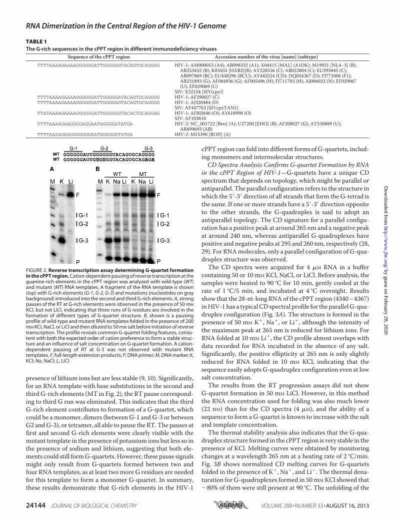
presence of lithium ions but are less stable (9, 10). Significantly,for an RNA template with base substitutions in the second andthird G-rich elements (MT in Fig. 2), the RT pause correspond-ing to third G run was eliminated. This indicates that the thirdG-rich element contributes to formation of a G-quartet, whichcould be amonomer, dimers (between G-1 andG-3 or betweenG2 andG-3), or tetramer, all able to pause the RT. The pauses atfirst and second G-rich elements were clearly visible with themutant template in the presence of potassium ions but less so inthe presence of sodium and lithium, suggesting that both ele-ments could still formG-quartets. However, these pause signalsmight only result from G-quartets formed between two andfour RNA templates, as at least twomoreG residues are neededfor this template to form a monomer G-quartet. In summary,these results demonstrate that G-rich elements in the HIV-1
cPPT region can fold into different forms of G-quartets, includ-ing monomers and intermolecular structures.CD Spectra Analysis Confirms G-quartet Formation by RNA
in the cPPT Region of HIV-1—G-quartets have a unique CDspectrum that depends on topology, which might be parallel orantiparallel. The parallel configuration refers to the structure inwhich the 5�-3� direction of all strands that form theG-tetrad isthe same. If one ormore strands have a 5�-3� direction oppositeto the other strands, the G-quadruplex is said to adopt anantiparallel topology. The CD signature for a parallel configu-ration has a positive peak at around 265 nm and a negative peakat around 240 nm, whereas antiparallel G-quadruplexes havepositive and negative peaks at 295 and 260 nm, respectively (28,29). For RNAmolecules, only a parallel configuration of G-qua-druplex structure was observed.The CD spectra were acquired for 4 �M RNA in a buffer
containing 50 or 10 mM KCl, NaCl, or LiCl. Before analysis, thesamples were heated to 90 °C for 10 min, gently cooled at therate of 1 °C/5 min, and incubated at 4 °C overnight. Resultsshow that the 28-nt-long RNAof the cPPT region (4340–4367)inHIV-1has a typical CD spectral profile for the parallelG-qua-druplex configuration (Fig. 3A). The structure is formed in thepresence of 50 mM K�, Na�, or Li�, although the intensity ofthe maximum peak at 265 nm is reduced for lithium ions. ForRNA folded at 10 mM Li�, the CD profile almost overlaps withdata recorded for RNA incubated in the absence of any salt.Significantly, the positive ellipticity at 265 nm is only slightlyreduced for RNA folded in 10 mM KCl, indicating that thesequence easily adopts G-quadruplex configuration even at lowsalt concentration.The results from the RT progression assays did not show
G-quartet formation in 50 mM LiCl. However, in this methodthe RNA concentration used for folding was also much lower(22 nM) than for the CD spectra (4 �M), and the ability of asequence to form aG-quartet is known to increase with the saltand template concentration.The thermal stability analysis also indicates that the G-qua-
druplex structure formed in the cPPT region is very stable in thepresence of KCl. Melting curves were obtained by monitoringchanges at a wavelength 265 nm at a heating rate of 2 °C/min.Fig. 3B shows normalized CD melting curves for G-quartetsfolded in the presence of K�, Na�, and Li�. The thermal dena-turation for G-quadruplexes formed in 50mMKCl showed that�80% of them were still present at 90 °C. The unfolding of the
TABLE 1The G-rich sequences in the cPPT region in different immunodeficiency viruses
Sequence of the cPPT region Accession number of the virus [name] (subtype)
TTTTAAAAGAAAAGGGGGGATTGGGGGGTACAGTGCAGGGG HIV-1: AM000053 (A4); AB098332 (A1); X04415 [MAL] (A1DK); M19921 [NL4–3] (B);AB253432 (B); K03455 [HXB2](B); AY228556 (C); AB023804 (C); EU293445 (C);AB097869 (BC); EU448296 (BCU); AY445524 (CD); DQ054367 (D); FJ771006 (F1);AB231893 (G); AF084936 (G); AF005496 (H); FJ711703 (H); AJ006022 (N); EF029067(U); EF029069 (U)
SIV: X52154 [SIV(cpz)]TTTTAAAAGAAAAGGGGGGATTGGGGGATACAGTGCAGGGG HIV-1: AF290027 (C)TTTTAAAAGAAAAGGGGGGATTGGGGAGTACAGTGCAGGGG HIV-1: AJ320484 (D)
SIV: AF447763 [SIVcpzTAN1]TTATAAAAGAAAAGGGGGGATTGGGGGGTACACTGCAGGAG HIV-1: AJ302646 (O); AY618998 (O)
SIV: AF103818TTTTAAAAGAAGGGGAGGAATAGGGGATATGA HIV-2: NC_001722 [Ben] (A); U27200 [EHO] (B); AF208027 (G); AY530889 (U);
AB499693 (AB)TTTTAAAAGAAGGGGGGGAATAGGGGATATGA HIV-2: M15390 [ROD] (A)
FIGURE 2. Reverse transcription assay determining G-quartet formationin the cPPT region. Cation-dependent pausing of reverse transcription at theguanine-rich elements in the cPPT region was analyzed with wild-type (WT)and mutant (MT) RNA templates. A fragment of the RNA template is shown(top) with G-rich elements (G-1, G-2, G-3) and mutations (nucleotides on graybackground) introduced into the second and third G-rich elements. A, strongpauses of the RT at G-rich elements were observed in the presence of 50 mM
KCl, but not LiCl, indicating that three runs of G residues are involved in theformation of different types of G-quartet structure. B, shown is a pausingprofile of wild-type and mutant RNA templates folded in the presence of 200mM KCl, NaCl, or LiCl and then diluted to 50 mM salt before initiation of reversetranscription. The profile reveals common G-quartet folding features, consis-tent with both the expected order of cation preference to form a stable struc-ture and an influence of salt concentration on G-quartet formation. A cation-dependent pausing of RT at G-3 was not observed with mutant RNAtemplates. F, full-length extension products; P, DNA primer; M, DNA marker; K,KCl; Na, NaCl; L, LiCl.
RNA Dimerization in the Central Region of the HIV-1 Genome
24144 JOURNAL OF BIOLOGICAL CHEMISTRY VOLUME 288 • NUMBER 33 • AUGUST 16, 2013
by guest on February 28, 2020http://w
ww
.jbc.org/D
ownloaded from

structure is likely prevented by the presence of potassium ions.Surprisingly, the melting curve for the structures formed in 10mM KCl showed that only 40% of them were unfolded at thehighest temperature, indicating that the G-quadruplex is verystable. The G-quadruplexes formed in 50 mM NaCl or LiClcompletely melted with Tm values of 68 and 59 °C, respectively.Because potassium is the dominant monovalent cation insidecells, these results suggest that G-quadruplex configurationcould be easily adopted by viral RNA sequence of the cPPTregion.Interacting RNA Strands with the HIV-1 cPPT Region
Sequence Are Selected by an Affinity Isolation Method—One,two, or four nucleic acid molecules can form a G-quadruplex.To test whether G-quartets that we detected are formed asintermolecular structures, we developed an affinity selectionmethod, with which the interactions between nucleic acids canbe tested by selection of interacting partners with one tagged bya poly(A) sequence. Magnetic beads conjugated with oligo-d(T)25 were used for affinity selection. The method has beenused to select mRNAs and was modified here by using bufferssuitable for G-quartet formation. The interacting partners aredistinguished in a denaturing gel stained with ethidium bro-mide. We tested the specificity of this approach using RNAstrands with the HIV-1 DIS synthesized by transcription invitro. The poly(A)-tagged RNA with the sequence 183–520 ofthe HIV-1 RNA genome (with the DIS at position 257–262)could select another RNA having the DIS (1–520) but devoid ofpoly(A). Non-tagged DIS RNA could not be selected by mag-netic beads in the absence of the poly(A)-tagged partner, dem-onstrating that the observed interactions are not the result ofnonspecific binding to the magnetic beads (Fig. 4A).To determine whether RNAmolecules with the sequence of
the of the HIV-1 cPPT region interact, the RNA strands corre-sponding to positions 4309–4396 of the HIV-1 NL4-3 RNAgenome were synthesized with poly(A) tails and were co-incu-bated with equivalent RNA strands but devoid of poly(A)sequence. Because the gag regions near DIS from HIV-1 MALand NL4-3 were already shown to form dimers throughG-quartet structure, we used RNA strands of these regions as
positive controls for dimerization (9, 10, 12). Our RNA strandshaving the relevant gag sequences (303–415 in HIV-1 MAL,290–403 in HIV-1 NL4-3) did not include the DIS. The RNApartners were combined and incubated to allow possibledimerization and subsequently used for affinity selection withmagnetic beads. As shown in Fig. 4B, the non-tagged RNAstrands of the gag region of the two different HIV-1 species(MAL and NL4-3) and non-tagged RNA strands of the cPPTregion were co-selected with corresponding poly(A)-taggedRNAs. This indicates that the G-rich cPPT region of the HIV-1RNA genome is a likely additional point of contact betweenthe two RNA genomes that augments dimerization initiatedthrough DIS.Dimerization of RNA Strands from the HIV-1 cPPT Region—
The complexes of interacting RNA partners from the cPPTregion selected by magnetic beads might have represented amixture of dimers and tetramers in which G-quartets areformed between two and four nucleic acid molecules, respec-tively. To determine what types of complexes are formedbetween these RNA molecules, we used a native gel analysisassay in which RNA templates of the cPPT region were radio-labeled, incubated in the presence of 1 M KCl or LiCl, and thenresolved in a native polyacrylamide gel. As a control, we usedRNA strands with the antisense sequence.An 88-nt HIV-1 fragment of the cPPT region (4309–4396)
including the G-rich segments self-associated to formmostly adimeric complex and a lesser amount of tetrameric complex,both with reduced electrophoretic mobility. In contrast, theantisense RNA template remained unfolded, which stronglysuggests that strand interaction resulted from structures thatcannot exclusively depend on A:U and G:C base pairs (Fig. 5).Surprisingly, the higher order structures were formed moreefficiently in the presence of LiCl than in the presence of KCl, a
FIGURE 3. CD spectral analysis of the G-rich RNA sequence near the cPPTin HIV-1. A, shown are CD spectra of the G-quadruplex-forming sequence indifferent salts at concentrations of 10 and 50 mM. The effects of K�, Na�, andLi� on the ellipticity signal are compared with the signal for RNA incubated inthe absence of salt. Salt concentrations are 50 mM KCl (red), 10 mM KCl(orange), 50 mM NaCl (dark blue), 10 mM NaCl (light blue), 50 mM LiCl (darkgreen), and 10 mM LiCl (light green) and no salt (gray). B, shown are normalizedCD melting temperature curves for G-quadruplexes formed in 50 mM KCl(red), 50 mM NaCl (blue), 50 mM LiCl (green), and 10 mM KCl (orange).
FIGURE 4. Affinity selection of RNAs interacting through intermolecularG-quartet structure. Poly(A)-tagged RNA templates were elongated at the3� end with an A25 polymer to select them with oligo(dT)25 magnetic beads.To determine whether RNA molecules can interact, the poly(A)-tagged RNAwas mixed with corresponding non-tagged RNA and incubated underdimerization conditions for 2 h. The oligo(dT)25 magnetic beads were addedto select the poly(A)-tagged RNAs, and after three rounds of washes, thesamples were eluted. The selected poly(A) RNAs and interacting partnerswere analyzed in a denaturing gel stained with ethidium bromide. A, a controlexperiment shows that the non-tagged RNA with DIS (RNA genomicsequence 1–520) can be selected with oligo(dT)25 magnetic beads only in thepresence of poly(A)-tagged RNA with DIS (183–520). No RNA was selected inthe absence of a poly(A)-tagged partner (line C). B, the RNAs of the gag region(RNA genomic sequence 303– 415 of the MAL isolate and 290 – 403 of theNL4-3) and cPPT region (4309 – 4396 of the NL4-3) could be isolated with thecorresponding poly(A)-tagged RNA partners after affinity selection with oli-go(dT)25 magnetic beads. AS, affinity selection; T, poly(A)-tagged RNA; P, non-tagged RNA partner; C, control.
RNA Dimerization in the Central Region of the HIV-1 Genome
AUGUST 16, 2013 • VOLUME 288 • NUMBER 33 JOURNAL OF BIOLOGICAL CHEMISTRY 24145
by guest on February 28, 2020http://w
ww
.jbc.org/D
ownloaded from

result not observed at the same concentration of RNA in theCDspectral analysis. However, the concentration of salt in bothexperiments was also significantly different (1 M for RNAdimerization and 50 mM for CD spectra).The higher rate of G-quartet dimer formation in the pres-
ence of LiCl is consistent with results obtained for G-quartetdimers formed from gag region sequences in which the yield ofdimers correlated inversely with the size of monovalent cation(i.e. Li� � Na� � K�) (9, 10). However, although the RNAG-quartet dimers of the gag region fold at a slower rate in thepresence of potassium ions, the complexes are much more sta-ble than those folded in the presence of lithium ions. Ourresults indicate that this is also the case with the G-richsequences of the cPPT region. A thermal dissociation experi-ment (Fig. 6) revealed that although only 12% dimerizationoccurred in the presence of potassium ions, the dimersremained stable when incubated for 8 min at temperaturesbetween 40 and 90 °C. Formation of dimers was more efficientin the presence of sodium ions, with�34% of the strands form-ing complexes, and they also remained stable during the incu-bation at higher temperatures. However, in the presence of lith-ium ions, althoughup to 49%of the strands formeddimers, theyexhibited substantial breakdown after incubation at 50 °C andwere completely disrupted after incubation at 90 °C. Theseresults demonstrate that complexes formed by the RNA tem-plate of the cPPT region display characteristics known forintermolecular RNA G-quartet structures.To confirm that the G-rich sequences contributed to
dimerization of the templates, we attempted to form dimerswith the G-rich sequences in the additional presence of differ-ent antisense 16-nt-long DNA oligonucleotides that wereexpected to prevent G-quartet formation by interacting withcomplementary regions of sense RNA template (Fig. 7).Because dimers weremore efficiently formed in the presence oflithium ions, we used this salt for this experiment. Inclusion ofa 5-fold molar excess of each DNA oligomer confirmed thatdimer formation is affected by DNA oligomers that bind theG-rich sequences. In particular, G-rich element G-1 within thecPPT and G-2 were indicated to be critical for RNA templatesdimerization. These results establish that dimer formationwithin the cPPT region depends onG residues and involves twoof the most conserved G-rich elements.
G-quartet Structure Near the cPPT Promotes TemplateSwitching by the RT—We previously showed that a majorrecombination hot spot in the HIV-1 gag region near DIS cor-relates with sequences rich in G residues that can formG-quar-tet structures (23, 24, 30).We showed enhanced strand transferresulting from G-quartet dimer formation that holds the tem-plates in close proximity and/or G-quartet monomer or dimerformation that increases frequency of RT RNase H by pausingthe RT (Fig. 8A).We have now acquired evidence that theG-quartet structure
formed in the cPPT region also promotes template switching bythe RT. We were initially encouraged in this expectationbecause the distribution of recombination breakpoints acrossmultiple HIV-1 genomes revealed several concentrations, oneof which was within a 200-nt-long sequence spanning fromnear the cPPT to the 3� end of the pol gene that contains runs ofG residues (31). To test the basis of the observed templateswitching, we constructed a reconstituted system to determinewhether G-quartet structure induces the RT to transfer thesynthesis of minus-strand DNA from one RNA template toanother. This system consisted of HIV-1 RT, HIV-1 NC, aprimer (DNA oligonucleotide), and two RNA templates repre-senting two copies of theHIV-1RNAgenomedenoted as donorand acceptor for primer strand transfer (Fig. 8B). The reactionwas initiated from a 32P-labeled oligo(dT) DNA primer
FIGURE 5. Dimerization of the HIV-1 cPPT region. The RNA templates ofsense (WT) and antisense (As) strands were incubated under dimerizationconditions for 2 h in the presence of 1 M KCl or LiCl. Formation of monomers(M), dimers (D), and tetramers (T) was analyzed on a non-denaturing gel run at4 °C in the presence of 10 mM KCl and 0.5 � Tris borate-EDTA. Li, lithium; K,potassium; U, unfolded RNA.
FIGURE 6. Cation-dependent association and thermal stability of the RNAdimer formed in the cPPT region. The RNA dimers were formed in parallel ata concentration of 4 �M in buffers containing 1 M KCl, NaCl, or LiCl. 1 volume of1 � Tris-EDTA buffer was added, and 15-�l aliquots were incubated at theindicated temperatures for 8 min. Thermal stabilities were measured by ana-lyzing samples in non-denaturing gels run at 4 °C. M, monomer; D, dimers.
FIGURE 7. Antisense oligonucleotide binding assay. Dimerization of thecPPT region was performed in the presence of a 5 M excess of different anti-sense DNA oligomers (a--g) binding to specific cPPT region sequences.
RNA Dimerization in the Central Region of the HIV-1 Genome
24146 JOURNAL OF BIOLOGICAL CHEMISTRY VOLUME 288 • NUMBER 33 • AUGUST 16, 2013
by guest on February 28, 2020http://w
ww
.jbc.org/D
ownloaded from
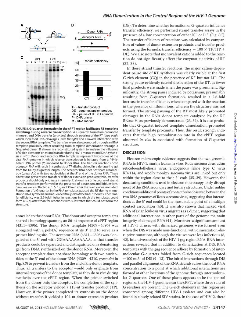
annealed to the donor RNA. The donor and acceptor templatesshared a homology spanning an 86-nt sequence of cPPT region(4311–4396). The donor RNA template (4309–4396) waselongated with a poly(A) sequence at its 3� end to serve as aprimer binding site. The acceptor RNA (4311–4396) was elon-gated at the 5� end with GGAAAAAAAAAA, so that transferproducts could be separated and distinguished on a denaturinggel from DNA synthesized on the donor RNA. Moreover, theacceptor template does not share homology with two nucleo-tides at the 5� end of the donor RNA (4309–4310, green dot inFig. 8B) to prevent transfers from the endof the donor template.Thus, all transfers to the acceptor would only originate frominternal regions of the donor template, as they do in vivo duringsynthesis over the cPPT region. When the primer switchedfrom the donor onto the acceptor, the completion of the syn-thesis on the acceptor yielded a 115-nt transfer product (TP).However, if the primer completed its synthesis on the donorwithout transfer, it yielded a 104-nt donor extension product
(DE). To determine whether formation of G-quartets influencetransfer efficiency, we performed strand transfer assays in thepresence of a low concentration of either K� or Li� (Fig. 8C).The transfer efficiency of reactions was calculated by compar-ison of values of donor extension products and transfer prod-ucts using the formula: transfer efficiency � 100 � TP/(TP �DE).We also note that monovalent cations added to the reac-tion do not significantly affect the enzymatic activity of RT(32, 33).In these strand transfer reactions, the major cation-depen-
dent pause site of RT synthesis was clearly visible at the firstG-rich element (GQ) in the presence of K� but not Li�. Thestrong pause evidently caused dissociation of the RT, as fewerfinal products were made when the pause was prominent. Sig-nificantly, the strong pause induced by potassium, presumablyresulting from G-quartet formation, resulted in a 2.6-foldincrease in transfer efficiencywhen comparedwith the reactionin the presence of lithium ions, wherein the structure was notformed. The strong pausing of the RT most likely promotedcleavages in the RNA donor template catalyzed by the RTRNase H, as previously demonstrated (23, 34). It is also proba-ble that G-quartet induced template dimerization, promotedtransfer by template proximity. Thus, this result strongly indi-cates that the high recombination rate in the cPPT regionobserved in vivo is associated with formation of G-quartetstructure.
DISCUSSION
Electron microscopic evidence suggests that the two genomicRNAs inHIV-1,murine leukemia virus, Rous sarcomavirus, avianreticuloendotheliosis virus, endogenous feline type C virusRD-114, and woolly monkey sarcoma virus are linked but onlywithin the region close to their 5� ends (35–39). However, thedenaturing conditions used in electron microscopy likely disruptmost of the RNA secondary and tertiary structures. Undermilderconditions additional pointsof contactwereobservedbetween thetwoRNAgenomes of Rous sarcomavirus, suggesting that interac-tions at the 5� end could be the most stable point of a multiplecontact association (40). It was also shown that nicked viralRNAof avian leukosis virusmigrates as a dimer, suggesting thatadditional interactions in other parts of the genome maintainintegrity of damaged RNA (41).Moreover, a significant amountof HIV-1 viruses with dimerized genomes were formed evenwhen the DIS was made non-functional with dimerization-dis-ruptive mutations, although the viruses were less infectious (8,42). Intensive analysis of theHIV-1 gag regionRNA-RNA inter-actions revealed that in addition to dimerization at DIS, RNAtemplates with the gag sequence adhere by formation of inter-molecular G-quartets folded from G-rich sequences located�100 nt 3� of DIS (9–13). The initial interactions through DISand parallel alignment of the RNA strands raises the local RNAconcentration to a point at which additional interactions arefavored at other locations of the genome through intermolecu-lar G-quartets. One of those places appears to be the centralregion of theHIV-1 genome near the cPPT, where three runs ofG residues are present. The G-rich elements in this region areconserved among different HIV-1 isolates and can also befound in closely related SIV strains. In the case of HIV-2, there
FIGURE 8. G-quartet formation in the cPPT region facilitates RT templateswitching during reverse transcription. A, G-quartet formation promotedminus-strand DNA transfer (gray line) through pausing of the RT (blue oval),which increased RNA cleavages (blue triangle) and allowed interaction withthe second RNA template. The transfers were also promoted through an RNAtemplate proximity effect resulting from template dimerization through aG-quartet dimer. B, shown is a reconstituted system to analyze the influenceof G-rich elements on strand transfer during HIV-1 minus-strand DNA synthe-sis in vitro. Donor and acceptor RNA templates represent two copies of theviral RNA genome in which reverse transcription is initiated from a 32P-la-beled DNA primer (P) annealed to donor RNA. The transfer reactions ontoacceptor RNA will result in synthesis of TP distinguished in a denaturing gelfrom the DE by its greater length. The acceptor RNA does not share a homol-ogy (green dot) with two nucleotides at the 5� end of the donor RNA. Thesealterations prevent end transfers of donor extension products; thus, transferproducts should only originate internally. C, shown is a time course of strandtransfer reactions performed in the presence of potassium and lithium ions.Samples were collected at 1, 5, 15, and 30 min after the reaction was initiated.Formation of a G-quartet in the RNA templates paused the RT during minus-strand DNA synthesis and influenced the yield of the final products. The trans-fer efficiency was 2.6-fold higher in reactions in which the templates couldform a G-quartet than for reactions with substrates that could not form thestructure.
RNA Dimerization in the Central Region of the HIV-1 Genome
AUGUST 16, 2013 • VOLUME 288 • NUMBER 33 JOURNAL OF BIOLOGICAL CHEMISTRY 24147
by guest on February 28, 2020http://w
ww
.jbc.org/D
ownloaded from

are two G-rich elements, G-1 and G-2, in this region, sufficientto formadimer, and our results indicate that they are critical forforming an intermolecularG-quartet.With an affinity selectionapproach and native gel analysis we show that two RNA mole-cules with the cPPT region sequence can self-associate, and thedimeric complex has similar features andproperties resemblingthose described for RNA dimers formed in the gag regionthrough an intermolecular G-quartet. The G-2 and G-3 ele-ments in theHIV-1 cPPT regionwere previously shown to formG-quartet structure in DNA, and it was suggested that the ele-ments are involved in formation of the cPPT DNA flap (43).Structurally, G-quartets can be polymorphic and adopt sev-
eral different forms depending on their sequence and concen-tration and the milieu of monovalent cations. Our results alsoshow this diversity, and whereas G-quartet dimers are evidentat a high concentration of RNA and salt, monomers are almostsolely formed in the presence of potassium ions at a low salt andRNA template concentration. Of course, our results cannotfully simulate the structural arrangements of the cPPT region ina virion. However, they reliably demonstrate that the sequenceshaving G-rich elements in the middle portion of the RNAgenome are capable of forming aG-quartet dimer and are likelya recombination-relevant position of interaction between thetwo viral genomes.Our results also confirm that G-quartets formed near the
HIV-1 cPPT can effectively pause synthesis by the RT andinduceDNAprimer strand transfer fromadonor to an acceptorRNA template, suggesting that a G-quartet formed near thecPPT facilitates RT template switching. In fact, an analysis of271 sequences of the HIV-1 groupM subtype indicates that thesequence near the 3� end of the pol gene, containing the cPPT,is a common site of recombinant breakpoints (31). Very likely,the increased rate of recombination in this region derives fromthe presence of G-quartets.Because, in our experiments monomers were formed more
efficiently than dimers, the stimulation of transfers in vitroprobably did not primarily result from a proximity effect ofdimerization. However, dimerization of templates through DISandG-quartets in gagwas previously shown to stimulate strandtransfer (24, 44). Moreover, in the context of whole genomes,the opportunity for multiple contact points should favordimeric over monomeric G-quartet formation. Consequently,we expect that dimerization is also a substantial contributor torecombination in the cPPT region. Significantly, the twogenomic RNAs adhering by formation of an intermolecularG-quartet near the cPPT would have a global effect on recom-bination as, together with the gag adherence point and possiblyothers, the adhering would maintain the proximity of the viralgenomes over a long distance. This would contribute toincreased recombination rates in many other locations.Previous studies showed that pol gene sequences and others
located downstream display recombination hot spots (30, 31,45). Because reverse transcription proceeds from the 3� to the5� end of the genomic RNA, the dimerization near cPPT wouldfacilitate recombination in downstream (3� on the template)regions only, as all interactions between genomes would be dis-rupted by synthesis of minus-strand DNA. Investigation of thedistribution of recombination breakpoints across multiple
HIV-1 genomes revealed the presence of a recombination hotspot lying �1 kb downstream of the cPPT, within the 600-nt-long sequence window of the first exon of tat, vpu, rev and thebeginning of env (31). In fact, this region together with the sec-ond exon of tat and rev exhibits the highest rate of recombina-tion throughout the whole genome. We propose that globalgenome spatial alignment, mediated by multiple sites ofdimerization, augments the recombination rate at this andother major hot spots. We also suspect that local dimerizationof genomic RNA in different regions plays a role in distinguish-ing RNA genomes from messenger RNA molecules.The ability of viral RNA genomes with binding-disruptive
mutations in DIS to form some dimers indicates that themech-anism of genome dimerization also involves other regions (8,42). Our results show that the sense RNA template of the cPPTregion has an ability to spontaneously dimerize throughG-quartet formation. Thus, we suggest that alignment ofHIV-1RNA in vivo is alsomediated by formation ofG-tetrads betweenviral genomes. Because self-association of RNA templatesthrough the G-quartet structure occurs at a slower rate thanthrough DIS, G-dimers would appear after interaction at DISand parallel alignment of two viral genomes, whichwould bringhomologous regions into proximity and initiate formation ofintermolecular G-quartets in gag, near cPPT, and likely in otherlocations. This is in agreement with an observation thatgenomic RNA dimerization undergoes maturation process, asgRNA dimers of newly released viral particles are less stablethan genomic RNA dimers of 48-h-old virions (8).Formation of G-quartet dimers between homologous sequences
throughout retroviral genomes could be a spontaneous reaction,similar to formation of double-strandedDNAor formation of hel-ices involving hybridization of complementary sequences. Anysequence with two runs of two or more G residues could poten-tially be a regionwhere theRNAgenomes associate, although thedistance between two G runs and the sequence context will besignificant factors determining the probability of the formationof G-quartet structure. For example, the G residues might beoccluded in stable hairpins so that formation of G tetrads can-not occur. Thus, more advanced analyzes with application ofcomputational approaches are needed to predict genomicregions more prone to form G-quartet dimers between associ-ated retroviral genomes.Moreover, the sequence requirementsfor formation of G-quartets are yet not fully understood, as thestructure might also involve some adenines, as was proposedfor the gag region (9, 10). The cPPT sequence also has a longtrack of A residues that might participate in formation of aquadruplex. The pausing of RT during reverse transcriptionoccurs primarily at G residues, but that does not rule outinvolvement of A residues, particularly those after G residues,forming a purine run.In summary, there is still little known about the mechanisms
and factors involved in dimerization of viral genomes despitethe fact that retroviral RNA dimerization is a key element invirus propagation and survival. Thus, viral genome dimeriza-tion might be a good target for inhibitors of replication ofHIV-1 and other retroviruses. However, understanding of thisprocess is prerequisite for development of suitable therapeuticapproaches.
RNA Dimerization in the Central Region of the HIV-1 Genome
24148 JOURNAL OF BIOLOGICAL CHEMISTRY VOLUME 288 • NUMBER 33 • AUGUST 16, 2013
by guest on February 28, 2020http://w
ww
.jbc.org/D
ownloaded from

Acknowledgment—We are grateful to Dr. Robert J. Gorelick of theNational Cancer Institute for NC used in these studies.
REFERENCES1. Moore, M. D., Fu,W., Nikolaitchik, O., Chen, J., Ptak, R. G., and Hu,W. S.
(2007) Dimer initiation signal of human immunodeficiency virus type 1.Its role in partner selection during RNA copackaging and its effects onrecombination. J. Virol. 81, 4002–4011
2. Moore, M. D., Nikolaitchik, O. A., Chen, J., Hammarskjöld, M. L., Rekosh,D., and Hu, W. S. (2009) Probing the HIV-1 genomic RNA traffickingpathway and dimerization by genetic recombination and single virionanalyses. PLoS Pathog. 5, e1000627
3. Clever, J. L., and Parslow, T. G. (1997) Mutant HIV-1 genomes with de-fects in RNA dimerization or encapsidation. J. Virol. 71, 3407–3414
4. Laughrea,M., Jetté, L.,Mak, J., Kleiman, L., Liang, C., andWainberg,M. A.(1997) Mutations in the kissing-loop hairpin of HIV-1 reduce ( . . . )genomic RNA packaging and dimerization. J. Virol. 71, 3397–3406
5. McBride, M. S., and Panganiban, A. T. (1996) The human immunodefi-ciency virus type 1 encapsidation site is a multipartite RNA element com-posed of functional hairpin structures. J. Virol. 70, 2963–2973
6. Skripkin, E., Paillart, J. C., Marquet, R., Ehresmann, B., and Ehresmann, C.(1994) Identification of the primary site of the human immunodeficiencyvirus type 1 RNA dimerization in vitro. Proc. Natl. Acad. Sci. U.S.A. 91,4945–4949
7. Paillart, J. C., Skripkin, E., Ehresmann, B., Ehresmann, C., andMarquet, R.(1996) A loop-loop “kissing” complex is the essential part of the dimerlinkage of genomic HIV-1 RNA. Proc. Natl. Acad. Sci. U.S.A. 93,5572–5577
8. Song, R., Kafaie, J., Yang, L., and Laughrea, M. (2007) HIV-1 viral RNA isselected in the form of monomers that dimerize in a three-step protease-dependent process. The DIS of stem-loop 1 initiates viral RNA dimeriza-tion. J. Mol. Biol. 371, 1084–1098
9. Marquet, R., Baudin, F., Gabus, C., Darlix, J. L., Mougel, M., Ehresmann,C., and Ehresmann, B. (1991) Dimerization of human immunodeficiencyvirus (type 1) RNA. Stimulation by cations and possible mechanism. Nu-cleic Acids Res. 19, 2349–2357
10. Sundquist, W. I., and Heaphy, S. (1993) Evidence for interstrand quadru-plex formation in the dimerization of human immunodeficiency virus 1genomic RNA. Proc. Natl. Acad. Sci. U.S.A. 90, 3393–3397
11. Awang, G., and Sen, D. (1993) Mode of dimerization of HIV-1 genomicRNA. Biochemistry 32, 11453–11457
12. Marquet, R., Paillart, J. C., Skripkin, E., Ehresmann, C., and Ehresmann, B.(1994) Dimerization of human immunodeficiency virus type 1 RNA in-volves sequences located upstream of the splice donor site. Nucleic AcidsRes. 22, 145–151
13. Ly, H., Nierlich, D. P., Olsen, J. C., and Kaplan, A. H. (1999) Moloneymurine sarcoma virus genomic RNAs dimerize via a two-step process. Aconcentration-dependent kissing-loop interaction is driven by initial con-tact between consecutive guanines. J. Virol. 73, 7255–7261
14. Siddiqui-Jain, A., Grand, C. L., Bearss, D. J., andHurley, L. H. (2002)Directevidence for a G-quadruplex in a promoter region and its targeting with asmall molecule to repress c-MYC transcription. Proc. Natl. Acad. Sci.U.S.A. 99, 11593–11598
15. Sun, D., Guo, K., and Shin, Y. J. (2011) Evidence of the formation of G-quadruplex structures in the promoter region of the human vascular en-dothelial growth factor gene. Nucleic Acids Res. 39, 1256–1265
16. Kumari, S., Bugaut, A., Huppert, J. L., and Balasubramanian, S. (2007) AnRNA G-quadruplex in the 5� UTR of the NRAS proto-oncogene modu-lates translation. Nat. Chem. Biol. 3, 218–221
17. Marcel, V., Tran, P. L., Sagne, C., Martel-Planche, G., Vaslin, L., Teulade-Fichou, M. P., Hall, J., Mergny, J. L., Hainaut, P., and Van Dyck, E. (2011)G-quadruplex structures in TP53 intron 3. Role in alternative splicing andin production of p53 mRNA isoforms. Carcinogenesis 32, 271–278
18. Gomez, D., Lemarteleur, T., Lacroix, L., Mailliet, P., Mergny, J. L., andRiou, J. F. (2004) Telomerase down-regulation induced by the G-quadru-plex ligand 12459 in A549 cells is mediated by hTERT RNA alternative
splicing. Nucleic Acids Res. 32, 371–37919. Didiot, M. C., Tian, Z., Schaeffer, C., Subramanian, M., Mandel, J. L., and
Moine, H. (2008) The G-quartet containing FMRP binding site in FMR1mRNA is a potent exonic splicing enhancer. Nucleic Acids Res. 36,4902–4912
20. Fisette, J. F.,Montagna, D. R.,Mihailescu,M. R., andWolfe,M. S. (2012) AG-rich element forms a G-quadruplex and regulates BACE1mRNA alter-native splicing. J. Neurochem. 121, 763–773
21. Brown, V., Jin, P., Ceman, S., Darnell, J. C., O’Donnell,W. T., Tenenbaum,S. A., Jin, X., Feng, Y., Wilkinson, K. D., Keene, J. D., Darnell, R. B., andWarren, S. T. (2001) Microarray identification of FMRP-associated brainmRNAs and altered mRNA translational profiles in fragile X syndrome.Cell 107, 477–487
22. Norseen, J., Johnson, F. B., and Lieberman, P. M. (2009) Role for G-qua-druplex RNA binding by Epstein-Barr virus nuclear antigen 1 in DNAreplication and metaphase chromosome attachment. J. Virol. 83,10336–10346
23. Shen, W., Gao, L., Balakrishnan, M., and Bambara, R. A. (2009) A recom-bination hot spot in HIV-1 contains guanosine runs that can form a G-quartet structure and promote strand transfer in vitro. J. Biol. Chem. 284,33883–33893
24. Shen, W., Gorelick, R. J., and Bambara, R. A. (2011) HIV-1 nucleocapsidprotein increases strand transfer recombination by promoting dimericG-quartet formation. J. Biol. Chem. 286, 29838–29847
25. Pandey, V. N., Kaushik, N., Rege, N., Sarafianos, S. G., Yadav, P. N., andModak, M. J. (1996) Role of methionine 184 of human immunodeficiencyvirus type-1 reverse transcriptase in the polymerase function and fidelityof DNA synthesis. Biochemistry 35, 2168–2179
26. Kikin, O., D’Antonio, L., and Bagga, P. S. (2006) QGRS Mapper. A web-based server for predicting G-quadruplexes in nucleotide sequences. Nu-cleic Acids Res. 34,W676–W682
27. Schaeffer, C., Bardoni, B., Mandel, J. L., Ehresmann, B., Ehresmann, C.,and Moine, H. (2001) The fragile X mental retardation protein binds spe-cifically to its mRNA via a purine quartet motif. EMBO J. 20, 4803–4813
28. Smargiasso, N., Rosu, F., Hsia, W., Colson, P., Baker, E. S., Bowers, M. T.,De Pauw, E., andGabelica, V. (2008)G-quadruplexDNAassemblies. Looplength, cation identity, and multimer formation. J. Am. Chem. Soc. 130,10208–10216
29. Paramasivan, S., Rujan, I., and Bolton, P. H. (2007) Circular dichroism ofquadruplex DNAs. Applications to structure, cation effects, and ligandbinding.Methods 43, 324–331
30. Dykes, C., Balakrishnan, M., Planelles, V., Zhu, Y., Bambara, R. A., andDemeter, L. M. (2004) Identification of a preferred region for recombina-tion and mutation in HIV-1 gag. Virology 326, 262–279
31. Fan, J., Negroni,M., and Robertson, D. L. (2007) The distribution ofHIV-1recombination breakpoints. Infect. Genet. Evol. 7, 717–723
32. Filler, A. G., and Lever, A. M. (1997) Effects of cation substitutions onreverse transcriptase and on human immunodeficiency virus production.AIDS Res. Hum. Retroviruses 13, 291–299
33. Yong, W. H., Wyman, S., and Levy, J. A. (1990) Optimal conditions forsynthesizing complementaryDNA in theHIV-1 endogenous reverse tran-scriptase reaction. AIDS 4, 199–206
34. Chen, Y., Balakrishnan,M., Roques, B. P., and Bambara, R. A. (2003) Stepsof the acceptor invasion mechanism for HIV-1 minus strand strong stoptransfer. J. Biol. Chem. 278, 38368–38375
35. Höglund, S.,Ohagen,A., Goncalves, J., Panganiban,A. T., andGabuzda,D.(1997) Ultrastructure of HIV-1 genomic RNA. Virology 233, 271–279
36. Bender, W., Chien, Y. H., Chattopadhyay, S., Vogt, P. K., Gardner, M. B.,and Davidson, N. (1978) High molecular weight RNAs of AKR, NZB, andwild mouse viruses and avian reticuloendotheliosis virus all have similardimer structures. J. Virol. 25, 888–896
37. Bender,W., andDavidson, N. (1976)Mapping of poly(A) sequences in theelectron microscope reveals unusual structure of type C oncornavirusRNA molecules. Cell 7, 595–607
38. Kung,H. J., Hu, S., Bender,W., Bailey, J.M., Davidson,N., Nicolson,M.O.,and McAllister, R. M. (1976) RD-114, baboon, and woolly monkey viralRNA’s compared in size and structure. Cell 7, 609–620
39. Murti, K. G., Bondurant, M., and Tereba, A. (1981) Secondary structural
RNA Dimerization in the Central Region of the HIV-1 Genome
AUGUST 16, 2013 • VOLUME 288 • NUMBER 33 JOURNAL OF BIOLOGICAL CHEMISTRY 24149
by guest on February 28, 2020http://w
ww
.jbc.org/D
ownloaded from

features in the 70S RNAs ofMoloney murine leukemia and Rous sarcomaviruses as observed by electron microscopy. J. Virol. 37, 411–419
40. Mangel, W. F., Delius, H., and Duesberg, P. H. (1974) Structure and mo-lecular weight of the 60–70 S RNA and the 30–40 S RNA of the Roussarcoma virus. Proc. Natl. Acad. Sci. U.S.A. 71, 4541–4545
41. Ortiz-Conde, B. A., and Hughes, S. H. (1999) Studies of the genomic RNAof leukosis viruses. Implications for RNA dimerization. J. Virol. 73,7165–7174
42. Shen, N., Jetté, L., Liang, C., Wainberg, M. A., and Laughrea, M. (2000)Impact of human immunodeficiency virus type 1 RNA dimerization onviral infectivity and of stem-loop B on RNA dimerization and reversetranscription and dissociation of dimerization frompackaging. J. Virol. 74,5729–5735
43. Lyonnais, S., Hounsou, C., Teulade-Fichou, M. P., Jeusset, J., Le Cam, E.,andMirambeau, G. (2002)G-quartets assemblywithin aG-richDNA flap.A possible event at the center of the HIV-1 genome.Nucleic Acids Res. 30,
5276–528344. Balakrishnan, M., Roques, B. P., Fay, P. J., and Bambara, R. A. (2003) Tem-
plate dimerization promotes an acceptor invasion-induced transfermech-anism during human immunodeficiency virus type 1 minus-strand syn-thesis. J. Virol. 77, 4710–4721
45. Zhuang, J., Jetzt, A. E., Sun, G., Yu, H., Klarmann, G., Ron, Y., Preston,B. D., and Dougherty, J. P. (2002) Human immunodeficiency virus type 1recombination. Rate, fidelity, and putative hot spots. J. Virol. 76,11273–11282
46. Williamson, J. R. (1994) G-quartet structures in telomeric DNA. Annu.Rev. Biophys. Biomol. Struct. 23, 703–730
47. Phan, A. T., and Patel, D. J. (2003) Two-repeat human telomericd(TAGGGTTAGGGT) sequence forms interconverting parallel and an-tiparallel G-quadruplexes in solution. Distinct topologies, thermody-namic properties, and folding/unfolding kinetics. J. Am. Chem. Soc. 125,15021–15027
RNA Dimerization in the Central Region of the HIV-1 Genome
24150 JOURNAL OF BIOLOGICAL CHEMISTRY VOLUME 288 • NUMBER 33 • AUGUST 16, 2013
by guest on February 28, 2020http://w
ww
.jbc.org/D
ownloaded from

Dorota Piekna-Przybylska, Gaurav Sharma and Robert A. BambaraSignificance for Viral Evolution
Mechanism of HIV-1 RNA Dimerization in the Central Region of the Genome and
doi: 10.1074/jbc.M113.477265 originally published online July 9, 20132013, 288:24140-24150.J. Biol. Chem.
10.1074/jbc.M113.477265Access the most updated version of this article at doi:
Alerts:
When a correction for this article is posted•
When this article is cited•
to choose from all of JBC's e-mail alertsClick here
http://www.jbc.org/content/288/33/24140.full.html#ref-list-1
This article cites 47 references, 21 of which can be accessed free at
by guest on February 28, 2020http://w
ww
.jbc.org/D
ownloaded from