mechanisms of tubulogenesis and endothelial phenotype...
TRANSCRIPT
Microvascular Research 99 (2015) 26–35
Contents lists available at ScienceDirect
Microvascular Research
j ourna l homepage: www.e lsev ie r .com/ locate /ymvre
Mechanisms of tubulogenesis and endothelial phenotype expressionby MSCs
Julie A. Rytlewski 1, M. Alejandra Aldon, Evan W. Lewis, Laura J. Suggs ⁎Department of Biomedical Engineering, The University of Texas at Austin, 107 W Dean Keeton, Stop C0800, Austin, TX 78712, USA
Abbreviations: MCBs, microcarrier beads; MMP, mamesenchymal stem cells; PECAM-1, platelet endotheliapolyethyleneglycol;VE-cadherin,vascularendothelialcadhgrowth factor; vWF, vonWillebrand factor.⁎ Corresponding author. Fax: +1 512 471 0616.
E-mail addresses: [email protected] (J.A. [email protected] (M. Alejandra Aldon), [email protected] (L.J. Suggs).
1 Present address: Division of Human Biology, Fred Hutc1100 Fairview Avenue N, D4-100, Seattle, Washington 98
http://dx.doi.org/10.1016/j.mvr.2015.02.0050026-2862/© 2015 Elsevier Inc. All rights reserved.
a b s t r a c t
a r t i c l e i n f oArticle history:Accepted 11 February 2015Available online 21 February 2015
Keywords:Mesenchymal stem cellsTubulogenesisVasculogenic mimicryFibrinHypoxiaNeovascularization
Stem cell-based therapies are a promising new avenue for treating ischemic disease and chronic wounds.Mesenchymal stem cells (MSCs) have a proven ability to augment the neovascularization processes necessaryfor wound healing and are widely popular as an autologous source of progenitor cells. Our lab has previouslyreported on PEGylated fibrin as a unique hydrogel that promotes spontaneous tubulogenesis of encapsulatedMSCs without exogenous factors. However, the mechanisms underlying this process have remained unknown.To better understand the therapeutic value of PEGylated fibrin delivery of MSCs, we sought to clarify therelationship between biomaterial properties and cell behavior. Here we find that fibrin PEGylation does notdramatically alter the macroscopic mechanical properties of the fibrin-based matrix (less than 10% difference).It does, however, dramatically reduce the rate of diffusion through the gel matrix. PEGylated fibrin enhancesthe tubulogenic growth of encapsulated MSCs demonstrating fluid-filled lumens by interconnected MSCs.Image analysis gave a value of 4320± 1770 μm total network length versus 618± 443 μm for unmodified fibrin.PEGylation promotes the endothelial phenotype of encapsulated MSCs—compared to unmodified fibrin—asevidencedbyhigher levels of endothelialmarkers (vonWillebrand factor, 2.2-fold; vascular endothelial cadherin,1.8-fold) and vascular endothelial growth factor (VEGF, up to 1.8-fold). Prospective analysis of underlyingmolecular pathways demonstrated that this endothelial-like MSC behavior is sensitively modulated by hypoxicstress, but not VEGF supplementation as evidenced by a significant increase in VEGF and MMP-2 secretion percell under hypoxia. Further gain-of-function studies under hypoxic stress demonstrated that hypoxia culture ofMSCs in unmodified fibrin could increase both vWF and VE-cadherin levels to values that were not significantlydifferent than cells cultured in PEGylatedfibrin. This result corroborated our hypothesis that the diffusion-limitedenvironment of PEGylated fibrin is augmenting endothelial differentiation cues provided by unmodified fibrin.However, MSC networks lack platelet endothelial cell adhesion molecule-1 (PECAM-1) expression, whichindicates incomplete differentiation towards an endothelial cell type. Collectively, the data here supports arevised understanding of MSC-derived neovascularization that contextualizes their behavior and utility as ahybrid endothelial–stromal cell type, with mixed characteristics of both populations.
© 2015 Elsevier Inc. All rights reserved.
Introduction
Autologous cell-based therapies are a developing strategy to addressischemic morbidities and promote perfusion of oxygen- and nutrient-deprived tissue. Currently, common targets include sites of acute injury,large or chronic wound beds, and critical limb ischemia (Fadini et al.,
trix metalloproteinase; MSCs,l cell adhesion molecule; PEG,erin;VEGF,vascularendothelial
ewski),[email protected] (E.W. Lewis),
hinson Cancer Research Center,109, USA.
2010). For these applications, the end-goal is to achieve revasculariza-tion, which is often the rate-limiting step in wound healing (Gibotet al., 2010). Establishing a robust blood supply better sustains thehigh metabolic demands of inflammation and tissue remodeling andpromotes more rapid resolution of the damaged tissue (Gibot et al.,2010). Healing outcomes have been further improvedwhenbiomaterialgels, foams, or scaffolds are co-delivered with the grafted stem cells(Kim et al., 2011). These biomaterials serve a threefold purpose: (1) toact as a cell delivery vehicle, (2) to enhance and direct stem cell behav-ior, and (3) to serve as bioactive filler that physically and biochemicallyintegrates with local tissues.
Our group has previously reported on PEGylated fibrin as a natural–synthetic polymer composite that promotes tubulogenesis of encapsu-lated bone marrow-derived mesenchymal stem cells (MSCs), withoutadded soluble factors (Zhang et al., 2010). Here, PEGylation slowsfibrinolysis and extends the therapeutic window of MSCs localized
Table 1Exogenous peptides and small molecules used in experimental groups.
Molecule Concentration Study Manufacturer
Cyclo-GRGDSP 500 μg/mL MIC-axis AnaSpecCyclo-GRGESP 500 μg/mL MIC-axis AnaSpecCytochalasin B 0.1 μM MIC-axis Sigma-AldrichColchicine 0.1 μM MIC-axis Sigma-AldrichHuman VEGF-165 50 ng/mL HIF-axis BioLegendCobalt chloride 75 μM HIF-axis Fisher Scientific
27J.A. Rytlewski et al. / Microvascular Research 99 (2015) 26–35
within the matrix. Additionally, bone marrow-derived MSCs serve as areadily available autologous progenitor population that are responsiveto substrate cues (Lutolf et al., 2009) and have a proven history of en-hancing angiogenic activity and wound closure (Falanga et al., 2007).While we have previously observed a dramatic increase in tubulogenicdevelopment of MSCs in PEGylated fibrin gels (compared to fibrin)(Rytlewski et al., 2012), the full breadth of differences in cellularbehavior has not yet been characterized, nor have the underlyingmechanisms. Understanding how PEGylation changes biomaterialproperties is critical to understanding why MSC network developmentis significantly improved. Furthermore, gaining a clearer understandingof this cell–gel system will facilitate a more targeted context for itspotential clinical implementation.
The series of in vitro studies presented here aims to characterizethe biophysical properties of PEGylated fibrin gels and subsequentcell behavior associated with neovascularization events: lumenalspace formation, production of matrix remodeling and paracrinefactors, and markers of an endothelial phenotype. Possible matrixcues were then isolated to determine which cues yielded specificneovascularization events.
Methods
Materials
Low-glucose Dulbecco's modified Eagle's medium (DMEM),phosphate buffered saline (PBS), fetal bovine serum (FBS), andGluta-MAX™-I (100×) were purchased from Invitrogen (Carlsbad, CA).Penicillin–streptomycin and trypsin/ethylenediaminetetra-aceticacid were purchased from ATCC (Manassas, VA). Sigma-Solohillmicrocarrier beads (MCBs) coated in porcine collagen were obtainedfrom Sigma-Aldrich (St. Louis, MO) as well as fibrinogen and thrombinfrom human plasma. Linear homo-difunctional polyethylene glycolsuccinimidylglutarate (PEG-(SG)2, 3400 Da) was purchased from NOFAmerica (White Plains, NY).
PEGylated fibrin gel fabrication
Gel fabrication followed our previously described protocol forenzymatically crosslinked PEGylated fibrin and fibrin gels (Rytlewskiet al., 2012; Zhang et al., 2010). Human fibrinogen was solubilizedin PBS (without calcium or magnesium, pH 7.8) at 8× the desiredfinal concentration. PEG-SG2 was similarly dissolved in PBS. Humanthrombin was reconstituted in nanopure ddH2O to 100 U/mL anddiluted to 25 U/mL with 40 mM CaCl2. Gel components were sterilizedwith 0.22 μm syringe filters. Gel components were mixed in thefollowing order (volume ratio): (1) fibrinogen, (1) PEG-SG2, (2) PBSfor rheology orMSC-seededMCBs for cell studies (4) thrombin. Gelationwas finalized at 37 °C for 15 min before rinsing with PBS (with calciumand magnesium).
Rheology
Gels were prepared without cells in 40 mm diameter non-stickmolds for a parallel plate rheometer configuration and were kepthydrated with PBS prior to and during testing. Strain sweeps weremeasured from 0.1–2.5% at 15 rad/s at a plate temperature of 37 °C.Storage and loss moduli were reported for statistical comparison at1% strain.
Cryogenic scanning electron microscopy
Cell-free gels were prepared in small-volume molds and kepthydrated in PBS (with calcium and magnesium). Gels were brieflyrinsed in ddH2O beforemounting on a cryogenic SEM stagewith carbontape. Mounted gels were then snap-frozen in liquid nitrogen and
fractured with a scalpel to expose cross-sectional structures. Gelswere transferred to the cryo prep unit within a vacuum cryo transfershuttle (Leica EM VCT100) to minimize crystal formation from airexposure. In the prep unit (Leica EM MED020), the stage tempera-ture was raised from−140 °C to−110 °C to sublimate water crystalsfrom the gels. Samples were then sputter coated with palladium andshuttle-transferred to the SEM for imaging. The SEM stage was keptat −120 °C to −123 °C for the duration of imaging.
Diffusional characterization
Syringes (without the plunger) were used as molds for containingfibrin or PEGylated fibrin gels and the test solute. Cell-free gels(500 μL volume) were formed at the 0 cm3 demarcation of the syringebarrel, gelled to completion at 37 °C, and rinsed with an equal volumeof PBS. After the PBS was aspirated, the slip-tips of the syringes werecarefully removed to expose the gel bottoms. Removal of the slip-tipsensured that diffusivity of the test solute was limited by the gel andnot by capillary resistance of the solvent through the syringe. Syringeswere vertically fixed in place and 500 μL of 2.5 mg/mL 10 kDadextran-Texas Red conjugate (Molecular Probes; Eugene, OR) wasadded directly on top of the gels. While fluorescence was not utilized,the purple appearance of Texas Red dye provided clear visualization ofthe dextran. Time-lapse photos of dye diffusion were taken every15 min for 7 h.
Expansion and maintenance of cells
Human bone marrow-derived mesenchymal stem cells (MSCs;Lonza; Basel, Switzerland)were cultured according to themanufacturer'sspecifications with growth medium in tissue culture-treated plasticflasks at 5000 cells/cm2. growth medium consisted of DMEMsupplemented with 10% FBS, 1% penicillin–streptomycin, and 2 mMGlutaMAX™-I. MSCs were tested by the manufacturer for trilineagedifferentiation potential and for positive expression of CD105,CD166, CD29, and CD44; cells were negative for CD14, CD34, andCD45. Population purity was greater than 95%.
For gel cultures, cells were seeded on MCBs according to ourpreviously described protocol, based on the modified bead-outgrowthassay developed by Nakatsu and Hughes (Nakatsu et al., 2007; Nakatsuand Hughes, 2008; Nehls and Drenckhahn, 1995). Briefly, cells atpassages 4–6 were trypsinized, centrifuged into a pellet, and re-suspended in media at a minimum concentration of 1.4 × 10⁵ cells/mL.MSCs were seeded at 7.0 × 10⁴ cells/mg MCB. Cells and MCBs weregently agitated every 30 min over 4 h and then transferred to ultra-low adhesion 6-well plates for further coating overnight. Cell-seededMCBs were strained through a 70 μm mesh and resuspended in300 μL growth media/mg MCB prior to encapsulation in gels. Gelswere thoroughly rinsed with growth media to remove cytotoxicunreacted PEG-SG2. Gel culture was carried out to day 7 with growthmedia unless otherwise specified.
Small molecules for functional assays
For some experimental groups, small molecules, proteins, orpeptides were added to culture media. Table 1 lists each molecule,

Table 2Antibodies used for western blot protein detection.
Antibody target 1°/2° Species Dilution Manufacturer
CD31 (clone P2B1) Primary Mouse 1:100 Abcam (ab24590)von Willebrand factor Primary Rabbit 1:1000 Abcam (ab6994)VE-cadherin Primary Rabbit 1:700 Abcam (ab33168)β-Actin Primary Rabbit 1:1000 Abcam (ab75186)Mouse IgG Secondary Rabbit 1:5000 Abcam (ab6728)Rabbit IgG Secondary Goat 1:5000 Santa Cruz Bio (sc-2004)
28 J.A. Rytlewski et al. / Microvascular Research 99 (2015) 26–35
the concentration used, the experimental study in which it wasapplied, and the manufacturer's information.
Hypoxic cell culture
Where indicated, MSCs were also cultured under 1% O2 and 2% O2
using a hypoxia chamber (Stemcell Technologies; Tukwila, WA),generously loaned to us by Dr. Aaron Baker. Hypoxic gas mixeswere composed of 5% CO2, 1% or 2% O2, and N2 balance (Praxair;Danbury, CT). Cell cultures were environmentally isolated insidethe hypoxia chamber with an open petri dish of water for humidityand kept at 37 °C. The chamber was purged with hypoxic gas for5 min and then purged again 90 min later to evacuate any residualoxygen. Thereafter, the chamber was purged every 48 h to maintainhypoxic cultures unless opened for assay endpoints.
Fluorescent cell staining for visualizing vacuolar compartments
Texas Red-conjugated dextran (10 kDa; Molecular Probes; Eugene,OR) was added to the media of gel cultures as a cell-impermeable dyefor pinocytic uptake (Davis and Camarillo, 1996). For day 7 endpointcultures, 1.25 mg/mL of dextran was added on day 4; for day 10endpoint cultures, dextran was added on day 7. After cultures wereterminated, MSCs were fixed and additionally stained with FITC-phalloidin (F-actin) and DAPI (nuclei).
Fluorescent cell staining for morphological quantification
On day 7 of gel culture, the samples were thoroughly rinsed withPBS (with calcium and magnesium) for 1 h. Calcein AM (Invitrogen;Carlsbad, CA) was added at 10 μM for 1 h to stain the cytoplasm of livecells. samples were again rinsed with PBS and then fixed with 4%neutral-buffered formalin for 30 min. samples underwent a final PBSrinse and stored at 4 °C overnight for imaging the next day.
Two-photon microscopy
Fluorescent z-stacks were collected of MSC outgrowth from indi-vidual microcarrier beads with an Ultima Multiphoton MicroscopySystem (Prairie Technologies; Middleton, WI). Simultaneous two-photon excitation of multiple fluorophores was achieved with atunable Ti:sapphire laser (Spectra-Physics Mai Tai HP; Newport;Irvine, CA) set to 720 nm. Z-slice thickness was adjusted to maintainisometric voxel dimensions. A thickness of 500–700 μm was imagedalong the z-axis for each image stack.
Three-dimensional morphological quantification
Image processing and three-dimensional morphological quantifica-tion followed our recently described method (Rytlewski et al., 2012).As before, z-stacks were preprocessed in ImageJ (Release 1.2.4; ImageJPlugin Project) and loaded into 3D Slicer (Release 3.6, 64-bit Linux).The Vascular Modeling Toolkit in 3D Slicer was used for imagesegmentation and 3D model generation. Centerline tracings of 3Dmodels were exported as large data clouds of x,y,z coordinates withcorresponding model radii. The data cloud was processed in MATLAB(Release 2008a for Macintosh) to calculate the average 3D networklength per MCB.
Cell proliferation
The CellTiter 96® Aqueous One Solution Cell Proliferation Assay(Promega; Madison, WI) was used to quantify the cell content of gels.On days 1, 3, 5, and 7 of gel culture, the culture supernatant wasremoved and saved for secreted protein analysis. Fresh growthmedia + 20% (v/v) CellTiter 96® solution was then added for 4 h.
Supernatants were transferred in triplicate to a 96-well plate.Absorbance was measured at 490 nm with a microplate reader(BioTek Synergy HT Multi-Mode Microplate Reader; Winooski, VT).
Secreted protein detection by ELISA
Quantikine ELISA kits (R&D Systems; Minneapolis, MN) wereprocured for VEGF, matrix metalloproteinase-2 (MMP-2), and MMP-9.Supernatants collected from cell proliferation samples were assayedaccording to the manufacturer's instructions. Optical density wasmeasured at 490 nm. Secreted protein quantities were normalized tocell number using the results of the matched-sample MTS assay.
Cellular protein detection by Western blot
Cells in gels were lysed with RIPA buffer (Santa Cruz Biotechnology;Dallas, TX) and homogenized with a soft tissue grinder (OMNIInternational; Kennesaw, GA). Cell–gel lysates were passed througha 21-gauge needle 20× for further homogenization and solid gelremnants were removed through centrifugation. Supernatantswere denatured in Laemmli buffer with 5% β-mercaptoethanolat 95 °C for 5 min. Denatured samples were separated with 10%mini-Protean® TGX™ precast gels (Biorad; Hercules, CA) at 20 μgprotein per gel lane and blotted onto PVDF membranes. membraneswere blocked for 1 h at room temperature with 5% (w/v) non-fatmilk in TBST and then incubated overnight with a primary antibodyat 4 °C. membranes were rinsed with TBST and incubated with anHRP-conjugated secondary antibody for 1 h at room temperature.After a second set of TBST rinses, 3 mL of SuperSignal West DuraChemiluminescent Substrate (Pierce Thermo Fisher Scientific; Rock-ford, IL) was added per membrane for 5 min prior to image capturewith a FluorChem CCD system (ProteinSimple; Santa Clara, CA). chemi-luminescent signal was quantifiedwith AlphaView software for statisti-cal analysis. For a list of antibodies and their dilutions, refer to Table 2.
Statistical analysis
Aone- or two-way analysis of variancewas used to determine signif-icance between experimental groups. Where significance was found,post-hoc tests were performed to further determine specific relation-ships of statistical significance. Tukey's correction for multiple compar-isons was applied where appropriate. P-values less than 0.05 wereconsidered statistically significant. All statistical tests were completedin Prism (Version 6.0 for Mac OS X; GraphPad, La Jolla, CA).
Results
Macro- and microscopic changes associated with fibrin PEGylation
Rheological characterization of PEGylated fibrin gels in Fig. 1Ashowed little change in their viscoelastic properties over unmodifiedfibrin gels when matched by fibrin concentration. At 10 mg/mL, fibringels (10F) had a statistically significant increase in their elastic modulusover PEGylated fibrin (10P) but the magnitude of this increase was lessthan 10% of the storage modulus. Both PEGylated fibrin and fibrin gelsdemonstrated increasing elastic moduli with increasing fibrin

*
10F 10P
a
a
f
A CB
D E
0h 7h
10F 10P
2.5h
10F 10P 10F 10P
gel
F
5F 5P7.5F 7.
5P10F
10P0
50
100
150
200
Mo
du
lus
(Pa)
Storage Modulus (G')Loss Modulus (G")
Fig. 1. Characterization of fibrin and PEGylated fibrin gel properties. Abbreviations: F, unmodified fibrin; P, PEGylated fibrin; 5, 7.5, 10 indicate mg/mL of fibrin. (A) Rheological analysis ofstorage and loss moduli for fibrin and PEGylated fibrin across three increasing fibrin concentrations. (B–E) Cryo SEM of fractured gel cross-sections at 10 mg/mL fibrin content. Images(D) and (E) are magnified views of the insets from (B) and (C), respectively. (F) Time-lapse photography: diffusion of 10 kDa dextran-Texas Red through fibrin and PEGylated fibringels within a syringe mold. *p b 0.05, versus fibrin control.
29J.A. Rytlewski et al. / Microvascular Research 99 (2015) 26–35
concentration, corresponding to the crosslinking density of the ma-trix (approximately 50 Pa, 75 Pa and 125 Pa for 5 mg/mL,7.5 mg/mL and 10 mg/mL 10P, respectively). Despite similar mechani-cal properties, cryo-SEM (Figs. 1B–E) revealed remarkable differencesin the fibrin gel microstructure introduced by PEGylation. Unmodifiedfibrin (10F) had a stereotypical fibrous and sponge-like appearance.PEGylated fibrin (10P), in contrast, had non-porous amorphous layersinterspersed within the fibrous architecture. This amorphous qualityis associated with a significant change in fibrin's diffusional characteris-tics. In Fig. 1F, time-lapse photography of labeled 10 kDa dextranshowed both slowed diffusion of the dextran solute and slowerfiltration of the PBS solvent through PEGylated fibrin (10P) overunmodified fibrin (10F) gels.
MSC in PEGylated fibrin form hollow tubes with endothelial markers
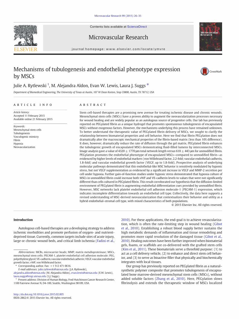
We have previously reported that networks formed by MSCs inPEGylated fibrin were significantly longer than those encouraged byfibrin alone (Rytlewski et al., 2012), indicating that PEGylated fibrin issuperior in promoting tubulogenesis. Here, we further characterizeimportant neovascular features of MSC networks and seek to correlatethese featureswith thematerial properties of PEGylated fibrinmatrices.The presence of intra- and inter-cellular vacuolar compartments is thefirst step in lumen formation and was the first key characteristic weexamined. Based on a lumenization study by George Davis's group onendothelial network development (Davis and Camarillo, 1996), gelculture media were doped with membrane-impermeable 10 kDadextran-Texas Red. Pinocytosed vesicles (containing the labeleddextran) are known to fuse with vacuolar compartments (K. Baylessand Davis, 2002). Hence, these lumenal spaces, if present, can bevisualized by the intracellular localization of the fluorescent dextran.Fluorescent microscopy of MSC networks in PEGylated fibrin (10P)
showed large intracellular compartments of fluorescent dextranthat spanned multiple cell lengths (Figs. 2A–C), suggesting that in-tercellular vacuole fusion had occurred. These large vacuolar spaceswith non-overlapping nuclei and narrow tube diameter are highlyreflective of cell-hollowing lumen assembly associated with smallcapillary development (Lubarsky and Krasnow, 2003). In contrast,lower magnification views of cells in fibrin gels (10F, Fig. 2D) donot demonstrate interconnected networks relative to similar viewsof cells in PEGylated fibrin (10P, Fig. 2E).
Mediators of MSC motility in PEGylated fibrin (10P) duringtubulogenesis and lumenization were identified by examining com-ponents of the matrix–integrin–cytoskeletal (MIC) axis (Davis et al.,2002). As in normal endothelial network development, MSCs exhib-ited a strong dependence on microtubule assembly and integrin-mediated binding. In Figs. 3B–D & G, colchicine (a tubulin inhibitor)caused a more dramatic decrease in network assembly than cytochala-sin B (an actin inhibitor), although both were statistically significant.Additionally, cyclic-RGD inhibition of cell motility (Figs. 3E–G) effec-tively eliminated network outgrowth, while control peptides of cyclic-RGE were not statistically different from controls. This result implicatesαvβ3 and α5β1 as likely integrins responsible for MSC adhesion toPEGylated fibrin and is consistent with current literature regardinghow endothelial cells interact with fibrin-based matrices.
Secreted and cellular proteins were analyzed to provide evidence ofendothelial-like MSC behavior. Total protein secretion of VEGF andMMP-2 was higher in fibrin gels than in PEGylated fibrin (not shown).However, the metabolic assay indicated a slower growth rate of MSCsin PEGylated fibrin (10P, Fig. 3H). When protein production wasnormalized to cell number, we found that MSCs in PEGylatedfibrin (10P) secreted significantly larger quantities of early (days 1and 3) VEGF and late (days 3, 5 and 7) MMP-2 than MSCs in fibrinalone (10F, Figs. 3I & J). Western blot of cellular proteins showed
50µm
green + blue
red
B
C
A
D E 10P10F
200µm
Fig. 2. Lumen development ofMSC networks in PEGylated fibrin. Two stitched frames of a single tubular structure. (A)Merged fluorescent channels from (B) and (C); (B) FITC-phalloidinlabeling of f-actin filaments with DAPI staining of nuclei and (C) dextran-Texas Red localization in vesicles, scale bar = 50 μm.
30 J.A. Rytlewski et al. / Microvascular Research 99 (2015) 26–35
that MSCs in both matrices (fibrin and PEGylated fibrin) undernormoxia also expressed vWF and VE-cadherin proteins (Fig. 5I),which are considered highly specific to endothelial populations(Pusztaszeri et al., 2006). Interestingly, PECAM-1 protein expressionwas not detected from either matrix (Supplemental material Fig. 1).
Hypoxia activation is more important to MSC proliferation and secretionthan direct VEGF stimulation
Potential stimuli from the microenvironment were tested topinpoint the key pathway responsible for catalyzing MSC trans-formation towards an endothelial-like cell type. The sensitivityof MSC tubulogenesis in PEGylated fibrin to chemically simulat-ed hypoxia (CoCl2) (Piret et al., 2002), true hypoxia (1% O2),and soluble VEGF (Figs. 4A–D) was assessed by quantificationof network development, cell proliferation, and angiogenic pro-tein production.
Induction of cellular hypoxia with 1% O2 was confirmed by ELISA forHIF-1α protein (Fig. 4E); protein was normalized to DNA content of thesample to account for different cell proliferation rates under normoxicand hypoxic conditions. Significant upregulation was demonstratedunder 1% hypoxia in PEGylated fibrin (10P) versus normoxia.As diagrammed in Fig. 4F, upregulation of HIF-1α is specificallyknown to stimulate neovascularization processes. Quantification ofMSC network lengths (Fig. 4G) indicated that VEGF, CoCl2, and 1% O2
all resulted in shorter networks than the control group (10P), though1% O2 was not statistically significant. Growth rate quantification,however, revealed that 1% O2 significantly reduced the number ofMSCs cultured in 10P from day 3 onward (Fig. 4H). When secretedVEGF andMMP-2were again analyzed, we found that only 1%O2 signif-icantly increased the production of both proteins over the controlnormoxic group (Figs. 4I & J). Others have similarly reported a lack of
MSC sensitivity to soluble VEGF and emphasized the importance ofother variables, such as cell density, over supplemental growth factors(Galas and Liu, 2014). From this study, we concluded that MSCs aresensitive to hypoxic stress in PEGylated fibrin gels. Despite limited cellproliferation, hypoxia did not significantly hinder the ability of thesecells to form tubular networks and encouraged further production ofsecreted proteins important in neovascularization.
Fibrin bioactivity induces baseline endothelial marker expression
After examining the morphological and functional consequencesof hypoxia in PEGylated fibrin matrices, we sought to refine thespecific roles of hypoxia and matrix cues on the expression of endo-thelial markers in MSC networks. This aim was subdivided into twoexperiments: a 2D study under normoxia to establish baselinebehavior and a 3D study under both normoxia and hypoxia toascertain any gain-of-function changes.
In the first experiment, MSCs were cultured as a two-dimensionalmonolayer on top of a thin gel (either 4 mg/mL fibrin, 4F, or 4 mg/mLPEGylated fibrin, 4P), eliminating gel-associated diffusion gradients(Sahai et al., 2012). Collagen was added as a third experimental groupto control for fibrin content (4 mg/mL collagen, 4C). Western blot wasperformed in triplicate (Fig. 5F) and the chemiluminescent signal wasnormalized to β-actin for vWF and VE-cadherin proteins. Statisticalanalysis revealed that MSCs on PEGylated fibrin (4P) and fibrin gels(4F) expressed similar quantities of both endothelial markers in 2D(Figs. 5G & H). MSCs on collagen (4C), however, had significantlydiminished endothelial protein expression compared to MSCs onPEGylated fibrin. These results imply that (1) PEGylation does notinterfere with the bioactivity of fibrin proteins and (2) fibrin alone isresponsible for a basal level of endothelial-like MSC character.

10P
10P
+Cyt
oB
10P
+Colch
10P
+cG
RGDSP
10P
+cG
RGESP0
2000
4000
6000
8000
Len
gth
of
MS
CN
etw
ork
s(µ
m)
** *
CytoB Colch
RGD RGE
10P
B
C
E F
D
A
10F
G
10F 10P0
20000
40000
60000
80000
Nu
mb
ero
fM
SC
s
Day 1Day 3Day 5Day 7
**
*
H
*
*
I
**
*
J
Fig. 3. Characterization of MSC behavior. Abbreviations: CytoB, cytochalasin B; Colch, colchicine; RGD, cyclic-GRGDSP; RGE, cyclic-GRGESP; 10F, 10 mg/mL unmodified fibrin; 10P,10 mg/mL PEGylated fibrin. (A–F) Fluorescent z-stack projections using standard deviation pixel intensities; images are representative of each group. (G) Average network length permicrocarrier bead as measured by our 3D quantification method. (H) Results of the Celltiter 96 assay. (I) VEGF secretion and (J) MMP-2 secretion normalized to cell number from (H).(G) *p b 0.05, versus 10P control; (H–J) *p b 0.05, versus 10F control at same time point.
31J.A. Rytlewski et al. / Microvascular Research 99 (2015) 26–35
PEGylation-associated hypoxic stress synergistically enhances fibrin cues
In the second experiment, typical 3D culture of MSCs in PEGylatedfibrin (10P) and fibrin (10F) were compared for endothelial markerexpression under normoxia (Figs. 5A & B, I–K). Western blot forvWF and VE-cadherin showed that PEGylated fibrin encourages signifi-cantly greater expression of both markers over unmodified fibrinalone (Figs. 5J & K). Since the 2D study indicated that fibrin contentencourages similar basal levels of vWF and VE-cadherin regardless ofPEGylation, the new difference observed in 3D culture can be attributedto PEGylation-associated changes to the matrix environment. Whenboth 3D matrices were cultured under 1% O2, MSC expression of vWFand VE-cadherinwas once again similar (Figs. 5C &D, L–N).We hypoth-esize that re-equilibration of theMSC endothelial character under 1%O2
is reflective of fibrin-encapsulated MSCs gaining function (Majumdaret al., 2013; Razban et al., 2012), PEGylated fibrin-encapsulatedMSCs experiencing diminished benefits from applied hypoxia, or acombination of both.
Morphological quantification was again applied to comparenetwork lengths. Again, under normoxia and hypoxia, cells in fibrin(10F) remained significantly shorter than cells in PEGylated fibrin
(10P, Fig. 5E). However, even with hypoxic stress and increasedendothelial marker expression, MSCs in fibrin networks were stillunable to form substantial networks. Additionally, both hypoxicgroups were statistically similar to their normoxic controls. Whilethis result is consistent with the study shown in Fig. 4, we wouldordinarily expect an increase in endothelial marker expression andangiogenic proteins with an increase in network length (Kumaret al., 2011; Simionescu et al., 2012). This suggests that there is anadditional feature of PEGylated fibrin that leads to extensivenetwork formation that may be independent of the endothelial-likeprotein expression of MSCs.
Discussion
The studies presented here sought to (1) characterize the extent ofendothelial-like character in MSC tubulogenesis and (2) understandhow the PEGylated fibrinmatrix influences these cellular outcomes. Ma-terial characterization showed that PEGylated fibrin is associated withincreased amorphous over fibrous gel character and restricted diffusion,which previous studies have linked with increased MSC tubulogenesis.Current studies find that, like normal endothelium, MSC networks in

10P
+1%
O 2
10P VEGF CoCl2 1% O2
A B C D
10P
10P
+VEGF
10P
+CoCl 2
10P
+1%
O 2
0
10000
20000
30000
Nu
mb
ero
fM
SC
s
**
** *
G H
10P
10P
+VEGF
10P
+CoCl 2
10P
+1%
O 20
2000
4000
6000
Len
gth
of
MS
CN
etw
ork
s(µ
m)
**
10P
10P
+VEGF
10P
+CoCl 2
10P
+1%
O 2
0
2
4
6
8
10
No
rmal
ized
MM
P-2
Sec
retio
n(p
g/c
ell)
*
*
*
*
10P
10P
+CoCl 2
10P
+1%
O 2
0
100
200
300
400
No
rmal
ized
VE
GF
Sec
ret io
n(f
g/c
ell)
*
** *
10P
Day 1Day 3Day 5Day 7
JI
EHypoxia HIF-1α
growth factorsproteinases
angiogenesis
VEGFMMPs
F
*
10P
Rat
io o
f H
IF-1
αto
DN
A
Fig. 4.VEGF and hypoxia stimuli study. Abbreviations: 10P, 10mg/mL PEGylated fibrin. (A–D) Fluorescent z-stack projections representative ofmorphological outcomes in (G). (E) HIF-1αproduction quantified by ELISA and normalized to total DNA content by nanodrop. (F) General mechanism of hypoxia-induced neovascularization, linking hypoxia with increased VEGFand MMP-2 production. (G) Average network lengths per microcarrier bead as measured by our 3D quantification method. (H) Results of Celltiter 96 assay indicated no mitogenicresponse to VEGF or CoCl2 but a significant decline in MSC proliferation under 1% O2. Hypoxic stress increased VEGF secretion (I) andMMP-2 production (J) of MSCs in PEGylated fibrin;values are normalized to cell numbers reported in (F). *p b 0.05, versus 10P control at same time point.
32 J.A. Rytlewski et al. / Microvascular Research 99 (2015) 26–35
PEGylated fibrin are also hollow, upregulate VEGF and MMP-2 produc-tion, and express vWF and VE-cadherin. Our studies have identifiedthat fibrin is responsible for a basal endothelial profile while hypoxiaserves to enhance differentiation ofMSCs further towards an endothelialphenotype. Robust vascular morphology, on the other hand, was mostsignificantly associated with fibrin PEGylation rather than the biochem-ical nature of the matrix or the oxygen tension in culture. These resultssuggest that morphology depends upon a physical matrix quality apart
from typical biochemical cues. We hypothesize that guidance tunnels,previously identified in normal neovascular sprouts, are able to remainpatent in semi-amorphous PEGylated fibrin and have a tendency tocollapse in fibrin. Unmodified fibrin exhibits a higher loss tangent atthe same concentration, leading to a greater degree of viscous flow forthe same value of storage modulus. This theory is supported by thetime-lapse video of MSC tubulogenesis (Supplemental Fig. 2) wherewe observe some cells retracing tunneled paths.

10F 10P0.0
0.2
0.4
0.6
0.8
VE
-cad
her
inP
rote
inE
xpre
ssio
n
10F 10P0
5
10
15
20
vWF
Pro
tein
Exp
ress
ion
*
4F 4P 4C0
1
2
3
4
vWF
Pro
tein
Exp
ress
ion
*
Normoxia
10F 10P0.0
0.1
0.2
0.3
0.4
0.5
VE
-cad
her
inP
rote
inE
xpre
ssio
n
*
G
4F 4P 4C0.0
0.2
0.4
0.6
0.8
VE
-cad
her
inP
rote
inE
xpre
ssio
n
*
H
Normoxia Hypoxia
A
B
C
D
10F
10P
10F
10P Normoxia Hypoxia
E
10F 10P0
5
10
15
vWF
Pro
tein
Exp
ress
ionM
Hypoxia
J
K
2D Culture 3D Culture
Normoxia
10F
10P
0
2000
4000
6000
8000
Len
gth
of
Net
wo
rks
(µm
)
* *
vWF
β-actin
VE-cad
4F 4P 4C
vWF
β-actin
VE-cad
10F 10P
vWF
β-actin
VE-cad
10F 10P
F I L
N
Fig. 5. Substrate cues versus hypoxic stress. Abbreviations: 10F, 10 mg/mL unmodified fibrin; 10P, 10 mg/mL PEGylated fibrin. (A–D) Fluorescent z-stack projections representativeof MSCs in PEGylated fibrin and fibrin under normoxic and hypoxic culture. (E) 3D morphological quantification of network length for groups shown in (A–D). (F–H) MSCs wereseeded on top of thin gel substrates and cultured for 7 days under normoxic conditions. Chemiluminescent signal from Western blots (shown in F) of vWF and VE-cadherinproteins was normalized to sample-appropriate β-actin signal in G and H, respectively. (I–N) Comparison of normoxia- and hypoxia-induced endothelial marker expression in 3D gelculture of MSCs. MSCs were seeded on microcarrier beads and encapsulated within gels, as previously described. Under normoxic conditions, chemiluminescent signal (shown in I) of vWFand VE-cadherin proteins was normalized to sample-appropriate β-actin signal in J and K, respectively. Under 1% O2 hypoxic conditions, chemiluminescent signal (shown in L) of vWF andVE-cadherin proteins was normalized to sample-appropriate β-actin signal in M and N, respectively. *p b 0.05, versus PEGylated fibrin in (G, H, J, K, M, N), versus normoxic control in (E).
33J.A. Rytlewski et al. / Microvascular Research 99 (2015) 26–35
The lack of PECAM-1 expression, however, led us to question the ex-tent ofMSC differentiation towards an endothelial cell type (Pusztaszeriet al., 2006). An adjunct study (Supplemental Fig. 3) demonstrated cleardifferences between MSCs and endothelial cells in their ability to form
networks and express endothelial markers. While endothelial cellsmore strongly express vascular markers such as VE-cadherin andvWF, MSCs are more capable of migrating through matrices and estab-lishing networks, a feature more typical of stromal than endothelial

34 J.A. Rytlewski et al. / Microvascular Research 99 (2015) 26–35
populations (Ghajar et al., 2010). Endothelial cells have a well-documented need for a supporting stromal population to fully formextended and stable networks; co-cultures are typically employedto rescue their lack of endogenously produced MMPs. Fibroblastsand MSCs have each been used as pericyte support cells in suchcases (Athanassopoulos et al., 2012; Ghajar et al., 2010; Lesmanet al., 2011; Oberringer et al., 2007). Interestingly, fibroblasts (usedas a negative control in this adjunct study) were also able to matchthe ability of MSCs to form networks and express vascular markers,suggesting two possibilities: that fibroblasts possess a greater degreeof phenotypic plasticity than previously anticipated (Alt et al., 2011;Blasi et al., 2011) or that a MSC networks retain fibroblastic featuresof their undifferentiated state alongside newly acquired endothelialbehaviors. To our knowledge, only one report has previously de-scribed the ability of fibroblasts to adopt endothelial character;their in vitro methodology employed traditional monolayer culturesupplemented by growth factors (Karlsson et al., 2009).
In literature, another neovascularization process has been similarlydescribed in which cells are similarly characterized as VE-cadherin(+) but PECAM-1 (−) and highly dependent on MMP-2 production:vasculogenic mimicry (Folberg and Maniotis, 2004; Hendrix et al.,2001; Hess et al., 2003). Vasculogenic mimicry is a unique processwhereby dense tumors overcome hypoxic stress by creating tumor-lined pseudovasculature. Described by Maniotis et al. in (1999),vasculogenic mimicry was the first evidence demonstrating thatnon-endothelialized microvessels are capable of transporting bloodwithout clotting (Maniotis et al., 1999). In terms of tissue engineeringstrategies, vasculogenic mimicry represents a neovascularization pro-cess where a single stromal-type population is driven (primarily) bymatrix cues and hypoxic stress towards endothelial-like structure andfunction (Vartanian, 2012; Zhao et al., 2012). Although these non-endothelialized networks are more leaky and less efficient than normalvasculature, they are functional enough to facilitate blood transport andtumor survival.
While we did not look at an exhaustive profile of endothelialmarkers (vWF, VE-cadherin, PECAM-1, VEGF and MMP-2 in thisstudy), the parallels between MSC tubulogenesis and vasculogenicmimicry provide a novel perspective towards understanding lesser-described stromal cell states. In cancer, these hybrid cell states arewell documented. In fact, the classical epithelial-to-mesenchymaltransition is often classified as “partial,” where epithelial cellsexhibit increased migration without complete loss of cell–celladhesion and polarity (Kalluri and Weinberg, 2009). Here, wedescribe adult stem cells exhibiting increased endothelial charac-teristics while retaining some of their original fibroblastic traits.We propose that transitional phenotypes in stem cell biology maysimilarly be a third state with functional utility and worthy offurther investigation.
In regard to therapeutic neovascularization, endothelial-likenetworks may provide a clinically feasible pseudovasculature fortemporarily sustaining grafts until the host vasculature is able toinfiltrate and remodel tissue. The ability of fibroblasts to matchendothelial-like MSC behavior suggests that this mechanism oftubulogenesis may be shared amongst other cells of stromal lineage.Tubulogenesis is a major mechanism of normal embryologicaldevelopment and organogenesis as well as a potent mechanism ofmetastatic invasion (Lubarsky and Krasnow, 2003; Nagle and Cress,2011). However, stromal tubulogenesis in non-diseased adult biologylacks documentation; further studies would be necessary todetermine whether this cell behavior is an artifact of in vitro cultureor simply a rare natural phenomenon. If tubulogenesis is in fact alatent but inducible stromal function, the work here has the poten-tial to open up new cell sources as therapeutically valuable forneovascularization strategies.
Supplementary data to this article can be found online at http://dx.doi.org/10.1016/j.mvr.2015.02.005.
Authorship contributions
JAR is the primary contributor in experimental design, execution,and manuscript preparation. MAA assisted in wet lab procedureswhile EWL performed computational analyses for morphologicalquantification. LJS is the primary investigator: she advised on experi-mental decisions and assisted in the manuscript preparation process.
Ethical standards
Experiments described within this manuscript comply withthe current laws of the United States of America, in which theywere performed.
Conflict of interest disclosures
The authors declare that they have no conflict of interest.
Acknowledgments
We gratefully acknowledge our sources of funding: the AmericanHeart Association (11GRNT7080002) for funding this project; the De-partment of Defense (NDSEG fellowship) and Cockrell School of Engi-neering (Thrust 2000 fellowship) for funding JR. We also would like tothank the ICMB Microscopy and Imaging Facility (UT Austin) for accessto cryo-SEM and confocal microscopes and Award NumberS10RR027950 from the National Center for Research Resources forproviding access to two-photon microscopy. The content here issolely the responsibility of the authors and does not necessarily rep-resent the official views of the National Center for Research Re-sources or the National Institutes of Health.
References
Alt, E., Yan, Y., Gehmert, S., Song, Y.-H., Altman, A., Gehmert, S., Vykoukal, D., Bai, X., 2011.Fibroblasts share mesenchymal phenotypes with stem cells, but lack their differenti-ation and colony-forming potential. Biol. Cell. 103, 197–208.
Athanassopoulos, A., Tsaknakis, G., Newey, S.E., Harris, A.L., Kean, J., Tyler, M.P., Watt, S.M.,2012. Microvessel networks in pre-formed in artificial clinical grade dermalsubstitutes in vitro using cells from haematopoietic tissues. Burns 38, 691–701.
Bayless, K., Davis, G., 2002. The Cdc42 and Rac1 GTPases are required for capillary lumenformation in three-dimensional extracellular matrices. J. Cell Sci. 115, 1123–1136.
Blasi, A., Martino, C., Balducci, L., Saldarelli, M., Soleti, A., Navone, S.E., Canzi, L., Cristini, S.,Invernici, G., Parati, E.A., Alessandri, G., 2011. Dermal fibroblasts display similarphenotypic and differentiation capacity to fat-derived mesenchymal stem cells, butdiffer in anti-inflammatory and angiogenic potential. Vasc. Cell 3, 5.
Davis, G.E., Bayless, K.J., Mavila, A., 2002.Molecular basis of endothelial cell morphogenesisin three-dimensional extracellular matrices. Anat. Rec. 268, 252–275.
Davis, G.E., Camarillo, C.W., 1996. An alpha 2 beta 1 integrin-dependent pinocyticmechanism involving intracellular vacuole formation and coalescence regulatescapillary lumen and tube formation in three-dimensional collagen matrix. Exp. CellRes. 224, 39–51.
Fadini, G.P., Agostini, C., Avogaro, A., 2010. Autologous stem cell therapy for peripheralarterial disease meta-analysis and systematic review of the literature. Atherosclerosis209, 10–17.
Falanga, V., Iwamoto, S., Chartier,M., Yufit, T., Butmarc, J., Kouttab, N., Shrayer, D., Carson, P.,2007. Autologous bonemarrow-derived culturedmesenchymal stemcells delivered ina fibrin spray accelerate healing inmurine and human cutaneous wounds. Tissue Eng.13, 1299–1312.
Folberg, R., Maniotis, A.J., 2004. Vasculogenic mimicry. APMIS 112, 508–525.Galas, R.J., Liu, J.C., 2014. Vascular endothelial growth factor does not accelerate
endothelial differentiation of human mesenchymal stem cells. J. Cell. Physiol.229, 90–96.
Ghajar, C.M., Kachgal, S., Kniazeva, E., Mori, H., Costes, S.V., George, S.C., Putnam, A.J., 2010.Mesenchymal cells stimulate capillary morphogenesis via distinct proteolyticmechanisms. Exp. Cell Res. 316, 813–825.
Gibot, L., Galbraith, T., Huot, J., Auger, F.A., 2010. A preexisting microvascular networkbenefits in vivo revascularization of a microvascularized tissue-engineered skinsubstitute. Tissue Eng. A 16, 3199–3206.
Hendrix, M.J., Seftor, E.A., Meltzer, P.S., Gardner, L.M., Hess, A.R., Kirschmann, D.A.,Schatteman, G.C., Seftor, R.E., 2001. Expression and functional significance ofVE-cadherin in aggressive human melanoma cells: role in vasculogenic mimicry.Proc. Natl. Acad. Sci. U. S. A. 98, 8018–8023.
Hess, A.R., Seftor, E.A., Seftor, R.E.B., Hendrix, M.J.C., 2003. Phosphoinositide 3-kinaseregulates membrane Type 1-matrix metalloproteinase (MMP) and MMP-2 activityduring melanoma cell vasculogenic mimicry. Cancer Res. 63, 4757–4762.

35J.A. Rytlewski et al. / Microvascular Research 99 (2015) 26–35
Kalluri, R., Weinberg, R.A., 2009. The basics of epithelial–mesenchymal transition. J. Clin.Invest. 119, 1420–1428.
Karlsson, L.K., Junker, J.P.E., Grenegard, M., Kratz, G., 2009. Human dermal fibroblasts: apotential cell source for endothelialization of vascular grafts. Ann. Vasc. Surg. 23,663–674.
Kim, C.H., Lee, J.H., Won, J.H., Cho, M.K., 2011. Mesenchymal stem cells improvewound healing in vivo via early activation ofmatrixmetalloproteinase-9 and vascularendothelial growth factor. J. Korean Med. Sci. 26, 726–733.
Kumar, G., Tison, C.K., Chatterjee, K., Pine, P.S., McDaniel, J.H., Salit, M.L., Young, M.F.,Simon, C.G., 2011. The determination of stem cell fate by 3D scaffold structuresthrough the control of cell shape. Biomaterials 32, 9188–9196.
Lesman, A., Koffler, J., Atlas, R., Blinder, Y.J., Kam, Z., Levenberg, S., 2011. Engineeringvessel-like networks within multicellular fibrin-based constructs. Biomaterials 32,7856–7869.
Lubarsky, B., Krasnow, M.A., 2003. Tube morphogenesis: making and shaping biologicaltubes. Cell 112, 19–28.
Lutolf, M.P., Gilbert, P.M., Blau, H.M., 2009. Designing materials to direct stem-cell fate.Nature 462, 433–441.
Majumdar, D., Bhonde, R., Datta, I., 2013. Influence of ischemic microenvironment onhuman Wharton's Jelly mesenchymal stromal cells. Placenta 34, 642–649.
Maniotis, A.J., Folberg, R., Hess, A., Seftor, E.A., Gardner, L.M., Pe'er, J., Trent, J.M., Meltzer,P.S., Hendrix, M.J., 1999. Vascular channel formation by human melanoma cellsin vivo and in vitro: vasculogenic mimicry. Am. J. Pathol. 155, 739–752.
Nagle, R.B., Cress, A.E., 2011. Metastasis update: human prostate carcinoma invasion viatubulogenesis. Prostate Cancer 2011, 249290.
Nakatsu, M.N., Davis, J., Hughes, C.C.W., 2007. Optimized fibrin gel bead assay for thestudy of angiogenesis. J. Vis. Exp. 3, 186.
Nakatsu, M.N., Hughes, C.C.W., 2008. An optimized three-dimensional in vitro model forthe analysis of angiogenesis. Methods Enzymol. 443, 65–82.
Nehls, V., Drenckhahn, D., 1995. A novel, microcarrier-based in vitro assay for rapidand reliable quantification of three-dimensional cell migration and angiogenesis.Microvasc. Res. 50, 311–322.
Oberringer, M., Meins, C., Bubel, M., Pohlemann, T., 2007. A new in vitro wound modelbased on the co-culture of human dermal microvascular endothelial cells andhuman dermal fibroblasts. Biol. Cell. 99, 197–207.
Piret, J.-P., Mottet, D., Raes, M., Michiels, C., 2002. CoCl2, a chemical inducer of hypoxia-inducible factor-1, and hypoxia reduce apoptotic cell death in hepatoma cell lineHepG2. Ann. N. Y. Acad. Sci. 973, 443–447.
Pusztaszeri, M.P., Seelentag, W., Bosman, F.T., 2006. Immunohistochemical expression ofendothelial markers CD31, CD34, von Willebrand factor, and Fli-1 in normal humantissues. J. Histochem. Cytochem. 54, 385–395.
Razban, V., Lotfi, A.S., Soleimani, M., Ahmadi, H., Massumi, M., Khajeh, S., Ghaedi, M.,Arjmand, S., Najavand, S., Khoshdel, A., 2012. HIF-1α overexpression inducesangiogenesis in mesenchymal stem cells. Biores. Open Access 1, 174–183.
Rytlewski, J.A., Geuss, L.R., Anyaeji, C.I., Lewis, E.W., Suggs, L.J., 2012. Three-dimensionalimage quantification as a new morphometry method for tissue engineering. TissueEng. Part C Methods 18, 507–516.
Sahai, S., McFarland, R., Skiles, M.L., Sullivan, D., Williams, A., Blanchette, J.O., 2012.Tracking hypoxic signaling in encapsulated stem cells. Tissue Eng. Part C Methods18, 557–565.
Simionescu, D.T., Chen, J., Jaeggli, M., Wang, B., Liao, J., 2012. Form follows function:advances in trilayered structure replication for aortic heart valve tissue engineering.J. Healthc. Eng. 3, 179–202.
Vartanian, A.A., 2012. Signaling pathways in tumor vasculogenic mimicry. Biochemistry(Mosc) 77, 1044–1055.
Zhang, G., Drinnan, C.T., Geuss, L.R., (null), 2010. Vascular differentiation of bone marrowstem cells is directed by a tunable three-dimensional matrix. Acta Biomater 6,3395–3403.
Zhao, N., Sun, B.-C., Sun, T., Ma, Y.-M., Zhao, X.-L., Liu, Z.-Y., Dong, X.-Y., Che, N., Mo, J., Gu,Q., 2012. Hypoxia-induced vasculogenic mimicry formation via VE-cadherinregulation by Bcl-2. Med. Oncol. 29, 3599–3607.