med. forum, vol. 25, no.3 march, 2014 · 2014-03-24 · med. forum, vol. 25, no.3 1 march, 2014...
TRANSCRIPT

Electro
nic Cop
y

Electro
nic Cop
y
Electro
nic Cop
y

Electro
nic Cop
y
Med. Forum, Vol. 25, No.3 March, 2014 ISSN 1029-385-X
Recognized by PMDC CONTENTS Recognized by HEC
Editorial
1. Vitamin D and its Effects on Crohn’s Disease - A Study 1
Mohsin Masud Jan
Original Articles
2. Hollow Abdominal Visceral Injuries following Minimally Invasive Gynecological Procedure:
A 16 Year Experience 2-5
1. Nazimuddin Jat 2. Farhat Bano 3. Iqbal Ahmed Memon 4. Pir Bakhsh Khokhar
5. Muhammad Ahmed Azmi
3. Efficacy of Intracoronary Bolus administration of Tirofiban in Acute Coronary Syndrome
Patients with No-Reflow Phenomenon during Percutaneous Coronary intervention (PCI) 6-9
1. Muhammad Nawaz Lashari 2. Muhammad Tanveer Alam 3. Tariq Ashraf
4. Mukhtiar Ahmad Pathan
4. Clinical Profile, Conventional Surgical Approaches and the Outcome of the Surgery in Juvenile
Nasopharyngeal Angiofibroma 10-13
1. Muhammad Siddique 2. Asmatullah Achakzai 3. Ijaz Ahamad 4. Sanaullah Tareen
5. Histological Study of Muscles Injuries to Observe the Effects of Environmental Pollutants on its
Recovery and Regeneration 14-19
1. Muhammad Riaz Sheikh 2. Azhar Masud Bhatti
6. Assessment of Antihyperlipidemic Properties of Aqueous Extract of Cassia fistula Leaves 20-23
1. Usman Nawaz 2. Nasim Ilyas 3. Adnan Jehangir 4. Soban Sadiq
7. Cut Throat Injury: One Year Study 24-26
1. Faheem A. Khan 2. Khalid A. Ashrafi 3. Itrat Jawaid 4. Wadood 5. Asif Abbasi
8. Cholesteatoma Clinical Outcome and Complications : A Study on Patients with Chronic Ear
Disease 27-30
1. Adnan Ejaz 2.Amjad Ali Khan 3. Qazi Mahfooz ul Haq
9. Hypertension as Independent Risk Factors for Acute Stroke 31-34
1. Javed Akhter Rathore 2. Mohammad Saleem 3. Bashir Ahmed Trumbu
10. Role of Oral and Topical Nasal Steroids in Prevention of Ethmoidal Nasal Polyp Recurrence
after Intranasal Polypectomy: A Comparative Study of 64 Cases 35-37
1. Ijaz Ahmad 2. Muhammad Siddique 3. Abdul Latif Kakar 4. Sanaullah Tareen
11. Age and Sex Distribution and Types of Foreign Body esophagus -- Mode of Presentation, Risk
Factors Involved, Management and Complications 38-40
1. Muhammad Amir Nadeem 2. Muhammad Shakaib 3. Hassan Iqbal 4. Tahseen-ul-Hassan Farooqi
12. Comparison on Hepatotoxicity Profile of Diclofenac Sodium & Diclofenac Potassium on
Rabbits 41-45
1. Sadaf Naeem, 2. Rahela Najam, 3. Nausheen Alam
13. Outcome of Trial of Labour with Previous One Ceasarean Section 46-49
1. Qamoos Razzaq 2. Shahida Aziz 3. Ruquiya Adil
14. Study to Estimate the Prevalence of Malaria Infection in Sukkur 50-53
1. Khalid Ahmed Qureshi 2. Arshad Hussain Laghari 3. Akhtar Hussain Samoo
15. The Effects of Insulin on the Volume, Absolute and Relative Weight Pf Liver in HFD/Streptozocin
Induced Diabetic Rats 54-58
1. Sahar Mubeen 2. Muhammad Rafique 3. Amber Ilyas
16. Socio-Demographic Profile of Female Victims of Vitriolage in Interior Sindh Pakistan 59-62
1. Mir Muhammad Sahito 2. Farzana Chang 3. Naseer Sheikh 4. M. Iqbal Mughal 5. Amber Mir
6. Fizzah Iqbal
17. Significance of CD Markers in the Classification, Patterns and Sub Typing of Non Hodgkin’s
Lymphoma 63-66
1. Nazir Pathan 2. Amin Fahim 3. Asghar Khan 4. Aneela Qureshi 5. Saba Khoja
Electro
nic Cop
y

Med. Forum, Vol. 25, No.3 March, 2014 ISSN 1029-385-X
18. Fatal Road Traffic Accidents in Karachi 67-70
1. Romela Naz 2. Imran Afzal 3. Zubaida Zain 4. Afshan Kamran 5. M. Iqbal Mughal
19. Fetomaternal Outcome of Pregnancies Complicated by Acute Appendicitis 71-73
1. Azad Ali Lashari 2. Kulsoom Bhatti 3. Tahmina Mahar 4. Rubina Hafeez
20. Comparison of the Efficacy of Partial Inferior Turbinectomy and Submucosal Diathermy on
Nasal Obstruction in Allergic Rhinitis 74-77
1. Muhammad Ismail Khan 2. Khalil Asad 3. Rafique Ahmad Khattak
21. Acute Septic Arthritis: Open Drainage Versus Needle Aspiration 78-81
1. Roohullah Jan 2. Zahid Askar
22. To Determine the Frequency of Modes of Delivery in Short-Statured Primigaravidae at Term 82-85
1. Shadab Akhtar 2. Saima Gillani
23. To Assess Vitamin D Levels in Patients Diagnosed as Fibromyalgia in Patients Attending Dow
University Hospital 86-88
1. Akhtar Ali Baloch 2. Munir Hussain Siddiqui 3. Babar Bashir 4. Uzma Majid 5. Afzal Qasim
6. Aurangzaib 7. Jawad-us-Salam 8. Muhammad Masroor 9. Qamar un Nisa
24. Attitude and Perception of Oral Health Problems in Pregnant Women 89-91
1. Muhammed Junaid Lakhani 2. Mohsin Girach 3. Wahab Kadri 4. Umair Hasan 5. Shaur Sheikh
6. Hareem Sultan 7. Hina Khan 8. Mehak Irshad Abro
Electro
nic Cop
y

Med. Forum, Vol. 25, No.3 March, 2014 1
Editorial Vitamin D and its Effects on Crohn’s
Disease - A Study Mohsin Masud Jan
Editor
Vitamin D deficiency has been linked to a host of
illnesses and conditions from heart disease and diabetes
to certain types of cancer.
Vitamin D supplements may help those with Crohn’s
disease overcome the fatigue and decreased muscle
strength associated with the inflammatory bowel
disease, according to new research.
Extra vitamin D “was associated with significantly less
physical, emotional and general fatigue, greater quality
of life and the ability to perform activities of daily
living,” said Tara Raftery, a research dietitian and
doctoral candidate at Trinity College Dublin. She is
scheduled to present the findings Saturday at the
Digestive Disease Week meeting in Orlando, Fla.
Raftery and her colleagues evaluated 27 patients who
had Crohn’s in remission. (Even in remission, fatigue
and quality of life can be problematic.) The patients
were assigned to take either 2,000 IUs (international
units) of vitamin D a day or a dummy vitamin for three
months.
Before and after the study, the researchers measured
hand-grip strength, fatigue, quality of life and blood
levels of vitamin D. “Hand-grip strength is a proxy
measure of muscle function,” Raftery said. “Muscle
function has been known to be reduced in Crohn’s
disease.” Besides boosting bone growth and
remodeling, vitamin D is thought to improve
neuromuscular and immune function, reduce
inflammation and help with other bodily tasks. Children
and adults aged 1 year to 70 are advised to get 600 IUs
a day; older adults, 800, according to the U.S. National
Institutes of Health (NIH). Vitamin D is found in fatty
fish such as salmon, in smaller amounts in cheese, egg
yolks and beef liver, and in fortified foods such as milk.
Sometimes called the sunshine vitamin, vitamin D is
also produced when the sun’s rays strike the skin.
Crohn’s can affect any part of the gastrointestinal tract,
but most commonly affects the end of the small bowel
and the beginning of the colon. Symptoms vary, but
may include persistent diarrhea, rectal bleeding,
abdominal cramps, and pain and constipation. About
700,000 Americans are affected, according to the
Crohn’s & Colitis Foundation of America. Its cause is
not well understood, but Crohn’s is thought to involve
heredity and environmental factors. Experts believe that
in those with Crohn’s, the immune system attacks
harmless intestinal bacteria, triggering chronic
inflammation and, eventually, the disease symptoms.
The daily vitamin D supplement benefitted participants
in many ways, Raftery found. “When levels of vitamin
D peaked at 30 ng/mL (75 nmol/L) or more [a level
considered healthy], muscle function in both the
dominant and non-dominant hands were significantly
higher than in those who had levels less than 30
ng/mL,” she said. Quality of life improved more for the
D-supplement group, too. Using a standard measure to
evaluate quality of life, the researchers found those who
achieved a healthy blood level of the vitamin scored 24
points higher than those not on supplements. A 20-point
difference is considered meaningful from a “real-
world” perspective, Raftery said.
Raftery now is testing vitamin D in a larger, year-long
study of 130 Crohn’s patients. The study results echo
those of other researchers, including John White,
professor of physiology at McGill University, Montreal.
He said the research findings “show collectively that
vitamin D acts in the intestine to stimulate the innate
immune system to defend against pathogenic bacteria,
and to enhance the barrier function of the intestinal
epithelium [the lining of the intestine].”
Other researchers, including Raftery, have also shown
vitamin D can help improve muscle strength, he said.
Vitamin D is getting a lot of attention in inflammatory
bowel disease treatments, said Dr. Neera Gupta, co-
chair of the Crohn’s & Colitis Foundation of America’s
pediatric affairs committee. More study is needed to
determine the benefits of maintaining vitamin D levels
higher than currently recommended, she said. Gupta
cautioned those with Crohn’s not to self-dose with
vitamin D. “Discuss your vitamin D status with your
primary gastroenterologist to determine whether or not
vitamin D supplementation is indicated in your
particular situation,” she said. White said supplements
are inexpensive and safer than too much sun exposure.
A daily intake of 2,000 IUs is considered safe, he said.
The safe upper limit for adults is 4,000 IUs, according
to the NIH. The data and conclusions of research
presented at medical meetings should be viewed as
preliminary until published in a peer-reviewed journal.
Electro
nic Cop
y

Med. Forum, Vol. 25, No. 3 March, 2014 2
Hollow Abdominal Visceral
Injuries following Minimally Invasive Gynecological
Procedure: A 16 Year Experience 1. Nazimuddin Jat 2. Farhat Bano 3. Iqbal Ahmed Memon 4. Pir Bakhsh Khokhar
5. Muhammad Ahmed Azmi 1. Assoc. Prof. of Surgery, Al-Tibri MC, Karachi 2. Asstt. Prof. of Surgery, Al-Tibri MC, Karachi
3. Prof. of Surgery, Al-Tibri MC, Karachi 4. Prof. of Community Medicine, Al-Nafees Medical College Islamabad
5. Prof. of Physiology, Al-Tibri MC, Karachi.
ABSTRACT
Objective: To study the pattern of hollow abdominal visceral injuries during minimally invasive gynecological
procedures.
Study Design: Retrospective, Descriptive Study.
Place and Duration of Study: This study was conducted at Fauji Foundation Hospital Karachi, Gulshan General
Hospital Karachi, Star General Hospital Karachi and Al-Tibri Medical College Hospital from July 1997 to
December 2013.
Materials and Methods: All patients admitted to above mentioned hospitals for minimally invasive gynecological
procedures electively or in emergency were included in the study.
Results: Total 3050 minimally invasive gynecological procedures were carried out in the above mentioned hospitals
and 77 (2.5 %) patients had complications following the procedures. Mean age was 28 years ranged from 20-55
years. 40 % patients had history of previous gynaecological surgery.Abdominal visceral injuries included small
bowel perforation 30 (38.96 %) patients, sigmoid colon perforation 20 (25.97%)patients. Both small bowel and
sigmoid colonic perforation 08 (10.38%) patients, Caecal perforation 01(1.29 %) patient and Urinary Bladder 01
(1.29%) patient. 52 % cases were diagnosed within two days of primary surgery and rest within 10 days.
Conclusion: All gynecologists must be skilled, vigilant and careful while doing minimally invasive gynecological
procedures that visceral injury should not happen or ready to deal if it happens accidentally.
Key Words: Bowel perforation. Illegally induce abortions, Dilatation and Curettage, Dilatation and Evacuation, and
Hysteroscopy.
INTRODUCTION
The visceral injury following minimally invasive
gynecological procedures are not common but can
happen and reported literature worldwide Incidences
0.3 % in premenopausal and 2.8% in post menopausal
patients1. They are known to happen during minimally
invasive gynecological procedures such as Dilatation &
Curettage(D&C), Dilation & Evacuation(D&E)and
Hysteroscopy. Although these procedures have very
low risk of complications and can also be performed as
inpatients and out patients2, but if complications occurs
patient will require major invasive procedures like
Laparoscopy and Laparotomy3. Common visceras
injuredare small bowel, large bowel, rectum and
bladder4. Most of the patients who had these
complications, the procedures were done by Mid Grade
trained operators5, so complication rate is higher than
normally reported. Approximately 1/3rd
of the injuries
can be diagnosed at the time of operation6. During
Dilatation & Curettage (D&C), if cervical canal is
narrow so chance of perforation is more. Canal should
be dilated to avoid uterine perforation7, but gut
perforation can be due to any instrumentation in the
uterine cavity resulting in great morbidities and
mortalities8. In Pakistan, most of their llegal Dilatation
& Curettage (D&C) are done by untrained mid wives so
having chances of more complications9. Even in trained
hands perforationto uterus and injury to visceras with
Dilatation & Curettage (D&C), Dilatation&
Evacuation(D&E) and hysteroscopy, can happen.
Uterus and bowel injuries reported incidenceis 3 % and
Uterus and bladder injuries is 1 % 10
.So, in order to
reduce the risk of uterine perforation different
precautions should be taken, such as uterine cervix
adequately dilated, severe uterine anteflexion or
retroflexion noted, and intrauterine adhesions should be
judged before procedures because if these rules are not
followed, Can lead to uterine perforation11
and other
complications. Such group of patients is more prone for
uterine perforation and other complications. Patient
who are nulliparous, post menopausal with markedly
retroverted uterus have more chances of perforation12
than the patients who are adequately assessed before
doing the procedure. Common complications during
minimally invasive gynecological proceduresare a)
excessive bleeding pervagina i.e revealed or concealed
b) Injury to abdominal viscera. c) prolapse of the bowel
through vaginal orifice. d) Infection in the uterus or
other pelvic organs. e) scarring of the uterus or cervix,
Original Article Visceral Injuries
Electro
nic Cop
y
Med. Forum, Vol. 25, No. 3 March, 2014 3
which may require further treatment.
MATERIALS AND METHODS
All patients admitted in gynae ward of three (03)
Hospitals of Karachi already mentioned, their history,
examination, diagnosis, primary procedure and
complications, hospital stay, treatment given &
intervention done were reviewed.
Fauji Foundation Hospital is 200 bed general
infirmaries. The catchment area of the hospital is a
populous neighborhood i.e Shah Faisal Colony Karachi.
The patient population belongs to retired & deceased
family members of military services along with general
population. It is fully equipped with all the latest
diagnostic & management facilities. Star general
&Gulshan hospitals are private concern facilities. They
have bed strength of 30 & 25 respectively. These
hospitals mostly deal with gynecological & obstetrical
patients. Fauji Foundation hospital Karachi had 1440
minimally invasive gynecological procedures done and
complication noted in 36 (2.5%) patients and Gulshan
general Hospital Karachi out of 1500 cases of
minimally invasive gynecological procedures 36 (2.5%)
patients had complications. At Star General Hospital
out of 180 cases of minimally invasive gynecological
procedures, 05 (0.34%) patients had complications.
Surgeons were involved once called by gynaecologist,
most of the time on first post operative day but rarely at
the time of surgery. All these patients were resuscitated
& blood was made available. Those patients who had
excessive bleeding P/V were managed by gynaecologist
or surgeon conservatively and cavity was packed.
Excessive Bleeding p/v patients had hysteroscopy if
bleeding point found, cauterization was done.
Patients with intraperitoneal bleed or with peritonitis
had laparoscopy, followed with whatever procedure
required. During laparoscopy, if no visceral injury
found the rent in uterine wall was repaired
laparoscopically and drain in the pelvis inserted after
peritoneal lavage. If bowel injury found, laparotomy
was done. Finding of laparoscopy was confirmed
during the laparotomy. If patient had faecal peritonitis
due to solitary small bowel perforation, exteriorization
of bowel done as ileostomy followed by peritoneal
lavage but, if sigmoid solitary perforation found, loop
colostomy was done with exteriorization of the injured
bowel followed by peritoneal lavage. If both small
bowel as well as sigmoid colon were injured,
perforation in small bowel brought out as ileostomy and
colonic perforation was repaired. If caecal perforation
found, tube ceacostomy was done left there for six
weeks and followed by gastrograffin enema and the
tube removed. All these patients had triple regime
antibiotic therapy (3rd
generation cephlosporins,
metronidazole & gentacin). During the procedures
surgeon made sure that blood and blood products are
available if needed. Most of the cases were done under
general anesthesia.
RESULTS
Mean age of the patient was 38 years but ages range
between 20-55 years. Out of 77 patients 05 (6.49%)
patients presented with excessive bleeding P/V, 04
(5.19%) patients with signs of shock because of
intraperitoneal bleed and 03 (3.89%) patients with
peritonism secondary to haemoperitonium, 60 (77.9 %)
patients presented with peritonitis within 48 hours after
primary procedure. 05 (6.5%) patients presented during
primary procedure with prolapsed of bowel through the
vaginal orifice. 30 (38.96%) patients hadileal
perforation and 20 (25.97%) patients had sigmoid
perforation and 08 (10.68%) patient had both ileal and
colonic perforation, 01(1.3%) patient had caecal
perforation and 01 had bladder perforation.
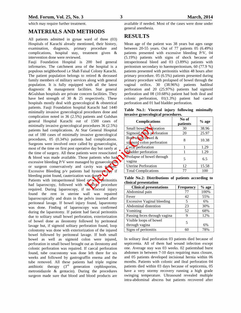
Table No.1: Visceral injury following minimally
invasive gynecological procedures.
Complications No of
patients % age
Small bowel perforation 30 38.96
Sigmoid colon perforation 20 25.97
Both small bowel &
sigmoid colon perforation 8 10.38
Cecal perforation 1 1.29
Bladder perforation 1 1.29
Prolapse of bowel through
vagina 5 6.5
Uterine Perforation 12 15.58
Total Complications 77 100
Table No.2: Distributions of patients according to
clinical presentation
Clinical presentations Frequency % age
Abdominal pain 77 100%
Fever 42 55%
Excessive Vaginal bleeding 5 6%
Abdominal distention 23 30%
Vomiting 52 68%
Passing feces through vagina 9 12%
Visible loops of bowel
through vagina 5
6%
Signs of peritonitis 60 78%
In solitary ileal perforation 03 patients died because of
septicemia. All of them had wound infection except
one. Average stay was 03 weeks. 02 patientshad burst
abdomen in between 7-10 days requiring mass closure,
and 05 patients developed incisional hernia within 06
months. Patients with colonic and ileal perforation 04
patients died within 03 days because of septicemia, 05
have a very stormy recovery running a high grade
swinging temperature. Ultrasound revealed multiple
intra-abdominal abscess but patients recovered after
Electro
nic Cop
y

Med. Forum, Vol. 25, No. 3 March, 2014 4
repeated ultrasound drainage and antibiotics and left the
hospital after six (06) weeks. Ceacal perforation had
tube ceacostomy done and removed without any
untoward effect.
Table No.3: Post Operative Complications
Postoperative
complications Frequency Percentage
Surgical site infections 28 38%
Postoperative pyrexia 14 19%
Postoperative diarrhea 8 11%
Wound dehiscence 2 3%
Enterocutaneous fistula 4 5%
Mortality due to Sepsis 7 10%
Pelvic abscess 5 7%
Inscisional Hernia 5 7%
Total 73 100%
Figure No. 1: Total and different gynecological procedure
showing 10 hysteroscopy, 45 dilation & dilation &
curettage and 22 Dilation & evacuation cases.
DISCUSSION
Hollow visceral injury incident following to minimally
invasive gynecological procedures is rare event and
lack of published information makes it difficult to
compare and review the findings. Our findings showed
that 2.52 % of all abdominal visceral injuries occurred
in minimally invasive gynecological procedures. The
other reported data showed variation ranging from 5-18
% 13
. The lower rate in our series is probably because of
the handing of all procedures by consultants. As
reported by many authors majority of patients were in
very poor general condition at the time of operation14
.
But still surgery was carried out because surgical
intervention is considered to be gold standard of
treatment of any visceral injury following minimally
invasive gynecological procedures 15
. These patients
present late to the primary physician with the problem
so intervention by primary Physician also resulted in
complication and these complication when arises both
the patient and family and physician fearing legal
consequences do not seek help from specialist center.
The few fortunate patients reaching health care facility
represent only the tip of an ice berg 9. The patients with
perforation of uterus have a history of previous pelvic
or abdominal surgery16
. In our series nearly 50% of
patients have previous pelvic or lower abdominal
surgery as reported also by Mesdaghinia E, et al.17
. In
our series perforation of uterus and complications
following Dilation &Curettage is reported 58.44 % in
45 patients but generally reported in literature injuries
to the viscera and the uterus in 20 % cases17
. We had 10
patients of hysteroscopy and only 01 perforation noted
but in literature reported 03 % complications following
hysteroscopy in safe hands and only 01 % uterine
perforation18
. Bowel injuring may occur during variety
of surgical procedures but smaller and substantial
number occurs during less extensive procedures such as
uterine curettage and laparoscopy6. The bowel may be
injured with the curette, ovum forceps or uterine sound,
or even the plastic cannula. Bowelperforation occurs
when the posterior vaginal wall is violated, allowing the
instrument to pierce the underlying structures. The
management of cases with intestinal injuriesfollowing
minimally invasive gynecological procedures poses
some major challenges to general surgeons and
gynecologists practicing in resource-limited
countries.As in our study the most of complication
occur during minor procedures such as Dilation &
Curettage (D&C).The major problem during
laparotomy is to decide whether to close the rent in the
bowel or do resection anastomosis. We made a rule to
do resection anastomosis if more than 50 % diameter is
involved and this is also reported same in literature19
.
Perforation of uterus and complications are more likely
to happen if surgeon is not very experienced, as in our
case maximum perforation occurred in Gulshan General
Hospital because the operator was not experienced, as
reported also by copper that 33 % of uterine perforation
occurred during the surgeon’s first procedure12
.
Minimally invasive gynecological procedures specially
D&C for abortion is the commonest procedure and in
countries where abortion is legalized, mortality and
morbidity related with the procedure declined
significantly. 20
CONCLUSION
It is recommended that minimally invasive
gynecological procedures should be carried out by a
trained& skilled operator to avoid complications. The
operator should have an adequate knowledge regarding
the size of uterus, wall thickness and the scaring on the
uterus. Early recognition, aggressive resuscitation and
early surgical management by institution are of
paramount importance if morbidity and mortality
associated with bowel perforation is to beavoided. The
gynecologist should be competent to deal with the
complications to reduce maternal morbidity and
mortality in the circumstances where surgical help is
not readily available.
Electro
nic Cop
y

Med. Forum, Vol. 25, No. 3 March, 2014 5
REFERENCES
1. Gentile GP, Siegler AM. Inadvertent intestinal
biopsy during laparoscopy and hysteroscopy: a
report of two cases. Fertil Steril 1981;36:402.
2. Lohr PA. Surgical abortion in the second trimester.
Reprod Health Matters 2008; 16:151-61.
3. Paschopoulos M, Polyzos NP, Lavasidis LG, et al.
Safety Issues of hysteroscopic surgery. Ann N Y
Acad Sci 2006;1092:229-234.
4. Mendez LE. Latrogenic injuries in gynaecologic
cancer surgery. Surg Clin North Am 2001;81:
897-923.
5. Mabula JB, Chalya PL, Mchembe MD, Kihnurwa
A, Massinde A, Chandika AB, et al. Bowel
Perforation secondary to illegally induced abortion:
a tertiary hospital experience in Tanzania. World J
Emerg Surg 2012;7: 29.
6. Krebs HB. Intestinal Injury in gynaecologic
surgery: a ten year experience. Am J Obstet
Gynaecol 1986;155:509-154.
7. Dabirashrafi H, Moghadami-Tabrizi N,
Mohammad K, Zandinejad Z. New technique for
reducing the risk of perforation of the uterus at the
beginning of dilatation and curettage. J Am Assoc
Gynecol Laparosc 1996;3:S7-8.
8. Kambiss SM, Hibbert ML, Macedonia C, Potter
ME. Uterine perforation resulting in bowel
infarction: Sharp traumatic bowel and mesenteric
injury at the time of pregnancy termination. Mil
Med 2000;165 (1):81- 82.
9. Rehman A, Fatima S, Gangat S, Ahmed A, Memon
IA, Soomro N. Bowel injuries secondary to
induced abortion: a dilemma. Pak J Surg 2007; 23:
122–125.
10. Pennysylvania Patient safety Advisory, Vol.6 No.
2-June 2009. Website: Http://www. Patientsafety
authority.org
11. Cooper JM, Brady RM. Intraoperative and early
postoperative complications of operative
hysteroscopy. ObstetricsGynecolClin North Am.
2000; 27(2):347-366.
12. Bradely LD. Complications in hysteroscopy
prevention treatment and legal risk. Curr Opin
Obstet Gynecol 2002;14(4):409-415.
13. Rana A, Pradhan N, Gurung G, Singh M. Induced
septic abortion: a major factor in maternal
mortality and morbidity. J Obstet Gynaecol Res
2004;30(1):3–8.
14. Bhattacharya S, Mukherjee G, Mistri P, Pati S:
Safe abortion – Still a neglected scenario: a study
of septic abortions in a tertiary hospital of Rural
India. Online J Health Allied Sci 2010; 9(2):7
15. Oludiran OO, Okonfua FE: Morbidity and
mortality from Bowel Injury secondary to induced
Abortion. Afr J Reprod Health 2003; 7 (3):65-68.
16. Rock JA, Jones HW. TeLinde’s Operative Gynecol
2011.
17. Mesdaghinia Elaheh. Archives of Trauma
Research. 2013;2(2):81-84.
18. Shveiky D, Rojansky N, Revel A,Benshushan
A, Laufer N, Shushan A. Complications of
hysteroscopic surgery: “beyond the learning curve
“J Minim Invasive Gynecol 2007; 14(2):218-22.
19. Perkins JD, Dent LL. Surgical Techniques;
Avoiding and repairing bowel injury in
gynecologic surgery; OBG Management 2004;
15-28.
20. Singh K, Ratnam SS. The influence of abortion
legislation on maternal mortality. Int J Gynaecol
Obstet 1998; 63S123-129.
Address for Corresponding Author:
Prof. Dr. Muhammad Ahmed Azmi,
A-815, Sector 11-A
North Karachi 75850
Karachi – Pakistan
Cell: 0333-2371281
E-mail: [email protected]
Electro
nic Cop
y

Med. Forum, Vol. 25, No. 3 March, 2014 6
Efficacy of Intracoronary
Bolus administration of Tirofiban in Acute Coronary
Syndrome Patients with No-reflow Phenomenon
during Percutaneous Coronary intervention (PCI) 1. Muhammad Nawaz Lashari 2. Muhammad Tanveer Alam 3. Tariq Ashraf
4. Mukhtiar Ahmad Pathan 1. Asstt. Prof. of Cardiology, DUHS&CH, Karachi 2. Asstt. Prof. of Medicine, DUHS&CH, Karachi
3. Asstt. Prof. of Cardiology, NICVD, Karachi 4. Senior Medical Officer, DUH Ojha Complex, Karachi
ABSTRACT
Objective: Currently in acute coronary syndrome, PCI is most common strategy. No-reflow phenomenon (NR) is
one of serious complication. Aim of this study was to evaluate role of intracoronary bolus administration of tirofiban
in acute coronary syndrome patients with no-reflow during PCI.
Study Design: It is prospective and observational study.
Place and Duration of Study: It is multicenter study, conducted in Karachi, Pakistan from August 2011 to
July 2013.
Patients and Methods: Total of 62 patients of acute coronary syndrome underwent for PCI and developed no-
reflow, received intracoronary bolus tirofiban were included. The angiographic definition of successful reperfusion
should include both TIMI 3 flow as well as MBG 2 or 3. No-reflow, assesed by thrombolysis in myocardial
infarction (TIMI) flow and myocardial blush grade (MBG) during treatment. Data were entered and analyzed using
SPSS-16 software. Statistical significance was defined as p-value <0.05.
Results: Out of 62 patients, 43 were males .The mean age was 51 ±13, range from 37 to 70 years. TIMI flow 1 and
11 seen in 17, 37 patients while MBG 1 and 11 seen in 20 and 33 patients before intracoronary bolus administration
of tirofiban. After bolus administration of tirofiban, TIMI flow 111 was seen in 61(98.387 %) out of 62 patients
while MBG 11 and 111 was also noted in 61(98.387 %) out of 62 patients. It showed better Thrombolysis In
Myocardial Infarction flow grades and TIMI myocardial perfusion grades (OR 0. 22, 95% CI 0 .12 -0 .39, p-value
<0.001) immediately after intracoronary bolus administration of tirofiban in reflow phenomenon patients
during PCI.
Conclusion: In patients with ACS, Intracoronary bolus adminstration of tirofiban is effective drug to improve no-
reflow during percutaneous coronary intervention especially when patient blood pressure is at lower-side.
Key Words: Intracoronary, bolus tirofiban, Acute coronary syndrome, No-reflow, PCI.
INTRODUCTION
No-reflow (NR) phenomenon could be defined as the
persistence of reduced flow and regional myocardial
dysfunction after the removal of an experimental
epicardial coronary occlusion1. So far, the precise
mechanisms of NR have not been fully clarified. The
optimal therapy for NR is still being explored. Some
studies2-4
suggested that the dysfunction of coronary
microcirculation perfusion was the central mechanism
of NR. And it would not occur until the lesion of
coronary micro-vascular endothelium to a certain extent
exists. It was a dynamic and persistent procedure. Once
the phenomenon occurred, the inflammation and lesion
of coronary micro-vascular endothelium would be
aggravated and the effect would sustain for weeks.
Restoration of myocardial perfusion rapidly could be
achieved by removing the micro-vascular obstruction
and recovering the ante-grade coronary flow of
occlusive vessel, and it has become a key of the
treatment for NR5. For NR, the mechanism of
conventional drugs was mainly for expanding the
coronary vessel, which might be beneficial to allowing
the formed micro-thrombus to get through the micro-
vascular network and removing the coronary occlusion.
The conventional pharmacological treatment for NR is
intracoronary (IC) administration of vasodilators (for
example, adenosine, verapamil, nitroglycerin, sodium
nitroprusside, etc.)6. On the basis of the mechanisms,
conventional drugs could not inhibit the sustained
thrombi caused by platelet aggregation when balloon
was dilating, which limited the effect7-8
. The effects of
these vasodilators in patients with NR were
contradictory and could not be sustained by large scale
clinical evidence9-10
. Platelet aggregation plays an
important role in the formation of embolization.
Glycoprotein inhibitors (GPI) block the final pathway
of platelet aggregation, combine with the glycoprotein
11b/111a receptors selectively and inhibit the
thrombinogen I competitively. And also, GPI could
inhibit the activation, adhesion and aggregation of
platelets. The pharmacological mechanisms of GPI
Original Article Acute Coronary Syndrome
Electro
nic Cop
y

Med. Forum, Vol. 25, No. 3 March, 2014 7
were contributed to the inhibitation of formation of
platelet thrombi, restoration of the ante-grade coronary
flow of occlusive vessel and reducing the incidence of
the ischemia event11-12
. Tirofiban is one kind of GPI,
which with high selectivity and short-acting
pharmacological mechanism13
. So far, there are some
randomized controlled trials investigated the treatment
of IC bolus administration of tirofiban for NR.
Therefore, the aim of this study was to evaluated the
efficacy of IC bolus administration of tirofiban for NR
during PCI.
MATERIALS AND METHODS
It is prospective, observational and multicentre study,
conducted at Karachi, Pakistan between August 2011 to
July 2013. Total of 62 patients of acute coronary
syndrome underwent for PCI and developed no-reflow,
received intracoronary bolus tirofiban were included.
38 patients had ST-elevation MI, 14 had non-ST
elevation MI and 10 had a USA ( Table 1). All patients
were given oral aspirin 300 mg, clopidogrel 150-300
mg and unfractioned heparin 5000 to 7500 units before
PCI. Tirofiban was administered as an intracoronary
bolus injection 10µg/kg over 01 min followed by
maintenance intravenous infusion at 0.15µg·kg–1·min
for 12 h. After PCI, all patients were managed in the
cardiac care unit with once-daily dose of aspirin (150-
300 mg) and clopidogrel (75 mg). A beta-blocker, statin
and an angiotensin-converting enzyme inhibitor (ACEI)
were also routinely prescribed to all patients. All
coronary angiograms were evaluated by authers after
PCI. Scores of thrombus in the PCI-targeted artery were
assessed as following: 0: no thrombus; 1: possible
thrombus; 2: the length of the thrombus is less than
50% of the vessel diameter; 3: the length of the
thrombus is half to twice the vessel diameter; 4: the
length of the thrombus is longer than twice the vessel
diameter10.
No-reflow in the PCI-targeted coronary
arteries was assessed by Thrombolysis In Myocardial
Infarction (TIMI) flow grade2. The TIMI myocardial
perfusion grade (TMPG) was used to assess myocardial
tissue-level perfusion3. TMPG was assessed only in the
area supplied by the PCI-targeted vessel. The
angiographic definition of successful reperfusion
should include both TIMI 3 flow as well as MBG 2
or 3.
Statistical Analysis: Data were entered and analyzed
using SPSS-16 software. Continuous data are expressed
as mean values ± SD. Student’s t-test was used to
analyze continuous variables. Categorical variables
were analyzed by chi-square test. P-value <0.05 was
considered statistically significant.
RESULTS
The baseline characteristics of the patients are shown in
Table 1. Out of 62 patients, 43 were males .The mean
age was 51 ±13, range from 37 to 70 years. As shown
in table. 11, TIMI flow1 and 11 was seen in 17, 37
patients while MBG (Table. 111) 1 and 11 seen in 20
and 33 patients before intracoronary bolus
administration of tirofiban. After bolus administration
of tirofiban TIMI flow 111 was found in 61(98.387 %)
out of 62 patients while MBG 11 and 111 was also
noted in 61(98.387 %) out of 62 patients. It was found
significantly better Thrombolysis In Myocardial
Infarction (TIMI) flow grades and TIMI myocardial
perfusion grades (OR 0.22, 95% CI 0.12 -0.39,
p-value <0.001) immediately after intracoronary bolus
administration of tirofiban during PCI.
Table No.1: Baseline Characteristics of patients.
Characteristics No.(62)
Age (years ) 51 ±13
Sex ( male ) 43
Hypertension 50
Diabetes 20
Current smoker 23
Prior MI 12
STEMI 38
NSTEMI 14
USA 10
Vessel.
LAD.
RCA.
LCX.
31
19
12
Table No.2: TIMI-Flow during PCI.
Timi-
Flow
Before I/C Tirofiban After I/C Tirofiban
0 08 00
1 17 01
11 37 00
111 00 61
I/C =Intracoronary
Table No.3: Myocadial Blush Grade during PCI.
Myocardial-blush
grade.
Before I/C
Tirofiban.
After I/C
Tirofiban.
0 07 00
1 20 01
11 33 08
111 02 53
I/C= Intracoronary.
DISCUSSION
The main findings of the present study are as follows.
Intracoronary bolus administration of tirofiban
is associated with an improved in no-reflow
phenomenon in form of TIMI flow and TMPG during
PCI like in other studies14-17
. Though TIMI is a classical
indicator of reperfusion during PCI, 18-20
it does not
mean that TIMI 3 flow represents a normal myocardial
perfusion.21
In other words, myocardial blush grade
(MBG) 01 might occur in the patients with TIMI 3 flow
Electro
nic Cop
y

Med. Forum, Vol. 25, No. 3 March, 2014 8
during PCI. It had been found that MBG was an
independent predictor of long-term mortality and could
be used to describe the effectiveness of myocardial
reperfusion.22
Van Hof et al.23
proposed that the
angiographic definition of successful reperfusion
should include both TIMI 3 flow as well as MBG 2 or
3. Moreover, Stone. et al.,24
suggested that MBG could
be used to stratify prognosis of survival in high risk
patients achieving TIMI 3 flow after intervention.
Theoretically, MBG is superior to TIMI when assessing
the myocardial perfusion during PCI. From the recent
researches, GPI has its obvious advantages in inhibiting
the formation of platelet thrombus, but bleeding event
was the main complication. Tirofiban is one kind of
GPI, which with high selectivity and short-acting
pharmacological mechanism.25
During PCI, IC bolus
administration of tirofiban might increase the local drug
concentration and improve the coronary flow.
Considering the particular mechanism and short half
life, IC tirofiban selectively blocks the final pathway of
the platelet aggregation, which might contribute to the
improving TIMI flow, MBG and reducing MACE.
In the present study 61.3% of patients had ST-elevation
ACS. Intracoronary tirofiban was administered
immediately after no-reflow phenomenon during PCI
because we hypothesized that local administration of
IIb/IIIa antagonist would have a faster and more
efficient action on the coronary thrombus and vascular
endothelium than the conventional intravenous bolus
injection.
Study Limitations: It was small sample data, the bias
should not be ignored. Also, the condition of patients,
the time and dosage of drugs might have influenced the
outcomes. Therefore, needs further powerful studies.
CONCLUSION
The treatment of IC bolus administration of tirofiban
was significantly effective to improve no-reflow
phenomenon during PCI in acute coronary syndrome
patients.
REFERENCES
1. Piana RN, Paik GY, Moscucci M, Cohen DJ,
Gibson CM, Kugelmass AD, et al. Incidence and
treatment of no-reflow after percutaneous coronary
intervention. Circulation 1994;89:2514-2518.
2. Niccoli G, Burzotta F, Galiuto L, Crea F.
Myocardial no-reflow in humans. J Am Coll
Cardiol 2009;54:281-292.
3. Chen YF, Yang YJ. No-reflow phenomenon after
percutaneous coronary intervention. Adv
Cardiovasc Dis Jan 2005;26:4-8.
4. Movahed MR, Butman SM: The pathogenesis and
treatment of no-reflow occurring during
percutaneous coronary intervention. Cardiovasc
Revase Med 2008;9:56-61.
5. 05.Resnic FS, Wainstein M, Lee MK, Behrendt D,
Wainstein RV, Ohno-Machado L et. al.: No-reflow
is an independent predictor of death and
myocardial infarction after percutaneous coronary
intervention. Am Heart J 2003;145:42-46.
6. Zhao HY. Effects of adenosine in reperfusion of
acute myocardial infarction. China Prac Med 2009;
4:71-72.
7. Kaplan BM, Benzuly KH, Kinn JW, Bowers TR,
Tilli FV, Grines CL, et al. Treatment of no-reflow
in degenerated saphenous vein graft interventions:
comparison of intracoronary verapamil and
nitroglycerin. Cathet Cardiovasc Diagn 1996; 39:
113-118.
8. Oldenburg O, Eggebrecht H, Herrmann J, Naber
CK, Haude M, Erbel R, et al. Dose-dependent
effects of in-tracoronary verapamil on systemic
and coronary hemodynamics. Cardiovasc Drugs
Ther 2000;14:651-655.
9. Huang D, Qian J, Ge L, Jin X, Jin H, Ma J, et al:
Restoration of coronary flow in patients with no-
reflow after primary coronary intervention of acute
myocardial infarction (RECOVER). Am Heart J
2012;164:394-401.
10. Warnhol A, Ostad MA, Heitzer T, Goldmann BU,
Nowak G, Munzel T. Effect of tirofiban on
percutaneous coronary intervention-induced
endothelial dysfunction in patients with stable
coronary artery disease. Am J Cardiol 2005; 95:
20-23.
11. Kouz R, Kouz S, Schampaert E, Rinfret S, Tardif
JC, Nguyen M, et al. Effectiveness and safety of
glycoprotein IIb/IIIa inhibitors in patients with
myocardial infarction undergoing primary
percutaneous coronary intervention: a meta-
analysis of observational studies. Int J Cardiol
2011;153:249-255.
12. Kimmelstiel C, Badar J, Covic L, Waxman S,
Weintraub A, Jacques S, et al. Pharmacodynamics
and pharmacokinetics of the platelet GPIIb /IIIa
inhibitor tirofiban in patients undergoing
percutaneous coronary intervention: implications
for adjustment of tirofiban and clopidogrel dosage.
Thromb Res 2005;116:55-66.
13. Zhang WZ, Song MC, Liang JY, Li JH, Lei HD,
Su JQ, et al. Effects of tirofiban in patients with no
reflow during PCI procedure. South China J
Cardiovasc Dis 2008;14:103-105.
14. Wei L, Li XQ, Jin EZ, Wang XY. Efficacy and
safety of intracoronary tirofiban on no-ref low
phenomenon after percutaneous coronary
intervention.J Clin Cardiol (China) 2011;27:25-29.
15. Wu J, Xu L, Du H, Wang YR: Influence of
tirofiban on no-reflow after percutaneous coronary
intervention in patients with acute coronary
syndrome. Chin J Evid Based Cardiovasc Med
2012;14:131-133.
Electro
nic Cop
y

Med. Forum, Vol. 25, No. 3 March, 2014 9
16. Zhang HY, Wang PX, Cao YJ, Wu ZG, Liu HS.
Clinical study of intracoronary injections of
tirofiban in acute myocardial infarction patients
with no reflow phenomenon after percutaneous
coronary intervention. J Clin Cardiol (China)
2011;27:25-29.
17. Zhang J, Jiang JG: Effects of intracoronary
injections of tirofiban in acute coronary syndrome
patients with no reflow phenomenon after
percutaneous coronary intervention. Anhui Med J
2012;2:155-158.
18. Geeganage C, Wilcox R, Bath PMW. Triple
antiplatelet therapy for preventing vascular events:
a systematic review and meta-analysis.BMC Med
2010;8:36.
19. Hamada S, Nishiue T, Nakamura S, Sugiura T,
Kamihata H, Miyoshi H, et al. TIMI frame count
immediately after primary coronary angioplasty as
a predictor of functional recovery in patients with
TIMI 3 reperfused acute myocardial infarction. J
Am Coll Cardiol 2001;38(3):666-671.
20. Fu W. Effects of tirofiban in elder patients of acute
myocardial infarction with no-reflow phenomenon
during PCI. Chin J Gerontol 2012;8:1565-1567.
21. Ndrepepa G, Tiroch K, Keta D, Fusaro M,
Seyfarth M, Pache J, et al. Predictive factors and
impact of no reflow after primary percutaneous
coronary intervention in patients with acute
myocardial infarction. Circ Cardiovasc Interv
2010;3(1):27-33.
22. Stone GW, Brodie BR, Griffin JJ, Morice MC,
Costantini C, Goar FST, et al. Prospective,
multicenter study of the safety and feasibility of
primary stenting in acute myocardial infarction: in-
hospital and 30-Day results of the PAMI stent pilot
trial. J Am Coll Cardiol 1998;31(1):23-30.
23. Vant Hof AW, Liem A, Suryapranata H, Hoorntje
JC, De Boer MJ, Zijlstra F. Angiographic
assessment of myocardial reperfusion in patients
treated with primary angioplasty for acute
myocardial infarction myocardial blush grade.
Circulation 1998;97(23):2302-2306.
24. Stone GW, Peterson MA, Lansky AJ, Dangas G,
Mehran R, Leon MB: Impact of normalized
myocardial perfusion after successful angioplasty
in acute myocardial infarction. J Am Coll Cardiol
2002;39(4):591-597.
25. Henriques JP, Zijlstra F, Vant Hof AW, De Boer
MJ, Dambrink JH, Gosselink M, et al.
Angiographic assessment of reperfusion in acute
myocardial infarction by myocardial blush grade.
Circulation 2003;107(16):2115-2119.
Address for Corresponding Author:
Dr. Muhammad Nawaz Lashari
Address: 1-C 7th
street,
Defence Housing Authority (DHA) Phase-1,
Karachi , Pakistan
Email: [email protected]
Cell No: 03002656269
Electro
nic Cop
y

Med. Forum, Vol. 25, No. 3 March, 2014 10
Clinical Profile, Conventional
Surgical Approaches and the Outcome of the Surgery
in Juvenile Nasopharyngeal Angiofibroma 1. Muhammad Siddique 2. Asmatullah Achakzai 3. Ijaz Ahmad 4. Sanaullah Tareen
1. Asstt. Prof. of ENT, BMC, Quetta 2.Senior Registrar of ENT, BMC, Quetta. 3. Assoc. Prof. of Radiology, BMC,
Quetta. 4. Epidemiologist, Fatima Jinnah Chest Hospital, Quetta.
ABSTRACT
Objective: To study the clinical profile of angiofibroma, various conventional surgical approaches and the outcome
of surgery.
Design: Prospective, analytical study.
Place and Duration of Study: This study was conducted at the Department of Otorhinolaryngology and Head &
Neck Surgery, Bolan Medical Complex Hospital, Quetta from January2008 to December2012.
Materials and Methods: This study included 31 patients of juvenile nasopharyngeal angiofibroma over a period of
5 years. All patients were treated by conventional surgical approaches following Fisch staging system. Twenty one
(21) patients were operated by lateral rhinotomy approach, Three (3) patients by transpalatal approach, five (5)
patients by Weber-Fergusson approach and two (2) by Mid-facial degloving approach. The patients were followed
up for a period of three (3) years for any recurrence.
Results: All patients were male with mean age 15.61±2.64 years. The patients presented with recurrent epistaxis,
nasal obstruction and nasopharyngeal mass apart from other symptoms and signs. Majority of the patients (80.64%)
came with stage II and IIIA disease. Lateral rhinotomy approach was used in majority of cases (67.74%).
Recurrence was observed in 5 cases (16.12%). Recurrence rate was less (one out of 21cases) with lateral rhinotomy
approach in comparison with other approaches. In transpalatal route recurrence was observed in two (2) cases out of
three (3) cases, while in Weber-Fergusson approach it was one out of five (5) and in Mid-facial degloving it was one
out of two (2) cases.
Conclusion: Juvenile nasopharyngeal angiofibroma is a disease of male adolescents. The patient most commonly
presents with recurrent epistaxis and nasal blockage with nasopharyngeal mass. Surgery is the treatment of choice.
Lateral rhinotomy approach gives an excellent exposure for most of these tumours with less chance of recurrence.
Key Words: Juvenile nasopharyngeal angiofibroma, Clinical profile, surgical approaches, Recurrence.
INTRODUCTION
Juvenile nasal angiofibroma (JNA) is a rare, highly
vascular, locally aggressive tumour that primarily
affects male adolescents.1It accounts for approximately
0.5% of all head and neck tumours.2 It originates from
sphenopalatine foramen3 and invades nasopharynx.
From here it may spread to the nose, paranasal sinuses,
pterygopalatine fossa, infratemporal fossa, orbit or skull
base and intracranial. Grossly, the tumour is pale red to
blue smooth mass often lobulated, noncapsulated,
sessile or pedunculated and covered by nasopharyngeal
mucosa.
The patient presents with recurrent epistaxis and nasal
blockage with intranasal mass.4 Cheek swelling, visual
changes, hearing impairment and neurological deficits
may be present sometimes. Diagnosis is made mainly
by history, clinical examination and imaging studies.
Preoperative biopsy is at best avoided for fear of
massive lethal bleeding. There are a variety of staging
criteria developed when evaluating juvenile
angiofibromas which include those developed by
Sessions, Chandler, Fisch, and Radkowski.5-9
The Fisch
staging is the most robust and practical.10
Treatment
options for juvenile nasopharyngeal angiofibroma
include surgery, radiation therapy, chemotherapy and
hormonal therapy.11
Surgery is the treatment of choice
for nasopharyngeal angiofibroma.4 Preoperative
angiography and embolization minimizes the
intraoperative blood loss.12
In surgical treatment of
juvenile nasopharyngeal angiofibroma the possibility of
recurrences and residual tumours is always there. The
present study focuses on the clinical profile of Juvenile
nasopharyngeal angiofibroma, various conventional
surgical approaches for this tumour and their outcome.
MATERIALS AND METHODS
The study was conducted in ENT and Head & Neck
Surgery Department of Bolan Medical Complex
Hospital, Quetta over a period of 5 years from January
2008 to December2012. Total patients were 31. All
patients underwent a complete workup and contrast
enhanced CT scan. Magnetic resonance imaging (MRI)
was performed in 2 cases. Since the facility of
Angiography and preoperative embolization were not
available, therefore, our patients received blood
transfusion during surgery and postoperatively.
Original Article Angiofibroma
Electro
nic Cop
y

Med. Forum, Vol. 25, No. 3 March, 2014 11
RESULTS
Fisch staging system was followed to stage the disease.
The patients with extensive intracranial involvement
and those with recurrent disease were excluded from
the study. All the patients were treated surgically using
various conventional surgical approaches like
Transpalatal, Lateral rhinotomy, Weber-Fergusson and
Mid-facial degloving approach. Lateral rhinotomy
approach was employed in majority of the patients as
most frequent approach. Postoperative specimens were
sent for histopathological examination to confirm the
diagnosis and all of them were reported as
angiofibroma. The Patients were followed-up
postoperatively for a period of 3years. During follow-
up, the symptomatic patients underwent a new contrast
enhanced CT scan to assess the presence and extent of
the recurrence.
The common presenting symptoms were recurrent
epistaxis (100%), nasal obstruction (100%) and nasal
discharge (87.09%). Nasal mass (83.87%), snoring
(80.64%), headache (45.16%), voice change (38.70%),
hyposmia (38.70%), and hearing impairment (29.03%),
swelling of cheek (16.12%) and diplopia (16.12%)
were also present in some patients. On clinical
examination a pinkish or bluish mass was found in
nasopharynx of all the patients, while anaemia, nasal
mass, mucopurulent nasal discharchage, palatal bulge,
nasal deformity, serous otitis media, conductive
deafness, facial asymmetry and proptosis were other
signs (Table 1). Four patients had stage I disease, 13
patients stage II tumour, 12 patients stage IIIA and 2
patients stage IIIB tumour (Table 2). Intracranial
extension was present in two patients but it was
extradural. Majority of the patients had stage II and
stage IIIA tumour as shown in Table2. In 17 (54.84%)
cases the tumour was right side and in 14 (45.16%)
cases tumour was on left side. Lateral rhinotomy
approach was employed in 21 patients. Three patients
underwent Transpalatal approach. Weber-Fergusson
approach was used in 5 cases and Mid-facial degloving
approach employed in 2 cases (Figure2). Complete
resection was possible in 29 cases (93.54%). In two
cases there was residual disease, and they were treated
by radiotherapy. Recurrence was observed in 5
cases(16.12%), 2 of them were operated by transpalatal
route , one by lateral rhinotomy, one by Weber-
Fergusson approach and one by Mid-facial degloving
approach. All of the recurrences were observed within
2years after surgery. Over all cure rate was 77.42% (n-
24). No mortality occurred in this series (Table3). No
major postoperative complication occurred except in
one case there was massive postoperative nasal
bleeding, which was managed by ipsilateral external
carotid artery ligation. Minor postoperative
complications were observed in some patients, who
included epiphora (6.45%), facial bruising (6.45%),
wound infection (9.67%), nasal crusts (12.90%), facial
numbness (9.67%) and palatal fistula (6.45%).
Table No1: Clinical features of angiofibroma.
Symptoms No. of cases Percentage Signs No. of
cases
Percentage
Epistaxis 31 100% Nasopharyngeal mass 31 100%
Nasal obstruction 31 100% Anaemia 28 90.32%
Nasal discharge 27 87.09% Mucopurulent nasal
discharge
27 87.09%
Nasal mass 26 83.87% Nasal mass 26 83.87%
Snoring 25 80.64% Palatal bulge 25 80.64%
Headache 14 45.16% Nasal deformity 18 58.06%
Voice change 12 38.70% Serous otitis media 9 29.03%
hyposmia 12 38.70% Conductive hearing loss 9 29.03%
Hearing
impairment
9 29.03% Facial asymmetry 5 16.12%
Swelling of cheek 5 16.12% Proptosis 5 16.12%
Diplopia 5 16.12%
Table No.2: Staging of the cases (According to Fisch
staging system).
I 4 12.91%
II 13 41.93%
IIIA 12 38.71%
IIIB 2 6.45%
IVA 0 0
IVB 0 0
Table No.3: Outcome of the Surgery
Outcome No. of
patients
Percentage
Completely cured 24 77.42%
Recurrence 5 16.12%
Residual disease 2 6.46%
Died 0 0
Total 31 100
Electro
nic Cop
y

Med. Forum, Vol. 25, No. 3 March, 2014 12
Figure No.1: Age Distribution
Figure No.2: Surgical approaches for angiofibroma
DISCUSSION
A young male presenting with symptoms epistaxis,
nasal obstruction and a nasopharyngeal mass is strongly
diagnostic of juvenile nasopharyngeal angiofibroma. In
present study all the patients were male and no female
case was reported. The mean age of the patients was
14.6years. For juvenile nasopharyngeal angiofibroma
the most common age group is the second decade of
life.13, 14
As reported earlier, the incidence of epistaxis
and nasal obstruction is more than 90% in patients with
angiofibroma.15
In this study all patients presented with
epistaxis, nasal obstruction and nasopharyngeal mass.
Other clinical features depend on size and extent of the
tumuor. Expansion and extension of the tumour may
lead to facial deformity, proptosis, palatal bulge,
headache, deafness, nasal mass, voice change, snoring
and nasal deformity. Most of these clinical features
were observed (Table 1). Diagnosis is made mainly by
clinical features, however, modern imaging techniques
allow accurate diagnosis and staging of juvenile
nasopharyngeal angiofibroma.6 Various management
options for control of juvenile nasopharyngeal
angiofibroma include radiotherapy, chemotherapy,
hormonal therapy and surgery.
Surgery is the treatment of choice for juvenile
nasopharyngeal angiofibroma.17
Surgical approaches for
angiofibroma range from extensive mid-facial
degloving to minimally invasive transnasal endoscopic
excision. An endoscopic approach is feasible for early
stage lesions (Fisch I and II) and conservative external
approaches are still useful in advanced stages (Fisch III
and IV). The open approaches proved helpful with
respect to exposure, safety, cosmetic outcome and low
morbidity. Preoperative angiography and embolization
minimize intraoperative blood loss12
, which was not
available at our center. So we used blood transfusion
during surgery. The conventional open approaches for
angiofibroma include transpalatal, lateral rhinotomy,
transmaxillary via mid-facial degloving, Weber-
Fergusson, LeFort 1 osteotomy and Maxillary swing
approach. We have used lateral rhinotomy approach in
majority of our patients and recurrence was observed in
one patient. Lateral rhinotomy approach is effective for
the exposure of the nasopharynx paranasal sinuses,
pterygopalatine fossa, and medial parts of the
infratemporal fossa. Lateral rhinotomy approach with
or without extension of incision can be used to remove
juvenile angiofibromas in majority of patients.18
Lateral
rhinotomy approach to nose and nasopharynx gives an
adequate exposure in almost all the cases of juvenile
nasopharyngeal angiofibroma.19
Surgical treatment,
specially the lateral rhinotomy approach and its
extensions, is recommended as the best method of
managing angiofibroma in most patients.20
Transpalatal
approach provides access to the nasopharynx, sphenoid,
sphenopalatine foramen and posterior nares. According
to Hosseini SM et al. the lowest recurrence rate is seen
either in the transpalatal approach when the tumour is
limited to the nasopharynx with extension to the nasal
cavity or Para nasal sinuses or with LeFort 1 approach
when skull base invasion is present.20
However, poor
results were observed by us with the transpalatal
approach. This approach was used in 3 cases of juvenile
nasopharyngeal angiofibroma. In one patient there was
residual disease and 2 patients came with recurrence.
Mid-facial degloving approach provides good exposure
to the maxillary antrum, nose, pterygopalatine fossa and
infra temporal fossa. When an open approach is used, a
midface degloving technique affords excellent exposure
even for advanced disease.21
Mid-facial degloving
approach was used in 2 patients. In one case there was
residual disease and in other case recurrence was
observed. Weber-Fergusson approach was employed in
5 patients and only in one patient recurrence was
observed. The main outcome measure was regular
follow-up for a period of 3 years. Recurrence of the
tumour was observed in 5 cases (16.12%) within two
years of surgery. Recurrence was less in patients with
lateral rhinotomy approach as compare to other surgical
approaches. The incidence of recurrence is in range of 6
to 16.66%.22,23
Age of the patient and stage of the
juvenile nasopharyngeal angiofibroma at presentation
are the two most important factors in predicting the
recurrence .24
As younger the age of the patient and
later the stage of angiofibroma, are the higher the
chances of recurrence. Hence, early diagnosis not only
Electro
nic Cop
y

Med. Forum, Vol. 25, No. 3 March, 2014 13
helps in better management but also prevents
recurrence of juvenile nasopharyngeal angiofibroma.
CONCLUSION
Juvenile nasopharyngeal angiofibroma is an uncommon
disease of male adolescents. It presents most commonly
with recurrent episodes of epistaxis, nasal obstruction
and a nasopharyngeal mass. Surgery is the treatment of
choice for Juvenile nasopharyngeal angiofibroma.
Lateral rhinotomy approach is effective for most of the
juvenile nasopharyngeal angiofibromas with less
chance of recurrence. Recurrence of juvenile
nasopharyngeal angiofibroma is observed within 2years
of surgery.
REFERENCES
1. Blount A, Riley KO, Woodworth BA. Juvenile
nasopharyngeal angiofibroma. Otolaryngol Clin
North Am 2011;44 (4):989-1004.
2. Bales C, Kotapa M, Loevner LA, Al-Rawi M,
Weinstein G, Hurst R, et al. Craniofacial resection
of advanced juvenile nasopharyngeal
angiofibroma. Arch Otolaryngol Head Neck Surg
2002;128:1071-1078.
3. Lloyd G, Howard D, Lund VJ, Savy L. Imaging for
juvenile angiofibroma. J Laryngol Otol 2000;114
(9):727-730.
4. Brentani MM, Butugono O. Endoscopic laser
assisted excision of juvenile nasopharyngeal
angiofibroma. Arch Otolaryngol Head Neck Surg
2003;129: 454-459.
5. Sessions RB, Bryan RN, Naclerio RM, Alford BR.
Radiographic staging of juvenile angiofibroma.
Head Neck Surg 1981;3:279-283.
6. Chandler JR, Goulding R, Moskowitz L, Quencer
RM. Nasopharyngeal angiofibroma. Ann Otol
Rhinol Laryngol 1984; 93:322-329.
7. Fisch U. The infratemporal fossa approach for
nasopharyngeal tumours. Laryngoscope 1983; 93:
36-44.
8. Andrews JC, Fisch U, Valavanis A, Aeppli U,
MaKek MS. The surgical management of extensive
nasopharyngeal angiofibromas with the
infratemporal fossa approach. Laryngoscope 1989;
99:429-437.
9. Radkowski D, Mc gill T, Healy GB, Jones DT.
Angiofibroma. Arch Otolaryngol Head Neck Surg
1996;122:122-129.
10. Gleeson M. Juvenile angiofibroma. In: Gilbert RW,
Gleeson M, Scott-Brown WG. Scott-Brown’s
Otorhinolaryngology Head and Neck Surg Hodder
Arnold 2008; 187: 2237-45.
11. Lee JT, Chen P, Safa A, Juliard G, Calcaterra TC.
The role of radiation in the treatment of advanced
juvenile angiofibroma. Laryngoscope 2002;112:
1213-20.
12. Tang IP, Shashindar S, Gopala Krishnan G,
Narayanan P. Juvenile nasopharyngeal
angiofibroma in a tertiary center: ten-year
experience. Singapore Med J 2009; 50 (3):261-264.
13. Mistry RC, Qureshi SS, Gupta S, Gupta S.
Nasopharyngeal angiofibroma: a single
institutional study. Indian J Cancer 2005;42:35-39.
14. Cansiz H, Guvenc MG, Sekercioglu N. Surgical
approaches to juvenile nasopharyngeal angio-
fibroma. J Craniomaxillofac Surg 2006;34:3-8.
15. Zaidi SH, Jafri JH. Juvenile nasopharyngeal
angiofibroma. Pakistan J Otolaryngol 1988;4:
77-84.
16. Paris J, Guel fucci B, Moulin G, Zanaret M, Triqlia
JM. Diagnosis and treatment of juvenile
nasopharyngeal angiofibroma. Eur Arch
Otolaryngol 2001;258 (3):120-4.
17. Llorente JL, Lopez F, Suarez V, Costales M,
Sudrez C. Evolution in treatment of juvenile
nasopharyngeal angiofibroma. Acta Otolaryngol
Esp 2011;62 (4):279-86.
18. Pradhan B, Thapa N. Juvenile angiofibroma and its
management. Nepal Med Coll J 2009;11(3):
186-188.
19. Hirani I, Muhammad IA, Farrukh MS, Alam J. Is
lateral rhinotomy, an adequate surgical approach
for juvenile nasopharyngeal angiofibroma? Pak
Otolaryngol 2009; 25 (1):3-5.
20. Hosseini SM, Borghei P, Borghei SH, Ashtiani
MT, Shirkhoda A. Angiofibroma: an outcome
review of conventional surgical approaches. Eur
Arch Otolaryngol 2005;262 (10):807-12.
21. Danesi G, Panciera DT, Harvey RJ, Agostinis C.
Juvenile nasopharyngeal angiofibroma: evaluation
and surgical management of advanced disease.
Otolaryngol Head Neck Surg 2008;138 (5):581-6.
22. Bremer JW, Neel HB 3rd
, De Santo LW, Jones GC.
Angiofibroma: treatment trends in 150 patients
during 40years. Laryngoscope 1986;96(12):1321-9.
23. Tandon DA, Bahadur S, Kacker SK, Goulatia RK.
Nasopharyngeal angiofibroma: a nine year
experience. J Laryngol Otol 1988; 102: 805-809.
24. Moorthy PNS, Reddy BR, Qaiyum HA, Madhira S,
Kolloju S. Management of juvenile nasopharyngeal
angiofibroma: A five year retrospective study.
Indian J Otolaryngol Head Neck Surg 2010;
62 (4):390-394.
Address for Corresponding Author:
Dr. Muhammad Siddique,
Asstt. Prof. of ENT, BMC, Quetta
Electro
nic Cop
y
Med. Forum, Vol. 25, No. 3 March, 2014 14
Histological Study of Muscle
Injuries to Observe the Effects of Environmental
Pollutants on its Recovery and Regeneration 1. Muhammad Riaz Sheikh 2. Azhar Masud Bhatti
1. Assoc. Prof. of Anatomy, Postgraduate Medical Institute, Lahore
2. Ex-Senior Demonstrator of Forensic Medicine, KEMU, Lahore, DHS (EPI), Punjab & EDO (Health), Gujranwala
ABSTRACT
Objective: Humans are exposed to environmental pollution, food contamination and Cigarette smoking. Environmental pollution in addition to its effects on different systems of body, it also effect on recovery and regeneration of muscular injuries. In this study under different environments the recovery period and regeneration of muscular injuries will be studied on the basis of muscle histology. Study Design: Experimental study. place and Duration of Study: This study was conducted in Animal room of Anatomy Department, Postgraduate Medical Institute, Lahore from May 2011 to December 2011. Materials and Methods: Study was carried out on total "100" animals. Control group comprises 28 animals exposed to (i) "Blunt trauma ", (ii) Incisional injury., (iii) thermal injury and (iv) chemical injury. Whereas other nine groups of animals following initiation of injury were exposed to heavy metal pollutants and non-heavy metal pollutants by orally, parentally or inhalation. Delayed wound healing was observed, because major factors limiting the ability of skeletal muscles to regenerate after trauma or diseases were a viable population of satellites cells, re-innervation and re-vascularization. Results: The experimental group animals became more lethargic, inactive, death rate was more. Death occurred earlier in group " 6" & 8 as compared to rest of groups ( P < 0.01). Injured muscle initially showed increase in circumference and then followed by resuming its normal size in two weeks. time interval. Conclusion: In control group wound healing occurred in normal time whereas those exposed to metallic and non-metallic environmental pollutants showed weight reduction and delayed wound heating. Key Words: Muscle, Injury, environment, pollutants, recovery, regeneration
INTRODUCTION
The word muscle is derived from Latin word. Mus. which means a mouse. The tail of mouse representing the tendon of the muscle skeletal muscles take part in, locomotion, speech, mastication etc. The Muscles have property of contractibility
1.
The muscles tissue is composed of excitable and contractile elongated cells called muscle cells. They are long cylindrical structure range 10---100Hm in diameter and form millimeters to many centimeters in length. The cell membrane is sarcolemma on its outer surface. In each fiber nuclei are 35 per mm of length. Nuclei are ovoid and situated peripherally those to sascolemma cytoplasm is occupied by parallel filamentous elements 1-3 um in diameter called Myofibrils. Section of skeletal muscle fibers show cross striations composed of light and dark bands. Myofibrils is further composed of actin and Myosin filaments. There is special smooth endoplasmic reticulum exist in the form of network of cisternal or membranous tubules which course between and around the myofibrils. These are longitudinal and transversally arranged tubules muscles are mesodermal in origin
2. Skeletal muscles are
voluntary muscles and under control of somatic nervous system. These muscles are situated and present at axial skeleton and appendicular skeleton. The skeletal muscles are red and white depending upon quantity of myoglobin. Red muscles are highly vascular as
compared to white muscles3.
A skeletal muscle capillary supply is very rich. Several capillaries have contact with each other muscle fiber. Each muscle fiber receives at least one motor nerve ending form the somatic nerves. One nerve fiber may innervate a single muscle fiber or many muscle fibers. A motor nerve fiber and muscle fiber together is motor unit (Richard S-snell-2005). A very peculiar property of skeletal muscle fibers is their power to regenerate following various kinds of degeneration causing injuries
4.
Regeneration: It is a process whereby lost specialized tissue is replaced by proliferation of surrounding undamaged specialized cells. There is no residual trace of previous injury. The causes of tissue loss or destruction are: 1. Traumatic excision, whether accidental or surgical a. Physical agents like trauma, extremes of temperature ( Burns & deep cold) b. Chemical agents poisons Arsenic, cyanide mercuric Salt etc. 2. Physical, Chemical and microbial agents a. Physical agents b. Chemical agents c. Microbial agents i.e virus, Bacteria and parasites 3. Ischemid leads to infrarction. Repair is the replacement of lost tissue by granulation tissue which matures to form scar tissue. Wound healing involved e.g movements of cells, division of
Original Article Muscle Injuries
Electro
nic Cop
y

Med. Forum, Vol. 25, No. 3 March, 2014 15
cells, rearrangement of tissues and biochemical changes. In wound healing following steps are important (i) wound contraction by which wound undergo reduction in size up to 80 %. So that only one-quarter to one-third of destroyed tissue has to be replaced. Contracting mechanism resides in granulation tissue at margins of wound i.e myofibroblast contract which form granulation tissue
5.
Wound healing occur by two phenomena’s (a) Healing by first intention means wounds with opposed edges. It is healing of clean, uninfected, surgical incision approximated by surgical sutures so called primary union or healing by first intension. (b) Healing by second intention: When there is more
extensive loss of cells and tissue occur in infarction,
inflammatory ulceration and surface wounds. There is
large tissue defect that must be filled. The original
architecture cannot be restored. Abundant granulation
tissue grows in from the margins to complete the repair.
This healing called secondary union or healing by
secondary intention. Extensive wound associated with
ischemia. Factor influence wound healing are6.
(i) Nutrition e.g low protein intake
(ii) Metabolic status e.g diabetes mellitus
(iii) Circulatory system i.e poor circulation
(iv) Harmon such as Glucocorticoids
(v) Infection
(vi) Movement
Environmental pollutants include Metallic and non
metallic. Metallic environment pollutants are lead,
mercury, Arsenic and Cadmium lead paints and lead
pipes to deliver water to homes, packed juices are the
great source of lead pollution. Average daily intake of
lead is 0.2mg. Lead effect on neuromuscular junction
and causes lead palsy i.e muscle weakness or palsy can
occur. Mercury has a number of important industrial
uses and poisoning from occupational exposure and
environmental pollution by mercury vapors, which is
0,024 per days found in air. Lethal dose in blood is 4-5
ug/ml7.
Arsenic is found in soil, water and air as a common
environmental toxicant. It is also in high concentration
in water. It is also found in fruit, vegetables (due to
pesticide spray) and fish. Arsenic effects on skin causes
necrosis, sloughing and hyper keratoris. Thus causes
atrophy and degeneration. Cadmium is one of the major
environmental pollutants. It is cumulative poison found
in plastic and house hold utensils, shell fish, animal
liver, kidneys, in cigarettes etc. Cadmium do
constriction of large arterioles in skeletal muscles8.
Peoples are exposed to non metallic environmental pollutants like air pollutants, solvents vapors and pesticides enter the body through inhalation. Five pollutants account for 98% of air pollution these are (i) Carbon Monoxide (52 %) (ii)Sulfur oxides (18 %) (iii) Hydrocarbons 121 (iv) particulate Matter (10 %) v. Nitrogen oxides (6 %). Solvent vapors such as gasoline, light fluids, Aerosol, Sprays floor and tile cleaners etc. Pesticides includes insecticides, rodenticides,
fungicides etc. These compound are manufactured for the sole purpose of destroying some form of life
9.
MATERIALS AND METHODS
This research was carried out on “100” Sprague dawly
strain albino rats housed in cages and fed on chicken
diet No 4 and water ad libilum. All rats were divided
into nine (9) groups. Control groups was comprised of “
28” animals and was divided into 4-sahgroups subject
to different injuries it include:-
Group-I Blunt injury
Group-II incisional injury
Group-III Thermal injury
Group-IV Chemical injury like application of acids
Other eight groups of animals were administered two
different doses of a specific pollutant for “one week” ;
two weeks, three weeks and four weeks and so were
further subdivided into 4-sub groups according to
nature of injury applied to gastrocnemius muscle of
lower limb under anaesthesia as that control group.
Each group was sacrificed after “ one, two, three and
four weeks. Research was done according to following
experimental design:
Sr.N
o.
Group Dose Sub groups as that
of control
1 Control
group
i. Blunt trauma
ii Incisional injury
iii Thermal injury
iv. Chemical injury
2. Lead
group
i. 2.5 mg/kg
body wt. orally
ii. 4mg body wt.
orally
i. ii iii, iv.
As control sp
3. Cadmium
Chloride
group
i. 0.1 mg/kg body
weight orally
ii. 1 mg/kg body
weight
i, ii, iii, iv
4. Arsenic
group
i. 1.5 mg/kg body
weight
ii. 30 mg/kg body
weight
i, ii, iii, iv
5. Mercury
group
1 mg /kg weight
orally
3 mg/kg body
weight orally
i, ii, iii, iv
6. Carbon
monoxide
i. 1000 PPM
(Parts per
million)/
30 minute /day
ii. 3000 PPM/30
minutes/day
inhalation
i, ii, iii, iv
7. Gasoline
group
i. 10 mg /kg
ii.100 mg/kg
i, ii, iii, iv
8. Sulfur
oxide
group
i. 1 PPM/10
minutes/ day
ii. 3 PPM/10
minutes/ day
i, ii, iii, iv
9. Organopho
sphorus
(pyrethram
ine group)
i. 25 mg/kg body
weight orally
ii.50 mg/kg body
weight
i, ii, iii, iv
Electro
nic Cop
y
Med. Forum, Vol. 25, No. 3 March, 2014 16
Injured muscle tissue was obtained “ First week (day
1-7 days), seconds third and fourth week interval post
injury than this tissue
was studied for following:
(1) cytology (2) Connective tissue
(3) Ratio of necrotic tissue and original surveying
muscle fibers (4) Nucleoli
stain used was Mallory (PTAH) schofield’s, silver
impregnation method, haemotoxyline and eosin stain,
van gerson’s stain.
RESULTS
Macroscopic: The experimental group animals became more lethargic inactive, death rate was more, death occurred earlier in group 6 & 8 as compared to rest of groups. There was gradual decrease in mean animals weight (P < 0.01) injured muscle showed initially increase in girth to (12-14mm) to and then following by resuming its normal size (8 mm) in two weeks time interval. Microscopic: In control group 4-types of injuries induced caused haemorrhage inflammation, non-necrotic degeneration and later regeneration were observed. In short time following blunt trauma, the damaged segments showed gross tearing and degeneration. A large number of mononuclear cells was seen in intercellular connective tissue and within damaged muscle Cells. By 24-48 hour there was an increase in the number of sarcolemmae nuclei .Some of which were likely of satellite cell origin. By day 3 regenerating muscle cells displayed central nuclei. By day 6
th further progression of regeneration was seen. On
days 14, 21 and 30 after trauma, the muscle appeared to have healed and no abnormalities could be found at the site of injury. In incisional injury healing occurred by contraction by which wound undergo reduction in size up to 80%. Contracting mechanism resides in granulation tissue at margins of would that is, myofibroblast. Which form granulation tissue. The only one quarter to one third of destroyed tissue was replaced by granulation tissue. Chemical and thermal injuries caused inflammatory ulceration and surface wounds with large tissue defect. Original architecture could not be restored even after 3
rd weeks. Abundant
granulation tissue growth was seen. Extensive wound ischemic was noted.
Group 1: Control
In groups 2 to 9 exposed to environmental pollutant
incisional injury in all groups showed initial contraction
to maximum reduction in size of wound, but one
quarter of wound showed delayed appearance of
granulation tissue up to 3rd
week and ultimately healed
in 4th
week. Blunt trauma haemorrhage followed by non
nectrotic regeneration phenomena was noted and
showed delayed healing up to 3rd
weeks.
Group 1 Control (i.) Blunt trauma.
Group 1 control (i) Healing after blunt trauma.
Group 2 control (ii) Incisional injury.
Electro
nic Cop
y

Med. Forum, Vol. 25, No. 3 March, 2014 17
Group 2 Control (ii) Healing after incisional injury.
In chemical and thermal injuries there was extensive
tissue loss, showed very much delayed healing
phenomena, due to inhibition to muscle cells
proliferation microangicpathy and nerve atrophy as
caused by pollutants. Wound healing occurred with
abundant evenly developed collagen tissue up to 4th
week time.
Group 3 Control (iii) Thermal injury.
Group 3 Control (iii) Healing after thermal injury.
Group 4 Control (iv) Chemical injury.
Group 4 Control (iv) Healing after chemical injury.
Group 5 Mercury group. Mercury exposed wound
healing.
Electro
nic Cop
y

Med. Forum, Vol. 25, No. 3 March, 2014 18
Group 8 sulphur oxide group. Wound healing after
sulphur oxide exposure.
Group 9. Organophosphorus group. Wound healing
after organophosphorus exposure.
DISCUSSION
This study highlighted the effect of environmental
pollution on regeneration of muscle injury. In control
group 4-types of injuries were induced and their normal
healing process is almost completed in “ 2 “ weeks.
Weight of animals remained constant during the period.
In groups “2 to 9” exposed to various pollutants
resulted in marked and gradual reduction in mean
weight of animals (P < 0.01) and frequent death
especially group 6 to 9. Each group after administration
of pollutants incisional and Blunt wound healed in two
weeks time, whereas other wounds which were
chemically and thermal induced were wider wounds
and showed damaged muscle cells and microangiopathy
as indicated by black margins of wound. The wound
healing delayed up to 4th
week the group “ 2” & “3”
wounds undergo regeneration in mid of 3rd
week
whereas group “ 4” to “9” resulted in much delayed
healing up to end of 4th
week.
It is correlated with GOODMAN and GILMAN’S10
pharmacological statements of arsenic causes skin
necrosis, sloughing and hyper karatosis. This causes
atrophy and degeneration. Gutierrzz JMJ,11
in his study
chemical injury induced by snake venom caused
disruption of the integrity of plasma membrane. Plasma
membrane was interrupted in many portions. The good
regeneration response may be explained by observation
that chemical injury does not affect blood vessels
nerves or basal lamina. Regeneration was completed at
the end of 4th
week. Bornemann et al proved that
vimentin is a useful marker for regenerated muscle
fibers. Zacharias LS.12
Snyder R studied muscle
regeneration is better in younger than older mice after
imposed injury due to more satellite cells observed in
younger muscle. Bichoff suggested that satellite cells
are myogenic stem cells that can be induced te enter the
cell cycle by an extract of crushed muscle. Wattig B,13
proved the acceleration of muscle regeneration by
nucleotide administration. Fisher BD, et al14
studied
that following acute blunt trauma, hemorrhage
inflammation, non-necrotic regeneration was observed.
Regeneration progressed at 6th
day and completed upto
2nd
week. Fisher concluded by its study of blunt injury
the regeneration progressed at “ 6th
“ day and completed
up to 2nd
week. Volodine15
in it's study the causes of
incomplete restoration of extensive skeletal muscle
injury are accumulation of collagen, progressive
secondary neurogenic atrophy, microangiopathy
disorder of tissue nutrition resulted in chronicity of
pathological process in the muscle. Hurme Timo et al16
proved that proliferation of satellite cells was extensive
in regenerating zone. Bodine –Fowler, Sue,17
suggested
direct injury caused by crushing pucturing, cutting,
ischemia the major limiting factor is viable population
of satellite cells, reinnervation and revascularization.
Caldwell et al,18
result of this study suggested that
presence of empty basement membrane tubes is not
essential for orientation of regenerating myoblast in
skeletal muscle.
CONCLUSION
The environmental metallic pollutant are of greatest
concern are lead, mercury, Arsenic, and cadmium. In
the past lead paint was available for use in home and
lead pipes to deliver water to homes. As a result, people
were exposed to lead on daily basis. Mercury is a
contaminant of our environment. Arsenic is found
naturally in high concentration in drinking water.
Cadmium has been classified as known human
carcinogen. Non metallic environmental pollutants like
carbon dioxide sulfur Oxide gases also affect human
health. Wound healing is delayed in those exposed to
these pollutants.
Electro
nic Cop
y

Med. Forum, Vol. 25, No. 3 March, 2014 19
REFERENCES
1. Guyton AC. Medical physiology. Philadelphia:
2004.p.821-2. 2. Junqueira, Carneiro Basic Histology. 11th ed.
2005.p.182-196. 3. Copenhave WM, Delly GE, Wood RL. Bailey's
text book of histology. Tokyo; 2005. 4. Richard S. Snell Clinical Neuroanatomy. 6
th ed.
2006.p.475-481.
5. Walter JB. Walter and Israel General Pathology.
7th
ed. 1996.p.165-178.
6. Gregory C. Sephal, Stephen C, Wood Ward
Rubin’s Pathology. 2004.p.84-116. 7. Abidi TS, Riaz M. Cadmium Induced
Nephrotoxicity. Pak J Med Res 1994;33;2:108. 8. Zhang C, Thind GS, Joshua IG, Fleming JT.
Cadmium-Induced Arteriolar Constriction in
skeletal Muscle Microcirculation. AJH 1993;6:325-
329. 9. Zhang C, Thind GS, Joshua IG, Fleming JT.
Cadmium-Induced Arteriolar Constriction in
skeletal Muscle Microcirculation. AJH 1993;6:
325-329. 10. Goodman, Gilman, editors. The Pharmacological
Basis of Therapeutics. Heavy metals and Heavy
metal Antagonists. 11th
ed. 2006.p.1753-1775. 11. Gutirrez JM, Nunez J, Diaz C, et al. (Instituto
Clodomoro Picado, Faculated de Microbiologia,
Brazil.) Skeletal muscle degeneration and
regeneration after injection of bothropstoxin II.
Exp Mol Pathol. 1991;55(3):217-229. 12. Zacharias JM, Anderson JE. Muscle Regenration
after imposed injury is Better in Younger than
older MDX dystrophic mice. J Neurol Sci
1991;104(2):190-196.
13. Wattig BG, Schlow M, Madauss F, Warzok HR,
Cervos-Navarro J. Acceleration of muscle
Regenration. Zentralba Pathol 1991;137(5):
409-413. 14. Fisher. B, Baracos TK, Shnitka SW, Mendryk,
REID DC. Ultrastructural events following acute
muscle trauma. Med Sci Sports Exercise 1990;
22(2):185-193.
15. Volodina AV, Gurko NS, Yu V, Kuprenskii,
Poznyakov OM. Analysis of the causes of
incomplete structural and functional restoration of
skeletal muscle after severe traumatic injury to the
limb. Patol Fiziol Eksp Ter 1991;0(6):50-53. 16. Hurme, Timo, Kalimo H. (Dep. Pathol.,Univ.
Turku Kiinamyllynakatu 10, SF 20520 Turkum
Finland). Activation of myogenic precursor cells
after muscle injury. Med Sci Sports Exercise 1992;
24(2):197-205. 17. Bodine-Flower, Sue. (Dep. Orthopaedic Surg.,
UCSD Sch Med., VA Med, Center, 3350 LA Jolla
Village Drive, San Diego, CA 92161, USA)
Skeletal muscle regeneration after injury: An
overview. J Voice 1994;8(1):53-62. 18. Caldwell CDJ, Mattey, Weller RO. Role of the
basement membrane in the regeneration of skeletal
muscle. Neuropathol Appl Neurobiol 1990; 16(3):
225-238.
Address for Corresponding Author:
Dr. Muhammad Riaz Sheikh
Assoc. Prof. of Anatomy,
Post graduate medical institute
6- Bird wood road. Lahore
Cell No.: 0333-4239975
0331-4235082
Postal address: 171-G-Block Gulshan-e-Ravi Lahore.
Elec
tronic
Copy

Med. Forum, Vol. 25, No. 3 March, 2014 20
Assessment of Antihyperlipidemic
Properties of Aqueous Extract
of Cassia Fistula Leaves 1. Usman Nawaz 2. Nasim Ilyas 3. Adnan Jehangir 4. Soban Sadiq
1. Asstt. Prof. of Pharmacology, PIMC, Peshawar 2. Asstt. Prof. of Biochemistry, WMC, Wah Cantt 3. Asstt. Prof.
of Pharmacology, IIMC, Rawalpindi 4. PG Student
ABSTRACT
Objective: To study antihyperlipidemic properties of aqueous extract of Cassia fistula leaves in mice.
Study Design: Experimental study.
Place and Duration of Study: This study was conducted at Pharmacology Laboratory of Islamic International
Medical College, Rawalpindi, and National Institute of Health, Islamabad from February to April 2011.
Materials and Method: 40 male Balb/c mice were used which were randomly divided into 4 groups i.e. Group A,
B, C and D, each group containing 10 mice. Duration of study was 8 weeks after acclimatization (1 week). Group A
served as control group (received standard mice diet), Group B as disease control (received 2% cholesterol diet) and
Group C & D as Cassia fistula groups (received 2% cholesterol diet + aqueous extract of Cassia fistula leaves 250
and 500 mg/kg respectively).
Results: At the end of 8 weeks, blood samples of all the mice were analyzed. In disease Group B, there was
significant rise in the levels of serum total cholesterol, triglycerides and LDL-C and decline in HDL-C. In Group C
and D (Cassia fistula groups), there was much less rise in cholesterol, triglycerides and LDL-C especially in Group
D. Also, there was significant increase in HDL-C level in group D.
Conclusion: The results demonstrate that administration of aqueous extract of Cassia fistula leaves, along with
cholesterol diet, not only prevented the rise in serum cholesterol, triglycerides and LDL-C levels but also increased
the HDL-C level, demonstrating antihyperlipidemic properties of the extract.
Key Words: Cassia fistula, total cholesterol, serum triglycerides, HDL-C (high density lipoprotein cholesterol),
LDL-C (low density lipoprotein cholesterol).
INTRODUCTION
Hyperlipidemia is the condition in which lipid or
lipoprotein levels are elevated in the plasma. There is a
related term “dyslipidemia” which also includes any
decreased lipid levels.1 Hyperlipidemia is the major
underlying pathology of atherosclerosis which is the
leading cause of death.2
Elevated LDL-C and reduced
HDL-C are major risk factors for ischemic heart
diseases.2 HDL-C serve as a good lipid and recruits
cholesterol from the vessels and inhibit the lipoproteins
oxidation, so has antiatherogenic effects.3
Drugs derived from the natural resources are found to
be less harmful to the human body due to biological
friendliness, in contrast to the synthetic drugs.4
Cassia
fistula is one such plant, which is called as „Amaltas‟ in
Pakistan and in English it is known as „Golden shower‟.
Numerous parts of Cassia fistula have been verified to
possess several medicine properties such as
hypoglycemic, hepatoprotective, antibacterial,
antifungal, antifertility, antitumor, wound healing,
hypolipidemic, antipyretic, analgesic, immune-
mudulator, laxative and purgative properties.5–13
This
study was conducted to find out antihyperlipidemic
properties of aqueous extract of Cassia fistula leaves.
MATERIALS AND METHODS
Chemicals/Instruments: 2g Cholesterol, extra pure
(Applichem, Germany), coconut oil, commercially
available kits (Randox) for biochemical analysis of
serum lipids, pre-coated TLC (Thin Layer
Chromatography) plate, silica gel GF254, toluene, ethyl
acetate, formic acid and methanol as solvent system.
The standard compounds used are ellagic acid, gallic
acid and protocatechuic acid. The instruments used
were incubator and centrifuge (Germany), TLC scanner
III (Camag, Switzerland) with win CATS software.
Preparation of 2% Cholesterol Diet: 2% cholesterol
diet was prepared at animal house of NIH. 2 grams
cholesterol powder along with coconut oil, was mixed
and mashed with 98 grams of standard mice diet and
the prepared diet was given pellet form.14, 15
Plant Materials and Preparation of the Extract: The
leaves of the Cassia fistula were collected from Amaltas
road, Wah Cantt. The plant was identified with the help
of a botanist at National Agriculture Research Council
(NARC), Islamabad. The leaves were dried in shade for
7 days and then crushed to form coarse powder.14, 15
Three beakers (1000ml each) were taken and 500ml
distilled water was put in each beaker, with proper
labeling. 100 grams of dried Cassia fistula leave
powder was soaked in 500 ml of distilled water in each
Original Article Antihyperlipidemic
effect of Cassia
Fistula
Electro
nic Cop
y

Med. Forum, Vol. 25, No. 3 March, 2014 21
beaker, with periodic stirring for 76 hours, and filtered
using Whatmann filter paper no. 1 and rota sucker. The
filtrate was evaporated at 55° C in hot air oven. The
extract was stored in air tight bottles, protected from
light and kept in refrigerator at 2-8 °C.16, 17
The yield of
aqueous extract of Cassia fistula with respect to original
dry plant material was about 25% (75g was obtained
from 300g).
Standardization & Phytochemical testing of the
Extract: The Cassia fistula leaves extract was tested for
phytochemicals by performing thin layer
chromatography and standard biochemical tests as
demonstrated by Goodwill et al.18
It was found that the
extract contains alkaloids, flavanols, and glycosides
including flavonoids, saponins, anthraquines and
triterpenoids. In addition to these some carbohydrates,
proteins and amino acids were also present.
Animals: 40 male Balb/c mice (weighing 20–40g, age
7–8 weeks) were procured for this study. They were
kept in the experimental research laboratory of NIH,
Islamabad.. Prior to the commencement of the
experiments, all animals were kept for one week (week
zero) under the same laboratory conditions, at a
temperature of 22 ± 2 °C, relative humidity of 70 ± 4%
and 12 hour light/day cycle. They received nutritionally
standard diet and tap water. The care and handling of
rats were in accordance with the internationally
accepted standard guidelines.
Experimental Procedure: At the start of week 1, the
mice were randomly divided into 4 groups i.e. Group A,
B, C and D, each group containing 10 mice. Group A
served as control group (received standard mice diet),
Group B as disease control (received 2% cholesterol
diet) and Group C & D as Cassia fistula groups
(received 2% cholesterol diet + aqueous extract of
Cassia fistula leaves, 250 & 500 mg/kg respectively).
Sample collection: At the end of week 8, blood
sampling of all the mice was done. 24 hours after
administration of the last dose of extract and after
overnight fasting, the animals were weighed and
anaesthetized under ether vapours. A sample of 1-2ml
blood was drawn through cardiac puncture from all
animals. Blood was transferred to the sterile
vacuotainers with gel and allowed to clot at room
temperature for one hour. It was then centrifuged for 10
mins at a speed of 3000 rpm. Serum was separated and
stored in sterile Eppendorf tubes at -20°C for analysis
of biochemical parameters.
Biochemical Analysis: The blood drawn was assessed
to measure serum total cholesterol, triglycerides, LDL-
C and HDL-C, using commercially available kit
(Randox, UK).
Statistical Analysis: The data obtained was analyzed
using SPSS 20. All data are shown as mean ± S.E.M.
One way ANOVA (Analysis of Variance) was applied
to observe group mean differences. Post Hoc Tukey test
was applied to observe mean differences among the
groups. P-value of <0.05 was considered as statistically
significant.
RESULTS
The results indicate that as compared to Group A, in
Group B there was significant rise in total cholesterol,
triglycerides and LDL-C, but the HDL-C levels fall. In
Groups C & D, in contrast to Group B, the Cassia
fistula leaves extract not only prevents the rise in serum
total cholesterol, LDL-C and triglyceride levels but also
causes increase in HDL-C level in dose dependent
manner. At the dose of 500mg/kg body weight, there
was no significant difference in the serum total
cholesterol, triglycerides and LDL-C of Groups D and
control Group A, also the HDL-C levels were
significantly higher in group D as compared to
Group A.
Table 01: Fasting lipid profile of all groups at the end of week 8
Groups Cholesterol (mg/dl) Triglycerides (mg/dl) LDL-C
(mg/dl)
HDL-C
(mg/dl)
Group A
(N=10)
Control 91.30 ± 2.43 73.00 ± 2.39 39.00 ± 1.57 32.10 ± 1.47
Group B
(N=10)
2 % Cholesterol diet 170.90 ± 2.60* 124.20 ± 2.34* 102.40 ± 1.89* 23.00 ± 1.12*
Group C
(N=10)
2 % Cholesterol Diet +
250 mg/kg CF
122.20 ± 2.76** 91.90 ± 2.18** 61.10 ± 2.79** 38.10 ± 1.23**
Group D
(N=10)
2 % Cholesterol Diet +
500 mg/kg CF
95.60 ± 2.78** 78.00 ± 2.00** 43.70 ± 1.48** 44.80 ± 2.17**
*p<0.05 when compared with group A (control), **p<0.05 when compared with group B
DISCUSSION
The Cassia fistula leaves extract contains alkaloids,
flavanols (tannins & proanthocyanidins) and glycosides
including flavonoids, saponins, anthraquines &
triterpenoids. These findings are also consistent with
the findings of Panda SK et al.19
The anthraquines have
stimulatory effect on gastrointestinal wall increasing
peristalsis and decreasing the contact time between
food and gut wall, decreasing cholesterol absorption.
Similarly, the high fiber contents in Cassia fistula
leaves might be involved in decreasing cholesterol
absorption from the gut.
Electro
nic Cop
y
Med. Forum, Vol. 25, No. 3 March, 2014 22
The tannins present in the extract might potentially
inhibit the activity of lipases found in mice, inhibiting
fat absorption. Previous studies using tannins from
grape seed extract showed their antihypercholesteromic
effects.20
Furthermore, antihyperlipidemic activity of
flavonoids
and proanthocyanidins
has been
demonstrated in different studies.21, 22
Figure No.1: Comparison of fasting lipid profile of all
groups at the end of week 8
The results of this study correlate with a study done in
2009 by Gupta and Jain, in which they have
demonstrated the antihyperlipidemic role of ethanolic
extract of Cassia fistula legumes in diet induced
hyperlipidemia.23
The results of present study are also
parallel with the finding of el-Saadany et al. who also
observed a decrease in cholesterol and triglyceride
concentrations in blood and liver of cholesterol fed rats
after administration of Cassia fistula legume extract.24
Another study performed by Christine et al. in 2011,
has demonstrated weight lowering effect of methanol
extract of Cassia fistula leaves and its possible role to
be used as a hypolipidemic drug.25
Some constituents of
Cassia fistula leaves, especially flavonoids,
anthraquinones and proanthocyanidins have strong anti-
oxidant activity, which may account for its inhibitory
effects on lipid peroxidation.7
The dose, of the Cassia fistula extract, used was quite
high. To reduce the dose, active principles of the extract
are needed to be separated and tested for
antihyperlipidemic activity, individually &
comparatively, and the component with maximum
activity should be selected.
CONCLUSION
The results of present study have demonstrated
antihyperlipidemic properties of aqueous extract of
Cassia fistula leaves, in dose dependent manner.
REFERENCES
1. Hyperlipidemia. [Internet] 2013 [cited 2013 June
17]. Available from: http://www. Vascular
web.org/vascularhealth/Pages/hyperlipidemia
2. Schenck-Gustafsson K. Risk factors for
cardiovascular disease in women. Maturitas 2009;
63(3):186-90.
3. Tsompanidi EM, Brinkmeier MS, Fotiadou EH,
Giakoumi SM, Kypreos KE. HDL biogenesis and
functions: role of HDL quality and quantity in
atherosclerosis. Atherosclerosis 2010;208(1):3-9.
4. Koehn FE, Carter GT. The evolving role of natural
products in drug discovery. Nature Reviews Drug
Discovery 2005;4:206-220
5. Gupta M, Mazumder UK, Rath N, Mukhopadhyay
DK. Antitumor activity of methanolic extract of
Cassia fistula Linn seed against Ehrlich ascites
carcinoma: J Ethnopharmacol 2000;72:151-156.
6. Wasu SJ, Muley BP. Hepatoprotective Effect of
Cassia fistula Linn. Ethnobotanical Leaflets
2009;13: 910-16.
7. Jehangir A, Nagi AH, Shahzad M, Zia A. The
hepatoprotective effect of Cassia fistula (Amaltas)
leaves in isoniazid and rifampicin induced
hepatotoxicity in rodents. Biomedica 2010;26:
25-29.
8. Duraipandiyan V, Ignacimuthu S. Antibacterial and
Antifungal activity of Cassia fistula Leaves: An
Ethno medicinal plant. J Ethnopharmacol 2007;
112:590-594.
9. Awal MA, Ahsan SM, Haque E, Asghar QH,
Ahmed M. In vitro Antibacterial Activity of Leaf
& Root Extract of Cassia fistula. Dinajpur Med
Coll J 2010;3: 10-13
10. Kannampali P, Chandrasekaran VRM, Kupannan
G, Sivanesan K. Effect of Cassia fistula leaf extract
on diethyl nitrosamine induced hepatic injury in
rats. Chemico Biological Interactions, Elsevier
2007;167:12-18.
11. Ali NH, Kazmi SU, Faizi S. Modulation of
humoral immunity by Cassia fistula & amoxy-
Cassia. Pak J Pharmaceutical Sci 2008
Jan:21(1):21-23
12. Panda SK, Brahma S, Dutta SK. Selective
antifungal action of crude extracts of Cassia fistula
Leaves: A preliminary study on Candida and
Aspergillus species. Malaysian Journal of
Microbiology. 2010:6(1):62-68
13. Alka C, Meera A. Evaluating the antifertility
potential of an aqueous extract from Cassia fistula
seeds in male rats. Fertility and Sterility. 2010
Mar:93(5):1706-1710
14. Farhana A, M Sadiq, Adnan J. Antihyperlipidemic
Effect of Acacia honey (Desi kikar), in Cholesterol
Diet induced Hyperlipidemia in Rats. Biomedica.
2011 Jun: 27: 62 – 67
15. Saghir MR, Sadiq S, Nayak S, Tahir MU.
Hypolipidemic effect of aqueous extract of Carum
carvi (black Zeera) seeds in diet induced
hyperlipidemic rats. Journal of Pharmaceutical Sci
2012;25(2):333-7.
Electro
nic Cop
y

Med. Forum, Vol. 25, No. 3 March, 2014 23
16. Bilal R, Zakaria M, Usman A, Zia A. Comparison
of simvastatin with Eugenia jambolana fruit pulp in
their effects on Alanine Transferase, Aspartate
Aminotransferase and Creatinine Phosphokinase
levels of hyperlipidaemic rats. J Pak Med Assoc
2011;61(12):1190-4.
17. Ranjith V, Saravana K, Vadivel S, Ramesh S,
Thejomoorthy S. Antibacterial Effect of Cassia
fistula Extract on Pathogenic Bacteria of
Veterinary Importance. Tamilnadu J Veterinary &
Animal Sci 2009;5(3):109-113.
18. Godswill N. Anyasor K. Ogunwenmo O. Olatunji
A. Oyelana Blessing E. Phytochemical constituents
and antioxidant activities of aqueous and methanol
stem extracts of Costus afer Ker Gawl.
(Costaceae). Afri J Biotechnol 2010;9(31):
4880-4884.
19. Panda SK, Padhi LP, Mohanty G. Antibacterial
activities and phytochemical analysis of Cassia
fistula (Linn.) leaf. J Advanced Pharmaceutical
Tech & Res 2011;2(1):62-7.
20. Tebib K, Besancon P, Rouaonet J. Dietary
grapeseed tannins affect lipproteins, lipoprotein
lipases and tissue lipids in rats fed
hypercholesteromic diet. J Nutr 1994;124:
2451-2457.
21. Anilla L, Vijaylakshmi NR. Flavonoids from
Emblica officinalis and Mangifera indica:
effectiveness for dyslipidemia. J Ethnopharmacol
2002;79:81-87.
22. Yamakoshi J., Kataoka S, Koga T. and Ariga T.
Proanthocyanidin rich extract from grap seeds
attenvates the development of aortic atherosclerosis
in cholesterol fed rabbits. Atheroschlerosis
1999;142:139-149.
23. Gupta UC, Jain GC. Study on hypolipidemic
activity of Cassia fistula legume in rats. Asian J
Exp Sci 2009;23:241-8.
24. Saadany SS, Massary RA, Labib SM, Sitohy MZ.
The biochemical role and hypocholestrolemic
potential of the legume of Cassia fistula in
hypercholestrolemic rats: Nahrung 1991;35:
807-835.
25. Christine L. Chichioco-Hernandez C. Marie F.
Leonido G. Weight-lowering effects of Caesalpinia
pulcherrima, Cassia fistula and Senna alata leaf
extracts. J Med Plants Res 2011;5(3):452-455.
Address for Corresponding Author:
Dr. Usman Nawaz, Assistant Professor of Pharmacology,
Pak International Medical College,
Sector B-2, Phase 5, Hayatabad, Peshawar
Email: [email protected]
Cell No: 03349689668
Electro
nic Cop
y

Med. Forum, Vol. 25, No. 3 March, 2014 24
Cut Throat Injury: One Year Study 1. Faheem A. Khan 2. Khalid A. Ashrafi 3. Itrat Jawaid 4. Wadood 5. Asif Abbasi
1. Senior Registrar of ENT, 2. Prof. of ENT, 3. Asstt. Prof. of ENT, 4. Senior Registrar of ENT,
5. Assoc. Consultant of ENT, KM&DC & Abbasi Shaheed Hospital
ABSTRACT
Objective: To evaluate the cause and pattern of cut throat injury.
Study Design: Prospective study
Place and Duration of Study: This study was conducted in Abbasi Shaheed Hospital and Karachi Medical and
Dental College from 01.09.2012 to 31.08.2013.
Materials and Methods: Total number of patients with cut throat were 42 patients, in which 33 were selected in
this study. During this period, those patients who came in emergency room in Abbasi Shaheed Hospital with cut
throat injury were included in this study.
Results: In this study male were 24 patients (72.72%) and female were 9 (27.27%). Male female ratio was 2.66:1.
Maximum incidence of age was between 21-30-years 9 cases (27.27%). Accidental cause was the main cause of cut
throat with 19 patients 57.57%. In 9 (27.27%) cases of suicidal, 7 (77.77%) patients was history of psychiatric
illness. Zone II was the commonest site which was 20 cases (60.60%) Skin and soft tissue is the commonest
structure involved which was 20 cases (60.60%). 26 patients (78.78%) were discharge from ward after treatment.
Conclusion: Proper assessment the cause and accurate treatment may prevent complication
Key Words: Cut throat injury, Neck injury, Neck trauma, Penetrating neck injury.
INTRODUCTION
Cut throat injuries and associated death are very
common in our society1. Cut throat may define as an
“injury inflicted by cutting or stabbing with a sharp
instrument2. The injuries may be open or incised or
incised looking injury in the neck due to sharp objects
which may be superficial or penetrating in nature may
be described by the term cut throat injuries 3,4
. Basic
knowledge of the anatomy of neck is essential to
understand for the assessment of injury and
management5. Neck is divided into three zones. Zone I
extend from thoracic outlet and clavicle to cricoid
cartilage, with in this zone lie great vessels, trachea,
esophagus, upper mediastinum, lung apices and
thoracic duct. Zone II extend from cricoid to angle of
the mandible and structures are carotid and vertebral
arteries, jugular veins, pharynx, larynx, esophagus and
trachea. Zone III from angle of mandible to skull base
and structures include distal extracranial carotid and
vertebral arteries and segment of the jugular veins.
Zone I and Zone III are protected by bone while Zone
III are not protected by bone, so risk of injury is
different in three zone4,6
.
Cut throat injury requires multidisciplinary approach
for effective management. Cut throat injury may be
homicidal, suicidal and accidental7. This requires the
close collaboration of otorhinolaryngologist, anesthetic
and psychiatrist8
. Anesthetic secures compromised
airway. Otorhinolaryngolist assesses the injuries and
repairs the injury. All the cases of cut throat patients are
attempted to suicide should have a psychiatric
consultation9 , because there is a possibility of a second
attempt. Similarly, victims of homicidal cut throat need
psychological support to overcome the trauma10.
Globally trauma currently account 10% of all disability
adjusted life years lost and this is expected to increase
to 20% by 202011
.In our country, there is no wide scale
study done. A retrospective study on cut throat injury
was done in a limited way1.
MATERIALS AND METHODS
This prospective study was conducted in Abbasi
Shaheed Hospital and Karachi Medical and Dental
College during the period of 1/9/2012 to 31/08/2013.
During this period, total number of patients with cut
throat was 42 patients , in which 33 were selected in
this study. Patients who came in emergency room in
Abbasi Shaheed Hospital and Karachi Medical and
Dental college with cut throat injury were included in
this study. All the patients were properly assessed and
properly examination done. Opinion was taken from
anesthetic for compromised airway. After assessment of
the injury repair the wound. Detail Performa prepared
for the study, in which detail history was taken and
examination findings were noted after taking consent
from patients and attendant. Psychiatrist opinion was
taken in homicidal, suicidal and accidental cases to
prevent second attempt and to overcome the trauma.
Relevant investigations were done where needed.
Inclusion Criteria: All patients who came with cut
throat included in this study.
Exclusion Criteria: Patients were excluded from this
study who
Refer from other hospital after repair the wound.
Received superficial abrasion or cut in throat.
Original Article Cut Throat
Electro
nic Cop
y
Med. Forum, Vol. 25, No. 3 March, 2014 25
RESULTS
Pie chart 01 shows male and female comparison, male
were 24 (72.72%) and female were 9 (27.27%). Male
female ratio was 2.66:1. Bar chart shows age incidence,
maximum incidence of age was between 21-30-years 9
patients (27.27%) followed by 31-40-years of age 8
(24.24%). Diagram shows that accidental injury was the
main cause of cut throat, 19 patients (57.57%). In table
01, out of 9 (27.27%) cases of suicidal cause, 7
(77.77%) patients were the history of psychiatric
illness. In pie chart 02, Zone II was the commonest site
which was 20 cases (60.60%) followed by Zone III, 11
cases (33.33%). In table 02, Skin and soft tissue is the
commonest structure involved which was in 20 patients
(60.60%). 26 patients (78.78%) were discharge from
ward after treatment and only 2 (2.06%) patients
expired during treatment shows in table 03.
Pie Chart No.1: Male Female Ratio
Bar Chart: Age Incidence
Diagram: Causes of cut throat injury
Table No. 01: Psychiatric illness Psychiatric illness No psychiatric illness
7 (77.77%) 2 (22.22%)
Male Female
5 (71.42%) 28.57%)
Pie Chart No.2: Zone Involvement
Table No. 02: Structure involved
Skin 02 (6.06%)
Skin and soft tissue 20 (60.60%)
Skin, soft tissue and cartilage 11(33.33%)
Table No.3: Outcome
Discharge 26(78.78%)
Expired during treatment 2(6.06%)
Brought dead 5 (15.15%)
DISCUSSION
Cut throat injury is a major health problem. WHO study
shows, each year 5 million people around the world die
as a result of this injury. As per WHO, it is estimated
that for a every death 10-20 gets hospitalized and 50-
100 receives emergency care12, 13
. Unfortunately, few
medical literatures are available. We should know the
cause behind the incidence, for proper assessment,
management and rehabilitation; it can be accidental,
homicidal or suicidal. Every case should take opinion
from Psychiatrist to prevent second attempt in case of
suicidal and overcome the trauma in accidental cases.
So, in this study evaluate the cause of cut throat injury
and pattern of injury.
In this study, total number of patients were 33 in which
24 (72.22%) were male and 9 (27.27%) were female.
Male female ratio was 2.66:1. Ozdemir B study showed
male predominates with 60% cases female were 40%14
.
Most of the victims between the age group of 21-30
years of age (27.27%) followed by 31-40 years of age
(24.24%). One study showed maximum number of
victims was between the age group of 21-30 years of
age. The reason is that between this age group tolerance
level is less. Only one case (3.03%) found above the
age of 61 above. Common causes of cut throat injury
was accidental (57.57%) followed by suicidal
(27.27%). Out of 9 cases of suicidal, 7 (77.77%) cases
had a history of psychiatrist illness. One study showed,
more than 90 percent people who kill themselves have a
diagnosable mental disorder, most commonly a
depressive disorder or a substance abuse disorder15
.
Psychiatrist opinion is very important part to prevent
Electro
nic Cop
y

Med. Forum, Vol. 25, No. 3 March, 2014 26
second attempt. In suicidal cut throat, male were more
as compared to female (77.77%). According to
Kochanek KD four time as many men as women die by
suicide16
. Accidental and homicidal cases also need
psychiatric opinion. Because, Post traumatic disorder
can develop at any age, including childhood, but
median age of onset is 23 years17
. One study showed,
about 19% of Vietnam veterans experienced Post
traumatic disorder at some point after the war18
. A
study reported 25 % of patients having made a second
attempt of suicide10
.Zone II is the most common area
involved (60.60%). Manilal Aich, et al. found
commonest site was zone II which was (74.62%)9.
Most of the patients came with skin and soft tissue
injury 60.60% followed by skin, soft tissue cartilage
involvement was 33.33%. 78.72% patients were
discharge well after treatment. Only 2 (6.06%) cases
were expired during treatment. Prompt assessment and
early management not only prevent death but also
prevent complication.19
CONCLUSION
Incidence of cut throat injury is increasing now days.
Incidence of complications and death may be decreased
by early recognition, prompt treatment to the patients.
Every patients should take psychiatric opinion to
prevent second attempt and overcome the trauma.
REFERENCES
1. Bhattacharjee N, Arefin SM, Mazumder SM, Khan
MK. Cut throat injury: a retrospective study of 26
cases. Bangladesh Med Res Counc Bull 1997;
23(3):87-90.
2. Kemal CJ, Patterson T, Molina DK. Deaths due to
sharp force injuries in Bexar County, Texas, with
respect to manner of death. Am J Forensic Med
Pathol 2013;34(3):253-9.
3. Penden M, McGee K, Sharma G. The injury chart
book: a graphical overview of the global burden of
injuries. Geneva: World Health Organization;
2002.
4. Ladapo AA. Open injuries of the anterior neck.
Ghana Med J 1979;18:182-186.
5. S. Moeng, K. Boffard. Penetrating Neck Injuries.
3S4candinavian J Surg 2002;34–40.
6. Duncan JAT. A case of severely cut throat. Br J
Anaesth 1975;47:1327-1329.
7. Demirci S, Dogan KH, Gunaydin G. Throat-cutting
of accidental origin. J Forensic Sci 2008;53(4):
965-7.
8. Herzog M, Hoppe F, Baier G, Dieler R: Injuries of
the head and neck in suicidal intention.
Laryngorhinootologie 2005;84(3):176-81.
9. Aich M, Alam AK, Talukder DC, Sarder MAR,
Fakir AY, Hossain M. Cut throat injury: review of
67 cases. Bangladesh J Otorhinolaryngol 2011;
17(1):5-13.
10. Amadasun JEO. Decision making in self-inflicted
life threatening neck injuries report of two cases. J
Oto-Rhino-Laryngol Head-Neck Surg 1999;2:
21-23.
11. Mury CJL, Lopez AD. Alternative projections of
mortality and disability by cause 1990-2000:
Global Burden of disease study. Lancet 1997;349:
1498- 504.
12. Krug EG, Sharma GK, Lozano R. The global
burden of injuries. Am J Public Health 2000;
90:523-6.
13. Modi JP, Pandy AS. Modis medical jurisprudence
and Toxicology, 20th
ed. Bombay, India;1977.p.
256 - 75.
14. Ozdemir B, Celbis O, Kaya A. Cut throat injuries
and honor killings: review of cases in eastern
Turkey. J Forensic Leg Med 2013 May;20(4):
198-203.
15. Conwell Y, Brent D. Suicide and aging I: patterns
of psychiatric diagnosis. Int Psychogeriatrics 1995;
7(2):149-64.
16. Kochanek KD, Murphy SL, Anderson RN, Scott C.
Deaths: final data for 2002. National Vital
Statistics Reports. 2004;53 (5):1-115.
17. Kessler RC, Berglund PA, Demler O, Jin R,
Walters EE. Lifetime prevalence and age-of-onset
distributions of DSM-IV disorders in the National
Comorbidity Survey Replication (NCS-
R). Archives of Gen Psychiat 2005;62(6):593-602.
18. Dohrenwend BP, Turner JB, Turse NA, Adams
BG, Koen KC, Marshall R. The psychological risk
of Vietnam for U.S. veterans: A revist with new
data and methods. Sci 2006;313(5789):979-982.
19. Shabbir Akhtar, Sohail Awan. Laryngotracheal
trauma: its management and sequelae. J Pak Med
Assoc 2008;58(5):241-3.
Address for Corresponding Author:
Faheem A. Khan
C-19, Block A, North Nazimabad,
Karachi 74700.
Cell: 03002148912
E-mail: [email protected]
Electro
nic Cop
y

Med. Forum, Vol. 25, No. 3 March, 2014 27
Cholesteatoma Clinical Outcome
and Complications: A Study on Patients with
Chronic Ear Disease 1. Adnan Ejaz 2.Amjad Ali Khan 3. Qazi Mahfooz ul Haq
1. Assoc. Prof. Azra Nahid Medical College Lahore 2. Asstt. Prof. of ENT, WMC Abbottabad
3. Prof. of Medicine FMDC, Abbottabad
ABSTRACT
Objectives: To identify the clinical behaviour and consequences of cholesteatoma and to formulate a strategy for
achieving early diagnosis, appropriate management and avoidance of all the grave complications.
Study Design: Hospital based descriptive type of study
Place and Duration of Study: This study was conducted in Shahina Jamil Hospital from April 2011 to Dec 2012.
Materials and Methods: 40 patients of any age and gender were included in the study. The age, gender, socio-
economic status and complications like hearing loss, facial nerve paralysis, intracranial spread, labyrinthitis, mastoid
fistula and ossicular chain status were recorded and analyzed.
Results: We found that 62.5% were male and 37.5% were female.75% of the patients belong to rural areas and 25%
belong to urban areas. All the patients (100%) presented with ear discharge and hearing loss. Ossicles were found
intact in all patients. Labyrinthitis was the most common complication (37.5%).
Conclusion: It was found that cholesteatoma was frequent among poor, male and young patients leading to necrosis
of the ossicles in all patients. Labyrinthitis, mastoid fistula and intracranial spread were common complication.
Key Words: Cholesteatoma, Clinical outcome, Complications.
INTRODUCTION
Normally skin does not present in the middle ear or
mastoid. When keratinizing squamous epithelium gets
into these air spaces, it can form a progressively
enlarging and destructive cystic lesion called a
cholesteatoma1 Papillar cholesteatoma represents the
presence of non neoplastic accumulation of keratinizing
stratified Squamous epithelium along with desquamated
keratin debris in the tympanic cavity and/or mastoid.
Once the squamous epithelium reaches these areas from
its origin in the external auditory canal or tympanic
membrane, a locally invasive and destructive process
typically ensues. The cholesteatoma of the middle ear is
a chronic otitis described as dangerous because of the
evolutionary risks and the potentially serious
complications 2. Cholesteatoma is locally destructive
and erode bone. A number of mechanisms have been
proposed to account for this behavior, including
secretion of osteolytic enzymes, pressure necrosis,
osteitis and surrounding chronic granulation tissue3, 4
.The rate of progression of the disease is usually
insidious. Surgery is the treatment. The goals of
surgical management include the eradication of disease,
restoration of hearing, and to the extent possible,
maintenance or restoration of normal anatomic
configuration5.
There is no single surgical treatment of choice for aural
cholesteatoma. The extent of cholesteatoma, the amount
of preoperative destruction, mastoid pneumatization
guide the surgeon in choosing the type of operation for
a particular ear, which may range from simple
extraction of cholesteatoma to radical mastiodectomy6.
Large erosive lesions that arise in the middle ear can
extend through the roof of the temporal bone to
compress the brain, and associated infection may cause
intracranial abscesses7.
MATERIALS AND METHODS
This study was a hospital based descriptive type of
study conducted in department of Otorhinolaryngology,
Shahina Jamil Hospital Abbottabad from 1st April 2011
to 31st Dec 2012, after approval from hospital ethical
committee. 40 patients of any age and gender were
included in the study.
The inclusion criteria included those patients of any age
and sex with chronic ear disease in which
cholesteatoma was found and confirmed
postoperatively by histopathology. The exclusion
criteria included all those patients who required mastoid
exploration for the second time due to the diagnosis of
cholesteatoma
A detailed history of the patient was taken pre-
operatively with special regard to clinical features of
complications i.e. facial weakness, vertigo, Nystagmus,
headache, pain or swelling behind the ear. A thorough
examination of the patients was done with otoscope and
microscope. Findings were noted in questionnaire.
Tunning fork tests was done pre-operatively and post
operatively to document qualitative status of hearing
and result were confirmed by doing pure tone
audiometry. Fundoscopy was performed to see any
papilodema indicative of some intracranial lesions.
Routine investigations and CT scan was done in those
Original Article Cholesteatoma
Electro
nic Cop
y

Med. Forum, Vol. 25, No. 3 March, 2014 28
patients in which intracranial complications were
suspected.
All patients underwent surgery and during surgery all
the complications which were suspected pre-operatively
were confirmed and cholesteatoma taken out during
surgery was sent for histopathology to pathology
department for confirmation of the diagnosis. All the
patient were discharged within 7-days, after the
removal of stitches and aural packs and were advised to
report back for follow up after 1 week, and after 1
month, till the ear become dry. At the completion of the
study all individual data was entered in questionnaire.
RESULTS
The study on patient with cholesteatoma was carried
out. Forty (40) patients were included in this study.
Patient selected for study were divided in 3 age
groups(Child, adult and old age) for convenience.
Table No.1: Demographic variable of the patients.
Age Group No of patients
(40) % age
Children (5-14 years) 5 12.5 %
Adults (15-40 years) 25 62.5 %
Old (41- Onward) 10 25 %
Sex
Male 25 62.5 %
Female 15 37.5 %
Income Group
Upper Class Nil -
Middle Class 8 20 %
Lower Class 32 80 %
Area Distribution
Urban 10 25 %
Rural 30 75 %
Duration of disease
in Years
1-5 25 62.5 %
6- 10 10 25 %
11- 15 5 12.5 %
Among 40 patients with cholesteatoma, 25 patients
(62.5%) were male and 15 patients (37.5%) were
female. Thirty two (32) patients (80%) belonged to
lower class while 8 patients (20%) belonged to middle
class and non belonged to upper class. In our study 30
patient (75%) were from rural area and 10 patients
(25%) were from urban areas, when duration of
symptoms of disease was recorded it was found that 25
patients (62.5%) had their duration between 1-5 years,
10 patients (25%) had duration of 6-10 years and only 5
patients (12.5%) had duration of 11-15 years.
During the study it was seen that most common
presenting features were discharge from the ear and
hearing loss i.e. 100%. Out of 40 patients with ear
discharge, 30 patients (75%), complaint of foul
smelling discharge and 25 patients (62.5%) complaint
of bleeding from the ear. 37.5% of patient complaint
of vertigo, 25% patient complaint of tinnitus, 25%
Complaint of headache, 20% complaint of neck
rigidity, 12.5% complaint of Otalgia and 12.5% were
having severe degree of hearing loss. In 15 patients
(37.5%) there was moderate degree hearing loss and
none of them had mild degree hearing loss.
Table No.2: Variables of clinical features and
complications of the patients. Clinical Features No. of
Patients (40) % age
Ear Discharge 40 100%
Hearing Loss 40 100%
Otalgia 5 12.5%
Bleeding from the Ear 25 62.5%
Headache 10 25%
Tinnitis 10 25%
Vertigo 15 37.5%
Neck Rigidity 8 20%
Fever 5 12.5%
Foal Smelling
Discharge
30 75%
Hearing Loss Degree
Mild 0 -
Moderate 15 37.5 %
Severe 25 62.5 %
Type of H/L
Mixed 8 20 %
Conductive 30 75 %
Sensorineural 2 5 %
Status of ossicles
Intact 0 0%
Necrosed 40 100 %
Complications
Facial Palsy 2 5 %
Intracranial Spread 10 25 %
Mastoid Fistula 10 25 %
Labyrinthitis 15 37.5 %
Out of 40 patients 30 (75%) were having conductive
hearing loss. 8 patients (20%) were having mixed
hearing loss and only 5% were having sensorineural
hearing loss. During mastoid exploration for
cholesteatoma the Ossicular chain was seen under
microscope and it was found that all the patients were
having intact ossicles. Regarding complications out of
40, 15 patients (37.5%) were having labyrinthitis, 10
patients (25%) had mastoid fistula, 10 patients (25%)
had intracranial complications and only 2 (5%) had
facial nerve paralysis.
DISCUSSION
Chronic suppurative otitis media with cholesteatoma is
persistent diseases which can cause even life
threatening complications if left untreated.
Electro
nic Cop
y
Med. Forum, Vol. 25, No. 3 March, 2014 29
Male to female ratio of our study was 5:3.In contrary to
this ratio, Hanne H Owen, Jorn Rosborg and Michael
Gaihede showed female to male ratio as 13:128.
Kikuchi M &Yamamoto E in 2002 concluded that the
male to female ratio was 4:1 in their study, which also
showed male preponderance9
In our study, disease was found more common in adults
with peak age incidence in 15-40 years (62.5%), and
only 5 patients (12.5%) were seen in the age group of 5-
14 years.Compareable to our study, a study
byArunabha Sengupta showed peak age incidence of
disease in 11-30 years10
. A study at Karachi by
Udipurwala et al in (1994)11
showed the mean age 24
years. Majority of the patient in our study belonged to
rural area (75%) and out of these 40 patients 32 (80%)
were from poor socio-economic setup. A similar study
was carried out at Mayo hospital Lahore, showed 88%
patients belonging to low income group12
. Arunabha
Sengupta also showed that majority (60%) of the people
are from poor socio-economic setup 10
. The reason for
higher incidence in poor socio-economic setup was
probably poor hygienic environment, malnutrition and
low immunity.
It was found that 62.5% of patients had the disease
from the last 1-5 years while 25% had disease for last
6-10years. This study was comparable to Grewel, who
in 2003 noted otorrhoea of long duration with an
average of 8 years13
The clinical presentations of CSOM in my study were
discharging ear and hearing loss in all patients (100%)
while 12.5% suffered from otalgia,25% presented with
headache. This is comparable to a study by David
Moffat in which the most common presenting
complaint was hearing loss (60%) 14
.In our study vertigo was present in 37.5% of the cases,
while in Grewel study it was only 18%13
.Tinnitus in our
Study was 25%, while it was 38% by Grevel13
.
In my study majority of patients (75%) had conductive
type of hearing loss but few patients (5%) showed
sensorineural hearing loss.
In my study pre-operative hearing loss was of severe
type in majority of the patients (62.5%).It was in
contrast to the study of Sakagami M & Seo T in 1999
who concluded that preoperative hearing level was mild
(34.2 +/-18.4 dB) 15
.The difference was probably due to
the reason that patient presentations in our setup was
late, due to prolonged duration of disease and long
period of topical antibiotic use.
In our study 25% of the patients had intracranial
complications which
Included meningitis, extradural abscess and cerebellar
abscess, which is
Comparable to the percentage of takin M & Osma U i.e.
28%16
. Different studies have noticed the common
intracranial complication as brain abscess followed by
meningitis and lateral sinus thrombus17, 18
. In these
studies the most common extra cranial complications
were sensorineural hearing loss followed by facial
paralysis and mastoid abscess18
.
Facial nerve paralysis was also seen in 5% of patients.
Sade and Fuchs (1994)19
noticed facial nerve paralysis
was more in adults than in children. Labyrinthitis
developed in 37.5% of patients, which is very high
percentage in contrary to Grewel percentage which was
only 11.46%13
. This high percentage was again due to
the reason that most of our patients presented late, due
to ignorance and illiteracy
Limitation of our study was that it was a hospital based
study which was not applicable to whole society and
number of patients under this study was less due to poor
referral by GPs and quacks.
CONCLUSION
In my study of 40 patients, cholesteatoma was found to
be the disease of poor class, more prevalent among
young age group and male persons and lead to necrosis
of the ossicles in all the patients. Among complications
labyrinthitis, mastoid fistula and intracranial spread was
seen in most of the patients, which should be prevented
with early diagnosis and prompt treatment of chronic
middle ear disease. However, there is need to conduct
such type of study in large number of patients of our
society.
REFERENCES
1. Olszewska E, Wagner M, Bernal-Sprekelsen M, et
al. Etiopathogenesis of cholesteatoma. Eur Arch
Otorhinolaryngol 2004;261:6–24.
2. Chihani M, Aljalil A, Touati M, Bouaity B,
Ammar H. Posttraumatic Cholesteatoma
Complicated by a Facial Paralysis. Oto-Rhino-
Laryngol Morocco 2012;10:11-13.
3. Axon PR, Fergie N, Saeed SR, Temple RH,
Ramsden RT. Petrosal cholesteatoma: management
considerations for minimizing morbidity. Am J
Otol 1999;20:505–510.
4. Profant M, Steno J. Petrous apex cholesteatoma.
Acta Otolaryngol 2000;120:164–167.
5. Charles C, Della Samtina Su cherl lee.
Reconstruction of canal wall down mastoidectomy.
Arch Otolaryngol and Head-Neck Surg 2006;
132:617–623.
6. Kennedy K, Vrabec J, Francis B. Cholesteatoma
Pathogenesis and surgical management.
Otolaryngol 1999.
7. Matanda RN, Muyunga KC, Sabue MJ, Creten W,
Van de Heyning P. Chronic suppurative otitis
media and related complications at the University
Clinic of Kinshasa. B-ENT 2005;1:57–62.
8. Owen HH, Rosborg J, Gaihede M. Cholesteatoma
of the external ear canal: etiological
factors,symptoms and clinical findings in a series
of 48 cases. BMC Ear Nose and Throat Disorders
2006;6:16-20.
Electro
nic Cop
y
Med. Forum, Vol. 25, No. 3 March, 2014 30
9. Kikuchi M, Yamamoto E, Shinohara S, Shiomi Y,
Fujiwara K, Shiomi Y, et al. Clinical evalution of
congenital Cholesteatoma of the middle ear.
Nippon Jibiinkoka Gakkai Kaiho 2003;106(8):
797-807.
10. Sengupta A, Anwar T, Ghosh D, et al. A study of
surgical management of chronic suppurative otitis
media with cholesteatoma and its outcome. Ind J
Otolaryngol Head Neck Surg 2010;62(2):171–176
11. Udipurrwala, et al. Pathological profile in chronic
suppurative otitis media, the regional
experience, JPMA 1994;44(10) 235-237.
12. Cheema KM, Maqbool M, Hameed A. Surgical
management of chronic suppurative otitis media.
Ann King Edward Med Coll 1998; 4(4)23-24.
13. Gerwel DS, Hathiram BT, Dwivedi A, Kumar L,
Sheth K, Srivastava SJ. Labyrinthine fitula: A
complication of cronic suppurative otitis media.
Laryngol Otol 2003;117(5);353-357.
14. Moffat D, Jones S, Smith. Petrous Temporal Bone
Cholesteatoma: A New Classification and Long-
Term Surgical Outcomes. Thieme Med Pub 2008;
18:107–116.
15. Sakagami M, Seo T, Mode M, Fukazawa K, Sone
M, et al. Cholesteatoma and otitis media with
intact ossicular chain. Auris Nasus Larynx 1999;
26(2).
16. Takin M Osma U, Meric F, Kolesteatomlu, kronik.
Otitis media: Olgularin klinik degerlendiril-
mesi. Topcu, I. Kulak-Burun-Bogaz-SIhtis-Derg
2002;9(4):263.6.
17. Muzaffar MR, Zaman K. Intracranial
complications of ear disease: Pak J Otolaryngol
1993:963-67.
18. Ali Z, Obaid MA.Mastoid Surgery PIMS
experience. J Surg 1994;8 & 9: 50-53.
19. Sade J, Fuch C. Cholesteatoma: ossicular
destruction in adults and children. J Laryngol
Otol 1994;108:541 -44.
Address for Corresponding Author:
Dr Adnan Ejaz.
Assoc. Prof. Azra Nahid Medical College Lahore
248 DI Nespak society Lahore
E-mail: [email protected]
Cell Co: 03234180527
Electro
nic Cop
y

Med. Forum, Vol. 25, No. 3 March, 2014 31
Hypertension as Independent
Risk Factors for Acute Stroke 1. Javed Akhter Rathore 2. Mohammad Saleem 3. Bashir Ahmed Trumbu
1. Asst. Prof. of Medicine, AJKMC, Muzaffarabad 2. Consultant Physician, DHQ Hospital Kotli AJK
3. Consultant Physician, AKCMH/SKBZ Muzaffarabad
ABSTRACT
Objective: The stroke is leading cause of death in world and many patients die of an acute stroke. This study was
conducted to determine whether the hypertension is independent risk factor for acute stroke.
Study Design: Prospective cross sectional
Place and Duration of study: This study was carried out at AK CMH/SKBZ Muzaffarabad from May 2012 to
May 2013.
Materials and Methods: In this study 220 patients having first ever acute stroke. Many clinical variables have been
investigated as risks factors for stroke.The hypertension was investigated as a risk factor for stroke. The evaluation
of patients included structured questionnaire, clinical, neurological examinations, laboratory tests, and computed
tomogram scan (CT) brain. The follow-up at 14 days was done for all patients.
Results: Out of 220 cases of acute stroke, 171 (77.73%) were hypertensive, and 49(22.27) % were normotensive.
The statistical significant risk factors for stroke were: hypertension (p=0.04) and Hypercholesterolemia (p=0.05); for
cerebral infarction (CI): (p=<0.001) and hypertension (p = 0.05) and Hypercholesterolemia (p=0.001); for
intracerebral hemorrhagic (ICH) stroke (p = <0.001). The low Glasgow coma scale (GCS) score (p=0.05) on
admission was associated with high mortality and worst outcome in hypertensive patients. Thus hypertension has
statistical significant association for both CI and ICH stroke patients.
Conclusion: This study confirms the statistical significant association of hypertension with acute stroke and
emphasizes the need in preventing and controlling of hypertension in order to avoid stroke and its mortality.
Key Words: acute stroke; hypertension, outcome
INTRODUCTION
The stroke is third leading cause of death worldwide
and 10% of patients with an acute ischemic stroke die
within 30 day1-3
It has major impact on mortality and
morbidity . Various risk factors have been associated
with stroke. The hypertension has important association
with stroke. The identification of hypertension and
other risk factors have therapeutic implications.4
Various studies show hypertension as major risk factor
for hemorrhagic stroke.4-6
MATERIALS AND METHODS
The patients who presented with a first-ever acute
stroke were prospectively studied. The WHO definition
was used to define stroke.7The ethics committee
approved this study. The stroke was diagnosed when
neurological deficits were confirmed on CT scan brain.
Patients with transient ischemic attack (TIAs) and
subarachnoid hemorrhage (SAH) were excluded. A 12-
lead ECG and echocardiography were done. Stroke
severity on admission was assessed with Glasgow coma
scale (GCS) 8The history of preexisting stroke risk
factors specifically hypertension were assessed. The
hypertension was defined as history of hypertension or
if the patient had antihypertensive treatment or had two
measurement of blood pressure BP >160/95 mm Hg or
single measurement of BP>180/110 Hg during
admission9, diabetes mellitus was defined as by
preadmission history of diabetes mellitus and venous
plasma glucose concentration of 7.0mmol/l after an
overnight fast on at least two separate measurement
and or 11.1 mmol/l two hour post prandially10
, current
cigarette smoking was defined as who smoked at least
one cigarette/tobacco per day for preceding three
months or more, Hypercholesterolemia defined as by
preadmission history with cholesterol >5 mmol/l, and
LDL-cholesterol >3 mmol/l11
and history of coronary
artery disease. The death due to stroke was measured as
outcome in all hypertensive patients. Our approach to
assess hypertension as independent factors in stroke
patients were consistent with international studies.12
Data based on structured questionnaires was prepared
and neurological examinations were performed. Blood
pressure levels were recorded on admission and after
hospitalization. The data entry and analyses were done
on software statistical package SPSS 20. Chi square test
was used to get p value. Hypertension was cross
tabulated as independent variable to stroke as
dependent variables to get p value. P value <0.05 was
considered significant.
RESULTS
During the May 2012 to May 2013, 220 patients (mean
age ± SD, 63.82± 9.87) range 45 to 85 years were
admitted with acute stroke. There were 119 males and
101 women (54.1% vs. 45.9%). The maximum
frequency of stroke was seen between ages 55-74.
Original Article Hypertension
with Stroke
Electro
nic Cop
y

Med. Forum, Vol. 25, No. 3 March, 2014 32
Mean systolic blood pressure was 163.85±28.53 and
mean diastolic blood pressure 102.46± 18.8.The GCS
was show
n in table. Out of 171 hypertensive patients 119(54.1%)
had cerebral infarctions (CI) and 50(22.9%) had
hemorrhagic stroke.
Table shows the characteristics of the 220 patients with
acute stroke. Hypertension was the most common risk
factor for stroke 171(77.72) followed by hyper-
cholesterolemia 145 (70.7 %). Others risk factors are
also shown in table .The mean fasting blood sugar was
6.49±1.31. mmol/l, mean random blood sugar
8.43±4.74 and mean cholesterol was 6.49±1.16mmol/l.
Out of 220 patients with acute stroke 35(15.9%) died.
Mortality was common between ages 55-74 years.
GCS score <1-8 significant association with mortality.
Our analysis revealed hypertension (p = <0.001) is
major and independent risk factor for both systolic
blood pressure (SBP) and diastolic blood pressure
(DBP).
Table No.1: Characteristics of stroke according to hypertension and other risk factors, demographics and
GCS score
Total Hypertensive P-value Normotensive P-Value
N (%) 220 171(77.72) 49(22.7)
Age (year) mean age ± SD 64.21±9.61 .941 62.29±110.75 .278
Age category 45-54 43 (19.5)
55-64 66 (30.0)
65-74 72 (32.7) .272 .488
75-84 36 (16.4)
>85 03 (1.4)
Male 119(54.1) 87 (73.1) .062
30 (25.2) <0.001
Female 101 (45.9) 86 (67.3) 15 (14.9)
Stroke subtype
Ischemic Infarct Brain 119(54.1) .041 40(18.1) <0.001
Hemorrhagic Stroke 50(22.9) .052 11(5) <0.001
Risk factors
SBP
DBP
171 (77.72) <0.001
<0.001
49 (22.27) .056
<0.001
Hypercholesterolemia 159 (72.3) 130 (81.8) .053 29 (18.2) .180
Smoking 132 (60.0) 104 (78.8) .949 28 (21.2) .964
Cardiac Disease 99 (45.0) 82 (82.8) .412 17 (17.2) .001
Diabetes 36 (16.3) 26 (72.2) .963 10 (27.8) .033
Atrial Fibrillation 23 (10.5) 15 (65.2) .159 08 (34.8) .001
Outcome
GCS 1-8 64 (29.1) .059 .962
9-12 103 (46.8)
13-15 53 (24.1)
Died in Hospital 35(15.9) 30(13.6) .003 5(2.27)
P-value asym. 2-sided
DISCUSSION
Out of 220 patients of stroke 171(77.72%) were
hypertensive. Our results showed that hypertension is
commonest risk factor for both ischemic and
intracerebral hemorrhagic stroke. The statistical
significant association of hypertension for stroke
subtype was: hypertension (CI; p= 0.041 vs.ICH; p =
Electro
nic Cop
y

Med. Forum, Vol. 25, No. 3 March, 2014 33
0.052) followed by Hypercholesterolemia (CI; p =
0.002 vs.ICH; p = 0.002).The low GCS score (p<0.001)
on admission in hypertensive was associated with worst
outcome. Hypertension and hypercholesterolemia have
statistical significant association with stroke in our
study and are interrelated each other as reported earlier. 4 6 12
Both SBP and DBP has significant association
with stroke and its subtypes .The other risks factors like
ischemic heart disease (IHD), smoking, diabetes
mellitus, are common modifiable vascular risk factors
for stroke as shown in previous epidemiological
studies13-15
Our study showed that hypertension and its level was
the most important independent potential risk factor for
all stroke subtype, particularly for intracerebral
hemorrhagic stroke as observed previously16
Although
hypertension is statistical significant in stroke in our
study but the hypertension is underestimated as risk
factor for stroke, because we used high cutoff point for
blood pressure of 160/90 mm Hg. Estimated actual
blood pressure is also problematic during study as it
might be raised in acute stroke phase. Subsequently
blood pressure might be lowered than usual because of
use of antihypertensive drugs and poor food and salts
intake. We used two means reading in order to avoid
these biases to minimum levels. Smoking has
significant association with ischemic stroke17
but was
insignificant in our study. The cholesterol has
significant association with stroke in our study as have
been shown in another study.18
Limitation of our study is using high cutoff point for
blood pressure estimation. In order to avoid bias in our
study for hypertension, history that relied on past
medical history was substantiated on examination and
investigations to establish its relationship to stroke. In
our study 16% died of stroke which is consistent with
previous studies in Pakistan19-20
and developed
countries.21
Our study confirms that hypertension is
statistical significant risk factor for acute stroke as
shown in previous studies.22-24
CONCLUSION
Stroke causes great morbidity and mortality. We reports
16 % mortality rate after acute stroke. Our findings
suggest that hypertension was statistical significant and
independent risk factor of ischemic and intracerebral
hemorrhagic stroke. The modifications of hypertension
as modifiable risk factor for primary prevention and
interventions that reduce blood pressure optimally can
play a beneficial role in secondary prevention of stroke.
REFERENCES
1. Rothwell PM, Coull AJ, Giles MF, Howard SC,
Silver LE, Bullet LM, et al. Change in stroke
incidence, mortality, case-fatality, severity, and
risk factors in Oxfordshire, UK from 1981 to 2004
(Oxford Vascular Study). Lancet 2004;363:1925.
2. Feigin VL. Stroke in developing countries: can the
epidemic be stopped and outcomes improved?
Lancet Neurol 2007; 6: 94–97.
3. Strong K, Mathers C, Bonita R. Preventing stroke:
saving lives around the world. Lancet Neurol 2007;
6:182–87.
4. Ariesen MJ, Claus SP, Rinkel GJ, Algra A. Risk
factors for intracerebral hemorrhage in the general
population: a systematic review. Stroke 2003;
34:2060–65.
5. Donnan GA, Hankey GJ, Davis SM. Intracerebral
haemorrhage: a need for more data and new
research directions. Lancet Neurol 2010;9:133–34.
6. O’Donnell M, Xavier D, Diener C, et al. Rationale
and design of Interstroke: a global case-control
study of risk factors for stroke. Neuroepidemiol
2010;35: 36–44.
7. Hatano S. Control of stroke in the community,
Methodological consideration and protocol of
WHO stroke register. Geneva WHO 1973;98.
document no. CVD/S/73
8. Sandercock PAG, Warlow CP, Starky IR.
Predisposing factors for cerebral infarction: the
Oxfordshire community stroke project. Br Med J
1989;298:75-81.
9. Caroline TM, Mackerback JP. Socioeconomic
difference in stroke among Dutch elderly women.
Stroke 1999;30:357-62.
10. Hamidon BB, Raymond AA.The Impact of
Diabetes mellitus on in-hospital strokes Stroke
mortality. J Postgraduate Med 2003;49:307-10
11. Togha M, Bakhtavar K. Factors associated with in-
hospital mortality following intracerebral
hemorrhage: a three-year study in Tehran, Iran.
BMC Neurol 2004;4:9.
12. McQueen MJ, Hawken S, Wang X, et al. For the
Interheart study investigators. Lipids, lipoproteins,
and apolipoproteins as risk markers of myocardial
infarction in 52 countries (the Interheart study): a
case-control study. Lancet 2008;372:224–33.
13. Sacco RL, Khatri M, Rundek T, et al. Improving
global vascular riskprediction with behavioral and
anthropometric factors. The multiethnic NOMAS
(Northern Manhattan Cohort Study). J Am Coll
Cardiol 2009; 54: 2303–11.
14. Hankey GJ. Potential new risk factors for ischemic
stroke: what is their potential? Stroke 2006;37:
2181–88.
15. Johnston SC, Mendis S, Mathers CD. Global
variation in stroke burden and mortality:
estimates from monitoring, surveillance, and
modelling. Lancet Neurol 2009; 8: 345–54.
16. Woo D, Haverbusch M, Sekar P. et al. Effect of
untreated hypertension on haemorrhagic stroke.
Stroke 2004;351703–1708.1708.
17. Teo KK, Ounpuu S, Hawken S, et al. On behalf of
the Interheart Study Investigators. Tobacco use and
Electro
nic Cop
y
Med. Forum, Vol. 25, No. 3 March, 2014 34
risk of myocardial infarction in 52 countries in the
Interheart study: a case-control study. Lancet 2006;
368: 647–58.
18. Prospective Studies Collaboration. Blood
cholesterol and vascular mortality by age, sex, and
blood pressure: a meta-analysis of individual data
from 61 prospective studies with 55 000 vascular
deaths. Lancet 2007; 370: 1829–39.
19. Razzak AA, Khan BA, Baig SM. Ischemic strokes
in young adults of South Asia. J Pak Med Assoc
2002; 52:417–22.
20. Fayyaz M,Hassan MA, Atique MH. Risk factors
and early prognosis in stroke. Ann King Edward
Med Coll 1999; 5:12–5.
21. Ezzati M, Hoorn SV, Rodgers A, Lopez AD,
Mathers CD, Murray CJL. The Comparative Risk
Assessment Collaborating Group. Estimates of
global and regional potential health gains from
reducing multiple major risk factors. Lancet 2003;
362: 271–80.
22. Tatu L, Moulin T, El Mohamad R, et al. Primary
intracerebral hemorrhages in the Besancon stroke
registry. Initial clinical and CT findings, early
course and 30 day outcome in 350 patients. Eur
Neurol 2000;43:209–214.
23. Inagawa T, Ohbayashi N, Takechi A, et al. Primary
intracerebral hemorrhage in Izumo City, Japan:
incidence rates and outcome in relation to the site
of hemorrhage. Neurosurgery 2003;531283–1297.
24. Song YM, Sung J, Lawlor D, et al. Blood pressure,
haemorrhagic stroke, and ischaemic stroke: the
Korean national prospective occupational cohort
study. BMJ 2004;328324–325.
Address for Corresponding Author:
Dr. Javed Akhtar Rathore, Assistant Professor of Medicine AJK Medical College
Muzaffarabad A.K
Consultant Physician & Head of Department of
Medicine & Supervisor FCPS Part-II
Trainee Medicine-Combined Military Hospital /Sheik
Khalifa Bin Zyad Hospital Muzaffarabad, A.K
Cell+92-355-8106847, 0300-5377451
Email: [email protected]
Electro
nic Cop
y

Med. Forum, Vol. 25, No. 3 March, 2014 35
Role of Oral and Topical Nasal
Steroids in Prevention of Ethmoidal Nasal Polyp
Recurrence after Intranasal Polypectomy:
A Comparative Study of 64 Cases 1. Ijaz Ahamad 2. Muhammad Siddique 3. Abdul Latif Kakar 4. Sanaullah Tareen 1. Assoc. Prof. of Radiology, BMC, Quetta 2. Asstt. Prof. of ENT, BMC, Quetta 3. Asstt. Prof. of ENT,
BMC, Quetta 4. Epidemiologist, Fatima Jinnah Chest Hospital, Quetta.
ABSTRACT
Objective: To study the effect of oral and intranasal topical corticosteroids on nasal polyp recurrence after
intranasal polypectomy.
Study Design: Perspective, comparative.
Place and Duration of Study: This study was conducted out in ENT and Head & Neck Surgery Department, Bolan
Medical Complex Hospital, Quetta, from March 2010 to March 2013.
Materials and Methods: Sixty four patients of both sexes with diagnosis of ethmoidal nasal polyp underwent
intranasal polypectomy. Then patients were divided into two groups, Group-A and Group-B. There were 32 patients
in each group. In group-A the patients received oral and intranasal topical steroids postoperatively while in group-B
patients did not receive oral and intranasal topical steroids. Both groups were followed-up for any recurrence of
polyps at interval of 3, 6 and 12 months.
Results: Recurrence rates in group-A were 6.25%, 12.5% and 21.87%, while the recurrence rates in group-B were
12.5%, 28.12% and 43.75% at interval of 3, 6 and12 months.
Conclusion: A postoperative short course of oral steroids followed by topical nasal steroid spray after intranasal
polypectomy can reduce the recurrence rate of ethmoidal nasal polyps significantly.
Key Words: Nasal polyps, Intranasal polypectomy, Oral steroids, Topical nasal steroids, Recurrence.
INTRODUCTION
Nasal polyps are a common pathology of unknown
aetiology with a high rate of recurrence after surgery.
The prevalence of nasal polyposis in general population
is between 1% and 4%.1It increases with age, reaching
a peak in those aged 50years and older. The male to
female ratio is about 2:1.2
The patient presents with
symptoms of nasal obstruction, hyposmia and postnasal
drip. The polyps are visible on anterior rhinoscopy as
pearly white, multiple grape like masses. In some cases,
there is proptosis or hypertelorism. CT Scan and in
some cases MRI of nose and paranasal sinuses are
routine investigations for ethmoidal polyps. Biopsy of
the postoperative specimen is necessary for
confirmation of diagnosis. Treatment of ethmoidal
polyps comprises of medical and surgical modalities.
The mainstay of medical management is intranasal and
oral systemic corticosteroids.3 The surgical treatment
comprises of intranasal polypectomy, intranasal
polypectomy with intranasal ethmoidectomy 4 ,
transantral ethmoidectomy, external ethmoidectomy
and Functional endoscopic sinus surgery (FESS).5
Functional endoscopic sinus surgery (FESS) is now
widely accepted for treatment of nasal polyps ,however,
it is not available in most of the centers. In such centers,
intranasal polypectomy still remains the practiced
option.6 Even after endoscopic sinus surgery higher
recurrence rates have been observed. Recurrence is one
of the problems facing every otolaryngologist in
management of cases. The rate of recurrence is variable
and different studies show that just over 40% present
for the first time and 5% had five or more
polypectomies.7 There is evidence that administration
of systemic steroids in the postoperative period for
patients who have polyps may have a significant impact
on their postoperative course.8 The use of intranasal
topical steroids is the best treatment for the prevention
of recurrence of nasal ethmoidal polyps.9 The purpose
of this study is to evaluate the effect of oral and
intranasal steroids on nasal ethmoidal polyps
recurrence after intranasal polypectomy.
MATERIALS AND METHODS
The study was conducted in ENT and Head & Neck Surgery Department, Bolan Medical Complex Hospital, Quetta, between March 2010 to March 2013. Sixty four patients of both genders with diagnosis of ethmoidal nasal polyps were included. Patients with history of diabetes mellitus, hypertension, peptic ulcer, herpes keratitis, glaucoma, osteoporosis, tuberculosis, psychiatric disorders, fungal allergic sinusitis, cystic fibrosis, aspirin sensitivity were excluded from this study. The age range of the patients was from 17 to 65 years. After complete workup all patients underwent intranasal polypectomy under general anesthesia. The
Original Article Polypectomy
Electro
nic Cop
y

Med. Forum, Vol. 25, No. 3 March, 2014 36
postoperative specimens were sent for histopathological examination to confirm the diagnosis. Then patients were divided into two groups, group-A and group-B. There were 32 patients in each group. The patients in group-A received 60mg of prednisolone for one week tapered over 3 weeks. This was followed by nasal steroid spray. Beclomethasone dipropionate two sprays, each containing 50µg delivered in each nostril twice a day for three months, while patients in group-B did not receive oral and nasal topical steroids postoperatively. All other treatment protocol was same for both groups. All patients were followed at interval of 3, 6 and 12 months for recurrence. Statistical analysis was performed using t-test. A probability value of p<0.05 was taken as the level of significance.
RESULTS
The study included 64 patients of the age 17 to 65 years with a mean age of 43.76 years. There were 43 male patients and 21 female patients and male to female ratio was 2:1. The mean age for group-A patients was 44.75 years and the mean age for group-B patients was 42.78 years. In group-A who received oral and nasal topical steroids in their postoperative period, the recurrence rate was 6.25% after three months of surgery. While in group-B who did not receive oral and nasal topical steroids recurrence was 12.5% after 3 months of surgery. After six months of surgery the total recurrence in group-A was 12.5% and in group-B total recurrence was 28.12%. After one year of follow-up the total recurrence in group-A of patients was 21.87% and in group-B of patients it was 43.75%) as depicted in figure 1. Table 1 and 2 show the recurrence rates during follow-up period. These results show a significant reduction in the recurrence of ethmoidal nasal polyps after one year in group-A in comparison with the group-B (p<0.02), which shows statistically significant difference.
Fig No. 1: Recurrence rate in group-A and group-B
of patients.
Table No.1: Recurrence rate in group-A of patients
Duration No. of patients Percentage
3 months 2 6.25%
6 months 4 12.5%
12 months 7 21.87%
Table No.2: Recurrence rate in group-B of patients
Duration No. of patients Percentage
3 months 4 12.5%
6 months 9 28.12%
12 months 14 43.75%
DISCUSSION
Several controlled studies have shown that topical
steroids can delay the recurrence of polyps after surgery
and with that postpone the need for new surgery.10,11,12
Recurrence rate in group-B of patients Systemic
steroids are more potent, and have shown to be more
effective at decreasing polyp eosinophilia when
compared with steroid sprays.13
A short course of
systemic steroid is equally effective as simple
polypectomy with a snare and it may serve as a medical
polypectomy.14
Bross- Soriano et al, reported that the
use of nasal topical steroids (fluticasone propionate)
after endoscopic surgery of polyps is effective in
reducing recurrence of nasal polyps (14% compared to
44% in control group).15
Kang et al, found that high
dose nasal topical corticosteroid therapy is more
effective than low dose topical therapy in preventing
recurrence of nasal polyps (7.1% opposed to44%).16
According to a study by Al-Husban et al, preoperative
short course of oral steroids followed by postoperative
topical nasal steroid sprays show significant reduction
in recurrence rate of nasal polyps after endoscopic nasal
polypectomy.17
Whichever technique of surgery is used,
there is quite a high incidence of recurrence of nasal
polypi after the operation, so surgery should be
followed by corticosteroid nasal spray.18
The rate of
recurrence is higher in patients with asthma, eczema
and aspirin hyper sensitivity.19
Medical therapy after
surgery is essential for preventing recurrence.20
In our study patients who received a short course of oral
steroids followed by topical nasal steroid sprays after
intranasal polypectomy had a low rate of recurrence as
compared to those who did not receive steroids
postoperatively. The observation of many international
studies are comparative with our this study.
CONCLUSION
A postoperative short course of oral corticosteroids
followed by topical nasal steroid sprays significantly
can reduce the recurrence rate of the ethmoidal nasal
polyps after intranasal polypectomy.
Recommendation: Further studies are required to
investigate the recurrence of polyps in postoperative
polypectomy steroid therapy.
REFERENCES
1. Bateman N, Fahy C, Woolford TJ. Nasal polyps:
still more questions than answers. J Laryngol Otol
2003; 117:1-9.
Electro
nic Cop
y

Med. Forum, Vol. 25, No. 3 March, 2014 37
2. Mygind N, Lund VJ. Nasal polyposis. In: Gilbert
RW, Gleeson M and Scott-Brown WG eds. Scott-
Brown’s Otorhinolaryngology Head and Neck
Surgery. Hodder Arnold 2008;187:1549-59.
3. Nores J, Avan P, Bonfils P. Medical management
of nasal polyposis: a study in a series of 152
consecutive patients. Rhinol 2003; 41(2):97-102.
4. Eichel BS. The intranasal ethmoidectomy. A 12
year prospective. Otolaryngol Head Neck Surg
1982;90: 540-543.
5. Schaefer SD, Manning S, Close LG. Endoscopic
paranasal sinsus surgery. Indications and
contraindications. Laryngoscope 1989;11:1-5.
6. Iqbal J, Raza SN, Naqvi NU, Azeem AQ, Raahat
ZM. Endoscopic sinus surgery versus conventional
intranasal polypectomy for ethmoidal nasal
polyposis. Pak J Otolaryngol 2007;23:3-5.
7. Drake-Lee AB, MC laughlan P. Clinical
symptoms, free histamine and IgE in patients with
nasal polyps. Int Archives of Allergy and Applied
Immunol 1982; 69:268-271.
8. Wright E, Agrawal S. Impact of perioperative
systemic steroids on surgical outcomes in patients
with chronic rhinosinusitis with polyposis:
evaluation with novel perioperative sinus
endoscopy (POSE) scoring system. Laryngoscope
2007; 117:1-7.
9. Stjäme P, Olsson P, Alenius M. Use of
mometasonefuroate to prevent polyp relapse after
endoscopic sinus surgery. Arch Otolaryngol Head
Neck Surg 2009; 135:296-302.
10. Dingsör G, Kramer J, Olsholt R, Sodersström T
.Flunisolide nasal spray 0.025% in the prophylactic
treatment of nasal polyposis after polypectomy. A
randomized double-blind parallel, placebo-
controlled study. Rhinol 1985; 23: 49-58.
11. Karlsson G, Rundcrantz H. A randomized trial of
intranasal beclomethasone dipropionate after
polypectomy. Rhinol 1982;20:144-8.
12. Rowe-Jones J, Medcalf M, Durham S, Richards D,
Mackay I. Functional endoscopic sinus surgery: 5
year follow up and results of a prospective,
randomized, stratified, double blind, placebo
controlled study of postoperative fluticasone
propionate aqueous nasal spray. Rhinol 2005;43:
2-10.
13. Jankowski R, Bouchoua F, Coffinet L, Vignaud
GM. Clinical factors influencing the eosinophil
infiltration of nasal polyps. Rhinol 2002; 40(4):
173-8.
14. Lildholdt T, Fogstrup J, Gammelgaard N,
Kortholom B, Ulsoe C. Surgical versus medical
treatment of nasal polyps. Acta Otolaryngologica
1988;105:140-3.
15. Bross-Soriano D, Arrieta-Gomez J, Calleros H.
Infection after endoscopic polypectomy using nasal
steroids. Otolaryngol Head Neck Surg 2004;130:
319-322.
16. Kang G, Yoon B, Jung JH, Cha HE, Kim ST. The
effect of high dose topical corticosteroid therapy on
prevention of recurrent nasal polyps after revision
endoscopic sinus surgery. Am J Rhinol 2008; 22:
497-501.
17. Al-Husban H, Nawasreh O, Al-Raggad D. The
efficacy of preoperative short course of oral
steroids followed by postoperative topical nasal
sprays on nasal polyp recurrence after endoscopic
nasal polypectomy. J of the Royal Med Services
2010; 17(4):56-60.
18. Lidholdt T, Runderantz H. Glucocorticosteroid
treatment for nasal polypi. Otolaryngol Head Neck
Surg 1997; 123:595-600.
19. Bolyard F, Gosset P, Lamblin C, Wallaret B. Cell
and cytokine profiles in nasal secretions from
patients with nasal polyposis: effects of topical
steroids and surgical treatment. Allergy 2001;56:
333-8.
20. Assanasen P, Naclerio RM. Medical and surgical
management of nasal polyps. Curr Opin
Otolaryngol Head Neck Surg 2001; 9: 27-36.
Address for Corresponding Author:
Dr. Ijaz Ahamad,
Assoc. Prof. of Radiology, BMC, Quetta
Electro
nic Cop
y

Med. Forum, Vol. 25, No. 3 March, 2014 38
Age and Sex Distribution and
Types of Foreign Body Esophagus - Mode of
Presentation, risk Factors involved, Management and
Complications 1. Muhammad Amer Nadeem 2. Muhammad Shakaib Faiz 3. Hassan Iqbal
4. Tahseen-ul-Hassan Farooqi 1. Asstt. Prof. of Otorhinolaryngology, 2. PGR of ENT, 3. Assoc. Prof. of ENT, 4. Prof. of ENT,
Nishtar Medical College & Hospital Multan
ABSTRACT
Objective: To find out the age and sex distribution, type of foreign bodies, their mode of presentation, site of
impaction, risk factors involved.
Study Design: Experimental study.
Place and Duration of Study: This study was carried out in the ENT Department, Nishtar Hospital, Multan from
January 2012 to December 2012.
Materials and Methods: A total of 100 cases presented with history of FB ingestion were included.
Results: Male patients with FB ingestion were 67% and female were 33%. Male to female ratio was 2:1. Patient’s
age ranges from 6 months to 70 years. Most of the patients were below the age of 10 years. Majority of the patients
belonged to lower socio-economic group and also from the rural areas. In total review of nature of foreign body,
regardless of age, coin was the commonest F.B (76%). In the present study rigid oesophagoscopy was done for
removal of all F.B.
Conclusion: When diagnosis is delayed, or presentation is late, complication rate increases. Do not let foreign body
to pass down spontaneously after 24 hour, after that it should be urgently removed under GA without further delay.
Key Words: Rigid endoscopy, Impaction, Eesophagoscopy.
INTRODUCTION
Foreign bodies of the digestive tract make a common
but serious problem dealt by the oto-laryngologist1. FB
in the oesophagus are clinically important entities
because of potential risk of oesophagial perforation
either spontaneously or iatrogenic during removal.
Most of the FBs pass on uneventfully in to the lower
gastrointestinal tract, excreted with feces and do not
require any intervention. About 10-20% of the FB get
impacted in upper gastrointestinal tract and require
removal2.
Common sufferers are children, peak incidence 6
months to 6 years, 3
followed by edentulous adults4,
prisoners and psychiatric patients. Most frequent
encountered FBs are coins, meat bolus, fish bones,
safety pins and dentures4. Impaction occurs either
because of the size and nature of the ingested material
or due to oesophogeal narrowing and most of the FB
get stuck at the level of cricopharyngeus5.
The time honoured method for removal of oesophageal
FB is rigid endoscopy under general anaesthesia6.
Alternative method of extraction of smooth FB includes
flexible oesphagoscopy, FB advancement with
bouginage and balloon extraction under fluoroscopy2,7.8
.
Jackson says that poor children who are not given
individual attention and who are left to feed themselves
at an early age are more liable to swallow a foreign
body 9 and this is very vital point in our society where
people tend to have many kids. Kids remain neglected
and unsupervised due to poor family planning.
Wearing of dentures is commonly associated with FB
ingestion in adults because a person is unable to detect
fish or meat bone in the mouth as early as a person with
normal pallate. Tough meat if improperly chewed may
become impacted. Dentures themselves are dislodged
and get stuck in the esophagus while the patient is
drunk or asleep. This dental prosthesis with wires is one
of the most difficult FB encountered and has a high
incidence of complications10
(JCPS).
Local conditions (Angulations or narrowings) of the
oesophagus may determine the impaction of FB 11
.
Meat bolus impaction may be because of benign
strictures as primary cause. In present series 4 cases are
reported to have benign strictures along with FB.
Rigid endoscopy has been the standard practice for
removing FB from the esophagus. Flexible endoscopy
as an alternative method is favoured by some
endoscopists but we do not have any such experience.
The success rate of FB removal by rigid endoscopy is
99%1. In our series it is 98%. No oesophagostomy was
done in present study. It is said to be 0.5% according to
Stewart 12
. Oesophageal perforation is a horrible
experience for any ENT surgeon. Incidence of
perforation during oesophagoscopy is 0.25% according
Original Article Foreign Body Esophagus
Electro
nic Cop
y

Med. Forum, Vol. 25, No. 3 March, 2014 39
to Palmar 13
. But fortunately we did not encounter such
complication.
The factor that consistently correlated with major
complication is the presence of FB in the oesophagus
for more than 24 hours 14
and its sharp edge nature,
sharp FB dentures, safety pins can pose serious
problems for the surgeons. That is why FB sould be
removed as early as possible and with great care.
The purpose of this study was to find out the age and
sex distribution, type of foreign bodies, their mode of
presentation, site of impaction, risk factors involved.
The role of rigid endoscopic removal of FB and the
problem associated with it are also discussed. Risk
factors involved in the impaction of FB are also
addressed.
MATERIALS AND METHODS
This study was carried out in the ENT Department,
Nishtar Hospital, Multan from Jan 2011 to Dec 2012.
A total of 100 cases presented with history of FB
ingestion were included.
RESULTS
Male patients with FB ingestion were 67% and female
were 33%. Male to female ratio was 2:1. Majority of
the patients belonged to lower socio-economic group
and also from the rural areas.
Patient’s age ranges from 6 months to 70 years. Most of
the patients were below the age of 10 years. (Table-1)
In total review of nature of foreign body regardless of
age, coin was commonest FB (76%) as show in
Table-2.
Types of foreign body in age groups are shown in
table-3.
In present study diagnostic oesophagoscopy was done
for removal of FB (Table-4).
Table-5 shows the types of foreign body in male and
female patients.
Table No.1: Age distribution (n=100)
Age group No. of cases Percentage
I 78 78.0
II 16 16.-
III 06 06.0
TableNo.2: Types of foreign body (n=100)
Type No. of cases Percentage
Coin 76 76.0
Meat bolus 07 07.0
Bone chip 05 05.0
Seed 04 04.0
Denture 03 03.0
Bunta 02 02.0
Magnet 01 01.0
Metallic washer 01 01.0
Fish bone 01 O1.0
Table No.3: Types of FB (n=100)
Type Group-I
(n=78)
Group-II
(n=16)
Group-III
(n-6)
Coins 74 (94.8%) 02 (12.5%) -
Bunta 02 (02.5%) - -
Magnet 01 (01.2%) - -
Steel
washer
01 (01.2%) - -
Bone
chip
- 05 (31.2%) -
Meat
bolus
- 05 (31.2%) 02 (33.3%)
Seed - 03 (18.7%) 01 (16.7%)
Fish
bone
- 01 (06.3%) -
Denture - - 03 (16.7%)
Table No.4: Types of FB in male and female patients
Type of FB Male
(n=67)
Female
(n=33)
Coin 55 (82.0%) 21 963.6%)
Bunta 02 (02.9%) -
Bone chip 03 (04.8%) 02 (06.1%)
Denture 02 902.9%) 01 (03.0%)
Meat bolus 02 (02.9%) 05 (15.2%)
Seed 02 (02.9%) 02 (06.1%)
Fish bone 01 (01.5%) -
Metallic washer - 01 (03.0%)
Table No.5: Results of x- rays (n=100)
Result No. of cases %age
FB visible 89 89.0
FB not visible 11 11.0
DISCUSSION
From the last 60 years there have been many accounts
in the literature concerning the swallowed foreign
bodies. This reflects fairly high incidence of the cases
world wide.
Most of the FBs in the oesophagus are seen in children.
In the study carried out by Maroof1 ,and Zeba Ahmad15
the incidence of the FB in children below 10 years was
62% which is comparable with the results in present
study. Other researches like Erbes and Babbitt16
reported an incidence of 80% and according to
Khan MA1 report it was 66%. This high incidence is
because of social status where people in the society are
illiterate having many children, so kids are not properly
looked after.
A second peak incidence of FB in oesophagus is seen
older age group. Which is shown in table 1 of study by
Hussain 17
. Whereas this study shows an incidence of
18%. This high incidence in old age group due to
endentulous persons or people with ill fitted dentures.
Old people are less propreoceptive to presence of bone
and other inedible in there food.
Electro
nic Cop
y

Med. Forum, Vol. 25, No. 3 March, 2014 40
In present study male to female ratio is 2:1,
which is consistent with other studies reported by
Hussain G, et al.18
Commonly encountered oesophageal FB are coins,
meat bolus, bone chip, fish bones and dentures,. But a
number of unusual FB included wire pieces, ear rings,
safety pins, lockets are also cited in the literature. The
cardinal symptoms are persistent FB sensation and
dysphagia due to size of FB or inflammatory reaction
and spasm caused by its presence.As compared to
adults,children aremore vague and some times present
with no symptoms.19
This study comparable with the others reports cited in
the literature concerning to clinical presentation.
Dysphagia was the commonest presenting symptom. If
proximal 1/3rd
of oesophageal is obstructed then
increased salivation and regurgitation my occur 2-6
.
In children respiratory symptoms like cough, chocking
may be the presenting complaints due to overflow of
oesophageal contents. Polling of saliva on indirect
laryngoscopy has been reported to be an accurate sign
of retained objects. But this sign is difficult to ellicit in
most of the children. Tenderness on pressing the cricoid
region is reliable sign in majority of the cases of
impacted FB.
X – ray is the main stay to confirm presence of radio-
opaque FB. If no FB is seen on X-ray like plastic, wood
pieces, seads and patient is symptomatic, endoscopy is
still performed. Air trapment in upper end of
oesophagus on X-ray soft tissue neck lateral view
indicated presence of non-opaque FB. Morbidity rate of
0.34 to 2% has been reported with rigid oesophgoscopy
and mortality rate of 0.05%.20
.
CONCLUSION
Late presentation and delay in diagnosis are main
causes of complications and mortality. Early diagnosis
and safe retrieval is key to avoid complications.
Keep toys, coins and edibles away from reach of
children. Endoscopy remains mainstay of diagnostic as
well as therapeutic tool and safe and effective method
of removal.
REFERENCES
1. Khan MA, Hameed A, Choudhry AJ. Management of foregin bodies in Esophagus. JCPSP 2004; 14(4):218-20.
2. Webb WA.Management of foreign bodies of the upper gastrointestinal tract;update. Gastrointest Endosc 1995;41:39-51.
3. Hachimi-Idrissi S, Come L, Vandenpias Y. Management of ingested foreign bodies in childhood:our experience and review of the literature. Eur J Emerg Med 1998;5:319-23..
4. Abdullah BJJ,Teong LK, Mahadevan J, et al. .Dental prosthesis ingested and impacted in the
oesophagus and orolaryngopharynx. J Otolaryngol 1998;27:190-4.
5. Masood A, Haq MI. Management of Oesophageal Foreign Bodies. Profes Med J 2005;12(3)308-311.
6. MacPherson RL, Hill JG, Othersen HB, Tagge EP, Smith CD. Esophageal foreign bodies in children; diagnosis,treatment and complication. AJR An J Roentgenol 1996;166:919-24.
7. Bondio WA, Jona JZ, Glicklich M, Cohen R. Esophageal bouginage technique for coin ingestion in children. J Pediatr Surg 1988;23:917-8.
8. Morrow SE, Bickler SW, Kennedy AP, Snyder CL, Sharp RJ, Ashcraft KW. Balloon Extraction of esophageal foreign bodies in children. J Pediatr Surg 1998;33:266-70.
9. Jackson CL. Foreign bodies in the esophagus. Am
J Surg 1957;93(2):308–312.
10. Nwaorgu OG, Onakoya PA, Sogebi OA, Kokong DD, Dosumu OO. Esophageal impacted dentures. J Natl Med Assoc 2004;96(10):1350-3.
11. Ginsberg GG. Management of Ingested foregin objects and food bolus impactions. Gastrointest Endosc 1995;41:33-8.
12. Stewart KC, Urschel JD, Fischer JD, Geeraert AJ, Lees GM, Mossey JF. Esophagotomy for incarcerated esophageal foreign bodies. Ann Surg 1995;61:252–253.
13. Palmer ED, Wirts CW. Survey of gastroscopic and Esophagoscopic Accidents. JAMA 1957;164(18): 2012-2015.
14. Chaikhouni A, Kratz JM, Crawford FA. Foreign bodies of the esophagus. Ann Surg1985;51:173-9.
15. Ahmad Z, Mutiullah S, Zahid T, Marfani S. Pak J Surg 2009;25(2):106-109.
16. Erbes J, Babbitt DP. Foreign bodies in the alimentary tract of infants and Children. App Ther 1965;7:1103-9.
17. Bunker PG. The role dentistry in problem of foreign body in the air and food passages. J Am Dent Assoc 1962;64:782-7.
18. Hussain G, Iqbal M, Ullah I. Esophageal Foreign body. An Experience with rigid esophgoscope. Gomal J Med Sci 2010;8(2):218-20.
19. Conners GP, Chamberlain JM, Ochsenschlager DW. Symptoms and spontaneous passage of esophageal coins[see coment]. Arch Pediat Adolesc Med 1995;(1):36-9.
20. Giordano A, Adams G, Boies L, Meyerhoff W. Current managementof esophageal foreign bodies. Arch Otolaryngol 1981;107:249-51.
Address for Corresponding Author:
Dr. Muhammad Amer Nadeem,
Assistant Professor of Otorhinolaryngology
Nishtar Medical College & Hospital Multan
E-mail: [email protected]
Phone number: 0300-6336902.
Electro
nic Cop
y
Med. Forum, Vol. 25, No. 3 March, 2014 41
Comparison on Hepatotoxicity
Profile of Diclofenac Sodium & Diclofenac Potassium
on Rabbits 1. Sadaf Naeem 2. Rahela Najam 3. Nausheen Alam
1. Asstt. Prof. of Pharmacology, University of Karachi 2. Assoc. Prof. of Pharmacology, University of Karachi
3. Assistant Prof. of Pharmacology, Federal Urdu University of Karachi.
ABSTRACT
Objective: Aim of this study was to determine the clinical hepatotoxicity of diclofenac sodium and of diclofenac
potassium, further to evaluate whether these drugs could elicit liver cell destruction and anemia, and which drug is
comparatively safer for prolong use.
Study Design: Experimental study.
Place and Duration of Study: This study was conducted in the Department of Pharmacology; Faculty of Pharmacy,
University of Karachi, Duration of study was 30 days.
Materials and Methods: Male 50 rabbits were equally divided into 5 groups, group A was served as control and the
group B & C were diclofenac sodium(0.8mg/kg/day and 1.5mg/kg/day), and group D & E were of diclofenac
potaasium (0.8mg/kg/day and 1.5mg/kg/day), treated. All the animals were caged in pair in an iron caged with free
access to grass and hay of standard diet and tap water for a period of 30 days. At the end of 30 days blood was
collected through cardiac puncture from each rabbit and was analyzed to determine the levels of SGOT, SGPT,
Bilirubin, ESR and Erythrocyte count.
Results: The experimental results suggest that SGOT and SGPT levels were significantly increased in diclofenac
sodium treated rabbits after 10 and 30 days (P < 0.01), while diclofenac potassium treated rabbits showed
significant result, (P < 0.05) only after 30 days of treatment.
The level of bilirubin was significantly increased in diclofenac sodium treated rabbits after 10 days and 30 days
(P < 0.01) and diclofenac potassium also showed significant result (P < 0.05) after 30 days treatment. Erythrocyte
count decreased in both control and treated rabbits after 10 days but control results are not significant. After 30 days
diclofenac sodium showed highly significant decreased count of erythrocytes (P < 0.01) but diclofenac potassium
showed only significant results (P < 0.05). E.S.R values significantly increased in diclofenac sodium and diclofenac
potassium treated rabbits after 10 days and 30 days.
Conclusion: Our study concluded that as compared to sodium, potassium salt of diclofenac is safer for prolong pain
management as the incidence of adverse effects were comparably lower in potassium salt..
Key Words: Diclofenac Sodium, Diclofenac Potassium, Hepatotoxicity, serum aminotransferases, Bilirubin.
INTRODUCTION
Drugs are an important cause of liver injury. More than
900 drugs, toxins, and herbs have been reported to
cause liver injury, and drugs account for 20-40% of all
instances of fulminant hepatic failure. The incidence of
drug-induced liver disease appears to be increasing,
reflecting the increasing number of new agents that
have been introduced into clinical use over the past
several decades. Thus, monitoring hepatic enzymes is
considered appropriate, especially with the drugs that
are reported to overt injury1. The nonsteroidal anti-
inflammatory drug diclofenac is used to treat pain,
inflammatory disorders and dysmenorrhea, and
commonly formulated in two different salts, diclofenac
sodium and potassium. To provide rapid pain relief,
diclofenac potassium was launched as an immediate-
release tablet2. In contrast to delayed release
preparations of the sodium salt, diclofenac-K is
formulated to dissolve under the acid conditions of the
stomach3.
Diclofenac causes rare but significant cases of serious
liver toxicity. The apoptotic effect of the drug has been
evaluated in humens after exposure to sub-cytotoxic
concentration of the drug4.
An elevation of ALT or AST was observed in patients
receiving diclofenac when compared to other NSAIDs.
Transaminase elevations were seen more frequently in
patients with osteoarthritis than in those with
rheumatoid arthritis. In addition to enzyme elevations
seen in clinical trials, post marketing surveillance has
found rare cases of severe hepatic reactions, including
liver necrosis, jaundice, and fulminant fatal hepatitis
with or without jaundice. Some of these rare reported
cases underwent liver transplantation. 5
Diclofenac was
found to generate protein adducts in the livers of treated
mice as well as in rat hepatocytes via protein acylation
by the drug glucuronide. In vitro experiments
with
cultured rat hepatocytes have shown, however, that the
covalent binding of diclofenac is neither the only nor
the major cause of acute cytotoxicity. Moreover,
it is
also suggested that diclofenac is cytotoxic to rat
Original Article Pharmacology
Electro
nic Cop
y

Med. Forum, Vol. 25, No. 3 March, 2014 42
hepatocytes due to cytochrome P-450 (CYP)-mediated
metabolism, by the formation of reactive metabolite(s)
by drug oxidation, which could be related
to drug
toxicity, has been reported.6
To the best of our knowledge there are few studies
regarding drug-induced hepatoxicity in pakistan, so this
study was designed to analyze the drug-induced
hepatotoxicity among two different salts of diclofenac
sodium & potassium, and to find the comparatively
safer drug for prolong use.
MATERIALS AND METHODS
Locally bred 50 male rabbits weighing range 1.03 to
1.7kg were used for the experiment. They were caged
in pair in an iron caged with free access to grass and
hay of standard diet and tap water. Food intake was
monitored weekly by giving weighed amount of food
and weighing the remaining food in the iron cage. Body
weight, food intake, water intake, skin color and
posture of all rabbits were monitored in pre-
experimental period.
Drug Administration: Diclofenac sodium &
diclofenac potassium both in 2 different doses
according to Paget & Barnes7dissolved in drinking
water and was given orally. Control rabbits were given
tap water. In the beginning of the experiment 40
rabbits were divided in to 5 groups, and labeled as:
1. Water treated (control).
2. Diclofenac sodium 0.8mg/kg/day treated.
3. Diclofenac sodium 1.5mg/kg/day treated.
4. Diclofenac potassium 0.8mg/kg/day treated.
5. Diclofenac potassium 1.5mg/kg/day treated.
Blood was collected through cardiac puncture from
each control rabbit in sodium citrate containing test
tubes. Centrifugation method was used to obtain
plasma. Plasma, samples were stored at 2-8oC for the
estimation of SGOT, SGPT, Bilirubin, ESR and
Erythrocyte count.
The dosing started from day 1 till day 30th
. At 10th
day
after the dosing, body weight, food intake, water intake,
behavioral monitoring and blood samples were
collected in 3.8% sodium citrate containing test tubes
by cardiac puncture. Centrifugation gave plasma, which
was used for the different tests.
On 30th
day of the dosing, body weight, food intake,
water intake was observed and then rabbits were
sacrificed and blood was collected in 3.8% sodium
citrate (anti-coagulant) containing test tubes. Blood was
centrifuged and plasma was collected to perform the
tests.
After separation of serum, liver enzymes SGOT, SGPT
& Bilirubin were estimated by Spectrophotometer by
using standard kit method. E.S.R is estimated by
Westergen’s tube method & RBC’s count by Haug
method.
Statistical analysis: Comparison of difference of mean
between diclofenac sodium in two different doses,
control group & diclofenac potassium was made by
using student's t-test. Rabbits liver enzymes like SGPT,
SGOT and Bilirubin and blood parameters like E.S.R
and Erythrocyte count, after 10day and 30day were
statistically analyzed by two way ANOVA using a
software “Minitab-15”. p value less than 0.05 were
considered statistically significant and p value less than
0.005 were considered highly significant.
RESULTS
Figure No.1: Data analyzed by two-way ANOVA
(df = 1, 36), fig 1a & 1b shows that SGOT and SGPT
were significantly increased (P<0.05) in diclofenac
sodium 0.8 and 1.5mg/Kg/day treated rabbits after 10
days and highly significant (P<0.01) results obtained in
rabbits after 30 days. But diclofenac potassium 0.8 and
1.5mg/Kg/day treated rabbits show (P<0.01) significant
effects only after 30 days of treatment.
0
10
20
30
40
50
60
70
80
90
100
control dc Na0.8mg dc Na1.5mg dc K0.8mg dc K1.5mg
SG
OT
(U/I)
Fig: 1a
0
10
20
30
40
50
60
70
80
90
100
control dc Na0.8mg dc Na1.5mg dc K0.8mg dc K1.5mg
SG
PT
(U/I)
Fig: 1b
Figure No.1: Effect of Diclofenac Sodium 0.8mg/Kg ,
1.5mg/Kg & Diclofenac Potassium 0.8mg/Kg , 1.5mg/Kg
on Rabbit Liver Enzyme SGOT & SGPT (U/I)
Fig 1a: Shows effect of 0.8mg/Kg/day &1.5mg/Kg/day diclofenac sodium and potassium on rabbit liver SGOT after10 and 30 days, while Fig 1b: Shows effect of diclofenac sodium & potassium 0.8mg and 1.5mg/kg/day on rabbit liver SGPT after10 and 30 days,
Electro
nic Cop
y

Med. Forum, Vol. 25, No. 3 March, 2014 43
Table 1: Show the effect of diclofenac sodium and diclofenac potassium at the dose of 0.8 and 1.5 mg/Kg/day on rabbit Bilirubin (mg/dl). ). Data analyzed by two-way ANOVA (df = 1, 36), shows that Bilirubin significantly increases (P<0.05) in diclofenac sodium 0.8 and 1.5mg/Kg/day treated rabbits after 10 days and highly significant (P<0.01) results obtained in rabbits after 30 days. diclofenac potassium 0.8 and 1.5mg/Kg/day treated rabbits show (P>0.05) insignificant effects after 10 days but highly significant effect (P<0.01) after 30 days treatment.
Table No.1: Effect of Diclofenac Sodium and
Potassium 0.8mg/Kg/day and 1.5mg/Kg /day on
Rabbit Bilirubin (mg/dl) on Day
10
on Day
30
two-way
ANOVA
(df = 1, 36)
control 0.4059
± 0.129
0.478
± 0.111
Diclofenac
Na 0.8mg/Kg/day
0.5496*
± 0.094
0.761*
*
± 0.09
Diclofenac
Na1.5mg/Kg/day
0.913**
± 0.16
1.147*
*
±0.162
Diclofenac
K 0.8mg/Kg/day
0.568*
± 0.139
0.829*
*
±0.189
Diclofenac
K1.5mg/Kg/day
0.829**
± 0.138
1.09**
± 0.195
F-Interaction
= 4.15,P <0.05
Values are mean + S.D. (n=10). Significant differences by
Newman-Keuls test *p<0.05, **p<0.01, as compared to
control rabbits, following data analyzed by Two Way
ANOVA df (1,36 ).
Figure No. 2. Fig 2a Shows, effect of 0.8mg/Kg/day &
1.5mg/Kg/day diclofenac sodium and potassium on
rabbit Erythrocyte count. Result shows Erythrocyte
count significantly decreases after10 days in diclofenac
sodium 0.8 & 1.5mg/Kg/day treated rabbits, and highly
significant (P<0.01) results obtained in rabbits after 30
days. But diclofenac potassium 0.8 and 1.5mg/Kg/day
treated rabbits show (P<0.01) significant effects only
after 30 days of treatment.
0
1
2
3
4
5
6
7
control dc Na0.8mg dc Na1.5mg dc K0.8mg dc K1.5mg
Ery
thro
cyte
Count *
106
/mm
3
Fig: 2a
0
2
4
6
8
10
12
14
control dc Na0.8mg dc Na1.5mg dc K0.8mg dc K1.5mg
E.S
.R(m
m)
Fig: 2b
Figure No.2: Effect of Diclofenac Sodium &
Potassium 0.8mg/Kg & 1.5mg/Kg on Rabbit
Erythrocyte Count /mm3& ESR(mm)
Fig 2a Shows, effect of 0.8mg/Kg/day & 1.5mg/kg/day
diclofenac sodium & potassium on rabbit Erythrocyte
count after10 and 30 days while fig 2b shows effect of
0.8mg/Kg/day & 1.5mg/Kg/day diclofenac sodium &
potassium on rabbit ESR after 10 and 30 days.
Fig 2b shows effect of 0.8mg/Kg/day & 1.5mg/Kg/day
diclofenac sodium and potassium on rabbit E.S.R
after10 and 30 days. diclofenac sodium 1.5mg/Kg/day
treated rabbits show significant result (P<0.05) after 10
day and highly significant (P<0.01) results obtained
after 30 days while diclofenac sodium 0.8mg/Kg/day
shows insignificant results after 10 days but significant
(P<0.05) after 30 days of treatment.. Diclofenac
potassium 0.8mg/Kg/day & 1.5mg/Kg/day show
(P>0.05) insignificant effects after 10 days but highly
significant effect (P<0.01) obtained after 30 days
treatment.
DISCUSSION
Diclofenac sodium and Diclofenc potassium belongs to
the arylalkanoic acid class of NSAID’s. They are
indicated in arthritic conditions and are also moderately
analgesic drugs since they can be used for a certain
period of time; the hepatotoxicity of diclofenac can
occur within 6-12 weeks of therapy. The possible
mechanism of diclofenac induced liver injury is due to
hypersensitivity and metabolic aberration.8As reported
earlier6 diclofenac can impair ATP synthesis by
mitochondrion which is in accordance to our result
indicating that they can cause hepatotoxicity over long
period of administration of these drugs. When we
administered diclofenac in the dose of 0.8mg/kg and
1.5mg/kg both profile show that the level of SGOT and
SGPT were significantly elevated. The toxicity may be
related to the impairment of ATP synthesis and also by
impairing NADPH which are required to reduce the
toxicity of hepatocytes.
Electro
nic Cop
y

Med. Forum, Vol. 25, No. 3 March, 2014 44
This toxicity is also related to a fact that diclofenac
sodium and potassium can form a toxic metabolite, and
can also cause binding of drug to hepatic proteins. 9The
toxic metabolite formed is 4’hydroxy diclofenac by the
action of CYP2CP10
.The results also showed that the
toxicity profile of diclofenac sodium and potassium
changed when the duration of therapy was increased.
So the levels of SGOT and SGPT were further
increased significantly after the period of 30 days
dosing, thus indicating that the hepatotoxicity is not
only dose dependent but is also duration dependent.
The increase in the liver enzymes i.e. transaminases
were not only significant but also the level of bilirubin
was found to be elevated after the administration of
these drugs.11
The increase level of bilirubin indicates
that the hepatotoxicity may be progressed towards liver
necrosis. The toxic effects of diclofenac and its
metabolites, along with hypersensitivity reactions may
be the suggested molecular mechanism of liver injury.
The reason of marked elevated transaminases in the
rabbits liver may be attributed to the fact that the
metabolic pathways of diclofenac results in the
formation of a metabolite that leads to acute lethal cell
injury.12
The increased level of bilirubin may also leads to
certain renal dysfunctions as increased clearance and
precipitation of bilirubin could lead to the renal
nephritis syndromes. This finding may also be related
to the study by Revai, Harmos 13
, who has reported that
there may be renal complications due to the use of
NSAID’s particularly diclofenac partially due to the
development of secondary membranous nephropathy.
This was also supported by the study that the renal
complications were reversed after the withdrawal of
diclofenac and showed response if treatment with
prednisolone was initiated.
The significant rise in the level of bilirubin could also
be related to the findings that the total erythrocyte count
and Hb was significantly reduced after the
administration of diclofenac. The increased hemolysis
of the R.B.C’s can also lead to the increased level of
bilirubin which could further be exaggerated by the
liver toxicity, as liver could not decreases the
concentration of bilirubin of serum through the
clearance mechanism. William14
reported that there
may be revised forms of hepatic injury induced by
diclofenac. In this type of injury there is a combined
failure of canalicular pumps and other intracellular
processes also that allow toxic bile acids to accumulate,
causing secondary injury to hepatocytes.
The reason of decreased count of erythrocytes with the
elevation in the levels of serum tranaminases and
bilirubin could also be due to the development of acute
immune hemolytic anemia 15
. The drug antibody can
react with the R.B.C’s leading to hemolysis. Another
finding shows that there may be the development of
IgM antibody that react strongly with the R.B.C’s. This
antibody was developed by the metabolite of diclofenac
metabolism i.e. 4-hydroxy diclofenac.16
This could also
support our finding that possibly the formation of
hydroxyl diclofenac has lead to the agglutination of
R.B.C’s in the blood of the rabbit, leading to elevated
level of bilirubin and was the major cause of decline in
R.B.C’s count.
The hepatotoxic drug reactions involve moderate to
severe injury to hepatocyte and is indicated by a clinical
picture that resembles viral hepatitis. This is
characterized by a rapid onset of malaise and jaundice
in association with elevated aminotranferase level
which may be at least 5 times as high as normal. This is
consistent with our findings indicating the rise in the
level of tranaminases was very significant and was
indicative of liver toxicity. The rise in liver
transaminases is so high that probably if the drug was
not stopped that death could have been reported. This is
also true because in the previous reports and
investigations on diclofenac clearly indicate that the
drug should be discontinued if the symptoms are to be
reversed otherwise the toxicity may be further
enhanced, and become fatal.17
The finding also show that diclofenac sodium is more
toxic as compared to diclofenac potassium, since the
level of transaminases were increased by diclofenac
sodium even after 10 days of treatment, whereas
diclofenac potassium produces significant toxicity after
30 days of treatment. This could be due to slight change
in the structure and because of the presence of
potassium instead of sodium, in ICF and the
impairment of ATP synthesis can leads to the swelling
of the cell which may be higher if structure has sodium,
as sodium is involved in the resting membrane potential
and in the generation of the action potential. This may
be due to increased solubility and permeability of
diclofenac potassium in blood than sodium salt. The
higher concentration of diclofenac potassium at the site
of action may sometimes be beneficial and well within
the therapeutic window. 18
These results suggest that diclofenac produces hepatic
injury but due to pharmacokinetic difference among
sodium and potassium salt of diclofenac the ratio of
producing toxicity is slightly lower with diclofenac
potassium. This work could be further expanded to
check the effect of diclofenac sodium and potassium
salts on cardiovascular system and on metabolic
pathways like carbohydrates and lipid metabolism for
further investigation.
CONCLUSION
Diclofenac is used commonly to treat mild to moderate
pain particularly when inflammation is present so liver
function should be monitored regularly during long
term treatment. Our study concluded that as compared
to sodium, potassium salt of diclofenac is safer for
Electro
nic Cop
y
Med. Forum, Vol. 25, No. 3 March, 2014 45
prolong pain management as there was low evidence of
hepatic injury.
REFERENCES
1. Lewis JH. Drug-induced liver disease. Med Clin
North Am 2000;5:1275–311.
2. Reiner V, Reiner A, Reiner G, Conti M. Increased
absorption rate of diclofenac from fast acting
formulations containing its potassium salt.
Arzneimittelforschung 2001;51:885–90.
3. Olson NZ, Sunshine A, Zighelboim I, DeCastro A.
Onset and duration of analgesia of diclofenac
potassium in the treatment of postepisiotomy pain.
Am J Ther 1997;4:239–46.
4. Gomez-Lechon MJ, Ponsoda X, O'Connor E,
Donato T, Castell JV, Jover R. Diclofenac induces
apoptosis in hepatocytes by alteration of
mitochondrial function and generation of ROS.
Biochem Pharmacol 2003; 66: 2155_67.
5. Simon LS. Biologic effects of nonsteroidal anti-
inflammatory drugs. Curr Opin Rheumatol 1997;
9:178–182.
6. Bort R, Ponsoda X, Jover R, et al. Diclofenac
toxicity to hepatocytes: A role for drug metabolism
in cell toxicity. J Pharmacol Exp Ther 1999; 288:
65-72.
7. Paget GE, Barnes JM. Evaluation of Drug.
Activities, Toxicity Tests. Pharmacometric
Academic Press: New York(London); 1964.
8. Walker AM. Quantitative studies of the risk of
serious hepatic injury in person using non steroidal
anti-inflammatory drugs. Arthritis Rheum 1997;40:
201-208.
9. Pumford NR, Myers TG, Davila JC, et al;
Immunochemical detection of liver protein adducts
of the non-steroidal anti-inflammatory drug
diclofenac. Chem Res Toxicol 1993;6(2):147-50.
10. Kretzrommel A, Boelsterli UA. Diclofenac
covalent protein binding is dependent on Acyl
Glucuronide formation and is inversely related to
P450 – mediated acute cell injury in cultured rat
hepatocytes: April 2002. PMID: 8532609.
11. Scully LJ, Clarke D, Barr RJ. Diclofenac induced
hepatitis. Dig Dis Sci 1993;38(4):744-51.
12. Najam R, Naeem S. A comparative study on liver
toxicity profile of diclofenac sodium and piroxicam
on rabbits. Med Forum 2011;22(4).
13. Revai T, Harmos G. Nephrotic syndrome and acute
interstitial nephritis associated with the use of
diclofenac. Transfuse Med Rev 4:69, 1997.
14. William M. Drug induced Hepatotoxicity. 2003;
349:474-485.
15. Salama A, Gottsche B, Mueller-Eckhardt C. Auto-
antibodies and drug or metabolite dependent
antibodies in patient with diclofenac induced
immune haemolysis. Dis Mon 2001;101:7889.
16. Bougie D, Johnson ST, Weitekamp LA, Aster RH.
Sensitivity to a metabolite of diclofenac as a cause
of acute immune hemolytic anemia. Srp Arh Celok
LEK 1999;125(9-10):291-4.
17. Laine L. Approaches to nonsteroidal anti-
inflammatory drug use in the high-risk patient.
Gastroenterol 2001;120:594–606.
18. Ahmed M. Iqbal M. et al. Comparison of
bioavailability & pharmacokinetic of diclofenac
sodium and potassium in healthy and E-coli
induced febrile rabbits. Pak J Zool 2010;42(4):
395-400.
Address for Corresponding Author:
Dr. Sadaf Naeem,
Asstt. Prof. of Pharmacology,
University of Karachi
Electro
nic Cop
y
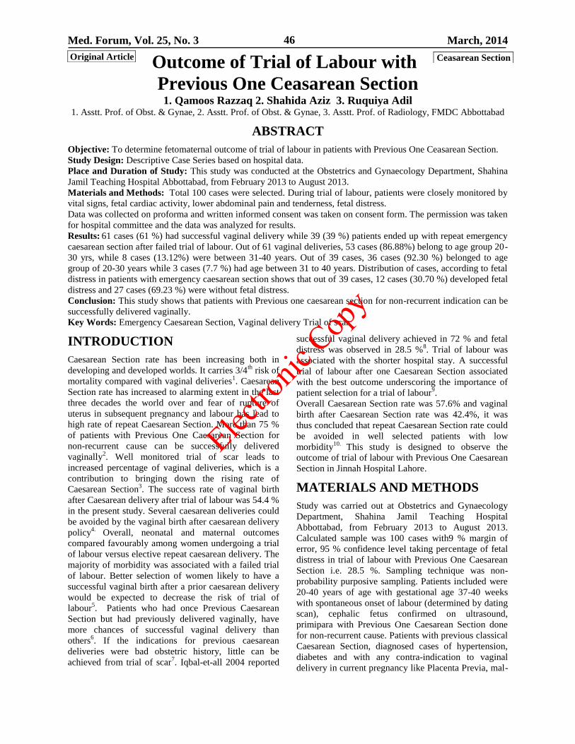
Med. Forum, Vol. 25, No. 3 March, 2014 46
Outcome of Trial of Labour with
Previous One Ceasarean Section 1. Qamoos Razzaq 2. Shahida Aziz 3. Ruquiya Adil
1. Asstt. Prof. of Obst. & Gynae, 2. Asstt. Prof. of Obst. & Gynae, 3. Asstt. Prof. of Radiology, FMDC Abbottabad
ABSTRACT
Objective: To determine fetomaternal outcome of trial of labour in patients with Previous One Ceasarean Section.
Study Design: Descriptive Case Series based on hospital data.
Place and Duration of Study: This study was conducted at the Obstetrics and Gynaecology Department, Shahina
Jamil Teaching Hospital Abbottabad, from February 2013 to August 2013.
Materials and Methods: Total 100 cases were selected. During trial of labour, patients were closely monitored by
vital signs, fetal cardiac activity, lower abdominal pain and tenderness, fetal distress.
Data was collected on proforma and written informed consent was taken on consent form. The permission was taken
for hospital committee and the data was analyzed for results.
Results: 61 cases (61 %) had successful vaginal delivery while 39 (39 %) patients ended up with repeat emergency
caesarean section after failed trial of labour. Out of 61 vaginal deliveries, 53 cases (86.88%) belong to age group 20-
30 yrs, while 8 cases (13.12%) were between 31-40 years. Out of 39 cases, 36 cases (92.30 %) belonged to age
group of 20-30 years while 3 cases (7.7 %) had age between 31 to 40 years. Distribution of cases, according to fetal
distress in patients with emergency caesarean section shows that out of 39 cases, 12 cases (30.70 %) developed fetal
distress and 27 cases (69.23 %) were without fetal distress.
Conclusion: This study shows that patients with Previous one caesarean section for non-recurrent indication can be
successfully delivered vaginally.
Key Words: Emergency Caesarean Section, Vaginal delivery Trial of scar.
INTRODUCTION
Caesarean Section rate has been increasing both in
developing and developed worlds. It carries 3/4th
risk of
mortality compared with vaginal deliveries1. Caesarean
Section rate has increased to alarming extent in the last
three decades the world over and fear of rupture of
uterus in subsequent pregnancy and labour has lead to
high rate of repeat Caesarean Section. More than 75 %
of patients with Previous One Caesarean Section for
non-recurrent cause can be successfully delivered
vaginally2. Well monitored trial of scar leads to
increased percentage of vaginal deliveries, which is a
contribution to bringing down the rising rate of
Caesarean Section3. The success rate of vaginal birth
after Caesarean delivery after trial of labour was 54.4 %
in the present study. Several caesarean deliveries could
be avoided by the vaginal birth after caesarean delivery
policy4.
Overall, neonatal and maternal outcomes
compared favourably among women undergoing a trial
of labour versus elective repeat caesarean delivery. The
majority of morbidity was associated with a failed trial
of labour. Better selection of women likely to have a
successful vaginal birth after a prior caesarean delivery
would be expected to decrease the risk of trial of
labour5. Patients who had once Previous Caesarean
Section but had previously delivered vaginally, have
more chances of successful vaginal delivery than
others6. If the indications for previous caesarean
deliveries were bad obstetric history, little can be
achieved from trial of scar7. Iqbal-et-all 2004 reported
successful vaginal delivery achieved in 72 % and fetal
distress was observed in 28.5 %8. Trial of labour was
associated with the shorter hospital stay. A successful
trial of labour after one Caesarean Section associated
with the best outcome underscoring the importance of
patient selection for a trial of labour9.
Overall Caesarean Section rate was 57.6% and vaginal
birth after Caesarean Section rate was 42.4%, it was
thus concluded that repeat Caesarean Section rate could
be avoided in well selected patients with low
morbidity10.
This study is designed to observe the
outcome of trial of labour with Previous One Caesarean
Section in Jinnah Hospital Lahore.
MATERIALS AND METHODS
Study was carried out at Obstetrics and Gynaecology
Department, Shahina Jamil Teaching Hospital
Abbottabad, from February 2013 to August 2013.
Calculated sample was 100 cases with9 % margin of
error, 95 % confidence level taking percentage of fetal
distress in trial of labour with Previous One Caesarean
Section i.e. 28.5 %. Sampling technique was non-
probability purposive sampling. Patients included were
20-40 years of age with gestational age 37-40 weeks
with spontaneous onset of labour (determined by dating
scan), cephalic fetus confirmed on ultrasound,
primipara with Previous One Caesarean Section done
for non-recurrent cause. Patients with previous classical
Caesarean Section, diagnosed cases of hypertension,
diabetes and with any contra-indication to vaginal
delivery in current pregnancy like Placenta Previa, mal-
Original Article Ceasarean Section
Electro
nic Cop
y

Med. Forum, Vol. 25, No. 3 March, 2014 47
presentation and good size baby determined on ultra
sound were excluded.100 pregnant patients, fulfilling
inclusion and exclusion criteria who were subsequent to
a previous delivery by caesarean and had regular ante-
natal check up reporting obstetric OPD were taken. An
informed consent was taken. Demographic variables
like name, age, address was noted. At 37 weeks of
gestation decision was taken regarding trial of labour
intravenous line was maintained and blood group and
cross matching was done pre-hand. Patients were
allowed to go in spontaneous labour. Patients were
closely monitored during trial of labour by vital signs,
fetal cardiac activity, lower abdominal pain and
tenderness, fetal distress. Facilities were made available
during whole trial of labour for emergency Caesarean
Section. Fetal and maternal outcome was vaginal
delivery and fetal distress. All this information is noted
on proforma. Data was analysed using SPSS version12.
Parity, maternal outcome in form of normal vaginal
delivery and fetal distress was presented by frequency
and percentage and gestational age was presented by
calculating mean + SD.
RESULTS
Total 100 patients were included in this study carried
out over a period of 6 months at obstetrics and
Gynaecology Department, Shahina Jamil Teaching
Hospital Abbottabad, from February 2013 to August
2013. Sample size of current study was 100 cases.
Table No 1: Distribution of cases by age having
successful vaginal delivery after previous one
caesarean section
Age (years) Cases Percentage
20-30 53 86.88
31-40 08 13.12
TOTAL 61 100.0
Mean + S.D 29.3+1.4
Out of 61 vaginal deliveries, 53 cases (86.88 %) belong
to age group of 20-30 years while 8 cases (13.12%)
were between 31-40 years.
Table No 2: Distribution of cases by age having
failed vaginal delivery (emergency caesarean
section) after previous one caesarean section
Age (year) Cases Percentage
20-30 36 92.30
31-40 03 07.70
Total 39 100.0
Mean+S.D 26.5+1.9
Out of 39 cases 36 cases (92.30%) belong to age group
of 20-30 years while 3 cases (7.7 %) had age between
31 – 40 years.
Table No. 3: Distribution of cases by gestational age
in cases of successful vaginal deliveries in previous
one caesarean section n=61
Gestational age
(weeks)
Cases Percentage
37-38 21 34.42
38+1-39 25 40.99
39+1-40 15 24.59
Total 61 100.0
Mean+s.d 39.3+ 1.2
Out of 61 cases of successful vaginal birth 21 cases
(34.42%) were between 37-38 weeks of gestation, 25
cases (40.99%) were between 38+1-39 weeks and 15
cases (24.59%) were between 39+1-40 weeks of
gestation.
Table No. 4: Distribution of cases by mode of
delivery following trial of labour in previous one
caesarean section
Mode of delivery Cases Percentage
Vaginal delivery 61 61.0
Emergency
Caesarean section
39 39.0
Total 100 100.0
Concerning the mode of delivery following trial of
labour, out of 100 cases 61 cases (61%) had successful
vaginal delivery while 39 (39%) patients ended up with
repeat emergency Caesarean Section after failed trial of
labour.
Table No. 5: Distribution of cases by fetal outcome
in terms of fetal distress in cases of vaginal delivery
n=61
Fetal distress Cases Percentage
Yes 03 04.92
No 58 95.08
Total 61 100.0
Table 5 shows out of 61 cases, 3 cases (4.9 %)
developed fetal distress while 58 cases (95.08 %) had
no sign of fetal distress.
Table No. 6: Distribution of cases by fetal outcome
in terms of fetal distress in cases of emergency
caesarean section
Fetal distress Cases Percentage
Yes 12 30.7
No 27 69.3
Total 39 100.0
Table 6 shows distribution of cases according to fetal
distress in patients with emergency Caesarean Section.
Out of 39 cases, 12 cases (30.70 %) developed fetal
distress and 27 cases (69.23%) had no fetal distress.
DISCUSSION
Caesarean Section has been increasing in both
developed and developing countries, it causes 3/4th
increased risk of mortality as compared to vaginal
Electro
nic Cop
y

Med. Forum, Vol. 25, No. 3 March, 2014 48
deliveries. High Caesarean Section rate has negative
impact on maternal health and obstetric care cost, so
there is a need to reduce it11,12
. Caesarean Section rate
can be reduced either by reducing primary Caesarean
rate or reducing repeat Caesarean rate by subjecting
those with previous one Caesarean to trial of labour
after thorough assessment and counselling. In current
study objective was to determine feto-maternal outcome
after trial of labour in patients with previous one
Caesarean Section. Successful vaginal births achieved
mostly in early ages than in later age. Another study by
Bujold et al concluded that successful vaginal delivery
is inversely related to maternal age13,14
.
In present study successful vaginal births occurred
between gestational age 37-39 weeks. Rate of
successful vaginal birth decreases with increasing
gestational age. Another study by Smith et al in
Cambridge University UK concluded that risk of
emergency Caesarean Section increased with increasing
gestational age15
. In this study, out of 100 cases 61 (61
%) achieved successful vaginal delivery while 39
(39%) eneded up with emergency Caesarean Section.
Another study conducted by Birgisdottir et al concluded
similar results16,17
.
Another study by Butt and Akhtar concluded that 62.8
% had successful vaginal delivery while 34.7 % had
emergency Caesarean Section18,19
. With successful
VBAC 3 babies (4.92 %) were found to have meconium
stained liquor but all babies were healthy and had
spontaneous cry while babies who were delivered
through emergency Caesarean Section 12 (30.7 %)
developed fetal distress out of 39. All survived and
discharged without any neurological deficit or
complication. In a study conducted by Homer
concluded babies whose mother attempted BBAC were
significantly less likely to require admission in neonatal
Intensive Care Unit20,21,22
. No maternal and peri natal
mortality seen.
CONCLUSION
This study showed that patients with Previous One
Caesarean Section for non-recurrent indication can be
successfully delivered vaginally. Antenatal booking and
follow up, careful case selection for trial of scar and
close observations during labour will achieve successful
maternal and peri natal outcome. VBAC also saves any
future Caesarean Section, as currently Previous Two
Caesarean Sections is an indication for elective
Caesarean Section in our setup.
Suggestions: To reduced the caesarean section rate
VBAC should be encouraged. Early booking, regular
antenatal visits and confinement in hospital should be
encouraged.
REFERENCES
1. Begum I, Khan A, Nasreen M, Khan S. Outcome
of Previous Caesarean Section. J Pak Med Assoc
2004; 43:130-3.
2. Hassan A. Trial of scar and vaginal birth after
Caesarean Section. J Ayub Med Coll Abbotabad
2005; 17:57-61.
3. Akhtar BT. Vaginal birth after previous one
Caesarean Section: term pregnancy outcome. J
Postgrad Med Inst 2005; 19: 175-81.
4. Tongsong T, Jitawong C. Success rate of vaginal
birth after Caesarean delivery at Mahraj Nakorn
Chiang Mai Hospital. J Med Assoc Thai 2003;86:
829-35.
5. Leobel G, Zelop CM, Egan JF, wax J. Maternal
and neonatal morbidity after elective repeat
Caesarean Delivery versus a trial of labour after
previous Caesarean Delivery in a community
teaching hospital. J Matern Fetal Neonatal Med
2004;15: 243-6.
6. Mehr-un-Nisa, Hassan L. Trend of vaginal delivey
afte one previous Caesarean Section in a Tertiary
Care Hospital. Pak J Med Res 2004; 43: 60-4.
7. Iqbal J, Nausheen F, Bhatti FA, Sheikh S. Vaginal
Birth after Caesarean Section (VBAC). Ann King
Edward Med Coll 2004;10: 187-9.
8. Tan PC, Subramanian RN, Omar SZ. Labour and
perinatal outcome in women at term with one
previous lower segment Caesarean: A Resview of
1000 consecutive cases. Aust NZ J Obstet
Gynaecol 2007; 47: 31-6.
9. Sohail R, Mehboob N, Zaman F. Trends of
delivery in patients with previous one Caesarean
Section. J Surg Pak 2004; 9: 39-41.
10. Bujold E, Hammoud AO, Hendler I, Berman S,
Blackwell S C, Duperron L, et al. Trial of labour in
patients with a previous Caesarean Section: does
maternal age influence the outcome? Am J Obset
Gynaecol 2004; 190: 1113-8.
11. Smith GCS, White IR, Pell JP, Dobbie R.
Predicting Caesarean Section and uterine rupture
among women attempting vaginal birth after prior
Caesarean Section. PLOS Med 2005;2:e 252.
12. Brigisdottir BT, Hardardottir H, Bjarnadottir R I,
Thorkelsson T. Vaginal birth after one previous
Caesarean Section. Laeknabladid 2008; 94: 591-7.
13. Homer CS. Birth after Caesarean over a nine year
period in one Australian state. Midwifery 2009; 19:
56-9.
14. Birara M, Gebrehiwot Y. Factors associated with
success of vaginal birth after one caesarean
section (VBAC) at three teaching hospitals in
Addis Ababa, Ethiopia: a case control study BMC
pregnancy and childbirth 2013; 13 : 31
Electro
nic Cop
y
Med. Forum, Vol. 25, No. 3 March, 2014 49
15. Al-Zirqi I, Stray-Pedersen B, Forsén L, Vangen S.
Uterine rupture after previous caesarean section.
BJOG 2010;117(7):809-20.
16. American College of Obstetricians and
Gynecologists. Vaginal birth after previous
cesarean delivery. ACOG Practice bulletin no. 115.
Obstet Gynecol. 2010 Aug;116(2 Pt 1):450-63.
Guideline summary
17. Dekker GA, Chan A, Luke CG, Priest K, Riley M,
Halliday J, et al. Risk of uterine rupture in
Australian women attempting vaginal birth after
one prior caesarean section: a retrospective
population-based cohort study. BJOG 2010;
117(11):1358-65.
18. Dodd JM, Crowther CA. Elective repeat caesarean
section versus induction of labour for women with
a previous caesarean birth. Cochrane Database of
Systematic Reviews 2012;5.
19. Gomez LR. Burgos J. Cobos P. Melchor JC. Osuna
C. Centeno Mdel M. et al. Oxytocin versus
dinoprostone vaginal insert for induction of labor
after previous cesarean section: a retrospective
comparative study. J Perinatal Med 2011;39(4):
397-402.
20. Harper LM. Cahill AG. Boslaugh S. Odibo AO.
Stamilio DM. Roehl KA. Et al. Association of
induction of labor and uterine rupture in women
attempting vaginal birth after cesarean: a survival
analysis. Am J Obst & Gynea 2012;206(1):51.e1-5.
21. Jozwiak M, Dodd JM. Methods of term labour
induction for women with a previous caesarean
section (Protocol). Cochrane Database of
Systematic Reviews 2012;4.
22. Ouzounian JG. Miller DA. Hiebert CJ. Battista LR.
Lee RH. Vaginal birth after cesarean section: risk
of uterine rupture with labor induction. Am J
Perinatol 2011;28(8):593-6.
Address for Corresponding Author:
Dr. Qamoos Razzaq,
Assistant Prof. Gynae. & Obstet,
FMDC, Abbottabad.
Electro
nic Cop
y

Med. Forum, Vol. 25, No. 3 March, 2014 50
Study to Estimate the
Prevalence of Malaria Infection in Sukkur 1. Khalid Ahmed Qureshi 2. Arshad Hussain Laghari 3. Akhtar Hussain Samoo
1. Asstt. Prof. of Pathology, 2. Asstt. Prof. of Biochemistry, 3. Asstt. Prof. of Physiology,
Ghulam Muhammad Mahar Medical college, Sukkur
ABSTRACT
Objective: The aim of the study to estimate the prevalence of malaria amongst the population with fever or history
of fever at sukkur.
Study Design: Cross sectional study
Place and Duration of Study: This study conducted at laboratory Ghulam Mohammad Mahar Medical College
Hospital Sukkur from January 2011 to December 2012.
Materials and Methods: Total 1746 clinical suspected case of malaria were included in the study. Patients
presenting with fever were screened, thick and thin blood films prepared on microscopic glass slide, stained with 5%
Giemsa’s stain.
Results: There were 344 cases below 11 years, 590 cases between 11 to 20 years.812 cases above 20 years. They
were 960 males and 786 females, with male to female ratio 1.2:1 .The prevalence of plasmodium slide positivity was
7.9% (138/1746). Among plasmodium slide positive, 60.5% (83/138) were positive for P.vivax and 39.5% (55/138)
were positive for P. falciparum.
Conclusion: Identification of malaria cases in early acute phase of disease is significant for proper curative
treatment. Anti mosquito eradication measures should be taken for breeding places of vector with personal
protection measures and awareness program for malaria should be initiated.
Key Words: Anopheles mosquito, P. falciparum, P. vivax.
INTRODUCTION
Malaria infection is a global public health problem. The name malaria is derived from the Italian mal aria meaning bad air.
1 The disease was also known as
paludism from the Latin palus (marsh).2 Both names
reflect that in ancient times, disease was believed that disease was spreading from marshy and swampy places.
3
Grassi, feletti and Welch (1890 - 1897) proved that malaria is transmitted by one human to another by bite of infected Anophelese mosquito. The disease affects 109 countries in the World.
4
WHO estimates that 216 million cases each year.5
Approximately over half of million people die from malaria, mostly children younger than 5 years old.
6
Estimated number of Annual malaria cases in Pakistan is 1.5 millions.
6,7 The development cycle of vector
requires hatching of eggs in standing water, in temperature between 16 °C to 38 °C. Rice crop cultivation provides breeding sites for parasite and influence incidence and prevalence of disease.
8,9 The
most common region of malaria reported by C.D.C was Africa (58%), Asia (18%) and South America (16%). Malaria is transmitted to man by intracellular plasmodium protozoa, P. falciparum, P. malaria, P. ovale and P. vivax. The predominant species in Pakistan is P. falciparum and P. vivax.
10
Children and adults are asymptomatic during initial phase of malaria infection. The usual incubation period for P. falciparum is 9-14 days and P. vivax is 12-17 days. The incubation period can be as long as 6-12 months for P. vivax. The disease is characterized by
fever, chills, sweats, fatigue, anemia, thrombocytopenia, headache and anorexia.
11,12
P. falciparum is most severe form of malaria and associated with more parasitemia. Fatality rate is up to 2.5% in adults and 30% in infants, if treatment is not started promptly. P. vivax results in parasitemia upto 2%. P. falciparum infects both Immature and mature erythrocytes. P. vivax infects primarily immature erythrocytes.
13,14
Hozhabri et al (2000) studies the prevalence of plasmodium slide positivity among the children treated for malaria at R.H.C Jhangara Sindh, Pakistan. They observed the prevalence of plasmodium slide positivity was 5.9%.
15
The prevalence of community based study done in rural sindh by Covel and Lieut (1934) epidemic of malaria. They found 40% malaria positivity at that time. Malaria diagnosis was based on splenetic rate (percentage of children between 2-10 years of age showing enlargement of spleen).
16
The prevalence for malarial infection studies has been conducted in many parts of the Pakistan, but still lacking in many areas. A present study was conducted to estimate the prevalence of malaria infection suspected cases in Sukkur.
MATERIALS AND METHODS
This study was under taken at Ghulam Muhammad Mahar Medical College (GMMMC) Hospital, Sukkur among suspected patients of malaria, during the period of two years (January 2011to December 2012). The patients were including in the study if they met the
Original Article Prevalence of Malaria
Electro
nic Cop
y

Med. Forum, Vol. 25, No. 3 March, 2014 51
eligibility criteria. The age grouping of the patients was divided in to 3 categories: 2 months to 10 years of age, 11 – 20 years and 21 years and above. The specimen was collected from patients after cleaning the skin with 70% rectified sprit to minimize the chance of contamination. 3 ml of intra venous blood was by drawn into EDTA vacutainer tube and information about age and sex were recorded. All the patients underwent a general examination for dehydration, anemia, spleen enlargement, jaundice and other abnormalities. Body temperature was also recorded by using a standard oral mercury thermometer. Thick and thin blood film were prepared through the through the blood sample in the pathology laboratory of the hospital. Thin film was fixed with 1 – 2 drops of methanol to avoid dehemoglobinization of RBCs while thick films were not fixed with methanol. The slides were pleased in staining jar, containing 5% Giemsa’s stain for 20 minutes. After staining, air dry film was examined. 100-200 fields were examined under oil immersion objective (100 X) before being declared negative. Thick blood film were examined for quickly scanning of parasite while thin blood film for identification of malaria parasite species. Those films that were malaria parasite positive, we identified different species such as P. falciparum
and P. vivax.
RESULTS
Total 1746 cases were screened during period of
January 2011 to December 2012. 960 cases were males
and 786 were females. Ages of patient ranged from
above 2 months to 80 years. Male to female ratio was
1.2:1. Mean age was 21 years. The malarial slide
positivity was 7.9% (138/1746). Among the positive
cases P. vivax were 60.2% (83/138) and P. falciparum
(55/138) were 39.8%.
In 83 positive cases of P. vivax, patients aged below 11
years were 60.6% (23/83), from 11 to 20 years were
55.8% (24/83 ) while in patients above 20 years were
63.2% (36/83). Among P. falciparum positive cases
(55/138), 39.4% (15/55) were under 11 years, 44.2%
(19/55) were aged from 11 to 20 years while 36.3%
(21/55) were above 20 years.
The seasonal variation of disease was also noted in this
study. It was observed that high infection rate of P.
falciparum was found in month of January while it was
lower in the month of April. Higher rate of infection
with P. vivax was observed in November and low in the
month of January.
Table No.1: Month wise prevalence of malaria infection in sukkur (years 2011& 2012)
Month Patients* MP
(+ve)**
Infection
(%)
P. vivax (%) P. falciparum (%)
January 157 17 10.82% 01 (0.63%) 16 (10.19%)
February 135 06 4.44% 02 (1.48%) 04 (2.96%)
March 212 05 2.34% 02 (0.94%) 03 (1.4%)
April 148 02 1.35% 01 (0.67%) 51 (0.67%)
May 94 06 6.37% 05(5.31%) 01 (1.06%)
June 119 09 7.56% 06 (5.04%) 03 (2.52%)
July 133 09 6.75% 07 (5.26%) 02 (1.50%)
August 130 08 6.15% 06 (4.61%) 02 (1.54%)
September 151 10 6.62% 06 (3.97%) 04 (2.64%)
October 124 20 16.12% 12 (9.67%) 08 (6.45%)
November 224 40 17.84% 31 (13.83%) 09 (4.01%)
December 119 06 5.04% 04 (3.36%) 02 (1.68%)
TOTAL 1746 138 7.9% 83 (4.75%) 55 (3.15%)
*Numbers of blood slide examined for malaria parasite. **Malaria parasite positive blood slide in patients.
Table No.2: Age and gender wise distribution of malaria infection
S.No Age (year) Male Female Total MP
+ve*
P.vivax
(%)
P. falciparum
(%)
01 Below 10 184 160 344 38 23 (60.6%) 15 (39.4%)
02 11 - 20 332 258 590 43 24 (55.8%) 19 (44.2%)
03 Above 21 444 368 812 57 36 (63.2%) 21(36.8%)
04 Total 960 786 1746 138 83 (60.2%) 55 (39.8%)
*Malaria parasite positive blood slide in patients.
DISCUSSION
Malaria is a major global health problem, occurring in
more then100 countries in the world17,24
, affects an
estimated 300 million people and causes more than a
million deaths per year.18
Falciparum malaria has high
mortality as it causes complication like cerebral
malaria, renal failure and algid malaria.19
In our study 1746 patients suspected for malaria were
screened during January 2011 to December 2012. Out
Electro
nic Cop
y
Med. Forum, Vol. 25, No. 3 March, 2014 52
of 1746 individuals 138 cases were positive for malaria
parasite. In positive cases 60.1% were indentified
positive with plasmodium vivax and 39.90% with
Plasmodium falciparum. Negligible mixed infection of
P. falciparum and P. vivax was also observed. Male
patients were more affected by plasmodium than female
may be due to maximum exposure to vector. Male to
female ratio were found in this study was 1.2:1 and
mean age was 21 years in patients positive with
malaria.
The positivity rate for malaria parasite was found 7.9%
(138/1746) in general population. A research work for
malaria in 2007 at Ayub teaching hospital Abbottabad
shows 7.2% malaria infection in general population.8 A
study about the prevalence of malaria infection in
human population in 3 district localities of Quetta,
Pakistan confirm finding of our study.17,18,21
Another
study conducted about the prevalence of malaria
parasite at Dera Murad Jamali in 2008 find out the
malaria parasite 40.4%. P. vivax infection was 71.7%
and 28.2% with P. falciparum out of positive cases.22
Among the positive cases we observed significantly
high positivity of P. vivax which was 60.2% (83/138)
then P. falciparum (55/138) that only were 39.8%. A
study conducted at Karachi and other areas in sindh,
Pakistan shows that malaria in patients with P. vivax
was 62.5% and patients were positive with P.
falciparum were only 36.0%.13,14
Another study was
carry out at Karachi and in others areas of sindh,
Pakistan showing the similar results. It shows two time
high P. vivax infection then P. falciparum.15,16
A study
in Mansehra in 2006, 160 cases of malaria in children
were investigated and results of study were revels the
high vivax infection.6
Study conducted in 2011 in
patients with fever in all provinces of Pakistan
including Islamabad. P. vivax ranged from 2.4% to
10.8% and P. falciparum ranged 0.1% to 3.8% which is
similar to this study.22
This study raises the roof of positivity of P. vivax and
P. falciparum in almost all age groups and do not show
significant difference. Positive cases of P. Vivax in
patients aged below 11 years were 60.6%, from 11 to
20 years were 55.8% while in patients above 20 years
were 63.2%. Among P. falciparum positive cases
39.4% were under 11 years, 44.2% were aged from 11
to 20 years while 36.3% were above 20 years.
However in our study seasonal variation also noted.
High prevalence of P. vivax malaria infection was
found during the month of October and November due
to humidity and optimum temperature in our region
while lowest infection rate were found in January.
Highest infection of P. falciparum found in January and
lowest in April. Study conducted at Dera Murad Jamali
shows highest infection of P. vivax in October and
lowest in January and infection with P. falciparum in
January while lowest in October.23
CONCLUSION
High slide positivity rate was observed in clinically
suspected cases of malaria. Identification of malaria in
acute stage are significant for morbidity and mortality.
Screening of suspected cases and anti-mosquito
eradication measures should be taken for breading sites
of vector, mass education and awareness programs may
be initiated.
REFERENCES
1. Abbasi KA, Sahikh SA. Comparative study of
cerebral malaria due to plasmodium vivax and
falciparum. Pak Paed J 1957;21:155-158.
2. Akber JU. Malaria in children at a children
Hospital. J Coll Phys Surg Pak 2002;7:20-22.
3. Akber JU, Shamsher AK, Sirajuddin HA. Malaria
in children: study of 160 cases at a private clinic in
Mansehra. J Ayub Med Coll 2006;18:44-45.
4. Bhalli MA, Samiullah. Falciparum malaria-a
review of 120 cases. J Coll Phys Surg Pak 2001;
11:300-303.
5. WHO/UNICEF. World Malaria Report Geneva,
2005.
6. Country Report Pakistan Vector- Borne Diseases in
Pakistan. Inter-Country Workshop on Developing a
regional strategy for integrated vector management
for malaria and other vector borne diseases,
Khartoum, Sudan, January 2003;21-23.
7. Durrani AB, Durrani I, Abbas N, Jabeen M.
Epidemiology of cerebral Malaria and its mortality.
J Pak Med Assoc 1997;47:213-215.
8. Idris M, Sarwar J, Fareed J. Pattern of malaria
infection diagnosed at Ayub Teaching Hospital
Abbottabad. J Ayub Med Coll 2007;19:35-36.
9. Jamal M, Jehan M, Nadir A. Malaria in pediatric
age group:a study of 200 cases. Pak Armed forces
Med J 2005;55:74-77.
10. Jan AH, Kiani TA. Haematozoan parasites in
Kashmirirefugees. Pak J Med Res 2001;40:10-12.
11. Khadim MT. Malaria a menace at Zhob Garrison.
Pak.Armed Forces Med. J. 2002 ; 52:203-207.
12. Korenromp EL. Roll back Malaria monitoring and
evaluation group & MERG Task Force on malaria
morbidity. Malaria incidence estimate at Country
level for the Year 2004- Proposed estimate and
draft report Geneva. Roll back Malaria, WHO
2004.
13. Mahmood KH. Malaria in Karachi and other areas
in Sindh. Pak Armed Forces Med J 2005;55:
345-348.
14. Nizamani NA, Kalar, Khushk IA. Burden of
malaria in Sindh, Pakistan: a two years surveillance
report. J Liaqat Uni Med Health Sci 2006;5:76-83.
15. Hozhabri S, Akhtar, Rahbar M, Ludy S. Prevalence
of plasmodium slides positivity among the children
Electro
nic Cop
y

Med. Forum, Vol. 25, No. 3 March, 2014 53
treated for malaria, Jhangara, Sindh. J Pak Med
Assoc 2000;50:401-405.
16. Murtaza G, Memon IA, Noorani AK. Malaria
prevalence in sindh. Med Channel 2004;10:
20-22.
17. Malaria Control Program (MCP). District wise
epidemiological data of malaria control program,
Balochistan, Pakistan, 2005.
18. Malaria Control Program (MCP). District wise
epidemiological data of malaria control Program,
Balochistan, Pakistan, 2006.
19. Mohammad N, Hussain. Prevalence of malaria in
general population of district Buner. J Pak Med
Inst 2003;17:75-80.
20. Saleem I, Pirzada AH, Rahman A and Noorul-
Iman. Cerebral malaria, an experience in NWFP,
Pakistan. J Med Sci 2006;14:35-39.
21. Sheikh AS, Sheikh AA, Sheikh NA, Paracha SM.
Endemicity of malaria in Quetta. Pakistan J Med
Res 2005;44:41-45.
22. Akbar JU, Shamshar AK, Sirajuddin HA. Malaria
in children: study of 160 cases at a private clinic in
Mansehra. J Ayub Med Coll Abbottabad 2007;19:
35-36.
23. Yasinzia MI, Kakarsulemankhel JK. Prevalance of
human malaria infection in Pakistan: District Dera
Murad Jamali. Pak J Sci 2008;60:2-4.
24. Zarchi AK, Mohmmodzadeh A, Vatani HA. A
Survey on malaria and some related factors in
south east of Caspian Sea. Pak J Med Sci 2006;
22:489-492.
Address for Corresponding Author:
Dr. Arshad Hussain Laghari.
Assistant Professor of Biochemistry,
Ghulam Muhammad Mahar Medical College, Sukkur
Cell No.: +92-301-2378134
Office No.: +92-71-9310780
E-mail: [email protected]
Electro
nic Cop
y

Med. Forum, Vol. 25, No. 3 March, 2014 54
The Effects of Insulin on the
Volume, Absolute and Relative Weight of Liver in
HFD/Streptozocin Induced Diabetic Rats 1. Sahar Mubeen 2. Muhammad Rafique 3. Amber Ilyas
1. Asstt. Prof. of Anatomy, DIMC, DUHS, Karachi 2. Prof. of Anatomy, Al-Tibri Medical College, Karachi
3. Asstt. Prof. of Anatomy, JM&DC, Karachi
ABSTRACT
Objectives: In present study, the effects of insulin on the volume, absolute and relative weight of liver was studied
in Wistar albino rats for a period of 4 weeks.
Study Design: Retrospective / observational study.
Place and duration of study: This study was conducted in the Animal House of DUHS and it took 8 months 1st
June 2011 to 1st Feb 2012 to be completed.
Materials and Methods: The Male Wistar albino rats which were randomized into 3 groups; each group containing
10 rats. Group A served as control, Group B as insulin treated and Group E as untreated diabetic rats. All the other
rats except the Group A were kept on in-house prepared High Fat Diet (HFD) throughout the study. After 2 weeks of
exclusive HFD, diabetes was induced by intraperitoneal (IP) injection of low dose streptozocin (STZ 3.5mg/100gm).
After the induction, one group was left untreated (Group E) and one group (Group B) was treated with insulin for 4
weeks. The rats were then, sacrificed, liver was isolated, weighed, and its dimensions were noted.
Results: The mean absolute liver weight (ALW) of rats was observed as 8.60 ± 2.54 gm, 13.18 ± 0.68 gm and
9.40 ± 3.18 gm in control, untreated and insulin treated groups respectively. And the mean percent liver weight
(PLW) was calculated as 2.99 ± 0.66%, 5.10 ± 0.73% and 3.99 ± 1.37% in control, untreated and insulin treated
groups respectively. Statistically significant difference was noted between ALW, PLW and liver volume of rats of
the three groups.
Conclusion: Insulin decreases the volume, absolute and relative weight of liver of diabetic rats when used for a
short period.
Key Words: Diabetic rat model, Type 2 diabetes, Diabetes induction, Insulin, Liver.
INTRODUCTION
The day to day researches reveal soaring rates of
obesity, non alcoholic fatty liver disease and diabetes
mellitus. This emerging prevalence is due to more
mechanized life style, unbalanced diet and physical
inactivity. Use of excessive fat in diet and no exercise
makes a person obese leading to a condition known as
insulin resistance (IR)1 which gives birth to lot more
diseases like obesity, diabetes mellitus type 2 (DM2)
and fatty liver disease. DM2 is a common, severe and
chronic form of metabolic disorder characterized by
hyperglycemia due to decline in insulin action i.e. IR
followed by the inability of insulin producing beta cells
to compensate for IR.1Increased morbidity and
mortality associated with the disease is due to complex
metabolic changes that lead to functional impairment of
many organs. About 1.3% population of the world is
suffering from DM22, 3
and its prevalence in our
community as well as in the world is increasing like an
epidemic. The Nation (issue 2008)4, Pakistan occupies
7th
position in WHO diabetes prevalence list. “In
Pakistan 6.9 million people are affected by diabetes and
the International Diabetes Federation has estimated that
this number will grow to 11.5 million by 2025 unless
measures are taken to control the disease.”4
Enlarged fatty liver is a well-known complication of
DM2 with an established prevalence of 21–78%, 5-7
and usually remains undiagnosed because of its silent
course in many of DM2 patients. Type 1 diabetes is not
associated with accumulation of fat if blood glucose
levels are well controlled, but type 2 diabetes may have
a 70% correlation with fat accumulation regardless of
blood glucose control.8
The fatty liver disease NAFLD is defined as fatty
infiltration of liver cells (hepatocytes) in patients are
taking no or insignificant alcohol (< 20 g/day).It can be
idiopathic or secondary to metabolic syndromes like
obesity, hypertension, hyperlipidemia and diabetes.3
Fatty liver is caused by a number of factors which result
in progressive hepatic failure.9
The factors are
summarized as Two hit theory.”The “First Hit” to liver
occurs when the enzymes involved in lipid metabolism
i.e. uptake, synthesis, oxidation, and export are altered.
In other words, the uptake of fatty acids by the liver
exceeds its oxidation capacity. Lipid synthesis in liver
is increased if diet consists of excess of sucrose, fats or
fructose. The increase in lipid synthesis and decrease in
lipid oxidation leads to the development of insulin
resistance, both hepatic and peripheral.10
The second hit
is due to oxidative stress, which causes peroxidation of
Original Article Insulin Reduced Liver
Electro
nic Cop
y

Med. Forum, Vol. 25, No. 3 March, 2014 55
lipids in the hepatic cell’s membrane and cytokine
production.9, 11
Nonalcoholic steatohepatitis (NASH) is a variant of
fatty liver in which fat in the hepatocytes is
accompanied by lobular inflammation and
steatonecrosis. The diagnosis can only be made in the
absence of alcohol abuse or other causes of liver
disease, particularly hepatitis C. Nonalcoholic
steatohepatitis has been associated most commonly
with obese women with diabetes.12
There is certainly a
higher prevalence in type 2 diabetic patients on
insulin.13
Insulin is an essential hormone which is involved in
glucose as well as lipid metabolism.14
It is synthesized
and released by the β cells of pancreas in the blood in
response to elevated blood glucose levels. The body
cells especially fat, muscle and liver cells have “insulin
receptors” on their surface which regulate entrance of
glucose into cells. As glucose levels in blood increase,
glucose is pushed into the cells where glucose is
converted into energy. Other mechanisms by which
insulin keeps blood glucose level in normal range is the
inhibition of glucose production by the liver.15
Unfortunately, people who are insulin-resistant cannot
utilize insulin efficiently and glucose, therefore, is
unable to enter the cells and accumulate in the blood. In
order to clear excess glucose from the blood, the body
signals the pancreatic β cells to produce excess insulin
which results in a condition referred to as
hyperinsulinemia (high level of insulin in blood).
Hyperinsulinemia, though compensatory, but has
various serious side effects on body. It suppresses fatty
acid oxidation which leads to high levels of fatty acids
in the form of triglycerides (TGs), to accumulate in the
blood.16
In 2002, Mason et al. proved that serum TGs
are usually increased after chronic intraperitoneal and
subcutaneous insulin infusion17
which in turn, causes
fatty acids to be deposited in the liver which leads to
development of HIR.18, 19
Thus, fatty infiltration and IR
can potentiate each other creating vicious cycle of
metabolic dysfunction.9
The underlying mechanism by
which obesity induces IR is the fat deposition in liver
(HIR) skeletal muscles and adipose tissue (peripheral
insulin resistance).20
The liver compensates for
hyperglycemia by decreasing its glucose secretion in
the presence of insulin. As the resistance of cells to
insulin increases, compensatory insulin secretion fails
and either fasting, postprandial or random glucose
concentrations increase. IR can progress to DM2 when
insufficient insulin is produced by β cells in a condition
of hyperglycemia.21
The cure for diabetes type 2 is not known yet, but
research is focused mainly on the medicines that can
halt organ damage or at least slow the process of
damage in DM2 patient diabetes. Insulin is the most
commonly prescribed drug in patients of diabetes. But,
studies reveal that insulin alone is detrimental to liver
cells because of its lipogenic effect.22
In view of above facts, the current research was
designed to see the effects of insulin on the liver of
untreated and insulin treated diabetic rats for a short
period.
MATERIALS AND METHODS
The study was conducted at Anatomy Department, Dow
International Medical College, Dow University of
Health Sciences, Karachi.
Duration of Study: The study took 8 months to be
completed from 1st June 2011 to 1
st Feb 2012.
Animals were obtained from the Animal House of Dow
University of Health Science. Thirty adult albino rats
(average body weight 160 ± 20 gm) were selected for
the study and were acclimatized for one week. They
were kept at room temperature 30 ± 1º C with a 12
hour light and dark cycle and were given balanced in-
house prepared HFD diet and water ad libitum. The
animals were divided into three groups each containing
10 animals. All rats were weighed on a digital scale at
the beginning of the experiment and weekly until
sacrificed. Also blood glucose levels were done with
glucometer (Accucheck Performa) in all rats every
week. Under aseptic condition, the blood sample for
glucose testing was taken from the base of the tail
because of easy visibility of veins at that area.
Experimental induction of diabetes type 2 was done in
2 phases: In the first phase, control rats (n=10) were
given free access to water and in-house prepared
standard diet while the other rats were kept on in-house
prepared HFD for 2 weeks. After 2 weeks of dietary
manipulation, all rats except the control group (n=10)
were given low dose STZ (manufactured by Bio Plus
Fine Chemicals, USA) (3.5mg/ 100 mg) intraperitonelly
(IP).1
STZ was prepared freshly before use by
dissolving the powder in cold citrate buffer (pH 4.5) for
immediate use within 10 minutes of preparation. Rats
were fasted for12-14 hours before IP injection of STZ.
The matched control group was given citrate buffer (0.1
ml/100mg).Four day after injection of STZ rats were
tested for blood glucose level. Rats having blood
glucose levels ≥200mg/dl were considered diabetic and
were included in the study.
After experimental induction of diabetes type 2, the rats
were divided into 3 groups; Control Group-A (n=10),
Untreated diabetic Group-E (n=10) and Insulin treated
Group-B (n=10).
HFD/STZ induced rats (Group B) were given IP
injection of insulin (0.3U/100 gm/day) 23
for 4 weeks.
The rats were allowed to have HFD till the end of the
experiment. After 4 weeks of treatment, the animals
were weighed and rats were sacrificed. The abdominal
cavity was dissected; liver exposed, carefully separated
from surrounding tissues and dissected out and placed
in a petri dish. Liver was examined for the significant
Electro
nic Cop
y
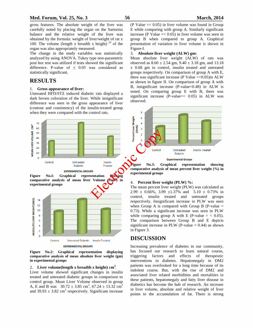
Med. Forum, Vol. 25, No. 3 March, 2014 56
gross features. The absolute weight of the liver was
carefully noted by placing the organ on the Sartorius
balance and the relative weight of the liver was
obtained by the formula: weight of liver/weight of rat x
100. The volume (length x breadth x height) 24
of the
organ was also appropriately measured.
The change in the study variables was statistically
analyzed by using ANOVA. Tukey type non-parametric
post hoc test was utilized if tests showed the significant
difference. P-value of ≤ 0.05 was considered as
statistically significant.
RESULTS
1. Gross appearance of liver: Untreated HFD/STZ induced diabetic rats displayed a
dark brown coloration of the liver. While insignificant
difference was seen in the gross appearance of liver
(contour and consistency) of the insulin-treated group
when they were compared with the control rats.
Figure No.I: Graphical representation showing
comparative analysis of mean liver Volume (cms3) in
experimental groups
Figure No.2: Graphical representation displaying
comparative analysis of mean absolute liver weight (gm)
in experimental groups
2. Liver volume(length x breadth x height) cm3:
Liver volume showed significant changes in insulin
treated and untreated diabetic groups in comparison to
control group. Mean Liver Volume observed in group
A, E and B was 30.72 ± 3.85 cm3, 67.24 ± 13.32 cm
3
and 39.93 ± 3.82 cm3 respectively. Significant increase
(P Value =< 0.05) in liver volume was found in Group
E while comparing with group A. Similarly significant
increase (P Value =< 0.05) in liver volume was seen in
group B when compared to group A. Graphical
presentation of variation in liver volume is shown in
Figure-I.
3. Absolute liver weight (ALW) gm: Mean absolute liver weight (ALW) of rats was
observed as 8.60 ± 2.54 gm, 9.40 ± 3.18 gm, and 13.18
± 0.68 gm in control, insulin treated and untreated
groups respectively. On comparison of group A with E,
there was significant increase (P Value =<0.05)in ALW
as shown in figure II. On comparison of group A with
B, insignificant increase (P-value=0.48) in ALW is
noted. On comparing group E with B, there was
significant increase (P-value=< 0.05) in ALW was
observed.
Figure No.3: Graphical representation showing
comparative analysis of mean percent liver weight (%) in
experimental groups
4. Percent liver weight (PLW) %: The mean percent liver weight (PLW) was calculated as
2.99 ± 0.66%, 3.99 ±1.37% and 5.10 ± 0.73% in
control, insulin treated and untreated groups
respectively. Insignificant increase in PLW was seen
when Group A is compared with Group B (P-value =
0.73). While a significant increase was seen in PLW
while comparing group A with E (P-value = < 0.05).
The comparison between Group B and E depicts
significant increase in PLW (P-value = 0.44) as shown
in Figure 3.
DISCUSSION
Increasing prevalence of diabetes in our community,
has focused our research to learn natural course,
triggering factors and effects of therapeutic
interventions in diabetes. Hepatomegaly in DM2
patients was overlooked for a long time because of its
indolent course. But, with the rise of DM2 and
associated liver related morbidities and mortalities in
these patients, hepatomegaly and fatty liver disease in
diabetics has become the hub of research. An increase
in liver volume, absolute and relative weight of liver
points to the accumulation of fat. There is strong
Electro
nic Cop
y
Med. Forum, Vol. 25, No. 3 March, 2014 57
association of type 2 diabetes with IR and fatty liver
disease. A lot of work has been done on fatty liver
disease associated with obesity, but less work is done
on increased liver span, its causes and consequences in
diabetes. It might be because human livers are not
easily available.
In the present study, liver weight and volume was
analyzed in untreated diabetic rats and insulin treated
diabetic rats. The blend of dietary manipulation and
chemicals was used to make a perfect experimental
model of DM2 as done previously by many renowned
scientists.21, 22, 24
It is known the fact that HFD creates an
animal model with glucose intolerance and
IR.22,24,25
The rats were fed HFD to induce IR followed
by a low dose of STZ (diabetogenic agent) which
specifically targets insulin secreting cells of pancreas
and is used by many researchers to create diabetic
model as it is simple, time saving and economical. 21, 24,
26Therats developed diabetic symptoms, increased
blood glucose levels and body weights which is
comparable with the studies of Ikebukuro 2002 and
Srinivasan K 2005, with no significant difference in
them. 22, 27
As far as the gross features of liver are concerned, no
significant changes were noted when compared to
control rats. Significant changes were observed in liver
volume, absolute and percent liver weight of treated
and untreated diabetic rats when compared with control
rats. The absolute and percent liver weight of untreated
and insulin treated rats increased despite of the facts
that mean body weight of these rats decreased. It might
be because of the fact that lipogenesis is decreased in
peripheral tissues while these lipogenic pathways are
activated in liver causing build up of fat in liver leading
to enlarged liver.28
The present observations are
consistent with other study. 29
The study variables showed a significant increase in
untreated diabetic rats which can be a consequence of
sustained increase in blood glucose levels as they were
not given any medicine to lower blood sugar. Increased
blood glucose is an another doubtful but, potential risk
factor for the development of fatty liver disease because
fat deposition is independent of blood glucose
levels30, 31
Insignificant increase is noted in the study variables of
insulin treated diabetic rats. The likely explanation for
the insignificant increase could be the IP administration
of insulin which by passes liver and therefore, less
insulin passes through liver to stimulate lipogenesis in
liver. The IP injection of insulin reduced high blood
glucose levels and therefore, insulin was no longer
deficient. Thus, the peripheral lipogenic pathways were
activated in the presence of insulin and the fat
transiently deposited in ectopic sites (i.e. liver) was now
redirected to its natural storage place, the peripheral
adipose tissue.22
These results suggest that short term
treatment with IP insulin does not increase absolute and
percent liver weight in diabetic rats.
Regardless of the true origin of fatty liver in diabetics,
be it our unhealthy life style, increase insulin resistance
or uncontrolled glucose, lipid dysmetabolism seems to
be the culprit. Despite its obvious importance,
pathogenesis of fatty liver is poorly understood mainly
because of ethical limitations. But, the questions need
to be addressed as the condition is increasing with the
increase of diabetes.
CONCLUSION
It is concluded from the study that short-term (4 weeks)
use of intraperitoneal injection of insulin decrease
absolute and relative weight as well as volume of the
liver in HFD/STZ induced diabetic rats.
REFERENCES
1. Arul B, Kothai R, Christina A. Hypoglycemic
Activity of Casearia esculenta Roxb. in Normal
and Diabetic Albino Rats. Iranian J Pharm Res
2006;1:47-51.
2. Ragunatha M, Ragunatha N. Diabetes mellitus and
Vitamin D. Nutrition News 1992;13:4-6.
3. Rossetti L, Glaccari A. Relative Contribution of
Glycogen Synthesis and Glycolysis to Insulin-
mediated Glucose Uptake. J Clin Invest
1990;85:1785-92.
4. http://www.nation.com.pk/pakistan-news-
newspaper-daily-english-online/karachi/15-Nov-
2008.
5. Sung KC, Kim SH. Interrelationship between Fatty
Liver and Insulin Resistance in the Development of
Type 2 Diabetes. J Clin Endocrinol Metab 2011;
10:2010-190.
6. Clark J, Diehl A. Hepatic steatosis and type 2
diabetes mellitus. Curr Diab Rep 2002;2:210-5.
7. Kumar KS, Malet PF. Nonalcoholic steatohepatitis.
Mayo Clin Proc 2000; 75:733-9.
8. Levinthal GN, Tavill AS. Liver Disease and
Diabetes Mellitus. Clin diabetes 1999;17(2):73-81.
9. Angulo P. Nonalcoholic Fatty Liver Disease. N
Engl J Med 2002;346:1221-31.
10. Joachim H, Kumar S. Mechanisms Linking
Obesity, Chronic Kidney Disease, and Fatty Liver
Disease: The Roles of Fetuin-A, Adiponectin, and
AMPK. J Am Soc Nephrol 2010;21:406-12.
11. Sass D, Chang P, Chopra K. Nonalcoholic Fatty
Liver Disease. Digestive Diseases and Sciences
2005;50:171–180.
12. Bacon BR, Farahvash MJ, Janney CG.
Nonalcoholic steatohepatitis: an expanded clinical
entity. Gastroenterol 1994;107:1103-9.
13. Wanless JR, Lentz JS. Fatty liver hepatitis
(steatohepatitis) and obesity: an autopsy study with
analysis of risk factors. Hepatol 1990;12:1106-10.
Electro
nic Cop
y
Med. Forum, Vol. 25, No. 3 March, 2014 58
14. Kushi A. Obesity and mild hyperinsulinemia found
in neuropeptide Y-U1 receptor deficient mice. Proc
Natl Acad Sci 1998;95:15659-64.
15. Anderwald C, Bernroider E, Krssak M, Stingl H,
Brehm A, G. Bischof M, et al. Effects of Insulin
Treatment in Type 2 Diabetic Patients on
Intracellular Lipid Content in Liver and Skeletal
Muscle. Diabetes 2002;51:3025-32.
16. Wolfrum C, Asilmaz E, Luca E, Friedman JM,
Stoffel M. Foxa 2 regulates lipid metabolism and
ketogenesis in the liver during fasting and in
diabetes. Nature 2004;432:1027-32.
17. Mason T M, Chan B, El Bahrani B, Goh T, Gupta
N, Gamble J, et al. The effect of chronic insulin
delivery via the intraperitoneal versus the
subcutaneous route on hepatic triglyceride
secretion rate in streptozotocin diabetic rats.
Atherosclerosis 2002;161(2):345-52.
18. Yki-Jarvinen H. Thiazolidinediones. N Engl J Med
2004;351:1106-18.
19. Marchesini G, Brizi M, Bianchi G, Tomassetti S,
Bugianesi E, Lenzi M, et al. Nonalcoholic fatty
liver disease: a feature of the metabolic syndrome.
Diabetes 2001;50:1844-50.
20. Kelley D, Williams K, Price J, Mc Kolanis T,
Goodpaster B, Thaete F. Plasma fatty acids,
adiposity and variance of skeletal muscle insulin
resistance in type 2 diabetes mellitus. J Clin
Endocrinol Metab 2001;86:5412-9.
21. McGarry JD. "Banting lecture 2001: dysregulation
of fatty acid metabolism in the etiology of type 2
diabetes." Diabetes 2002;51(1):7-18.
22. Srinivasan K, Viswanad B, Asrat L, Kaul CL,
Ramarao P. Combination of high-fat diet-fed and
low-dose streptozotocin-treated rat: a model for
type 2 diabetes and pharmacological screening.
Pharmacol Res 2005;52(4):313-20.
23. Reed MJ, Meszaros K, Entes LJ, Claypool MD,
Pinkett JG, Gadbois TM, et al. A new rat model of
type 2 diabetes: the fat-fed, streptozotocin-treated
rat Metabolism 2000;49(11):1379-510.
24. Habib-ur-Rehman M, Tahir M, Lone KP, Sami W.
Ethanol Induced Hepatotoxicity in Albino Rats. J
Coll Phys Surg Pak 2011;21 (10): 642-3.
25. Zhang F, Ye C, Guo Li, Ding W, Zhou W, Zhu H,
et al. The Rat Model of Type 2 Diabetes Mellitus
and its Glycometabolism Characters. Exp Anim
2003;52(5): 401-07.
26. Mahmoud AA, Zuhair Bani I, Khaled R, Abu-
Halaweh Sami A, Al-Essa Mohamed K. Induction
of Diabetes Mellitus in Rats Using Intraperitoneal
Streptozotocin: A Comparison between 2 Strains of
Rats. Eur J Med Res 2009;32(3):398-402.
27. Ikebukuro K, Adachi Y, Yamada Y, Fujimoto S,
Seino Y, Oyaizu H. Treatment of Streptozotocin –
induced diabetes mellitus by transplantation of islet
cells Plus bone marrow cells via portal vein in rats.
Transplantation 2002;73(4):512-8.
28. Akbarzadeh A, Norouzian D, Mehrabi MR,
Jamshidi Sh, Farhangi A, Verdi A Allah, et al.
Induction of diabetes by streptozotocin in rats.
IJCB 2007;22(2): 60-4.
29. Zafar M, Naqvi SN. Effects of STZ-Induced
Diabetes on the Relative Weights of Kidney, Liver
and Pancreas in Albino Rats: A Comparative
Study. Int J Morphol 2010;28(1):135-42.
30. Zimmerman HJ, MacMurray FG, Rappaport H,
Alpert LK: Studies of the liver in diabetes mellitus.
J Lab Clin Med 1950;36:922-7.
31. Jaques WE. The incidence of portal cirrhosis and
fatty metamorphosis in patients dying with diabetes
mellitus. N Engl J Med 1953;249 442-5.
Address for Corresponding Author:
1. Dr.Sahar Mubeen
Assistant Professor of Anatomy,
DIMC, DUHS, Karachi
Postal Address: C-201, Saima Mall and Residency,
Main Rashid Minhas Road, Karachi.
Email address: [email protected]
Phone Number: 0334-3617614
Electro
nic Cop
y
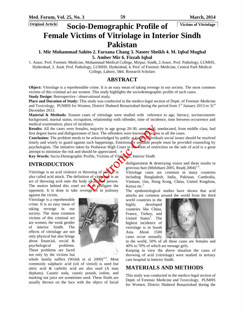
Med. Forum, Vol. 25, No. 3 March, 2014 59
Socio-Demographic Profile of
Female Victims of Vitriolage in Interior Sindh
Pakistan 1. Mir Mohammad Sahito 2. Farzana Chang 3. Naseer Sheikh 4. M. Iqbal Mughal
5. Amber Mir 6. Fizzah Iqbal 1. Assoc. Prof. Forensic Medicine, Muhammad Medical College, Mirpur, Sindh, 2.Assoc. Prof. Pathology, LUMHS,
Hyderabad, 3. Asstt. Prof. Pathology, LUMHS, Hyderabad, 4. Prof. of Forensic Medicine, Central Park Medical
College, Lahore, 5&6. Research Scholars
ABSTRACT
Object: Vitriolage is a reprehensible crime. It is an easy mean of taking revenge in our society. The most common
victims of this criminal act are women. This study highlights the sociodemographic profile of such cases.
Study Design: Retrospective / observational study.
Place and Duration of Study: This study was conducted in the medico-legal section of Deptt. of Forensic Medicine
and Toxicology, PUMHS for Women, District Shaheed Benazirabad during the period from 1st January 2013 to 31
st
December 2013.
Material & Methods: Sixteen cases of vitriolage were studied with reference to age, literacy, socioeconomic
background, marital status, occupation, relationship with offender, time of incidence, time between occurrence and
medical examination, place of incidence.
Results: All the cases were females, majority in age group 20-30, unmarried, uneducated, from middle class, had
first degree burns and disfigurement of face. The offenders were known to victims in all the cases.
Conclusion: The problem needs to be acknowledged by public at large. Individuals social issues should be resolved
timely and wisely to guard against such happenings. Emotionally unstable people must be provided counseling by
psychologists. The initiative taken by Peshawar High Court in the form of restriction on the sale of acid is a great
attempt to minimize the risk and should be appreciated.
Key Words: Socio-Demographic Profile, Victims of Vitriolage, Interior Sindh
INTRODUCTION
Vitriolage is an acid violence or throwing of acid. It is
also called acid attack. The definition of vitriolage is an
act of throwing acid onto the body of another person.
The motive behind this cruel act is to disfigure the
opponent. It is done to take revenge or in jealousy
against the victim.
Vitriolage is a reprehensible
crime. It is an easy mean of
taking revenge in our
society. The most common
victims of this criminal act
are women, the weak gender
of interior Sindh. The
effects of vitriolage are not
only physical but also brings
about financial, social &
psychological problems.
These problems are faced
not only by the victims but
whole family suffers (Welsh et al 2009)3-4
. Most
commonly sulphuric acid (oil of vitriol) is used but
nitric acid & carbolic acid are also used (A man
depkans). Caustic soda, caustic potash, iodine, and
marking nut juice are sometimes used. These fluids are
usually thrown on the face with the object of facial
disfigurement & destroying vision and these results in
grievous hurt (Mithiharn 2005, Readj 2004)1-2
.
Vitriolage cases are common in many countries
including Bangladesh, India, Pakistan, Cambodia,
Vietnam, Oas, Hong Kong, China, United Kingdom,
Kenya etc.5
The epidemiological studies have shown that acid
attacks are common around the world from the third
world countries to the
highly developed
countries like China,
France, Turkey, and
United States5. The
highest incidence of
vitriolage is in South
Asia. About 1500
cases occur annually
in the world, 50% of all these cases are females and
40% to 70% of which are teenage girls.
Keeping in view the above situation the cases of
throwing of acid (vitriolage) were studied in tertiary
care hospital in Interior Sindh.
MATERIALS AND METHODS
This study was conducted in the medico-legal section of
Deptt. of Forensic Medicine and Toxicology, PUMHS
for Women, District Shaheed Benazirabad during the
Original Article Victims of Vitriolage
Electro
nic Cop
y

Med. Forum, Vol. 25, No. 3 March, 2014 60
period from 1st January 2013 to 31
st December 2013.
Sixteen cases of vitriolage were studied with reference
to age, literacy, socioeconomic background, marital
status, occupation, relationship with offender, time of
incidence, time between occurrence and medical
examination, place of incidence.
RESULTS
Out of 16 vitrolage victims included in the study most
of the victims (50%) were in the age group of 20 – 30,
followed by 25% in the age group 15 – 20 years, 12.5%
in the age group of 30 – 40 and same number in 40 – 50
years. Table No.1. The maximum numbers of victims
were unmarried (62.5%), while married were 05
(31.2%), and one victim ( 6.25%) was divorcee. Table
No.2. The majority of victims were uneducated
(44.75%), 4 (25%) were matriculate, same number
were intermediate and 1 victim (6.25%) was graduate.
Table No.3. Most of victims were of middle class
(62.5%), 6 (37.5%) were from middle class and none in
upper class. Table No. 4. In our study most of the
victims had first degree burn of (75%), while 4 victims
(25%) had 2nd
degree burns. Table No.5. Out of 16
vitriolage victims, 10 victims shown disfigurement of
face accounting (62.5%) followed by unilateral corneal
opacity in 4 victims (25%), while 2 victims (12.5%)
had bilateral corneal opacities as shown in Table No.6.
Offenders were known to victims in all the cases. None
of the victims was engaged in any job.
Table No.1: Age of victims of vitriolage
S.No. Age in
years
No. of
Victims
Percentage
1. 15 – 20 04 25.00
2. 20 – 30 08 50.00
3. 30 – 40 02 12.50
4. 40 – 50 02 12.50
Total 16 100.00
Table No.2: Marital status of victims of vitriolage
S.No. Marital
Status
No. of
Victims
Percentage
1. Married 05 31.20
2. Unmarried 10 62.50
3. Divorcee 01 6.25
Total 16 100.00
Table No.3: Educational status of victims of
vitriolage
S.No. Level of
Education
No. of
Victims
Percentage
1. Matriculation 04 25.00
2. Intermediate 04 25.00
3. Graduate 01 6.25
4. Illiterate 07 43.75
Total 16 100.00
Table No.4: Economic status of victims of vitriolage
S.No. Economic
status
No. of
Victims
Percentage
1. Lower class 06 37.0%
2. Middle class 10 62.50
3. Upper class 00 00.00
Total 16 100.00
Table No.5: Clinical Presentations of victims of
vitriolage
S.
No.
Clinical
Presentation
No. of
Victims
Percentage
1. 1st degree Burn 12 75.00
2. 2nd
degree Burn 04 25.00
3. 3rd
degree Burn 00 00.00
Total 16 100.00
Table No.6: Complications in victims of vitriolage
S.
No.
Complication No. of
Victims
Percentage
1. Disfigurement of
face 10 62.50
2. Unilateral corneal
opacity 04 25.00
3. Bilateral corneal
opacity 02 12.50
Total 16 100.00
DISCUSSION
Acid is used as an easily available weapon, especially
against women in most of the developing as well as
developed countries. Vitriolage is at rise in developing
countries, including Pakistan. Pakistani women are
treated in uncivilized manners. They are often victim of
such violent incidences. They are bound in cultural
restrictions and gender based injustice. The vitriolage
cases are reflection of the limitations imposed upon
them, Any female going a little away from such
limitations they have to experience grave consequences
like vitriolage. Victims suffer unbearable pain and
unimaginable mental torture (anguish). The dark aspect
of the fact is that most families conceal the facts such as
name of victim, name of assailant, address & social
reason behind this crucial act and photographs. The
victim families talked to us only on assurance of
secrecy.
In a Reuters report around 1,500 acid attacks are
reported each year and 80 percent of them target
women. Yet the number is most likely higher, as many
victims are afraid to speak out. According to Monira
Rahman, CEO of the Acid Survivors Association in
Bangladesh, most of the women and girls she supported
were attacked by men who viewed them as
commodities and therefore believed they were justified
in disfiguring them and violating their rights. She writes
until women have real equality, they will continue to
Electro
nic Cop
y

Med. Forum, Vol. 25, No. 3 March, 2014 61
suffer not only from poverty but also be subjected to
violence.6
The Acid Survivors Foundation (ASF) predicts that 150
acid attacks occur in Pakistan each year, with many
more going unreported.24
The number of acid attacks in Colombia has increased
in recent in years. A woman in India may be attacked
over a dowry or caught without a veil in Pakistan, but in
Colombia often attacked out of anger over her
independence. Europe is also not an exception. Such
incidences are also seen there.9
NHS statistics for England do not separate out acid
attacks. In 2011-12 there were 105 hospital admissions
in England for "assault by corrosive substance", but the
category covers more than just acid.12
According to The Aurat Foundation, a women's rights
organization, more than 8,500 acid attacks, forced
marriages and other forms of violence against women
were reported in Pakistan in 2011.7
In 2010, at least 8,000 acid attacks, forced marriages
and other forms of violence against women were
reported, according to The Aurat Foundation.
Acid burnings are among the most horrific crimes
against women in Pakistan that are now criminalized in
a landmark set of laws passed by the parliament. They
stand to protect millions of women from common forms
of abuse in a conservative, Muslim country with a
terrible history of gender inequality. This is a big
achievement for the women of Pakistan, civil society
and the organizations that have been working for more
than 30 years to get women friendly bills passed. The
new laws explicitly criminalized acid attacks and
mandated that convicted attackers would get a
minimum sentence of 14 years that could extend to life,
and pay a minimum fine of about $11,200.8
Previously, victims had to prosecute attacks as
attempted murder or disfigurement and were largely
unsuccessful, said Valerie Khan, head of the Acid
Survivors Foundation.9
According to Dawn News the Peshawar High Court in
October, 2013 has banned the open sale of acid. Chief
Justice Peshawar High Court, Justice Dost Muhammad
Khan, remarked that acid was being used as a form of
weapon & women were being targeted with it.10
Acid attacks are seen to be on the rise in Khyber
Pakhtunkhwa. In year 2012, 150 acid attack cases were
registered in Pakistan, 30 of which were reported from
KP’s Mardan district alone. According to Acid
Survivors Foundation, the data shows an upward trend
in this form of violence targeting women. Some 45 per
cent of the acid attacks are the result of family feuds
while 17 per cent are linked to refusals to marriage
proposals. Moreover, a report by the Aurat Foundation
released in January 2013 said that although the total
number of reported cases of violence against women
had decreased by 12 per cent, a deeper analysis showed
a significant 89 per cent increase in cases of acid-
throwing, followed by a 62 per cent rise in domestic
violence.10
The Asian Human Rights Commission (AHRC) is
working on acid throwing cases as part of its
involvement in denouncing human rights violations
across Asia. It has commented that acid attacks are
serious concern in Pakistan.11
It is a crime with a marked gender skew. Experts say
that women and girls are victims in 75-80% of cases.
Of the female victims, about 30% are under 18.12
In our study 62.5% victims were unmarried,
constituting the major group of victims. The reason
behind is their refusal for marriage because of their
illegitimate relations. Females are the victims in most
of the cases in our study, being the week gender of
sindhi community. It is accordance with study of
Bandyopadhyay13
.
In our study most of victims were uneducated (50%),
while 25% were matriculate & three (18.75%) were
intermediate and only one girl was graduate (6.25%).
The majority of victims belong to lower middle class
(62.5%), while 37.5% victims belonged to lower and
none from upper class. As in our society mostly girls
from lower middle class go out for work in factories,
fields, schools or for domestic work, therefore more
prone for encounters with men. It results in disputes
consequent upon refusal for illegitimate relations or
marriage.
In our study majority of females suffered from
disfigurement of face (62.5%). Howard health policy
review is also in accord suggesting that the main motive
of offenders was to disfigure the face.17
. Stating that
perpetrators of these acid throwing attacks at their
victims, usually were at their faces for burning them.18
It causes permanent scaring of face,13,14,15,18,20
25% of
victims had unilateral and 12.5% bilateral corneal
opacity as studied by Bandyopathyay13
& Bahl19
. The
long term consequences of these attacks cause
blindness, according to acid survivors foundation in
Pakistan the most notable effects of an acid attack is the
life long disfigurement20,22
.
In our study all the victims were females of all ages.
This is comparable to articles by barrister Afzal
Hussain14
, stating that cases of women being the victim
were more common & same comment in study of
Naveed & Sloan.23
.
CONCLUSION
The problem needs to be acknowledged by public at
large. Individual’s social issues should be resolved
timely and wisely to guard against such happenings.
Emotionally unstable people must be provided
counseling by psychologists. The initiative taken by
Peshawar High Court in the form of restriction on the
sale of acid is a great attempt to minimize the risk and
should be appreciated.
Electro
nic Cop
y

Med. Forum, Vol. 25, No. 3 March, 2014 62
REFERENCES
1. Welsh J. It was like burning in hell: A Comparative
exploration of acid attack violence, Carolina Papers
on Int Health 2009;1-115.
2. Kaur AD, Sandhu HS, Aggarwal KK, Oberoi SS.
An unusual case of vitriolage. J Forensic Med &
Toxicol 2007;7(2). http://pafmat.com/20072_
7.htm; retrieved on 3.01.2014.
3. Mathiharan H. Modi’s Medical Jurisprudence &
Toxicology. 23rd
ed. Lexis Nexis Butterworth;
2005.p.949-75
4. Reddy KSN. The Essentials of Forensic Medicine
and Toxicology. 24th
ed. Hyderabad, India: K
Saguna Devi; 2004.p.462.
5. Avon Global Center for Women and Justice at
Cornell Law School, the Committee on
International Human Rights of the New York City
Bar Association, the Cornell Law School
International Human Rights Clinic, and the Virtue
Foundation(2011) Combating Acid Violence in
Bangladesh, India and Cambodia". Avon foudation
for women. pp.1-64.Retrieved 6 March 2013
6. Nurbanu UC. Bangladeshi Woman, Forced To
Return To Husband After Acid Attack. The
Huffington Post , Posted: 28.11.2012; http://www.
huffingtonpost.com/2012/11/28/nurbanu-acid-
attack_n_2206621.html; Retrieved on 24.12.2013
7. Younus F. Dead: Pakistani Acid Victim Commits
Suicide. The Huffington Post , Posted: 28.03.2012;
http://www.huffingtonpost.com/2012/03/28/fakhra-
younus-dead-pakistan-acid_n_1384612.html.
Retrieved on 24.12.2013.
8. Ahmed M. Pakistan Women Rights Laws Ban
Horrific Acid Burnings. http://www.
huffingtonpost.com/2011/12/13/pakistan-women-
rights-laws_n_1146263.html; Retrieved on
24.12.2013.
9. The Americas acid attacks are rising in Columbia.
Washington post: posted on 8.3.2012;
http://www.washingtonpost.com/world/the_amerio
cas/acid_attack_rising_in
columbia/2012/08/03/e8c85528-c843-11e1-9634-
0dcc540e7171-story.html. Retrieved on
24.12.2013.
10. Khan V. PHC bans open sale of acid in KP,
Historical Court Decision. October 9th, 2013;
http://acidsurvivorspakistan.org/; retrieved on
24.12.2013.
11. Acid attacks a serious concern in Pakistan (Edited
text of a statement issued by the Asian Human
Rights Commission: AHRC-STM-018-2010).
Ethics in Action 2010; http://www.human
rights.asia/resources/journals-
magazines/eia/eiav4a1/3-acid-attacks-a-serious-
concern-in-pakistan; retrieved on 24.12.2013.
12. Castella TD. How many acid attacks are there? BC
News Magazine; http://www.bbc.co. uk/news/
magazine-23631395; Retrieved on 24.12.2013.
13. Bandyopadhyay, Mridula, Khan MR. Loss of face:
violence against women in South Asia' in Lenore
Manderson. Bennett LR, editor. Violence Against
Women in Asian Societies. Routledge; 2003. ISBN
978-0-7007-1741-5.
14. Guerrero, Linda (October 2012). "Burns due to
acid assaults in Bogotá, Colombia". Burns: 1–6.
doi:10.1016/j.burns.2012.10.022
15. Acid Violence in Uganda: A Situational Analysis.
Acid Survivors Foundation Uganda. November
2011. pp. 1–21. Retrieved 6 March 2013.
16. Olaitan, Peter B, Jiburum BC. Chemical injuries
from assaults: An increasing trend in a developing
country. Ind J Plastic Surg 2008;41(1):20–23.
17. Acid attacks: Bangladesh’s efforts to stop the
violence. Harvard Health Policy Review 2008;3(1):
1-4. Retrieved 2008-06-18.
18. CNN.com - Bangladesh combats an acid onslaught
against women - November 11, 2000. Archived
from the original on September 22, 2007. Retrieved
2008-03-13.
19. Bah l, Taru, Syed MH.. Encyclopaedia of the
Muslim World. Anmol Publications PVT. LTD
2004. ISBN 978-81-261-1419-1.
20. Iranian woman blinded by acid attack pardons
assailant as he faces same fate. The Guardian.
2011-07-31. Retrieved 2013-09-21.
21. Bangladesh combats an acid onslaught against
women. November 11, 2000. Archived from the
original on 2007-09-22. Retrieved 2008-03-13.
22. Hussain BA. Proposed legislation on acid violence.
23. Naveed B, Sloan S. May 13;2010 Asia:vitrolage
24. Freeman J. Acid attacks in Pakistan 2013;
http://www.disruptivewomen.net/2013/08/10/acid-
attacks-in-pakistan/
Address for Corresponding Author:
Dr. Mir Muhammad Sahito
Assoc. Prof. Forensic Medicine
Muhammad Medical College, Mirpur, Sindh
Electro
nic Cop
y

Med. Forum, Vol. 25, No. 3 March, 2014 63
Significance of CD Markers in the
Classification, Patterns and Sub
Typing of Non Hodgkin’s Lymphoma 1. Nazir Pathan 2. Amin Fahim 3. Asghar Khan 4. Aneela Qureshi 5. Saba Khoja
1. Assist. Prof. of Pathology, LUM&HS, Jamshoro 2. Assist. Prof. of Pathology, Al-Tibri Medical College, Karachi
3. Assist. Prof. of Pathology, Isra University, Hyderabad 4. Assoc. Prof. of Pathology, Al-Tibri Medical College,
Karachi 5. Assist. Prof. of Pathology, Isra University, Hyderabad
ABSTRACT
Objective: Aim of this study is Immunohistochemical analysis of H&E diagnosed cases of NHL for confirmation,
classification and differentiation on the basis of phenotypic expression of CD3, CD30, CD45 and CD20 markers.
Study Design: Prospective study.
Place and Duration of Study: This study was conducted in the Pathology department of Liaquat University of
Medical and Health Sciences Jamshoro (LUMHS) during October 2010 to March 2012
Materials and Methods: The sample consisted of all the specimens received in the Pathology department of
Liaquat University of Medical and Health Sciences Jamshoro (LUMHS) during the above period.
Immunohistochemical stains including CD3, CD30, CD20 and CD45 were used for classification and differentiation
of cases of NHL.
Results: Out of one hundred and eighty (180) H & E diagnosed cases of Non Hodgkins Lymphoma, only 142
(78.8%) were positive for CD20 and were confirmed as B cell NHL; however 6 (3.3%) cases showed positivity with
CD30 and were confirmed as large T-cell NHL. 38(21.2%) cases showed positivity for CD3 and all 180 (100%)
cases were positive for CD45 and were confirmed as NHL.
Conclusion: It is concluded that Immunohistochemistry is helpful in differentiation of NHL. Cases of B cell NHL
occur more frequently than T cell NHL. Furthermore NHL is more common in males and mostly presents with nodal
involvement.
Key Words: CD markers, Immunohistochemistry, Non Hodgkin’s Lymphoma.
INTRODUCTION
Malignant tumors are composed of abnormal cells
having multiple genetic mutations thus resulting in the
escape from cell cycle regulatory genes1. Non-
Hodgkin’s Lymphoma (NHL) is one of the many
malignant tumors arising from lymphoid organs. The
yearly incidence of non-Hodgkin’s lymphoma is
variable in different geographical location and is found
to be around 3% to 4% in developed countries2. Non-
Hodgkin's lymphoma is encountered more in males
than the females and commonly encountered between
sixth and seventh decade of life3. In Pakistan, NHL
accounts for 4th commonest malignant tumor in males
with the incidence of 6.1%4. WHO classifies NHL into
many B cell and T cell subtypes, consisting of several
distinct lymphoid neoplasm5.
The classification of the NHL is based on various
parameters including histomorphologic changes on
biopsy specimen, immunohistochemistry and flow
cytometry6,7
. NHLs classification is based on the origin
of neoplastic lymphocytic cells and is divided into the
B-cell and T-cell lymphoma. Lymphoma of the B cells
accounts about 90% of NHLs, whereas rest of the 10%
are T-cell lymphoma8.
The steps involved in the diagnosis of lymphoma
includes hematoxlin and eosin stained biopsy slides,
Immunohistochemical markers along with other
diagnostic tools, some of which may be either invasive
or non-invasive leads to final confirmation of the
disease. This helps out to have a proper therapeutic
plan9. The classification of both Hodgkins disease and
NHL is continuing to evolve incorporating not only
histopathologic data, but also immunophenotypic,
genotypic and clinical characteristics to design a
treatment plan using varying combinations of
chemotherapy, radiotherapy and immunotherapy10
.
The principle of the Immunohistochemistry or IHC
involves the use of antibodies for detecting antigens in
a tissue sample. Lymphoid as well as hematopoietic
diagnosis and classification can be done easily with the
help of IHC11
. Two different types of dyes including
chromogenic or fluorescent means are used in IHC for
detecting target antigens, however experimental design
will be needed to decide the type of readout. In
fluorescent method, fluorophore is used for the
conjugation of primary and secondary antibody which
is later detectable by fluorescent microscopy. In
contrast chromogenic detection involves enzymes
mainly horseradish peroxidase (HRP) or alkaline
phosphatase (AP). This causes formation of colored,
insoluble precipitates when substrates, such as DAB
and NBT/BCIP are added respectively.12
Original Article Non Hodgkin’s
Lymphoma
Electro
nic Cop
y

Med. Forum, Vol. 25, No. 3 March, 2014 64
So this study is planned for the Immunohistochemical
analysis of H&E diagnosed cases of NHL for
confirmation, classification and differentiation on the
basis of phenotypic expression of CD3, CD30, CD45
and CD20 markers.
MATERIALS AND METHODS
This prospective study was conducted in the Pathology
department of Liaquat University of Medical and
Health Sciences Jamshoro (LUMHS) during October
2010 to March 2012. All sample received during above
period were included in this study. All diagnosed cases
of NHL on H&E staining of all ages and either gender
were included in this study. Biopsies were taken from
nodal or extranodal sites and were processed for gross
and microscopic examination. After the routine
processing and paraffin embedding, H & E and special
staining; the immunohistochemical analysis was
performed. The antibodies used in immunohisto
chemical staining included CD3, CD30, CD20 and
CD45. The antibodies were ordered from DAKO
DENMARK. All cases were analyzed for age, sex
distribution (male vs female), site (nodal vs extranodal)
and subtypes of NHL. Seven cases (28 slides) of
positive and negative control of H&E diagnosed NHL
were included for the staining (performed in batches).
RESULTS
The sample size of this study consisted of one hundred
and eighty (180) cases of H & E diagnosed NHL
diagnosed on staining. Out of total 180 cases of NHL,
patients with age below 60 years were 146 (81.1%)
while the patients with age above 60 years were 34
(18.9%) and the mean age was 46 years. Table 1 shows
age wise distribution of all these patients.
Out of total 180 cases of NHL, 110 (61.1%) cases were
seen in male while 70 (38.9%) female were affected.
Gender distribution is shown in Table 2.
Table No.1: Age Distribution of Patients
Characteristic No. of Cases Percentage
Age < 60 years 146 81.1
Age ≥ 60 years 34 18.9
Table No.2: Gender Distribution
Characteristic No. of Cases Percentage
Male 110 61.1
Female 70 38.9
Out of total 180 cases of NHL, majority were found
involving lymph node accounting 124 (68.8%) while 56
(31.2%) on extra nodal sides. Site of presentation is
shown in Table 3.
Immunohistochemical staining of CD20 show that out
of 180 cases of Non Hodgkin lymphoma 142 (78.8%)
were seen positive for CD 20 and were confirmed as B
cell NHL. Immunohistochemical staining of CD30
show that out of 180 cases of NHL, only 6 (3.3%) cases
show positivity with CD 30 and were confirmed as
large T-cell NHL. Immunohistochemical staining of
CD3 show that out of 180 cases of NHL, 38(21.2%)
cases show positivity and were confirmed as T-cell
NHL. Immunohistochemical staining of CD45 showed
that out of 180 cases of NHL all 180 (100%) cases
show positivity with CD45 and were confirmed as
NHL. Results of Immunohistochemical staining are
shown Table 4.
Table No.3: Site of Presentation
Characteristic No. of Cases Percentage
Nodal 62 68.8
Extranodal 28 31.2
Table No.4: Immunohistochemistry Results
CD
Markers
Positive
cases
%age Negative
cases
%age
CD-20 142 78.9 38 21.1
CD-30 6 3.3 174 96.6
CD-3 38 21.1 142 78.8
CD-45 180 100 0 0
DISCUSSION
Diagnosis of Non-Hodgkin’s Lymphoma on H&E staining seems to be quite helpful but presence of Reed-Sternberg like cells or round cells in NHL make it difficult to diagnose. In these circumstances, immunoassaying is very much helpful. The present study included 190 lymph node biopsies, which were diagnosed on H&E by the Pathologists as Non-Hodgkin’s Lymphoma, but remained doubtful and need confirmation and further sub typing of NHL, as is needed for the better management plans. In this study 61.1% patients with NHL were male while 38.9% were female. Aftab et al in a study has also reported male predominance which is in agreement with findings of our study
13. NHL affects males more
when compared with females. However literature of the available data shows remarkable variation in the gender distribution. Male to female ratio of NHL in the developed and developing countries is reported as 1.4:1 and 4.5 to 3.1 respectively
14. The study conducted by
Zeba et al also reported male predominant and support our study
15.
In the present study 68% patients have nodal involvement while 31.2% patients have extra nodal involvement. Study conducted by Arora et al reported 33% of cases having extra nodal presentation and 67% have nodal presentation
16. Another study conducted in
china found that, tumors in lymph nodes were seen in 42.8% and extra nodal 57.2% and proposed that autoimmune etiology is thought to be involved in extra nodal marginal zone lymphomas and these are reported to be more common in East Asian countries like Japan and China than in Pakistan
17.
In our study out of 180 cases of Non-Hodgkin’s
Lymphoma diagnosed on panel of CD markers, 142
Electro
nic Cop
y

Med. Forum, Vol. 25, No. 3 March, 2014 65
(78.8%) were seen positive for CD20 and confirmed as
B-cell NHL and 38 (21.2%) showed positivity for CD3
and diagnosed as T-cell NHL. Study conducted by
Yasmeen Bhurgri reported 80% of cases as B-cell NHL
and 20% as T-cell NHL support our study18
. Another
study conducted in Saudi Arabia also revealed majority
of cases of B-cell phenotype; while only 14% of the
cases were T-cell lymphoma19
.
The CD20 marker is expressed in all the maturation
levels of B cells except stem cells and plasma cells;
however it is present in both the normal and malignant
B cells20
. CD3 is composed of at least five different
polypeptide chains closely associated with T cell
antigen receptor and with each other21
. CD3 is a useful
marker for the detection of T-cell lymphoma22
.
Polyclonal CD3 along with CD20 distinguishes
between T-cell and B cell lymphomas23
.
The CD30 antigen is an isoglycoprotein whose
expression is used for detecting the anaplastic large cell
lymphomas (ALCL). This distinct variant of T cell
NHL shows proliferation of large pleomorphic cells in a
cohesive pattern and is strongly positive for CD3024
. In
the present study CD3 and CD30 were positive in 6
(3.3%) cases of ALCL, while the study conducted by
Krol and Gulsah reported 8 (<1%) and 1.2% cases of
ALCL respectively which is in accordance with our
findings25,26
.
Immunohistochemical staining of CD45 showed that
out of 180 cases of NHL all 180 (100%) cases show
positivity with CD45 and were confirmed as NHL.
CD45 glycoprotein expression is positive in all
lymphocytic cells. However CD45 expression in low-
grade B-cell NHL is not well reported in the literature27
.
CD45 is a transmembrane protein-tyrosine phosphatase
and is found on the B and T cells, thymocytes and
macrophages. Thus it is useful in differentiating
lymphomas from non lymphoid tumors as CD45 shows
positivity for malignant B and T cells. Furthermore
expression of CD20 antigen is generally restricted to
the B-cell lineage while CD3 stains only cells of T-cell
origin28
. Thus CD20 and CD3 only help in
differentiating B and T cell Lymphomas.
CONCLUSION
It is concluded that Immunohistochemistry is helpful in
diagnosing the types of NHL. Cases of B cell NHL
occur more frequently than that of T cell NHL.
Furthermore NHL is more common in males and
mostly presents with nodal involvement.
REFERENCES
1. Mikdat B, Fatma A, Muradiye A, Hacer H, Sibel
Y, Metin C, et al. A potential association between
the number of CA repeats in the promoter region of
the ADAMTS9 gene with lymphatic metastasis of
breast cancer. Turk J Med Sci 2013;43:671-677.
2. Naseem SS, Nagi AH, Ashraf M, Bilal S, Akhlaq
M, Henna N, et al. A pattern of lymphadenopathies
seen in a tertiary care hospital in Lahore, Pakistan.
Turk J Med Sci 2011;41(2):353-358.
3. Sathiya M and Muthuchelian K. Significance of
Immunologic Markers in the Diagnosis of
Lymphoma. Acad J Cancer Res 2009; 2 (1): 40-50.
4. Hanif M, Zaidi P, Kamal S, Hameed A. Institution-
based cancer incidence in a local population in
Pakistan: nine year data analysis. Asian Pacific J
Cancer Prev 2009;10: 227-30.
5. Ekstram SK. Epidemiology and etiology of non-
Hodgkin lymphoma - a review. Acta Oncologica
2006;45: 258-71.
6. Buckner CL, Christiansen LR, Bourgeois D,
Lazarchick JJ and Lazarchick J. Case Reports:
CD20 Positive T-cell Lymphoma/Leukemia: A
Rare Entity with Potential Diagnostic Pitfalls.
Annals Clinic & Lab Sci 2007; 37(3): 263-267.
7. Lee M, Cha HJ, Yoon DH, Suh C and Huh J. EBV-
positive diffuse large B-cell lymphoma of the
elderly with aberrant expression of CD3 and TIA-
1. Blood research 2013; 48(2): 156-160.
8. Kurtoğlu E, Yıldırım M, Karakılınç H, Yıldız M,
Göktaş S, Süren D, et al. Clinicopathological
properties of Non-Hodgkin Lymphomas in the
south-west of Turkey. Cancer Res J 2013;1(2):
15-19.
9. Hingorjo MR, Syed S. Presentation, Staging and
Diagnosis of Lymphoma: A Clinical Perspective. J
Ayub Med Coll Abbottabad 2008;20(4): 100-103.
10. Young GAR, Iland HJ. Clinical perspectives in
lymphoma. Int Med J 2007;37:478–84.
11. Higgins RA, Blankenship JE, Kinney MC.
Application of immunohistochemistry in the
diagnosis of non-Hodgkin and Hodgkin lymphoma.
Arch Pathol Lab Med 2008; 132(3): 441-461.
12. Rosen ST, Winter JN and Gordon LI. Non-
Hodgkin lymphoma. Cancer management: A
multidisciplinary approach. In: Pazdur R, Wagman
LD, Camphausen KA, Hoskins WJ, editors. 11th
ed. CMP Medica Lawrence KS; 2008.p.719-774.
13. Aftab K, Bhurgri Y, Pervez S. Small B cell Non-
Hodgkins Lymphoma in Pakistan. J Pak Med
Assoc 2006; 56(1): 22-25.
14. Aziz Z, Rehman A, Akram M, Saeed A. Non-
Hodgkin's lymphoma in Pakistan: a
clinicopathological profile of 175 patients. J Pak
Med Assoc 1999; 49: 11-15.
15. Aziz Z, Sana S, Saeed S, Akram M. Applicability
of international prognostic index in non Hodgkin's
lymphoma in Pakistan. J Ayub Med Coll
Abbottabad 2004; 16: 15-20.
16. Arora N, Manipadam MT, and Nair S. Frequency
and distribution of lymphoma types in a tertiary
care hospital in South India: analysis of 5115 cases
using the World Health Organization 2008
Electro
nic Cop
y

Med. Forum, Vol. 25, No. 3 March, 2014 66
classification and comparison with world
literature. Leukemia & Lymphoma 2013;54(5):
1004-1011.
17. Zhang YN, Zhou XG, Zhang SH, Wang P, Zhang
CH, Huang SF. Clinicopathologic study of 369 B-
cell non-Hodgkin lymphoma cases, with reference
to the 2001 World Health Organization
classification of lymphoid Neoplasms 2005;34(4):
193-7.
18. Bhurgri Y, Pervez S, Bhurgri A, Faridi N,
Usman A, Kazi LAG, et al. Increasing Incidence of
Non-Hodgkin’s Lymphoma in Karachi,1995-2002
Asian Pac J Cancer Prev 2005; 6(3): 364-369.
19. Ollberding NJ, BriseisAschebrook-Kilfoy DB,
Caces SM, Smith DD, Weisenburger, Chiu BC.
Dietary patterns and the risk of non-Hodgkin
lymphoma. Public Health Nutrition 2013; 1-7.
20. Thomas DA, O'Brien S, Jorgensen JL, Cortes J,
Faderl S, Garcia-Manero G, et al. Prognostic
significance of CD20 expression in adults with de
novo precursor B-lineage acute lymphoblastic
leukemia. Blood 2009; 113(25): 6330-6336.
21. Allison JP, Havran WL. The immunobiology of T
cells with invariant gamma delta antigen receptors.
Annu Rev Immunol 1991; 9: 679-705.
22. Cabecadas JM, Isaacson PG. Phenotyping of T-cell
lymphomas in paraffin sections--which antibodies?
Histopathology 1991; 19: 419-424.
23. Ahmed HG, Adam TM, Basama NK, Agabeldor
AA. Utility of CD3 and CD30 in Immuno-
phenotyping of Lymphomas among Sudanese
patients. J Cancer Sci Ther 2011;3:116-119.
24. AL–Mammori, RaheemTuama O, AL-Thahab AE,
and Al-Awad AS."Determination of
Chemoattractant factor (Interleukin-8) and Gamma
in.terferon (IFN-γ) among Lymphoma patients
(Hodgkin's and Non Hodgkin)." J Pharm and Biol
Sci 2013; 5(2): 75-78.
25. Krol ADG, Cessie SI, Snijder S, Kluin-Nelemans
JC, Kluin PM, Noordijk EM. Primary extranodal
non-Hodgkin’s lymphoma (NHL): the impact of
alternative definitions tested in the Comprehensive
Cancer Centre West population-based NHL
registry. Ann Oncol 2003; 14: 131-9.
26. Gülşah K, Işınsu K, Saba K, Duygu K, Zeynep B.
Is CD117 Expression Related with Plasma Cell
Differentiation, IRF4/MUM1 and CD38
Expressions in Lymphoid Neoplasms? Turk J Med
Sci 2008; 38 (6): 511-517.
27. Carulli G, Cannizzo E, Zucca A, Buda G, Orciuolo
E, Marini A, et al. CD45 expression in low-grade
B-cell non-Hodgkin's lymphomas. Leuk Res 2008;
32(2): 263-7.
28. Sarma A, Barmon D, Sharma JD, Kataki AC,
Bhuyan C. Primary Non-Hodgkin's Lymphoma of
the Uterine Cervix: Report of Three Cases with
Immunohistochemical Study. J Cancer Res 2012;
1: 1-4.
Address for Corresponding Author:
Dr. Amin Fahim
House No: A-21, SAU Colony
Tando Jam, Sindh, Pakistan
E-mail: [email protected]
Cell # 0331-3504341
Electro
nic Cop
y

Med. Forum, Vol. 25, No. 3 March, 2014 67
Fatal Road Traffic Accidents in
Karachi 1. Romela Naz 2. Imran Afzal 3. Zubaida Zain 4. Afshan Kamran 5. M. Iqbal Mughal
1. Asstt. Prof. of Forensic Medicine, Sir Syed M&DC for Girls, Karachi 2. Assoc. Prof. Forensic Medicine,
JM&DC, Karachi 3. Asstt. Prof. Forensic Medicine, Shifa College of Medicine, Islamabad 4. Asstt. Prof.
of Pharmacology, Altamash Dental College, Karachi, 5. Prof. of Forensic Medicine, CPMC, Lahore.
ABSTRACT
Objective: Fatalities due to road traffic accidents are a major cause of mortality all over the world. The aim of our
study is to look at socio-demographic profile of such cases in order to find some remedial measures to minimize the
brunt.
Study Design: Retrospective / observational study.
Place and Duration of Study: This study was conducted at the Emergency Department, Sir Syed Hospital,
Qayyumabad, Karachi from January to June, 2013.
Materials and Methods: Study was based upon 56 victims of RTA. It includes all those who died during treatment
or found dead on arrival in the emergency department. The demographic data of these cases was recorded.
Results: Males victims were 71% & maximum number of victims (37.5%) was in age group 30-39 years. Most of
the accidents (38%) took place between 1200hrs & 1800hrs. Most of the injuries were found on the head (66%) and
then chest 14%, lower extremities 21%, upper extremities 19%. The most common external injuries were abrasions,
bruises and lacerations. Skull was the most common bone to be fractured (44%). The head injury constituted major
cause of death (53%) followed by hemorrhagic shock (35%). Pedestrians and two wheeler riders were mostly
affected individuals followed by cyclists, drivers of three wheeler, light and heavy motor vehicles.
Conclusion: The incidence of traffic accidents is at rampant. The accidents risks can be minimized by enhancement
of road visibility, good traffic control with properly working road traffic signals. Fatalities of Traffic accidents
victims can be reduced by providing good pre-hospital care, establishment of well equipped emergency trauma
centres in all big hospitals.
Key Words: Road Traffic Fatalities, Vehicles, Injuries, Karachi.
INTRODUCTION
Accidents are very common in big cities as there are
many modes of transport and roads are overburdened.
We hear as well as read in newspapers about such
happenings almost daily. These accidents often result in
loss of life and material. These are caused by the
carelessness / negligence of the drivers and their
ignorance about the traffic rules.
Like most cities, road accidents are also a problem in
Karachi, Pakistan. It is the largest city in Pakistan in
terms of area and population and it is significantly
facing the issue. According to WHO, road traffic
accidents will be the 3rd
greatest cause of the fatalities
by 20201. Fatal RTAs for about 90% occurs on the
roads of developing countries despite that the number
of vehicles they have are less than half of the globule
vehicles2. WHO reports that India has the highest score
of deaths due to RTAs in the world ranking. According
to data for 2007, the number of people died in India due
to RTAs is 114,590and the next in this ranking is China
with 89,455 deaths3. According to a WHO report in
2009 in Pakistan road traffic injuries lead to 25.3 deaths
per 100,0004.
The patient logs of the two largest government hospitals
in Karachi were studied between December 1993 and
February 1994 to identify those persons most likely to
be injured in a road traffic accident and to identify the
vehicle types which were most likely to be involved.
The 18% of people involved in accidents died.
Pedestrians and motorcyclists were the most common
victims accounting for 46% amongst all injured and
51% of all deaths. Although buses and trucks were less
than 4% of the registered vehicles in Karachi but they
were the striking vehicle among 49% of all injured and
65% of all died. Road traffic accidents dispropor-
tionately affect wage earners. Specific interventions to
improve the road safety concerning trucks and buses is
needed5.
According to official data compiled by the Accident
Department of Karachi Traffic Police, a total of 97 fatal
accidents were recorded during January-April this year
(2013), which is lower than the corresponding period of
last year6.
By latest information RTA is the main cause of injury,
disablement & mortality all over the world which
perplex a great burden on natural health system7. RTAs
are also the leading cause of mortality in young &
adults less than 45 years of age and more so in males.
The consequences of injuries substantiated in RTAs
depends on variety of reasons including venturous
driving location & condition of the roads (roads without
speed breakers, cat eyes, sign boards etc.), type of
vehicle, condition of the driver, time of accident &
Original Article Road Accident
Electro
nic Cop
y

Med. Forum, Vol. 25, No. 3 March, 2014 68
below standard safety designs8,9
. Head injury is the
main cause of death in RTAs.
In Pakistan epidemiological data on RTAs have been
reported from emergency departments, intensive &
surgical care units of some particular hospitals & police
stations8. Necessary medical care in the 1
st hour after an
injury to the victim can reduce the number of deaths
from RTAs & increase the probability of survival.
According to research in Europe about 50% of the
deaths from RTAs occur at the place of accidents or on
the way to the hospital, 15% victims die within 4 hrs
after an accident & 35% die after 4 hours9.
MATERIALS AND METHODS
This Observational retrospective study was carried out
in the emergency department of Sir Syed Hospital
Qayyumabad Karachi. This hospital is located in a
thickly populated area & is traversed by 2 busiest roads
in the city. This hospital is a 350 bedded hospital and is
the main public hospital in Qayyumabad and most of
the residents are taking medical facilities from this
hospital. The study was based on deaths due to RTAs
during period from January 2013 to June 2013.
Study was based upon 56 victims of RTA. It includes
all those who died during treatment or found dead on
arrival in the emergency department. The demographic
data of these cases was recorded. On admission
informed consent was taken from relatives or attendants
of those patients who were received unconscious. A
detailed medical history was obtained from the persons
who survived or from the relatives. From medical
charts, type of injury & its severity was assessed; while
other details were obtained from medical staff and from
police. Definition of road traffic accident used in this
study was as follows “A collision involving at least 1
vehicle in motion on a public or a private road that
result in at least 1 person being injured or killed”
RESULTS
Total 56 deaths from RTA were registered in Sir Syed
Hospital with in the period of study. Male victims were
71% (Table No.1) & maximum number of victims
(37.5%) was in age group 30-39 years (Table No.2).
Most of the accidents (38%) took place between
1200hrs & 1800hrs. Most of the injuries were found on
the head (66%) and then chest 14%, lower extremities
21%, upper extremities 19% (Table No.3). The most
common external injuries were abrasions, bruises and
lacerations. Skull was the most common bone to be
fractured (44%) (Table No.4). The head injury
constituted major cause of death (53%) followed by
hemorrhagic shock (35%) (Table No. 5). Pedestrians
and two wheeler riders were mostly affected individuals
followed by cyclists, drivers of three wheeler, light and
heavy motor vehicles.
Table No. 1: Sex distribution of cases (N=56)
Sr.No. Gender No. of cases %age
1 Males 40 71.43
2 Females 16 28.57
Total 56 100.00
Table No. 2: Age distribution of cases
Sr. No. Gender No. of cases %age
1 0-9 02 3.57
2 10-19 05 8.93
3 20-29 08 14.29
4 30-39 21 37.50
5 40-49 08 14.28
6 50-59 07 12.50
7 >60 05 8.93
Total 56 100.00
Table No. 3: Distribution of site of fatal injuries
Sr.
No.
Site of fatal injury No. of
cases
%age
1 Head 37 66.07
2 Chest 08 14.29
3 Lower extremities 12 21.43
4 Upper extremities 11 19.64
Table No.4: Distribution of fractures in RTA victims
Sr.
No.
Site of fracture No. of
cases
%age
1 Skull 25 44.64
2 Ribs 07 12.50
3 Lower extremities 11 19.64
4 Upper extremities 10 17.86
5 Spine 05 8.93
Table No.5: Cause of fatal injuries in RTA victims
Sr.
No.
Cause of death No. of
cases
%age
1 Head injury 30 53.57
2 Hemorrhagic shock 20 35.72
3 Spinal cord injury 04 7.14
4 Complications 02 3.57
Total 56 100.00
DISCUSSION
The study was undertaken over a period of 6 months in
the emergency department of Sir Syed Hospital,
Qayyumabad, Karachi. The study shows that majority
of the deceased (71.43%) were males & it is consistent
with other studies like that in Kenya or in other low
socio-economic countries12
. This is due to more
exposure of males to roads because most of their work
is outdoor and many of them being the only supporter
of their families which causes an adverse economic
impact on their families13
. Majority of victims (37.5%)
were in the age of 20-49 years and this is consistent
with other studies14,15
. Severe injuries by road crashes
are also associated with rainy seasons; this finding is
Electro
nic Cop
y
Med. Forum, Vol. 25, No. 3 March, 2014 69
similar to that reported in case-control study in Iran &
Hong Kong16,17
. The most commonly effected part of
the body was head & chest followed by lower
extremities, this finding is similar to that finding in
New Delhi18
where the most common pattern of injury
was head (18.9%), followed by fractures of lower limbs
(17.8%). Majority of fractures were observed in skull in
all type of road users. Most of the accidents occurred
between afternoons & evening (1200hrs-1800hrs) with
the continuance of early night hours, this finding of our
study is similar to other studies.20,21,22
According to
Aygencel et al19
these hours correspond to the time
range that people actively work & travel and then go
back to their homes after finishing their work.
In late 90s the traffic officers used to visit schools and
colleges to guide the students about traffic laws and
informing them about the signal system and zebra
crossings, but recently this practice has been
abandoned.6
As the number of vehicles is increasing day by day with
the same length and number of roads, lack of attention
of drivers and foot travelers and unawareness of traffic
rules followed by absence of foot paths and non
working of traffic signals and poor traffic control on
signals are the main causes of accident. Fast driving at
night under the influence of alcohol also increases the
number of road crashes.
In our study 64 % victims died on the scene or during
transportation. This was the common observation
among different researchers23
.
A study by Saha Dev et al24
conducted in New Delhi
showed that most of the victims were brought to the
hospital alive, a delay in emergency services, shortage
of pre hospital teams, absence of nurses or doctors &
formalities by police were the possible reasons of
fatalities due to RTAs.
The experts have advised the motorcyclists to always
wear helmets and desist from over speeding, besides the
pedestrian also need to be very careful while crossing
the roads.
The deadly roads of Karachi where traffic accidents
have become the order of the day happen to be the
signal-free corridors. These include Shara e Faisal,
Quaidabad Chowrangi, Dawood Chowrangi, Korangi
Road, Korangi Crossing, Korangi Double Road,
Korangi Industrial Area, Jail Road, Karimabad Signal,
Rashid Minhas Road, Sohrab Goth, Sakhi Hasan
Chowrangi, New Karachi, M.A Jinnah Road, Numaish
Chowrangi to Sea-Breeze Plaza, Mauripur Road, Hub
River Road, Garden Road and Abul Hassan Isphani
Road.6
CONCLUSION
The incidence of traffic accidents is at rampant. The
accidents risks can be minimized by enhancement of
road visibility, good traffic control with properly
working road traffic signals. Fatalities of Traffic
accidents victims can be reduced by providing good
pre-hospital care, establishment of well equipped
emergency trauma centres in all big hospitals.
REFERENCES
1. Maas AIR, Stoccheti N, Bullock R. Moderate and
Severe traumatic brain injury in adults.
2. World Health Organization RT injuries, fax sheet;
358 (online) Sep: 2011 (cited 2012 Feb 28)
available from URL: http:/www.who.int/media
centre/factsheets/fs358/en/index.html
3. Gafarov KZ. Basic principle of child injury
prevention. Karpol, Kazan 1998.
4. World Health Organization, Global Status on road
safety: Time for action (online) 2010 (cited 2012
Feb 28). Available from URL: http://who/libdoc.
who.int/publications/2009/9789241563840_eng.pdf.
5. Road traffic injuries in Karachi: the disapportionate
role of buses and trucks; http://www.ncbi.
nlm.nih.gov/pubmed/9444028 (Accessed 2 Dec
2013)
6. 113 dead in road accidents this year (2013)
http://www.thenews.com.pk/Todays-News-4-
171398-113-dead-in-road-accidents-this-year
(Accessed: 2 Dec 2013)
7. http://www.worldlifeexpenctancy.com/causeofdeat
h/roadtrafficaccidents/by-country
8. Naghavi M, Shahraz S, Bhalla K, Jafari N,
Pourmalek F, Bartels D, et al. Adverse health
outcomes of road traffic injuries, Iran after rapid
motorization. Arch Iran Med 2009;12(3):284-94.
9. Pathak A, Desania NL. Verma R. Profile of road
traffic accidents and head injury in Jaipur. J Indian
Acad. Forensic Med 2008;30:6-9.
10. Hassan Q Bashir RM, Shah M. Physical trauma. A
leading cause of medicolegal cases at DHQ
Hospital Abbotabad. J Ayub Med Coll Abbotabad
2010;22:156-8.
11. Reducing the severity of road injuries through post
impact care. Brussels, European transport safety
council, Post impact care working party, 1999.
12. Odero WO, Kibosia JC. Incidence and
characteristics of injuries in Eldoret, Kenya. East
Afr Med J 1995;72(11):706-10.
13. Salagado MSL, Comlombage SM. Analysis of
fatalities in road accidents. Forensic Sci Int 1988;
36:91-6.
14. Andrews CN, Kobusingye OC, Lett R. Road traffic
accident injuries in Kampala East afr Med J 1999;
76 (4):189-94.
15. Odero W, Garner P, ZWi A. Road traffic injuries in
developing countries:a comprehensive review of
epidemiological studies. Trop Med Int Health
1997;2 (5): 445-60
16. Majdzadeh R, Khalagi K, Naraghi K, Motevalian
A, Eshraghian MR. Determinants of traffic injuries
Electro
nic Cop
y

Med. Forum, Vol. 25, No. 3 March, 2014 70
in drivers and motorcyclists involved in an
accident. Accid Anal Prev 2008;40(1):17-23.
17. Azmani W, Rusli MA, Ismail AA, Hashi M.
Factors and Road accidents during festive seasons
in Kelantan. NCD Malaysia 2005;4:24-7.
18. Montazeri A, Road traffic related mortality in Iran:
a descriptive study. Public Health 2004;118:110-3.
19. Aygencel G, Mahamet K, Mahamet E, Telatar G.
Review of traffic accident cases presenting to an
adult emergency service Turkey. J Forensic and
Legal Med 2008;15:1-6.
20. Jha N, Srinivas DK, Roy G, Jagdish S.
Epidemiological study of road traffic cases: A
study from South India. Ind J Comm Med
2004;29:20-4.
21. Menon A, Pai VK, Rajeev A. Pattern of fatal head
injuries due to vehicular accidents in Mangalore. J
Forensic Med 2008;15:75-7.
22. Verma PK, Tewari KN. Epidemiology of head
traffic injuries in Delhi: results of survey. Regional
Health Forum 2004;8(1):7-14.
23. Henriksson E, Ostrom M, Erikson A.
Preventability of vehicle related fatalities Accident
Analysis Prevention 2001;33:467-75.
24. Sahdev P, Lacqua MJ, Singh B, Dogre TD. Road
traffic fatalities in Delhi: cause injury patterns and
incidence of preventable death. Accidents Analysis
Preven 1994;26:377-84.
Address for Corresponding Author:
Dr. Romela Naz,
Asstt. Prof. Forensic Medicine,
Sir Syed Medical and Dental College for Girls,
Karachi.
Electro
nic Cop
y
Med. Forum, Vol. 25, No. 3 March, 2014 71
Fetomaternal Outcome of
Pregnancies Complicated by Acute Appendicitis 1. Azad Ali Lashari 2. Kulsoom Bhatti 3. Tahmina Mahar 4. Rubina Hafeez 1. Asstt. Prof. of Surgery 2. Asstt. Prof. Obst. & Gynae 3 & 4. Senior Registrars, Obst. & Gynae,
Ghulam Muhammad Mahar Medical College, Sukkur
ABSTRACT
Objective: To observe the frequency and Fetomaternal outcome of pregnancies with acute appendicitis.
Study Design: Prospective / observational study
Place and Duration of Study: This study was conducted in gynecology and surgical department of Ghulam
Muhammad Mahar Medical College Teaching Hospital Khairpur Sindh from January 2010 to December 2012.
Materials and Methods: All pregnant ladies admitted in Gynae and surgical department with history of acute pain
in abdomen and strong suspicious of acute appendicitis on the basis of history, clinical examination and ultrasound
findings after exclusion of other gynecological and surgical causes of acute abdomen during pregnancy were
included in the study for following variables: Presentation, duration of symptoms, operative findings and
complications associated with disease and operative procedure were noted. Data was collected on pre-designed
Performa and analyzed on SPSS version 15.
Results: During 3 year study period total 8700 Obstetric admission and cases with strong suspicious of acute
appendicitis in pregnancy was 20 (0.22%), most women belongs to age between 18-40 years. More cases seen 2nd
trimester 11(55%), duration of symptoms < 24 hours seen in 85% and >24 hours seen in 15% of cases. Abdominal
pain was leading symptom present in 80% of cases while lower abdominal tenderness was leading sign seen in 90%.
On surgery signs of acute appendicitis seen in 75%, normal looking appendix in 10%, while perforated appendix
with moderate pus in peritoneal cavity seen in 15% of cases. One maternal death was seen in study population due to
septicemia, most probably because of late presentation.
Conclusion: The evaluation of a pregnant woman presenting with acute abdominal pain warrants a careful workup
due to the possible risks for the fetus and mother if appendix perforates.
Key Words: Pregnancy, acute appendicitis, perforation, maternal mortality.
INTRODUCTION
Acute appendicitis is the most common general surgical
problem encountered during pregnancy.1
Suspected cases of acute appendicitis in pregnancy are
considered surgical emergency due to the potentially
devastating out come for both mother and unborn fetus
if appendix perforates.2
In non gynecological cause of
acute abdomen the most common cause is acute
appendicitis with an incidence of 1500-1700
pregnancies.3 Of those women who are afflicted with
acute appendicitis during pregnancy, the incidence by
trimester is 32%, 42% and 26%.4
Pregnant women are less likely to have a classic
presentation of appendicitis than non pregnant women.
The most common symptom of appendicitis i.e. right
lower quadrant pain, occurs close to MC Burney,s
point in the majority of pregnant women, regardless of
the stage of pregnancy.5
The diagnosis is particularly challenging during
pregnancy because of the relatively high prevalence of
abdominal/gastrointestinal discomfort, anatomic
changes related to the enlarged uterus, and the
physiologic leukocytosis of pregnancy. Appendiceal
rupture occur more frequently in the third trimester,
possibly because these changes and reluctance to
operate on pregnant women delay diagnosis and
treatment.6 Maternal mortality rate can reach 4% while
fetal death can be seen in up to 43% of perforated
appendix.7
The definitive treatment for suspected acute
appendicitis in pregnant patient is emergent
appendisectomy, even if intra-operatively the appendix
is grossly normal. Immediate surgical intervention with
in the first 24 hours is warranted in any case of
suspected or confirmed acute appendicitis in a pregnant
woman to avoid perforation and resultant severe
complications.8
The aim of this study was to observe the frequency and
fetomaternal outcome of pregnancy with acute
appendicitis.
MATERIALS AND METHODS
The present study was conducted combined in Gynae
and Surgical department of Teaching Hospital Khairpur
Sindh, during three year periods. Total number of 20
cases with strong suspicious of acute appendicitis in
pregnancy were included in the study after exclusion of
other gynecological, obstetrical and surgical causes of
acute abdomen. In all cases a detailed history regarding
gestational age, onset of abdominal pain, duration of
symptoms were taken. A complete general physical and
abdominal examination was performed. All base line
investigations e.g.: complete blood picture, urine D/R,
abdominal and pelvic ultrasound were performed and
noted. Pre-operative antibiotics was started all cases.
Original Article Acute Appendicitis
Electro
nic Cop
y
Med. Forum, Vol. 25, No. 3 March, 2014 72
All the patients were counseled about the disease and
surgical procedure laparatomy.
The abdomen was opened through a right paramedian
incision. In all cases appendix was removed regardless
of its gross appearance was normal in two cases. In two
cases the pregnancy was terminated concomitantly by
caesarean section. These both cases presented with 4-5
days history of pain in abdomen in 3rd
trimester and
previous history of caesarean section. On operation
moderate amount of frank pus was found in peritoneal
cavity.
Progesterone was used in all patients whose pregnancy
was continued. All the analysis was performed by using
SPSS version 15.
RESULTS
Amongst the 20 cases of acute appendicitis during
pregnancy, the age range of patients was 18-40 years
and parity was 1-8 children. More cases seen in 2nd
trimester 11(55%). The duration of symptom was <24
hours found in 85% of cases. (table: 1)
The main complaint of the patient was abdominal pain
was seen in 16(80%) of cases, the pain generally
beginning in the upper abdomen and shifting to the
right lower quadrant associated with nausea and
vomiting. Lower abdominal tenderness was present in
18(90%) of cases, only in 7(35%) of patients positive
Psoas and Rovsing signs.(table: 1)
Table No.1: Demographic characteristics of study
population N= 20
Parameters No: %age
Total Obstt: patients 8700 -
No: of appendicitis pt: 20 0.22%
Age of women 18-40 yrs -
Gestational age(trimester
wise)
-1st trimester
-2nd
trimester
-3rd
trimester
06
11
03
30%
55%
15%
Duration of symptoms 17 (<24 hrs)
03 (>24 hrs)
85%
15%
Presenting symptoms
-Abd: pain shifting to RLQ:
-Rt lower quadrant pain only
- Nausea and vomiting
- Fever >100oF
-Constipation
-Lower abd: tenderness
-Rebound tenderness
-Positive Psoas & Rosing
sign
16
04
15
14
13
18
05
07
80%
20%
75%
70%
65%
90%
25%
35%
All women underwent laparatomy for appendisectomy.
During operation signs of acute appendicitis was seen
in 15(75%) cases, 3(15%) found perforated appendix
with moderate frank pus collected in peritoneal cavity
and only in 2(10%) cases appendix looks normal.
(table: 2)
Table No. 2: Operative findings N=20
Finding Number %age
-Acute appendicitis
(red, inflamed appendix with
minimal
Fluid present in lower abdomen)
15 75%
-Perforated appendix with
moderate
Pus in peritoneal cavity
03 15%
-Normal looking appendix 02 10%
Appendix was removed in all cases regardless its
normal appearance. In two cases concomitantly
caesarean section was performed due to presentation of
patients in 3rd
trimester, late presentation, perforated
appendix and previous baby was delivered by
abdominal route.
The seriousness of appendicitis in pregnancy is
reflected in both maternal and fetal outcome, and can be
directly related to the rapidity with the therapeutic
regimen is carried out. The patients who were operated
as early as possible after admission to the hospital had
few post-operative complications, as compared with
those who went longer before operation, morbidity
including fever, ileus and wound infection as twice as
common and they also face prolonged hospital stay.
In patients whose appendix was removed during first
trimester, one had miscarriage occurred during hospital
stay period and remaining delivered of living baby at
term. Fourteen of 20 patients operated during the 2nd
and 3rd
trimester 2 had premature deliveries occurred
within the 4-5th
post-operative days and who died in the
neonatal period and thus must be considered to be
related to the appendicitis and the operative procedure
in the mother (table: 3). This all above happened in
those patients who presented late and also her operation
was occurred late after admission in the hospital.
Table No. 3: Fetomaternal outcome N=20
Variables Number %age
Fetal outcome
-Miscarriage
-Preterm delivery
-Term birth
01
02
17
05%
10%
85%
Maternal outcome
-Morbidity(wound infection)
-Prolong hosp: stay(8-
10)days
-Maternal death
05
12
01
25%
60%
05%
There was one maternal death seen in study population.
The patient of 35 weeks pregnant admitted the hospital
after 4 days of abdominal pain. The uterus was found
irritable, preterm labour, abruption placentae and acute
appendicitis was provisional diagnosis. Twenty four
hours after admission a diagnosis of ruptured appendix
Electro
nic Cop
y

Med. Forum, Vol. 25, No. 3 March, 2014 73
was made and laparatomy was done, wide spread
peritonitis resulted from ruptured supporative
appendicitis. She delivered still born fetus on 3rd
post-
operative day and died on 4th
post-operative day due to
septicemia.
DISCUSSION
Early surgical intervention has shown to be vital in
minimizing both maternal and fetal morbidity and
mortality.
The incidence of 0.2% in our study also correlates with
other studies where incidence of appendicitis in
pregnancy is 0.1 to 0.17%.9
Burwell 10
found it three times more common in the
first trimester, but in our study more cases seen in 2nd
trimester which consistent with study by Tracey.11
The
classical symptoms of pain in abdomen shifting to the
right lower quadrant , accompanied by nausea and
vomiting was present in 80% of cases.
The risk of delay in diagnosis is associated with a
greater risk of complications such as perforation,
infection, preterm labour and risk of fetal and maternal
loss.12
Maternal mortality has been reported from none to
2%.8, 11
An un-ruptured appendix carries a fetal loss of
1.5% to 9% while this rate increases up to 36% with
perforation.13
fetal loss rate 15% was seen in this study.
The risk of premature delivery is the greatest during the
first week after surgery as seen in our study. However,
maternal mortality is very low and if occur it is directly
related with delay in diagnosis and intervention.
CONCLUSION
The accurate diagnosis of appendicitis during pregnancy requires a high level of suspicious and clinical skills, and not merely relying on the classic signs and diagnostic testing. Early surgical intervention is essential in pregnancy with acute appendicitis. Suspected cases of this condition require serial physical examination as well as general surgeon and Obstetric consultation, since they are most qualified to evaluate all aspects of gravid patient and maternal physiology.
REFERENCES
1. Tamir TL, Bongard FS, Klein SR. Acute
appendicitis in the pregnant patient Am J Surg
1990; 160:571.
2. Hodjati H, Kazerooni T. Location of the appendix
in the gravid patient: A re-evaluation of
established concept. Int J Gynecol Obstet. 2003;
81:245-247.
3. Babaknia A, Parsa H, Woodruff JD. Appendicitis
during pregnancy. Obstet Gynecol 1977; 50:40-44.
4. Clio NB, Amini D, Landy HJ. Appendicitis and
cholecystitis in pregnancy, Clin Obstet Gynecol
2009;52(4):586-596.
5. Popkin CA, Lopez PP, Cohn SM, et al. The
incision of choice for pregnant women with
appendicitis is through MC Burney,s point. Am J
Surg 2002;183: 20.
6. Bickell NA, Aufses AH JR, Rojas M, Bodian C.
How time affects the risk of rupture in appendicitis.
JAM Coll Surg 2006; 202: 401.
7. Mazze RI, Kallen B. Appendisectomy during
pregnancy: a Swedish registry of 778 cases. Obstet
Gynecol 1991;77: 835-40.
8. Anderson B, Nielsen TF. Appendicitis in
pregnancy: Diagnosis, management and
complications. Acta Obstet Gynecol Scand 1999;
78:758-762.
9. Mourad J, Elliott JP, Erickson L, Lisboa L.
Appendicitis in pregnancy: new information that
contradicts long-held clinical beliefs. Am J Obstet
Gynecol 2000; 182: 1027-9.
10. Burwell JC and Brooks JB. Acute appendicitis in
pregnancy. Am J Obstet Gynecol 1959;78: 772-75.
11. Tracey M, Fletcher HS. Appendicitis in pregnancy.
Am Surg 2000; 66: 555-9.
12. AL- Mulhim AA. Acute appendicitis in pregnancy:
A review of 52 cases. Int Surg 1996; 81:295-7.
13. AL-Fozan H, Tulandi T. Safety and risks of
laparoscopy in pregnancy. Curr Opin Obstet
Gynecol 2002; 14: 375-9.
Address for Corresponding Author:
Dr. Kulsoom Bhatti
Assistant Professor of Obstetrics & Gynaecology,
Ghulam Muhammad Mahar Medical College Teaching
Hospital Khairpur Sindh
Cell #: 03083234318
Email: [email protected]
Electro
nic Cop
y

Med. Forum, Vol. 25, No. 3 March, 2014 74
Comparison of the Efficacy of
Partial Inferior Turbinectomy and Submucosal
Diathermy on Nasal Obstruction in Allergic Rhinitis 1. Muhammad Ismail Khan 2. Khalil Asad 3. Rafique Ahmad Khattak
1. Senior Registrar of ENT, Mufti Mehmood Memorial Teaching Hospital/Gomal MC, D.I.Khan
2. District Specialist ENT, DHQ Teaching Hospital/ Kohat, KPK
3. Assoc. Prof. of ENT, Khalifa Gul Nawaz Teaching Hospital/Bannu MC, Bannu, KPK
ABSTRACT
Objective: The purpose of this study was to compare the effects of bilateral partial inferior turbinectomy and sub-
mucosal diathermy of inferior turbinates on nasal obstruction in patients with allergic rhinitis.
Study Design: Prospective comparative study
Place and Duration of Study: This study was conducted at Departments of ENT, Mufti Mehmood Memorial
(MMM) Teaching Hospital and District Headquarter Teaching Hospital (DHQ), Lakki Marwat from January 2009 to
June 2013.
Materials and Methods: Subjects of either sex, aging more than 10 years with symptomatic bilateral enlarged
inferior turbinates due to allergic rhinitis attending ENT OPD were included. They were divided into two groups. An
alternate sequential entry was made to group A and group B. Patients in group A were offered bilateral partial
inferior turbinectomy (PIT) while those in group B underwent mono-polar sub-mucosal diathermy (SMD) of inferior
turbinates. Data was collected at first visit prior to intervention, second at 1 month and third at 3 months after
surgery. Anterior rhinoscopy and visual analogue scale were used to assess treatment outcomes.
Results: Out of 96 patients with 48 patients in each group, males (58%.3) out-numbered the females (41.7%) in both
groups. Mean age of the patients in group 1 was 21.88±7.528 range (10-35) years and in group 2 was a 19.75±6.525
range (10-33) year. The mean nasal obstruction score was lower in group 1 than in group 2 at 1 month, and 3 months
after surgery suggesting a good outcome, although statistically non-significant (p=value 0.177, 0.198 respectively).
Conclusion: Both bilateral PIT as well as SMD are effective in improving nasal obstruction in patients with
hypertrophied inferior turbinates due to allergic rhinitis.
Key Words: Hypertrophied inferior turbinate, Sub-mucosal diathermy, Turbinectomy.
INTRODUCTION
Chronic nasal obstruction due to hypertrophied inferior
turbinates is a relatively common occurrence in patients
suffering from allergic rhinitis, and when more
conservative treatment fails (nasal steroids,
antihistamines, etc), turbinate surgery may be
indicated.1 Allergic rhinitis generally leads to increased
thickness of the medial mucosal layer which could be
attributed to hypertrophy of the lamina propria that
houses sub-epithelial inflammatory cells, venous
sinusoids and sub-mucosal glands or it could be due to
an increase in the size of the bony structure of the
inferior turbinate. 2
Various surgical techniques have
been tried to reduce the size of the inferior turbinate to
improve nasal airways such as turbinectomy,
turbinoplasty, extra mucosal or sub mucosal
electrocautery, radiofrequency ablation (RFA), laser-
assisted resection or ablation, and cryosurgery.3
Turbinectomy is a partial or complete resection of the
inferior turbinate with or without the guidance of an
endoscope. 4
The exposed raw mucosal edges and bone
from this procedure may lead to nasal crusting with
need for postoperative debridement. The most common
complications of bilateral turbinectomy are bleeding
and empty nose syndrome.5
Sub-mucosal diathermy (SMD) is a very common
thermal technique which involves passing a probe just
below the mucosal surface lining of the turbinate bones
and cauterizing using heat energy to shrink the size of
theses structures. This preserves most of the mucosal
lining and allows for preservation of normal function.
This technique has the advantage that it can be done in
the office setting under local anesthesia and it has rare
and minor complications.6
The purpose of this study was to compare the effects of
bilateral partial inferior turbinectomy and sub-mucosal
diathermy on nasal obstruction in patients with
hypertrophied inferior turbinates due to allergic rhinitis.
MATERIALS AND METHODS
The study was approved by the Ethics and Research
Committee of the hospital. Informed consent was
obtained from all patients that participated in the study.
This prospective comparative study was conducted at
Departments of ENT, Mufti Mehmood Memorial
(MMM) Teaching Hospital and District Headquarter
Teaching Hospital (DHQ), Lakki Marwat from January
2009 to June 2013. Subjects of either sex, aging more
Original Article Turbinectomy
Electro
nic Cop
y

Med. Forum, Vol. 25, No. 3 March, 2014 75
than 10 years with symptomatic enlarged inferior
turbinates due to allergic rhinitis attending ENT OPD
were included. Criteria for enlarged inferior turbinates
due to allergic rhinitis from the history included
symptoms of nasal obstruction, rhinorrhoea, sneezing
and ocular pruritis. Examination criteria included nasal
mucosal oedema, clear nasal discharge, post nasal drip,
oedematous and grayish enlarged inferior turbinates.
Patients with other nasal pathology, including nasal
polyps, septal perforation and gross septal deviations
were excluded from the study. Data was collected at
first visit prior to intervention. During this time patients
were asked about their nasal symptoms on a proforma
and they were briefed on how to scale their level of
nasal obstruction on a 10-point VAS where 0 represents
no pain and 10 represents severe nasal obstruction.
Scoring criteria on VAS was as follows: no nasal
obstruction-0, mild obstruction-1, moderate
obstruction-2, fairly bad obstruction-3 and severe
obstruction-4. Patients were divided into two groups.
An alternate sequential entry was made to group A and
group B. Patients in group A were offered bilateral
partial inferior turbinectomy (PIT) while those in group
B underwent bilateral mono-polar sub-mucosal
diathermy (SMD). A standardized anaesthetic protocol
was followed for all patients undergoing partial inferior
turbinectomy and patients <15 years undergoing SMD.
Partial inferior turbinectomy was performed by sharp
dissection. After medializing the inferior turbinate, it
was crushed with a straight artery forcep before cutting
it down with a sharp turbinectomy scissor all along.
After securing haemostasis, both nasal cavities were
packed with gauze soaked in Furacin skin ointment for
24 hours. Silastic splints were put in both nasal cavities
for one week to prevent nasal synechiae.
Sub-mucosal diathermy was performed after injecting a
volume of 2.5-5ml of 2% xylocaine with1:80,000 into
muco-cutaneous junction of the inferior turbinate.
Turbinates were cauterized at 2-3 spotted areas of the
turbinate using a monopolar power with an isolated
cautery needle. The needle was inserted deep to the
conchal bone and withdrawn gradually while the power
on. The strength and length of cauterization is judged
by the amount of discolouration of turbinate. No nasal
packs or splints were used routinely after SMD and the
patients were all discharged the same day from hospital.
The evaluation was performed by anterior rhinoscopy at
1 and 3 months after surgery. The symptomatic
improvement was rated by the visual analog scale
(VAS).
Data collection: A Performa was used for each patient
having following variables noted and entered into the
data sheet of SPSS 17: gender and age as demographic
and independent variables and nasal obstruction score
preoperatively, 1 month and 3 months after surgery as
study and dependent variables.
Data analysis: Age and gender were expressed as
frequency and percentage. Nasal obstruction score
preoperatively, 1 month and 3 months after surgery
were expressed as mean and standard deviation and
their differences between the groups were analyzed by
Two-Sample Independent T Test. P value of < 0.05 was
considered as statistically significant.
RESULTS
Out of 96 patients with 48 patients in each group, males
(58%.3) out-numbered the females (41.7%) in both
groups. Mean age of the patients in group 1 was
21.88±7.528 range (10-35) years and in group 2 was a
19.75±6.525 range (10-33) years. The difference in
mean age between the two groups was statistically non
significant (p= 0.144) as determined by Two-Sample
Independent T Test. Further the age was stratified into
five categories with maximum number of patients in
both the study groups was in the age group 10-15 years
(Table-1).
Table-2 shows analysis of research variables. The mean
nasal obstruction score was lower in group 1 than in
group 2 preoperatively, at 1 month, and 3 months after
surgery, although statistically non-significant (p=value
0.203, 0.177, 0.198 respectively) as determined by
Two-Sample Independent T Test.
Table No.1: Age and Gender wise distribution of the
patient
Age
(Years)
No. of
Patients
(%age)
Gender No. of
Patients
(%age)
GROUP A (PIT)
>10-15 12 (25.00%) Male 29 (60.4%)
16-20 11 (22.90%)
21-25 09 (18.75%)
26-30 07 (14.60%) Female 19 (39.6%)
31-35 09 (18.75%)
Mean
age
21.88±7.528
Total 48 (100%) 48 (100%)
GROUP B (SMD)
>10-15 15 (31.25%) Male 27 (56.2%)
16-20 13 (27.10%)
21-25 08 (16.65%)
26-30 09 (18.75%) Female 21 (43.8%)
31-35 03 (06.25%)
Mean
age
21.88±7.528
Total 48 (100%) 48 (100%)
Electro
nic Cop
y
Med. Forum, Vol. 25, No. 3 March, 2014 76
Table No.2: Mean Nasal Obstruction Score preoperatively, at 1 month & 3 months after surgery on VAS
(n=48 each group)
MNOS= Mean Nasal Obstruction Score, DF=Degree of Freedom.
DISCUSSION
There is currently no consensus on patient selection for
hypertrophied inferior turbinates in allergic rhinitis.
Furthermore, the long-term effects on nasal airflow
dynamics, nasal physiology and long-term
complications remain to be studied. The evidence base
for turbinate surgery is weak and surgeons empirically
offer surgery where the predominant symptom is nasal
obstruction. Few authors have utilized objective
measurements of nasal airflow such as rhinomanometry
to select suitable patients for surgery. 7
In our study the age range of the patients is almost
similar to that reported in literature.8-10
On the other
hand in the studies by Chen et al and Leong and
colleagues, all of the patients were from pediatric age
group 11,12
. Contrary to our reports in the study by
Batra PS et al, all of the patients were from adult
group.13
All of the above studies also had male
preponderance like our study but more females had
been reported in another study. 14
But contrary to these
reports, more than half of the patients i.e 15 out of 18
were male in a study by Ashoor from Bahrain.15
These
differences in gender distribution may be attributed to
genetic and geographic differences.
The surgical technique used for turbinate surgery plays
an important role in post-operative improvement in
nasal airways. Only patients with inferior turbinate
hypertrophy due to thickness of the mucosal layer could
benefit from SMD. If the hypertrophy is due to an
increase in bony structure, only turbinoplasty is the
solution to improve the nasal airways.15
Functional
results of SMD were less than partial turbinectomy
although statistically non-significant. Mean nasal
obstruction was reduced to 4.35 from 5.27 after surgery
in group A and to 5.08 from 6.02 after diathermy.
Almost similar results have been reported by other two
studies as well.8,15
In one of these studies,15
the
postoperative improvement in nasal breathing after PIT
was reported for 96% of patients 2 weeks, and for 88%
2 months after surgery. Similarly diathermy showed
good results in 78% of cases 2 weeks, and 76% 2
months after surgery. In our series 15% of the SMD
patients were advised to undergo operative revision
while only 2% of the PIT patients were advised revision
surgery. These results are better than those reported in
other study where 20% of PIT patients were advised
revision surgery.8 In agreement to our reports,
significant results were observed at 1 month after
turbinectomy in all patients in a study by Cavaliere and
colleagues. 16,17,20
Probably the better results for partial turbinectomy than
diathermy on nasal airways could be due to two
reasons. First it involves removal of both mucosal as
well as bony parts of the inferior turbinate and second
there are decreased chances for re-growth of turbinate
after the more radical procedure of turbinectomy.18,19
CONCLUSION
In this study we concluded that both bilateral partial
inferior turbinectomy as well as sub-mucosal diathermy
are effective in improving nasal obstruction in patients
with hypertrophied inferior turbinates due to allergic
rhinitis.
Suggestions: Further multi centre randomized
controlled trials are needed to evaluate the role of
inferior turbinate surgery for nasal obstruction in
allergic rhinitis after failed medical treatment.
REFERENCES
1. Hol MK, Huizing EH. Treatment of inferior
turbinate pathology: a review and critical
evaluation of the different techniques. Rhinol 2000;
38(4):157-66.
2. Farmer SE, Eccles R. Chronic inferior turbinate
enlargement and the implications for surgical
intervention. Rhinol 2006;44(4):234-8.
3. Bhandarkar ND, Smith TL. Outcomes of surgery
for inferior turbinate hypertrophy. Curr Opin
Otolaryngol Head Neck Surg 2010;18(1):49-53.
4. Ozcan KM, Gedikli Y, Ozcan I, Pasaoglu L, Dere
H. Microdebrider for reduction of inferior
turbinate: evaluation of effectiveness by computed
tomography. J Otolaryngol Head Neck Surg 2008;
37(4):463-8.
5. Chhabra N, Houser SM. The diagnosis and
management of empty nose syndrome. Otolaryngol
Clin North Am 2009; 42(2):311-30.
6. Schumacher MJ. Nasal dyspnea: the place of
Rhinomanometry in its objective assessment. Am J
Rhinol 2004; 18(1):41-6.
Nasal
Obstruction
Score
Preoperatively At 1 month after surgery At 3 months after
surgery
Group 1 Group 2 Group 1 Group 2 Group 1 Group 2
MNOS 5.27 6.02 4.81 5.60 4.35 5.08
Std. Deviation 2.811 2.914 2.750 2.944 2.605 2.901
D F 1 1 1
p-value 0.203 0.177 0.198
Electro
nic Cop
y

Med. Forum, Vol. 25, No. 3 March, 2014 77
7. Leong SC, Eccles R. Inferior turbinate surgery and
nasal airflow: evidence-based management. Curr
Opin Otolaryngol Head Neck Surg 2010; 18(1):
54-9.
8. Fradis M, Golz A, Danino J, Gershinski M,
Goldsher M, Gaitini L, Malatskey S, Armush W.
Inferior turbinectomy versus submucosal
diathermy for inferior turbinate hypertrophy. Ann
Otol Rhinol Laryngol 2000; 109(11):1040-5.
9. Jose J, Coatesworth AP. Inferior turbinate surgery
for nasal obstruction in allergic rhinitis after failed
medical treatment. Cochrane Database Syst Rev
2010; 8;(12):CD005235.
10. Chhabra N, Houser SM.Surgical options for the
allergic rhinitis patient. Curr Opin Otolaryngol
Head Neck Surg 2012; 20(3):199-204.
11. Chen YL, Liu CM, Huang HM. Comparison of
microdebrider-assisted inferior turbinoplasty and
submucosal resection for children with
hypertrophic inferior turbinates. Int J Pediatr
Otorhinolaryngol 2007; 71(6):921-7.
12. Leong SC, Kubba H, White PS. A review of
outcomes following inferior turbinate reduction
surgery in children for chronic nasal obstruction.
Int J Pediatr Otorhinolaryngol 2010; 74(1):1-6.
13. Batra PS, Seiden AM, Smith TL. Surgical
management of adult inferior turbinate
hypertrophy: a systematic review of the evidence.
Laryngoscope 2009; 119(9):1819-27.
14. Mottola G, Iemma M. Comparison of the
effectiveness and safety of radiofrequency
turbinoplasty and traditional surgical technique in
treatment of inferior turbinate hypertrophy.
Otolaryngol Head Neck Surg 2005; 133(6):972-8.
15. Ashoor AA. Efficacy of submucosal diathermy in
inferior turbinate hypertrophy. Bahrain Med Bull
2012; 34(1); 31-4.
16. Cavaliere M, Mottola G, Iemma M. Comparison of
the effectiveness and safety of radiofrequency
turbinoplasty and traditional surgical technique in
treatment of inferior turbinate hypertrophy.
Otolaryngol Head Neck Surg 2005; 133(6):972-8.
17. Sapci T, Sahin B, Karavus A, Akbulut UG.
Comparison of the effects of radiofrequency tissue
ablation, CO2 laser ablation, and partial
turbinectomy applications on nasal mucociliary
functions. Laryngoscope 2003;113(3):514-19.
18. Bandos RD, Rodrigues de Mello V, Ferreira MD,
Rossato M, Anselmo-Lima WT. Clinical and
ultrastructural study after partial inferior
turbinectomy. Braz J Otorhinolaryngol 2006;
72(5):609-16.
19. Barbosa Ade A, Caldas N, Morais AX, Campos
AJ, Caldas S, Lessa F. Assessment of pre and
postoperative symptomatology in patients
undergoing inferior turbinectomy. Braz J
Otorhinolaryngol 2005;71(4):468-71.
20. Chen YL, Tan CT, Huang HM. Long-term efficacy
of microdebrider-assisted inferior turbinoplasty
with lateralization for hypertrophic inferior
turbinates in patients with perennial allergic
rhinitis. Laryngoscope 2008;118(7):1270-74.
Address for Corresponding Author:
Dr. Muhammad Ismail Khan
Senior Registrar of ENT,
Mufti Mehmood Memorial Teaching Hospital/
Gomal MC, D.I.Khan
Cell No: 0311-9686262
E-mail: [email protected]
Iqbal Medical Complex Opposite DHQ Hospital,
South Circular Road Dera Ismail Khan
Elec
tronic
Copy
Med. Forum, Vol. 25, No. 3 March, 2014 78
Acute Septic Arthritis: Open
Drainage versus Needle Aspiration 1. Roohullah Jan 2. Zahid Askar
1. Medical Officer, Dept. of Orthopedics &Traumatology 2. Prof. of Orthopedics & Traumatology,
Khyber Teaching Hospital, Peshawar, Khyber Pakhtunkhwa
ABSTRACT
Objective: This study aimsto compare the effectiveness of open drainage with needle aspiration in acute septic
arthritis in our local population.
Study Design: Randomized controlled trial.
Place and Duration of study: This study was conducted in Orthopedics and Trauma unit, Khyber Teaching
Hospital, Peshawar from Feb 2009 to Jan 2010.
Materials and Methods: There a total of 114 patients that were randomly assigned to group A and B with
57 patients in each group. Group A was subjected to open drainage and Group B to needle aspiration. The
effectiveness of intervention was assessed by the reduction of at least one grade of pain from the baseline on 7th
post-
operative day.
Results: In group A 26 (45.61%) and 31 (54.38%) patients had Grade 2 and Grade 3 pain respectively whereas in
group B 28(49.12%) and 29(50.88%) patients had Grade 2 and Grade 3 pain respectively(p value 0.0025).Open
drainage and needle aspiration in group A and B were effective in 49 (85.96%) and 39(68.42%) patients respectively
(p value 0.0025).
Conclusion: Open drainage is more effective than needle aspiration in patients with acute septic arthritis.
Key Words: Septic Arthritis; Open drainage; Needle Aspiration.
INTRODUCTION
Acute septic arthritis or suppurative arthritis is an infection of the joint by microorganisms resulting in purulent effusion into the joint capsule.
1
Knee and hip joints are most commonly involved in acute septic arthritis (ASA) in adults and children respectively. The annual incidence of acute septic arthritis ranges from 2 to 10 percent per 100,000 population.
2The incidence of septic arthritis has been
noted 34% higher in males as compared to females.3
Acute septic arthritis is an orthopedic emergency4 and
delayed or inadequate treatment can lead to significant morbidity and mortality.
5
The prognosis is directly related to host factors, the virulence of the infecting organism, and the promptness of treatment.
4 Despite of more effective antibiotics and
advanced methods of joint drainage, about one third of patients need amputation, arthrodesis, prosthetic surgery or functional impairment due to cartilage degradation and irreversible bone loss
6, systemic sepsis
and death.7
Acute septic arthritis was a fatal disease whose mortality was 50% in 1874
8 but due to
development of various antibiotics and general progress in the management of septic patients, now a days, the mortality rate ranges from 10 to 15%.
9
There are a variety of methods to drain the purulent fluid from the infected joint including needle aspiration, tidal irrigation, arthroscopy, and arthrotomy. There is no set of universally accepted criteria for choosing the drainage method to clean the joint of harmful degradative products, to control the infection, and to preserve the articular cartilage and function of the joint.
10 However the treatment principle is pus
evacuation in any possible technique.11
The comparative studies between repeated needle aspiration and open surgical drainage of ASA has been done previously but this comparison has controversies.
12,13
Some authors have concluded that aspiration is a satisfactory method for all joints except the hip
12 and
open surgical drainage is necessary especially in children
2 and others that the hip joint can be
satisfactorily aspirated and claim that the technique of aspiration has become the usual practice in several pediatric orthopedic departments.
11
The current study was designed to determine the best treatment option for ASA in our local population after comparing the open drainage and needle aspiration.
MATERIALS AND METHODS
This study was conducted at Orthopedics and Trauma unit, Khyber Teaching Hospital, Peshawar from August 02, 2011 to February 02, 2012.All patients 10 years old and above, with ASA of knee, hip, elbow and shoulder joints with Grade 2 (moderate) to Grade 3 (severe) pain were included in the study. Patients with infected prosthetic joints (hemi arthroplasty and total arthroplasty), with adjacent osteomyelitis preceding joint infection evident by plan radiographs of the effected joint and those with pre-existing osteoarthritis, rheumatoid arthritis and gout were excluded from the study.Patients with diabetic mellitus were also excluded from the study due to their peripheral neuropathies and altered sensations of pain . The study was conducted after approval from hospitals ethical and research committee. The purpose and benefits of the study were explained to all patients and
Original Article Septic Arthritis
Electro
nic Cop
y

Med. Forum, Vol. 25, No. 3 March, 2014 79
if agreed upon, a written informed consent was obtained. All patients were worked up with detailed history and clinical examination followed by routine baseline pre-operative investigations. The patients were randomly allocated in two groups by lottery method. Patients in group A were subjected to open drainage for ASA and patients in group B were subjected to needle aspiration for ASA. Needle aspiration was done under local anesthesia and repeated after 24 hours. Patients in open drainage group were prepared for surgery under general anesthesia on next OT day and a suction drain was put in place to keep draining the joint fluid and was removed after 24 hours. Post operatively, all patients in both groups were kept under observations for 24 hours and discharged on the 2
ndpost-operative day if
indicated. All the patients were followed up after 7 days to determine intervention effectiveness in terms of reduction in at least one grade of pain from the baseline preoperative grade .Chi square test was used to compare the effectiveness between needle aspiration and open drainage. P value of < 0.05 was considered significant. Effectiveness was stratified among age, gender, baseline grade of pain and joint involved to see the effect modifications.
RESULTS
The total number of patients in Group A and B each was 57.The overall mean age in group A and B was 31.89 years + 14.24SD and 33.01years + 14.63SD respectively with insignificant p value of 0.3266. Comparison of open drainage versus needle aspiration is presented in graphic and tabular form. Results are stratified according to age, gender and joint involved.
Table No. 1: Mean age and standard deviation of
patients with acute septic arthritis in group A (open
drainage) & B (needle aspiration)
Group
Mean + SD P value Male Female Total
Group A
33.14 + 15.45
28.32 + 10.20
31.89 + 1424
0.3266
Group B
30.67 + 14.85
28.80 + 14.31
33.01 + 14.63
P value = 0.5900
Graph No. 1: Gender Distribution Of Patients With Acute
Septic Arthritis In Group A (Open Drainage) & B (Needle
Aspiration)
Table No. 2: Frequency of joint involvement in
patients with acute septic arthritis in group A (open
drainage) & B (needle aspiration)
Joint Involved Group A Group B P value
Shoulder Joint 3
(5.26%)
2 (3.51%)
0.831 Elbow Joint 12
(21.05%)
10 (17.54%)
Hip Joint 20
(35.08%)
23 (40.35%)
Knee Joint 22
(38.59%)
22 (38.59%)
P value = 0.791
Graph No. 2: Base line pre operative grade of pain in
patients of open drainage (group A) and needle aspiration
(group A) of acute septic arthritis
Table No. 3: Stratification of effectiveness of open
drainage (group a) and needle aspiration (group b) in
patients with acute septic arthritis according to age
Age Groups
(years)
Group
P value Group A Group B
10-20 10 (20.40%) 18 (46.15%)
0.342
21-30 21 (42.85%) 13 (33.33%)
31-40 9 (18.36%) 3(7.69%)
41-50 5 (10.20%) 2 (5.13%)
51-60 3 (6.12%) 2(5.13%)
61 and
above
2 (4.75%) 1 (2.56%)
Total 49 (100%) 39 (100%)
Graph No. 3: Effectiveness of open drainage (group A)
and needle aspiration (group B) in patients with acute
septic arthritis
Electro
nic Cop
y

Med. Forum, Vol. 25, No. 3 March, 2014 80
DISCUSSION
Septic arthritis is a substantial public health problem,
accounting for 0.2-0.7% of hospital admissions.
However, despite the availability of effective
antibiotics, the appropriate approach to adjunctive
therapy remains controversial. Although early drainage
is essential to minimize the risks of permanent loss of
articular function, it is unclear whether the optimal
approach involves arthroscopic lavage or daily
arthrocentesis; surgeons appear to prefer surgical lavage
because their training routinely considers septic arthritis
to be a closed-space infection comparable to an abscess,
whereas rheumatologists appear to prefer daily
arthrocentesis because of its ease and non-invasive
nature. There is a paucity of prospective data
comparing the two approaches, and the literature is
largely retrospective.15
In our study majority of patients presented with Knee
and hip septic arthritis in both groups. In a study
conducted at china, septic arthritis of hip and knee was
mostly reported affected joints (33% and 35.4%.).5 In a
local study by Shabir M,16
hip joint was most
commonly involved (40.8%) followed by knee joint
(31.8%). In our study the involvement of different
joints was insignificant regarding the effectiveness of
open drainage and needle aspiration (p value 0.831).
Removal of purulent material from affected joint is
considered essential in the effective management of
septic arthritis, although this is based on expert opinion
rather than any randomized controlled trial17
. This can
either be achieved surgically by arthroscopy or open
arthrotomy, or through closed needle aspiration. There
is controversy regarding which method is better, and a
systematic review of the literature in 2007 did not
reveal any prospective studies in adults addressing this
question18
.
In our study, open drainage was more effective
(85.96%) as compared to needle aspiration (68.42%) in
terms of improvement in at least one grade of pain on
Visual Analogue Scale at 5th day follow up and this
was also statistically significant(0.0025). This
correlates well with Kang SN et al 14
who has reported
arthrotomy as the best method of treatment and has
reported that open drainage was successful in 92.59%
with failure in only 7.41% and needle aspiration was
successful in 54.55% with failure in 45.45% of cases.
Similarly in a local study by Shabir M16
, open drainage
was more successful (57.9%) as compared to needle
aspiration (48.8%).
In our study the values for distribution of gender (p
value 0.5900), mean ages of patients (p value 0.3266),
effectiveness according to age (p value 0.342), gender
(0.755), base line grade of pain (0.022) and joint
involvement were statistically insignificant. This
observation has also been noted by Kocher MS et al.21
CONCLUSION
Open drainage is more effective than needle aspiration
in patients with acute septic arthritis in terms of
improvement in at least one grade of pain on Visual
Analogue Scale at 7th day follow up. The best
treatment option for ASA in our local population after
comparing the open drainage and needle aspiration is
open drainage of infected joint in terms of improvement
of pain.It is recommended to other orthopedic surgeons
to adopt the open drainage as a first line of treatment
option for all patients with acute septic arthritis.
REFERENCES
1. Margaretten ME, Kohlwes J, Moore D, Bent S.
Does this adult patient have septic arthritis? JAMA
2007;297(13):1478-88.
2. Torre IGL. Advances in the Management of Septic
Arthritis. Infect Dis Clin N Am 2006;20:773–88.
3. Iqbal K, Ashfaq K, Tago IA, Soomro Y.
Organisms responsible for acute septic arthritis at
different age groups. Pak J Orthopaedic Surg
2008;20(2):125-9.
4. Visser S, Tupper J. Septic until proven otherwise:
Approach to and treatment of the septic joint in
adult patients. Canadian Family Physician 2009;
55:374-5.
5. Morina F, Bytyci C, Krasniqi R, Shabani E,
Mustafa A. Management of 44 cases of septic
arthritis in children. J Chin Clin Med 2009;4(5):
282-5.
6. Mathews CJ, Kingsley G, Field M, Jones A,
Weston VC, Phillips M, et al. Management of
septic arthritis: a systematic review. Ann Rheum
Dis 2007;66:440–5.
7. Holtom PD, Borges L, Zalavras CG. Hemato-
genous septic ankle arthritis. Clin Orthop Relat Res
2008;466:1388–91.
8. Kariminasab MH, Azar MS, Saravi MS. Surgical
Intervention for Treatment of Septic Arthritis in
Infancy and Childhood; A Retrospective Study.
Arch Iranian Med 2009;12 (4):409-11.
9. Abid N, Bhatti M, Azharuddin M, Islam M. Septic
Arthritis in a Tertiary Care Hospital. J Pak Med
Assoc 2006;56(3):95-8.
10. El-Sayed AMM. Response to Uri Givon. Re:
Treatment of early septic arthritis of the hip in
children: comparison of results of open arthrotomy
versus arthroscopic drainage; Letter to the editor. J
Child Orthop 2008;2:497.
11. Givon U, Gane A. Re: Treatment of early septic
arthritis of the hip in children: comparison of
results of open arthrotomy versus arthroscopic
drainage; Letter to the editor. J Child Orthop
2008;2:499.
12. Ravindran V, Logan I, Bourke1BE. Medical vs
surgical treatment for the native joint in septic
Electro
nic Cop
y

Med. Forum, Vol. 25, No. 3 March, 2014 81
arthritis: a 6-year, single UK academic centre
experience; concise report. Rheumatol 2009;48:
1320–2.
13. Lavy CB. Septic arthritis in western and sub-
saharan african children; A review. Int Ortho
2007;31:137–44.
14. Kang SN, Sanghera T, Mangwani J, Paterson JMH,
Ramachandran M. The management of septic
arthritis in children; review article. J Bone Joint
Surg 2009 Sept;91B(9):1127-33.
15. Manadan AM, Block JA. Daily needle aspiration
versus surgical lavage for the treatment of bacterial
septic arthritis in adults. Am J Ther 2004;11(5):
412-5.
16. Shabir M. Acute septic arthritis and its
management. J Postgrad Medic Inst 2004;18(2):
269-74.
17. Coakley G, Mathews C, Field M, Jones A,
Kingsley G, Walker D, et al. BSR & BHPR, BOA,
RCGP and BSAC guidelines for the management
of the hot swollen joint in adults. Rheumatol
(Oxford) 2006;45:1039–41.
18. Mathews CJ, Coakley G. Septic arthritis: current
diagnostic and therapeutic algorithm. Current
Opinion in Rheumatol 2008;20:457-62.
19. Ravindran V, Logan I, Bourke BE. Medical vs
surgical treatment for the native joint in septic
arthritis: a 6 year, single UK academic centre
experience. Rheumatol 2009;48(10):1320-2.
20. del Val del Amo N, Erausquin Arruabarrena C,
Blanco R, PinillosAransay V, MartínezTaboada V,
Rodríguez Valverde V. Study of 112 patients with
septic arthritis caused by pyogenic organisms and
fungi: changes in the clinical spectrum during the
last 2 decades. Rev Clin Esp 1997;197(8):540-4.
21. Kocher MS, Mandiga R, Murphy JM, Goldmann
D, Harper M. Sundel R, et al. A clinical practice
guideline for treatment of septic arthritis in
children: efficacy in improving process of care and
effect of outcome of septic arthritisof the hip. J
Bone Joint Surg 2003;85-A(6):994-9.
Address for Corresponding Author:
Dr. Roohullah Jan
House No. 406, Street No. 4, Sector F-9,
Phase-6, Hayatabad,
Peshawar, KhyberPakhtunkhwa.
Cell No. 03339286929
Email: [email protected]
Electro
nic Cop
y

Med. Forum, Vol. 25, No. 3 March, 2014 82
To Determine the Frequency
of Modes of Delivery in
Short-Statured Primigaravidae at Term 1. Shadab Akhtar 2. Saima Gillani
1. Registrar of Obst & Gynae 2. Prof. of Obst & Gynae, Khyber Teaching Hospital,Peshawar, Khyber Pakhtunkhwa
ABSTRACT
Objective: The aim of this study was to determine the frequency of modes of delivery in short-statured
primigaravidae at term.
Study Design: Descriptive cross sectional study.
Place and Duration of Study: This study was conducted in the Department of Obstetrics and Gynaecology, Khyber
Teaching Hospital, Peshawar, from Apr 2010 to Mar 2011.
Materials and Methods: 369 primigravidae,15-35 years old with singleton pregnancy, cephalic presentation and
with height less than 152 cm were randomized for the study. Pelvic assessment was done clinically in all and
radiologically in some cases. Monitoring of labour was done and modes of deliveries were recorded.
Results: Mean age and height was 25.57years + 3.22SD and 148.9cm + 2.1SD respectively. Normal vaginal
delivery occured in 96(26.02%) while 32(8.67%) patients had instrumental vaginal delivery (13 (3.52%) vacuum
and 19 (5.15%) outlet forceps). Elective and emergency cesarean section was done in 28 (7.59%) and 207 (56.09%)
patients respectively.
Conclusion: Primigravidae with short stature constitutes a high risk group for poor progress in labour. Emergency
cesarean section is more common in short statured primigravidae.
Key Words: Short stature, Primigravida, Modes of delivery, Cesarean section.
INTRODUCTION
In many developing countries most women deliver at home or in health facilities without operative capacities
1,2,3. Identification before labour of women at
risk of dystocia and timely referral to a district hospital for delivery is one strategy to reduce maternal and perinatal mortality and morbidity
1,3,4. In a country like
Pakistan, maternal and perinatal/ morbidity and mortality are high
5. Anthropometric measurements
which are not costly, none invasive may be used as predictors to pick up women at risky labour
6,7,8. It is a
well-established fact that the height of mother is correlated to the size of the pelvis
9,10,11. Several studies
have demonstrated that mothers with CPD are shorter than those who have normal vaginal deliveries
11,12,.
Timed optimally, a cesarean delivery for CPD is best for the mother as well as her fetus
13,14. To facilitate this
it is imperative that CPD is diagnosed sufficiently early.
15,16 The consequences of late detection are
particularly grave in the developing world where the mother may go into labor in a setting where facilities for performing cesarean section are inadequate
1,2,3,5. In
such situations, it is vital that women at potential risk of CPD are identified prior to the onset of labor to facilitate referral to a center where a cesarean delivery can be performed
17,18 There is, however, no consensus
on the height below which CPD is likely to occur.19,20,21
Several studies have used a cut-off value of 150cm for height to predict CPD
1,8. However, this will not be
appropriate for all ethnic populations. Yet the recognition and prediction of possible CPD is necessary at every birth to prevent the serious complications
associated with undiagnosed disproportion.1,9,22,23
Many studies and literature stress on the use of height as an indicator for identification of CPD.
1,2,6,7,24 Maternal
height is related strongly to the ability of primiparous women to be delivered vaginally without great difficulty.
25,26,27,28
MATERIALS AND METHODS
Primigravidae, 15 to 35 years old with height less than 152 cm and at term with singleton pregnancy and cephalic presentation were included in the study. After taking an informed consent, a questionnaire was used to collect data and the mode of delivery was recorded. Detailed history was taken from every patient to exclude those having diseases and/or complications of pregnancy influencing the mode of delivery like diabetes, hypertension, intrauterine growth restriction, oligo/polyhydramnios, antepartum hemorrhage. Maternal height and weight were recorded. A thorough abdominal and pelvic examination was done. Based on clinical pelvimetry an impression was made whether pelvis was grossly inadequate, borderline or adequate. In borderline &/or doubtful cases radiological pelvimetry was advised. In cases with inadequate pelvis ,findings were confirmed by radiological pelvimetry. However it was not done in laboring patients.Cases with grossly inadequate pelvic diameters either clinically or radiologically were scheduled for elective cesarean section taking into consideration patient’s wishes. Patients having borderline pelvis were given trial of labour.Progress of labour was charted on partogram. Patients with primary or secondary arrest of
Original Article Delivery in
Short-Statured
Primigaravidae
Electro
nic Cop
y

Med. Forum, Vol. 25, No. 3 March, 2014 83
labour in first stage were delivered through emergency cesarean section.Patients with P/V findings favorable for instrumental vaginal delivery were delivered through outlet forceps or ventouse (vacuum). Mode of delivery for each case was recorded in the proforma.
RESULTS
There were 369 primigravidae patients in the study
fulfilling the inclusion criteria. Age range was 15 to 35
years as set forth in inclusion criteria. The mean age
was 25.57 years + 3.22SD. Mean height was 148.9cm +
2.1SD. Among all cases 41(11.11%) were not in labour
while 207 (56.09%) were in early labour and 121
(32.79%) were in advanced labour. Mean gestational
age was 38.1wks + 1.43SD by dates and was 39.1wks +
1.09SD by scan.
Table No. 1: Modes of delivery in short statured
primigravidae
Mode Of Delivery Frequency
NVD with Episiotomy n=102 (27.64%)
Outlet Forceps Delivery n= 19 (5.16%)
Vacuum Vaginal Delivery n=13 (3.52%)
Emergency C Section n=207(56.09%)
Elective C Section n= 28 (7.59%)
Total n= 369(100%)
Figure No.1: Clinical Pelvimetry Findings in short
statured primigravidae
Clinical pelvimetry findings are given in Figure No. 1.
Trial of labour was given to 341(92.41%) cases,
including those with borderline pelvis either clinically
or radiologically. Mean total duration of labour in these
cases was 9hours + 1.04SD. Mean duration of first
stage of labour was 7.5 hours + 1.3SD and that of the
second stage was 1.5hours + 0.56 SD. Out of those
given trial of labour, 124 (33.60%) of cases were
having secondary arrest of cervical dilatation (Table
No.3). Mean dilatation of cervix at which arrest of
labour occurred was 6cm + 1.0SD. There was failure of
descent of head in 35 (9.49%) cases. Mean head station
at which arrest occurred was -1 + 0.2SD.
Among those given trial of labour 207 ( 60.70%) ended
up in emergency cesarean section while 134 (39.30%)
were delivered vaginally.
Modes of delivery for all cases is given in Table No.1.
Indication for all elective cesarean section cases was
contracted pelvis diagnosed clinically and confirmed
radiologically. Indications for emergency cesarean
section is given in tabular form.(Table No. 2)
Table No. 2 :Indications for emergency cesarean
section in short statured primigravidae
Indication Number
of Cases
Percent
Primary dysfunctional labour 8 3.86%
Secondary arrest 124 59.90%
2nd
Stage arrest 25 12.08%
Fetal Distress 9 4.35%
Failed Vacuum 0 0%
Failed Forceps 6 2.90%
Failed descent 35 16.91%
Total 207 100%
Table No.3: Mode of delivery according to height in short statured primigravidae
Height Range
(Centimeters)
Elective
C.Section
Emergency
C.Section
Normal
vaginal
delivery
Outlet
forceps
delivery
Vacuum
vaginal
delivery
141-143, n = 19 16 (84.21%) 2 (10.53%) 0 (0%) 1(5.26%) 0 (0%)
144-146, n= 48 9 (18.75%) 23 (47.92%) 10 (20.83%) 6(12.5%) 0 (%)
147-149, n= 121 3 (2.48%) 74 (61.15%) 32 (26.45%) 9 (7.44%) 1 (0.83%)
150-152, n= 181 0 (0%) 108(59.67%) 54(29.83%) 9 (4.97%) 10 (5.52%)
Total 28 (7.59%) 207(56.09%) 96 (26.02%) 25(6.77%) 413(3.52%)
DISCUSSION
Short height in females is generally associated with a
small pelvis. Pelvic size plays a very important role in
obstetrics. It is a valuable tool for prediction about
mode of delivery. Hence height of the pregnant women
which is readily measurable in antenatal clinics is of
utmost importance in the antenatal assessment. In our
study we recorded the frequency of modes of delivery
in short statured primigravidae with term singleton
Electro
nic Cop
y

Med. Forum, Vol. 25, No. 3 March, 2014 84
pregnancies. Age range chosen was between 15 to 35
years thus excluding teen and advanced age
pregnancies which are themselves associated with
various pregnancy and labour complications.
We did clinical pelvis assessment in all our cases since
various studies shows that it is superior to radiological
pelvimetry.11,12
However cases with inadequate findings
were supported by CT & or X ray pelvimetry. Clincal
pelvimetry findings in more than 90% cases were
consistent with radiological findings. Since various
studies.11,12,24
have shown that radiological pelvimetry
underestimates the pelvic capacity in majority of cases,
cases with borderline findings on clinical pelvimetry
were given trial of labour. This approach was not only
cost effective, but also reduced the frequency of
elective cesarean section. Same was the
recommendation of JI Adinma and AO Agbai.25
Table No.4: Pelvic adequacy according to height Height Range
(Centimeters)
Inadequate
n (%)
Borderline
n (%)
Adequate
n (%)
141-143
N= 19
13(68.42%) 3(15.78%) 3(15.78%)
144-146
N=48
19(39.58%) 16(33.33%) 13(27.08%)
147-149
N= 121
13(10.74%) 6(4.95%) 102(84.30%)
150-152
N= 181
3(1.66%) 10(5.52%) 168(92.82%)
Total
(N= 369)
48(13%) 35(9.48%) 286(77.52%)
Although X Ray Pelvimetry has a limited role in the
diagnosis of cephalopelvic disproportion and is
considered obsolete nowadays.We, however did it in
some cases due to poor financial circumstances of the
patient and in some emergency cases presenting in early
labour in the evening times where facilities for CT
Pelvimetry was not available in our Hospital. But we
did not decide the mode of delivery solely on the basis
of X Ray Pelvimetry findings unless the pelvis was
clinically inadequate
When we looked at the relation of different heights with
the mode of delivery within our study group, we found
that at the height below 148 cm the rate of operative
delivery, both instrumental and cesarean section was
drastically increased supporting the fact that as the
height of the mother decreases the rate of cesarean
section rises. This fact has been confirmed in a number
of studies. 2,4,10,14,16,20
In Pakistan so far no study has been conducted to
identify the relation between height and mode of
delivery. So a cut off point for height below which the
risk of cesarean section increases has never been
identified. We selected a cut-off point based on the
average height of women in our population which falls
between 145 to 165 cm. More data about the
anthropometric measurement in the Pakistani pregnant
women is needed to establish the normal distribution of
maternal height in our population.
Since most of the short statured women are favored by
nature by producing small babies, fetal weight was not
given any consideration in this study. However we feel
that wherever possible pelvic capacity should be related
to the fetal size while deciding about the mode of
delivery.
Rate of emergency caesarean section in our study was
56.09%, the major indication being secondary arrest of
cervical dilatation. This is close to the rate (53.2%)
reported by Kathleen M Merchant29
. In majority of
cases, cervical dystocia (59.90%) was the indication for
emergency cesarean section indicating that there was
failure of descent of head and hence no mechanical
stimulus/stretch for the cervix to dilate due to
inadequate pelvis.
Adequacy of pelvis should be assessed in every short
statured primigravida once she reaches term. Clinical
pelvimetry should be done in every short statured
primigravidae presenting to health care facility at term.
In case of any doubt about pelvic adequacy radiological
assessment of the pelvis should be done preferably
through CT pelvimetry. However in our setup,
considering the fact that most of our patients belong to
low socioeconomic class, it may not be cost effective in
each case. Since labour is the best predictor of pelvic
adequacy, unless the pelvis is grossly contracted
clinically or radiologically, every primigravida with
short stature needs to be given a well monitored trial of
labour. In case of any signs of poor labour progression
or fetal compromise, a timely decision regarding the
preferred mode of delivery should be undertaken, either
vaginal delivery or cesarean section.
The results of this study will be shared with other
obstetricians and will help in developing a protocol for
management of labour and delivery in short statured
primigravidae at term.
CONCLUSION
This study concludes that maternal height is a simple
indicator for pelvic adequacy. Short stature is
associated with a small pelvis. The risk of cesarean
section and instrumental vaginal delivery is higher in
short statured primigravidae. Therefore, these women
need to be delivered in a health care facility where
labour can be monitored closely and timely decision
could be taken regarding mode of delivery. This will
Electro
nic Cop
y
Med. Forum, Vol. 25, No. 3 March, 2014 85
help reduce maternal and fetal morbidity and mortality
in our country.
REFERENCES
1. Oboro VO, Ande AB, Olagbuji BN, Ezeanochie MC, Aderoba A, Irhibogbe I. Influence of maternal height on mode of delivery in Nigerian women. Niger Postgrad Med J 2010;17(3):223-6.
2. Roosmalen J, Brand R. Maternal height and the outcome of labor in rural Tanzania. Int J Gynecol Obstet 1992;37:169-77.
3. Moller B, Lindmark G. Short stature: an obstetric risk factor? A comparison of two villages in Tanzania. Acta Obstet Gynecol Scand 1997;76: 394-7.
4. Bogaert LJ. The relation between height, foot length, pelvic adequacy and mode of delivery. Eur J Obestet Gynecol Reprod Biol 1999;82:195-9.
5. McGuinness BJ, Trivedi AN. Maternal height as a risk factor for cesarean section due to failure to progress in labour. Aust N Z J Obstet Gynaecol 1999;39:152-4.
6. Everett VJ. The relationship between height and cephalopelvic disproportion in Dar-es-Salaam. East Afr Med J 1975;52:251-6.
7. Tsu VD. Maternal height and age: risk factors for cephalopelvic disproportion in Zimbabwe. Int J Epidemiol 1992;21:941-6.
8. Wongcharoenkiat N, Dittakarn B. Maternal height and the risk of Cesarean delivery in nulliparous women. J Med Assoc Thai 2006;89(4):65-9.
9. Parsons MT, Winegar A, Siefert L, Spellacy WN. Pregnancy outcomes in short women. J Reprod Med 1989;34:357-61.
10. Frame S, Moore J, Peters A, Hall D. Maternal height and shoe size as predictors of pelvic disproportion: an assessment. Br J Obstet Gynaecol 1985;92:1239-45.
11. Suonio S, Saarikoski S, Raty E, Vohlonen I. Clinical assessment of the pelvic cavity and outlet. Arch Gynecol 1986;239:11-6
12. Liselele HB, Boulvain M, Tshibangu KC. Maternal height and external pelvimetry to predict cephalopelvic disproportion in nulliparous African women: a cohort study. BJOG 2000;107:947-52.
13. Scott RT, Strickland DM, Hankins GD, Glistrap LC. Maternal height and weight gain during pregnancy as risk factors for caesarean section. Mil Med 1989;154:365-7.
14. Camilleri AP. The obstetric significance of short stature.Eur J Obstet Gynecol Reprod Biol 1981;12: 347-56.
15. Sokal D, Sawadogo L, Adjibade A. Operations Research Team. Short stature and cephalo-pelvic
disproportion in Burkina Faso West Africa. Int J Gynaecol Obstet 1991;35:347-50.
16. Dajardin B, Cutsem R, Lambrechts T. The value of maternal height as a risk factor to dystocia: a meta-analysis. Trop Med Int Health 1996;1:510-20.
17. Prasad M, Al-Taher H. Maternal height and labour outcome. J Obstet & Gynecol 2002;22:513-5.
18. Glenning PP. The Short Primigravida..Austr NZJ Obstet Gynaecol 1965;5(3):174-7.
19. Mahomed K, Muchini B, Mudzamiri S, et al. Maternal height–how high is the risk of short stature? J Obstet Gynaecol 1995;15(2):76-80.
20. Chan BC, Lao TT. The impact of maternal height on intrapartum operative delivery: A reappraisal. J Obstet & Gynaecol Res 2009;35:307–14.
21. Kwawukume EW, Gosh TS, Wilson JB. Maternal height as a predictor of vaginal delivery. Int J Gynecol Obstet 1993;41:27-30.
22. Kara F, Yesildagler N. Maternal height as a risk factor for caesarean section. Arch Gynecol Obstet 2005;271:336-7.
23. Kappel B, Eriksen G, Hansen KB, Hvidman L, Krag-Olsen B, Nielsen J, et al. Short stature in Scandinavian women. An obstetrical risk factor. Acta Obstet Gynecol Scand 1987;66:153-8.
24. Rozenhole AT, Ako SN, Leke RJ, Boulvain M. The diagnostic accuracy of external pelvimetry and maternal height to predict dystocia in nulliparous women: a study in Cameroon. BJOG 2007;114: 630-5.
25. Adinma JI, Agbai AO, Anolue FC.Relevance of clinical pelvimetry to obstetric practice in developing countries. West Afr J Med 1997;16(1): 40-3.
26. Kirchengast S, Hartmann B. Short stature is associated with an increased risk of cesarean deliveries in low risk population. Acta Medica Lituanica 2007;14:1-6.
27. Nourah H, Alganmi S, Bardan A. The risk of cesarean delivery in short Saudi women. Int J Clin Med 2012;3:238-41.
28. Desai P, Hazra M, Trivedi LB. Pregnancy outcome in short statured women. J Ind Med Assoc 1989; 87:32-4.
29. Merchant KM, Villar J, Kestler E. Maternal height and newborn size relative to risk of intrapartum caesarean delivery and perinatal distress. BJOG 2001;108:689-96.
Address for Corresponding Author:
Dr. Shadab Akhtar,
House No. 406, Street No.04 Sector F-9,
Phase- 6, Hayatabad, Peshawar, Khyber Pakhtunkhwa.
Cell No. 0333-9286929
Email: [email protected]
Electro
nic Cop
y

Med. Forum, Vol. 25, No. 3 March, 2014 86
To Assess Vitamin D Levels in
Patients Diagnosed as Fibromyalgia
in Patients Attending Dow University Hospital 1. Akhtar Ali Baloch 2. Munir Hussain Siddiqui 3. Babar Bashir 4. Uzma Majid 5. Afzal
Qasim 6. Aurangzaib 7. Jawad-us-Salam 8. Muhammad Masroor 9. Qamar un Nisa 1. Assoc. Prof. of Medicine, DIMC, DUHS, Karachi 2, 3 & 4. Asstt. Profs. of Medicine, DIMC, DUHS, Karachi
5. Asstt. Prof. of Cardiology, Ojha Institute of Chest Diseases, DUHS, Karachi 6. Assoc. Prof. of Medicine, DUHS,
Karachi 7. Asstt. Prof. of Neurology, DIMC, DUHS, Karachi 8. Prof. of Medicine, DUHS, Karachi
9. Asstt. Prof. of Neurology, DIMC, DUHS, Karachi
ABSTRACT
Objective: To assess the Vitamin D levels in patients diagnosed as fibromyalgia in patients attending Dow
University Hospital Karachi.
Study Design: Descriptive study
Place and Duration of Study: This study was conducted at Dow University Hospital, Karachi from 21st January
2013 to 30th December 2013.
Materials and Methods: 83 patients were selected from the outpatient department of medicine OPD. Results:
among 83 patients who were enrolled in the study 60 (72.71%) were male and 23were (27.71) were females ages
were between 7 and 70 years. the vitamin D levels ranged between 2 and 58with mean vitamin D level found to be
12,58 which is counted as moderate deficiency.
Results: 83 patients with fibromyalgia were included in study. Among them who were enrolled in the study 60
(72.28%) were male and 23 (27.71%) were females .The subjects included in study age between 17 and 70 with
mean age of 39.69 years. The fibromyalgia was gauged on clinical assessment. Vitamin D levels were classified.
Vitamin D levels ranged between 2 and 58 with mean 12.58. As objective of study to assess vit D levels in
diagnosed fibromyalgia patients in our population it was found that relationship between two parameters in
significant.
Conclusion: Vitamin D deficiency is frequently diagnosed in patients with fibromyalgia and nonspecific
musculoskeletal pain in study population.
Key Words: Vitamin D deficiency, fibromyalgia, musculoskeletal pain.
INTRODUCTION
Fibromyalgia (FM) is a common chronic widespread
pain disorder that has a worldwide prevalence of
between 0.5% and 5 %.1 In 1990 American college of
rheumatology (ARC) gave criteria that helped to
increase the recognition and classification of the
disorder as: At least 3 months of widespread pain
defined as axial pain and pain above and below the
waist and on the right and left sides of the body. Pain in
11 of 18 tender point sites determined by digital
palpation.2,5
In 2010, the ACR accepted a clinical case
definition to diagnose the disorder:1. Widespread pain
index (WPI) ≥7 and symptom severity (SS) scale score
≥5 or WPI 3 - 6 and SS scale score ≥9.2. Symptoms
have been present at a similar level for at least 3
months.3 The patient does not have a disorder that
would otherwise explain the pain3,4
. Comparing to 1990
criteria this criteria did not include a physical or tender
point examination, but required that other disorders that
would otherwise explain the pain are ruled out. The
proposed criteria taking into account other fibromyalgia
Symptoms besides pain and are intended to also assess
fibromyalgia symptom-related severity. Vitamin D
deficiency is a common problem in Middle Eastern as
well as Bangladeshi, Indian, and Pakistani populations.5
Vitamin D deficiency is defined by most experts as
25-Hydroxy Vitamin D level less than 20ng/ml
(50nmol/ml).7,8
With the use of such definitions, it has
been estimated that one billion people worldwide have
Vitamin D deficiency or insufficiency.9 Fibromyalgia
as well as non-specific muscle diseases is associated
with vitamin D deficiency6. Studies of fibromyalgia and
persistent nonspecific musculoskeletal pain have found
that a low level of vitamin D is common.10
As
Vitamin D plays an important role in nonspecific
musculoskeletal pain and fibromyalgia, and it has also
been described in previous studies that fibromyalgia
patients were found to have vitamin D deficiency10,11
.
MATERIALS AND METHODS
This is a descriptive study data was obtained from
medicine OPD at Dow University of Health Sciences,
Karachi from the period of January 1st 2013 to 30
th June
2013. Informed consent was taken from the patients.
Pretested self-administrated questionnaire was used to
collect data. 83 patients diagnosed with fibromyalgia
were sent to the laboratory to check vitamin d levels.
Original Article Vit. D Deficiency
in Fibromyalgia
Electro
nic Cop
y

Med. Forum, Vol. 25, No. 3 March, 2014 87
After aseptic measures 5ml blood was drawn. The
serum 25-ohd concentrations was measured by
electrochemiluminescence method. The reference range
for 25-ohd was = or > 30 ng/ml as normal, vitamin d
deficiency was defined as serum 25-ohd levels < 20
ng/ml while a level between 20.1-20.9 ng/ml was
defined as insufficiency. The data was entered in SPSS
and analyzed using SPSS version 19.p value at 95%
confidence interval was calculated with <0.05 taken as
significant. Female patients diagnosed as fibromyalgia
according to ARC criteria with normal CBC, ESR,
serum calcium, phosphate and alkaline phosphates were
included in the study. Females with history of systemic
illnesses, history of surgery and hospitalization in last 1
year. Females on hormone replacement therapy,
glucocorticoids, bisphosphonates and other drugs
affecting bone mineralization were excluded from the
study.
RESULTS
83 patients with fibromyalgia were included in study.
Among them who were enrolled in the study 60
(72.28%) were male and 23 (27.71%) were females
.The subjects included in study age between 17 and 70
with mean age of 39.69 years (table 1). The
fibromyalgia was gauged on clinical assessment.
Vitamin D levels were classified (table 2). Vitamin D
levels ranged between 2 and 58 with mean 12.58
(table 3). As objective of study to assess vit D levels in
diagnosed fibromyalgia patients in our population it
was found that relationship between two parameters in
significant.
Table No. 1: Mean age of patients
Mean Standard
Deviation
95% Confidence Interval
Lower Upper
39.69 14.42 35.54 42.84
Table No. 2: Vitamin D levels classification
Normal vitamin D level >30
Mild vitamin D deficiency 15 to 20
Moderate vitamin D deficiency levels 11 to 15
Severe vitamin D deficiency level <10
Vitamin D toxicity >150
Table No.3: Mean Vitamin D level
Mean Standard
Deviation
95 % Confidence Interval
Lower Upper
12.58 11.58 10.19 14.97
DISCUSSION
Fibromyalgia is a complex problem which has revealed
symptoms of low levels of Vitamin D along with
anxiety and depression12
. Vitamin D controls over 200
genes and it plays a pivotal role for development and
growth of the body. In Pakistan the prevalence of
Vitamin D deficiency is on account of poor diet,
cultural practices and poverty. Lack of exposure to
sunlight is the major cause of Vitamin D. Every food
does not naturally contain Vitamin D and the one that
does contain is not adequate to meet the requirements
of adults and children alike13,14
. Our observations
revealed that off 83 patients, 63 (75.9%) had vitamin D
deficiency (fibromyalgia) and 20 (24.9%) had vitamin
D insufficiency. The patients exhibiting vitamin D
deficiency were mostly less than 40 years of age. One
study does reveal that younger patients are more
vulnerable to Vitamin D deficiency15
. In our study the
mean age of patients with decreased levels of vitamin D
was 39.69 which is consistent with the findings of
another local study16
. Similar results were seen in a
study conducted in India which showed prevalence of
vitamin deficiency to be of 70%-100% with
no difference of prevalence of in rural or urban
population24
.
Vitamin D levels below normal are found to be linked
with nonspecific musculoskeletal pain (R) the results of
which are similar to our study. Plotnikoff GA et al
suggests that physicians should disregard lab reports
lower limit range and that the serum level of vitamin D
should be at least 20ng/ml17
. Holick et al suggested that
vitamin D deficiency can be treated by administering
the patient to an oral dose of Vitamin D of 50,000 IU
once a week for 56 days18
. One study conducted
revealed that despite abundant sunlight all the year
round, subjects with serum 25 hydroxy vitamin D levels
below 10 ng/ml revealed normal serum calcium
concentration19
.
Vitamin D deficiency has also been found to be related
to cardiovascular diseases20
which may serve as
evidence towards linking of hypo-vitaminosis to
fibromyalgia. More over vitamin D deficiency has been
linked with inflammatory reactions, increased stores of
calcium in coronary artery, impaired function of
endothelium and increased vascular stiffness.22
The use of vitamin D and calcium supplementation has
been found to reduce the risks of fractures in
appropriately given doses. However, benefit or harm of
vitamin D supplementation for prevention of cancer has
not been clarified21
. In a study that was conducted to
see the effects of vitamin D supplementation on diffuse
musculoskeletal pain found vitamin D supplementation
to be associated with decreased pain scores and
improved quality of life.23
Our study showed that large number of patients was in
the hypovitaminosis range.
Patients revealing myositis, fibromyalgia and chronic
fatigue should be considered for Vitamin D deficiency
and osteomalacia. Health professionals should be well
educated and trained, whereas awareness should be
created amongst the masses highlighting the severity of
the matter. Increased skin pigmentation, ageing and
obesity are all associated with vitamin D levels.
Electro
nic Cop
y

Med. Forum, Vol. 25, No. 3 March, 2014 88
There is a need for a prospective long term study to
provide stronger evidence regarding the true prevalence
and association of vitamin D deficiency in patients with
fibromyalgia.24
CONCLUSION
Vitamin D deficiency is frequently diagnosed in
patients with fibromyalgia and nonspecific
musculoskeletal pain in our population hence health
professionals should be well educated and trained,
whereas awareness should be created amongst the
masses highlighting the severity of the matter.
REFERENCES
1. Choy, et al. Available from A patient survey of the
impact of fibromyalgia and the journey to
diagnosis. BMC Health Services Res 2010;10:102.
2. Wolfe F, Smythe HA, Yunus MB, Bennett RM,
Bombardier C, et al. The American College of
Rheumatology 1990 criteria for the classification
of fibromyalgia: report of the multicenter criteria
committee. Arthritis Rheum 1990; 33:160-72.
3. The American College of rheumatology 2010
criteria for clinical classification of fibromyalgia
http://www.rheumatology.org/Practice/Clinical/Cla
ssification/Fibromyalgia/2010_Fibromyalgia_Diag
nostic_Criteria_-_Excerpt/
4. American College of Rheumatology. Arthritis Care
& Research. 2010;62(5):600–610.
5. Hussein FS. Efficacy of daily and monthly high-
dose calciferol in vitamin D–deficient nulliparous
and lactating women, Am J Clin Nutr 2007;85(6):
1565-1571.
6. Humeira B. Myalgias or non-specific muscle pain
in Arab or Indo-Pakistani patients may indicate
vitamin D deficiency, Clin Rheumatol 2009;28:
971–973.
7. Holick MF. High prevalence of vitamin D
inadequacy and implications for health. Mayo Clin
Proc 2006;81:353-73.
8. HA, Giovannucci E, et al. Estimation of optimal
serum concentrations of 25-hydroxyvitamin D for
multiple health outcomes. Am J Clin Nutr 2006;
84:18-28.
9. Holick MF. Vitamin D deficiency. NEJM 2007;
357:266-81.
10. Bhatty SA, Shaikh NA, Irfan M, Kashif SM,
Vaswani AS, Sumbhai A. Gunpat. JPMA 2010;
60:949.
11. AlAllaf AW, Mole PA, Paterson CR, Pullar T.
Bone health in patients with fibromyalgia.
Rheumatol 2003;42:1202-6.
12. Armstrong DJ, Meenagh GK, Bickle I, Lee AS,
Curran ES, Finch MB. Vitamin D deficiency is
associated with anxiety and depression in
fibromyalgia. Clin Rheumatol 2007;26: 551-4.
13. Holick MF, Chen TC. Vitamin D deficiency: a
worldwide problem with health consequences. Am
J ClinNutr 2008; 87: 1080-6.
14. Iqbal R, Khan AH. Possible causes of vitamin D
deficiency (VDD) in Pakistani population residing
in Pakistan. J Pak Med Assoc 2010;60:1-2.
15. Zuberi LM, Haque N, Jabbar A, Habib A. Vitamin
D Deficiency in Ambulatory patients. JPMA 2008;
58: 482-4.
16. Bhatty SA, Shaikh NA, Irfan M, Kashif SM,
Vaswani AS, Sumbhai A. Gunpat. Vitamin D
deficiency in Fibromyalgia. JPMA 2010;60:949.
17. Plotnikoff GA, Quigley JM. Prevalence of severe
hypovitaminosis D in patients with persistent,
nonspecific musculoskeletal pain. Mayo Clin Proc
2003;78:1463-70.
18. Holick MF. Sunlight and vitamin D for bone health
and prevention of autoimmune diseases, cancers,
and cardiovascular disease. Am J Clin Nutr 2004;
80:1678-88.
19. Sedrani SH. Low 25-hydroxyvitamin D and normal
serum calcium concentrations in Saudi Arabia:
Riyadh region. Ann Nutr Metab 1984;28:181-5.
20. McGreevy C, Williams D. New Insights About
Vitamin D and Cardiovascular Disease A Narrative
Review. Ann Intern Med 2011;155:820-6.
21. Chung M, Lee J, Terasawa T, Lau J, Trikalinos
TA. Vitamin D With or Without Calcium
Supplementation for Prevention of Cancer and
Fractures: An Updated Meta-analysis for the U.S.
Preventive Services Task. Ann Intern Med 2011;
155:827-38.
22. GR, Gupta A. Vitamin D deficiency in India:
prevalence, causalities and interventions.
Nutrients 2014;6(2):729-75.
23. Kunadian V, Ford GA, Bawamia B, Vitamin D
deficiency and coronary artery disease: A review of
the evidence. Am Heart J 2014;167(3):283-291.
24. Le Goaziou MF, Kellou N, Flori M, Vitamin D
supplementation for diffuse musculoskeletal pain:
Results of a before-and-after study. Eur J Gen Pract
2014;20(1):3-9.
Address for Corresponding Author:
Dr. Munir Hussain Siddiqui
B 59, Block N, North Nazimabad Karachi
Cell No. 03452160441
Electro
nic Cop
y

Med. Forum, Vol. 25, No. 3 March, 2014 89
Attitude and Perception of Oral
Health Problems in Pregnant Women 1. Muhammed Junaid Lakhani 2. Mohsin Girach 3. Wahab Kadri 4. Umair Hasan
5. Shaur Sheikh 6. Hareem Sultan 7. Hina Khan 8. Mehak Irshad Abro 1. Assoc. Prof. of Oral Maxillofacial Surgery, JM&DC, Karachi 2. Prof. of Oral Maxillofacial Surgery, JM&DC,
Karachi 3. Assoc. Prof. of Oral Maxillofacial Surgery, JM&DC, Karachi 4. Asstt. Prof. of Periodontology, JM&DC,
Karachi 5,6,7 & 8. House Officers, Dept. of Oral Maxillofacial Surgery, FJM&DC, Karachi
ABSTRACT
Objective: The objective of this study was to assess attitude towards oral and dental health during pregnancy and to
examine their self-care practices in relation to oral and dental health.
Study Design: cross-sectional descriptive and analytical study
Place and Duration of Study: This study was conducted at the Jinnah Medical & Dental College, Karachi from
January 2013 to September 2013
Materials and Methods: This was a cross-sectional descriptive and analytical study conducted at the Jinnah
Medical & Dental College Karachi (JMDC). The study group comprised of 118 pregnant women attending the
Jinnah Medical & Dental College OPD from January 2013 to September 2013 using convenience sampling method.
A 15 item questionnaire was used, after it was pre-tested and validated. The data entry was done by the house
officers of JMDC. Statistical analysis was done using SPSS version16.
Results: 65% of the total patients were found to have dental caries on overall intraoral examination out of these 52%
patients had pain on percussion. 44% of the patients presented with gingivitis and bleeding on probing while 21%
presented with clinical periodontitis with mobility. 22.8% of the pregnant women presented with gingival
enlargements out of which 13.55% had localized while 9.3% had generalized gingival enlargements. 62% of the
pregnant women said they brush twice daily while 27% brush once daily only 3% women said that they brush their
teeth occasionally. None of them did flossing. Only 10% of the women had regular dental checkups after every six
months.
Conclusion: This study observed the oral health care practices of the pregnant women. The study highlighted the
lack of awareness of maintenance of oral health. Intensive oral and dental health education in pregnancy can lead to
improved oral and dental health, and ultimately safe pregnancy outcomes.
Key Words: Pregnancy, Oral health care, Attitudes, Perception.
INTRODUCTION
Oral hygiene maintenance and regular dental checkups
are important factors for prevention of complications
like; gingivitis, periodontitis and tooth loss.
Unfortunately oral health is ignored in majority of
population due to lack of education, poverty and lack of
awareness programs.
Good oral hygiene is often overlooked during
pregnancy.1 A number of periodontal changes
including; bleeding gums, gingivitis, localized or
generalized gingival enlargement may be seen during
pregnancy.2 This is caused by hormonal alteration
during pregnancy. Hormones like progesterone and
estrogen may affect the metabolism, immunology and
size of blood vessels all leading to periodontal
problems.3 The gingivitis which is caused by the
hormonal changes in pregnancy is known as pregnancy
gingivitis.4 Neglecting oral hygiene during pregnancy
may result in caries progression and tooth ache.
The objective of this study was to assess attitude
towards oral and dental health during pregnancy and to
examine their self-care practices in relation to oral and
dental health. The problem is well recognized but only
a limited work is done locally. This study was carried
out at the Jinnah Medical and Dental College, Karachi.
MATERIALS AND METHODS
This was a cross-sectional descriptive and analytical
study conducted at the Jinnah Medical & Dental
College Karachi (JMDC). The study group comprised
of 118 pregnant women attending the Jinnah Medical &
Dental College OPD from January 2013 to September
2013 using convenience sampling method. A 15 item
questionnaire was used, after it was pre-tested and
validated. The data entry was done by the house
officers of JMDC. Statistical analysis was done using
SPSS version 16.
RESULTS
65% of the total patients were found to have dental
caries on overall intraoral examination out of these 52%
patients had pain on percussion.
44% of the patients presented with gingivitis and
bleeding on probing while 21% presented with clinical
periodontitis with mobility 22.8% of the pregnant
women presented with gingival enlargements out of
Original Article Oral Health
Electro
nic Cop
y

Med. Forum, Vol. 25, No. 3 March, 2014 90
which 13.55% had localized while 9.3% had
generalized gingival enlargements.
62% of the pregnant women said they brush twice daily
while 27% brush once daily only 3% women said that
they brush their teeth occasionally. None of them did
flossing. Only 10% of the women had regular dental
checkups after every six months.
13% of the women had a habit of using betel nuts while
only 3% consumed smokeless tobacco.
DISCUSSION
Routine dental care such as brushing at least twice
daily, use of floss, brushing after meals, and dental
checkup at least twice a year was found to be poor
among the pregnant women in this study. In our study
62% of the pregnant women said they brush twice daily
while 27% brush once daily only 3% women said that
they brush their teeth occasionally. None of them did
flossing. Only 10% of the women had regular dental
checkups after every six months in comparison with a
postnatal survey conducted in Australia,5 26.4% of
pregnant women did not receive dental care at least
twice yearly. This raises serious concerns as pregnant
women may need extra oral and dental care due to
susceptibility to gum disease during pregnancy. Studies
have shown that gum disease may contribute towards
the birth of low birth weight babies and premature
births.6,7,8
In our study 65% of the total patients were found to
have dental caries on overall intraoral examination out
of these 52 patients had pain on percussion. This may
be due to the fear of getting the dental treatment during
pregnancy may harm the fetus. The new research data
shows women should not fear any dental intervention
during pregnancy; indeed, specialists believe that
common treatment during pregnancy is not harmful for
pregnant women or the unborn baby.9
44% of the patients presented with gingivitis and
bleeding on probing while 21% presented with clinical
periodontitis and mobility, pregnant women are more
susceptible to periodontal disease like gingivitis
because of female reproductive hormonal influences.10
In this study 22.8% of the pregnant women presented
with gingival enlargements out of which 13.55% had
localized while 9.3% had generalized gingival
enlargements. Gower in his study described that the
hormonal imbalance coincident with pregnancy
heightens the organism’s response to irritation;
however, bacterial plaque and gingival inflammation
are necessary for subclinical hormone alterations
leading to gingival enlargement.11
13% of the women had a habit of using betel nuts while
only 3% consumed smokeless tobacco. Knowledge
intervention in this area might be necessary.
This study shows that proper education on oral and
dental healthcare among the pregnant women may lead
to correct practice of oral and dental health. Pregnancy
is a time when women may be more motivated to make
health changes. Therefore, maintaining good oral health
during pregnancy is important, apart from reducing the
risk of adverse pregnancy outcomes, but it also
improves general health of both the mother and her
infants.12
CONCLUSION
This study observed the oral health care practices of the
pregnant women. The study highlighted the lack of
awareness of maintenance of oral health. Intensive oral
and dental health education in pregnancy can lead to
improved oral and dental health, and ultimately safe
pregnancy outcomes.
REFERENCES
1. Gajendra S, Kumar JV. The oral health and
pregnancy: a review. N Y. 15. State Dent J 2004;
70:40-44.
2. Suresh L, Radfar L. Pregnancy and lactation. Oral
Surg Oral Med Oral Pathol Oral Radiol Endod
2004;97(6):672-82.
3. Loe H, Silness J. The periodontal disease in
pregnancy: the prevalence and the severity. Acta
Odontol Scand 1963;21:533-51.
4. Palmer R, Soory M. The modifying factors:
diabetes, puberty, pregnancy, menopause and
tobacco smoking. In: Linde J, Karring T, Lang NP,
editors. Clinical Periodontology and Implant
Dentistry. 4th
ed. Oxford: Blackwell Munksgaard;
2003.p.184–86.
5. Thomas NJ, Middleton PF, Crowther CA. Oral and
dental health care practices in pregnant women in
Australia: a postnatal survey. BMC Pregnan
Childbirth 2008;8:13.
6. Khader YS, Ta’ani Q. Periodontal diseases and the
risk of preterm birth and low birth weight: a meta-
analysis. J Periodontol 2005;76(2):161-165.
Electro
nic Cop
y

Med. Forum, Vol. 25, No. 3 March, 2014 91
7. Mokeem SA, Molla GN, Al-Jewair TS. The
prevalence and relationship between periodontal
disease and pre-term low birth weight infants at
King Khalid University Hospital in Riyadh, Saudi
Arabia. J Contemp Dent Pract 2004;5(2):40-56.
8. Sánchez AR, Kupp LI, Sheridan PJ, Sánchez DR.
Maternal chronic infection as a risk factor in
preterm low birth weight infants: the link with
periodontal infection. J Int Acad Periodontol 2004;
6(3):89-94.
9. American Academy of Periodontology 2010; Baby
steps to a healthy pregnancy and on time delivery.
[Online] Available from: http://www.perio.org/
consumer/ pregnancy.htm#4 [Accessed 2 August
2013]
10. Deasy MJ, Vogel RI. Female sex hormonal factors
in periodontal disease. Ann Dent 1976;35(3):42-46.
11. Sooriyamoorthy M, Gower DB. Hormonal
influences on gingival tissue: relationship to
periodontal disease. J Clin Periodontol 1989;16:
201-20.
12. Silk H, Douglass AB, Douglass JM, Silk L. Oral
health during pregnancy. Am Fam Physician 2008;
77(8):1139-1144.
Address for Corresponding Author:
Dr. M. Junaid Lakhani,
Associate Professor,
Dept. of Oral Maxillofacial Surgery,
Jinnah Medical and Dental College, Karachi
R/o 22-23 Shaheed-e-Millat Road, Karachi
Cell No.:0300-8222287
Electro
nic Cop
y

Electro
nic Cop
y