medial arthrotomy. options - kneecourse.com tka... · the safest distance for splitting the vm...
TRANSCRIPT
1
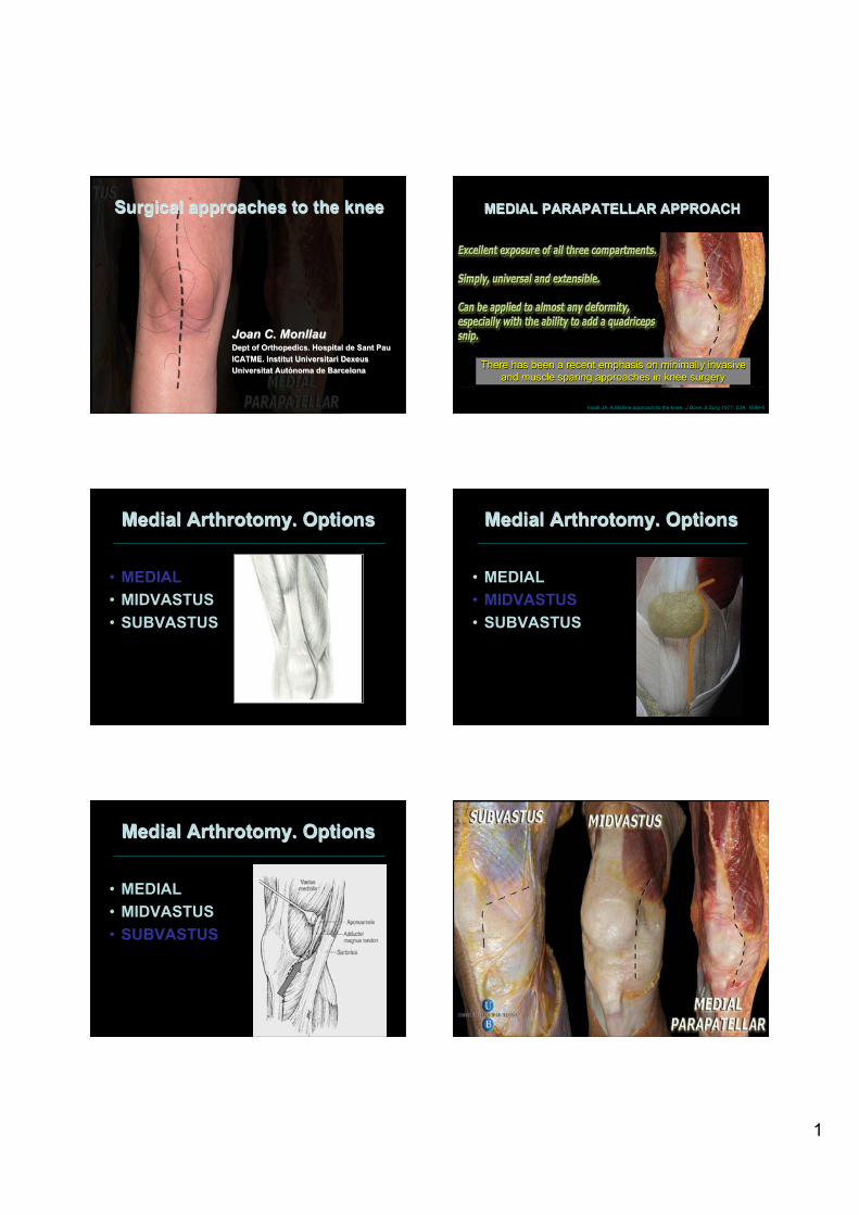
Surgical approaches to the knee
Joan C. Monllau Dept of Orthopedics. Hospital de Sant Pau ICATME. Institut Universitari Dexeus Universitat Autònoma de Barcelona
MEDIAL PARAPATELLAR APPROACH
Insall JA. A Midline approach to the knee. J Bone Jt Surg 1971; 53A: 1584-6
There has been a recent emphasis on minimally invasive and muscle sparing approaches in knee surgery
Medial Arthrotomy. Options
• MEDIAL • MIDVASTUS • SUBVASTUS
Medial Arthrotomy. Options
• MEDIAL • MIDVASTUS • SUBVASTUS
Medial Arthrotomy. Options
• MEDIAL • MIDVASTUS • SUBVASTUS

2
Anatomical Points of Interest
• Infrapatellar branch of the Saphenous nerve
MEDIAL SURGICAL APPROACHES Saphenous Nerve Infrapatellar Branch
TA - Adductor Tubercle LPFM - Medial Epicondyle GMM - Medial Gastrocnemius Tubercle
MEDIAL SURGICAL APPROACHES Bony Landmarks
MEDIAL SURGICAL APPROACHES Vastus Medialis ANATOMY
• Two portions Lieb FJ 1968
– Vastus Medialis Longus – Vastus Medialis Obliquus (VMO)
* VMO fibers runs in an oblique direction and so limits lateral patellar motion
MEDIAL SURGICAL APPROACHES Medial Retinaculum Medial PatelloFemoral Ligament (green arrow) Medial PatelloTibial Ligament (white arrows)
• Has been described as an alternative to traditional approaches which involve a large arthrotomy with partial division of the quadriceps mechanism.
• The most common application of this approach is in TKR, particularly when using a MIS.

3
Subvastus (Southern) approach for primary total knee arthroplasty Hofmann AA et al. Clin Orthop. 1991
Once the Medial retinaculum is transected, the extensor mechanism can be mobilized…
Release deep fibres of the MCL for better exposure
Remove part of the fat pad
Release synovial
SUBVASTUS vs MEDIAL PARAPATELLAR

4
MIDVASTUS
It runs between the fibers of VML and VMO Engh GA, Ammmeen DJ The midvastus approach to the knee. The Journal of Knee Surgery. 2003 Maestro A et al. The midvastus surgical approach in the knee arthroplasty. Int Orthop. 2000 Haas SB et al. Minimally Invasive Total Knee Replacement Through a Mini Midvastus approach. Clin Orthop. 2004
MIDVASTUS vs SUBVASTUS
Some surgeons believe that the subvastus approach completely avoids damage to the quadriceps mechanism and therefore would be associated with improved muscle function when compared with midvastus. In this prospective study no substantive differences have been found between the two approaches.
A comparison of subvastus and midvastus approaches in minimally invasive total knee arthroplasty. Bonutti PM et al.
J Bone Joint Surg Am. 2010
Mini Medial Approaches Advantages / disadvantages
• Subvastus approach
– Intact extensor mechanism – Decreasing pain – More limited (MIS) – Need for large medial and
lateral skin flaps – Postoperative hematoma
• Midvastus approach
– Preserve genicular art. blood supply to the patella
– Contraindicated in limited preoperative flexion
– Postoperative hematoma
Violation of the descending genicular artery
The safest distance for splitting the VM during medial parapatellar arthrotomy is 15 mm from the superior pole of the patella due to the course of the descending genicular artery.
• The incidence of patellar tendon avulsion has been reported to range from 0.17% to 1.4% in primary TKR
Rand JA et al. Clin Orthop 1989 Lynch AF et al. J Arthroplasty 1987
• Probably significantly higher in complicated cases, particularly in revision arthroplasty
Extensor Mechanism

5
Extended Approaches Objectives
• In some cases an extended approach might be needed
• Achieving wide exposure while protecting the extensor mechanism
• Proximal or distal
Extended Approaches Proximal exposures
• Coonse-Adams • Quad Snip • Quadplasty
Extended Approaches Proximal exposures
• Coonse-Adams • Quad Snip • Quadplasty
• Coonse-Adams • Quad Snip • Quadplasty
Extended Approaches Proximal exposures
- ↓ range of motion - delay starting resistance exercises - provokes an extensor lag
Proximal Exposures
Problems
• Tubercle Osteotomy
Extended Approaches Distal exposures
6
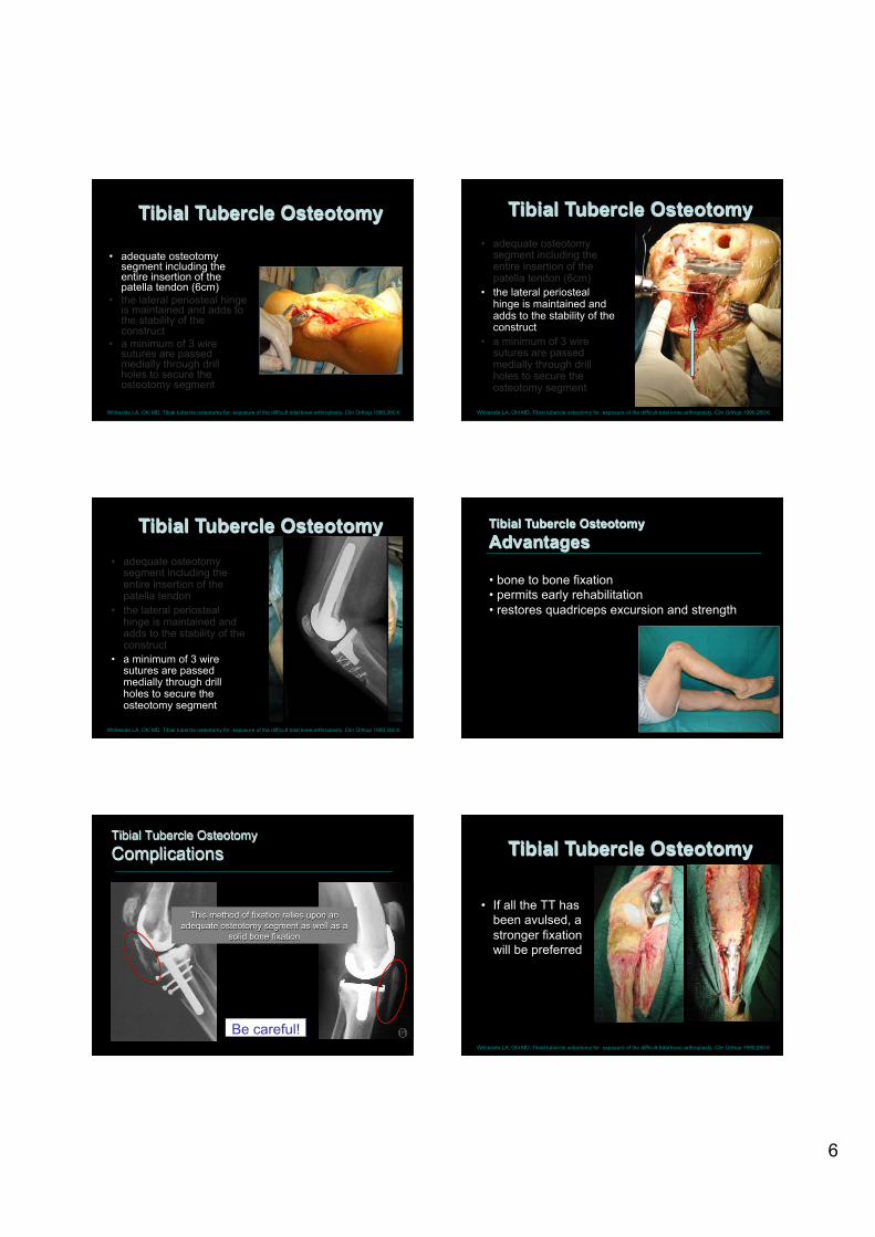
Surgical Technique Tibial Tubercle Osteotomy
• adequate osteotomy segment including the entire insertion of the patella tendon (6cm)
• the lateral periosteal hinge is maintained and adds to the stability of the construct
• a minimum of 3 wire sutures are passed medially through drill holes to secure the osteotomy segment
Whiteside LA, Ohl MD. Tibial tubercle osteotomy for exposure of the difficult total knee arthroplasty. Clin Orthop 1990;260:6
Surgical Technique Tibial Tubercle Osteotomy
Whiteside LA, Ohl MD. Tibial tubercle osteotomy for exposure of the difficult total knee arthroplasty. Clin Orthop 1990;260:6
• adequate osteotomy segment including the entire insertion of the patella tendon (6cm)
• the lateral periosteal hinge is maintained and adds to the stability of the construct
• a minimum of 3 wire sutures are passed medially through drill holes to secure the osteotomy segment
Tibial Tubercle Osteotomy
Whiteside LA, Ohl MD. Tibial tubercle osteotomy for exposure of the difficult total knee arthroplasty. Clin Orthop 1990;260:6
• adequate osteotomy segment including the entire insertion of the patella tendon
• the lateral periosteal hinge is maintained and adds to the stability of the construct
• a minimum of 3 wire sutures are passed medially through drill holes to secure the osteotomy segment
• bone to bone fixation • permits early rehabilitation • restores quadriceps excursion and strength
Tibial Tubercle Osteotomy Advantages
Tibial Tubercle Osteotomy Complications
Be careful!
This method of fixation relies upon an adequate osteotomy segment as well as a
solid bone fixation
Surgical Technique Tibial Tubercle Osteotomy
Whiteside LA, Ohl MD. Tibial tubercle osteotomy for exposure of the difficult total knee arthroplasty. Clin Orthop 1990;260:6
• If all the TT has been avulsed, a stronger fixation will be preferred

7
To sum up • Sub and Midvastus approaches provide
excellent exposure and allows quicker advancement in rehab after TKR
• Particularly useful when using MIS • TT osteotomy may be useful in revision
cases to avoid extending ext mechanism injuries
Both exposures have a learning curve
Thank You!!!