medial longitudinal arch of runners
TRANSCRIPT
CLINICAL SCIENCE
Rearfoot alignment and medial longitudinal archconfigurations of runners with symptoms andhistories of plantar fasciitisAna Paula Ribeiro,I Francis Trombini-Souza,I Vitor Tessutti,I Fernanda Rodrigues Lima,II Isabel de Camargo
Neves Sacco,I Sılvia Maria Amado JoaoI
IUniversity of Sao Paulo, Physical Therapy, Speech and Occupational Therapy Department, School of Medicine, Sao Paulo, Brazil. IIUniversity of Sao Paulo,
Rehabilitation in Sport Rheumatology Department, School of Medicine, Sao Paulo, Brazil.
OBJECTIVE : To evaluate and compare rearfoot alignment and medial longitudinal arch index during static posturesin runners, with and without symptoms and histories of plantar fasciitis (PF).
INTRODUCTION: PF is the third most common injury in runners but, so far, its etiology remains unclear. In theliterature, rearfoot misalignment and conformations of the longitudinal plantar arch have been described as riskfactors for the development of PF. However, in most of the investigated literature, the results are still controversial,mainly regarding athletic individuals and the effects of pain associated with these injuries.
METHODS: Forty-five runners with plantar fasciitis (30 symptomatic and 15 with previous histories of injuries) and 60controls were evaluated. Pain was assessed by a visual analogue scale. The assessment of rearfoot alignment and thecalculations of the arch index were performed by digital photographic images.
RESULTS: There were observed similarities between the three groups regarding the misalignments of the rearfootvalgus. The medial longitudinal arches were more elevated in the group with symptoms and histories of PF,compared to the control runners.
CONCLUSIONS: Runners with symptoms or histories of PF did not differ in rearfoot valgus misalignments, butshowed increases in the longitudinal plantar arch during bipedal static stance, regardless of the presence of painsymptoms.
KEYWORDS: Plantar fasciitis; Pain; Rearfoot; Plantar arch; Runners.
Ribeiro AP, Trombini-Souza F, Tessutti V, Lima FR, Sacco ICN, Joao SMA. Rearfoot alignment and medial longitudinal arch configurations of runnerswith symptoms and histories of plantar fasciitis. Clinics. 2011;66(6):1027-1033.
Received for publication on February 3, 2011; First review completed on February 28, 2011; Accepted for publication on March 12, 2011
E-mail: [email protected] / [email protected]
Tel.: 55 11 3091-8424
INTRODUCTION
At the end of the 20th century, the practice of runningincreased considerably 1,2 and, as a result, there wereincreases in the incidence of injuries in the lower limbs.3 Aretrospective study with 2002 runners, showed that plantarfasciitis (PF) was the third most common injury in runners4
for about 20% of the athletes.5 Tauton et al.4 reported thatout of 267 cases of investigated PF in various sportmodalities, 160 involved only running. This finding resultedin an increased interest in investigating causal factors of thisinjury.
PF is characterized by musculoskeletal disorders of theplantar fascia from inflammatory and degenerative origins,
such as in the medial tubercule of the calcaneus, the mostcommon clinical symptoms of which are typical pain of theinferior and medial calcaneal areas.6,7 According to Greveet al.,8 morning pain is an important evaluation criterion.There are several intrinsic and extrinsic factors related toPF.9 However, some specific intrinsic factors have beenexplored more thoroughly for the development of PF,among them obesity,10 decreases in the range of motion ofankle dorsiflexion,10–12 plantar longitudinal arch configura-tions,6,13–15 rearfoot pronations4,9,16 and increased plantarloads.17–19 However, some controversies regarding thesefactors still remain, mainly regarding their involvement inthe etiology of this injury.9
Some studies attributed the influences of plantar long-itudinal arch and rearfoot pronation to the development ofPF.6,14,20–22 The pioneering research of Hicks 23,24 demon-strated that the height and length of the plantar longitudinalarch could be implicated in the development of PF. Hicksstudied a model in different lower limbs of cadavers whichcharacterized the tension forces absorbed by the plantar
Copyright � 2011 CLINICS – This is an Open Access article distributed underthe terms of the Creative Commons Attribution Non-Commercial License (http://creativecommons.org/licenses/by-nc/3.0/) which permits unrestricted non-commercial use, distribution, and reproduction in any medium, provided theoriginal work is properly cited.
CLINICS 2011;66(6):1027-1033 DOI:10.1590/S1807-59322011000600018
1027

fascia and found important functions of the longitudinalarches for plantar load distributions. Most previous gaitstudies observed that the pain stimulus for the feet ofindividuals with PF promoted changes in foot roll-overpatterns, thus causing load reductions in the rearfoot andload increases in other plantar regions, such as the midfoot,possibly owing to the protective mechanisms of pain.19
However, during bipedal static support, studies whichevaluated athletes with PF still have contradictory resultsconcerning the types of the longitudinal arch and rearfootangles, especially when the pain stimulus is underconsideration.
Using subjective static evaluations, Tauton et al.4
observed that the plantar longitudinal arch was moreelevated in runners with PF and that 55% demonstratedexcessive rearfoot pronation; other studies also reportedsimilar findings.6,14,25 An elevated arch could induce greaterstiffness of the feet, which would result in an inefficientcapacity to dissipate foot impact forces with the ground and,thus, place the plantar fascia in a position of greatermechanical stress.14 Excessive pronation, for instance, couldlead to greater loads on the medial areas of the calcaneusand greater tensions in the plantar area;6,14,25 however, painwas not quantified in theses studies. On the other hand,studies which evaluated the plantar longitudinal arch,9,11
rearfoot alignment9 and calcaneal pain with long-distancerunners did not demonstrate significant differences regard-ing these factors when compared to controls.
In a study of non-athletes, Prichasuk and Sbhadra-bandhu26 observed that 82 individuals with symptomaticcalcaneal spurs had lower plantar arches, which wereconsidered important factors for the development of PF. Ina retrospective review of 1000 X-rays of subjects withcalcaneal pain, Shama et al.16 found that 81% demonstratedrearfoot pronation.
A more recent study by Pohl et al.,15 revealed that femalerunners with a history of PF had lower medial longitudinalarches and rearfoot valguses, similar to control runners.However, they pointed out the need for future studies toevaluate runners with pain symptoms, as their subjects hadover two years of diagnoses, which is a phase for possibleremissions of these symptoms. It is well known thatcomplaints of pain most often occur during the acute phase.According to Wearing et al.,19 symptomatic feet make someadaptations during gait to reduce the loads on the rearfoot.Karr27 also reported that the adaptations of the foot could bedemonstrated not only by the lowering of the arch, but alsoin its elevation, associated with the onset of micro-traumasof the plantar fascia.
Considering the controversial results regarding the effectsof the presence or absence of pain on the rearfoot andlongitudinal plantar arch configurations in runners with PF,the present study was conducted. The importance of theseresults relies on a better understanding of the rearfootangles and medial longitudinal arch indices during thestatic posture of runners with pain symptoms and historiesof PF. Another question which motivated the present studywas: would pain caused by PF induce postural adaptationsof the feet and ankle? The hypotheses were that theconditions of symptomatic PF would be associated withdecreases in rearfoot pronations and elevated longitudinalarches. It was expected that lower arches and excessiverearfoot pronations in runners with histories of PF,compared to control runners, would be found.
MATERIALS AND METHODS
ParticipantsOne hundred and five recreational runners of both
genders, ranging in age from 20 to 55 years,18 wereevaluated over the period of 1 year. For inclusion in thisstudy, the runners had to have been running at least 20 kmweekly for at least one year,28 had to be experienced inrunning long distance competitions, had to have regularrearfoot strike patterns, and had to have maximum leglength discrepancies of 1 cm. The exclusion criteria for bothgroups included histories of previous foot surgeries, traumaor fractures of less than three months previously, neuro-pathies, obesity and musculoskeletal disorders, such asarthritis, tendinitis, bursitis, ankylosing spondylitis and heelspurs.
The control runners were recruited after reading thestudy proposal published in electronic media. The PFrunners were recruited from the Rehabilitation Center inSport Rheumatology of the University Hospital in SaoPaulo, Brazil. Diagnoses were made by the same experi-enced physician, which were confirmed by ultra-soundimages taken by an experienced technician. All runnerssigned a term of informed consent approved by the LocalEthics Committee (protocol 1227/07).
Forty-five runners had diagnoses of unilateral PF con-firmed by ultrasonography to verify thickening of theplantar fascia, hypoechoic changes, perifascial fluid collec-tion and bony spurs.29 Thirty runners had symptoms of heelpain; the PF group with symptoms (PFS) of more than fourmonths’ duration was considered to be in the acute phase ofinjury, which was also clinically diagnosed by ultra-sound.They must have had pain from palpation of the plantarfascia, complaints of pain in the morning during standingand after assuming sitting and standing positions for longdurations.18,30 Fifteen runners had previous histories of PF(PFH group), with the time of the first diagnosis within amean (SD) of 1.5 (3.3) years. These runners were free frompain symptoms in the heel for more than two months andwere considered to be in the remission phase of the injury.
The control group (CG) was composed of 60 runners withno histories or symptoms of PF. The mean weekly trainingvolumes were 40 (12) km for the PFH group and 45 (10) kmfor the PFS group, whereas their mean time practice runningwas 7 (5) and 6 (5) years for the PFH and PFS, respectively.For the CG, the mean weekly training volume was 45(6) kmand the mean weekly time of training of 4 (3) years. Themean running speed reported by the subjects, regardingtheir last 10-km competition, was 11.7 (0.6) km/h.
Initial assessmentsAll runners were interviewed using a previously devel-
oped questionnaire to characterize their histories and clarifytheir exclusion criteria. This questionnaire was divided intofour items: personal data, anthropometric characteristics, PFdata and physical activity data for running.
Pain assessmentsThe level of pain in all subjects was assessed by a 10 cm
visual analogue scale (VAS). Before the plantar pressuremeasurements, the subjects rated the pain they felt at themoment of evaluation, ranging from none to unbearable.According to Jensen et al.,31 the scale is valid and reliableand has already been used in several PF studies.18,19
Rearfoot and plantar archRibeiro AP et al.
CLINICS 2011;66(6):1027-1033
1028
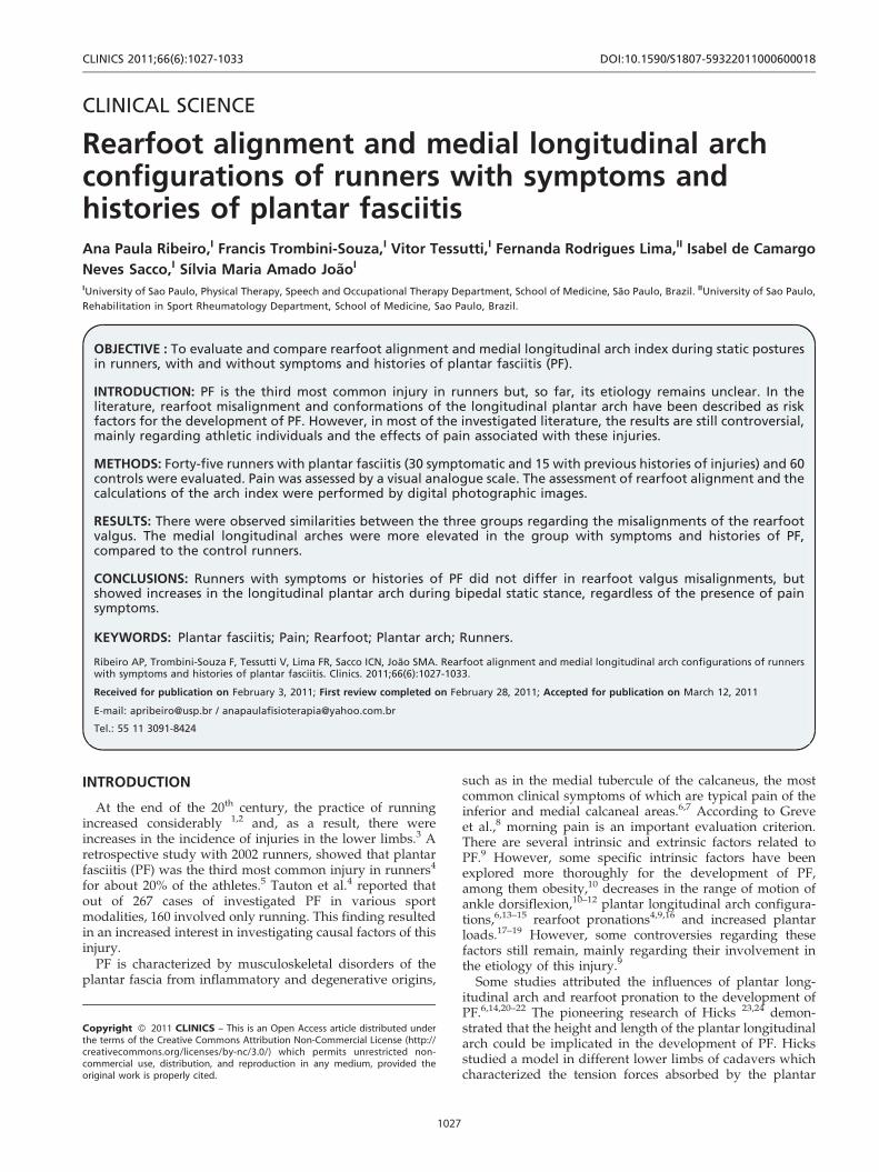
Rearfoot alignment assesmentsTo evaluate the alignment of the calcaneal tendons in the
posterior view of the frontal plane, the runners stood over a45 cm platform, keeping their feet 7.5 cm apart. With adematographic pen and white marks of 9 mm, the followinganatomical points were identified on the inferior andposterior aspects for both legs: 1) the posterior calcanealtuberosity; 2) the second point above the calcaneus; and, 3)the lower third of the leg.32,33 (Figure 1A). The center of eachmarker in the medial-lateral axis was obtained with a digitalcaliper, a metallic device with graduations in cm, used tomeasure the distances between the two symmetricallyopposing sides with a ruler. The images were then obtainedwith a digital camera positioned anterior and perpendicularto the subjects at a distance of 90 cm and at a height of45 cm.
AutoCAD 2005H software was used to quantify calcanealtendon alignments. To obtain these measures, a line wasfirst drawn from the first (3 cm) to the second (7 cm)markers. Then, another line was drawn from the highestmarker to the floor (22 cm), which passed through thecenter of the third marker (13 cm; Figure 1B).32,33 Theintersections of the extensions of these lines resulted inangles which were classified as normal (0 –5 ), varus (,0 )and valgus (.5 ) alignment values.34
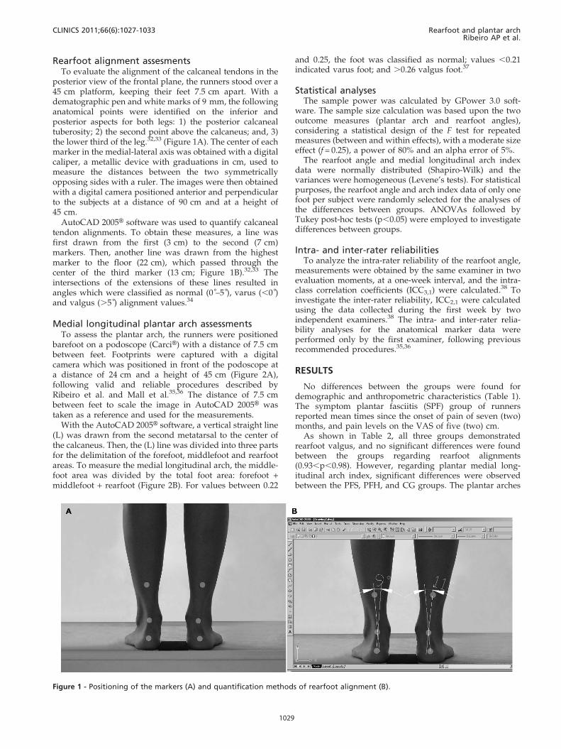
Medial longitudinal plantar arch assessmentsTo assess the plantar arch, the runners were positioned
barefoot on a podoscope (CarciH) with a distance of 7.5 cmbetween feet. Footprints were captured with a digitalcamera which was positioned in front of the podoscope ata distance of 24 cm and a height of 45 cm (Figure 2A),following valid and reliable procedures described byRibeiro et al. and Mall et al.35,36 The distance of 7.5 cmbetween feet to scale the image in AutoCAD 2005H wastaken as a reference and used for the measurements.
With the AutoCAD 2005H software, a vertical straight line(L) was drawn from the second metatarsal to the center ofthe calcaneus. Then, the (L) line was divided into three partsfor the delimitation of the forefoot, middlefoot and rearfootareas. To measure the medial longitudinal arch, the middle-foot area was divided by the total foot area: forefoot +middlefoot + rearfoot (Figure 2B). For values between 0.22
and 0.25, the foot was classified as normal; values ,0.21indicated varus foot; and .0.26 valgus foot.37
Statistical analysesThe sample power was calculated by GPower 3.0 soft-
ware. The sample size calculation was based upon the twooutcome measures (plantar arch and rearfoot angles),considering a statistical design of the F test for repeatedmeasures (between and within effects), with a moderate sizeeffect (f = 0.25), a power of 80% and an alpha error of 5%.
The rearfoot angle and medial longitudinal arch indexdata were normally distributed (Shapiro-Wilk) and thevariances were homogeneous (Levene’s tests). For statisticalpurposes, the rearfoot angle and arch index data of only onefoot per subject were randomly selected for the analyses ofthe differences between groups. ANOVAs followed byTukey post-hoc tests (p,0.05) were employed to investigatedifferences between groups.
Intra- and inter-rater reliabilitiesTo analyze the intra-rater reliability of the rearfoot angle,
measurements were obtained by the same examiner in twoevaluation moments, at a one-week interval, and the intra-class correlation coefficients (ICC3,1) were calculated.38 Toinvestigate the inter-rater reliability, ICC2,1 were calculatedusing the data collected during the first week by twoindependent examiners.38 The intra- and inter-rater relia-bility analyses for the anatomical marker data wereperformed only by the first examiner, following previousrecommended procedures.35,36
RESULTS
No differences between the groups were found fordemographic and anthropometric characteristics (Table 1).The symptom plantar fasciitis (SPF) group of runnersreported mean times since the onset of pain of seven (two)months, and pain levels on the VAS of five (two) cm.
As shown in Table 2, all three groups demonstratedrearfoot valgus, and no significant differences were foundbetween the groups regarding rearfoot alignments(0.93,p,0.98). However, regarding plantar medial long-itudinal arch index, significant differences were observedbetween the PFS, PFH, and CG groups. The plantar arches
Figure 1 - Positioning of the markers (A) and quantification methods of rearfoot alignment (B).
CLINICS 2011;66(6):1027-1033 Rearfoot and plantar archRibeiro AP et al.
1029
were more elevated in both groups with PF (PFS and PFH),compared to the controls (p = 0.008). The reliability analysesfor the rearfoot alignment angles for the PFS group resultedin ICC values of 0.96 and 0.98, respectively, for the intra-and inter-rater reliabilities.
DISCUSSION
The purpose of the present study was to evaluate andcompare rearfoot alignment and medial longitudinal archindices during static postures in runners with and withoutsymptoms and histories of PF. The results indicated that allgroups demonstrated similar rearfoot valgus misalign-ments. However, the plantar medial longitudinal archindices were higher for both the PF groups (PFS andPFH), compared to the controls, but no differences werefound between the PF groups.
The similarity of the valgus misalignments for runnerswith PFS and PFH corroborated the findings of Pohl et al.,15
who, besides doing static evaluations, also performeddynamic analyses during running. They did not find
differences regarding the valgus misalignments of thecalcaneus or the variables related to the peaks, times andmovement excursions of calcaneal eversion in a group offemale runners with PF histories. However, they empha-sized the importance of studies which investigated painsymptoms, which could have invariably affected the results.
In the present study, the aspects of pain wereconsidered and, as a result, it was observed that valgusmisalignments remained the same for the PF and controlgroups. It was expected that the symptom of pain wouldlead to less support on the medial heel as an analgesicstrategy during bipedal standing positions. Consequently,the reduction in weight bearing on this region of thecalcaneus would decrease the effects on the rearfootvalgus. However, similarly to controls, the presence ofpain in the symptomatic runner group was not enough toreduce natural support that caused this static valgusrearfoot behavior.
Rome et al.9 also reported similar findings whenevaluated runners with complaints of calcaneal painwere compared to control groups. They did not observe
Figure 2 - Positioning of the subject on the podoscope (A). Description of the calculation of the medial longitudinal arch index. L:vertical straight line, A: rearfoot area, B: midfoot area. C: forefoot area (B).
Table 1 - Descriptive statistics. Means (SD) of demographic and anthropometric data.
Variable Gender
PFS group
(n = 30; 11 F; 19 M)
PFH group (n = 15;
5 F; 10M)
Control group
(n = 60; 20 F; 40M) p-value
Age (years) Female
Male
44.0(9.1)
46.0(7.0)
34.0(4.0)
40.0(6.0)
38.0(8.0)
36.0(5.0)
0.188
0.200
Body mass (kg) Female
Male
57.8(9.5)
78.0(9.2)
62.0(9.5)
75.4(8.3)
55.8(7.0)
71.9(9.4)
0.579
0.236
Height (m) Female
Male
1.56(6.1)
1.75(4.6)
1.76(7.8)
1.66(5.6)
1.71(9.0)
1.79(5.6)
0.167
0.100
Body mass index (kg/
m2)
Female
Male
23.43(1.8)
25.5(2.3)
22.3(3.2)
23.3(1.8)
21.7(1.7)
23.5(2.6)
0.304
0.082
*ANOVAs two-way. p , 0,05 statistically significant.
PFS: plantar fasciitis with symptoms, F: female, M: male, PFH: previous history of plantar fasciitis.
Rearfoot and plantar archRibeiro AP et al.
CLINICS 2011;66(6):1027-1033
1030
differences in rearfoot alignments; however, a limitation ofthis study was that they did not assess the level andduration of pain associated with PF.
The innovative aspects of the present study were to betterunderstand how runners with pain levels of 5 (2) cm, over aduration of seven months, did not show differences in theirrearfoot alignments compared to runners with histories ofPF and controls. These findings suggest that valgusmisalignments of the rearfoot do not appear to be a riskfactor involved in the development of PF in runners. Incontrast, Tauton et al.4 reported excessive rearfoot pronationin 55% of athletes with PF. However, their assessments onlyincluded subjective clinical measures of alignment whichwere visually determined and were less valid and reliable.
Prichasuk and Sbhadrabandhu,26 employed quantitativeX-ray measures to investigate the calcaneal lateral tilt anglesin non-athletic subjects with symptomatic calcaneal spurs.They found that, compared to controls, the symptomaticgroup demonstrated decreases of tilt angles and their resultscorroborated those of Shama et al.16 One possible explana-tion for the differences between their results and those in thepresent study could be the sample selection, as only runnerswithout diagnoses of calcaneal spurs were evaluated. It ispossible that the presence of spurs in the chronic stages ofPF could result in excessive rearfoot valgus misalignmentsduring static postures.
One limitation of the present study was that dynamicanalyses of the rearfoot and midfoot were not carried out.Even though, Pohlet al.15 had already reported that rearfootmovements did not change in female runners with a historyof PF, the inclusion of dynamic measures would beimportant for the demonstration of movements of thesesegments with the presence of pain. Also, these authors didnot try to assess the relationships between midfoot prona-tion and PF, as previously reported by Chang et al.39 Theinclusion of these assessments could have explained thedynamic involvement of the rearfoot and midfoot duringconditions of pain in runners with PF. According to Riddleet al.,10 there are potential relationships between theincreases in pain and decreases in ankle range of motion,which are of extreme importance for the foot rockingmechanisms during running.
Cornwall and McPoil40 postulated that excessive prona-tion could occur as a result of structural changes in theplantar arches, or to compensatory mechanisms, whichcould result from decreases in dorsiflexion. However, as inthe diabetic neuropath, the literature is not yet clearregarding the relationship between the smaller ankle rangeof motion and the foot roll-over mechanism.41 This studyalso evaluated the behaviors of the plantar arch with the
presence of pain in runners with PF. According to Huangand Kitoaka,42 the crucial function of the plantar fascia is tomaintain the integrity of the plantar arch. Thus, theliterature hypothesizes that lowered plantar arches couldinduce greater stretching of the plantar fascia.6,15,21,43,44
In the present study, the initial hypothesis was that thepresence of pain would result in increases of the plantarmedial longitudinal arch index. However, it was observedthat plantar arch configurations appeared to be more relatedto the genesis of PF and not specifically to pain symptoms.Compared to the CG, arches were more elevated in thegroups with PF, both symptomatic and with histories. Apossible explanation for these findings could be the fact thatthe elevation of the plantar arch would lead to greaterstrains of the plantar fascia to maintain arch architectureduring static positions. The maintenance of this posture forlong periods could lead to micro-traumas of the plantarfascia and, consequently, to the onset of PF, which ischaracterized by periods of crises and remissions.45
It is well documented that individuals with PF havedecreases in their ankle dorsiflexion range of motion10,12
associated with less flexibility of the triceps surae musculargroup,12 and decreases in extension of the toes.46 Thesechanges could suggest that the plantar fascia would be keptin a more shortened position and, thus, would be exposed togreater tensions to be able to support the plantar arch, asfound in the present study. Based on this, the majority oftreatments for PF recommend the use of shoe wedges withthe objective of supporting the plantar medial longitudinalarch and to relax the plantar fascia, which could result inrelief of the pain symptoms.47–50
Another proposed intervention which reinforces theprevious idea, is the support of the plantar medial long-itudinal arch by means of functional bandages.51 Accordingto Wearing et al.19, the thickening of the plantar fascia andthe levels of pain associated with PF are factors which couldimpose stresses on the static structures of the plantar arch.However, the present findings demonstrate that thepresence of pain in the group with PF, did not promotechanges in the configuration of the plantar arch, whichsuggests that more elevated plantar arches could be betterrelated to the development of PF.
Messier and Pittala52 evaluated the plantar arch of 15runners with PF by means of plantar impressions and didnot find statistically significant differences, compared to theCG. However, they reported the tendency of more elevatedplantar arches in the PF group. In the present study, 45runners, 30 with symptoms and 15 histories of PF wereevaluated and the plantar impressions were obtained withpodometry, whose reliability35 and validity36 are well
Table 2 - Descriptive statistics [mean (SD)] for the measures of alignment of the rearfoot and plantar medial longitudinalarch index between groups of runners with symptoms and histories of plantar fasciitis.
Measure
1
PFS group (n = 30)
2
PFH group (n = 15)
3
Control group (n = 60) p-value*
Rearfoot angle 6.9(3.2) 6.7(4.2) 7.2(5.5) 0.971 (1-2)
0.982 (1-3)
0.931 (2-3)
Plantar medial longitudinal
arch index
0.17(0.08) 0.17(0.07) 0.22(0.05) 0.984 (1-2)
0.009 (1-3)
0.008 (2-3)
*ANOVAs two-way. Post-Hoc de Tukey. p , 0,05 diferenca estatıstica significante.
PFS: plantar fasciitis, PFH: previous history of plantar fasciitis.
CLINICS 2011;66(6):1027-1033 Rearfoot and plantar archRibeiro AP et al.
1031

established. With a larger sample size, the results demon-strated more elevated arches in both groups of runners withPF, as was reported by Messier and Pittala.52
According to Krivikas,14 a more elevated arch, besidespromoting greater stresses on the plantar fascia, whosefunction is to provide support to the plantar arch, wouldreduce the attenuation of the impact forces of the calcaneuson the ground. This mechanism would lead to greater loadson the medial and posterior areas of the feet. Taking intoconsideration the fact that running could cause highrepetitive impacts on the calcaneus and, that if theseimpacts were repeated about 625 times/km,53 elevatedplantar medial longitudinal arches would cause greatertensions and micro-traumas on the plantar fascia in runners.
According to Karr,27 both elevated and lowered archescould predispose individuals in the development of PF. Inthe present study, the runners with symptoms and historiesof PF demonstrated higher elevated configurations of theplantar medial longitudinal arch, compared to the controls.These findings are in disagreement with those reported byPohl et al.,15 who found lower plantar longitudinal arches infemale runners with PF, although they did not take painsymptoms into consideration. Two factors could explain thedifferences between these results. First, they only evaluatedrunners with histories of PF with a mean onset time of 2.5years. Second, they did not provide data regarding theapplied interventions. This could explain if the plantarmedial longitudinal arch was being re-structured and, thus,the differences between the present results, in which thesubjects in the PF groups received treatments over a shorterperiod of time (mean = 6 months).
Based on these findings, the relevance of this study wasthat it attempted to clarify that the presence of pain did notaffect rearfoot misalignments and plantar medial long-itudinal arch configurations of runners with PF. Wearinget al.19 observed that pain symptoms promoted adaptationsin the foot roll-over mechanisms during gait in individualswith PF. However, Ribeiro et al.54 observed that painsymptoms did not promote any adaptations in foot roll-overmechanisms during running in recreational runners withPF. In the present study, the evaluations were carried out inbipedal static support and did not find any effects of pain onplantar arch shapes or rearfoot alignments. It is important tonote that the elevated architecture of the plantar arch inrunners with PF could lead to greater strain on the plantarfascia during static and, mostly, dynamic activities, such asrunning, because of the repetitive foot impacts with theground during practice. Chronically, these stresses couldcause micro-traumas in the plantar fascia and probably leadto the progression of symptoms, or even to the onset of PF.
A limitation of this study was that dynamic analyses ofthe rearfoot and midfoot were not included; thus, futurestudies are necessary for a better understanding of PF inrunners. Regarding the plantar arch, studies evaluatinginterventions employing wedges, functional bandages andother physical therapy resources are necessary for a betterunderstanding of the mechanical effects on the plantarmedial longitudinal arch configurations in individuals withPF.
CONCLUSIONS
Runners with symptoms or histories of PF did not differin their rearfoot valgus alignment but showed increases in
longitudinal medial plantar arches during bipedal staticsupport, regardless of the presence of pain.
ACKNOWLEDGEMENTS
The authors are thankful to all the runners who participated in the study
and also acknowledge the financial support of Brazilian Government
Funding Agency CAPES (Coordenacao de Aperfeicoamento de Pessoal de
Ensino Superior).
REFERENCES
1. De Wit B, De Clercq D’ Aerts P. Biomechanical analysis of the stancephase during barefoot and shoed running. J Biomech. 2000;33: 269-78,doi: 10.1016/S0021-9290(99)00192-X.
2. Tillman M, Fiolkowski P, Bauer J. In-shoe plantar measurements duringrunning on different surfaces: Changes in temporal and kineticparameters. Sports Engineering. 2002;5:121-8, doi: 10.1046/j.1460-2687.2002.00101.x.
3. van Gent RN, Siem D, van-Middelkoop M, van-Os AG, Bierma-ZeinstraSMA, Koes BW. Incidence and determinants of lower extremity runninginjuries in long distance runners: A systematic review. Br J Sports Med.2007; 41:469-480, doi: 10.1136/bjsm.2006.033548.
4. Taunton JE, Ryan MB, Clement DB, McKenzie DC, Lloyd-Smith DR.Plantar fasciitis: A retrospective analysis of 267 cases. Phys Ther Sports.2002;3:57-65, doi: 10.1054/ptsp.2001.0082.
5. Clement DB, Taunton JE, Smart GW, McNicol KL. A survey of overuserunning injuries. Phys Sports Med. 1981;9:47-58.
6. Kwong PK, Kay D, Voner RT, White MW. Plantar fasciitis: Mechanicsand pathomechanics of treatment. Clin Sports Med. 1987;7:119-26.
7. Greve JM, Grecco MV, Santos-Silva PR. Comparison of radial shock-waves and conventional physiotherapy for treating plantar fasciitis.Clinics. 2009;64:97-103, doi: 10.1590/S1807-59322009000200006.
8. Tisdel CL, Donley BG, Sferra JJ. Diagnosing and treating plantar fasciitis:A conservative approach to plantar heel pain. Cleve Clin J Med.1999;66:231-5.
9. Rome K, Howe T, Haslock I. Risk factors associated with thedevelopment of plantar heel pain in athletes. The Foot. 2001;11: 119-25,doi: 10.1054/foot.2001.0698.
10. Riddle DL, Pulisic M, Pidcoe P, Johnson RE. Risk factors for Plantarfasciitis: A matched case-control study. J Bone Joint Surg Am. 2003; 85-A:872-7.
11. Warren BL. Anatomical factors associated with predicting plantarfasciitis in long-distance runners. Med Sci Sports Exerc. 1984;16: 60-3.
12. Kibler WB, Goldberg C, Chandler TJ. Functional biomechanical deficitsin running athletes with plantar fasciitis. Am J Sports Med. 1991;19:66-71,doi: 10.1177/036354659101900111.
13. Warren BL, Jones CJ. Predicting plantar fasciitis in runners. Med SciSports Exerc. 1987;19:71-3.
14. Krivickas L. Anatomical factors associated with overuse sports injuries.Sports Med. 1997;24:132-46, doi: 10.2165/00007256-199724020-00005.
15. Pohl M, Hamil J, Davis I. Biomechanical and anatomic factors associatedwith a history of plantar fasciitis in female runners. Clin J Sport Med.2009;19:372-6, doi: 10.1097/JSM.0b013e3181b8c270.
16. Shama SS, Kominsky SJ, Lemont H. Prevalence of non-painful heel spursand its relation to postural foot position. J Am Podiatry Assoc.1983;73:122-3.
17. Bedi HS, Love BR. Differences in impulse distribution in patients withplantar fasciitis. Foot Ankle Int. 1998;19:153-6.
18. Wearing SC, Smeathers JE, Urry SR. The effect of plantar fasciitis onvertical foot-ground reaction force. Clin Orthop Relat Res. 2003;175-85,doi: 10.1097/01.blo.0000057989.41099.d8.
19. Wearing SC, Smeathers JE, Sullivan PM, Yates B, Urry SR, Dubois P.Plantar fasciitis: Are pain and fascial thickness associated with archshape and loading? Phys Ther. 2007;87:1002-8, doi: 10.2522/ptj.20060136.
20. Taunton JE, Ryan MB, Clement DB, McKenzie DC, Lloyd-Smith DR,Zumbo BD. A retrospective case-control analysis of 2002 runninginjuries. Br J Sports Med. 2002b36:95-101, doi: 10.1136/bjsm.36.2.95.
21. Viel E, Esnault M. The effect of increased tension in the plantar fascia: Abiomechanical analysis. Phys Theory Pract. 1989;5:69-73.
22. Huang YC, Wang LY, Wang HC. The relationship between the flexibleflatfoot and plantar fasciitis ultrasonographic evaluation. Chang GungMed J. 2004;27:443-8.
23. Hicks J. The foot as a suppor. Acta Anat. 1955;25:34-45, doi: 10.1159/000141055.
24. Hicks JH. The mechanics of the foot. II: The plantar aponeurosis and thearch. J Anat. 1954;88:25-30.
25. Chandler TJ, Kibler WB. A biomechanical approach to the prevention,treatment and rehabilitation of plantar fasciitis. Sports Med. 1993;15:344-52, doi: 10.2165/00007256-199315050-00006.
Rearfoot and plantar archRibeiro AP et al.
CLINICS 2011;66(6):1027-1033
1032
26. Prichasuk S, Subhadrabandhu T. The relationship of pes planus andcalcaneal spurs to plantar heel pain. Clin Orthop Relat Res. 1994;306:192-6.
27. Karr SD. Subcalcaneal heel pain. Orthop Clin North Am. 1994; 25:161-75.28. Tessutti V, Trombini-Souza F, Ribeiro AP, Nunes AL, Sacco ICN. In-shoe
plantar pressure distribution during running on natural grass andasphalt in recreational runners. J Sci Med Sport. 2010;13:151-5, doi: 10.1016/j.jsams.2008.07.008.
29. Karabay N, Toros T, Hurel C. Ultrasonographic evaluation in plantarfasciitis. J Foot and Ankle Surgery. 2007;46:442-6, doi: 10.1053/j.jfas.2007.08.006.
30. Liddle D, Rome K, Howe T. Vertical ground reaction forces in patientswith unilateral plantar heel pain: A pilot study. Gait Posture. 2000;11: 62-6, doi: 10.1016/S0966-6362(99)00053-3.
31. Jensen MP, Karoly P, Braver S. The measurement of clinical painintensity: A comparison of six methods. Pain 1986;27:117-26.
32. Cornwall MW, McPoil TG. Influence of rearfoot postural alignment onrearfoot motion during walking. The Foot. 2004;14:133-3, doi: 10.1016/j.foot.2004.02.003.
33. McPoil TG, Cornwall MW. Relationships between three static angles ofthe rearfoot and the pattern of rearfoot motion during walking. J OrthopSports Phys Ther. 1996;23:370-5.
34. Eng JJ, Pierrynowski MR. The effect of soft foot orthotics on three-dimensional lower-limb kinematics during walking and running. PhysTher. 1994;74:836-44.
35. Ribeiro AP, Tombini-Souza F, Iunes DH, Monte Raso VV. Inter- andintra-examiner reliability of photopodometry and intra-examiner relia-bility of photopodoscopy. Rev Bras Fisioter. 2006;10:435-9, doi: 10.1590/S1413-35552006000400012.
36. Mall N, Mack Hardaker W, Nunely J, Queen R. The reliability andreproducibility of foot type measurements using a mirrored foot photobox and digital photography compared to caliper measurements.J Biomech. 2007;40:1171-6, doi: 10.1016/j.jbiomech.2006.04.021.
37. Cavanagh P, Rodgers M. The arch index: A useful measure fromfootprints. J Biomech. 1987;20:547-51, doi: 10.1016/0021-9290(87)90255-7.
38. Weir JP. Quantifying test-retest reliability using the intraclass correlationcoefficient and the SEM. Strength Cond J. 2005;19:231-40.
39. Chang R, van Emmerick R, Hamil J. Kinematics and anti-phasecoordination of the rearfoot-forefoot couple in chronic plantar fasciitis.In: Proceedings of the 10th Annual International Conference onFoot Biomechanics and Orthotic Therapy. November. 2007. San Diego,CA;16-8.
40. Cornwall MW, McPoil TG. Plantar fasciitis: Etiology and treatment.J Orthop Sports Phys Ther. 1999;29:756-60.
41. Bacarin TA, Sacco IC, Hennig EM. Plantar pressure distribution patternsduring gait in diabetic neuropathy patients with a history of foot ulcers.Clinics. 2009;64:113-20, doi: 10.1590/S1807-59322009000200008.
42. Huang CK, Kitoaka HB, An KN. Biomechanical stability of the arch. FootAnkle. 1993;14:353-7.
43. Kosmahl EM, Kosmahl HE. Painful plantar heel, plantar fasciitis, andcalcaneal spurs: Etiology and treatment. J Orthop Sports Phys Ther.1987;9:17-24.
44. Huang YC, Wang LY, Wang HC. The relationship between the flexibleflatfoot and plantar fasciitis ultrasonographic evaluotion. Chang GungMed J. 2004;27:443-8.
45. Imamura M, Imamura S, Carvalho AE, Mazagao RA, Cassius DA,Fischer AA. Plantar fasciitis: A new treatment approach. Arch Phys MedRehabil. 2003;84: E4, doi: 10.1016/S0003-9993(03)00526-4.
46. Allen RH, Gross MT. Toe flexors strength and passive extension range ofmotion of the first metatarsophalangeal joint in individuals with plantarfasciitis. J Orthop Sports Phys Ther. 2003;33:468-78.
47. Kogler GF, Solomonidis SE, Paul JP. Biomechanics of longitudinal archsupport mechanisms in foot orthoses and their effect on plantaraponeurosis strain. Clin Biomech. 1996;11:243-52, doi: 10.1016/0268-0033(96)00019-8.
48. Landorf KB, Keenan AM, Herbert RD. Effectiveness of different types offoot orthoses for the treatment of plantar fasciitis. J Am Podiatr MedAssoc. 2004;94:542-9.
49. Pfeffer G, Bacchetti P, Deland J. Comparison of custom and prefabricatedorthoses in the initial treatment of proximal plantar fasciitis. Foot AnkleInt. 1999;20:214-21.
50. Jamali B, Walker M, Hoke B, Echternach J. Windlass taping technique forsynptomatic relief of plantar fasciitis J Sport Rehabil. 2004;13:228-43.
51. Saxelby J, Betts RF, Bygrave CJ. Low-Dye’ taping on the foot in themanagement of plantar-fasciitis. The Foot. 1997;7:205-9, doi: 10.1016/S0958-2592(97)90037-7.
52. Messier SP, Pittala KA. Etiologic factors associated with selected runninginjuries. Med Sci Sports Exerc. 1988;20:501-5.
53. Frederick EC. Biomechanical consequences of sport shoe design. ExercSport Sci Rev. 1986;14:375-400, doi: 10.1249/00003677-198600140-00016.
54. Ribeiro AP, Trombini-Souza F, Tessutti VD, Lima FR, Joao SMA, SaccoICN. The effects of plantar fasciitis and pain on plantar pressuredistribution of recreational runners. Clin Biomech. 2011; 26:194-9, doi: 10.1016/j.clinbiomech.2010.08.004.
CLINICS 2011;66(6):1027-1033 Rearfoot and plantar archRibeiro AP et al.
1033