mediastinal mass
DESCRIPTION
mediastinal massTRANSCRIPT

Tumors of the Mediastinum*
Beau V. Duwe, MD; Daniel H. Sterman, MD. FCCP; andAli I. Musani, MD, FCCP
Tumors of the mediastinum represent a wide diversity of disease states. The location andcomposition of a mass is critical to narrowing the differential diagnosis. The most common causesof an anterior mediastinal mass include the following: thymoma; teratoma; thyroid disease; andlymphoma. Masses of the middle mediastinum are typically congenital cysts, including foregutand pericardial cysts, while those that arise in the posterior mediastinum are often neurogenictumors. The clinical sequelae of mediastinal masses can range from being asymptomatic to producingsymptoms of cough, chest pain, and dyspnea. This article will review the anatomy of the mediastinumas well as the different clinical, radiographic, and prognostic features, and therapeutic options of themost commonly encountered masses. (CHEST 2005; 128:2893–2909)
Key words: bronchogenic; cysts; enterogenous; germ cell tumor; goiter; lymphoma; mediastinum; neuroblastoma; neurogenictumor; pericardial; teratoma; thyroid; thymoma
Abbreviations: AFP � �-fetal protein; ALL � acute lymphoblastic leukemia; BMT � bone marrow transplant;GCT � germ cell tumor; hCG � human chorionic gonadotropin; HD � Hodgkin disease
T he mediastinum is demarcated by the pleuralcavities laterally, the thoracic inlet superiorly,
and the diaphragm inferiorly. It is further compart-mentalized into anterior, middle, and posterior divi-sions based on structural landmarks seen on thelateral radiograph. This has important implicationsfor diagnosing suspected masses1 (Table 1). Theanterior mediastinum contains the thymus, fat, andlymph nodes. The middle mediastinum contains theheart, pericardium, ascending and transverse aorta,brachiocephalic veins, trachea, bronchi, and lymphnodes, while the posterior mediastinum consists ofthe descending thoracic aorta, esophagus, azygousvein, autonomic ganglia and nerves, thoracic lymphnodes, and fat.
The likelihood of malignancy is influenced primar-ily by the following three factors: mass location;patient age; and the presence or absence of symp-toms. Although more than two thirds of mediastinaltumors are benign, masses in the anterior compart-
ment are more likely to be malignant.2 In the studyby Davis et al3 of 400 patients with mediastinalmasses, malignancy was seen in 59%, 29%, and 16%,respectively, of anterior, middle, and posterior me-diastinal masses. Age is an important predictor ofmalignancy as well with many of the lymphomas andgerm cell tumors (GCTs) presenting between thesecond and fourth decade of life. Last, symptomaticpatients are more likely to have a malignancy. InDavis et al,3 85% of patients with a malignancy weresymptomatic at presentation, compared to 46% ofpatients with benign neoplasms.
The most common symptoms at presentation wereas follows: cough (60%); chest pain (30%); fevers/chills (20%); and dyspnea (16%). Most symptomscan be categorized into the following two groups:localizing symptoms (Table 2); and systemic symp-toms (Table 3). Localizing symptoms are secondaryto tumor invasion. Common localizing symptomsinclude respiratory compromise; dysphagia; paralysisof the limbs, diaphragm, and vocal cords; Hornersyndrome; and superior vena cava syndrome.4 Sys-temic symptoms are typically due to the release ofexcess hormones, antibodies, or cytokines. A classicexample is hypercalcemia, which is caused by aparathyroid adenoma.
The initial workup of a suspected mediastinal massinvolves obtaining posteroanterior and lateral chestradiographs. This can provide information pertainingto the size, anatomic location, density, and composi-tion of the mass (Table 1). CT scanning is used tofurther characterize mediastinal masses and their
*From the Departments of Internal Medicine (Dr. Duwe) andPulmonary, Allergy, and Critical Care Medicine (Drs. Stermanand Musani), Hospital of the University of Pennsylvania, Phila-delphia, PA.Manuscript received December 6, 2004; revision accepted April1, 2005.Reproduction of this article is prohibited without written permissionfrom the American College of Chest Physicians (www.chestjournal.org/misc/reprints.shtml).Correspondence to: Ali I. Musani, MD, Assistant Professor,Pulmonary, Allergy and Critical Care Medicine, Associate Direc-tor, Interventional Pulmonology Program, Hospital of theUniversity of Pennsylvania, Philadelphia, PA 19104; e-mail:[email protected]
www.chestjournal.org CHEST / 128 / 4 / OCTOBER, 2005 2893
Downloaded From: http://journal.publications.chestnet.org/ on 03/03/2014

relationship to surrounding structures as well as toidentify cystic, vascular, and soft-tissue structures.4In rare circumstances, fluoroscopy, barium swallow,angiograph, CT angiography, and three-dimensionalreconstruction may provide additional information.The role of MRI is primarily in ruling out orevaluating a neurogenic tumor.5 MRI is also valuableto evaluate the extent of vascular invasion or cardiacinvolvement.
Although nuclear scans and biochemical studiescan be used to further characterize a lesion, tissuediagnosis is almost always required. If a mass is likelyto be benign after initial workup, it can be removedsurgically without biopsy. Otherwise, a diagnosticbiopsy specimen can be obtained by transthoracic ortransbronchial needle aspiration, mediastinoscopy,anterior mediastinotomy, or video-assisted thoracicsurgery, depending on the anatomic location andradiographic appearance of the lesion.
Tumors of the Anterior Mediastinum
Thymoma
Thymomas are the most common neoplasm of theanterior mediastinum with an incidence of 0.15 casesper 100,000.6–9 Although rare in children, thymomasrepresent 20% of anterior mediastinal neoplasms inadults.10,11
Thymomas as a group have a wide spectrum of histo-logic diversity and are classified based on cell type pre-dominance as lymphocytic, epithelial, or spindle cell vari-ants. There is a strong association between histologicsubtype and invasiveness as well as prognosis.12–14 As aresult, the World Health Organization15 devised a newclassification system to group thymomas based on cyto-logic differences, which may be helpful in determiningtreatment regimens and predicting survival (Table 4).
Most thymomas are solid tumors, but up to onethird may have components that are necrotic, hem-orrhagic, or cystic.7,16 Thirty-four percent of thymo-mas invade through their own capsules, extendinginto surrounding structures.8,17–20 Likewise, trans-diaphragmatic extension into the abdomen and me-tastasis into the ipsilateral pleura and pericardium
Table 1—Differential Diagnosis of a Mediastinal Massby Anatomic Location*
Anterior Middle Posterior
Thymoma Lymphoma Neurogenic tumorTeratoma, seminoma Pericardial cyst Bronchogenic cystLymphoma Bronchogenic cyst Enteric cystCarcinoma Metastatic cyst XanthogranulomaParathyroid adenoma Systemic granuloma Diaphragmatic herniaIntrathoracic goiter MeningoceleLipoma Paravertebral abscessLymphangiomaAortic aneurysm
*From Baum and Crapo.122
Table 2—Localizing Symptoms Secondary to TumorInvasion of Surrounding Structures*
Involved Anatomic Structure Localizing Symptom
Bronchi/trachea Dsypnea, postobstructive pneumonia,atelectasis, hemoptysis
Esophagus DysphagiaSpinal cord/vertebral column ParalysisRecurrent laryngeal nerve Hoarseness, vocal cord paralysisPhrenic nerve Diaphragmatic paralysisStellate ganglion Horner syndromeSuperior vena cava Superior vena cava syndrome
*From Baum and Crapo.122
Table 3—Systemic Syndromes Secondary to PrimaryMediastinal Tumors and Cysts*
Syndrome Tumor
Myasthenia gravis, RBC aplasia,hypogammaglobulinemia,Good syndrome, Whippledisease, megaesophagus,myocarditis
Thymoma
Multiple endocrineadenomatosis, Cushingsyndrome
Carcinoid, thymoma
Hypertension Pheochromocytoma,ganglioneuroma,chemodectoma
Diarrhea GanglioneuromaHypercalcemia Parathyroid adenoma,
lymphomaThyrotoxicosis Intrathoracic goiterHypoglycemia Mesothelioma, teratoma,
fibrosarcoma, neurosarcomaOsteoarthropathy Neurofibroma, neurilemoma,
mesotheiomaVertebral abnormalities Enteric cystsFever of unknown origin LymphomaAlcohol-induced pain HDOpsomyoclonus Neuroblastoma
*From Baum and Crapo.122
Table 4—World Health Organization Classification ofThymomas*
Class ofThymoma Cytologic Features
Type A Spindle cell, medullaryType AB MixedType B1 Lymphocyte rich, lymphocytic, predominantly
cortical, organoidType B2 CorticalType B3 Epithelial, atypical, squamous, well-differentiated
thymic carcinoma
From Wilkins et al.15
2894 Reviews
Downloaded From: http://journal.publications.chestnet.org/ on 03/03/2014

can occur,7,9,18 although lymphogenous and hema-togenous spread is rare.16,17
The Masaoka clinical staging system is based onthe degree of invasion of the tumor through thecapsule into the surrounding structures, which hasimportant implications for prognosis21 (Table 5). Inthe study by Okumura et al,12 the Masaoka stagingsystem was shown to be useful as an independentpredictor of survival in patients with thymoma.
Typically, a thymoma is an incidental finding on achest radiograph.10,22,23 One third of patients mani-fest symptoms of chest pain, cough, or dyspnearelated to tumor compression or invasion.16 Metas-tasis is uncommon; however, parathymic syndromes,which include myasthenia gravis, hypogammaglobu-linemia, and pure RBC aplasia, may develop.17
Myasthenia gravis is most frequent in women andis associated with thymoma. Symptoms include dip-loplia, ptosis, dysphagia, weakness, and fatigue.Thirty percent to 50% of patients with thymomashave myasthenia gravis, compared to 10 to 15% ofpatients with myasthenia gravis who have a thymo-ma.24,25 Pathogenesis is thought to occur via myloidcell lineages derived from the thymus that recognizeantigens on the neuromuscular junction producingautoantibodies.26 These autoantibodies bind to ace-tylcholine receptors of the neuromuscular junction,causing muscle fatigue.26 Thymectomy can alleviatesymptoms; however, this benefit is often delayed formonths after surgery. Given the association betweenthymoma and myasthenia gravis, the serum anti-acetylcholine receptor antibody level should be mea-sured in all patients with a suspected thymoma torule out myasthenia gravis before surgery.27,28
Hypogammaglobulinemia and pure RBC aplasiaare present in 10% and 5% of patients with athymoma, respectively.7 Good syndrome is diag-nosed in patients with a thymoma and combinedB-cell/T-cell immunodeficiency.29 Thymoma is alsoassociated with various other autoimmune disorders,such as systemic lupus erythematosus, polymyositis,and myocarditis.3,7,18,30
Thymomas appear on a chest radiograph as awell-defined lobulated mass in the anterosuperiormediastinum, typically anterior to the aortic root.7,18
Further evaluation with contrast-enhanced thoracicCT scanning usually reveals an encapsulated, well-defined, soft-tissue mass, often with hemorrhage,necrosis, or cyst formation31 (Fig 1). They can alsoappear predominantly cystic with a nodular compo-nent.32
Surgical excision can be used for diagnosis; how-ever, the sensitivity of ultrasonography and CT scan-guided fine-needle aspiration is increasing. Andersonand colleagues33 reported a success rate of 95%using ultrasonographically guided fine-needle aspira-tion. The success of fine-needle aspiration is opera-tor-dependent and contingent on the skill of theimmunohistologist. Thus, the results in the study byAnderson et al33 may overstate the true success ofpreoperative diagnosis. Tissue diagnosis may occursimultaneously with total resection of the mass if athymoma is strongly suspected on the basis of clinicaland radiologic evidence.34
Surgical resection remains the standard of care forboth noninvasive and invasive thymomas as it pro-vides the best prognosis. Adjunctive chemotherapyand radiation treatment is used for locally invasive ormetastatic disease, or inoperable tumors. Addition-ally, although it is commonly accepted that resectionalone is sufficient treatment for stage I disease, thereis no consensus regarding the role for postoperativeradiation therapy in patients with stage II disease.35
According to Curran et al,36 of 117 patients,postoperative radiotherapy showed no survival ben-efit for those patients with stage I disease but did forpatients with stage II and III disease. The 5-yearmediastinal relapse rate for patients with stage II orIII disease treated with surgery alone was 53%, whilepatients who received treatment with total resectionand radiotherapy experienced no relapses. A smallerretrospective study by Eralp et al37 of 36 patientswith stage II or III disease also showed a benefit forpostoperative radiation therapy. While these studieshad positive results, other institutional reviews35,38
have shown no benefit to postoperative radiotherapy.A larger randomized controlled trial would be usefulto assess the benefit of postoperative radiation ther-apy in patients with stage II thymomas.
Thymoma is generally responsive to chemotherapy
Table 5—Masaoka Staging System of Thymoma*
Stage Degree of Invasion 5-yr Survival Rate, %
1 Complete encapsulation macroscopically and no capsular invasion microscopically 96–1002 Invasion into the surrounding fatty tissue or mediastinal pleura macroscopically or
invasion into the capsule microscopically86–95
3 Invasion into neighboring organs macroscopically 56–694a Pleural or pericardial dissemination 11–504b Lymphogenous or hematogenous metastasis
*From Shamji et al.21
www.chestjournal.org CHEST / 128 / 4 / OCTOBER, 2005 2895
Downloaded From: http://journal.publications.chestnet.org/ on 03/03/2014

as well. In locally invasive or bulky disease, preoper-ative cisplatin-based chemotherapy, with or withoutpostoperative radiotherapy, may offer the best prog-nosis.39 Kim et al40 examined 23 patients with locallyadvanced, unresectable disease who underwentthree courses of induction chemotherapy with cis-platin, doxorubicin, cyclophosphamide, and pred-nisone. The 7-year disease-free and overall survivalrates were 77% and 79%, respectively.40
Other chemotherapeutic agents and regimens areless efficacious. Thus, these alternative regimensshould only be used in patients who cannot toleratecisplatin and doxorubicin or as second-line therapy inthose who have relapsed.41
The following features are associated with poorprognosis: metastasis; large tumor size (ie, � 10 cm);tracheal or vascular compression; age � 30 years;epithelial or mixed histology; and the presence of ahematologic paraneoplastic syndrome.42 As theabove prognostic factors suggest, both histologicsubtype and disease stage appear to be important inpredicting survival. Currently, stage is used princi-pally to guide treatment; however, controversy re-
garding the use of chemotherapy and radiation ther-apy in patients with different stages of thymoma mayreflect that histologic subtype ought to play animportant role in determining which treatment mo-dalities are most appropriate. The creation of theWorld Health Organization classification system forthymoma in 1999 has given further insight into thepotential importance of histologic subtype on prog-nosis; however, the Masaoka staging system is stillcurrently used to stratify 5-year survival rates (Table 5).
Thymic Carcinoma
Thymic carcinomas are a heterogeneous group ofaggressive, invasive epithelial malignancies.3 Theirincidence is rare, occurring predominantly in mid-dle-aged men. Most patients present with cough,shortness of breath, and chest pain.43 Fatigue, weightloss, and anorexia are common, while superior venacava syndrome and cardiac tamponade have beendescribed.44–46
Histologically, thymic carcinomas are large, firm,infiltrating masses with areas of cystic change andnecrosis. They are classified as low grade or highgrade, with squamous cell-like and lymphoepitheli-oma-like variants being the most common celltypes.47 In contrast to thymomas, thymic carcinomasare cytologically malignant, with typical features ofcellular necrosis, atypia, and mitoses.44 Radiograph-ically, thymic carcinomas are heterogeneous withnecrosis and calcifications (Fig 2) and can be associ-ated with pleural and pericardial effusions.
Treatment and prognosis depend on the cancerstage and grade. The Masaoka staging system usedfor thymomas is not useful as a prognostic tool inthymic carcinoma.48 Morphologic features that por-tend a poor prognosis include the following: infiltra-tion of the tumor margin; absence of a lobulargrowth pattern; presence of high-grade atypia andnecrosis; and � 10 mitoses per high-power field.21
Complete surgical resection is the treatment ofchoice and can be curative.49 Chemotherapy andradiation therapy have roles in treating unresectabletumors.36,40,50
Yoh et al51 examined 18 patients with thymiccarcinomas. Patients with unresectable disease weretreated with cisplatin, vincristine, doxorubicin, andetoposide. The overall response rate was 42% with1-year and 2-year survival rates of 80% and 56%,respectively.51 Superior to previous chemotherapeu-tic regimens, the regimen of cisplatin, vincristine,doxorubicin, and etoposide warrants additional studyby a randomized controlled trial for its use in thetreatment of thymic carcinoma.
Figure 1. A 36-year-old man with an invasive thymoma. Acontrast-enhanced CT scan shows a heterogenous high-attenu-ated solid upper portion (arrow) with a small calcification in theleft anterior aspect of the main pulmonary artery.
2896 Reviews
Downloaded From: http://journal.publications.chestnet.org/ on 03/03/2014

Thymic Carcinoid
Thymic carcinoid is a malignant tumor, which ishistologically similar to carcinoid tumors found atother sites. Its highest incidence is in the fourth andfifth decades of life.44 Thymic carcinoid is associatedwith Cushing syndrome and multiple endocrine neo-plasia syndrome.7 According to a prospective studyof patients with endocrine neoplasia syndrome type 1by Gibil et al,52 thymic carcinoid developed in 8% ofpatients.
Thymic carcinoid presents as a large, lobulated,invasive mass of the anterior mediastinum with orwithout hemorrhage and necrosis.53 Metastasis iscommon, with spread to regional lymph nodes aswell as distant metastasis developing in two thirds ofpatients.53 The treatment is complete surgical resec-tion. For a locally invasive tumor, radiotherapy andchemotherapy are used despite minimal effect.53,54
The prognosis of these tumors is poor but difficult toassess. In a retrospective study by Tiffet et al,55 therewas no association between prognosis and histologicfeatures.
Thymolipoma and Nonneoplastic Thymic Cysts
Thymolipoma is a rare, benign, slowly growingtumor of the thymus gland that occurs in youngadults of both sexes.2 CT scans and MRI studiesshow a characteristic fat density. The treatment ofchoice is surgical excision.
Thymic cysts are rare tumors of unclear etiology.They can be congenital or acquired, and are associ-ated with inflammation or with an inflammatoryneoplasm, such as Hodgkin disease (HD).56 Congen-ital thymic cysts are remnants of the thymopharyn-geal duct.57 Inflammatory cysts probably arise froman inflamed thymic parenchyma. Radiographically,they appear as simple homogenous cysts (Fig 3).Microscopically, thymic cysts may be identical tocystic thymic neoplasms. Thus, thorough samplingand examination are essential.58 Surgical excision iscurative.
Mediastinal GCTs
Mediastinal GCTs are derived from primitivegerm cells that fail to migrate completely during
Figure 3. A 30-year-old woman with a unilocular thymic cyst. Acontrast-enhanced CT scan shows a homogeneous cystic masswith a partially enhanced wall (arrows).
Figure 2. A 46-year-old man with a thymic carcinoma. Acontrast-enhanced CT scan shows a necrotic mass with anirregularly shaped enhancing wall in the right anterior mediasti-num.
www.chestjournal.org CHEST / 128 / 4 / OCTOBER, 2005 2897
Downloaded From: http://journal.publications.chestnet.org/ on 03/03/2014
early embryonic development.59–61 GCTs are foundin young adults and represent 15% of anteriormediastinal masses found in adults.2 MalignantGCTs are more common (� 90%) in men. A medi-astinal GCT should prompt a search for a primarygonadal malignancy.
GCTs are classified into the following three groupsbased on cell type: benign teratomas; seminomas;and embryonal tumors. The embryonal tumors, alsocalled malignant teratomas or nonseminomatousGCTS, are diverse and include choriocarcinomas,yolk sac carcinomas, embryonal carcinomas, andteratocarcinomas.62 These tumors often produce se-rologic markers such as �-fetal protein (AFP) andhuman chorionic gonadotropin (hCG), which can beuseful in the diagnostic evaluation.2
Mediastinal Teratomas (Benign)
Consisting of tissue from at least two of the threeprimitive germ layers, benign teratomas are the mostcommon mediastinal GCT.63 Ectodermal tissues,which usually predominate, include skin, hair, sweatglands, and tooth-like structures. Mesodermal tis-sues, such as fat, cartilage, bone, and smooth muscleare less common, as are endodermal structures likerespiratory and intestinal epithelium.64 The majorityof mediastinal teratomas are mature teratomas thatare histologically well-defined and benign.63 If ateratoma contains fetal tissue or neuroendocrinetissue, it is defined as immature and malignant. Inchildren, the prognosis is favorable, but it can oftenrecur or metastasize.65
Most patients are completely asymptomatic. Likeother mediastinal masses, presenting symptoms in-clude cough, dyspnea, and chest pain. Digestiveenzymes secreted by intestinal mucosa or pancreatictissue found in the teratoma can lead to the ruptureof the bronchi, pleura, pericardium, or lung.2 A rareresult of a ruptured mediastinal teratoma is theexpectoration of hair or sebum.66,67 Mature terato-mas do have the potential in rare circumstances toundergo malignant transformation into a variety ofmalignancies. Reports68 of rhabdomyosarcoma, ade-nocarcinoma, leukemia, and anaplastic small celltumors have all been identified as arising frommature or immature teratomas.
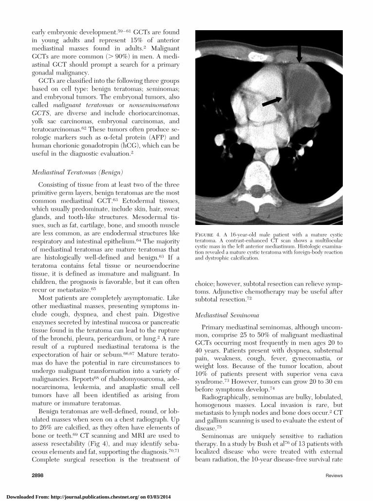
Benign teratomas are well-defined, round, or lob-ulated masses when seen on a chest radiograph. Upto 26% are calcified, as they often have elements ofbone or teeth.69 CT scanning and MRI are used toassess resectability (Fig 4), and may identify seba-ceous elements and fat, supporting the diagnosis.70,71
Complete surgical resection is the treatment of
choice; however, subtotal resection can relieve symp-toms. Adjunctive chemotherapy may be useful aftersubtotal resection.72
Mediastinal Seminoma
Primary mediastinal seminomas, although uncom-mon, comprise 25 to 50% of malignant mediastinalGCTs occurring most frequently in men ages 20 to40 years. Patients present with dyspnea, substernalpain, weakness, cough, fever, gynecomastia, orweight loss. Because of the tumor location, about10% of patients present with superior vena cavasyndrome.73 However, tumors can grow 20 to 30 cmbefore symptoms develop.74
Radiographically, seminomas are bulky, lobulated,homogenous masses. Local invasion is rare, butmetastasis to lymph nodes and bone does occur.2 CTand gallium scanning is used to evaluate the extent ofdisease.75
Seminomas are uniquely sensitive to radiationtherapy. In a study by Bush et al76 of 13 patients withlocalized disease who were treated with externalbeam radiation, the 10-year disease-free survival rate
Figure 4. A 16-year-old male patient with a mature cysticteratoma. A contrast-enhanced CT scan shows a multilocularcystic mass in the left anterior mediastinum. Histologic examina-tion revealed a mature cystic teratoma with foreign-body reactionand dystrophic calcification.
2898 Reviews
Downloaded From: http://journal.publications.chestnet.org/ on 03/03/2014

was 54%, with an actuarial survival rate of 69%.There is, however, debate as to the role of chemo-therapy and surgical resection. A retrospective studyby Bokemeyer et al77 showed that chemotherapyalone led to a 90% 5-year disease-free survival rateand that additional radiation offered only a slightsurvival advantage, while patients treated with justradiation initially had a much higher rate of diseaserecurrence. In patients with locally advanced dis-ease, the preferred treatment includes chemother-apy followed by the surgical resection of residualdisease.78
Mediastinal Nonseminomatous GCTs
Nonseminomatous malignant GCTs comprise aheterogeneous group of masses that includes embry-onal cell carcinomas, endodermal thymus tumors,choriocarcinomas, yolk sac tumors, and mixed GCTswith multiple cellular components. These tumors areoften symptomatic and malignant, and predomi-nantly affect young men.2 In addition, they can beassociated with hematologic malignancies, and 20%of patients have Kleinfelter syndrome.79,80
At diagnosis, 85% of patients are symptomatic,which includes complaints of chest pain, hemoptysis,cough, fever, or weight loss. Gynecomastia can de-velop as a result of �-hCG secretion from certaintumor types.62,81
These tumors are large, irregularly shaped, withareas of central necrosis, hemorrhage, or cyst forma-tion82 (Fig 5). Measuring AFP and �-hCG levels isimportant in making the diagnosis. An elevated AFPlevel is suggestive of an endodermal sinus tumor orembryonal carcinoma and is sufficient, in the pres-ence of a mediastinal mass, to establish the diagno-sis.62,81
Chemotherapy with bleomycin, etoposide, andcisplatin is the current standard of care for patientswith nonseminomatous malignant GCTs.83 Follow-ing chemotherapy, � 5% of patients have total res-olution of their malignancy with normalized serummarkers. Patients with residual tumor undergo sur-gical resection, although studies have shown that thenormalization of tumor markers prior to surgeryportend a better prognosis.83,84 In contrast to pureseminomas, nonseminomatous GCTs carry a poorerprognosis; patients with these tumors have a 5-yearoverall survival rate of 48%, compared to 86% inpatients with seminomas.85
Mediastinal Goiter
In patients undergoing thyroidectomy, the inci-dence of mediastinal goiter is 1 to 15%.86 Mostgoiters are euthyroid and are found incidentallyduring a physical examination. Radiographically, me-
diastinal goiters are encapsulated, lobulated, hetero-geneous tumors.2 A classic finding on a CT scan iscontinuity of the cervical and mediastinal compo-nents of the thyroid. If the goiter contains functionalthyroid tissue, then scintigraphy with a radioactiveisotope of iodine can be diagnostic.2
Surgical resection is recommended since theselesions are not usually amenable to needle biopsy,and malignancy develops in a significant number.Nearly all substernal goiters can be removed easilythrough a cervical incision minimizing surgical mor-bidity.87
Mediastinal Parathyroid Adenoma
The mediastinum is the most common location atwhich an ectopic parathyroid tumor may develop.Overall, 20% of parathyroid adenomas develop in themediastinum, with 80% occurring in the anteriormediastinum.88
These tumors are encapsulated, round, and usually� 3 cm in size, so that they may not be identified ona CT scan. Thus, MRI or nuclear scans with 99mTcand 201Ti are more effective for the diagnosis ofparathyroid adenomas.89 Surgical resection is cura-tive.
Figure 5. A 59-year-old man with a nonseminatous malignantGCT. A contrast-enhanced CT scan shows a heterogeneouslow-attenuating anterior mediastinal mass compressing the pul-monary artery.
www.chestjournal.org CHEST / 128 / 4 / OCTOBER, 2005 2899
Downloaded From: http://journal.publications.chestnet.org/ on 03/03/2014

Primary Mediastinal Lymphoma
Primary mediastinal lymphoma is a rare entitycomprising only 10% of lymphomas in the mediasti-num. Lymphoma usually occurs in the anteriormediastinum and is part of more widespread disease.HD represents approximately 50 to 70% of medias-tinal lymphomas, while non-Hodgkin lymphomacomprises 15 to 25%.90,91 The three most commontypes of mediastinal lymphoma include nodular scle-rosing HD, large B-cell lymphoma, and lymphoblas-tic lymphoma.2
HD
HD has an incidence of approximately 2 to 4 casesper 100,000 people per year, with a bimodal distri-bution of incidence peaking in young adulthood andagain after age 50 years.92 For mediastinal-predom-inant disease, prevalence peaks in young womenduring the third decade of life, while it is unaffectedby age in men.93 HD is divided into four subtypes,including nodular sclerosing, lymphocyte-rich, mixedcellularity, and lymphocyte depleted HD, with thenodular sclerosing subtype representing more thantwo thirds of cases.94
Most patients experience constitutional symptoms(B symptoms), including fevers, night sweats, andweight loss. For patients with mediastinal involve-ment, cough, dyspnea, chest pain, pleural effusions,and superior vena cava syndrome may occur.93
The presence of Reed-Sternberg cells are patho-gnomonic of HD. These cells contain bilobed nucleicontaining prominent eosinophilic nuclei. The classicimmunohistochemical profile is biomarker positivityfor CD15 and CD30 cells.95
The chest radiograph finding is abnormal in up to76% of patients with HD, often showing enlarge-ment of the prevascular and paratracheal nodes.96–98
A CT scan examination is usually sufficient to iden-tify lymphoma; however, in certain circumstances,such as after radiation treatment, MRI may be betterin distinguishing scars from residual disease96 (Fig6). A positron emission tomography scan may also beuseful in staging and following disease progression.99
Still widely used is the Ann Arbor staging systemfor HD. This system has important implications fordetermining prognosis and types of treatment (Table6). In 1989, the Ann Arbor staging system wasmodified at a meeting in Cotswold, England, toseparate out patients with bulky disease due to itsprognostic significance.
The treatment of HD is separated into the treat-ment of early-stage disease (ie, stage I and II disease)and late-stage disease (ie, stage III and IV disease).Based on the Cotswold modifications, early-stagedisease can be further subclassified into favorable
and unfavorable, depending on the degree of tumorburden. For patients with favorable stage I or IIdisease, extended-field radiation alone used to be thestandard of care. Hagenbeek et al100 conducted a
Figure 6. A 39-year-old man with nodular sclerosis HD. Acontrast-enhanced CT scan shows an anterior mediastinal mass inwhich the central portion is cystic (black arrow). The rightparatracheal lymph node is enlarged (white arrow).
Table 6—Ann Arbor Staging System With CotswoldModifications for HD*
Stage Characteristics
1 Involvement of one lymph node region orlymphoid structure
2 Two or more lymph node regions on same side ofthe diaphragm
3 Lymph nodes on both sides of the diaphragm4 Involvement of extra nodal sitesModifications
A No symptomsB Fever, night sweats, weight loss � 10% in 6 moX Bulky disease (greater than one third widening of
the mediastinum or � 10 cm diameter of nodalmass
E Involvement of single, contiguous, or extra nodal site
*From Yung and Linch.94
2900 Reviews
Downloaded From: http://journal.publications.chestnet.org/ on 03/03/2014

randomized controlled trial in which 762 patientswith favorable stage I or II HD were randomized toreceive either combination therapy with six cycles ofepirubicin, bleomycin, vinblastine, and prednisone,and involved field radiation, or to receive subtotalnodal irradiation alone. The complete remission ratewas similar among patients in both groups, while therelapse rate was significantly higher in the radiation-alone group.100 Thus, the use of combined involved-field radiation and chemotherapy is quickly becom-ing the standard of care. For patients with stage I orII HD with bulky tumors, treatment consists ofchemotherapy followed by radiation.101 Patients withstage III or IV HD are treated primarily withchemotherapy. Canellos et al102 showed that ABVDwas superior to MOPP in preventing relapse.
Patients who relapse may benefit from a bonemarrow transplant (BMT), while those who have hada good response to standard-dose second-line che-motherapy benefit the most.103 For patients withHD, autologous BMT is superior to allogeneic BMTsince the relapse rate for both is similar, and thenonrelapse mortality rate is 48% for patients whohave undergone allogeneic BMT and 27% for thosewho have undergone autologous BMT.104
Patients with stage I and II HD have cure rates of� 90%. Patients with stage IIIA HD have a cure rateof 30 to 90% with standard treatment. Stage IIIBHD offers a cure rate of 60 to 70%, while stage IVHD has a cure rate of 50 to 60% (2,101). Amongpatients with advanced disease, a prognostic indexwas created by the International Prognostic FactorProject on Advanced Hodgkin’s Disease that wasbased on the total number of unfavorable featuresfrom among seven potential features found at diag-nosis, as follows: serum albumin level, � 4 g/dL (or40 g/L); hemoglobin level, � 10.5 g/dL (105 g/L);male gender; age � 45 years; stage IV disease; WBCcount, � 15,000 cells/�L; and lymphocyte count,� 600/�L and/or � 8% of the WBC count.105
Non-Hodgkin Lymphoma
Although there are many classes and grades ofnon-Hodgkin lymphoma, lymphoblastic lymphomaand large B-cell lymphoma are the most commonsubtypes to affect the mediastinum.106 The overallincidence of non-Hodgkin lymphoma is greatest inwhite men with a mean age of 55 years.106 However,the mean ages of presentation for lymphoblasticlymphoma and primary large B-cell lymphoma are28 and 30 to 35 years, respectively.106,107
Lymphoblastic lymphoma is highly aggressive,arising from thymic lymphocytes.108 Common symp-toms include cough, wheezing, shortness of breath,superior vena cava syndrome, cardiac tamponade, or
tracheal obstruction, and can involve the mediasti-num, bone marrow, CNS, skin, or gonads.108 It isoften confused with T-cell acute lymphoblastic leu-kemia (ALL) because bone marrow involvementwith blasts is relatively common.109,110
Primary mediastinal B-cell lymphoma is a diffuselarge B-cell lymphoma derived from the thymus.Common symptoms at presentation include chestpain, cough, dysphasia, superior vena cava syndrome,phrenic nerve palsy, and hoarseness.107 The involve-ment of extrathoracic structures and bone marrow isless common at presentation than for lymphoblasticlymphoma. However, on the recurrence of disease,involvement of the liver, kidneys, and brain canoccur.111,112
Computer tomography scanning is used to charac-terize the lesion and to determine the extent ofinvasion. The middle and posterior mediastinalnodes are involved more often than the anteriorones.2 Tissue diagnosis should be obtained beforetreatment. Flow cytometry and cytogenetic analysiscan be used to help render a definitive diagnosis.113
Treatment for mediastinal non-Hodgkin lym-phoma depends on the stage, histologic subtype, andextent of the disease. For lymphomblastic lym-phoma, the treatment regimens are often similar toALL due to its propensity to involve the marrow.Treatment with intensive chemotherapy programswith maintenance-phase chemotherapy is superior toshort-term chemotherapy without a maintenancephase. In a study by Kobayashi et al,114 patients withALL and those with lymphoblastic lymphoma whoreceived short-term chemotherapy had a cure rate of78% but a relapse rate of 72% with only a 7% 7-yearsurvival rate. Intrathecal chemotherapy is also nec-essary to prevent CNS relapse. CNS irradiation isoften part of prophylactic treatment to prevent CNSrecurrence, while mediastinal irradiation has beenused as well. Many patients go on to relapse evenafter treatment. As a result, BMT is a commonlyemployed treatment for patients with lymphoblasticlymphoma. Levine et al115 demonstrated in a retro-spective analysis of 204 patients with lymphoblasticlymphoma who had been treated with either alloge-neic or autologous BMT that although there werefewer relapses at 5 years with allogeneic BMT(relapse rate, 46% vs 56%, respectively), the inci-dence of treatment-related mortality in patients whounderwent allogeneic BMT made the overall survivalbenefit insignificant.
Patients with primary mediastinal B-cell lym-phoma can be treated with conventional chemother-apy; however, there may be an additional benefit totreatment with high-dose chemotherapy and in-volved-field radiation.107,116 Currently, if patients failto have a full response to standard chemotherapy,
www.chestjournal.org CHEST / 128 / 4 / OCTOBER, 2005 2901
Downloaded From: http://journal.publications.chestnet.org/ on 03/03/2014

high-dose chemotherapy and/or radiation therapyare considered. After relapse, many patients aretreated with high-dose chemotherapy and autolo-gous BMT.107
Tumors of the Middle Mediastinum
Mediastinal Cysts
Mediastinal cysts comprise 12 to 20% of medias-tinal masses and are found in the middle compart-ment of the mediastinum.117–119 Despite a similarincidence, children are more often symptomatic atpresentation due to compression on the surroundingstructures.120 The most common type of mediastinalcyst are foregut cysts, which are derived as anembryonic abnormality, with enterogenous cysts (50to 70%) and bronchogenic cysts (7 to 15%) being themost common subtypes.2
Bronchogenic Cysts
Bronchogenic cysts are formed during embryonicdevelopment as an anomalous budding of the laryn-gotracheal groove.121 These cysts are lined withciliated, pseudostratified, columnar epithelium, andcontain bronchial glands and cartilaginous plates.2Approximately 40% of bronchogenic cysts are symp-tomatic resulting in cough, dyspnea, or chest pain.121
Radiographically, bronchogenic cysts can be iden-tified on plain radiographs (Fig 7a) but are bestdefined by CT scanning. These cysts are well-de-fined round masses with a homogenous densitysimilar to water; however, some bronchogenic cystsare mucoid and can give the impression of being asolid mass.120 Bronchogenic cysts are nonenhancing,and, when there is a direct communication with thetracheobronchial tree, air-fluid levels may be seen.122
MRI can differentiate the lesion from other masses(Fig 7, bottom, B, 8).
Tissue is often required to make a definitivediagnosis of a bronchogenic cyst. This can be accom-plished by tracheobronchial, endoscopic, or thoro-scopic needle aspiration. Most bronchogenic cystsare removed surgically or are drained by needleaspiration. The treatment of asymptomatic cysts iscontroversial as surgery is not without risk, yet thesecysts can grow to cause symptoms in the future.123
Enterogenous Cysts
Enterogenous cysts arise from the dorsal foregutand are lined by squamous or enteric (alimentary)epithelium and may contain gastric or pancreatictissue. Esophageal duplication cysts are located in orare attached to the esophageal wall. Twelve percent
Figure 7. Top, A: a 49-year-old man with a bronchogenic cyst. Achest radiograph shows a rounded mass (arrow) that displaces theright primary bronchus superiorly. Bottom, B: a 49-year-old manwith a bronchogenic cyst. A sagittal T1-weighted magneticresonance image shows a high-signal intensity cyst with a fluid-fluid level due to infection (arrow).
2902 Reviews
Downloaded From: http://journal.publications.chestnet.org/ on 03/03/2014

of patients with esophageal duplication cysts haveassociated malformations, mostly of the GI tract.124
Symptoms of enterogenous cysts are similar tothose of other mediastinal cysts. They are oftenasymptomatic, but if they contain gastric or pancre-atic mucosa, there is the added risk of hemorrhage orrupture of the cyst from mucosal secretions. Radio-graphically, it can be difficult to distinguish thesefrom bronchogenic cysts, although they are moreoften calcified (Fig 9). The presence of cartilage
suggests the presence of a bronchogenic cyst.121
Most cysts should be surgically excised, and video-assisted thoracic surgery is the treatment ofchoice.125
Neuroenteric Cysts
Neuroenteric cysts are characterized by the pres-ence of both enteric and neural tissue in surgicalspecimens.126 Most of these cysts form in the poste-rior mediastinum above the level of the main carina.The close association of the foregut and notochordduring embryogenesis possibly explains this ana-tomic location. Neuroenteric cysts are associatedwith multiple vertebral anomalies, such as scoliosis,spina bifida, hemivertebra, and vertebral fusion.Almost all are discovered by age 1 years due tosymptoms from tracheobronchial compression.2Neurologic symptoms may be caused by intraspinalextension. Complete surgical excision is curative.127
Pericardial Cysts
Pericardial cysts are part of a larger group ofmesothelial cysts. They form as a result of a persis-tent parietal recess during embryogenesis.121 Theyare estimated to occur in 1 of 100,000 people.Although most are congenital, a few cases of ac-quired pericardial cysts do exist. They are oftenasymptomatic and are identified in the fourth to fifthdecade of life. Rarely, cardiac compression mayoccur, causing hemodynamic compromise.95 Radio-graphically, pericardial cysts are well-marginatedspherical or tear drop-shaped masses that character-istically abut the heart, anterior chest wall, anddiaphragm.2 The most common location of pericar-dial cysts is at the right cardiophrenic angle (70%),followed by the left cardiophrenic angle (22%).128
On CT scans, these masses appear as unilocular andnonenhancing (Fig 10, 11). As with most mediastinalcysts, surgical removal is the treatment of choice,although clinically asymptomatic patients may beobserved without intervention.
Lymphangiomas
Lymphangiomas are rare congenital abnormalitiesof the lymphatic vessels. Typically, they are isolatedsolitary masses, but they can be more widespread orassociated with chromosomal abnormalities.129
These lesions are benign in nature and are found inthe cervical region 75% of the time. In 10% of cases,the cysts extend into the mediastinum and areassociated with chylothorax and hemangiomas.129
Although these tumors are commonly identified inchildren before the age of 2 years, when the mass isisolated to the mediastinum it is often not identified
Figure 8. A 37-year-old woman with a bronchogenic cyst. Acoronal T1-weighted magnetic resonance image shows a cyst withhigh-signal intensity contents (arrow).
Figure 9. A 10-year-old female patient with a duplication cyst. Acontrasted-enhanced CT scan shows a thin-walled water-attenu-ation cyst adjacent to the esophagus (arrow).
www.chestjournal.org CHEST / 128 / 4 / OCTOBER, 2005 2903
Downloaded From: http://journal.publications.chestnet.org/ on 03/03/2014

until it has gotten large enough to cause compressivesymptoms.130 Such symptoms include chest pain,cough, and dyspnea. Radiographically, these lesionsappear cystic and can be confused with pericardialcysts, although lymphangiomas are more likely tohave a loculated appearance.130 The use of lym-phangiographic contrast media combined with CTscanning can also differentiate these lesions.129 Totalresection is optimal; however, in cases complicatedby chylothorax, there is some evidence suggestingthat additional radiotherapy may be of some bene-fit.131 Lymphangiomatosis seen in young women istypically a more progressive form of disease in whichmultiple tumors are found and invade multiple organstructures, including the lung, heart, and bone.132
Tumors of the Posterior Mediastinum
Neurogenic Tumors
Neurogenic tumors are derived from tissue of theneural crest, including cells of the peripheral, auto-nomic, and paraganglionic nervous systems. Ninety-five percent of posterior mediastinal masses arise inthe intercostal nerve rami or the sympathetic chainregion.133 They are classified on the basis of cell typeand comprise approximately 12 to 21% of all medi-astinal masses, although 95% occur in the posteriorcompartment.134 Seventy percent to 80% of neuro-genic tumors are benign, and nearly half are asymp-tomatic; however, they can occasionally cause com-pressive or neurologic symptoms.133,135,136
Nerve Sheath Tumors
These benign, slowly growing tumors comprise 40to 65% of neurogenic mediastinal masses. Neurile-
momas or schwannomas constitute 75% of this groupof masses. These tumors are firm, encapsulatedmasses consisting of Schwann cells. Neurofibromasare nonencapsulated, soft, and friable, and are asso-ciated with Von Recklinghausen neurofibromato-sis.137,138 They are often asymptomatic and are dis-covered incidentally.
Radiographically, nerve sheath tumors are sharplymarginated spherical masses. Being adjacent to thespine, they can cause erosion and deformity of theribs and ventral bodies as they increase in size. Lowattenuation on CT scans can indicate hypocellularity,cystic changes, hemorrhage, or the presence of lipidwithin myelin.2 Ten percent of these tumors growthrough the intervertebral foramina and create adumbbell appearance on radiographs.139 MRI isused to rule out intraspinal extension.
The surgery of choice for removal of these tumorsis thoroscopy, or thorocotomy when the former is notan option.133,134 For tumors invading the vertebralbody or foramina, en bloc resection can beachieved.140 There may be a role for postoperativechemotherapy or radiation therapy when total resec-
Figure 11. A 54-year-old woman with a pericardial cyst. Acontrast-enhanced CT scan shows a large thin-walled cystic massat the level of the aortic arch (black arrow). The innominate veinis compressed by this mass (white arrow).
Figure 10. A 37-year-old man with a pericardial cyst. A contrast-enhanced CT scan shows a thin-walled water-attenuation cyst (arrow).
2904 Reviews
Downloaded From: http://journal.publications.chestnet.org/ on 03/03/2014

tion is not possible. Postoperative complicationsinclude Horner syndrome, partial sympathectomy,recurrent laryngeal nerve damage, and paraplegia.134
Malignant Tumors of Nerve Sheath Origin
Malignant nerve sheath tumors are spindle cellsarcomas of the posterior mediastinum, and includemalignant neurofibromas, malignant schwannomas,and neurogenic fibrosarcomas. They affect men andwomen equally in the third to fifth decade of life andare closely associated with neurofibromatosis, with a5% risk of sarcomatous degeneration.141 Pain andnerve deficits are common. Complete surgical resec-tion is the optimal treatment, but, in patients withunresectable tumors, adjuvant chemotherapy andradiation are options.
Autonomic Ganglionic Tumors
Tumors of the autonomic nervous system arisefrom neuronal cells rather than from the nervesheath. They form a continuum ranging from benignencapsulated ganglioneuroma to aggressive malig-nant nonencapsulated neuroblastoma. Derived fromembryologic origins, these tumors arise in the adre-nal glands or in the sympathetic ganglia. However,ganglioneuromas and ganglioneuroblastomas arisemostly in the sympathetic ganglia of the posteriormediastinum.142 Fifty percent of neuroblastomasarise in the adrenal glands and up to 30% in themediastinum.142,143
Ganglioneuroma: Ganglioneuromas are benign tu-mors composed of one or more mature ganglioniccells. Arising from the nerve ganglion cells, they arethe most benign and differentiated of the autonomicganglionic tumors.144 Most patients are asymptom-atic and receive diagnoses in the second or thirddecade of life.145 Radiographically, the tumors areoblong and well-marginated, occurring along theanterolateral aspect of the spine and spanning threeto five vertebrae145 (Fig 12). CT scanning is notparticularly helpful as the mass can be homogenousor heterogeneous. Complete surgical resection isideal.146
Ganglioneuroblastoma: Ganglioneuroblastomashave histologic features of both ganglioneuromas andneuroblastomas. They are the least common type ofneurogenic tumor. Prognosis depends on histologicappearance.2 Both sexes are equally affected in thefirst decade of life.147 Symptoms may arise due tolarge tumor size, intraspinal extension, and metasta-
sis. Staging is similar to that for neuroblastoma, asdescribed in the following section.
Neuroblastoma: Neuroblastoma is a disease ofyoung children, with 95% occurring in patients � 5years of age.143,148 Neuroblastomas are highly aggres-sive and readily metastasizing tumors that are com-posed of small round cells arranged in sheets orpseudorosettes.149 They are nonencapsulated lesions,often exhibiting hemorrhage, necrosis, or cystic degen-eration. Symptoms include pain, neurologic deficits,Horner syndrome, respiratory distress, and ataxia.148,149
Neuroblastomas have the highest propensity of anytumors in its class to produce vasoactive substances thatcan cause hypertension, flushing, and diarrhea.142
Grossly, these tumors appear as an elongatedparaspinous mass, sometimes impinging on adjacent
Figure 12. A 20-year-old woman with a posterior mediastinalganglioneuroma. Top, A: a contrast-enhanced CT scan image thatshows a mass with mixed attenuation and calcifications. Bottom,B: acoronal T2-weighted magnetic resonance image that showsan 8-cm mass with heterogeneous signal intensity.
www.chestjournal.org CHEST / 128 / 4 / OCTOBER, 2005 2905
Downloaded From: http://journal.publications.chestnet.org/ on 03/03/2014

structures and causing skeletal damage.150,151 On CTscans, 80% of these tumors have calcification.151 Aswith all neurogenic tumors, MRI is useful to determinethe extent of intraspinal involvement.146 Radionuclideimaging with 123I metaiodobenzylguanide can also beused to detect primary and metastatic disease.152
Treatment for neuroblastoma depends primarilyon the stage of disease (Table 7). Treatment forlimited-stage disease is surgical resection. For pa-tients with stage I disease, resection is usually cura-tive. For patients with partially resectable stage IIand III disease, treatment includes postoperativechemotherapy and radiation. For patients with stageIV disease, there is much controversy over the role ofsurgery; however, some studies153 have suggestedthat delayed surgery after initial treatment withchemotherapy and radiation results in a better out-come than initial surgical intervention. In addition,there are ongoing studies looking at the role ofradioactive 131I metaiodobenzylguanide therapy incombination with chemotherapy in patients withadvanced-stage disease.154 Poor prognostic factors inneuroblastoma include large tumor size, poorly dif-ferentiated cell type, advanced stage, extrathoracicorigin, and presentation in an elderly patient.138
ACKNOWLEDGMENT: We thank the following people fortheir contributions: Jin Mo Goo, MD, Department of Radiology,Seoul National University College of Medicine, for the contribu-tion of Figures 1 to 6 and 11, which were originally published inthe Journal of Computed Assisted Tomography in 2003; Mi-Young Jeung, MD, Department of Radiology, University ofStrasbourg, for the contribution of Figures 6 to 10, which wereoriginally published in Radiographics in 2002; Allen Forsythe,MD, for the contribution of Figure 12, which was published inRadiographics in 2004. It was only through their contributionsthat we were able to produce this study.
References1 Fraser RS, Pare JAP, Fraser RG, et al. The normal chest. In:
Fraser RS, Pare JAP, Fraser RG, et al, eds. Synopsis ofdiseases of the chest. 2nd ed. Philadelphia, PA: WB Saun-ders, 1994; 1–116
2 Strollo DC, Rosado-de-Christenson ML, Jett JR, et al.
Primary mediastinal tumors: Part 1. Tumors of the anteriormediastinum. Chest 1997; 112:511
3 Davis RD Jr, Newland Oldham H Jr, Sabiston DC Jr.Primary cysts and neoplasms of the mediastinum: recentchanges in clinical presentation, methods of diagnosis, man-agement and results. Ann Thorac Surg 1987; 44:229–237
4 Silverman NA, Sabiston DC Jr. Mediastinal masses. SurgClin North Am 1980; 60:757–777
5 Grillo HC, Ojemann RG, Scannell JG, et al. Combinedapproach to “dumbbell” intrathoracic and intraspinal neuro-genic tumors. Ann Thorac Surg 1983; 36:402–407
6 Wychulis AR, Payne WS, Clagett OT, et al. Surgical treat-ment of mediastinal tumors. J Thorac Cardiovasc Surg 1972;62:379–391
7 Rosai J, Levine GD. Tumors of the thymus. In: FirmingerHI, ed. Atlas of tumor pathology. Washington, DC: ArmedForces Institute of Pathology, 1976; 34–212
8 Lattes R. Thymoma and other tumors of the thymus: ananalysis of 107 cases. Cancer 1962; 15:1224–1260
9 Engels EA, Pfeiffer RM. Malignant thymoma in the UnitedStates: demographic patterns in incidence and associationswith subsequent malignancies. Int J Cancer 2003; 105:546–551
10 Mullen B, Richardson JD. Primary anterior mediastinaltumors in children and adults. Ann Thorac Surg 1986;42:338–345
11 Gerein AN, Srivastava SP, Burgess J. Thymoma: a ten-yearreview. Am J Surg 1978; 136:49–53
12 Okumura M, Ohta M, Tateyama H, et al. The World HealthOrganization histologic classification system reflects theoncologic behavior of thymoma: a clinical study of 273patients. Cancer 2002; 94:624–632
13 Nakagawa K, Asamura H, Matsuno Y, et al. Thymoma: aclinicopathologic study based on the new World HealthOrganization classification. J Thorac Cardiovasc Surg 2003;126:1134–1140
14 Lardinois D, Rechsteiner R, Lang RH, et al. Prognosticrelevance of Masaoka and Muller-Hermelink classificationin patients with thymic tumors. Ann Thorac Surg 2000;69:1550–1555
15 Wilkins EW Jr, Edmunds L Jr, Castleman B. Cases ofthymoma of the Massachusetts General Hospital. J ThoracCardiovasc Surg 1966; 52:322–330
16 Lewis JE, Wick MR, Scheithauer BW, et al. Thymoma: aclinicopathologic review. Cancer 1987; 60:2727–2743
17 Verstandig AG, Epstein DM, Miller WT, et al. Thymoma-report of 71 cases and a review. Crit Rev Diagn Imaging1992; 33:201–230
18 Zerhouni EA, Scott WW, Baker RR, et al. Invasive thymo-mas: diagnosis and evaluation by CT. J Comput AssistTomogr 1982; 6:92–100
19 Yokoi K, Miyazawa N, Mori K, et al. Invasive thymoma withintracaval growth into right atrium. Ann Thorac Surg 1992;53:507–509
20 Masaoka A, Monden Y, Nakahara K, et al. Follow-up studyof thymomas with special reference to their clinical stages.Cancer 1981; 48:2485–2492
21 Shamji F, Pearson FG, Todd TR, et al. Results of surgicaltreatment for thymoma. J Thorac Cardiovasc Surg 1984;87:43–47
22 Cohen DJ, Ronnigan LD, Graeber GM, et al. Managementof patients with malignant thymoma. J Thorac CardiovascSurg 1984; 87:301–307
23 Laurent E, Latrabe V, Lecesne R, et al. Mediastinal masses:diagnostic approach. Eur Radiol 1998; 8:1148–1159
24 Osserman KE, Genkins G. Studies in myasthenia gravis:review of a 20-year experience in over 1200 patients. Mt
Table 7—Staging of Neuroblastoma andGanglioneuroblastomas*
Stage Characteristics
I Well-circumscribed, noninvasive ipsilateral tumorsII Local invasion without extension across the midline,
ipsilateral regional lymph node involvementIII Tumor extension across the midline and involvement of
bilateral regional lymph nodesIV Metastatic diseaseIVS Clinical stage I or II and metastatic disease limited to the
liver, skin and bone marrow
*From Baum and Crapo.122
2906 Reviews
Downloaded From: http://journal.publications.chestnet.org/ on 03/03/2014

Sinai J Med 1971; 38:497–53725 Marx A, Muller-Hermelink HK, Strobel P. The role of
thymomas in the development of myasthenia gravis. Ann NY Acad Sci 2003; 998:223–236
26 Drachmnan DB. Myasthenia gravis. N Engl J Med 1994;330:1797–1810
27 Lennon VA, Jones G, Howard F, et al. Auto antibodies toacetylcholine receptors in myasthenia gravis. N Engl J Med1983; 308:402–403
28 Howard FM Jr, Lennon VA, Finley J, et al. Clinical corre-lation of antibodies that bind, block or modulate humanacetylcholine receptors in myasthenia gravis. Ann N Y AcadSci 1987; 505:526–538
29 Souadjian JV, Enriquez P, Silverstein MN, et al. Thespectrum of diseases associated with thymoma. Arch InternMed 1974; 134:374–379
30 Kelleher P, Misbah SA. What is Good’s syndrome? Immu-nological abnormalities in patients with thymoma. J ClinPathol 2003; 56:12–16
31 Kim JH, Goo JM, Lee HJ, et al. Cystic tumors in the anteriormediastinum: radiologic-pathological correlation. J ComputAssist Tomogr 2003; 27:714–723
32 Rosado de Christenson ML, Galobardes J, Moran CA.Thymoma: radiologic-pathologic correlation. Radiographics1992; 12:151–168
33 Anderson T, Lindgren PG, Elvin A. Ultrasound guidedtumor biopsy in the anterior mediastinum. Acta Radiol 1992;33:310–311
34 Morgenthaler TI, Brown LR, Colby TV, et al. Symposiumon intrathoracic neoplasms: part IX. Mayo Clin Proc 1993;68:1110–1123
35 Singhal S, Shrager JB, Rosenthal DI, et al. Comparison ofstages I-II thymoma treated by complete resection with orwithout adjuvant radiation. Ann Thorac Surg 2003; 76:1635–1641
36 Curran WJ Jr, Kornstein MJ, Brooks JJ, et al. Invasivethymoma: the role of mediastinal irradiation following com-plete or incomplete surgical resection. J Clin Oncol 1988;6:1722–1727
37 Eralp Y, Aydiner A, Kizir A, et al. Resectable thymoma:treatment outcome and prognostic factors in the late ado-lescent and adult age group. Cancer Invest 2003; 21:737–743
38 Blumberg D, Port JL, Weksler B, et al. Thymoma: amultivariate analysis of factors predicting survival. AnnThorac Surg 1995; 60:908–913
39 Thomas CR, Wright CD, Loehrer PJ. Thymoma. J ClinOncol 1999; 17:2280–2289
40 Kim ES, Putnam JB, Komaki R, et al. Phase II study of amultidisciplinary approach with induction chemotherapy,followed by surgical resection, radiation therapy, and con-solidation chemotherapy for unresectable malignant thymo-mas: final report. Lung Cancer 2004; 44:339–379
41 Daniele O, Fornasiero A. Ifosfamide in thymic neoplasms.Oncology 2003; 65:44–45
42 Gamondes JP, Balawi A, Greenland T, et al. Seventeen yearsof surgical treatment of thymoma: factors influencing sur-vival. Eur J Cardiothorac Surg 1991; 5:124–131
43 Hernandez-Ilizaliturri FJ, Tan D, Cipolla D, et al. Multimo-dality therapy for thymic carcinoma (TCA): results of a30-year single-institution experience. Am J Clin Oncol 2004;27:68–72
44 Truong LD, Mody DR, Cagle PT, et al. Thymic carcinoma:a clinicopathologic study of 13 cases. Am J Surg Pathol 1990;14:151–166
45 Tamura Y, Kuroiwa T, Doi A, et al. Thymic carcinomapresenting as cranial metastasis with intradural and extracra-
nial extension: case report. Neurosurgery 2004; 54:209–21146 Yaqub A, Munn NJ, Wolfer RS. Thymic carcinoma present-
ing as cardiac tamponade. South Med J 2004; 97:212–21347 Suster S, Rosai J. Thymic carcinoma: a clinicopathologic
study of 60 cases. Cancer 1991; 67:1025–103248 Blumberg D, Burt ME, Bains MS, et al. Thymic carcinoma:
current staging does not predict prognosis. J Thorac Cardio-vasc Surg 1998; 115:303–338
49 Ritter JH, Wick MR. Primary carcinoma of the thymusgland. Semin Diagn Pathol 1999; 16:18–31
50 Loehrer PJ, Kim KM, Aisner SC, et al. Cisplatin plusdoxorubicin plus cyclophosphamide in metastatic or recur-rent thymoma. J Clin Oncol 1994; 12:1164–1168
51 Yoh K, Goto K, Ishii G, et al. Weekly chemotherapy withcisplatin, vincristine, doxorubicin, and etoposide is an effec-tive treatment for advanced thymic carcinoma. Cancer 2003;98:926–931
52 Gibril F, Chen YJ, Schrump DS, et al. Prospective study ofthymic carcinoids in patients with multiple endocrine neo-plasia type 1. J Clin Endocrinol Metab 2003; 88:1066–1081
53 Wick MR, Bernatz PE, Carney JA, et al. Primary mediastinalcarcinoid tumors. Am J Surg Pathol 1982; 6:195–205
54 Economopoulos GC, Lewis JW Jr, Lee MW, et al. Carcinoidtumors of the thymus. Ann Thorac Surg 1990; 50:58–61
55 Tiffet O, Nicholson AG, Ladas G, et al. A clinicopathologicstudy of 12 neuroendocrine tumors arising in the thymus.Chest 2003; 124:141–146
56 Graeber GM, Thompson LD, Cohen DJ, et al. Cystic lesionsof the thymus. J Thorac Cardiovasc Surg 1984; 87:295–300
57 Indeglia RA, Shea MA, Grage TB. Congenital cysts of thethymus gland. Arch Surg 1967; 94:149–152
58 Suster S, Rosai J. Multilocular thymic cyst: an acquiredreactive process. Am J Surg Pathol 1991; 15:388–398
59 Parker D, Holford CP, Begent RH. Effective treatment formalignant mediastinal teratoma. Thorax 1983; 38:897–902
60 Bohle A, Studor UK, Sonntag RW, et al. Primary orsecondary extragonadal germ cell tumor? J Urol 1986;135:939–943
61 Recondo J, Libshitz HI. Mediastinal extragonadal germ celltumors. Urology 1978; 11:369–375
62 Javadpour N. Significance of elevated serum alpha fetopro-tein (AFP) in seminoma. Cancer 1980; 45:2166–2168
63 Nichols CR. Mediastinal germ cell tumors: clinical featuresand biologic correlates. Chest 1991; 99:472–479
64 Crussi-Gonzalez F. Extragonadal teratomas. In: HartmannWH, ed. Atlas of tumor pathology. Washington, DC: ArmedForces Institute of Pathology, 1982; 77–94
65 Carter C, Bibro MC, Touloukian RJ. Benign clinical behav-ior of immature mediastinal teratoma in infancy and child-hood. Cancer 1982; 49:398–402
66 Adebonojo SA, Nicola ML. Teratoid tumors of the medias-tinum. Am Surg 1976; 42:361–365
67 Thompson DP, Moore TC. Acute thoracic distress in child-hood due to spontaneous rupture of large mediastinalteratoma. J Pediatr Surg 1969; 4:416–423
68 Donadio AC, Motzer RJ, Bajorin DF, et al. Chemotherapyfor teratoma with malignant transformation. J Clin Oncol2003; 21:4285–4291
69 Lewis BD, Hurt RD, Payne WS, et al. Benign teratoma ofthe mediastinum. J Thorac Cardiovasc Surg 1983; 86:727–731
70 Graeber GM, Shriver CD, Albur RA, et al. The use ofcomputed tomography in the evaluation of mediastinaltumors. J Thorac Cardiovasc Surg 1986; 91:662–666
71 Moeller KH, Rosado-de-Christenson ML, Templeton DA.Mediastinal mature teratoma: imaging features. AJR Am JRoentgenol 1997; 169:985–990
www.chestjournal.org CHEST / 128 / 4 / OCTOBER, 2005 2907
Downloaded From: http://journal.publications.chestnet.org/ on 03/03/2014

72 Arai K, Ohta S, Suzuki M, et al. Primary immature medias-tinal teratoma in adulthood. Eur J Surg Oncol 1997; 23:64–67
73 Polansky SM, Barwick KW, Revie CE. Primary mediastinalseminoma. AJR Am J Roentgenol 1979; 132:17–21
74 Hainsworth J. Diagnosis, staging, and clinical characteristicsof the patient with mediastinal germ cell carcinoma. ChestSurg Clin N Am 2002; 12:665–672
75 Hosono M, Machida K, Honda N, et al. Intense Ga-67accumulation in pure primary mediastinal seminomas. ClinNucl Med 2003; 28:25–28
76 Bush SE, Martinez A, Bagshaw MA. Primary mediastinalseminoma. Cancer 1981; 48:1877–1882
77 Bokemeyer C, Droz JP, Horwich A, et al. Extragonadalseminoma: an international multicenter analysis of prognos-tic factors and long term treatment outcome. Cancer 2001;91:1394–1401
78 Bukowski RM, Wolf M, Kulander BG, et al. Alternatingcombination chemotherapy in patients with extragonadalgerm cell tumors: a Southwest Oncology Group study.Cancer 1993; 71:2631–2638
79 Dexeus FH, Logothetis CJ, Chong C, et al. Genetic abnor-malities in men with germ cell tumors. J Urol 1988; 140:80–84
80 Nichols CR, Hoffman R, Einhorn LH, et al. Hematologicmalignancies associated with primary mediastinal germ celltumors. Ann Intern Med 1985; 102:603–609
81 Hori K, Uematsu K, Yasoshima H, et al. Testicular semi-noma with human chorionic gonadotropin production.Pathol Int 1997; 47:592–599
82 Lee KS, Im JG, Han CH, et al. Malignant primary germ celltumors of the mediastinum: CT features. AJR Am J Roent-genol 1989; 153:947–951
83 Wright C, Kesler K. Surgical techniques and outcomes forprimary nonseminomatous germ cell tumors. Chest SurgClin N Am 2002; 12:707–715
84 Walsh GL, Taylor GD, Nesbitt JC, et al. Intensive chemo-therapy and radical resections for primary non-seminoma-tous mediastinal germ cell tumors. Ann Thorac Surg 2000;69:337–343
85 International Germ Cell Consensus Classification. A prog-nostic factor-based staging system for metastatic germ cellcancers: International Germ Cell Cancer CollaborativeGroup. J Clin Oncol 1997; 15:594–603
86 Kathic M, Wang C, Grillo H. Substernal goiter. Ann ThoracSurg 1985; 39:391–399
87 Allo MD, Thompson NW. Rationale for the operativemanagement of substernal goiters. Surgery 1983; 94:969–977
88 Clark O. Mediastinal parathyroid tumors. Arch Surg 1988;123:1096–1100
89 Oates E. Improved parathyroid scintigraphy with Tc 99mMIBI, a superior radio tracer. Appl Radiol 1994; 23:37–40
90 Strickler JG, Kurtin PJ. Mediastinal lymphoma. SeminDiagn Pathol 1991; 8:2–13
91 Lichtenstein AK, Levine A, Taylor CR, et al. Primarymediastinal lymphoma in adults. Am J Med 1980; 68:509–514
92 Cartwright R, Brincker H, Carli PM, et al. The rise inincidence of lymphomas in Europe. Eur J Cancer 1999;35:627–633
93 Vaeth JM, Moskowitz SA, Green JP. Mediastinal Hodgkin’sdisease. AJR Am J Roentgenol 1976; 126:123–126
94 Yung L, Linch D. Hodgkin’s lymphoma. Lancet 2003;361:943–951
95 Kornstein MJ, DeBlois, et al. Pathology of the thymus andmediastinum. 1st ed. Philadelphia, PA: WB Saunders, 1995
96 Costello P, Jochelson M. Lymphoma of the mediastinumand lung. In: Taveras JM, Ferrucci JT, eds. Radiology:diagnosis, imaging, intervention (vol 1). Philadelphia, PA:Lippincott-Raven 1996; 1–13
97 Keller AR, Kaplan HS, Lukes RJ, et al. Correlation ofhistopathology with other prognostic indicators in Hodgkin’sdisease. Cancer 1968; 22:487–499
98 Castellino RA, Blank N, Hoppe RT, et al. Hodgkin disease:contributions of chest CT in the initial staging evaluation.Radiology 1986; 160:603–605
99 Schiepers C. Filmont JE. Czernin J. PET for staging ofHodgkin’s disease and non-Hodgkin’s lymphoma. EurJ Nucl Med Mol Imaging 2003; 30(suppl):S82–S88
100 Hagenbeek A, Carde P, Noordijk E, et al. Prognostic factortailored treatment of early stage Hodgkin’s disease: resultsfrom a prospective randomized phase III clinical trial of 762patients [abstract]. Blood 1997; 90:585
101 DeVita VT, Maack PM, Harris NL. Hodgkin’s disease. In:DeVita VT, Hellman S, Rosenberg SA, eds. Cancer: princi-ples and practice of oncology. 5th ed. Philadelphia, PA:Lippincott-Raven, 1997; 2242–2283
102 Canellos GP, Anderson JR, Propert KJ, et al. Chemotherapyof advanced Hodgkin’s disease with MOPP, ABVD, orMOPP alternating with ABVD. N Engl J Med 1992; 327:1478–1484
103 Moskowitz C. An update on the management of relapsedand primary refractory Hodgkin’s disease. Semin Oncol2004; 31:54–59
104 Hale GA, Phillips GL. Allogeneic stem cell transplantationfor the non-Hodgkin’s lymphomas and Hodgkin’s disease.Cancer Treat Rev 2000; 26:411–427
105 Hasenclever D, Diehl V. A prognostic score for advancedHodgkin’s disease: International Prognostic Factors Projecton Advanced Hodgkin’s Disease. N Engl J Med 1998;339:1506–1514
106 Sutcliff SB. Primary mediastinal malignant lymphoma. Se-min Thorac Cardiovasc Surg 1992; 4:55–67
107 van Besien K, Kelta M, Bahaguna P. Primary mediastinalB-cell lymphoma: a review of pathology and management.J Clin Oncol 2001; 19:1855–1864
108 Thomas DA, Kantarjian H. Lymphoblastic lymphoma. He-matol Oncol Clin North Am 2001; 15:51–95
109 Murphy S. Childhood non-Hodgkin’s lymphoma. N EnglJ Med 1978; 299:1446–1148
110 Murphy SB. Classification, staging and end results of treat-ment of childhood non-Hodgkin’s lymphomas: dissimilari-ties from lymphomas in adults. Semin Oncol 1980; 7:332–339
111 Kirn D, Mauch P, Shaffer K, et al. Large-cell and immuno-blastic lymphoma of the mediastinum: prognostic featuresand treatment outcome in 57 patients. J Clin Oncol 1993;11:1336–1343
112 Lazzarino M, Orlandi E, Paulli M, et al. Primary mediastinalB-cell lymphoma with sclerosis: an aggressive tumor withdistinctive clinical and pathologic features. J Clin Oncol1993; 11:2306–2313
113 Cheson BD. Hodgkin’s disease and the Non-Hodgkin’slymphomas. In: Lenhard RE Jr, Osteen RT, Gansler T, eds.Clinical Oncol. Atlanta, GA: American Cancer Society 2001;497–516
114 Kobayashi T, Tobinai K, Shimoyama M, et al. Long-termfollow up results of adult patients with acute lymphocyticleukemia or lymphoblastic lymphoma treated with short-term, alternating non-cross-resistant chemotherapy. JpnJ Clin Oncol 1999; 29:340–348
115 Levine JE, Harris RE, Loberiza JO, et al. A comparison ofallogeneic and autologous bone marrow transplantation for
2908 Reviews
Downloaded From: http://journal.publications.chestnet.org/ on 03/03/2014

lymphoblastic lymphoma. Blood 2003; 101:2476–2482116 Aviles A, Garcia EL, Fernandez R, et al. Combined therapy
in the treatment of primary mediastinal B-cell lymphoma:conventional versus escalated chemotherapy. Ann Hematol2002; 81:368–373
117 Pun YW, Moreno BR, Prieto VJ, et al. Multicenter experi-ence of video-assisted thoracic surgery to treat mediastinalcysts and tumors. Archivos de Bronconeumologia 2002;38:410–414
118 Wychulis AR, Payne WS, Clagett OT, et al. Surgical treat-ment of mediastinal tumors: a 40 year experience. J ThoracCardiovasc Surg 1971; 62:379–392
119 Whooley BP, Urschel JD, Antkowiak JG, et al. Primarytumors of the mediastinum. J Surg Oncol 1999; 70:95–99
120 Ahrens B, Wit J, Schmitt M, et al. Symptomatic broncho-genic cyst in a six month old infant: case report and reviewof the literature. J Thorac Cardiovasc Surg 2001; 122:1021–1023
121 Takeda S, Miyoshi S, Minami M, et al. Clinical spectrum ofmediastinal cysts. Chest 2003; 124:125–132
122 Crapo JD, Glassroth J, Karlinsky J, et al. Baum’s textbook ofpulmonary diseases. Philadelphia, PA: Lippincott Williams& Wilkins, 2004; 883–912
123 Kumar A, Aggarwal S, Halder S, et al. Thorascopic excisionof mediastinal bronchogenic cyst: a case report and review ofliterature. Ind J Chest Dis Allied Sci 2003; 45:199–201
124 O’Neill JA. Foregut duplications. In: Fallis JC, Filler RM,Lemoine G, eds. Current topics in general thoracic surgery:an international series. New York, NY: Elsevier, 1991;121–123
125 Cioffi U, Bonavina L, De Simone M. Presentation andsurgical management of bronchogenic and esophageal du-plication cysts in adults. Chest 1998; 113:1492–1496
126 Superina RA, Ein SH, Humphreys RP. Cystic duplicationsof the esophagus and neurenteric cysts. J Pediatr Surg 1984;19:527–530
127 Rescorla FJ, Grosfeld JL. Gastroenteric cysts and neuren-teric cysts in infants and children. In: Shields TW, LoCiceroJ III, Ponn RB, eds. General thoracic surgery (vol 2). 5th ed.Philadelphia, PA: Williams & Wilkins, 2000; 2415–2422
128 Feigin D, Fenoglio JJ, McAllister HA, et al. Pericardial cysts:a radiologic-pathologic correlation and review. Radiology1977; 125:15–20
129 Shahriari A, Odell JA. Cervical and thoracic components ofmultiorgan lymphangiomatosis managed surgically. AnnThorac Surg 2001; 71:694–696
130 Nakazato Y, Ohno Y, Nakata Y, et al. Cystic lymphangiomaof the mediastinum. Am Heart J 1995; 129:406–409
131 Johnson DW, Klazynski PT, Gordon WH, et al. Mediastinallymphangioma and chylothorax: the role of radiotherapy.Ann Thorac Surg 1986; 41:325–338
132 Rostom AY. Treatment of thoracic lymphangiomatosis. ArchDis Child 2000; 83:138–139
133 Kumar A, Kumar S, Aggarwal S, et al. Thoracoscopy: thepreferred approach for the resection of selected posteriormediastinal tumors. J Laparoendosc Adv Surg Tech A 2002;12:345–353
134 Reeder LB. Neurogenic tumors of the mediastinum. SeminThorac Cardiovasc Surg 2000; 12:261–267
135 Shapiro B, Orringer MB, Gross MD. Mediastinal paragan-gliomas and pheochromocytomas. In: Shields TW, LoCiceroJ III, Ponn RB, eds. General thoracic surgery (vol 2). 5th ed.Philadelphia, PA: Williams & Wilkins; 2000: 2333–2355
136 Saenz NC. Posterior mediastinal neurogenic tumors ininfants and children. Semin Pediatr Surg 1999; 8:78–84
137 Wain JC. Neurogenic tumors of the mediastinum. ChestSurg Clin N Am 1992; 2:121–136
138 Shields TW, Reynolds M. Neurogenic tumors of the thorax.Surg Clin North Am 1988; 68:645–668
139 Aughenbaugh GL. Thoracic manifestations of neurocutane-ous diseases. Radiol Clin N Am 1984; 22:741–756
140 Mazel CH, Grunenwald D, Laudrin P, et al. Radical excisionin the management of thoracic and cervicothoracic tumorsinvolving the spine: results in a series of 36 cases. Spine2003; 28:782–792
141 Ducatman BS, Scheithauer BW, Piepgras DG, et al. Malig-nant peripheral nerve sheath tumors: a clinicopathologicstudy of 120 cases. Cancer 1986; 57:2006–2021
142 Gale AW, Jelihovsy T, Grant AF, et al. Neurogenic tumorsof the mediastinum. Ann Thorac Surg 1974; 17:434–443
143 Davis S, Rogers MAM, Pendergrass TW. The incidence andepidemiologic characteristics of neuroblastoma in theUnited States. Am J Epidemiol 1987; 126:1063–1074
144 Forsythe A, Volpe J, Muller R. Posterior mediastinal gangli-oneuroma. Radiographics 2004; 24:594–597
145 Benjamin SP, McCormack LJ, Effler DB, et al. Primarytumors of the mediastinum. Chest 1972; 62:297–303
146 Wang YM, Li YM, Sheih CP, et al. Magnetic resonanceimaging of neuroblastoma, ganglioneuroblastoma, and gan-glioneuroma. Acta Pediatr Surg 1995; 36:420–424
147 Adams A, Hochholzer L. Ganglioneuroblastoma of theposterior mediastinum: a clinicopathologic review of 80cases. Cancer 1981; 47:373–381
148 Grosfeld JL, Baehner RL. Neuroblastoma: an analysis of 160cases. World J Surg 1980; 4:29–38
149 Page DL, DeLellis RA, Hough AJ. Atlas of tumor pathology:tumors of the adrenal. Washington, DC: Armed ForcesInstitute of Pathology, 1986; 219–260
150 Bar-Ziv J, Nogrady MB. Mediastinal neuroblastoma andganglioneuroma: the differentiation between primary andsecondary involvement on the chest roentgenogram. AJRAm J Roentgenol 1975; 125:380–390
151 Stark DD, Moss AA, Brasch RC, et al. Neuroblastoma:diagnostic imaging and staging. Radiology 1983; 148:101–105
152 Hoefnagel CA. Radionuclide therapy in children with neu-roblastoma. Hell J Nucl Med 2002; 2:107–110
153 Castel V, Tovar JA, Costa E, et al. The role of surgery instage IV neruoblastoma. J Pediatr Surg 2002; 37:1574–1578
154 Mastrangelo S, Tornesello A, Diociaiuti L, et al. Treatmentof advanced neuroblastoma: feasibility and therapeutic po-tential of a novel approach combining 131-I-MIBG andmultiple drug chemotherapy. Br J Cancer 2001; 84:460–464
www.chestjournal.org CHEST / 128 / 4 / OCTOBER, 2005 2909
Downloaded From: http://journal.publications.chestnet.org/ on 03/03/2014