medical entomology: a reemerging field of research to...
TRANSCRIPT

Clinical Infectious Diseases
S30 • CID 2017:65 (Suppl 1) • Laroche et al
Clinical Infectious Diseases® 2017;65(S1):S30–8
Medical Entomology: A Reemerging Field of Research to Better Understand Vector-Borne Infectious DiseasesMaureen Laroche,1 Jean-Michel Bérenger,1 Pascal Delaunay,2 Remi Charrel,3 Bruno Pradines,1,4,5 Franck Berger,6 Stéphane Ranque,1 Idir Bitam,7 Bernard Davoust,1 Didier Raoult,1 and Philippe Parola1
1Aix Marseille Université, CNRS 7278, IRD 198 (Dakar, Sénégal), Inserm 1095, Assistance Publique–Hôpitaux de Marseille (AP-HM), Unité de Recherche en Maladies Infectieuses et Tropicales Emergentes (URMITE), IHU Méditerranée Infection, Marseille, 2Service de Parasitologie-Mycologie, Hôpital de l’Archet, Centre Hospitalier Universitaire de Nice, Inserm U1065, Centre Méditerranéen de Médecine Moléculaire, Université de Nice-Sophia Antipolis, and 3UMR “Emergence des Pathologies Virales” (EPV: Aix-Marseille Université, IRD 190, Inserm 1207, EHESP), AP-HM, IHU Méditerranée Infection, 4Unité Parasitologie et Entomologie, Institut de Recherche Biomédicale des Armées, 5Centre National de Référence du Paludisme, and 6GSBDD Marseille-Aubagne, Centre d’épidémiologie et de santé publique des armées, Marseille, France; and 7Laboratoire Biodiversité et Environnement: Interactions Génomes, Faculté des Sciences Biologiques Université des Sciences et de la Technologie Houari Boumediene, Bab Ezzouar, Algeria
In the last decade, the Chikungunya and Zika virus outbreaks have turned public attention to the possibility of the expansion of vector-borne infectious diseases worldwide. Medical entomology is focused on the study of arthropods involved in human health. We review here some of the research approaches taken by the medical entomology team of the University Hospital Institute (UHI) Méditerranée Infection of Marseille, France, with the support of recent or representative studies. We propose our approaches to technical innovations in arthropod identification and the detection of microorganisms in arthropods, the use of arthropods as epi-demiological or diagnostic tools, entomological investigations around clinical cases or within specific populations, and how we have developed experimental models to decipher the interactions between arthropods, microorganisms, and humans.
Keywords. ticks; mosquitoes; fleas; lice; bedbugs.
Arthropods are invertebrate animals encompassing >1 million species and representing >80% of all living animal species [1]. Some of these, which we designate as vectors, can effect the active transmission of a pathogenic microorganism (virus, bacterium, parasite) from one vertebrate to another while tak-ing their blood meal [2]. Mosquitoes are the main vectors of human disease and are known in tropical countries to cause hundreds of individual deaths due to malaria or dengue fever every year. Moreover, in the last decade, the Chikungunya virus (CHIKV) and Zika virus (ZIKV) outbreaks have turned public attention to some examples of the expansion of vec-tor-borne infectious diseases throughout the globe, including parts of the New World [3, 4]. Moreover, the consequences of tick infestation are well known to veterinarians, but their medical importance has reemerged with the description of Lyme disease in the 1980s and the description of several tick-borne rickettsioses over the last 15 years [5]. Ticks are now recognized as the second main vector of human infectious diseases worldwide.
Understanding vector biology helps anticipate the emer-gence of vector-borne diseases. The last CHIKV and ZIKV out-breaks are representative of this. Indeed, with such widespread anthropophilic mosquito vectors, and in the context of travelers spreading viruses, it is logical to anticipate related outbreaks. Despite our alerts [4, 6], CHIKV and ZIKV became a “public health emergency of international concern” many months or even years after our first reports [7].
The discipline of medical entomology is focused upon insects, and more globally, arthropods that impact human health. It includes many links with veterinary entomology and environmental sciences, in a “one-health” concept. Knowledge of vector biology, arthropod monitoring, and control of vec-tor populations remains essential to preventing and surveying vector-borne infectious diseases. Research and monitoring in medical entomology are therefore essential in fighting arthro-pod-borne diseases.
Here, we present a number of research projects conducted by the medical entomology research teams of the University Hospital Institute Méditerranée Infection in Marseille, France. We limit this report to recent or representative studies: techno-logical innovations for arthropod identification and the detec-tion of the microorganisms they carry; the use of arthropods as diagnostic or epidemiological tools; entomological investiga-tions of cases or within specific populations; and experimental models used to decipher the interactions between arthropods, pathogens, and humans.
S U P P L E M E N T A R T I C L E
© The Author 2017. Published by Oxford University Press for the Infectious Diseases Society of America. All rights reserved. For permissions, e-mail: [email protected]: 10.1093/cid/cix463
Correspondence: P. Parola, Unité de Recherche en Maladies Infectieuses et Tropicales Emergentes (URMITE), IHU Méditerranée Infection 19-21 Boulevard Jean Moulin, Marseille 13005, France ([email protected]).
Received 27 February 2017; editorial decision 9 May 2017; accepted 15 May 2017.

Understanding Vector-Borne Infectious Diseases • CID 2017:65 (Suppl 1) • S31
EMERGENCE OF MALDI-TOF IN ENTOMOLOGY
Vector control remains essential in fighting transmitted dis-eases. The identification of vectors during entomological inves-tigations, and a knowledge of their distribution, contribute to estimating the risk of infectious diseases in a studied area, and to planning vector control and the protection of exposed pop-ulations. In addition, regarding individual cases, the identifica-tion of an arthropod collected on a patient is also important. If a vector is recognized, this can influence the care of the patient, but can also suggest an entomological investigation of the case. For example, at our laboratory, we receive ticks collected and sent by both patients and doctors, to evaluate the risks of trans-mission of human pathogens [8].
Morphological identification based on dichotomic keys is today the most common method used to identify arthropods [2]. However, this approach requires both entomological exper-tise and comprehensive documentation. Detailed identifica-tion keys are available for some arthropods (eg, mosquitoes, ticks) and are generally organized by geographic area [9–11], but access to these documents is limited [12]. Moreover, sev-eral events can alter or compromise crucial morphological criteria, such as the deterioration of the specimen during col-lection or transportation, or the engorgement of the specimen. Morphological criteria can also be absent for the immature stages, but also within a species complex where the species are morphologically indistinguishable [13]. All these events can lead to a misidentification that may have an impact on the interpretation of the infectious risk. Furthermore, in the past 30 years, the number of experts in systematics has decreased, and some arthropod families have become orphans. Indeed, individual entomologists can hardly become experts on a wide range of arthropod families.
During the last 15 years, molecular biology (polymerase chain reaction [PCR], sequencing) approaches have been used for arthropod identification. These methods require molecular biology facilities and, depending on the arthropod, different genes are targeted for the identification [14–17]. Indeed, there are no ideal universal primers to identify any arthropod with certainty, and this approach is completely dependent on the comprehensiveness and reliability of the GenBank database to which the obtained sequences are compared [17].
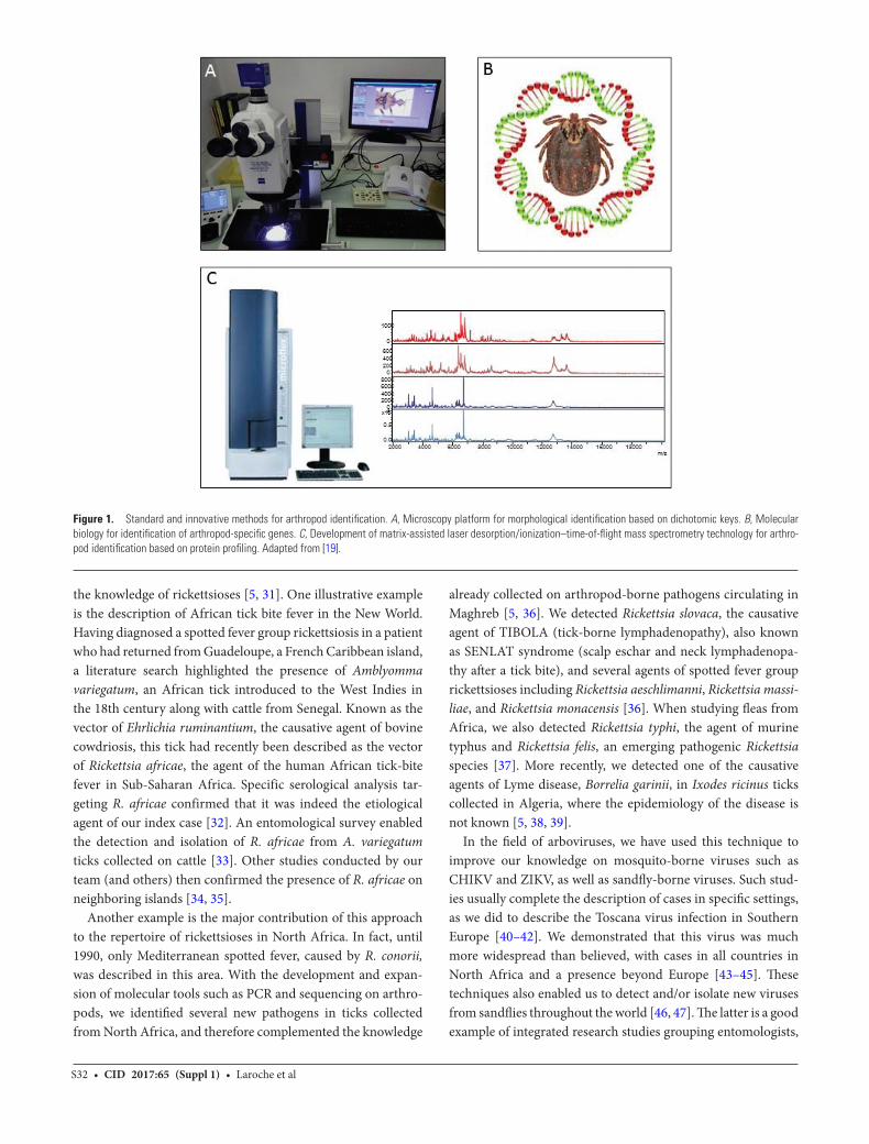
For the last 4 years, our team has developed the use of matrix-assisted laser desorption/ionization–time-of-flight mass spectrometry (MALDI-TOF MS) for arthropod identifi-cation. This technology is based on the thorough comparison of protein profiles of the submitted samples with a database of reference spectra, and has revolutionized the field of clinical microbiology. It is now routinely used for the rapid identifica-tion of bacteria and fungi from clinical samples [18]. The use of MALDI-TOF MS for the identification of arthropods involves several steps, including (i) determination of the body part of
the arthropod which will be used for MS analyses (ideally the smallest, to save the remaining body parts for further analyses); (ii) construction of a database of reference protein profiles of definitely identified specimens; (iii) completion of blind tests to check if the specimens are correctly identified when compared to the database; (iv) continual updating of the database with new species (Figure 1) [19]. We have applied this method to the identification of several hematophagous arthropods: mos-quitoes [20, 21], ticks [22], bedbugs, and triatomines (unpub-lished), using the spectra obtained from their legs, as well as fleas [23] and lice, using their cephalothorax. MALDI-TOF identification was recently validated on sand flies using their thorax, legs, and wings [24]. To date, our database includes ref-erence spectra for >60 arthropod species. This technology has been transferred to our laboratory in Dakar, Senegal, where it enabled the identification of the Ceratopogonidae, which are small insects of veterinary importance whose identification can be challenging [25]. These skills have been taught to laboratory technicians who are now fully trained to identify ticks collected on patients by MALDI-TOF MS, using the legs, while the rest of the body is used for pathogen detection through molecular biology.
More recently, we showed that MALDI-TOF MS was able to distinguish Rickettsia conorii–infected Rhipicephalus sanguineus ticks and noninfected ticks [26, 27], as well as Borrelia croci-durae–infected Ornithodoros sonrai ticks [28]. Based on these promising results, MALDI-TOF MS was challenged to detect Plasmodium parasites directly from Anopheles mosquitoes. Experimentally, Plasmodium berghei–infected mosquitoes were successfully distinguished from noninfected mosquitoes based on the spectra from their cephalothorax [29]. Furthermore, the identification of blood meal sources of malaria vectors is key information to obtain to better understand host/vector inter-actions and malaria epidemiology in endemic areas. Abdomen proteins from Anopheles gambiae that were artificially engorged on 7 distinct types of vertebrate blood were submitted for MALDI-TOF MS, resulting in the accurate determination of feeding patterns of freshly engorged mosquitoes up to 24 hours post–blood meal [30].
THE USE OF ARTHROPODS IN CATALOGING VECTOR-BORNE INFECTIOUS DISEASES
Over the last 15 years, the development of molecular tools has enabled the use of arthropods as a tool for epidemiological and geographical monitoring of the microorganisms they carry. This method has been used to obtain specific information regarding the epidemiology of a targeted microorganism, to increase the catalogue of known infectious diseases in a geographical area of interest, and to alert clinicians and microbiologists to the presence of a pathogen in a specific area. As a reference center for rickettsiology, we have used this approach to contribute to
S32 • CID 2017:65 (Suppl 1) • Laroche et al
the knowledge of rickettsioses [5, 31]. One illustrative example is the description of African tick bite fever in the New World. Having diagnosed a spotted fever group rickettsiosis in a patient who had returned from Guadeloupe, a French Caribbean island, a literature search highlighted the presence of Amblyomma variegatum, an African tick introduced to the West Indies in the 18th century along with cattle from Senegal. Known as the vector of Ehrlichia ruminantium, the causative agent of bovine cowdriosis, this tick had recently been described as the vector of Rickettsia africae, the agent of the human African tick-bite fever in Sub-Saharan Africa. Specific serological analysis tar-geting R. africae confirmed that it was indeed the etiological agent of our index case [32]. An entomological survey enabled the detection and isolation of R. africae from A. variegatum ticks collected on cattle [33]. Other studies conducted by our team (and others) then confirmed the presence of R. africae on neighboring islands [34, 35].
Another example is the major contribution of this approach to the repertoire of rickettsioses in North Africa. In fact, until 1990, only Mediterranean spotted fever, caused by R. conorii, was described in this area. With the development and expan-sion of molecular tools such as PCR and sequencing on arthro-pods, we identified several new pathogens in ticks collected from North Africa, and therefore complemented the knowledge
already collected on arthropod-borne pathogens circulating in Maghreb [5, 36]. We detected Rickettsia slovaca, the causative agent of TIBOLA (tick-borne lymphadenopathy), also known as SENLAT syndrome (scalp eschar and neck lymphadenopa-thy after a tick bite), and several agents of spotted fever group rickettsioses including Rickettsia aeschlimanni, Rickettsia massi-liae, and Rickettsia monacensis [36]. When studying fleas from Africa, we also detected Rickettsia typhi, the agent of murine typhus and Rickettsia felis, an emerging pathogenic Rickettsia species [37]. More recently, we detected one of the causative agents of Lyme disease, Borrelia garinii, in Ixodes ricinus ticks collected in Algeria, where the epidemiology of the disease is not known [5, 38, 39].
In the field of arboviruses, we have used this technique to improve our knowledge on mosquito-borne viruses such as CHIKV and ZIKV, as well as sandfly-borne viruses. Such stud-ies usually complete the description of cases in specific settings, as we did to describe the Toscana virus infection in Southern Europe [40–42]. We demonstrated that this virus was much more widespread than believed, with cases in all countries in North Africa and a presence beyond Europe [43–45]. These techniques also enabled us to detect and/or isolate new viruses from sandflies throughout the world [46, 47]. The latter is a good example of integrated research studies grouping entomologists,
Figure 1. Standard and innovative methods for arthropod identification. A, Microscopy platform for morphological identification based on dichotomic keys. B, Molecular biology for identification of arthropod-specific genes. C, Development of matrix-assisted laser desorption/ionization–time-of-flight mass spectrometry technology for arthro-pod identification based on protein profiling. Adapted from [19].

Understanding Vector-Borne Infectious Diseases • CID 2017:65 (Suppl 1) • S33
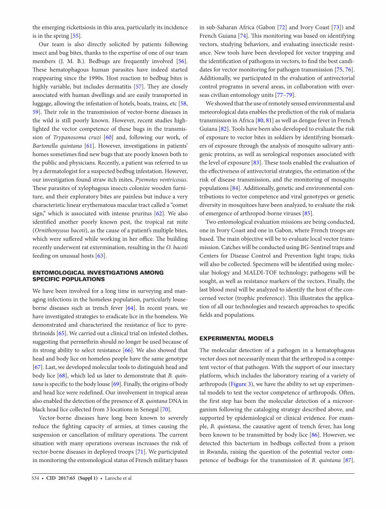
virologists, parasitologists, ecologists, epidemiologists, and medical and veterinary doctors. Classic techniques (cell culture, electron microscopy, seroneutralization) were automated and combined with molecular techniques such as real-time PCR and next-generation sequencing [48]. Collection of phleboto-mines in the field (Figure 2) [41] can be oriented from data on (i) Leishmania parasites, (ii) human/canine leishmaniasis cases, (iii) seroprevalence results for phleboviruses, and (iv) previous data indicative of phlebovirus isolation or detection. During the last decade, our group has discovered and isolated >50% of the newly described viruses transmitted by phlebotomines in the Old World: Punique and Medjerda Valley viruses in Tunisia [48, 49], Massilia virus in France [50], and Zerdali, Toros, and Adana viruses in Turkey [40, 47]. Similar studies have also iso-lated new viruses in Iran and various countries of the Balkan region (Bosnia-Herzegovina, Albania, Croatia) (unpublished data). Subsequent to the discovery of new viruses, studies aimed at providing evidence for their public health importance are increasingly being conducted; such studies have shown extremely high levels of exposure for populations living in cer-tain regions [51–53].
All this information plays a crucial role in raising the aware-ness of medical doctors about the presence of pathogens and associated arthropod-borne diseases.
ENTOMOLOGICAL INVESTIGATIONS ASSOCIATED WITH CLINICAL CASES
These investigations have been conducted primarily in the field of rickettsiology, particularly in cases where unusual seasonal or ecological parameters were encountered. Such investigations may enable a better understanding of vector behaviors, as seen when we showed that the aggressiveness toward humans of the brown dog tick, Rhipicephalus sanguineus, was significantly greater at warmer temperatures. This may explain why cases of Mediterranean spotted fever caused by R. conorii and trans-mitted by Rh. sanguineus are encountered during the warmest months in the Mediterranean area [54]. Also, when investigat-ing 2 cases of infection with Rickettsia sibirica mongolitimonae in the same family living in the south of France, we were able to identify Rhipicephalus pusillus as the potential vector, which is a tick associated with wild rabbits that can occasionally parasitize dogs and cats. These results may explain the epidemiology of
Figure 2. Centers for Disease Control and Prevention light traps, adapted for sandfly trapping. Placed in horse stables (A), near rabbit hutches and henhouses (B), and in quiet places in the shade of human habitations (C), where dogs sleep. D, Engorged female Phlebotomus perniciosus sandfly trapped in a horse stable. Adapted from [93].
S34 • CID 2017:65 (Suppl 1) • Laroche et al
the emerging rickettsiosis in this area, particularly its incidence is in the spring [55].
Our team is also directly solicited by patients following insect and bug bites, thanks to the expertise of one of our team members (J. M. B.). Bedbugs are frequently involved [56]. These hematophagous human parasites have indeed started reappearing since the 1990s. Host reaction to bedbug bites is highly variable, but includes dermatitis [57]. They are closely associated with human dwellings and are easily transported in luggage, allowing the infestation of hotels, boats, trains, etc [58, 59]. Their role in the transmission of vector-borne diseases in the wild is still poorly known. However, recent studies high-lighted the vector competence of these bugs in the transmis-sion of Trypanosoma cruzi [60] and, following our work, of Bartonella quintana [61]. However, investigations in patients’ homes sometimes find new bugs that are poorly known both to the public and physicians. Recently, a patient was referred to us by a dermatologist for a suspected bedbug infestation. However, our investigation found straw itch mites, Pyemotes ventricosus. These parasites of xylophagous insects colonize wooden furni-ture, and their exploratory bites are painless but induce a very characteristic linear erythematous macular tract called a “comet sign,” which is associated with intense pruritus [62]. We also identified another poorly known pest, the tropical rat mite (Ornithonyssus bacoti), as the cause of a patient’s multiple bites, which were suffered while working in her office. The building recently underwent rat extermination, resulting in the O. bacoti feeding on unusual hosts [63].
ENTOMOLOGICAL INVESTIGATIONS AMONG SPECIFIC POPULATIONS
We have been involved for a long time in surveying and man-aging infections in the homeless population, particularly louse-borne diseases such as trench fever [64]. In recent years, we have investigated strategies to eradicate lice in the homeless. We demonstrated and characterized the resistance of lice to pyre-thrinoids [65]. We carried out a clinical trial on infested clothes, suggesting that permethrin should no longer be used because of its strong ability to select resistance [66]. We also showed that head and body lice on homeless people have the same genotype [67]. Last, we developed molecular tools to distinguish head and body lice [68], which led us later to demonstrate that B. quin-tana is specific to the body louse [69]. Finally, the origins of body and head lice were redefined. Our involvement in tropical areas also enabled the detection of the presence of B. quintana DNA in black head lice collected from 3 locations in Senegal [70].
Vector-borne diseases have long been known to severely reduce the fighting capacity of armies, at times causing the suspension or cancellation of military operations. The current situation with many operations overseas increases the risk of vector-borne diseases in deployed troops [71]. We participated in monitoring the entomological status of French military bases
in sub-Saharan Africa (Gabon [72] and Ivory Coast [73]) and French Guiana [74]. This monitoring was based on identifying vectors, studying behaviors, and evaluating insecticide resist-ance. New tools have been developed for vector trapping and the identification of pathogens in vectors, to find the best candi-dates for vector monitoring for pathogen transmission [75, 76]. Additionally, we participated in the evaluation of antivectorial control programs in several areas, in collaboration with over-seas civilian entomology units [77–79].
We showed that the use of remotely sensed environmental and meteorological data enables the prediction of the risk of malaria transmission in Africa [80, 81] as well as dengue fever in French Guiana [82]. Tools have been also developed to evaluate the risk of exposure to vector bites in soldiers by identifying biomark-ers of exposure through the analysis of mosquito salivary anti-genic proteins, as well as serological responses associated with the level of exposure [83]. These tools enabled the evaluation of the effectiveness of antivectorial strategies, the estimation of the risk of disease transmission, and the monitoring of mosquito populations [84]. Additionally, genetic and environmental con-tributions to vector competence and viral genotypes or genetic diversity in mosquitoes have been analyzed, to evaluate the risk of emergence of arthropod-borne viruses [85].
Two entomological evaluation missions are being conducted, one in Ivory Coast and one in Gabon, where French troops are based. The main objective will be to evaluate local vector trans-mission. Catches will be conducted using BG-Sentinel traps and Centers for Disease Control and Prevention light traps; ticks will also be collected. Specimens will be identified using molec-ular biology and MALDI-TOF technology; pathogens will be sought, as well as resistance markers of the vectors. Finally, the last blood meal will be analyzed to identify the host of the con-cerned vector (trophic preference). This illustrates the applica-tion of all our technologies and research approaches to specific fields and populations.
EXPERIMENTAL MODELS
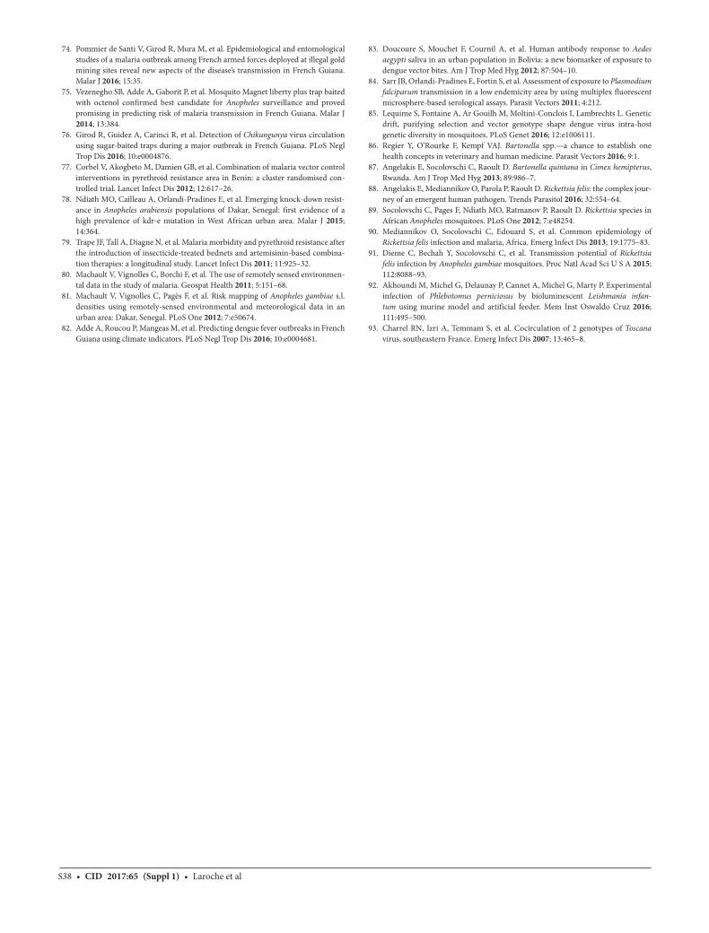
The molecular detection of a pathogen in a hematophagous vector does not necessarily mean that the arthropod is a compe-tent vector of that pathogen. With the support of our insectary platform, which includes the laboratory rearing of a variety of arthropods (Figure 3), we have the ability to set up experimen-tal models to test the vector competence of arthropods. Often, the first step has been the molecular detection of a microor-ganism following the cataloging strategy described above, and supported by epidemiological or clinical evidence. For exam-ple, B. quintana, the causative agent of trench fever, has long been known to be transmitted by body lice [86]. However, we detected this bacterium in bedbugs collected from a prison in Rwanda, raising the question of the potential vector com-petence of bedbugs for the transmission of B. quintana [87].

Understanding Vector-Borne Infectious Diseases • CID 2017:65 (Suppl 1) • S35
We confirmed this hypothesis with an experimental model using Cimex lectularius fed with B. quintana–infected blood through an artificial feeding device [61]. Furthermore, these data formed one of the first demonstrations of the potential vector role of bedbugs.
Another illustrative example of an experimental model contributing to knowledge of the emerging pathogen involves R. felis, an emerging pathogen described in 2002 [88]. In recent years, a growing number of cases have been reported around the world, and R. felis has been detected in several arthropods,
Figure 3. Pictures of arthropods of medical importance reared in the University Hospital Institute Méditerranée Infection insectary: 1. Cimex lectularius (bedbug); 2. Rhodnius prolixus (Triatominae); 3. Triatoma infestans (Triatominae); 4. Aedes albopictus larva; 5. Anopheles gambiae; 6. Aedes albopictus; 7. Anopheles stephensi; 8. Male Amblyomma variegatum; 9. Female Amblyomma variegatum; 10. Male Rhipicephalus sanguineus; 11. Female Rhipicephalus sanguineus; 12. Ctenocephalides felis (cat flea); 13. Pediculus humanus corporis (body louse). Adapted from [19]. See video at https://www.youtube.com/watch?v=BkpWb7CNTQs&sns=tw.

S36 • CID 2017:65 (Suppl 1) • Laroche et al
particularly in fleas. However, the only long-established vector has been the cat flea, Ctenocephalides felis. In 2012, R. felis was detected in mosquitoes in Africa, including Anopheles malaria vectors [89]. Moreover, the distribution of fevers of unknown origin associated with R. felis (up to 15% of cases) overlapped with malaria-endemic areas [90]. We artificially fed An. gambiae mosquitoes with blood or culture medium infected with R. felis. The bacterium was later detected in the mosquitoes, particu-larly in their saliva and salivary glands. Molecular detection of the bacterium’s DNA in mice following exposure to R. felis–in-fected An. gambiae bites revealed the ability of this mosquito to transmit this Rickettsia from one vertebrate to another. This work was the first evidence of transmission of human patho-genic bacteria by mosquitoes and introduced An. gambiae as a potential vector of R. felis in Africa [91].
Last, an experimental model of infection of Phlebotomus perniciosus with bioluminescent Leishmania infantum parasites highlighted the variability in infection intensity due to several factors such as the vector and the parasite species, but also the infection method. Artificial feeding was described as the most efficient approach to obtain high parasite loads in the exposed flies [92].
Experimental models also enabled us to assess the powerful antifeeding and insecticidal efficiency of the dinotefuran-per-methrin-pyriproxyfen ectoparasiticide on Triatoma infestans. Indeed, T. infestans bugs are vectors of Trypanosoma cruzi, the causative agent of Chagas disease, for which dogs are reservoir hosts. The prevention of domestic infection of dogs is a crucial step in the protection of domestic animals and humans (unpub-lished data).
PERSPECTIVES
The medical entomology studies conducted by our team have enabled us to decipher many aspects of vector-borne human diseases. The vast majority of our master’s and doctoral stu-dents involved in these studies come from developing countries, predominantly Africa. Most of them are financially supported by the Méditerranée Infection Foundation, and plan to create research units in their countries after being trained in Marseille.
The performance of our insectarium is based on the large diversity of arthropods we breed. It enables quick answers to epidemiological and clinical questions involving known and emerging pathogens or arthropod vectors, by developing experimental models and entomological surveys. The MALDI-TOF tool is continually challenged with identifying new arthropods as well as with the additional detection of their associated microorganisms. Indeed, we recently developed the MALDI-TOF identification of triatomines, the giant bugs responsible for the transmission of T. cruzi, the etiological agent of Chagas disease in the Americas. We also aim to assess the performance of MALDI-TOF in the concomitant identification
of 2 vector/pathogen couples, mosquitoes/Dirofilaria species and Ct. felis fleas/Bartonella species. Moreover, to improve our knowledge of vector-borne diseases, we are still conducting experimental models of infection. We have shown that An. gambiae was able to transmit R. felis, but this bacterium was detected in Aedes albopictus from Gabon by quantitative PCR, and Ae. albopictus cells support R. felis growth. These elements raise the question of the ability of Ae. albopictus mosquitoes to transmit R. felis [7]. This question will be answered by an experimental model of infection where Ae. albopictus mosquitoes will be put in a situation to reveal the acquisition and transmission of R. felis. Finally, in the national context of the French plan for a better understanding and knowledge of Lyme disease and other tick-borne diseases, we plan to create a so-called tick clinic, where patients bitten by ticks and suspected of having been infected by a tick-borne agent will have access to a doctor, microbiologists, and entomologists to investigate their condition.
NotesSupplement sponsorship. This article appears as part of the supplement
“Emerging Concepts and Strategies in Clinical Microbiology and Infectious Diseases,” sponsored by IHU Méditerranée Infection.
Potential conflicts of interest. All authors: No reported conflicts of interest. All authors have submitted the ICMJE Form for Disclosure of Potential Conflicts of Interest. Conflicts that the editors consider relevant to the content of the manuscript have been disclosed.
References1. Giribet G, Edgecombe GD. Reevaluating the arthropod tree of life. Annu Rev
Entomol 2012; 57:167–86.2. Mathison BA, Pritt BS. Laboratory identification of arthropod ectoparasites. Clin
Microbiol Rev 2014; 27:48–67.3. Simon F, Savini H, Parola P. Chikungunya: a paradigm of emergence and globali-
zation of vector-borne diseases. Med Clin North Am 2008; 92:1323–43, ix.4. Musso D, Gubler DJ. Zika virus. Clin Microbiol Rev 2016; 29:487–524.5. Parola P, Paddock CD, Socolovschi C, et al. Update on tick-borne rickettsioses
around the world: a geographic approach. Clin Microbiol Rev 2013; 26:657–702.6. Parola P, de Lamballerie X, Jourdan J, et al. Novel Chikungunya virus variant in
travelers returning from Indian Ocean islands. Emerg Infect Dis 2006; 12:1493–9.7. Parola P, Musso D, Raoult D. Rickettsia felis: the next mosquito-borne outbreak?
Lancet Infect Dis 2016; 16:1112–3.8. Aubry C, Socolovschi C, Raoult D, Parola P. Bacterial agents in 248 ticks removed
from people from 2002 to 2013. Ticks Tick Borne Dis 2016; 7:475–81.9. Faran ME, Linthicum KJ. A handbook of the Amazonian species of Anopheles
(Nyssorhynchus)(Diptera: Culcidae). 1981.10. Brunhes J, Rhaim A, Geoffroy B, Angel G, Hervy JP. Les moustiques de l’Afrique médi-
terranéenne: logiciel d’identification et d’enseignement [Software in French]. 2000.11. Estrada-Pena A, Bouattour A, Camicas JL, Walker AR. Ticks of domestic animals
in the Mediterranean region. University of Zaragoza. 2004.12. Cuisance D, Antoine Rioux J. Current status of medical and veterinary ento-
mology in France: endangered discipline or promising science? Comp Immunol Microbiol Infect Dis 2004; 27:377–92.
13. Yssouf A, Almeras L, Raoult D, Parola P. Emerging tools for identification of arthropod vectors. Future Microbiol 2016; 11:549–66.
14. Norris DE. Genetic markers for study of the anopheline vectors of human malaria. Int J Parasitol 2002; 32:1607–15.
15. Fang Q, Keirans JE, Mixson T. The use of the nuclear protein-encoding gene, RNA polymerase II, for tick molecular systematics. Exp Appl Acarol 2002; 28:69–75.
16. Smith JL, Fonseca DM. Rapid assays for identification of members of the Culex (Culex) pipiens complex, their hybrids, and other sibling species (Diptera: culici-dae). Am J Trop Med Hyg 2004; 70:339–45.
17. Lv J, Wu S, Zhang Y, et al. Assessment of four DNA fragments (COI, 16S rDNA, ITS2, 12S rDNA) for species identification of the Ixodida (Acari: Ixodida). Parasit Vectors 2014; 7:93.

Understanding Vector-Borne Infectious Diseases • CID 2017:65 (Suppl 1) • S37
18. Seng P, Rolain JM, Fournier PE, La Scola B, Drancourt M, Raoult D. MALDI-TOF-mass spectrometry applications in clinical microbiology. Future Microbiol 2010; 5:1733–54.
19. Laroche M, Almeras L, Berenger J-M, Raoult D, Parola P. Agents zoonotiques vectorisés étudiés à l’Institut Hospitalo-Universitaire Méditerranée Infection. Bulletin de l’Académie vétérinaire de France, Vol 168, No. 3. Paris: Académie vétérinaire de France, 2015:245.
20. Yssouf A, Socolovschi C, Flaudrops C, et al. Matrix-assisted laser desorption ion-ization–time of flight mass spectrometry: an emerging tool for the rapid identifi-cation of mosquito vectors. PLoS One 2013; 8:e72380.
21. Yssouf A, Parola P, Lindström A, et al. Identification of European mosquito spe-cies by MALDI-TOF MS. Parasitol Res 2014; 113:2375–8.
22. Yssouf A, Flaudrops C, Drali R, et al. Matrix-assisted laser desorption ioniza-tion-time of flight mass spectrometry for rapid identification of tick vectors. J Clin Microbiol 2013; 51:522–8.
23. Yssouf A, Socolovschi C, Leulmi H, et al. Identification of flea species using MALDI-TOF/MS. Comp Immunol Microbiol Infect Dis 2014; 37:153–7.
24. Lafri I, Almeras L, Bitam I, et al. Identification of Algerian field-caught phle-botomine sand fly vectors by MALDI-TOF MS. PLoS Negl Trop Dis 2016; 10:e0004351.
25. Sambou M, Aubadie-Ladrix M, Fenollar F, et al. Comparison of matrix-assisted laser desorption ionization-time of flight mass spectrometry and molecular biol-ogy techniques for identification of Culicoides (Diptera: ceratopogonidae) biting midges in senegal. J Clin Microbiol 2015; 53:410–8.
26. Yssouf A, Almeras L, Terras J, Socolovschi C, Raoult D, Parola P. Detection of Rickettsia spp in ticks by MALDI-TOF MS. PLoS Negl Trop Dis 2015; 9:e0003473.
27. Yssouf A, Almeras L, Berenger JM, Laroche M, Raoult D, Parola P. Identification of tick species and disseminate pathogen using hemolymph by MALDI-TOF MS. Ticks Tick Borne Dis 2015; 6:579–86.
28. Fotso Fotso A, Mediannikov O, Diatta G, et al. MALDI-TOF mass spectrometry detection of pathogens in vectors: the Borrelia crocidurae/Ornithodoros sonrai paradigm. PLoS Negl Trop Dis 2014; 8:e2984.
29. Laroche M, Almeras L, Pecchi E, et al. MALDI-TOF MS as an innovative tool for detection of Plasmodium parasites in Anopheles mosquitoes. Malar J 2017; 16:5.
30. Niare S, Berenger JM, Dieme C, et al. Identification of blood meal sources in the main African malaria mosquito vector by MALDI-TOF MS. Malar J 2016; 15:87.
31. Parola P, Paddock CD, Raoult D. Tick-borne rickettsioses around the world: emerging diseases challenging old concepts. Clin Microbiol Rev 2005; 18:719–56.
32. Parola P, Jourdan J, Raoult D. Tick-borne infection caused by Rickettsia africae in the West Indies. N Engl J Med 1998; 338:1391.
33. Parola P, Vestris G, Martinez D, Brochier B, Roux V, Raoult D. Tick-borne ricket-tiosis in Guadeloupe, the French West Indies: isolation of Rickettsia africae from Amblyomma variegatum ticks and serosurvey in humans, cattle, and goats. Am J Trop Med Hyg 1999; 60:888–93.
34. Kelly PJ. Rickettsia africae in the West Indies. Emerg Infect Dis 2006; 12:224–6.35. Parola P, Attali J, Raoult D. First detection of Rickettsia africae on Martinique, in
the French West Indies. Ann Trop Med Parasitol 2003; 97:535–7.36. Kernif T, Socolovschi C, Bitam I, Raoult D, Parola P. Vector-borne rickettsioses in
North Africa. Infect Dis Clin North Am 2012; 26:455–78.37. Leulmi H, Socolovschi C, Laudisoit A, et al. Detection of Rickettsia felis, Rickettsia
typhi, Bartonella species and Yersinia pestis in fleas (Siphonaptera) from Africa. PLoS Negl Trop Dis 2014; 8:e3152.
38. Kernif T, Messaoudene D, Ouahioune S, Parola P, Raoult D, Bitam I. Spotted fever group rickettsiae identified in Dermacentor marginatus and Ixodes ricinus ticks in Algeria. Ticks Tick Borne Dis 2012; 3:380–1.
39. Benredjem W, Leulmi H, Bitam I, Raoult D, Parola P. Borrelia garinii and Rickettsia monacensis in Ixodes ricinus ticks, Algeria. Emerg Infect Dis 2014; 20:1776–7.
40. Alkan C, Erisoz Kasap O, Alten B, de Lamballerie X, Charrel RN. Sandfly-borne phlebovirus isolations from Turkey: new insight into the sandfly fever Sicilian and sandfly fever Naples species. PLoS Negl Trop Dis 2016; 10:e0004519.
41. Charrel RN, Izri A, Temmam S, et al. Cocirculation of 2 genotypes of Toscana virus, southeastern France. Emerg Infect Dis 2007; 13:465–8.
42. Charrel RN, Bichaud L, de Lamballerie X. Emergence of Toscana virus in the Mediterranean area. World J Virol 2012; 1:135–41.
43. Es-Sette N, Ajaoud M, Bichaud L, et al. Phlebotomus sergenti a common vector of Leishmania tropica and Toscana virus in Morocco. J Vector Borne Dis 2014; 51:86–90.
44. Alkan C, Allal-Ikhlef AB, Alwassouf S, et al. Virus isolation, genetic characteriza-tion and seroprevalence of Toscana virus in Algeria. Clin Microbiol Infect 2015; 21:1040.e1–9.
45. Bichaud L, Dachraoui K, Piorkowski G, et al. Toscana virus isolated from sand-flies, Tunisia. Emerg Infect Dis 2013; 19:322–4.
46. Alkan C, Zapata S, Bichaud L, et al. Ecuador Paraiso Escondido virus, a new flavi-virus isolated from new world sand flies in Ecuador, is the first representative of a novel clade in the genus flavivirus. J Virol 2015; 89:11773–85.
47. Alkan C, Alwassouf S, Piorkowski G, et al. Isolation, genetic characterization, and seroprevalence of Adana virus, a novel phlebovirus belonging to the Salehabad virus complex, in Turkey. J Virol 2015; 89:4080–91.
48. Bichaud L, Dachraoui K, Alwassouf S, et al. Isolation, full genomic characteriza-tion and neutralization-based human seroprevalence of Medjerda Valley virus, a novel sandfly-borne phlebovirus belonging to the Salehabad virus complex in northern Tunisia. J Gen Virol 2016; 97:602–10.
49. Zhioua E, Moureau G, Chelbi I, et al. Punique virus, a novel phlebovirus, related to sandfly fever Naples virus, isolated from sandflies collected in Tunisia. J Gen Virol 2010; 91:1275–83.
50. Charrel RN, Moureau G, Temmam S, et al. Massilia virus, a novel Phlebovirus (Bunyaviridae) isolated from sandflies in the Mediterranean. Vector Borne Zoonotic Dis 2009; 9:519–30.
51. Sakhria S, Bichaud L, Mensi M, et al. Co-circulation of Toscana virus and Punique virus in northern Tunisia: a microneutralisation-based seroprevalence study. PLoS Negl Trop Dis 2013; 7:e2429.
52. Sakhria S, Alwassouf S, Fares W, et al. Presence of sandfly-borne phleboviruses of two antigenic complexes (sandfly fever Naples virus and sandfly fever Sicilian virus) in two different bio-geographical regions of Tunisia demonstrated by a microneutralisation-based seroprevalence study in dogs. Parasit Vectors 2014; 7:476.
53. Alwassouf S, Maia C, Ayhan N, et al. Neutralization-based seroprevalence of Toscana virus and sandfly fever Sicilian virus in dogs and cats from Portugal. J Gen Virol 2016; 97:2816–23.
54. Parola P, Socolovschi C, Raoult D. Deciphering the relationships between Rickettsia conorii conorii and Rhipicephalus sanguineus in the ecology and epidemiology of Mediterranean spotted fever. Ann N Y Acad Sci 2009; 1166:49–54.
55. Edouard S, Parola P, Socolovschi C, Davoust B, La Scola B, Raoult D. Clustered cases of Rickettsia sibirica mongolitimonae infection, France. Emerg Infect Dis 2013; 19:337–8.
56. Delaunay P, Blanc V, Del Giudice P, et al. Bedbugs and infectious diseases. Clin Infect Dis 2011; 52:200–10.
57. Delaunay P, Blanc V, Dandine M, et al. Bedbugs and healthcare-associated derma-titis, France. Emerg Infect Dis 2009; 15:989–90.
58. Delaunay P. Human travel and traveling bedbugs. J Travel Med 2012; 19:373–9.59. Bernardeschi C, Le Cleach L, Delaunay P, Chosidow O. Bed bug infestation. BMJ
2013; 346:f138.60. Salazar R, Castillo-Neyra R, Tustin AW, Borrini-Mayorí K, Náquira C, Levy MZ.
Bed bugs (Cimex lectularius) as vectors of Trypanosoma cruzi. Am J Trop Med Hyg 2015; 92:331–5.
61. Leulmi H, Bitam I, Berenger JM, et al. Competence of Cimex lectularius bed bugs for the transmission of Bartonella quintana, the agent of trench fever. PLoS Negl Trop Dis 2015; 9:e0003789.
62. Del Giudice P, Blanc-Amrane V, Bahadoran P, et al. Pyemotes ventricosus derma-titis, southeastern France. Emerg Infect Dis 2008; 14:1759–61.
63. Alexander JO. Arthropods and human skin. London: Springer Science & Business Media, 2012.
64. Brouqui P, Raoult D. Arthropod-borne diseases in homeless. Ann N Y Acad Sci 2006; 1078:223–35.
65. Drali R, Benkouiten S, Badiaga S, Bitam I, Rolain JM, Brouqui P. Detection of a knockdown resistance mutation associated with permethrin resistance in the body louse Pediculus humanus corporis by use of melting curve analysis genotyp-ing. J Clin Microbiol 2012; 50:2229–33.
66. Benkouiten S, Drali R, Badiaga S, et al. Effect of permethrin-impregnated under-wear on body lice in sheltered homeless persons: a randomized controlled trial. JAMA Dermatol 2014; 150:273–9.
67. Veracx A, Rivet R, McCoy KD, Brouqui P, Raoult D. Evidence that head and body lice on homeless persons have the same genotype. PLoS One 2012; 7:e45903.
68. Drali R, Boutellis A, Raoult D, Rolain JM, Brouqui P. Distinguishing body lice from head lice by multiplex real-time PCR analysis of the Phum_PHUM540560 gene. PLoS One 2013; 8:e58088.
69. Drali R, Sangaré AK, Boutellis A, et al. Bartonella quintana in body lice from scalp hair of homeless persons, France. Emerg Infect Dis 2014; 20:907–8.
70. Boutellis A, Veracx A, Angelakis E, et al. Bartonella quintana in head lice from Sénégal. Vector Borne Zoonotic Dis 2012; 12:564–7.
71. Pages F, Faulde M, Orlandi-Pradines E, Parola P. The past and present threat of vector-borne diseases in deployed troops. Clin Microbiol Infect 2010; 16:209–24.
72. Mourou JR, Coffinet T, Jarjaval F, et al. Malaria transmission in Libreville: results of a one year survey. Malar J 2012; 11:40.
73. Girod R, Orlandi-Pradines E, Rogier C, Pages F. Malaria transmission and insec-ticide resistance of Anopheles gambiae (Diptera: Culicidae) in the French military camp of Port-Bouët, Abidjan (Côte d’Ivoire): implications for vector control. J Med Entomol 2006; 43:1082–7.
S38 • CID 2017:65 (Suppl 1) • Laroche et al
74. Pommier de Santi V, Girod R, Mura M, et al. Epidemiological and entomological studies of a malaria outbreak among French armed forces deployed at illegal gold mining sites reveal new aspects of the disease’s transmission in French Guiana. Malar J 2016; 15:35.
75. Vezenegho SB, Adde A, Gaborit P, et al. Mosquito Magnet liberty plus trap baited with octenol confirmed best candidate for Anopheles surveillance and proved promising in predicting risk of malaria transmission in French Guiana. Malar J 2014; 13:384.
76. Girod R, Guidez A, Carinci R, et al. Detection of Chikungunya virus circulation using sugar-baited traps during a major outbreak in French Guiana. PLoS Negl Trop Dis 2016; 10:e0004876.
77. Corbel V, Akogbeto M, Damien GB, et al. Combination of malaria vector control interventions in pyrethroid resistance area in Benin: a cluster randomised con-trolled trial. Lancet Infect Dis 2012; 12:617–26.
78. Ndiath MO, Cailleau A, Orlandi-Pradines E, et al. Emerging knock-down resist-ance in Anopheles arabiensis populations of Dakar, Senegal: first evidence of a high prevalence of kdr-e mutation in West African urban area. Malar J 2015; 14:364.
79. Trape JF, Tall A, Diagne N, et al. Malaria morbidity and pyrethroid resistance after the introduction of insecticide-treated bednets and artemisinin-based combina-tion therapies: a longitudinal study. Lancet Infect Dis 2011; 11:925–32.
80. Machault V, Vignolles C, Borchi F, et al. The use of remotely sensed environmen-tal data in the study of malaria. Geospat Health 2011; 5:151–68.
81. Machault V, Vignolles C, Pagès F, et al. Risk mapping of Anopheles gambiae s.l. densities using remotely-sensed environmental and meteorological data in an urban area: Dakar, Senegal. PLoS One 2012; 7:e50674.
82. Adde A, Roucou P, Mangeas M, et al. Predicting dengue fever outbreaks in French Guiana using climate indicators. PLoS Negl Trop Dis 2016; 10:e0004681.
83. Doucoure S, Mouchet F, Cournil A, et al. Human antibody response to Aedes aegypti saliva in an urban population in Bolivia: a new biomarker of exposure to dengue vector bites. Am J Trop Med Hyg 2012; 87:504–10.
84. Sarr JB, Orlandi-Pradines E, Fortin S, et al. Assessment of exposure to Plasmodium falciparum transmission in a low endemicity area by using multiplex fluorescent microsphere-based serological assays. Parasit Vectors 2011; 4:212.
85. Lequime S, Fontaine A, Ar Gouilh M, Moltini-Conclois I, Lambrechts L. Genetic drift, purifying selection and vector genotype shape dengue virus intra-host genetic diversity in mosquitoes. PLoS Genet 2016; 12:e1006111.
86. Regier Y, O’Rourke F, Kempf VAJ. Bartonella spp.—a chance to establish one health concepts in veterinary and human medicine. Parasit Vectors 2016; 9:1.
87. Angelakis E, Socolovschi C, Raoult D. Bartonella quintana in Cimex hemipterus, Rwanda. Am J Trop Med Hyg 2013; 89:986–7.
88. Angelakis E, Mediannikov O, Parola P, Raoult D. Rickettsia felis: the complex jour-ney of an emergent human pathogen. Trends Parasitol 2016; 32:554–64.
89. Socolovschi C, Pages F, Ndiath MO, Ratmanov P, Raoult D. Rickettsia species in African Anopheles mosquitoes. PLoS One 2012; 7:e48254.
90. Mediannikov O, Socolovschi C, Edouard S, et al. Common epidemiology of Rickettsia felis infection and malaria, Africa. Emerg Infect Dis 2013; 19:1775–83.
91. Dieme C, Bechah Y, Socolovschi C, et al. Transmission potential of Rickettsia felis infection by Anopheles gambiae mosquitoes. Proc Natl Acad Sci U S A 2015; 112:8088–93.
92. Akhoundi M, Michel G, Delaunay P, Cannet A, Michel G, Marty P. Experimental infection of Phlebotomus perniciosus by bioluminescent Leishmania infan-tum using murine model and artificial feeder. Mem Inst Oswaldo Cruz 2016; 111:495–500.
93. Charrel RN, Izri A, Temmam S, et al. Cocirculation of 2 genotypes of Toscana virus, southeastern France. Emerg Infect Dis 2007; 13:465–8.