medical management of childhood lead poisoning · medical management of lead poisoning in children...
TRANSCRIPT
Chapter 8 Medical Management of Childhood Lead Poisoning Contents In Brief: Recommended Actions Based on Blood Lead Level (CDC, 2012) .............................. 8.2
Introduction .............................................................................................................................. 8.3
Diagnostic and Follow-up Testing ............................................................................................ 8.3
Clinical Assessment of Children with Lead Poisoning .............................................................. 8.4
What the Erythrocyte Protoporphyrin (EP) Test Measures ................................................ 8.6
Iron Deficiency, Pica and Lead Poisoning ......................................................................... 8.7
Chelation Therapy ............................................................................................................. 8.9
Lead Education for the Family ................................................................................................ 8.11
Coordinating Care .................................................................................................................. 8.11
Monitoring a Child’s Neurodevelopmental Progress ............................................................... 8.12
References ............................................................................................................................ 8.13
(P-00660, 11/2014)
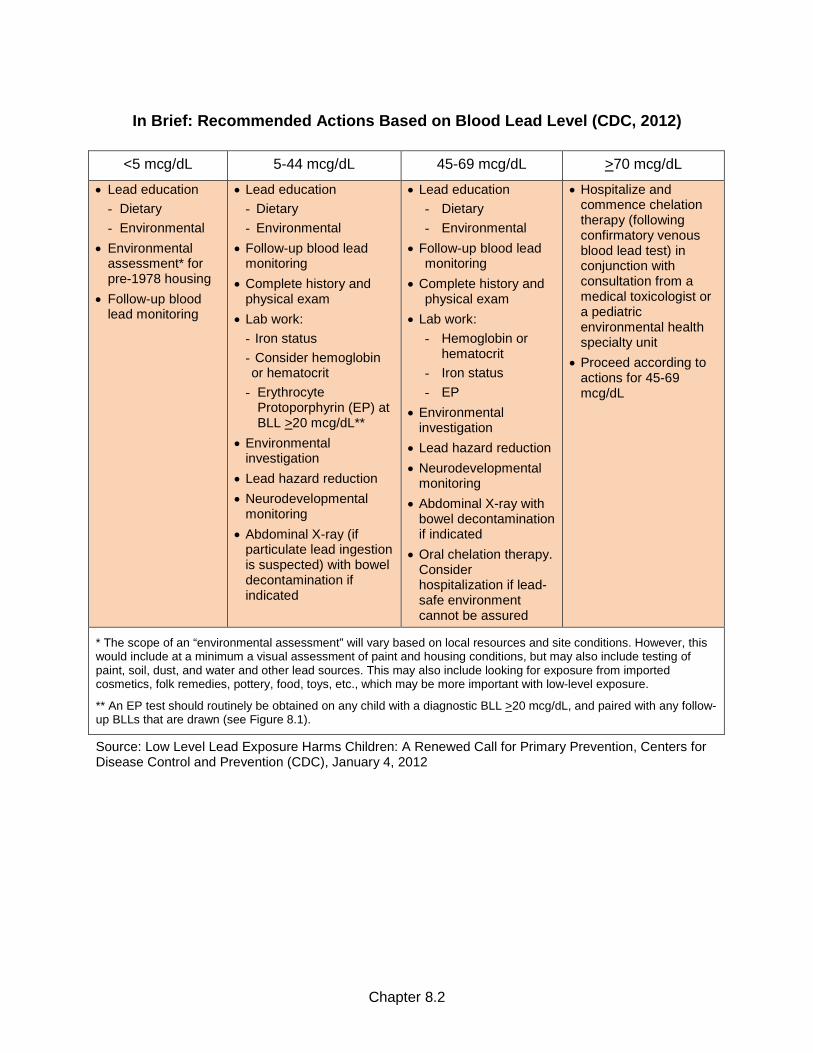
In Brief: Recommended Actions Based on Blood Lead Level (CDC, 2012)
<5 mcg/dL 5-44 mcg/dL 45-69 mcg/dL >70 mcg/dL
• Lead education - Dietary - Environmental
• Environmental assessment* for pre-1978 housing
• Follow-up blood lead monitoring
• Lead education - Dietary - Environmental
• Follow-up blood lead monitoring
• Complete history and physical exam
• Lab work: - Iron status - Consider hemoglobin or hematocrit
- Erythrocyte Protoporphyrin (EP) at BLL >20 mcg/dL**
• Environmental investigation
• Lead hazard reduction • Neurodevelopmental
monitoring • Abdominal X-ray (if
particulate lead ingestion is suspected) with bowel decontamination if indicated
• Lead education - Dietary - Environmental
• Follow-up blood lead monitoring
• Complete history and physical exam
• Lab work: - Hemoglobin or
hematocrit - Iron status - EP
• Environmental investigation
• Lead hazard reduction • Neurodevelopmental
monitoring • Abdominal X-ray with
bowel decontamination if indicated
• Oral chelation therapy. Consider hospitalization if lead-safe environment cannot be assured
• Hospitalize and commence chelation therapy (following confirmatory venous blood lead test) in conjunction with consultation from a medical toxicologist or a pediatric environmental health specialty unit
• Proceed according to actions for 45-69 mcg/dL
* The scope of an “environmental assessment” will vary based on local resources and site conditions. However, this would include at a minimum a visual assessment of paint and housing conditions, but may also include testing of paint, soil, dust, and water and other lead sources. This may also include looking for exposure from imported cosmetics, folk remedies, pottery, food, toys, etc., which may be more important with low-level exposure.
** An EP test should routinely be obtained on any child with a diagnostic BLL >20 mcg/dL, and paired with any follow-up BLLs that are drawn (see Figure 8.1).
Source: Low Level Lead Exposure Harms Children: A Renewed Call for Primary Prevention, Centers for Disease Control and Prevention (CDC), January 4, 2012
Chapter 8.2

Introduction Clinicians have an important role in preventing lead exposure and in managing lead-exposed children. This role is described in Low Level Lead Exposure Harms Children: A Renewed Call for Primary Prevention (CDC, 2012, January 4), and includes: 1. Screening questions, outreach and education to minimize exposures prior to blood lead
testing; 2. Emphasizing healthy nutrition and/or dietary supplements to reduce absorption; 3. Blood lead testing to promptly identify exposed children, for whom primary prevention has
failed; 4. Intervening appropriately when clinically indicated; 5. Overseeing ongoing monitoring of children with elevated blood lead levels (EBLLs), defined
as levels at or above the reference value (i.e., 5 mcg/dL); 6. Coordinating efforts with parents and local and state authorities to minimize risks to
individual children and to assist communities in their primary prevention efforts. Medical management of lead poisoning in children has been largely predicated on a secondary prevention model, i.e., intervention after an elevated blood lead level has been detected, usually prior to the onset of symptoms. Screening programs are the main vehicle for identifying children with lead poisoning. Once a child is identified to be at risk for lead poisoning, early detection is provided by physicians through blood lead screening tests (see Chapter 5: Screening and Diagnosis of Lead Poisoning).
Diagnostic and Follow-up Testing A diagnosis of lead poisoning is made based on a venous blood test. When a child has a capillary blood lead test >5 mcg/dL, a diagnostic venous blood test should be obtained to assure accuracy. See Table 8.1 for the recommended schedule for obtaining a confirmatory venous sample (CDC, 2012). In general, the higher the blood lead level (BLL), the sooner the confirmatory test should be done. The CDC recommends that BLLs of 10 – 44 mcg/dL are confirmed within 1 week – 1 month, noting that the higher the BLL on the screening test, the more urgent the need for confirmatory testing. Children whose BLL is at the upper end of this range should receive a confirmatory test in approximately one week if possible. Table 8.1. Recommended schedule for obtaining a confirmatory venous sample (CDC, 2012) Blood lead level (mcg/dL)
Time to confirmation testing
≥ 5‒9 1 – 3 months
10–44 1 week – 1 month*
45–59 48 hours
60–69 24 hours
≥ 70 Urgently as emergency test
* The higher the BLL on the screening test, the more urgent the need for confirmatory testing. Source: Low Level Lead Exposure Harms Children: A Renewed Call for Primary Prevention, Centers for Disease Control and Prevention, January 4, 2012.
Chapter 8.3

In the event that it’s not possible to obtain a confirmatory venous sample from the child, a second capillary sample drawn within 12 weeks of the initial screening test can be considered a confirmatory test. This is consistent with the standard surveillance definitions used by the CDC to classify confirmed and unconfirmed elevated BLLs. If the second capillary test result is elevated, all follow-up tests should be performed on venous samples. When lead poisoning is diagnosed, follow-up tests should be performed on venous blood samples to monitor the child’s BLL and to evaluate the effectiveness of interventions. The scheduling of follow-up tests depends on the diagnostic BLL (see Table 8.2). Even in the best laboratories, variations in test results of +2 mcg/dL are normal and are well within the acceptable lab error. Multiple blood lead tests are needed over time to examine the true trend in a child’s actual BLLs. Blood lead levels that rise may be indicative of an unrecognized source of exposure, inappropriate abatement activities, failure to mitigate the identified hazard, or the redistribution of lead stores within the child’s body. Table 8.2. Schedule for follow-up blood lead testinga (CDC, 2012) Venous blood lead level (mcg/dL)
Early follow-up testing (2 – 4 tests after identification)
Later follow-up testing after blood lead level declining
≥ 5–9 3 months * 6 – 9 months
10–19 1 – 3 months * 3 – 6 months
20–24 1 – 3 months * 1 – 3 months
25–44 2 weeks – 1 month 1 month
≥ 45 As soon as possible As soon as possible a Seasonal variation of BLLs exists and may be more apparent in colder climate areas. Greater exposure in the summer months may necessitate more frequent follow-ups. * Some case managers or clinicians may choose to repeat blood lead tests on all new patients within a month to ensure that their BLL is not rising more quickly than anticipated.
Source: Low Level Lead Exposure Harms Children: A Renewed Call for Primary Prevention, Centers for Disease Control and Prevention, January 4, 2012.
Clinical Assessment of Children with Lead Poisoning Table 8.3 provides a summary of the components of clinical assessment. These components are based on the experience of clinicians who have treated lead-poisoned children, and should not be seen as rigid rules but as a guide for clinical decisions. Today, most children with lead poisoning have no symptoms. The detrimental effects of BLLs below 45 mcg/dL are often subclinical and may include neurodevelopmental impairment often apparent only at a later age. It is critical that the primary care provider (PCP) and case manager not equate the absence of clinical symptoms, physical abnormalities, or abnormal laboratory results with an absence of toxicity.
Chapter 8.4

Table 8.3. Components of clinical assessment
Component Content Action Steps
Medical History Ask about: • Symptoms (most children with lead
poisoning are asymptomatic). • Developmental history. • Mouthing activities. • Pica behaviors. • Previous BLL measurements. • Family history of lead poisoning.
If there are delays or lags in developmental progress, the child should be referred to an early intervention program for further assessment.
Environmental History
Ask about: • The age, condition and how long they
have lived at the primary residence. • Remodeling, renovation, or repainting
within the last year in the home. • Ask the same questions about other
places the child spends time (including secondary homes and daycare) or previous residences.
• Occupational and hobby histories of adults with whom the child spends time.
• Use of imported dishes, cosmetics, toys, medicines.
• Local environmental risk factors that may be provided by the local health department.
Refer to the local health department for further assessment, environmental investigation, and lead hazard reduction.
Nutritional History
Ask about: • Usual foods eaten and eating patterns. • WIC or other food program participation. • The child’s iron status, using appropriate
laboratory tests.
Provide treatment for iron deficiency if diagnosed.
Refer for nutritional counseling.
Refer to WIC if income eligible.
Physical Examination
Pay particular attention to the neurodevelopmental examination and the child’s psychosocial and language development.
Findings of any delay in language, neurobehavioral or cognitive problems should result in referral to appropriate programs.
During early school years, further examinations are necessary to facilitate entry into appropriate educational programs.
Source: Adapted from Screening Young Children for Lead Poisoning, Centers for Disease Control and Prevention, 1997.
Chapter 8.5

What the Erythrocyte Protoporphyrin (EP) Test Measures Protoporphyrin is the last precursor in synthesis of heme, the oxygen-carrying component of red blood cells (erythrocytes). Small amounts of protoporphyrin are normally present in erythrocytes, hence the term erythrocyte protoporphyrin. Pathological conditions that impair heme synthesis also cause elevations in EP concentrations. The majority (90%) of EP in the blood is bound to zinc, and is referred to as zinc protoporphyrin (ZP). Because the life span of erythrocytes in the blood stream is 90-120 days, the result of an individual EP test reflects the average effect on heme synthesis over 90-120 days. This makes the EP tests an ideal partner with BLLs, which can fluctuate over a shorter period of time, to tell the story of lead poisoning. The terminology associated with EP test results can be confusing. You may see an EP result referred to as erythrocyte protoporphyrin (EP), zinc protoporphyrin (ZP), or free erythrocyte protoporphyrin (FEP). Technically these terms refer to different substances. However, in practice they are often used interchangeably, and test results can generally be interpreted in identical fashion. The confusion is the result of early methods of laboratory analysis and historical gaps in knowledge about the nature of blood protoporphyrin. Reporting Units – EP test results are most commonly reported as: • µmol EP/mol Heme: molar ratio of protoporphyrin to heme • µg EP or ZP/dL Whole Blood: micrograms per deciliter of whole blood concentration units of
EP or ZP When reported in µg/dL (or mcg/dL) reporting units, EP and ZP results are approximately equivalent. A value exceeding 35 µg/dL is widely accepted as indicative of pathology. Results exceeding 70 µmol/mol are accepted as indicative of pathology. An EP level higher than the threshold value does not indicate the reason for the elevation; further tests for iron deficiency and/or lead poisoning must be performed. Usefulness of Erythrocyte Protoporphyrin Testing – Lead in the blood begins to cause an increase in EP at levels of 15-20 mcg/dL. As the lead level rises, the EP level rises exponentially. Paired results of EP and BLL can provide information on the effect, extent, and duration of lead exposure. An elevated BLL along with a normal or near-normal EP may indicate that the lead exposure has been recent and/or short term. An elevated EP level with a minimal increase in BLL may indicate a higher past lead exposure and a continuing body burden of lead. Elevation of both EP and BLL may indicate prolonged and ongoing lead exposure. Figure 8.1 is an article written by Dr. Margaret Layde, former Assistant Professor Medical College of Wisconsin, Downtown Health Center, Milwaukee, which describes the usefulness of the EP test in monitoring children with a BLL >20 mcg/dL and post-chelation. In general, an EP test should routinely be obtained on any child with a diagnostic BLL >20 mcg/dL, and paired with any follow-up BLLs that are drawn.
Chapter 8.6
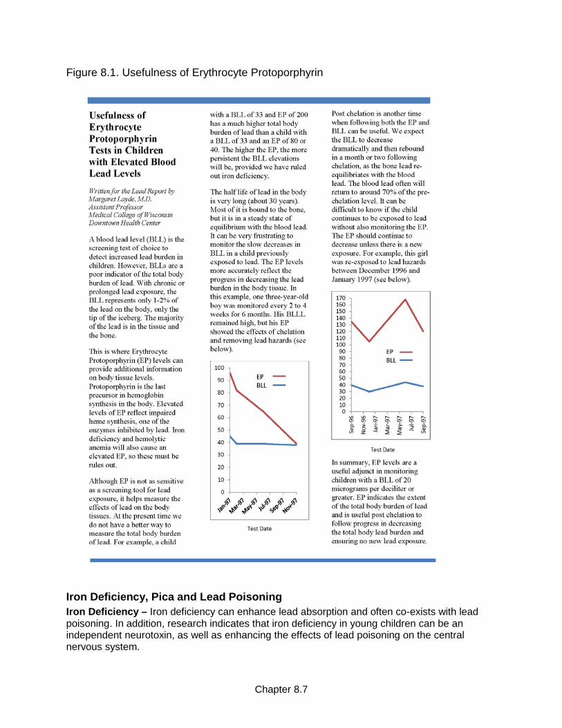
Figure 8.1. Usefulness of Erythrocyte Protoporphyrin
Iron Deficiency, Pica and Lead Poisoning Iron Deficiency – Iron deficiency can enhance lead absorption and often co-exists with lead poisoning. In addition, research indicates that iron deficiency in young children can be an independent neurotoxin, as well as enhancing the effects of lead poisoning on the central nervous system.
Chapter 8.7

Adequate iron intake lowers lead absorption, and should be considered an essential secondary tool to protect children from absorbing lead they ingest from their environments. While lead exposure does not begin to disrupt red blood cell production until BLLs reach approximately 40 mcg/dL, low iron stores promote absorption of lead at any BLL. Many U.S. children 1 to 2 years of age have daily iron intake below recommended amounts. When exposed to lead hazards, these children may see the lasting effects on cognitive development due to both iron deficiency and the long-lasting negative effects due to lead. All children with BLLs >5 mcg/dL should be evaluated for iron deficiency. Several tests are used to determine the iron status of a child, but they vary as to their sensitivity and specificity in identifying the cause of iron deficiency. An EP test is a good screening tool but must be followed by other diagnostic tests to determine the exact cause of iron deficiency. An increase in EP is the first biochemical change in erythrocytes due to insufficient iron levels. The advantage of the EP test to measure iron sufficiency is that it reflects iron status in bone marrow, and is more stable than other tests. The disadvantages are that EP is slow to change as a result of dietary iron supplements, and it is non-specific as to the cause of the deficit. Otitis media and respiratory infections in children can cause EP elevations, and are a complicating factor in interpreting test results. The EP can also be elevated due to liver disease and malignancy. Hematocrit and hemoglobin are crude measures of iron status, reflecting only cases of frank anemia. Serum iron and iron binding capacity (transferrin saturation) and ferritin are the most sensitive indicators of iron status. An abnormally low ratio of serum iron to iron binding capacity (transferrin saturation) of 0.2 is consistent with iron deficiency. The serum ferritin level, however, is the most definitive and accurate indication of overall iron status, although it is an acute phase reactant and may be falsely elevated in sick children; a value <12 mcg/dL indicates iron deficiency If iron deficiency is diagnosed, treatment should begin along with treatment of the lead exposure. [Note: Children receiving BAL (dimercaprol) as a chelating agent should not be treated for iron deficiency until the drug therapy is completed.] See Chapter 9 for more information on nutrition and childhood lead poisoning. Pica – Although formal pica definitions vary, the behavior common to all definitions of pica is a pattern of deliberate ingestion of nonfood items. For example, MedlinePlus Medical Encyclopedia defines pica as “a pattern of eating non-food materials, such as dirt or paper.” According to the DSM-5, to be diagnosed with pica a person must display: • Persistent eating of non-nutritive substances for a period of at least one month.
• The eating of non-nutritive substances is inappropriate to the developmental level of the individual.
• The eating behaviour is not part of a culturally supported or socially normative practice.
• If occurring in the presence of another mental disorder (e.g. autistic spectrum disorder), or during a medical condition (e.g. pregnancy), it is severe enough to warrant independent clinical attention.
Note: Pica often occurs with other mental health disorders associated with impaired functioning.
Chapter 8.8

Materials ingested pica can be benign or potentially harmful. There is a wide range of items associated with pica, such as chalk, newsprint, ice, pencil erasers, paint chips, dirt, clay, and pottery. Pica has long been recognized as a risk factor for lead poisoning. Lead poisoning can be associated with pica if the child is consuming substances that are contaminated with lead, such as lead-contaminated soil or paint chips. Pica is sometimes associated with anemia and other nutritional deficiencies, e.g., iron or zinc. Pica is most often observed in pregnant women, immigrant communities, and young children. Parents may underreport their child’s pica behavior because of embarrassment, or they may not be aware that the behavior is worth reporting (Rose, E. A., Porcerelli, J. H., & Neale, A. V., 2000). Pica is often discovered when a complication, such as lead poisoning, is diagnosed and careful questioning follows about eating habits. An open discussion with the family about favorite foods and nonfood substances might aid in the diagnosis. If pica is suspected, but parents are unaware of the behavior, physicians should ask that parents and caregivers keep records of observations of the child’s solitary play. Abdominal radiographs may be useful in determining whether children are currently ingesting lead-contaminated non-food items, including paint chips. They are particularly useful when children have an unexpected acute rise in BLL or are not responding to case management as expected. Treatment for pica may vary by patient and suspected cause (e.g., child, developmentally disabled or pregnant) and may emphasize psychosocial, environmental and family-guidance approaches. Diagnosis and treatment of pica should begin by assessing and addressing any missing nutrients or other medical problems, such as lead poisoning. Behavior-based treatment options can be useful, such as teaching the child through positive reinforcement which foods are good and which ones they should not eat.
Chelation Therapy The single most important factor in management of childhood lead poisoning is reducing the child’s exposure to lead; some children, however, will benefit from chelation therapy. The CDC recommends chelation therapy for children with a venous BLL >45 mcg/dL to reduce the BLL more quickly. Chelation therapy is not a substitute for effective and rapid environmental interventions and should only be used as part of an integrated environmental and medical approach to treating children with lead poisoning. Children receiving chelation therapy for treatment of lead poisoning require special care and consideration by the health care team. Primary care providers should consult with an expert in the management of lead chemotherapy prior to initiating chelation therapy. The PCP can contact the Poison Center at 1-800-222-1222 for information on treatment. Public health nurses should be in communication with the child’s physician regularly, to discuss the plan of care, follow-up, and to assure that the child is in a lead-safe environment when receiving chelation. There are several drugs, or chelating agents, that can be used in the treatment of lead poisoning. These drugs are capable of binding or chelating lead and deplete the soft and hard (skeletal) tissues of lead and thus reduce its acute toxicity. All drugs have potential side effects and must be used with caution. The American Academy of Pediatrics Committee on Drugs published “Treatment Guidelines for Lead Exposure in Children,” which contains a good summary of chelation with the most commonly used agents.
Chapter 8.9

A commonly used oral chelating agent is succimer (generic name). The product name for this drug is Chemet. The abbreviation for its chemical name is DMSA. Research found that chelation therapy with Succimer lowered average BLLs for about six months but resulted in no benefits in cognitive, behavioral and neuromotor endpoints. When to Start Chelation Therapy – As noted above, a child with a venous BLL >45 mcg/dL should be removed from the source(s) of lead exposure and treated promptly with appropriate chelating agents. If the BLL is between 45-69 mcg/dL, a second venous BLL should be drawn before initiating chelation to assure that therapy is based on the most recent and reliable information possible. If the BLL is >70 mcg/dL, the child should be hospitalized and chelation therapy should be initiated immediately while the second venous BLL is pending. The Child’s Environment during Chelation – Clinicians and public health professionals should assure that the child is living in a lead-safe environment before chelation is started. The initiation of outpatient chelation may need to be delayed until a lead-safe environment can be found. If the child receives chelation as a hospital patient, discharge may need to be delayed until the child’s home is determined to be lead-safe or an alternate lead-safe location can be found for the child to stay temporarily upon discharge. Chelation Information for the Family – Families whose children are receiving chelation therapy need adequate information for chelation to have a successful outcome. This education should address the following topics: • The need and importance of a lead-safe environment during and after chelation. It is often
difficult for families to secure a lead-safe place at the same time the child is hospitalized or started on a new medicine. However, it is one of the most important tasks for them to undertake during this time.
• The name of the drug, dose, route of administration, schedule, and side effects of the chelating agent being used. This is especially important if the parent is responsible for administering an oral chelating drug to the child.
• The importance of follow-up blood lead and EP testing (see below). Blood Lead and Erythrocyte Protoporphyrin Tests after Chelation – Post-chelation venous blood lead and EP levels should be obtained every few weeks for several months. Within a month or two after chelation is completed, the BLL may rebound to around 70 percent of the pre-chelation level as lead is released from the bone and re-enters the bloodstream. If the BLL rebounds to 45 mcg/dL or greater, chelation may need to be repeated. The EP level, in combination with a BLL, can be useful in differentiating between post-chelation blood lead rebound and ongoing exposure to lead. The EP level should continue to decrease after chelation unless there is a new exposure. An increase in both the BLL and EP level after chelation is an indication of re-exposure to lead. Further investigation should be done in this situation to identify the ongoing, or new, source of lead exposure. Unapproved “Chelation” Drugs – In October 2010, the U.S. Food and Drug Administration (FDA) warned eight companies to stop selling so-called “chelation” products that claim to treat a range of disorders from autism to Alzheimer’s disease. The FDA said the companies have not proven their products are safe and effective in treating autism spectrum disorder, cardiovascular disease, macular degeneration, Parkinson’s disease or any other serious illness. Some of the companies also claim their products can detect the presence of heavy metals in the body in an attempt to justify the need for chelation therapy. FDA said consumers should avoid non-
Chapter 8.10
prescription products offered for chelation or detoxification. FDA-approved chelating agents are available by prescription only and are approved for use in specific indications such as the treatment of lead poisoning and iron overload. The agency says even the prescription medications carry significant risks, and they should only be used with medical supervision.
Lead Education for the Family Health care providers should be aware that even at the lowest BLLs, there is action they can take to help the parents prevent a further increase in the child’s BLL. They should offer information to the parents about the meaning of the elevated blood lead test results, sources of lead exposure, steps the parents can take to protect their child, and the need for ongoing medical follow-up and blood lead testing. Education is one of the most important components of medical management. (See Chapter 4 for a detailed discussion about education strategies and resources.)
Coordinating Care Children with lead poisoning require comprehensive services to address a range of needs. This is best accomplished with a team of professionals. The local health department (LHD) provides case management services and environmental investigation of the child’s home. When lead hazards are identified in the home, the property owner is responsible for eliminating the lead hazards identified as causing the child’s lead poisoning. The physician provides ongoing assessment through age-appropriate physical exams, follow-up venous blood lead tests, chelation therapy if appropriate, and long-term monitoring for the development of cognitive, learning and behavioral deficits. The caregiver must be diligent in implementing steps to prevent ongoing lead exposure and provide support for the child. If a child presents without symptoms, the child’s PCP and case manager may have trouble convincing the child’s caregiver of the importance of suggested interventions. The PCP and case manager should manage each child individually, taking into consideration the child’s BLL and the ability of caregivers to cooperate and implement interventions. Public health staff typically coordinates the follow-up care provided to the child and family. LHD staff should assure that the PCP is included in discussing, planning and providing services so the public and private health care systems function as a team. Effective interactions between private health care providers and public health will lead to the most effective treatment of a child with lead poisoning. These interactions begin with communication about the results of screening and follow-up tests, and extend to physical examinations, developmental assessments, nutritional assessments, environmental interventions, and education and referrals. Both the child and the family benefit from efforts by public health and health care providers to complement each other’s work. When a diagnosis of lead poisoning is made, identification and control of ongoing sources of lead exposure for the child should be the highest priority. Health care providers should coordinate patient care with the LHD to assure prompt investigation and control of the sources of lead exposure. Although housing-based intervention services are typically outside the clinician’s role, medical and environmental interventions should be implemented simultaneously to best protect the child. Childhood lead exposure typically results from sub-standard housing. Families with limited housing choices are more likely to live in deteriorated housing, and may also face other
Chapter 8.11
problems such as unemployment, poverty, lack of routine medical care, poor nutrition, and instability. Families of children with lead poisoning may also need other support, and a follow-up team can refer them to other services for which they may be eligible.
Monitoring a Child’s Neurodevelopmental Progress Long-term follow-up of lead-poisoned children requires attention to the developmental and neurobehavioral effects of lead poisoning. Neurodevelopmental monitoring should continue long after a child is initially diagnosed with lead poisoning as many deficits will not manifest themselves until after a child starts school. This can be challenging, depending on the consistency of the child's source of medical care. Health care providers should include a child’s history of lead poisoning in the problem list maintained in the child’s medical record. If a child changes his or her PCP, this will ensure the information is transmitted to the next provider. Public health involvement routinely ends when the child's BLL drops and the source of lead exposure has been eliminated. However, public health professionals can play a key role in assuring that the family and the primary health care provider are aware that the neurodevelopmental status of the lead-poisoned child should be evaluated on an ongoing basis. (See Chapter 10 for a detailed discussion of neurodevelopmental surveillance for lead-poisoned children.)
Chapter 8.12

References American Academy of Pediatrics. (1995), Treatment Guidelines for Lead Exposure in Children, Committee on Drugs, Pediatrics, 96(1):155-160. http://pediatrics.aappublications.org/content/96/1/155.full.pdf American Academy of Pediatrics. (2005). Lead Exposure in Children: Prevention, Detection, and Management. Pediatrics, 116(4):1036-1046. http://pediatrics.aappublications.org/content/116/4/1036.full.pdf American Academy of Pediatrics (2012, May 16). AAP Commends CDC for Recognizing That for Children, There is No Safe Level of Lead Exposure [Press release]. http://www.aap.org/en-us/about-the-aap/aap-press-room/Pages/AAP-Statement-CDC-Revised-Lead-Exposure-Guidelines.aspx Centers for Disease Control and Prevention. (1997). Screening Young Children for Lead Poisoning: Guidance for state and local public health officials. Atlanta: CDC. Centers for Disease Control and Prevention. (2002). Managing Elevated Blood Lead Levels Among Children: Recommendations from the Advisory Committee on Childhood Lead Poisoning Prevention. Atlanta: CDC. http://www.cdc.gov/nceh/lead/CaseManagement/caseManage_main.htm Centers for Disease Control and Prevention (2012, January 4), Low Level Lead Exposure Harms Children: A Renewed Call for Primary Prevention, Report of the Advisory Committee on Childhood Lead Poisoning Prevention of the Centers for Disease Control and Prevention. Atlanta: CDC. http://www.cdc.gov/nceh/lead/ACCLPP/Final_Document_030712.pdf Centers for Disease Control and Prevention. (2012, June 7). CDC Response to Advisory Committee on Childhood Lead Poisoning Prevention Recommendations in “Low Level Lead Exposure Harms Children: A Renewed Call for Primary Prevention.” Atlanta: CDC. http://www.cdc.gov/nceh/lead/ACCLPP/CDC_Response_Lead_Exposure_Recs.pdf Rose, E. A., Porcerelli, J. H., & Neale, A. V. (2000). Pica: Common but Commonly Missed. Journal of American Board of Family Practitioners, 13(5):353-358.
Chapter 8.13