medical techology in context: maternal and child …
TRANSCRIPT

MEDICAL TECHOLOGY IN CONTEXT: MATERNAL AND
CHILD HEALTH TECHNOLOGY AT GHANA’S CAPE
COAST TEACHING HOSPITAL
by
ALLY WALDRON
A THESIS
Presented to the Departments of Human Physiology and Global Health
and the Robert D. Clark Honors College in partial fulfillment of the requirements for the degree of
Bachelor of Science
June 2019

ii
An Abstract of the Thesis of
Ally Waldron for the degree of Bachelor of Science in the Department of Human Physiology to be taken June 2019
Title: Medical Technology in Context: Maternal and Child Health Technology at Ghana’s Cape Coast Teaching Hospital
Approved: _______________________________________
Dr. Melissa Graboyes
This thesis draws on ethnography and science and technology studies to
consider the use of medical technology within the context of Cape Coast Teaching
Hospital in southern Ghana. All too often the transfer and integration of medical
technologies to the global south are based on the simplistic assumption that the
advantages of foreign technology are self-evident and universal. However, this thesis
presents evidence against the idea that medical technology remains static as it travels to
different healthcare contexts. Through ethnographic observation and analysis, I explore
how medical technology in Cape Coast Teaching Hospital (CCTH) has the capacity to
change the dynamics of the clinical space while also being changed by the health staff,
patients, and families with which they interact. To demonstrate this phenomenon, I
investigate three medical technologies involved in maternal and newborn health at
CCTH. I show how the ultrasound machine, pulse oximeter, and neonatal incubator,
change in this context to fit the needs of health professionals and patients while also
working to change the way people relate to each other and their illnesses. Through
exploration of these three concrete examples of medical technology within maternal and
child health, this thesis shows that context matters in how medical technology operates
and is operated within the clinical space. This awareness of medical technology in
context pushes for a change in international politics and ideologies surrounding global
health.
iii
Acknowledgements
I would like to thank my thesis committee for their support and expertise
throughout this process. Thank you to my primary advisor, Dr. Melissa Graboyes for
sharing her knowledge and pushing me to engage with my research on a deeper level.
Dr. Graboyes’ guidance was vital and her dedication to her field is an inspiration to me.
Thank you to Dr. Clare Evans, my second reader, for her kind words of support and
comprehensive critiques that helped me situate the purpose of my research within the
broader picture of global health. Thank you to Dr. Jeffery Measelle, my third reader,
for his thought provoking insights and engaging questions.
I would like to express my gratitude to the staff, students, and patients at Cape
Coast Teaching Hospital. Medaase for the privilege to learn about medicine, empathy,
and ingenuity in your community. My time spent at CCTH pushed me to reconsider the
characterization of medicine as a neutral institution, a perspective I will continue to
challenge as I pursue a career in the field. For this, I’m forever indebted to the people I
met in Cape Coast.
I would also like to take this opportunity to thank my family for their
unwavering support and interest in my writing. Thank you to my sister for listening to
me complain and dragging me away from my writing when I needed a break. Thank
you to my parents for fostering my curiosities and pushing me to pursue my passions.
My family was my safe harbor when I felt overwhelmed. It meant so much to me when
they asked to read my drafts. I was so excited to share my writing with them; their
comments lifted my confidence in my work.
iv
Table of Contents
Introduction: Akwaaba and the Hum of the Hospital 1 Preface 1 Cape Coast, Ghana 2
Chapter 1: Background 5
The Impossibility of Neutral Health Science 5 Medical Technology Travels 6 “Appropriate Technology” for Developing Nations 9 Medical technology’s path to Cape Coast, Ghana 12
Chapter 2: Methods 18
Chapter 3: The Fetal Ultrasound 25 Oregon, United States 25 Cape Coast, Ghana 25 Technology Overview 26 Ultrasound Mediates Interactions Between Clinicians and Patients 30 Ultrasound Affects Perceptions of Illness and Treatment 31 Context Attributes Ultrasound with New Meaning and Purpose 33 Ultrasound as Broken and Limited Technology 40
Chapter 4: The Pulse Oximeter 44
Oregon, United States 44 Cape Coast, Ghana 44 Technology Overview 45 Pulse Oximetry Mediates Clinical Interactions 48 The Pulse Oximeter Affects Perceptions of Illness and Treatment 49 Pulse Oximeter Gained New Meaning and Purpose 51 Pulse Oximetry as a Broken and Limited Tool 52
Chapter 5: The Neonatal Incubator 55 Oregon, United States 55 Cape Coast, Ghana 56 Technology Overview 56

v
Incubator Mediates Clinical Interactions 61 Incubator Affects Perceptions of Illness and Treatment 65 Incubators Gain New Meaning and Purpose 66 The Incubator as a Broken and Limited Technology 68
Chapter 6: Discussion 73 Context Matters 73 Brokenness and Ingenuity 76 Clinical Distance 77 Limitations 79
Chapter 7: Conclusion and Nante yiye 80 Bibliography 82
vi
List of Accompanying Materials
1. Undergraduate Research Symposium Poster
Introduction: Akwaaba and the Hum of the Hospital
Preface
Transfer of technology in all spaces, but especially of biomedical equipment, is
often indiscriminately characterized as a positive product of globalization and
international aid. Inserting foreign medical technologies into the clinical spaces of the
global south is expected to lead directly to health equity. The export of medical
technologies to the global south is based on the simplistic assumption that the
advantages of foreign technology are self-evident and universal. Medical technology
granted this power is presumed to be fit for all, a universal lifesaver, a product of
advanced scientific knowledge with the ability to transcend cultures to improve health
and wellbeing around the world. This comes with the assumption that medical
technology remains static as it transverses borders, that it retains its original purpose
and meaning as it moves through cultures.
However, this thesis interrogates the idea of the global movement of technology
as an unalloyed good. This thesis considers the ideologies, assumptions, and
expectations that travel with the medical equipment in order to question the
characterization of medical technology as a static source of health care. Exploration of
foreign medical devices in a unique clinical context reveals that medical technology can
have influence beyond the expected clinical effects. Awareness of the way context
matters for the way medical technology is experienced by doctors, nurses, students, and
patients calls for the reconsideration of the current methods of technology transfer as
global health aid.

2
Cape Coast, Ghana
The rain falls heavy and slow as if the drops are getting stuck in the inky black
of the night. It sounds like thunder and smells like home. When the sun rises, the
morning is cast in scorching heat that dries the red dirt roads into frozen rivers. I step in
footprints of dried mud as I walk alongside the busy main road towards Cape Coast
Teaching Hospital. Children in matching school uniforms chase a chicken out from its
shady spot under a car frame. The music that drifts out of open taxi windows seems to
get caught up in the wind to rustle the palm fronds above me. A woman crosses the
street with a full bucket of water balanced on her head and a sleeping baby on her back.
I turn at the corner by the roasted corn stand, and the sprawling, green trimmed
buildings of the Cape Coast Teaching Hospital come into view. As I walk up the
graveled driveway toward the entrance, I can almost feel the building humming. It hums
a tune so different than the reprise of street noises behind me.
The one-story hospital wards are linked by a breezeway with a floor worn so
smooth it reflects the sun. Outside each ward, families gather in the grass under bold red
signs for Female Surgery, Maternity, Labor and Delivery, Paediatrics. The people
sitting in the plastic chairs and leaning against the wall of the open-air waiting room are
there six hours later when I leave for the day. Hospital gowns and sheets are washed in
tubs by the patients’ families and laid out in the sun to dry. The breeze funnels down the
hallway, blowing the noise of the waiting area across the rainbow of baby clothes hung
on a clothesline. A group of young medical students, white coats gleaming, walk out of
the Paediatrics ward. I hold the door to the ward open as a man carries a toddler with a
full leg cast inside. I let the door swing closed behind me and it takes a second for my
3
eyes to adjust to the sudden lack of sunlight. Hope, a medical student I met on my first
day here greets me, “akwaaba, eti sen [welcome, how are you]”, before shaking my
hand. Our handshake is broken in the typical Ghanaian way as our thumbs and middle
fingers snap together.
I spent the summer of 2017 in Cape Coast, Ghana, shadowing doctors, medical
students and nurses in the Female Surgery, Labor and Delivery, and Paediatrics wards
of Cape Coast Teaching Hospital (CCTH). I had the privilege to learn about what it
means to care for patients and their families in the face of scarce resources and
understaffing. I had the opportunity to see medicine operate in a context strikingly
different from that which I was familiar. I was welcomed into a group of medical and
nursing students learning to be healthcare providers in an environment that was
constantly pushing back at them. I witnessed medicine working in a way that made me
realize biomedical science is not a neutral body of knowledge. Compared to my
experiences clinically shadowing in Oregon, I saw that the role of medical knowledge
and technology is not universal; it does not remain static as it travels the world.
In Oregon, I was used to seeing medical technology at the center of every
doctor-patient interaction: the pediatrician squinting at the electronic patient chart, the
heart-lung machine spinning in the middle of the operating room, the neonatal
incubators with their spider webs of wires. In Oregon, these devices always functioned
flawlessly as expected pieces of the clinical landscape. I always found myself fascinated
by these innovative medical devices, only seeing their ability to improve the
productivity and availability of healthcare. But my experience in Ghana made me
consider how these medical devices function within the global health framework. What
4
happens when medical technology designed and first used in the global north is
transferred internationally to different healthcare systems? How does medical
technology affect existing health care arrangements and relationships and in turn how is
it affected by them? How is this medical technology adapted and appropriated within
this new context? What does this process say about global health aid and the
relationship between the global north and south?
5
Chapter 1: Background
The Impossibility of Neutral Health Science
Biomedical knowledge is translated into different languages and guided across
borders to be shared, but the translation is expected to be transparent, the information
communicated is assumed to be universal. This idea of neutral science pervades all
aspects of the healthcare field. Medical competence is understood as the ability to see
past the individual patient’s subjectivity to get at the underlying physiological problems
that will lead to diagnoses and treatment. Health professions students are instructed to
take on an objective view of medicine, to not let their patient interactions be affected by
social class, race, sexuality, or gender. In this vein, many claim medical technology is
“scientific and beyond culture, and is thus eminently portable.”1 Medical technology is
expected to be transferable, to be placed in a foreign healthcare system to carry out the
diagnostic or therapeutic actions for which it was designed. Cultural barriers may
require altering of a given machine or technique, but the technology itself is neutral, its
purpose, methods and effects are expected to be static in all locations.
However, this portrayal of medical technology as a neutral force is not widely
accepted with the fields of medical anthropology or STS. Biomedicine in anthropology
is understood “in terms of experience and perception, performance and practice, power
relations and local biologies.”2 Joseph Rouse, a philosopher of science and society,
1 Claire Wendland, A Heart for the Work: Journeys Through an African Medical School (Chicago: University of Chicago Press, 2010), 7. 2 Bernhard Hadolt, Viola Hörbst, and Babette Müller-Rockstroh, “Biomedical Techniques in Context: On the Appropriation of Biomedical Procedures and Artifacts” Medical Anthropology, 31:3 (2012): 180, DOI: 10.1080/01459740.2011.636410.
6
argues for an analysis of science that acknowledges that “the traffic across the
boundaries erected between science and society is always two-way.”3 The questions we
ask in science, how we apply them, who applies them, which results are circulated, are
all questions that require exchanges between science and society. In health science, this
inseparable tie between knowledge and culture becomes even more evident. As medical
anthropologist Claire Wendland explains, “because medical science is so thoroughly
embodied, learned, and practiced on the real bodies of real people, it has been more
difficult to maintain the illusion that biomedical knowledge is culture free and
disembodied.”4 In the clinical setting, science is charged with the values, theories,
practices and social institutions of its Western origin.5 As health science travels, it
comes into contact with contextual aspects of other cultures to cause effects beyond the
expected clinical outcomes.
Medical Technology Travels
In this increasingly globalized world, one of the most observable ways
biomedicine is transferred is through the movement of medical technology. The WHO
summarizes medical technology as “an instrument, apparatus or machine that is used in
the prevention, diagnosis or treatment of illness or disease, or for detecting, measuring,
restoring, correcting or modifying the structure or function of the body for some health
3 Joseph Rouse, “What Are Cultural Studies of Scientific Knowledge?” Configurations 1, no 1 (1993): 4, doi:10.1353/con.1993.0006. 4 Wendland, A Heart for the Work, 8. 5 Andrew Cunningham and Birdie Andrews, Western Medicine as Contested Knowledge ( New York: Manchester University Press, 1997), 11.
7
purpose.”6 However, in anthropological terms, medical technologies are “fundamental
elements of individual and collective attempts to order lives and bodies in health and
sickness.”7 In a clinical setting, medical technology is embedded with the ways we see
the human body, express and evaluate illnesses, and interact with patients and health
professionals. With this understanding, the transfer of medical technology also means
the transfer of social institutions and foreign perceptions of disease. Along with medical
equipment’s physical pieces and instruction manuals comes the western idea that all
diseases are manageable with modern medical solutions and that reasonableness can
only be established through scientific means.8 With the transfer of medical technology
consideration is overwhelmingly given to the receiving facility’s infrastructure,
maintenance, and management. Little consideration is given as to how the purpose of
the technology, who uses it, and how it is used changes from location to location and
how this might affect the local healthcare system.
The export of medical equipment to the global south has the power to act on the
public healthcare sector on a grand scheme. Infiltration of foreign biomedical
technology into nations of the global south has been argued as a source of cultural
imperialism as the market of the global north works to extend dominant power
positions. Biomedicine has also been shown to aggravate socioeconomic inequalities,
diminish primary care infrastructure, and overshadow the role of structural violence and
6 World Health Organization, Medical Devices : Managing the Mismatch (Geneva, World Health Organization, 2010), 2. 7 Hadolt, Biomedical Techniques in Context, 179. 8 Siegrid Tautz et al, “Between Fear and Relief: How Rural Pregnant Women Experience Foetal Ultrasound in a Botswana District Hospital,” Social Science and Medicine 50 (2000): 692.
8
vulnerabilities in health. Fabricating medical technology as culturally neutral allows
markets of the global north to more readily trade, sell, and export their products. It is
critical to highlight that whether viewed as neutral or culturally charged, medical
technologies are always seen as products in export, artifacts of scientific knowledge that
must travel over both physical and ideological distances to reach countries of the global
south.
Medical technology in export to the global south also has a critical influence on
a more focused scale within the healthcare context. Studies in STS and medical
anthropology have explored how medical technology can be and do different things in
different clinical contexts. Medical anthropologists argue that medical technology
“should be studied as subject to and part of the processes of sociocultural adaptation and
appropriation in different societal settings.”9 Medical technology is invented and built
to help health professionals diagnose and treat patients with more efficiency and
accuracy. It is designed to show off the most recent scientific achievements in
protecting and sustaining the human body. Medical technology is exported to improve
health statistics, to turn a profit, to save lives and stop the spread of disease.10 However
in clinical settings of the global south, where conditions are harsh, resources limited,
and culture dynamic, medical equipment can play a variety of roles as science and
society constantly intersect. In this process, neither medical technology nor its new
context remain unaffected. As technologies are exported to diverse locations, they alter
9 Hadolt, Biomedical Techniques in Context, 181. 10 Babette Müller-Rockstroh, “Appropriate and Appropriated Technology: Lessons Learned from Ultrasound in Tanzania,” Medical Anthropology 31, no. 3 (May 2012): 198. doi:10.1080/01459740.2011.639105.
9
existing practices, identities, and institutional structures. At the same time, these
technologies are shaped by the political, economic, and sociocultural forces of the local
context.11 This process of mutual shaping that comes with the movement of medical
technology is a critical element that is often overlooked in global health policy. More
research is needed regarding the underlying social, cultural, and institutional context of
where the technology originated and where it will operate after export.
“Appropriate Technology” for Developing Nations
In discussing the dynamic role of medical technologies, it is critical to consider
how these technologies arrive in Ghana and other countries of the global south
alongside the idea of “appropriate technology for developing nations”.12 Appropriate
technology can be used as a term that portrays the global south as underdeveloped,
inferior, and in need of low tech, less complex devices. The “appropriate” medical
technology that travels from the global north to Ghana is often determined by
conceptualizations that portray the country as impoverished, uneducated, and in
desperate need of outside expertise. As a result, aid agencies and medical technology
manufacturers often promote the transfer of technology that is inexpensive, less
advanced, outdated, and basic.
According to the WHO, “appropriate technology” for developing countries
includes equipment that is, “inexpensive, portable, and solidly constructed.”13 In 2015,
11 Hadolt, Biomedical Techniques in Context, 180. 12 World Health Organization, Medical Devices: Managing the Mismatch (Geneva, World Health Organization, 2010), 11. 13 Ibid., 11.
10
the WHO along with UNFPA, UNICEF, and the World Bank updated their
collaborative working publication “Interagency List of Priority Medical Devices for
Essential Interventions for Reproductive, Maternal, Newborn and Child Health.”14 This
document was developed in Denmark and Switzerland by the heads of these four aid
agencies based on review of their own previous publications. In one of these
publications reviewed, titled “Medical Devices: Managing the Mismatch”, the WHO
outlined the medical device agenda with the “crucial 4 As: Availability, Accessibility,
Appropriateness, and Affordability.”15 Consistent with the common perspective of
Africa’s condition, international experts see the solution as one of a technological
embrace that comes with globalization and leads to improved measurable health
outcomes. This push for a global embrace of medical technology comes with the idea
that medical technology is “fit for all”, that medical technology is the universal
solution.16
With the WHO definition of appropriate technology, medical technology in
export becomes an object of aid; an object that carries with it the idea that African
countries need a helping hand from the West. Müller-Rockstroh notes this dynamic,
aptly writing, “Africa’s rebirth, international reports imply, now requires donors as
midwives and Africa to do the labor.”17 Underlying this perspective of the power of
medical technology is a goal toward further globalization that will come when African
14 World Health Organization, “Interagency List of Medical Devices for Essential Interventions for Reproductive, Maternal, Newborn and Child Health,” (Geneva, World Health Organization, 2015). 15 World Health Organization, Medical Devices: Managing the Mismatch, 23. 16 Ibid., 15. 17 Babette Müller-Rockstroh, “Appropriate and Appropriated Technology: Lessons Learned from Ultrasound in Tanzania,” Medical Anthropology 31, no. 3 (May 2012): 201. doi:10.1080/01459740.2011.639105.
11
countries follow the path laid out for them by wealthier nations. This form of
development seems to require the packaging of Africa and its people into neat images
and statistics to be placed in front of biotechnical researchers, international policy
makers, and technology companies.
However, the term appropriate technology can also be a phrase used when the
local context and underlying determinants of health are thoughtfully considered before
integrating a device as suitable. For medical anthropologist Paul Famer, a model of
appropriate technology is one that is founded on understandings of local history, culture
and social institutions, and community desires. For Farmer, appropriate technology
should be used as a term that empowers the local community by considering underlying
determinants of health as well as serving the already sick. With this perspective of
appropriate technology, the global south should have medical technology that best
addresses the causes of morbidity and mortality, not technology that is the cheapest or
most cost effective. Many models advanced in the global development field are founded
on the idea that there are only limited funds available so that projects must choose
between high technology interventions or preventative services. And in Famer’s view,
the term appropriate technology used by large global development institutions is a
“means of justifying the unfair partition of the world’s wealth.”18
18 Farmer, Paul. Infections and Inequalities: The Modern Plagues. Berkeley : University of California Press, 1999.

12
Medical technology’s path to Cape Coast, Ghana
Ghana has become a major market for and recipient of foreign medical
technologies. The perception that technology is the only right avenue towards better
public health and therefore toward further development is not only readily accepted by
the public, but attractive to international donors, technology companies, and national
governments. As a public government owned referral and teaching facility, Cape Coast
Teaching hospital is financed and supplied through the Ghana Ministry of Health,
internally generated funds, and donations. Placed within clinical settings, medical
equipment is expected to work toward quick, visible improvements in the public health
sector, which is appealing to the Ministry of Health (MOH). Medical equipment’s path
to the ward floors of Cape Coast Teaching Hospital can be considered across three main
categories: internally generated funds used for purchase by the state, international donor
funding for specific programs, and private physical donations of perceived needed
equipment.
Ghana Ministry of Health Technology Purchase
The Ghanaian national health budget for medical technology is made up of state
funds as well international aid. For health technology companies, Ghana can be seen as
a paradoxical market. Ghana can be considered risky for business in regards to
maintenance, communication, payments, and operating skill. But at the same time,
Ghana is a vastly open market with huge potential due to the massive need for improved
health services. The globalized view of medical technology as the modern element of
public health has influenced how countries allocate health sector funds.
13
Just after I arrived in Cape Coast, the Ministry of Health had finished a national
campaign centered on maternal and child health. For CCTH, this involved developing
and equipping an Obstetrics and Gynecology Emergency block. A ceremony was held
to commemorate the opening of this section of the Accident and Emergency ward.
Green and yellow ribbons were stretched across a newly purchased ultrasound machine
that was nestled between two hospital beds. The ribbons were cut after a speech about
how the ultrasound machine will encourage more mothers to come to the hospital to
give birth and allow emergency personnel to better direct expectant mothers to other
wards of the hospital.
Over the past 10 years, Ghana Health Service (GHS) annual reports show that
increasing amount of health budget funds have been allocated towards the purchase and
maintenance of medical equipment. And in 2014, the GHS developed the Equipment
Maintenance Fund for “maintenance and replacement of medical equipment.”19 Before I
began shadowing, I met with the deputy director of administration to discuss in which
wards I would like to shadow. With every other description of a ward, the deputy
director proudly announced different types of equipment the hospital had purchased: the
hemodialysis machines in the dialysis unit, the new infant incubator in the pediatrics
ward. The attractiveness of each ward seemed to be based partly on the equipment
operating on the floor.
This increased purchase of medical equipment does not come without
disadvantages and barriers. The MOH does not always closely consider individual
hospital needs when allocating the health budget and using international aid often
19 Ghana Health Service, “Ghana Health Service 2014 Annual Report.” GHANA 1, 2015, 42.
14
comes with predetermined conditions. The purchase of medical equipment from
wealthy countries by countries of the global south often requires loans tied to structural
adjustment programs or other outlined conditions that can further promote the
imperialistic relationship between donor and receiving country. In an introductory
address of a 2016 annual report, the GHS writes, “the inability to address issues of
inadequate financing and the pattern of erratic fund flow over successive years is
hampering service delivery efforts [….] the majority of donated funds were earmarked
for implementing only particular programmes.”20 In this way, health and development
becomes dependent on material conditions that amplify relations of power and
production.
Donations from Health Technology Companies
In addition to the national monetary budget for medical technology, large
physical donations come from outside sources. These sources include NGO’s,
international aid agencies, and medical equipment companies. While well intentioned,
these international donations of equipment can siphon authority away from the national
health systems. When medical equipment is donated, the local authorities often have
little say over where the equipment goes and who operates it. This often means there is
less of a foundation, such as trained operators and maintenance strategies, to support the
equipment. Another idea to consider is that these physical donations are often restricted
by specific conditions the local health system is expected to follow.
20 Ghana Health Service, “Ghana Health Service 2016 Annual Report,” 2017.
15
For example, in a few months before my arrival in Cape Coast, Midwex, a
communication and medical technology company based in the United Arab Emirates,
donated CCTH’s first digital X-ray machine. In an article published by a Ghana news
source, the hospital’s board director is seen smiling next to the Midwex Managing
Director, a poster in the background advertising Midwex as “Your technology partner in
Africa”. In an article published by Midwex regarding this donation, it is mentioned that
the X-ray was donated on the condition that CCTH purchase “a certain quantity of
consumables” from Midwex. 21 By the time I arrived in Cape Coast in July, the machine
was out of service due to inadequate replacement parts.
Additionally, the location and type of equipment donation from large companies
is constricted by loan and insurance agreements that make the donation possible. An
example of this can be outlined in the ultrasound transfer project Philips Medical
Systems started with Ghana in 1996. Due to the large market potential, but high risk in
terms of secure payment, Philips Medical Systems enrolled in a financing program with
the Dutch Investment Bank. The Bank granted the necessary budget and helped develop
an insured loan to protect the company if payment was delayed or could not be made.
However, conditions existed within this agreement that limited the designs of medical
technology and type of health facility to which these technologies could be placed.
Philips Medical Systems’ has maintained a presence in Ghana and other African
counties today with the company’s annual flagship “Cairo to Cape Town Roadshow”,
of which Accra, Ghana is the sixth stop. In an advertisement published in The Herald
Ghana the event focuses on two key challenges facing Africa today, one of which is
21 Amiroune, Khalil. “Our First X-Ray Project in Ghana.” Midwex, 2018.
16
“the revitalization of African healthcare infrastructure” which Philips contributed to
with their brand new VISIQ ultra-mobile ultrasound device that plugs into tablets, a
device not very common in public hospitals.22 Overall, donations from large companies
and international aid organizations can prevent the local health administrations from
having authority over what and how medical technology is implemented.
Equipment donations from local and international health organizations
Medical technology also ends up on the ward floors of CCTH through private
philanthropic donation. In the Labor and Delivery, and Paediatrics wards, donations
received in the past few years included neonatal incubators, rapid diagnostic tests, a
defibrillator, and an oxygen concentrator. Sources of these donations to CCTH include
local philanthropies, US medical schools, individual traveling doctors and foreign
volunteers. Despite well-intentions, the context within which the medical technology
operates is often overlooked in this donation process. These donations of medical
devices often come without service contracts for maintenance and repair. The device
might have certain operating requirements such as a specific electricity voltage or
continuous source of oxygen that cannot be supplied with the hospital. As a result,
many private equipment donations break down or never come to function as prescribed
in the clinical setting. The presence of broken or out of service technology, even though
it cannot function as prescribed, is still involved in the mutual shaping between health
technology and clinical context.
22 “PHILIPS Advances Quality Healthcare in Ghana.” The Herald Ghana. July 2014.
17
For example, the University of Utah donated two neonatal incubators to CCTH,
a seemingly fitting donation for a hospital working to expand its newborn ICU services.
However, the incubators had voltage requirements above what the hospital’s power grid
ever reached. These incubators were never powered on within the walls of CCTH.
Instead, they were pushed into the corner of the NICU, their plastic covers removed to
be used as linen storage and spare parts for other incubators.
While medical equipment and monetary donations are given with the intent to
strengthen the local health system, the local context of the receiving setting is often left
unconsidered. The majority of medical technology is designed to operate where there is
stable electricity, and amenities such as purified water and pressured gas, which are not
always available at CCTH. The majority of medical technology is also designed to
operate within the cultural and social institutions of the global north. But what happens
when the medical technology is placed in a drastically different context?
18
Chapter 2: Methods
In this research, this new context is Cape Coast Teaching Hospital in Ghana.
This thesis is based on ethnographic field notes recorded while clinical shadowing at
Cape Coast Teaching Hospital paired with years of shadowing experience in Oregon.
Cape Coast Teaching Hospital (CCTH) is a 400 bed capacity referral hospital serving
the Central region and bordering parts of the neighboring regions. The hospital was
established by the Ministry of Health in 1998 and was transformed into a teaching
hospital in 2013. CCTH is a teaching facility for medical students from the University
of Cape Coast as well as a large population of nursing students. CCTH provides a
comprehensive range of services including general inpatient and outpatient services
within each ward, a pharmacy, physical therapy clinic, dialysis center, imaging center,
laboratory, and public health outreach program.
While in Cape Coast, I shadowed health professionals and students in the
Female Surgery, Paediatrics, and Maternity wards four days a week for a month. I was
at the hospital for 5 or 6 hours during the day and also shadowed during two night
shifts. As an undergraduate student, I did not perform any medical procedures or offer
patient instructions; my presence on the ward floor was purely observational. However,
it is critical to point out that as a foreign student on the ward floor, I affected the clinical
space. Just as medical technology cannot be neutral, nor can another person’s presence,
something that will be discussed later.
Shadowing in my case meant accompanying doctors, nurses, and students as
they went about their daily job tasks. I wore hospital scrubs and introduced myself as a
university student from the United States. I followed doctors during bedside morning
19
rounds, during which doctors would present a patient and ask questions of the small
group of medical students accompanying him (I did not shadow or meet any female
doctors during my time in Ghana). In the afternoons, I spent most of my time with
Ghanaian nursing and medical students, the only health care staff, besides one charge
nurse, left to care for patients on the ward floor after the doctors left. During this time,
the nursing and medical students graciously welcomed me into their daily activities.
The medical students were mostly third and fourth year students from Cape Coast
University at the hospital for their clinical training. The nursing students I interacted
with ranged from 1 year to 3 years of training and were preparing for their yearly
exams. I watched these students adeptly adjust what they had learned in the classroom
and in training to the clinical world to essentially run the ward on their own. They
improvised with insufficient resources, using plastic water bottles full of honey as
antibiotic solution and cutting open intravenous saline bags to have sterile liquid to
clean wounds. They timed each other’s patient history checklists and tested each other
on protocol. They took vitals by hand every few hours and never panicked when the
power cut out.
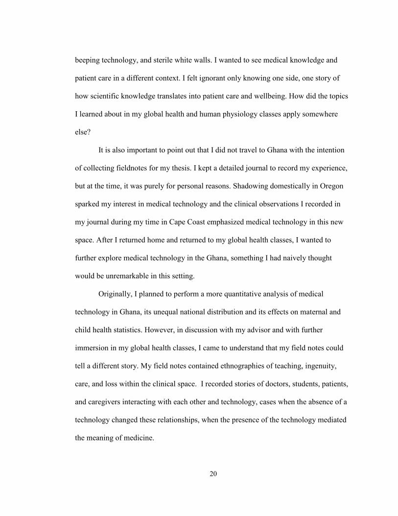
I did not travel to Ghana with humanitarian intentions. As an undergraduate
student who has taken global health classes and experienced volunteering abroad in
other countries, I understood that at my level of education, I was not traveling to Cape
Coast to volunteer any services in the clinical setting. However, I do acknowledge that
the conversation surrounding suggested clinical experience for medical school
admissions contributed to my desire to travel abroad. I was also curious to see medicine
when it was stripped to its core, when it could not hide behind electronic patient charts,
20
beeping technology, and sterile white walls. I wanted to see medical knowledge and
patient care in a different context. I felt ignorant only knowing one side, one story of
how scientific knowledge translates into patient care and wellbeing. How did the topics
I learned about in my global health and human physiology classes apply somewhere
else?
It is also important to point out that I did not travel to Ghana with the intention
of collecting fieldnotes for my thesis. I kept a detailed journal to record my experience,
but at the time, it was purely for personal reasons. Shadowing domestically in Oregon
sparked my interest in medical technology and the clinical observations I recorded in
my journal during my time in Cape Coast emphasized medical technology in this new
space. After I returned home and returned to my global health classes, I wanted to
further explore medical technology in the Ghana, something I had naively thought
would be unremarkable in this setting.
Originally, I planned to perform a more quantitative analysis of medical
technology in Ghana, its unequal national distribution and its effects on maternal and
child health statistics. However, in discussion with my advisor and with further
immersion in my global health classes, I came to understand that my field notes could
tell a different story. My field notes contained ethnographies of teaching, ingenuity,
care, and loss within the clinical space. I recorded stories of doctors, students, patients,
and caregivers interacting with each other and technology, cases when the absence of a
technology changed these relationships, when the presence of the technology mediated
the meaning of medicine.
21
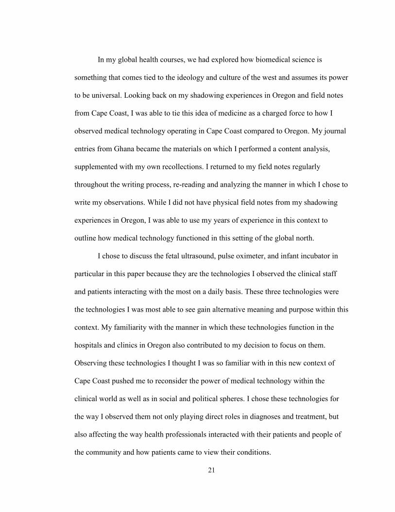
In my global health courses, we had explored how biomedical science is
something that comes tied to the ideology and culture of the west and assumes its power
to be universal. Looking back on my shadowing experiences in Oregon and field notes
from Cape Coast, I was able to tie this idea of medicine as a charged force to how I
observed medical technology operating in Cape Coast compared to Oregon. My journal
entries from Ghana became the materials on which I performed a content analysis,
supplemented with my own recollections. I returned to my field notes regularly
throughout the writing process, re-reading and analyzing the manner in which I chose to
write my observations. While I did not have physical field notes from my shadowing
experiences in Oregon, I was able to use my years of experience in this context to
outline how medical technology functioned in this setting of the global north.
I chose to discuss the fetal ultrasound, pulse oximeter, and infant incubator in
particular in this paper because they are the technologies I observed the clinical staff
and patients interacting with the most on a daily basis. These three technologies were
the technologies I was most able to see gain alternative meaning and purpose within this
context. My familiarity with the manner in which these technologies function in the
hospitals and clinics in Oregon also contributed to my decision to focus on them.
Observing these technologies I thought I was so familiar with in this new context of
Cape Coast pushed me to reconsider the power of medical technology within the
clinical world as well as in social and political spheres. I chose these technologies for
the way I observed them not only playing direct roles in diagnoses and treatment, but
also affecting the way health professionals interacted with their patients and people of
the community and how patients came to view their conditions.
22
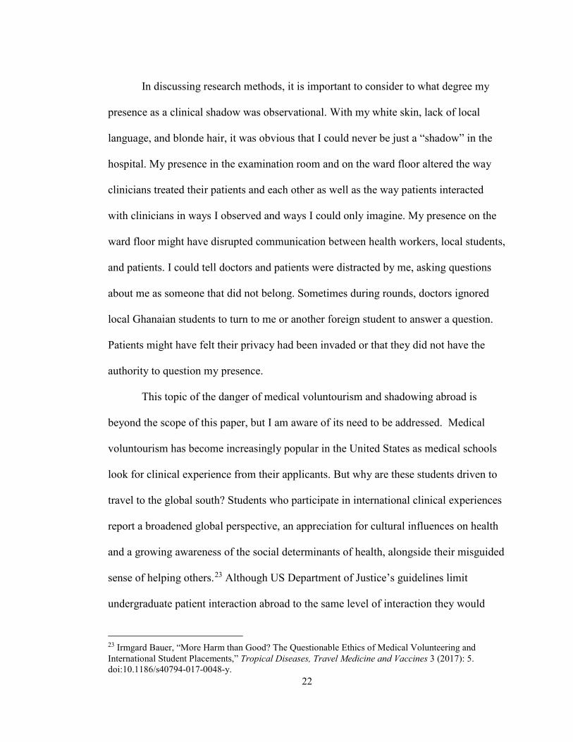
In discussing research methods, it is important to consider to what degree my
presence as a clinical shadow was observational. With my white skin, lack of local
language, and blonde hair, it was obvious that I could never be just a “shadow” in the
hospital. My presence in the examination room and on the ward floor altered the way
clinicians treated their patients and each other as well as the way patients interacted
with clinicians in ways I observed and ways I could only imagine. My presence on the
ward floor might have disrupted communication between health workers, local students,
and patients. I could tell doctors and patients were distracted by me, asking questions
about me as someone that did not belong. Sometimes during rounds, doctors ignored
local Ghanaian students to turn to me or another foreign student to answer a question.
Patients might have felt their privacy had been invaded or that they did not have the
authority to question my presence.
This topic of the danger of medical voluntourism and shadowing abroad is
beyond the scope of this paper, but I am aware of its need to be addressed. Medical
voluntourism has become increasingly popular in the United States as medical schools
look for clinical experience from their applicants. But why are these students driven to
travel to the global south? Students who participate in international clinical experiences
report a broadened global perspective, an appreciation for cultural influences on health
and a growing awareness of the social determinants of health, alongside their misguided
sense of helping others.23 Although US Department of Justice’s guidelines limit
undergraduate patient interaction abroad to the same level of interaction they would
23 Irmgard Bauer, “More Harm than Good? The Questionable Ethics of Medical Volunteering and International Student Placements,” Tropical Diseases, Travel Medicine and Vaccines 3 (2017): 5. doi:10.1186/s40794-017-0048-y.
23
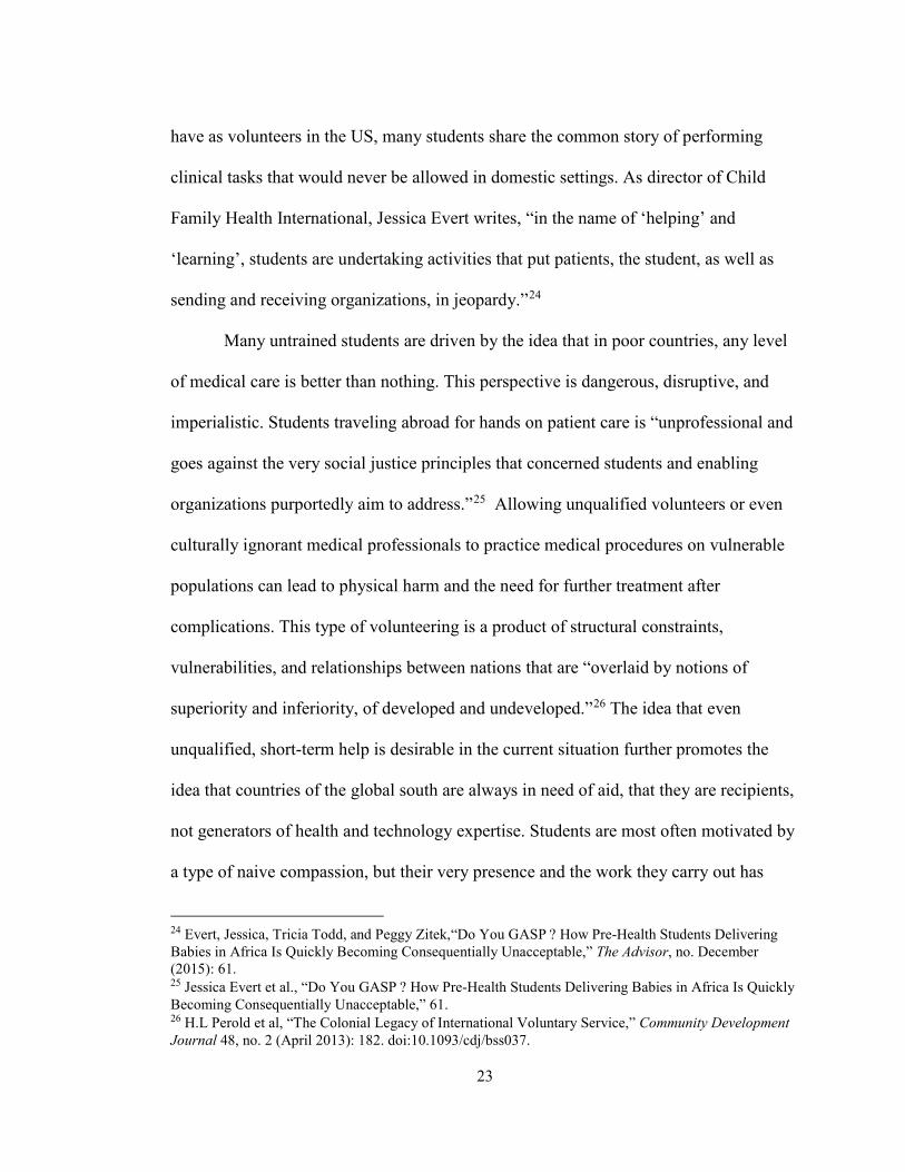
have as volunteers in the US, many students share the common story of performing
clinical tasks that would never be allowed in domestic settings. As director of Child
Family Health International, Jessica Evert writes, “in the name of ‘helping’ and
‘learning’, students are undertaking activities that put patients, the student, as well as
sending and receiving organizations, in jeopardy.”24
Many untrained students are driven by the idea that in poor countries, any level
of medical care is better than nothing. This perspective is dangerous, disruptive, and
imperialistic. Students traveling abroad for hands on patient care is “unprofessional and
goes against the very social justice principles that concerned students and enabling
organizations purportedly aim to address.”25 Allowing unqualified volunteers or even
culturally ignorant medical professionals to practice medical procedures on vulnerable
populations can lead to physical harm and the need for further treatment after
complications. This type of volunteering is a product of structural constraints,
vulnerabilities, and relationships between nations that are “overlaid by notions of
superiority and inferiority, of developed and undeveloped.”26 The idea that even
unqualified, short-term help is desirable in the current situation further promotes the
idea that countries of the global south are always in need of aid, that they are recipients,
not generators of health and technology expertise. Students are most often motivated by
a type of naive compassion, but their very presence and the work they carry out has
24 Evert, Jessica, Tricia Todd, and Peggy Zitek,“Do You GASP ? How Pre-Health Students Delivering Babies in Africa Is Quickly Becoming Consequentially Unacceptable,” The Advisor, no. December (2015): 61. 25 Jessica Evert et al., “Do You GASP ? How Pre-Health Students Delivering Babies in Africa Is Quickly Becoming Consequentially Unacceptable,” 61. 26 H.L Perold et al, “The Colonial Legacy of International Voluntary Service,” Community Development Journal 48, no. 2 (April 2013): 182. doi:10.1093/cdj/bss037.
24
unintended consequences. The movement of health technology can be analyzed in this
same vein as it becomes an object of aid that can be and do more than was originally
expected in clinical settings.
In this paper, I will add to the conversation of medical technology in global
contexts that is pushing to change international politics surrounding global health.
Through ethnographic observation, I will explore how medical technology in Cape
Coast Teaching Hospital (CCTH) has the capacity to change the dynamics of the
clinical space while also being changed by the health staff, patients, and families with
which they interact. To demonstrate this phenomenon, I investigate three medical
technologies involved in maternal and newborn health at CCTH. I show how the
ultrasound machine, pulse oximeter, and neonatal incubator, change in this context to fit
the needs of health professionals and patients while also working to change the way
people relate to each other and their illnesses. Through exploration of these three
concrete examples of medical technology within maternal and child health, I show that
context matters in how medical technology operates and is operated within the clinical
space.
25
Chapter 3: The Fetal Ultrasound
Oregon, United States
The lights are dimmed and soft music plays from speakers in the ceiling of the
hallway. The couple is met by an ultrasound technician in bubble gum pink scrubs. She
chats with the couple as she ushers them into the room illuminated by the glowing
screen of the ultrasound machine’s monitor. The expectant mother lies down on the
examination table, the white paper crinkling under her as she makes small talk with the
technician. The technician squirts clear gel over her belly, moves the transducer device
across her abdomen, taps on the keyboard, and a grey and white blur appears on the
screen. The technician asks questions from the mother, how’s your pregnancy going so
far, any discomfort or concerns. While the woman responds, the technician silently
assesses the fetal echo on the monitor to check for any abnormalities. Only then does
she swivel the monitor screen so that the couple can see. She congratulates the couple
on their first baby, pointing out the head, beating heart, and “ten tiny cute toes” before
printing out copies of “baby’s first picture” for the couple to take home. The father
mentions that the baby has his nose as the technician turns the monitor back to examine
the fetus from the head down to toes, taking measurements of bone growth, noting
anatomical landmarks, and estimating amniotic fluid volume.
Cape Coast, Ghana
She tells the young medical student that she has been nauseous, bleeding, and
having stomach pains since last night. She has been sitting in a plastic chair in the
26
waiting area for hours, sweating and watching people come and go. She is shown to a
metal examination table behind privacy screens where a nurse palpates her abdomen,
asking her where the pain is centered. Her physical symptoms along with her medical
history, leads the nurse to suspect a ruptured ectopic pregnancy. The nurse hands the
women a pink card and tells her she needs to “get a video done” as she motions her in
the general direction of the imaging room. A nursing student takes the women’s elbow,
walking her across the breezeway to join a line of women in varying stages of
pregnancy waiting outside. Finally, it is her turn as the nursing student leads her inside,
telling her she must change clothing and lay completely still. The woman lies down
before a nurse prepares her for the ultrasound. The doctor walks over to greet the
women, and begins the examination, his eyes focused on the monitor screen in front of
him. The woman stares up at the ceiling as the doctor concludes his silent examination
by writing on the pink card.
Technology Overview
For decades, health professionals in the global north have regarded ultrasound as
the “most important antepartum diagnostic technique available.”27 It is a popular, non-
invasive technique that uses ultrasound waves to create images of the fetus and
intrauterine environment. Ultrasound imaging can reveal fetal anatomy, growth and
development, pathologies, sex, and activity patterns. By emitting and receiving high
frequency sound waves across tissues, the machine’s transducer is able to generate a
continuous stream of images that show up on the machine’s screen. The sound waves
27 Lisa Mitchell, Baby’s First Picture: Ultrasound and the Politics of Fetal Subjects (Toronto: University of Toronto Press, 2001), 4.
27
travel until they hit a boundary between tissue types, like between soft tissue and bone,
at which point they are reflected back to the transducer. The different speed of
deflection and reception of the ultrasound waves show up as different gray scale colors
depending on the different densities of body tissues. Bones of the skull and legs reflect
the waves at different intensities than soft tissues like organs and fat or fluid. The
reflected waves are picked up by the transducer and relayed to the machine which
calculates the distance and intensities of the echoed signals to form a two dimensional
image.28 Since this technology was first developed in the 1950s, it has become a routine
and expected aspect of prenatal care for millions of expectant mothers in the global
north. In the United States, women have up to five ultrasounds during their pregnancy.29
As ultrasound scans have become a standard feature of maternal health services
in countries of the global north, its purpose has moved beyond the medical context.
Ultrasound’s diagnostic images are not just a technical matter, they are socially and
culturally constructed to have a variety of meanings. Ultrasound images “are highly
ambiguous and must be interpreted” by trained sonographers.” 30 Studies have revealed
that most expectant couples cannot recognize early fetal anatomy on the ultrasound
image without the sonographer’s interpretation and explanation. Despite the fact that
many parents cannot interpret the little grey blur of the ultrasound image, the
technology is emotionally compelling and firmly lodged in Western culture to say a lot
about the fetus as a person. Clinicians use ultrasound as a means to observe and assess
28 S Campbell, “A Short History of Sonography in Obstetrics and Gynaecology,” Facts, Views & Vision in ObGyn 5, no. 3 (2013): 215. http://www.ncbi.nlm.nih.gov/pubmed/24753947. 29 Mitchel, Baby’s First Picture, 7. 30 Ibid., 6.
28
fetal development, age, and position. When these clinical assessments are then
translated into laymen’s terms for the parents, ultrasound has the ability to transform the
fetus into a socially meaningful being. Mitchel captures the hybrid capabilities of
ultrasound, writing, “as sonographers search for the very anomalies that may suspend or
constrain fetal personhood, they are constructing that personhood by talking about the
image and encouraging parents to see and to bond with a sentient and acting ‘baby’”.31
Ultrasound is also thought to stimulate the parents’ emotional bond to the fetus and
reduce anxiety. In this way, the technology influences women into complying with
prenatal care recommendations.32
Ultrasound technology is becoming increasingly popular in the global south due
to its diagnostic capabilities and promotion as appropriate technology by international
donors and aid organizations. In 1985, the WHO presented policy advocating for
ultrasound as a technology “fit for all” that should be disseminated to primary health
care facilities.33 Transfer of ultrasound technology to nations characterized by high
child and maternal mortality rates became a critical aspect of international commitment
to the Millennium Development Goals. However, in Ghana, ultrasound’s mode of
transfer turned it into an exclusive object of maternal health. In the 1980s, ultrasound
machines traveled to Ghana in the luggage of white medical missionaries who worked
in private mission hospitals in rural areas. Therefore, the majority of health
professionals working in public sector state hospitals “encountered ultrasound either as
31 Mitchel, Baby’s First Picture, 118. 32 Ibid., 4. 33 Kurjak, A, and B Breyer. “The Use of Ultrasound in Developing Countries.” Ultrasound in Medicine & Biology 12, no. 8 (August 1986): 611–21. http://www.ncbi.nlm.nih.gov/pubmed/3532475.
29
merely hear-say or in written form in their British textbooks.”34 This unequal
distribution of ultrasound technology between private and public health care centers
became one of many contributing factors leading to the emigration of health
professionals and patients from public to private health care.35 Despite the Ministry of
Health’s campaigns to purchase more maternal health equipment for public hospitals,
the unequal pattern of distribution remains today.
The transfer policies that historically sought and continue to seek to export
ultrasound to Ghana as a means to improve maternal and newborn health statistics often
fail to consider ultrasound technology in context. Instead, policies view the technology
and the context in isolation. According to the WHO, once the issues of cost and
sustainable supply are resolved, the ultrasound has the universal capacity to
significantly improve maternal health.36 Viewing the transfer of ultrasound technology
as a neutral object of aid overlooks the possible consequences and appropriations that
can arise within the local clinical environment as a result of new technology placement.
The ultrasound machine is experienced differently than its counterpart in America by
the health professionals, medical and nursing students, and pregnant women of CCTH.
Within the maternity block (labor and delivery and antenatal wards) ultrasound
technology changes and is changed by the local clinical context of CCTH.
34 Müller-Rockstroh, Babette. “Ultrasound Travels The Politics of a Medical Technology in Ghana and Tanzania.” Universitaire Pers Maastricht, 2007, 71. 35 Müller-Rockstroh, Babette. “Ultrasound Travels The Politics of a Medical Technology in Ghana and Tanzania,” 77. 36 World Health Organization,“Interagency List of Priority Medical Devices for Essential Interventions for Reproductive, Maternal, Newborn and Child Health,” 2016.
30
Observations and conversations within the Labor and Delivery and Maternity
wards, indicate that the presence of fetal ultrasound technology moved beyond its
intended diagnostic purpose. The process of ultrasound’s institutionalization into the
local world of CCTH mediated relationships between health professionals, students, and
patients. Ultrasound influenced the way patients viewed their pregnancies and how
doctors treated their patients. Even the presence of a broken, unusable ultrasound
affected the clinical setting. Ultrasound’s transfer to CCTH acted to add to the device’s
meaning and purpose both within the clinical and public health context.
Ultrasound Mediates Interactions Between Clinicians and Patients
Hierarchical dynamics between doctor and patient
When women became patients for ultrasound, it seemed to make the power
disparities of the doctor-patient relationship even more apparent. During rounds,
medical students presented each mother’s case and then the doctor would ask the patient
questions about their food intake, pain levels, and any other concerns. Compared to
doctor-patient interactions in Oregon, the Ghanaian doctors often ran through these
questions with basic, even impatient responses. But when women entered the imaging
room or an ultrasound was wheeled to their bedside, the presence of the machine
became a barrier that further highlighted the separation between unknowing patient and
highly skilled physician. The doctor claimed his space behind the ultrasound machine,
his eyes fixated on the monitor, which was only visible to him. There were only brief
words exchanged between doctor and patient and once the exam began, the only thing
connecting the two people was the transducer probe the doctor moved across the
31
women’s abdomen. The monitor, fixed in place on this ultrasound model, was never
made visible to the women lying on the examination table. Printed images were only
shown and explained to the women to convince her to seek treatment or change a
behavior.
In Oregon, the dynamics between ultrasound operator and patient are different
because the context within which the ultrasound operates is different. In Oregon,
ultrasound is an expected part of prenatal care with an additional psychosocial element.
Expectant parents in the US see ultrasound as an opportunity to see and bond with their
baby before it is born. Also, ultrasound machines in the US are operated by ultrasound
technicians, not medical doctors as is done at CCTH. These combined contextual
factors mean there is less of a divided knowledge and power base between ultrasound
operator and pregnant patient in the US. There is more communication regarding the
ultrasound as the technician translates the ultrasound from medical to lay terms.
Ultrasound Affects Perceptions of Illness and Treatment
Making women into ultrasound patients
At CCTH, the presence of ultrasound pushed for health professionals to fit
pregnant women into patients for the technology. In Cape Coast, the majority of women
were not present in the imaging room for a regularly scheduled ultrasound. Only once
did I see a women demand an ultrasound to prove she was pregnant (however this
observation could be explained by my limited time in the ward). Pregnant women are
moved through the clinical space of the maternity ward or emergency block before they
32
become ultrasound patients. Usually, in the later stages of pregnancy, gestational age is
determined using the date of the patient’s last menstrual period and palpation of the
uterus. Doctors obtain fundal height by palpating the women’s abdomen, using what
one medical student called “hand knowledge” to feel where the upper edge of the
uterus, called the fundus, sits in relation to bodily landmarks like the pelvic bone.
However, when the fetus is turned transversely or the muscles of the abdomen contract,
establishing fundal height by hand became impossible. When these physical barriers are
paired with perceived ignorant patient knowledge or lying regarding their last menstrual
period, women are sent for an ultrasound to determine gestational age. Women are also
passed on to ultrasound when palpation or other physical symptoms point to possible
abnormalities or to determine if a cesarean section is needed during labor. Once women
became ultrasound patients, the technology has continued effects as restricted
availability and operation by doctors influence the dynamics between clinicians and
their patients.
Altered patient treatment protocol
The restricted availability of clinicians to perform ultrasound influenced clinical
protocol. After 4:00 pm, through the night, and on Sundays, ultrasound operators were
on call but not necessarily present on hospital grounds. When a nurse or medical student
thought one of their patient’s needed an ultrasound, they had to consider the
mobilization of resources required. The medical student or nurse had to contact their
superior, a charge nurse or even a resident doctor at home, who would then call the on
call ultrasound operator. To request using the ultrasound required a balance between
knowing enough about their patient’s condition to convince their superiors and needing
33
more information to warrant the resource. Sometimes, the presence of ultrasound
technology in these situations when the operator was not on the ward floor prevented
pregnant women from receiving the diagnoses and treatment they required. On the night
I stayed in the maternity ward for a night shift, a pregnant woman was admitted through
the emergency block in the early hours of the morning with what the young intern
thought was obstructed labor. In discussion with a few nurses, he mentioned wanting an
ultrasound immediately. But the senior nurses asked if he was sure, did he really want
to call in for an ultrasound or could it wait until the morning. With these comments, the
intern deemed the patient’s case was not an emergency despite the fact that had this
patient arrived during the day, when a resident doctor was present, she would have most
likely been sent for an emergency ultrasound. Instead, the woman was transferred to the
maternity ward to continue laboring and wait until a doctor arrived in the morning to
perform an ultrasound. The doctor arrived on time for his shift and performed the
ultrasound. The prolonged obstructed labor caused the women to suffer from an
obstetric fistula, but the baby was born healthy. This example shows that when
operational resources are limited, ultrasound has the power to alter what is considered
an emergency and change the way clinicians refer their patients for ultrasound imaging.
Context Attributes Ultrasound with New Meaning and Purpose
A video for women
In Ghana, ultrasound comes into contact with patients who do not have the same
perspectives of biomedical science that western patients do. At CCTH, having an

34
ultrasound examination became “doing a video” or “getting a video”. In the global
north, it is a common assumption that ultrasound provides pregnant women with
knowledge about viability, development, sex and position of their baby. However,
observing women get ultrasound scans in Cape Coast disrupted the idea that this
knowledge, and what women get out of ultrasound, is universal. Local bodies of
knowledge changed the way ultrasound was understood by pregnant women. In Cape
Coast, ultrasound became less of a way for women to emotionally connect and bond
with their babies and more of a mode to further medical care, to move them through the
hospital. Rarely was the ultrasound image explained to the women during the
examination, unless it was being used as a way to change her behavior or get her to
comply with treatment. Clinicians would refer to the ultrasound, telling the patient to
eat healthier, stay off their feet, drink more water, or take their medications because the
baby was too small or amniotic fluid too low. And rarely did women request an
ultrasound or schedule one themselves; instead, receiving an ultrasound was something
done at the request of nurses or doctors with no questions asked.
Ultrasound was experienced as something used in traumatic moments or to
confirm the worst. Some women called the transducer a torch that could see everything.
Other women said the ultrasound examination was actually treatment that stopped
bleeding or changed the position of the baby. These perspectives can be partially
explained by the lack of communication between doctor and patient during the
ultrasound exam. Women had to ask to see the baby, for the monitor, and the whole
machine in some cases, to be turned so they could see. But with the strict gap in
expertise and authority that divided doctor and patient, this request was rarely voiced.
35
The doctor would often explain fetal anatomy on the screen to medical students, his
nurses, and me as we stood behind him. But all while the monitor faced him, the wires
and paneling of the back or side of the monitor facing the patient. With less explanation
and different clinical uses, ultrasound loses its element of psychosocial entertainment it
held in the global north and becomes a strictly clinical tool associated with patient fear
and emergency.
Limited technology as a push factor for clinicians
As previously discussed, ultrasound technology is still concentrated within
private sectors due to colonial history and modes of international technology transfer.
As Müller-Rockstroh explains, “the general shortage [of ultrasound machines]
aggravated the disproportionate distribution of staff in urban and rural areas of the
country, a remnant from colonialism that had never been fully overcome despite
attempts by the postcolonial government.”37 In a general sense, this pattern contributes
to emigration of maternal and newborn healthcare professionals to private healthcare
facilities or to other countries to work with the technology that best serves their
profession. The resulting unequal distribution of health professionals is in the reverse
distribution of patients these doctors and nurses are to care for. The lack of properly
functioning or adequate ultrasound equipment paired with the knowledge that it was
within reach elsewhere seemed to contribute to job dissatisfaction and low morale
among doctors and nurses. Other factors such as better wages and work environment
also contribute to this movement of health professionals away from public health
37 Müller-Rockstroh, Babette. “Ultrasound Travels The Politics of a Medical Technology in Ghana and Tanzania,” 80.
36
facilities, and in this context, ultrasound becomes a technological factor that aggravates
this pattern.
At CCTH, many of the medical students expressed desires to leave Cape Coast
after graduation to work in larger facilities in Accra or at privately owned hospitals.
These comments often arose in frustration after, for example, an ultrasound machine
only printed images with black and white stripes or when the line in the corridor outside
the imaging room was so long someone’s patient simply left the hospital rather than
wait. One medical student said he has read about fetal ultrasound in many textbooks at
university, but as a third year student had not yet used one in the clinical setting.
Another more senior student agreed with this comment explaining that when he was
finally tasked to use one to determine if a women needed a cesarean section, found
himself feeling unprepared and nervous despite his advanced experience at the hospital.
These medical students sometimes turned to me with exasperated questions like “is this
what happens in America?” When these questions and situations arose, I could not help
but see in my head the Maternal Fetal Medicine clinic in Oregon with its hallway of
private rooms, each equipped with its own advanced ultrasound machine. And the fact
that even when I was a high school student, I had the ability to see ultrasound in action,
to have its technology explained to me. But here in CCTH, the absence of the
technology had an influence on students who read about ultrasound in textbooks, a
situation that seemed to elevate ultrasound as a foreign technology. While the lack of
ultrasound contributed as a push factor, the valued presence of the limited technology
was also employed a pull factor for health professionals.
37
Functioning ultrasound technology as a magnet for clinicians
As an advanced technology, ultrasound was used to attract health professionals
and students to CCTH. Ultrasound gained a new script as a magnet to keep and bring
doctors and nurses to the country and the public health sector. Ultrasound technology
was something used to attract even me, a foreign undergraduate student, to shadow in
the particular wards with the machines. In this way, ultrasound become part of the
hospital’s national image, a way for Ghana’s public health sector to keep up with the
advanced medical technology used around the world and to make visible their country’s
ability to provide up to date medical care.
Ultrasound becomes a teacher
Combined with the absence of senior staff, ultrasound also became a teacher, a
source of information for medical students. Unlike senior doctors and nurses who sent
patients to ultrasound only for particular suspected abnormalities or emergencies,
students wanted to send many more of their patients for ultrasound. However, these
medical students called for ultrasound for a variety of other reasons such as to confirm
gestational age or lie of the baby. Even when medical students seemed confident in the
issues they detected, they still sent women for an ultrasound to “fine tune their hands”
as one medical student put it. Ultrasound was able to provide them with feedback on
their palpation, calculations, and diagnostic skills like a senior clinician would have if
present. In this way, ultrasound then prepares medical students to work accurately when
the technology is not available and verifies their clinical skills.
38
Ultrasound as a public health booster
In the imaging room at CCTH, an ultrasound sound machine sat unused next to
a supply cabinet in the corner. When I asked a nursing student why no one was using
that one despite the long line outside, she responded that it was donated, sent over by a
volunteer doctor from Germany after he left, but had recently started malfunctioning. I
asked if it could be fixed and the nursing student replied that it probably could but there
was no one to fix it. With the spare parts unavailable and no set plan for maintenance,
the machine remained broken.
In Ghana, ultrasound moves between being a device placed to improve maternal
and newborn health statistics in extreme cases to a purchased device used to boost the
national public healthcare system. Ultrasound takes on a different purpose when it shifts
from being a donated gift to a commodity purchased by the state. Despite the fact that
foreign donations are free, accepting donations of ultrasound technology often resulted
in more costs later to repair broken or inconsistent machines due to a lack of spare parts,
maintenance instructions, or the local infrastructure to do so. But often when the
Ministry of Health purchases the ultrasound technology with its own resources with
direction of local hospital boards, ultrasound becomes something that offers elevated
authority and coordination within the local healthcare system and attracts health
professionals to stay in public facilities. Ultrasound becomes an item of prestige as its
value is partially from people knowing of the technology’s presence not just how it is
being clinically utilized.
Purchasing ultrasound technology as a commodity can give the local healthcare
system and hospital administration a type of power, prestige, and authority that is lost
39
when ultrasound is donated. Depending on the nature of the donation, the authority of
where to put the ultrasound, who is trained to operate it, and what level of the
technology can remain with foreign donors. However, purchasing the ultrasound
technology can allow the local systems to make demands. When ultrasound is
purchased by the MOH, preparation and coordination is required to make the best use of
monetary investments. Ultrasound received as a gift “does not make any demands with
regard to prior knowledge, information or experience but it may also obstruct the
spreading ultrasound knowledge beyond individual hospitals.”38 Donations are often
scattered throughout the country without the opportunity or time to create a foundation
to support the technology, such as training and maintenance routes. But when the
technology is purchase by the state, there is more opportunity for preparation and
coordination so that the expensive machine can operate more sustainably within the
local context. This often means maintenance plans are coordinated and funds are set
aside to maintain the technology. This preparation also distributes knowledge and
training to a wider range of clinicians and hospitals. When the state plans to purchase an
ultrasound, they have the ability to train operators and find where it is most needed,
where it can operate most successfully.
Purchasing the ultrasound technology also meant the acquisition of more
advanced technology that aimed to keep doctors and nurses more satisfied with their
ability to provide patient care. This boosted health professionals’ morale and ultimately
patient care while keeping trained clinicians where they were desperately needed. In
38 Müller-Rockstroh, Babette. “Ultrasound Travels The Politics of a Medical Technology in Ghana and Tanzania,” 105.
40
this way, the prestige the hospital gained from placement of ultrasound technology
translated to individual doctors’ prestige. At CCTH, in the maternity ward, a new
ultrasound machine had been purchased as part of a MOH campaign for maternal
health. Many of the medical students were excited to see the clear fetal images it was
rumored to produce and tried to switch resident assignments with others to see it.
Overall, ultrasound technology as a gift and a commodity do not differ much in regards
to the clinical purposes with which they serve their patients. However, they differ in
their capacity to empower or exclude the local health officials and recognize the
autonomy of Ghanaian ministries.
It is important to keep in mind that this planning and preparation does not
always occur when the MOH purchases equipment. Thus, it is important to keep in
mind that this distinction between gift and purchased commodity is not always so clear
in reality. Often, the Ministry of Health purchases and installs equipment without the
careful consideration required for the device to function properly. Or the funds the
MOH uses to purchase equipment comes from international donors and is already
earmarked for certain equipment.
Ultrasound as Broken and Limited Technology
However, these productive effects only hold true when the technology is reliably
functioning, which is not always the case at CCTH. And when this advanced
technology that is expected to boost morale and patient care malfunctions, the
technology caused the opposite effects. A broken ultrasound machine held just as much
power to shape with the clinical space as a fully functioning device.
41
The ultrasound machine that occasionally reported only flickering static stripes
on the monitor affected the attitudes of health professional and patients, how people
moved through the ward, and how their time was spent while in the hospital interacting
with the device. According to the charge nurse in the maternity ward, the hospital had
sent out for someone to come fix the malfunctioning ultrasound, but that had been
weeks ago and no progress had been made. One morning, when the machine
malfunctioned, the doctor simply stood up and left the room in frustration, leaving the
women lying there with the ultrasound gel on her abdomen and a long list of other
women waiting outside to be seen by the doctor. Patient frustration matched that of the
healthcare staff and often led to patients simply leaving the hospital without being seen
by a doctor or getting the imaging they required. When staff knew the ultrasound
machine was acting out, they reconsidered who needed an ultrasound, only sending the
emergency cases or patients that could pay immediately to imaging. Patients were also
aware of the limited functionality and availability of the ultrasound machines, which
caused feelings of desperation and anxiety among women in the maternity ward.
A few of the medical students tried to fix the monitor, tinkering with the panels
and wires in the back in an attempt to clear the image of its stripes. However, these
students were reprimanded by their resident who felt they were wasting time when they
should have been preparing for rounds. The hospital had contacted the company that
manufactured the ultrasound machine requesting repairs weeks before and no progress
had been made. The medical students took it upon themselves to tinker with the
machine when they had time, but were not able to repair it in the time I was in Cape
Coast.
42
In countries where women have access to ultrasound as a routine element of
prenatal care, ultrasound is a way to reassure women that there are no problems while
transforming the fetus into the image of a baby with which they can connect. American
ultrasound technicians maintain dialogue in a way that blurs distinctions between the
technology’s clinical diagnostic capabilities and psychosocial entertainment. But in
regions like Cape Coast, where ultrasound has been transferred and inserted into a
foreign local context, the technology takes on new meaning while also transforming the
clinical world around it. Ultrasound was transferred to Cape Coast with the hopes of
improving maternal health statistics, which have actually worsened in the last ten years,
as regional maternal mortality rates are higher than they were in 2012.39 The Ghana
Health Service attributes this increase in maternal mortality to a decline in antenatal
coverage, lack of skilled birth attendants, and increased teenage pregnancy rates. In
context, ultrasound altered relationships between doctors and their patients, changed
how pregnancy and related pathologies were viewed, and functioned as a “broken”
technology. Ultrasound became a magnet for health professionals, a source of authority
for hospitals and clinicians, and a stand in teacher for medical students. By allowing a
view of the fetus, ultrasound influenced hospital protocol, heightened the expertise and
authority doctors held over patients, and influenced the way women viewed their
pregnancies. When the ultrasound devices failed to operate, the technology caused
frustration and worsened patient care, but also allowed for local ingenuity in its repair.
39 Dr. Nsiah,Asare, Anthony. “The Health Sector in Ghana: Facts and Figures 2017,” 2017, 52.
43
These examples support the idea that local context matters for how a medical
technology will come to function in the clinical setting.
Pediatrics Ward, Cape Coast Teaching Hospital
The nurse walks between mismatched cribs under a ceiling fan that pushes the
90 degree air around. Faded Disney character decals plaster the walls of the pediatrics
ward. Winnie-the-Pooh in pale yellow watches over children with necrotizing fasciitis,
casted limbs, and burns. Dora the Explorer waves to children with yellow fever,
typhoid, malaria, and pneumonia. In the one-roomed NICU, the top of Cinderella’s head
peels off the wall as if to keep watch over the tiny babies lined up below. In the closet
sized Recently Resuscitated Unit, Buzz Light-year stands guard over three babies as the
nurses moves between their plastic isolettes, his presence adding to the crowded feel of
the room. A nurse blows up a nitrile glove like a balloon and pops the index finger into
the open mouth of a screaming baby who begins to suck on the makeshift pacifier. A
little boy in an open-backed hospital gown runs after a toy truck he sent careening down
the hall. At every child’s bedside is a mother. They kneel on the floor, arms draped into
cribs, they lean against the wall trying to see their children past the heads of doctors,
they sit at the foot of beds rubbing backs and humming softly.

44
Chapter 4: The Pulse Oximeter
Oregon, United States
The computer cart enters the room first, pushed by a nurse who is followed by
three doctors. The intern presents the patient’s case, reading from the computer how she
came into the ER for a high fever and vomiting, but turns out her heart had been failing
for a while and she is in desperate need of a new one. The intern finishes speaking and
the room is silent except for the continuous echo of beeping from the toddler’s pulse
oximeter. The device is clipped to her finger, the only one without chipped purple nail
polish. The beeping is irregular; it matches the constant echo of the beeping in the
neighboring room before speeding up for a few seconds then slowing down. The intern
glances up at the monitor displaying pulse rate and oxygen saturation as if to avoid eye
contact with the patient’s mother. The nurse repositions the oxygen cannula across the
girl’s face. After a few questions are whispered across the sleeping patient, the group
exits the room. The nurse returns to her seat behind a wall of computer monitors. The
screens display the vital signs from every patient in the unit; the tones of pulse rates and
saturation warnings beep like an unorganized marching band.
Cape Coast, Ghana
A women stands just outside the Recently Resuscitated Unit (RRU) looking in at
her baby, her hand pressed against the window. Five days ago, her daughter was born
with hydrocephalus; the baby’s head has swelled to three times the size of her twig thin
body. Her heart stopped and was restarted this morning and she was moved from the

45
NICU to the RRU. She is completely paralyzed but still cries, a strange strangled sound,
big eyes open wide and unblinking. She is wrapped in a rainbow of hand-knit blankets,
a portable pulse oximeter clipped to her impossibly tiny foot. The battery powered pulse
oximeter is the only one on the ward floor. A nurse watches the small glowing screen
on the device as she explains to the baby’s mother, that she will put the baby back on
oxygen if her blood oxygen saturation drops below 85 percent. The mother squints to
watch the number on the device. A nursing student appears at the doorway anxiously
asking for the pulse oximeter for another patient down the hall. The nurse glances down
at the pulse oximeter on the foot of the baby one last time before pulling it off. She
hands it to the other nurse as the device starts to blink low battery red.
Technology Overview
Pulse Oximetry is a non-invasive technique used to monitor oxygen levels in the
blood. The pulse oximeter can detect low oxygen saturation in the blood, called
hypoxemia. Hypoxemia is a risk factor that correlates with a variety of childhood and
newborn causes of morbidity and mortality and is hard to detect until visible cyanosis,
the blue coloring of tissues, is observed. Hypoxemia is one of the major complications
of acute lower respiratory infection, the leading cause of death in children under 5 years
of age in Ghana and many other countries around the world. Hypoxemia is also
correlated with heart disease and heart defects, perinatal asphyxia, sepsis, pneumonia,
and apnea. High blood oxygen saturation, called hyperoxemia, is also correlated with
morbidities in children and infants such as brain injury and blindness. Therefore,
46
monitoring oxygen saturation levels so they do not drop too low or climb too high is a
critical element of patient care.40
In the global north, a pulse oximeter is clipped to the finger or toe of a patient to
continually monitor the pulse rate and blood oxygen saturation. In the hospital setting,
this pulse oximeter is often connected to a monitor at the patient’s bedside and is part of
continuous monitoring of other vital signs.41 Each pulse oximeter device consists of a
computerized unit, probe, and a sensor that is in contact with the skin. Pulse oximetry
“uses the different light absorption spectra between oxygenated and de-oxygenated
hemoglobin to determine arterial blood oxygen levels.”42 Hemoglobin is the protein in
blood that holds onto oxygen so it can be released to organs and other tissues. The
device uses the absorbance of red and infrared light after it passes through the body
tissues and blood to determine the amount of hemoglobin saturated with oxygen.
Comparison of light absorbance at different wavelengths allows the technology to
estimate the concentrations of oxygenated and de-oxygenated hemoglobin. Oxygenated
hemoglobin absorbs more infrared light while de-oxygenated hemoglobin absorbs more
red light from the device. The ratio between absorbed red verses infrared light is used to
determine the level of blood oxygenation, a ratio that appears as a percent on the
device’s screen.43
40 Duke, T., R. Subhi, D. Peel, and B. Frey. “Pulse Oximetry: Technology to Reduce Child Mortality in Developing Countries.” Annals of Tropical Paediatrics 29, no. 3 (September 18, 2009): 167. 41 Durand, M, and R Ramanathan. “Pulse Oximetry for Continuous Oxygen Monitoring in Sick Newborn Infants.” The Journal of Pediatrics 109, no. 6 (December 1, 1986): 1053. 42 Nitzan, Meir, Ayal Romem, and Robert Koppel. “Pulse Oximetry: Fundamentals and Technology Update.” Medical Devices (Auckland, N.Z.) 7 (2014): 231. 43 Nitzan et al., “Pulse Oximetry: Fundamentals and Technology Update,” 1058.

47
Since it was introduced in the 1980s, the pulse oximeter has become a widely
used monitoring technology in clinical and surgical settings. In the global north, pulse
oximetry is a basic standard of care used to continually monitor pulse rate and oxygen
levels. The pulse oximeter is the device responsible for the audible beeps that echo
throughout hospitals. In high-income countries, the pulse oximeter is an expected piece
of medical equipment in ambulances, emergency departments, recovery rooms, and
operating rooms. It is one of the first things health professionals clip onto their patients
and it remains attached to patients until they are discharged.44
As a relatively basic, portable device with the power to improve healthcare
across a broad level of clinical departments, many global health campaigns have pushed
for the transfer of pulse oximeters to nations of the global south.45 To address the issue
of high surgical deaths in developing countries, the WHO launched the Safe Surgery
Saves Lives campaign in 2007. This campaign included a published “Surgical Safety
Checklist” that required a pulse oximeter to be present in every operating room.46 And a
few years later the Global Pulse Oximetry project was launched to further “promote the
use of pulse oximeters in every operating room in the world.”47 Multiple studies have
shown that the WHO’s campaign has meant that most of the focus on pulse oximeters in
low resource regions involves their placement in operating rooms. This focus overlooks
the range of uses the device can serve in other clinical settings and the many barriers
44 Chang, Meayoung. “Optimal Oxygen Saturation in Premature Infants.” Korean Journal of Pediatrics 54, no. 9 (September 2011): 362. 45 Duke, et al., “Pulse Oximetry: Technology to Reduce Child Mortality in Developing Countries,” 170. 46 World Health Organization. “WHO Surgical Safety Checklist.” WHO, 2009. https://www.who.int/patientsafety/safesurgery/checklist/en/. 47 World Health Organization, Pulse Oximetry Training Manual, 2011.
48
involved in implementation.48 49 These pulse oximeters “designed for use in resource
poor settings are rugged and reliable.”50 However, this can often mean the devices are
less accurate, more sensitive to movement, and require battery replacements.51
At CCTH, there was only one functioning pulse oximeter for approximately 40
patients in the pediatrics ward. In this section, I will explore how this affected the
purpose and meaning of the pulse oximeter device namely: it’s new clinical and social
purpose within the ward; how it mediated interactions between health professionals and
patient caregivers; influenced the way health professionals, students, and patients
viewed illnesses and treatments; how the “brokenness” and limited nature of the pulse
oximeter reorganized how patients and clinicians moved through the ward.
Pulse Oximetry Mediates Clinical Interactions
A bridge between professional medicine and local understanding
In addition to reporting numbers with clinical significance, the pulse oximeter
mediated how doctors and nurses interacted with their patients and patients’ caregivers.
The head nurse in the RRU explained that she would often go get the oximeter just to
show caregivers how the low number on the screen when it was clipped to their child
meant something had to change to raise it higher. By pointing out the number and
48 Herbert, Lara J., and Iain H. Wilson. “Pulse Oximetry in Low-Resource Settings.” Breathe 9, no. 2 (December 1, 2012): 93. doi:10.1183/20734735.038612. 49 Verwey, S, and P D Gopalan. “An Investigation of Barriers to the Use of the World Health Organization Surgical Safety Checklist in Theatres.” South African Medical Journal 108, no. 4 (March 28, 2018): 336. 50 “First WHO Global Forum on Medical Devices: Context, Outcomes and Future Actions.” World Health Organization, 2011. http://apps.who.int/medicinedocs/documents/s23217en/s23217en.pdf. 51 King, et al. “Opportunities and Barriers in Paediatric Pulse Oximetry for Pneumonia in Low-Resource Clinical Settings: A Qualitative Evaluation from Malawi and Bangladesh.” BMJ Open 8, no. 1 (2018): 9.
49
explaining its general meaning, nurses were able to outline the often complicated nature
of hypoxemia and its treatment. The pulse oximeter was used as a tool to reveal to
caregivers clinical information while commanding respect for the clinician’s actions. In
this way, the oximeter became a device that could bridge the gap between
professionalized medicine and common understandings of wellbeing. It was a non-
invasive technique that aided medical professionals in explaining to parents or
caregivers that their child needed supplemental oxygen, medication, or a surgical
procedure. This open form of communication seemed to decrease the knowledge and
authority disparity between health professionals and patient family members as it
sparked open communication and a desire to reach a common level of understanding.
With the pulse oximeter’s numbers as a mediator, doctors and nurses seemed more
willing to take the time to explain treatment decisions and the patient’s condition.
The Pulse Oximeter Affects Perceptions of Illness and Treatment
Parent caregiver perceptions
The pulse oximeter worked to change caregiver understandings of disease and
perceptions of care. One medical student explained that without the pulse oximeter it is
harder to get parents to accept oxygen treatment for their child due to the association of
the treatment with death. Many children are put on oxygen when they are very ill and
later die, causing a community association between the oxygen cannula and death. But
the numbers on the oximeter act as a direct feedback tool, as proof the child needs
oxygen. Showing parents the exact figure glowing on the pulse oximeter and explaining
what the number signified helped clinicians explain the role of oxygen in sustaining
50
their child’s life. Most family members readily accepted the explanation of the low
number on the pulse oximeter and seemed to appreciate the health professionals that
took the time to explain the situation. Additionally, with the pulse oximeter, children are
put on oxygen earlier. This pattern is something the medical student mentioned he
hoped would reverse the way oxygen treatment is viewed as parents saw the oximeter’s
numbers improving during oxygen treatment.
Confidence for clinicians
The availability of the oximeter to assess their patients also gave clinicians more
confidence in their referral and treatment decisions. In young infants especially, low
blood oxygen saturation is hard to detect and monitor with just a physical examination.
Having the pulse oximeter provided reassurance for medical students and nurses,
medical professionals within the clinical setting who were often treated as inferior by
senior doctors. A third year nursing student in the RRU explained that having a visible
number that reports “how the blood is” would determine what treatment steps she
would take next or how the current treatment was working without having to consult a
senior clinician. When the blood oxygen saturation level dropped below 85%, the baby
was immediately put on oxygen until the number rose back up. If the oxygen saturation
level was maintained at a healthy level, the nursing student knew the baby’s position
was suitable. With the pulse oximeter to show them how their treatments were working,
the medical students and nurses had more confidence in presenting their patient to
doctors during rounds because they had a concrete grasp of their patients’ vitals.
51
Pulse Oximeter Gained New Meaning and Purpose
From a continual monitor to a spot-check device
Due to the limited presence of pulse oximetry devices in the pediatric ward, it
became a spot-check device rather than a continual monitor. In just 30 minutes, the
device moved from the finger of a little boy with pneumonia to the toe of a premature
infant to the finger of a teenager with heart disease. The device is clipped on the
patient’s extremity and the patient is urged to remain still as a number appears on the
screen with audible beeps of pulse rate. The numbers are recorded in the patient’s chart
after a few minutes and the clip is removed and placed on the next patient. In most
tertiary care centers of the global north, an oximeter device is clipped to every patient in
surgical, emergency, recovery, and intensive care units. In the US, the device takes
measurements over time and alerts clinicians when numbers move out of normal range.
At CCTH in the pediatrics ward, there was only one working device while I was there.
This meant there was one pulse oximeter for nearly 30 children and 10 babies. To allow
for the technology to serve each infant or child that might need it, the device was used
for only a few minutes on each patient. Instead of taking measurements over long
periods of time, the oximeter was used to take snapshot measurements.
A means to ration limited supplemental oxygen supply
At CCTH, there is a limited amount of concentrated oxygen available for
supplemental oxygen treatment. This means oxygen treatment was reserved for patients
who were in critical need. Due to these circumstances, the pulse oximeter became a way
to ration oxygen to patients who had extreme hypoxemia and could stably use the
52
supplemental oxygen and therefore determined what constituted as critical need. When
oxygen was not available or the numbers on the device did not deem the patient critical
enough for supplemental oxygen, the pulse oximeter also allowed for the timely and
effective use of other treatments. These maneuvers to increase oxygen saturation
included assisted ventilation with room air, suctioning the airway, or repositioning the
patient. In the RRU, the nurse showed me how she clipped the oximeter on the foot of
the baby with hydrocephalus and asked me to watch the device. If the oxygen saturation
number dropped below 85 percent in the few minutes we monitored it, she explained,
the baby would be placed back on supplemental oxygen. If the number was in the low
90’s we would just reposition the baby’s neck and suction out her airway to clear it.
Pulse Oximetry as a Broken and Limited Tool
Mistrust of technology
In addition to the single functioning pulse oximeter, there was a collection of
unusable devices in the pediatric ward. Most of these broken devices were portable and
battery powered, the type of modified technology considered best suited to low resource
settings.52 These devices were unusable and sitting at the bottom of the storage closet
due to lack of local replacement batteries that fit the machine and broken sensor probes.
It is interesting to consider why these devices were never thrown away, but stored
despite their uselessness in the clinical space. A third year nursing student explained
that one of the pulse ox devices gathering dust in the closet kept presenting readings so
52 Herbert, Lara J., and Iain H. Wilson, “Pulse Oximetry in Low-Resource Settings,” Breathe 9, no. 2 (December 1, 2012): 90.

53
inaccurate that if they were true the patient would be dead. Before it simply stopped
turning on, another pulse oximeter device was discovered to be off (when readings were
compared to arterial blood gas) by a constant margin. So every time this malfunctioning
device was used, health professionals had to mentally adjust the numbers on the screen
before recording the blood saturation. The dependability of this method quickly became
a concern and the device was shut away in the storage closet. For senior health
professionals, these inaccurate readings and malfunctions of the devices led to a tone of
mistrust regarding pulse oximetry. Less experienced nurses and students placed a high
level of importance on the numbers the pulse oximeter presented, but senior health staff
took the numbers as just a small element of their overall assessment of the patient. In a
way the unusable pulse oximeters packed away in the closet became a physical
reminder of the unpredictability and fickleness of the device.
Movement through the ward floor
The brokenness of other pulse oximeter devices and therefore limited
availability of the device influenced how patients and clinicians moved through the
clinical space. Since there was only one charged oximeter on the ward floor, nurses
often had to move between rooms searching for the device. Running around looking for
the device was most often done by more novice clinicians like nursing and medical
students who also tended to rely more on the device for vital signs. However, the pulse
oximeter was most often used in the recently resuscitated unit (RRU) where the babies
had to be very closely monitored. This meant a nurse would often appear at the doorway
of the RRU asking for the device or a clinician would have to leave the RRU in search
of the device to bring it back. The pulse oximeter also determined where patients were
54
placed in the ward. The patients that were being spot-checked using the device were
placed near each other and close to the RRU. Once their oxygen saturation was
determined to be stable enough, in addition to other physical vital signs, the children
were moved farther from the RRU where they were monitored less closely.
At CCTH, the pulse oximeter is not a device expected to be clipped to every
patient or found in every room integrated into the monitoring equipment. It is not a
device used to continually monitor patients, calling for attention only when something
goes wrong. In CCTH’s pediatrics ward, the pulse oximeter is a portable, battery-
powered device that, due to its singular presence, moves around the ward depending on
where it is needed. When available, the pulse oximeter offered health staff information
regarding their patients’ blood oxygen saturation and heart rate, which allowed for
better tailored treatment decisions. The device was more than just a technology used to
monitor patient vitals because in this unique environment, it became a changeable
object while also working as an object for change. Its audible beeping and flashing
numbers moved patients and medical staff through the ward, rationed oxygen, instilled
confidence in health professionals, and bridged the gap between professionalized
medicine and caregivers’ understandings of their children’s wellbeing. This example
reveals that a unique clinical context can change the way even the most small,
straightforward medical devices are experienced by doctors, nurses, and their patients.
55
Chapter 5: The Neonatal Incubator
Oregon, United States
The incessant beeping of machines and constant rasp of ventilators seem to be
amplified by the overhead fluorescent lights. Alarms orchestrate the movement of
nurses and doctors between the rows of plastic incubators and radiant warmers that line
the walls and aisles of the large room. The walls are painted a pale yellow as if in an
attempt to soften the sterile feel of the unit. Every time a baby is born the tune of the
happy birthday song wafts out of speakers in the ceiling. Amid the hustle and bustle of
clinical staff, medical equipment, and parents, the babies’ cries can barely be heard.
One tiny baby sleeps under the plastic hood of his incubator, his body so obscured by a
ventilator, tubes, dressings, and monitor pads that the only clearly visible pieces of him
are his bare toes. A blue and green banner with his name and a bible verse stitched into
the fabric is taped to the side of his incubator, a brown teddy bear and a small hotwheels
car are nestled in the corner. His mother sits in a chair next to the machine, her face
level with her son’s, trying to catch his eye through the plastic barrier between them. At
the back of the room, a nurse explains to new parents that their severely premature
daughter will be kept safe and sound inside the incubator, which she describes as a
technological womb. Across the room, a team of nurses and doctors cheer as parents
carry their baby out across the threshold of the NICU. In another incubator, a baby
wails under the bright blue glow of phototherapy lights. She struggles to push her cries
past the pacifier in her mouth, tiny fists grasping at the feeding tube taped to her cheek.
56
In a few hours, her mother will arrive and it will take three nurses and a respiratory
therapist to place the baby in her mother’s arms.
Cape Coast, Ghana
The ceiling fan hangs from the ceiling at a slight angle, making a rhythmic
ticking noise as it spins. At first, only one baby in the incubator cries, pulling at the
feeding tube in his nose. His fussing wakes up the second baby next to him in the
incubator and soon they are both crying, the noise emerging from the incubator as a
muffled echo. Another incubator holds three babies, all sleeping soundly despite the
chaos in the neighboring incubator. The three sleeping babies appear blurry at the edges
due to the thick plastic barrier of their incubator. A nurse cradles a baby in a nest of
blankets and places her on a mattress under a bank of lights that act as a makeshift
radiant warmer. A group of doctors exits the neonatal unit and a nurse motions for the
mothers waiting outside to come through. Some mothers chat quietly to each other and
their babies, tickling toes through the access ports of incubators and updating mothers
who missed visiting hours the day before. But there are pockets of silence in the little
room, spaces of emptiness where a mother once stood watching her baby sleep.
Technology Overview
An incubator is an enclosed device that provides a protected and controlled
environment for high-risk, premature babies to develop. Babies are considered
premature when they are born at least three weeks before their projected due date. These
57
premature infants do not have fully developed organs, leaving them highly vulnerable to
a variety of complications, including infection and hypothermia. Many studies have
shown that infant survival rates significantly increase when the surrounding
environment is ideal.53 Incubators are implemented to mimic the environment of the
womb to create this ideal environment to help premature infants develop in a sterile area
with optimal levels of temperature, humidity and oxygen concentration.
One of the major causes of infant morbidity and mortality in premature infants is
hypothermia. After birth, infants are no longer in the temperature stable maternal
environment and quickly lose heat to their new surroundings. Newborns and especially
premature infants experience rapid heat loss because they have a large surface area to
body mass ratio, underdeveloped subcutaneous fat, and thin, permeable skin.
Physiological effects of hypothermia include metabolic stress, infection, dehydration,
slowed heart rate, respiratory distress, brain bleed, and death. According to the WHO,
moderate hypothermia calls for immediate warming and is recognized at a body
temperature of 89.6 to 96.6° F. When an infant reaches severe hypothermia, a body
temperature less than 89.6° F, “outlook is grave, skilled care is urgently needed”
(WHO, 47). Newborns lose heat mainly through evaporation of amniotic fluid, but this
can also occur if the baby is placed on or near a cool surface or moving air. Within the
first few minutes after birth, skin temperature of the baby typically falls by 4 degrees.
This means a newborn loses body heat at the same rate a naked adult would at a
freezing temperature of 32° F. A baby is placed in an incubator to prevent this rapid loss
53 Day, Richard L., et al. "Body Temperature and the Survival of Premature Infants." Pediatrics 34.2 (1964): 171-81.

58
of body heat that can lead to a variety of other complications by providing optimal
temperature and humidity levels.
The invention and implementation of the incubator into hospitals, first in Europe
and then the United States, in the early 20th century transformed premature infants from
“weaklings” to “fighters”.54 Caring for this new category of person and the technology
needed to sustain infants who should still be developing in the womb demanded the
development of a specialized field of medicine now known as neonatology. In the
global north, this advanced specialized and technological care is now standard treatment
for premature babies. In the NICU, the incubator acts as an enclave for the complex
technology implemented to monitor and sustain the premature infant. The plastic
capsule and web of wires of the incubator are a hallmark of neonatal intensive care units
in the global north. Blood pressure monitors, ventilation equipment, catheters,
nasogastric tubes, and IV lines pass through the incubator and groups of skilled nurses
and doctors assemble around it. Like ultrasound, the incubator has been studied as a
technology that holds meaning outside of the clinical space.
The incubator has been studied in countries of the global north as an integral
part of NICU culture and early parent-child relationships. The incubator has been
explored as a technological barrier that limits bonding between parent and infant. The
clear walls of the incubator invite the outsider to look in, but prevent the parent from
physically touching their child. Sometimes parents go weeks or months before holding
54 Mittal, Hitu, Lini Mathew, and Ashish Gupta. “Design and Development of an Infant Incubator for Controlling Multiple Parameters,” International Journal of Emerging Trends in Electrical and Electronics. Vol. 11, 2015, 42.

59
their baby for the first time. And when the small circular doors on the side of the
incubator are opened, this only permits a parent’s hand to enter. And when the baby is
finally placed in the arms of his or her parent for the first time, a web of wires and tubes
come with the baby. As advanced technological territory, the incubator can also lead
parents to feel unqualified or out of place in their baby’s highly technical life. In a study
on technology in the NICU, Manen aptly summarizes this idea writing, “the experiential
sensibility of maternity and paternity may become tenuous, fleeting, and elusive as true
contact remains out of reach”.55 With their baby inside the incubator, the parent remains
on the outside, separated from their child by both the physical barrier of the incubator as
well as the perceptions of the advanced technology surrounding it.
As large, very expensive devices that require steady inputs of clean water and
electricity, incubators are a rare element of newborn care in low resource settings. There
is a severe lack of study surrounding how incubators function in clinical settings of the
global south, as most publications simply highlight the health statistics that call for
“appropriate technology” for developing countries. International reports most often do
not name neonatal incubators, which cost between $15,000 and $50,000, as
“appropriate technology for developing countries” as the technology is not considered
“available, accessible, and affordable”.56 Ghana’s high infant mortality rate has sparked
numerous campaigns focused on improving neonatal care through actions. According to
the WHO, around 14 percent of under-five deaths in Ghana occur due to the
55 Van Manen, Michael, “Technics of Touch in the Neonatal Intensive Care,” Medical Humanities, no. 38 (2012): 93. 56 World Health Organization, Medical Devices: Managing the Mismatch (Geneva, World Health Organization, 2010), 23.

60
complications of preterm births. In Ghana, 14 percent (140,000) of babies were born
prematurely and 8,400 of these babies died in their first 30 days of life. In Ghana,
causes of preterm birth include infection, malaria, HIV, and the high adolescent
pregnancy rate. In Cape Coast, preterm death has been stuck at a high plateau for nearly
a decade. In a five-month period in 2017, 166 out of 406 births were premature babies
and 50 of these babies died at CCTH, pushing CCTH well above the national average
for premature infant deaths.57
Ideas for how to solve this problem depend on multiple actors. In a report
introducing the Newborn Care Programme, the Ghana Health Service calls for
improved care of the preterm infant, naming Kangaroo Care, which involves close skin-
to-skin contact between mother and newborn “as an effective method of providing
additional care to these vulnerable babies”.58 However, the Medical Director of CCTH
blames “insufficient incubators and lack of space” as responsible for the high rate of
preterm death and praises the donation of an incubator to the ward.59 The Ghana Health
Service suggests turning to alternative methods of preterm infant care, while the
hospital board views attainment of new incubator technology as the path to improved
care. Despite the drastic need to improve premature infant care, there is a disparity
between approaches that affects the way incubator technology is utilized.
In the following section, I will explore how the infant incubator operates within
the high-risk infant care context of CCTH. While I was shadowing at CCTH, the
57 Dr. Nsiah,Asare, Anthony, “The Health Sector in Ghana: Facts and Figures 2017,” 2017. 58 “GHS: Newborn Care Programme,” Ghana Health Service, 2017. 59 “Cape Coast Teaching Hospital Gets Incubator.” Ghana News Agency, 2017. https://www.gbcghana.com/1.11284602.
61
neonatal intensive care unit was a small room within the pediatrics ward. In this room,
there were three enclosed infant incubators, 7 open isolettes, two unused incubators and
a makeshift radiant warmer. This room served the over 700 infants requiring intensive
care that came through the hospital in 2017. The incubator technology affected the
clinicians, patients, and parents through its presence, absence, and brokenness in the
pediatrics ward. The few incubators available affected the movement of healthcare staff
as well as relationships between doctors and patients, and patients and their caregivers.
The brokenness and limited availability of the machine altered when it was utilized for
patients and led to the development of alternative techniques of care for preterm infants.
Incubator Mediates Clinical Interactions
Crowding connects mothers
Each incubator in the neonatal room holds three babies and most of the open
isolettes hold two babies. There are three tiny babies sleeping in the middle incubator,
one baby is on oxygen, one is so thin the flutter of her heartbeat can be seen across her
chest, the other sleeps soundly, his arm laid across the baby in the middle. A small
group of women are gathered just outside the room and when the doctor leaves, finished
with his rounds, they seem to move as one through the swinging doors and into the
room. They each walk up to the middle incubator, positioned so that each mother hovers
above her own baby, their arms touch, body’s leaning against each other.
Due to the limited number of incubators and large number of high-risk babies,
no incubator was occupied by just one baby. For most of the time I shadowed in the
62
ward, there were three babies in each incubator. This meant there were three mothers, or
grandmothers or aunts if the mother had died, in close physical and emotional proximity
to each other. For days and even weeks, these mothers, grandmothers, and aunts visited
their babies and went through the distressing experience of having a high-risk baby in
the hospital together. Mothers with babies who had been in the high-risk room for
weeks supported new mothers whose babies were recently transferred. Some women
were afraid to touch their babies because they appeared so fragile, but the more
experienced NICU caretakers encouraged them to bond. They ate meals and hung
laundry together on the lawn outside the pediatrics ward. They updated each other if
one woman could not make it to see her baby. These mothers and mother figures met at
an incubator, but formed circles of emotional support that extended outside of the NICU
and across generations. This community bonding seemed to act as a support system that
helped them process the trauma and challenges of mothering babies inside an incubator.
In this way, the incubator in the context of CCTH became an object that brought
caretakers together, bonding them under the stress of trying to mother in this high risk,
high tech environment.
Barriers between parent and baby
The incubator retained its capacity to physically and emotionally separate
mother from baby in Ghana. After a healthy, full term baby was delivered at CCTH, it
was most often placed on the bed next to the mother in the maternity ward. But if the
baby required a controlled environment or close observation, he or she was transferred
to the high risk room in the pediatrics ward down the hall and placed in an incubator if
space was available. One mother argued against her baby being transferred into the
63
incubator exclaiming that if he went back, she would not get to see him until he was
discharged. For mothers, visiting their babies meant first having the strength to stand
and walk down the hallway to the pediatrics ward. Once there, they would often have to
wait in line for a nurse to give them an update on their baby. After all of this, mothers
were permitted to go back to the crowded high-risk room to see their babies. But once
there, with her baby in view, the mother still encountered factors that prevented her
from bonding with her child. First, there was the obvious plastic barrier of the incubator
that prevented most physical touch. In addition, there was the less tangible barrier of
unfamiliar and intimidating medicine and technology. This gap between medicalized
baby and lay caretaker was amplified by the constant presence of healthcare staff, and
the tubes and wires surrounding the baby. Many women stood just outside the room or
up against the wall, as if these barriers created another physical gap between mother and
baby. In some instances, doctors and nurses would chastise women for trying to hold
their baby or getting in the way of medical staff. Even as the incubator technology
moved across countries, institutions, and cultures from the global north to the global
south, it maintained its powerful ability to act as a barrier between mother and child and
to push mothers to doubt their role as caretakers.
Elevated professional medicine
When morning rounds begin, the nurses usher many of mothers standing at their
babies’ incubators out into the hall to make room for the doctors. A few mothers who
are breastfeeding remain inside. The group of doctors moves from one end of the room
to the other, reading patient charts and discussing treatment plans. A medical student
64
silently draws blood from two babies in the corner incubator and soft crying fills the
room. The group of doctors make it to the end of the room where two mothers sit,
feeding their babies. Each of the doctors greets the women, but once conversation
regarding this baby’s pneumonia and that baby’s weight begins, the mothers are
ignored.
Like the ultrasound machine, the infant incubator acted as a piece of equipment
that further heightened the disparity of power and knowledge between medical
professional and patient. As discussed above, mothers were often pushed out of the
clinical space surrounding the infant incubators with both direct words and subtle
feelings of unfamiliarity. There was little effort put in to explain the medical
proceedings involving the incubator, which most likely contributed to the feelings of
distance mothers felt from their babies. Very rarely did parents approach doctors to ask
questions regarding their baby. Unlike in Oregon where medical staff made efforts to
welcome parents, both Mom and Dad, into the intimidating high-tech environment,
during rounds in Cape Coast, nurses and doctors often told mothers to “back away” or
“come back another time.” Mothers could only stay for a few hours at a time before
they were ushered out and fathers were rarely in the NICU. However, the mothers never
went far when they left the NICU, many stood just outside the room or grouped together
in the grass just outside the pediatrics ward doors.

65
Incubator Affects Perceptions of Illness and Treatment
Qualifying babies for the incubator
In the middle of the night, a mother arrived in a taxi and was admitted to the
maternity ward with obstructed, early labor. Her son was born severely premature via
an emergency cesarean section; he was so small his whole body fit in one of the
doctor’s hands. Instead of being immediately transferred into an incubator due to his
low birth weight and underdevelopment, the baby was left with his mother in the
maternity ward until arrangements could be made for him in the neonatal ward. The
next day, a baby had to be moved from an incubator to the make shift radiant warmer to
make room for the new baby who was considered higher risk. The baby previously
being cared for under the makeshift warmer was released onto the general pediatrics
floor.
Due to its limited availability, the presence of incubator technology affected
what qualified babies as high risk. To improve premature birth outcomes, the WHO
recommends unstable babies weighing less than 2,000 grams or less should be placed in
a “thermo neutral environment either under radiant warmers or in incubators.” 60 But at
CCTH, there were not enough incubators or warmers to hold every unstable baby that
met these criteria. The incubators became a rationed resource where only the most
unstable, high-risk infants were placed. This forced the doctors and nurses to assess
each potentially high risk baby and determine if their condition was detrimental enough
to warrant them a space in an incubator. As a result, many babies who should have had
60 World Health Organization. “Thermal Protection of the Newborn: A Practical Guide.” Geneva, 1997.
66
constant care in a thermo-controlled, sterile environment, were instead in open isolettes
on the general pediatrics floor or in the maternity ward with their mothers.
Mothers’ perceptions
The unexpected birth of a premature infant is traumatic and the use of the
incubator technology affected the way mothers viewed their sick infants. Many mothers
remarked on the fact that first seeing their babies inside the incubator made them seem
even more fragile and vulnerable. One nursing student explained to me that many
mothers associated the incubator with infant decline and death. Only the most high-risk
babies are placed in incubators and some get even sicker or die there. However, many
mothers also talked about the strength of their babies to fight and get healthier within
the neonatal unit. With this perspective, the incubator became a facilitator of the baby’s
progression from fragile premature infant to courageous fighter. In this way, incubators
at CCTH affected mothers’ perceptions of their sick infants in two drastically different
ways. The incubator both magnified the perceived vulnerability of the preterm infant,
but also pushed some mothers to see their babies’ strength while becoming a relied
upon tool for the babies’ survival.
Incubators Gain New Meaning and Purpose
Mediators of alterations to traditional practices
At CCTH, the incubators gained new meaning as part of public health
campaigns to improve infant mortality rates. The Ghana Health Service’s “Newborn
Care Programme” to improve thermal care pushed to modify the way mothers, birth
attendants, and health staff cared for premature infants. With the purchase of two new

67
incubators as part of this “Newborn Care Programme” came the push for other aspects
of this campaign to be implemented as well. The Newborn Care Programme
recommended delayed bathing, immediate wrapping and skin-to-skin contact.61 For
mothers and family members, this campaign pushed against some traditional birth and
bathing practices.
Early bathing is a major contributor to hypothermia in newborns, and according
to the Ghana Health Service should be delayed to help the newborn retain body heat.62
However, in Ghana, bathing the baby immediately after birth is associated with helping
the baby sleep and feel clean, shaping the baby’s head, and decreasing body odor later
in life. It is believed that the fluids on the baby’s skin will enter their body and cause
body odor later in adulthood. A charge nurse in the neonatal unit explained that for at-
home births, babies are bathed immediately and immediate skin-to-skin contact is
uncommon. Often the baby is left unwrapped as the birth attendant tends to delivering
the placenta, because until the placenta is delivered, the consequences of leaving the
newborn unattended are less serious than leaving the mother unattended.
Under this national campaign, clinicians were encouraged to improve close
monitoring of infant body temperature, resuscitation training, and delay in hospital
bathing. The campaign also called for clinicians to recommend the use of Kangaroo
Mother Care, which involves early skin-to-skin contact between mother and newborn,
as an approach to maintaining infant body temperature both at the hospital and for at
home births. One nursing student was charged with explaining direct skin-to-skin
61 Ghana Health Service, “Newborn Care Programme,” 2015. 62 Ghana Ministry of Health. “Ghana National Newborn Health Strategy and Action Plan,” 2014.
68
contact to new mothers in the maternity ward. The nurse explained to me that skin to
skin contact is not a common practice because mothers are exhausted and ill or due to
beliefs that the baby was too fragile or needed to be cleaned first. She said skin-to-skin
contact within the hospital is encouraged to a higher degree when the neonatal unit and
its incubators are full. Despite it not being a common practice, most of the mothers in
the maternity ward readily accepted Kangaroo Mother Care and expressed their
happiness in getting to keep their baby closer to them as opposed to in the neonatal unit.
Many medical students explained that because the alternative bathing guidelines
pushed to change traditional birth practices, they had not been very successful in
reducing premature infant morbidity and mortality. Despite the push for preventative
practices to reduce hypothermia and alternative treatment approaches, the neonatal unit
and its incubators remained crowded. In this way, incubators gained meaning as part of
the national campaign to improve high-risk infant care and treatment approaches at
CCTH.
The Incubator as a Broken and Limited Technology
Creation of improvised incubators
Due to the price of purchase and resources required to maintain incubator
technology, the incubator to baby ratio was high at CCTH. Incubators were a precious
resource so when the equipment broke or malfunctioned, local resources were used to
try to fix the incubator. When the top hood of one of the incubator’s cracked, a new top
was refitted to work. The new hood was from a long out of service incubator that has
69
been kept in storage. A local technician was able to resize the old incubator hood to fit
securely onto the bottom shell of the functioning incubator. Just because some piece of
medical equipment is broken did not mean it was useless. Old, broken equipment was
kept on the ward floor because it still has use as a source of spare parts and meaning as
source of technological pride. The inclination to fix the incubator also highlights the
creativity and ingenuity of a population often stereotyped as needing outside help.
To maintain newborn babies’ body temperature when the available incubators
and space were at capacity, hospital staff put together a makeshift incubator. This
provisional warmer consisted of a bank of fluorescent lights leaned against the wall next
to a low-rise hospital bed. Babies would be wrapped in blankets and placed on the
mattress under the lights. A medical student described this set up as “better than
nothing” but explained that because it is not enclosed like an incubator, the baby is at a
higher risk for infection and dehydration. There is nothing to prevent the baby from
breathing in the germs and dust in the ambient air either. And without ability to control
humidity, the baby’s skin will lose more water to the environment. It is also impossible
to maintain a consistent the temperature of with this radiant warming technique.
However, the radiant warmer provided the warm environment that most low
birth weight, high-risk babies require to develop. Additionally, many mothers and
nurses preferred the makeshift incubator to the enclosed, plastic incubators. Babies
placed in the makeshift warmer were not closed off from the outside world by a
physical barrier as they were in the enclosed incubators. This granted nurses easier
access to the baby for clinical tasks that had to be performed routinely such as taking
blood, monitoring blood pressure, and administering medication. And mothers with
70
babies in the makeshift warmer had more access to interact with and touch their babies
compared to mothers with babies in the enclosed incubators. The typical incubator is
designed to be an external plastic womb, a piece of technology that provides a warm,
clean, safe place for the infant until it can survive in the outside world. But when this
technology is absent, innovative ideas to keep the baby warm like this one came into
play.
Storage and spare parts
There were two unused incubators pushed up against the wall in the high-risk
baby room. One of these incubators held neatly folded piles of blankets and packages of
sterile gloves, and the other was being used as a hard surface to write in the patients’
charts. One of the incubators never even functioned within the walls of CCTH because
they required an electricity voltage the hospital’s power grid could not sustain. The
other incubator had been functional, but its temperature sensor broke down a few
months prior. The company that donated the machine no longer made spare parts for the
outdated version. When I asked why the machines weren’t just thrown out, the nurse
replied that they were very expensive machines, that they planned to bring in a local
repairman to try and get it working again. These incubators were broken,
nonfunctioning pieces of medical technology, but they were not thrown away. Instead,
they were repurposed in order to maintain their station in the clinical space. It is
interesting to consider why the equipment was not simply thrown away or moved to
storage. This propensity to keep non-functioning equipment on the ward floor speaks to
the mindset that technology is a critical aspect of neonatal care. As an outsider, I
initially saw the broken incubators as useless; they took up space in the cramped unit
71
and served no true purpose. But for the staff at CCTH, even the nonfunctioning
incubators seemed to represent their ability to provide healthcare with highly technical
medical equipment while also embodying a culture of technological tinkering and
repurposing. The simple presence of the incubator technology, both broken and
functioning, seemed like a way to show with pride that CCTH was capable of
integrating advanced medical equipment into neonatal patient care. This ideology that
infant care requires the presence of technology is rooted in Western biomedicine and is
transferred to the global south along with foreign medical equipment and international
public health campaigns.
The infant incubator maintained its function to mimic the safe, controlled
environment of the womb, but also adopted new meaning within the neonatal unit of
CCTH. The limited availability of incubators paired with the high number of premature
patients mediated interactions between clinicians, babies, and caretakers. While the
incubator acted as a barrier between mother and baby and highlighted the disparities of
expertise and power between doctor and caretaker, it facilitated supportive bonds
between mothers. The incubators altered the way both clinical staff and mothers
perceived the condition of the premature infants. New meaning was attached to the
incubator as part of a national public health campaign to improve newborn care that
pushed against common birthing practices, but also introduced skin-to-skin contact to
improve thermal care. Broken incubator technology also had the capacity to alter the
clinical space as equipment was fixed, repurposed, and imitated. The inclination and
ability to repurpose parts to fix a broken incubator and construct a makeshift warmer to
imitate the incubator technology undercuts the idea that the answer to Africa’s

72
healthcare barriers must come from the West. The above examples show that the staff
and local engineers in Cape Coast have the capacity to physically adapt medical
technology to allow it to function in its new context.
73
Chapter 6: Discussion
Context Matters
The lifesaving effects of medical technology in Ghana and around the world are
undeniable. The invention, implementation, and spread of technology and techniques
have played a role in improving health outcomes over the past two centuries in
countries all over the globe. In the clinical setting, goals of medical technology include
giving medical professionals increased information, accuracy, and efficiency in
diagnosing and treating their patients. However, inserting this medical technology into a
locality different from the one it was designed for altered the way the Ghanaian
healthcare system operated as well as how the technology operated. The infiltration of
health technology into the clinical environment of Ghana had a broad spectrum of
influences on the communities with which it interacted. Within Cape Coast Teaching
Hospital, pieces of equipment mediated how people interacted with each other, their
illness, and their caretakers. The presence, absence, or brokenness of a machine altered
hospital protocol and the ultimate meaning of the machine for health professionals,
students, and patients. The design or availability of a technology worked to determine
who operated the technology and how the patient or family member viewed their
disease within their own lived experience.
The dynamic functionality of medical technology within unique contexts
occurred in a variety of ways at CCTH. I discussed four main modes in which context
mattered for the way each medical technology operated. First, each technology
mediated clinical interactions between doctors, nurses, patients and caregivers. Second,
74
the presence of the devices affected both clinical and lay perceptions of illness and
treatment. Third, all three technologies gained additional meaning and associations
among clinical staff and patients. And lastly, each technology retained a capacity to
influence the clinical space as a broken or limited piece of equipment.
In the section on fetal ultrasound technology, ultrasound’s designation as a
technology fit for all was challenged by discussion of the device’s adapted dynamics
within the context of CCTH. In the maternity ward, ultrasound technology mediated
clinical interactions as it emphasized the doctor-patient knowledge and authority
inequalities, increasing the social gap between professionalized medicine and lay
patient. Ultrasound technology affected perceptions of illness and treatment by altering
what was labeled as an emergency case and inciting health staff to fit pregnant women
into patients for the technology. The ultrasound moved from being a routine prenatal
tool to become an emergency video for women, a teacher for medical students, and a
commodity for the public health system. Unreliable, malfunctioning ultrasound
technology also had the capacity to interact with the clinical environment by affecting
health professional’s attitudes, how patients were moved through the ward and eliciting
ingenuity in repair efforts.
In the pulse oximeter section, it was outlined that even the most basic device can
change and be changed by the clinical context. In the United States, the pulse oximeter
is a common tool used to measure blood oxygen saturation and continually monitor
patients across a broad range of conditions. In Cape Coast, the device lost these
characteristics and gained new functionality within the pediatrics ward. The pulse
oximeter mediated clinical interactions by bridging the gap between professionalized
75
medicine and lay caregiver thus decreasing the doctor-patient power disparity. The
pulse oximeter influenced perceptions of illness and treatment as a source of confidence
for healthcare staff in their decisions and a means to change caregiver perceptions of
oxygen treatment. Even as a basic device, the pulse oximeter gained new meaning to
become a spot check device and a way to ration scarce oxygen resources. The limited
availability of the pulse oximeter device was a factor in determining how patients and
nurses moved through the ward, while its brokenness led to mistrust of the technology.
In the incubator section, the advanced technology was introduced as an integral
aspect of NICU culture, an expensive device used to mimic the controlled environment
of the womb for premature and high-risk babies in the United States. The incubator has
the potential to improve infant care in the global south, but its high cost and need for
consistent inputs of water and electricity often mean the device is not considered an
“appropriate technology” for resource poor healthcare centers. In Cape Coast, the
incubator maintained its capacity to obstruct bonding between mother and baby, but
evolved in operation and experience within the clinical context. The incubators in the
high risk baby room mediated interactions between people by connecting mothers going
through similar emotional trauma, disconnecting mothers from their babies, and
elevating the authority of medical professional over patient caregiver. The presence of
the incubator technology affected both mother and clinical perceptions of illness and
treatment. The incubator also gained new meaning as it was associated with a national
public health campaign to improve newborn thermal care. As an advanced, expensive
technology, the incubator also had dynamic influences as a broken and limited device.
A broken incubator was used as a storage space, but also elicited ingenuity from local

76
staff and became a source of pride within the hospital.
Brokenness and Ingenuity
The functionality of broken technology is not only an inhibitory element, but a
factor with the capacity to create constructive change within the healthcare system. The
ideas surrounding broken medical equipment differ between Oregon and Cape Coast. In
Oregon, malfunctioning or inaccurate technology was promptly removed from the
clinical space. Oregonian healthcare staff did not take on the task of tinkering with the
equipment. The technology either operated with its predetermined function or it did not,
in which case it was replaced. In Cape Coast, the distinction between a working
technology and useless machine was less clear. A broken piece of equipment was
rarely immediately removed from the ward floor. Malfunctioning technology elicited
improvisation and ingenuity from the local healthcare staff who took it upon themselves
to tinker with the equipment in an attempt to repair it or grant it another purpose. Often
times the malfunctions came as expected patterns in certain devices and clinicians
already knew what adjustments were required. Equipment beyond repair with limited
ties to maintenance institutions was repurposed or used for spare parts. When
inoperable devices retained their presence in the ward, they became monuments of the
hospital’s ability to integrate highly technological elements into patient treatment.
Thinking about African healthcare staff as innovators and fixers allows for the
reconsideration of the presentation of African healthcare as underdeveloped and in
desperate need of Western assistance, a topic this paper alludes to but was unable to
discuss in depth and one that warrants further research. Medical equipment received as
donations from the global north was continually malfunctioning, or in extreme cases,
77
could never be powered on within the walls of CCTH. The many broken and faulty
pieces of donated equipment also call for the reevaluation of medical technology
donation. What is donated and how it is donated speaks volumes about the dynamics
between sending and receiving institutions.
Clinical Distance
Comparison across the three medical devices reveals the power of medical
technology to either generate or diminish distance between patients and health
professionals. Ultrasound technology and the neonatal incubator created further distance
between people by acting as communication barriers, reducing the need for physical
patient contact, and increasing reliance on medical machine over trained person. In
contrast, the pulse oximeter worked to pull patient and clinician closer together by
mediating open communication, requiring consistent physical contact, and calling for
human interpretation.
The ultrasound machine created space between patient and doctor by reducing
the need for physical contact and verbal communication. Availability of the ultrasound
diminished the need for palpation of a woman’s abdomen. Information normally gained
from palpation such as gestational age and position of the baby could be gained during
an ultrasound exam instead. During the ultrasound exam, the only thing connecting
doctor and patient seemed to be the transducer the doctor guided across the patient’s
abdomen. Conversation was rarely exchanged across the monitor’s screen. Even though
the doctor had opened up a window to peer into the insides of his patient, the distance
between clinician and patient seemed amplified.
78
The neonatal incubator also functioned to generate space between health
professionals, caregivers, and patients. The presence of the advanced technology
seemed to push doctors to place sick babies inside the incubator rather than with their
mothers. This reliance on technological care over human care not only separated baby
from mother, but also distanced doctors from their patients and patients’ caregivers.
Inside the incubator, vital signs normally assessed by hand hourly by nurses were
recorded by equipment integrated into the incubator. The plastic canopy and monitoring
technology of the incubator also distanced the high-risk babies from the outside world.
For a nurse to check the baby’s blood oxygen saturation level or for a mother to change
a diaper meant closing a large distance between technological womb and human touch.
In contrast to these space-generating technologies, the pulse oximeter functioned
to minimize distance between health professional, patient, and caregiver. The use of the
pulse oximeter functioned to decrease distance between health professional and
caregiver by mediating open communication. Additionally, the pulse oximter’s shift
from a continual monitor to a spot check device at CCTH meant increased patient
contact. In Oregon, the wide availability and accuracy of the technology meant nurses
were only drawn into the patient’s room when the monitoring screens in the hall
alarmed for abnormal oxygen saturation. In this context, nurses could monitor patients
from a distance, using and trusting the technology to determine when closer patient
contact was required. In Cape Coast, the limited availability of the device meant nurses
were in constant patient contact, moving the device from one patient to the next. And
because the portable pulse oximeter was known to malfunction, nurses were also
79
carefully observing their patient’s physical symptoms in combination with the pulse
oximeter’s numbers to ensure accurate assessment.
Limitations
Along with discussion of the results, this research has limitations that need to be
addressed. One area of limitation is rooted in the method of qualitative data collection
and use. I used my years of shadowing experience in Oregon to outline how medical
technology operated in the global north, but I did not have field notes recorded from
these experiences. Field notes from Oregon to directly compare to my Cape Coast field
notes would have made my methods more reliable and open for analysis by other
researchers. Additionally, while my field notes were taken in Cape Coast with an
emphasis on medical technology, they were not recorded with a predetermined thesis
topic as a guide. Instead the notes were used in combination with recollections to
construct a research question and analysis.
My presence as a foreign student in Cape Coast also brings up potential
limitations to this research. English is the official language in Ghana, but some patients
spoke Fante or Twi. The information I gained through translation could have been
biased or altered by the medical student or nurse translating for me. Knowledge of the
local languages could have reduced this limitation and revealed more qualitative
information about how technology affected the clinical space. My physical presence on
the ward floor also affected clinical and patient interactions. As previously discussed,
my presence as a foreign student was not a neutral factor and could have influenced the
way I saw people and medical equipment interact. These limitations point to the need
for further research on the topic of medical technology in different contexts.

80
Chapter 7: Conclusion and Nante yiye
All too often the transfer and integration of medical technologies in the global
south are based on the simplistic assumption that the advantages of foreign technology
are explicit and universal. Through discussion of medical technologies in the context of
Cape Coast Teaching Hospital’s paediatrics and maternity wards, I have aimed to show
that these devices are both changeable objects and objects for change within the clinical
setting. By exploring how the fetal ultrasound, pulse oximeter, and infant incubator gain
and lose meaning and purpose, I have presented evidence against the idea that medical
technology can be transferred globally and remain a static element of healthcare with
direct paths to health equity.
Medical technology is undoubtedly a pivotal element of the healthcare system
and patient wellbeing in Ghana, but as a culturally charged product, carries
consequences beyond advertised medical purposes. In a place to which it had been
exported, the medical equipment at CCTH was not just a one-way solution for improved
health statistics as was originally intended. Not everything changes in the process of
traveling medical technology, some aspects stay robust while others diminish, evolve,
and transform. The dynamic experiences and use of medical technologies emerged by
following the ultrasound, pulse oximeter and incubator into the working clinical context
of Cape Coast Teaching Hospital. The three technologies of maternal and child health
take on new meaning and purpose as they are integrated into the jobs, community, and
illness narratives at CCTH.
These conclusions call into question the current use of medical technology as an
exported object of global health aid and national development. This awareness that

81
context matters for the way medical technology is experienced and integrated
contradicts the idea that foreign medical equipment donations to the global south is a fit
for all answer to health and wellbeing. With these three examples of medical technology
in context, talking about “appropriate technologies for developing countries” becomes
policy talk rather than an observed reality. 63
When medical technology travels from one nation to another, it encounters the
cultural, social, political and economic institutions of a new context. With this
understanding, medical technology cannot remain static as it travels, it cannot be
expected to maintain the same meaning and purpose within a new context. In Cape
Coast, medical technology became a warped mirror of personal wellbeing, an accessible
teacher, a symbol of ingenuity, a source of pride, a token of miscommunication, and a
bridge of understanding.
Medicine is not a neutral institution and the way it is exported can speak to the
way we characterize people from other parts of the world. Exporting medical
technology deemed appropriate for the global south often means differentiating these
nations as the poor other, in need of support and products from the global north. With
this tiered perspective of global health aid, it is difficult to foresee global health and
development equality as a likely outcome. I hope that by exploring the way medicine
travels, this thesis not only pushes for a deeper understanding of Ghana and its people,
but a truer understanding of what biomedicine is and can become in the hands of all the
world’s people.
63 WHO, “Medical Devices: Managing the Mismatch,” 2010, 2.
82
Bibliography
Amiroune, Khalil. “Our First X-Ray Project in Ghana.” Midwex, 2018.
Bauer, Irmgard. “More Harm than Good? The Questionable Ethics of Medical Volunteering and International Student Placements.” Tropical Diseases, Travel Medicine and Vaccines 3 (2017): 5. https://doi.org/10.1186/s40794-017-0048-y.
Campbell, S. “A Short History of Sonography in Obstetrics and Gynaecology.” Facts, Views & Vision in ObGyn 5, no. 3 (2013): 213–29.
“Cape Coast Teaching Hospital Gets Incubator.” Ghana News Agency, 2017.
Chang, Meayoung. “Optimal Oxygen Saturation in Premature Infants.” Korean Journal of Pediatrics 54, no. 9 (September 2011): 359–62. https://doi.org/10.3345/kjp.2011.54.9.359.
Cunningham, Andrew, and Birdie Andrews. Western Medicine as Contested Knowledge. New York: Manchester University Press, 1997.
Duke, T., R. Subhi, D. Peel, and B. Frey. “Pulse Oximetry: Technology to Reduce Child Mortality in Developing Countries.” Annals of Tropical Paediatrics 29, no. 3 (September 18, 2009): 165–75, https://doi.org/10.1179/027249309X12467994190011.
Durand, M, and R Ramanathan. “Pulse Oximetry for Continuous Oxygen Monitoring in Sick Newborn Infants.” The Journal of Pediatrics 109, no. 6 (December 1, 1986): 1052–56. https://doi.org/10.1016/S0022-3476(86)80298-0.
Dr. Nsiah-Asare, Anthony. “Ghana Health Service 2016 Annual Report,” 2016.
Dr. Nsiah,Asare, Anthony. “The Health Sector in Ghana: Facts and Figures 2017,” 2017.
Evert, Jessica, Tricia Todd, and Peggy Zitek. “Do You GASP ? How Pre-Health Students Delivering Babies in Africa Is Quickly Becoming Consequentially Unacceptable.” The Advisor, no. December (2015): 61–65.
Farmer, Paul. Infections and Inequalities: The Modern Plagues. Berkeley : University of California Press, 1999
“First WHO Global Forum on Medical Devices: Context, Outcomes and Future Actions.” World Health Organization, 2011.
“Ghana Health Service 2014 Annual Report.” GHANA 1, 2015.
83
“Ghana Health Service: National Health Research Agenda 2015-2019.” Ghana National Health Research Agenda, 2015.
Ghana Ministry of Health. “Ghana National Newborn Health Strategy and Action Plan,” 2014.
“GHS: Newborn Care Programme.” Ghana Health Service, 2017.
Hadolt, Bernhard, Viola Hörbst, and Babette Müller-Rockstroh. “Biomedical Techniques in Context: On the Appropriation of Biomedical Procedures and Artifacts.” Medical Anthropology 31, no. 3 (May 2012): 179–95. https://doi.org/10.1080/01459740.2011.636410.
Herbert, Lara J., and Iain H. Wilson. “Pulse Oximetry in Low-Resource Settings.” Breathe 9, no. 2 (December 1, 2012): 90–98. https://doi.org/10.1183/20734735.038612.
“Interagency List of Medical Devices for Essential Interventions for Reproductive, Maternal, Newborn and Child Health.” Biomedical Technology. I.World Health Organization. ISBN 978: 92–94. Accessed January 18, 2018.
King, Carina, Nicholas Boyd, Isabeau Walker, Beatiwel Zadutsa, Abdullah H Baqui, Salahuddin Ahmed, Mazharul Islam, et al. “Opportunities and Barriers in Paediatric Pulse Oximetry for Pneumonia in Low-Resource Clinical Settings: A Qualitative Evaluation from Malawi and Bangladesh.” BMJ Open 8, no. 1 (2018): e019177. https://doi.org/10.1136/bmjopen-2017-019177.
Kurjak, A, and B Breyer. “The Use of Ultrasound in Developing Countries.” Ultrasound in Medicine & Biology 12, no. 8 (August 1986): 611–21.
Manen, Michael Van. “Technics of Touch in the Neonatal Intensive Care.” Medical Humanities, no. 38 (2012): 91–96. https://doi.org/10.1136/medhum-2012-010204.
Mitchell, Lisa. Baby’s First Picture: Ultrasound and the Politics of Fetal Subjects. Toronto: University of Toronto Press, 2001.
Mittal, Hitu, Lini Mathew, and Ashish Gupta. “Design and Development of an Infant Incubator for Controlling Multiple Parameters.” International Journal of Emerging Trends in Electrical and Electronics. Vol. 11, 2015.
Müller-Rockstroh, Babette. “Appropriate and Appropriated Technology: Lessons Learned from Ultrasound in Tanzania.” Medical Anthropology 31, no. 3 (May 2012): 196–212. https://doi.org/10.1080/01459740.2011.639105.
Müller-Rockstroh, Babette. “Ultrasound Travels The Politics of a Medical Technology in Ghana and Tanzania.” Universitaire Pers Maastricht, 2007.
84
Nitzan, Meir, Ayal Romem, and Robert Koppel. “Pulse Oximetry: Fundamentals and Technology Update.” Medical Devices (Auckland, N.Z.) 7 (2014): 231–39. https://doi.org/10.2147/MDER.S47319.
Perold, H., L. A. Graham, E. M. Mavungu, K. Cronin, L. Muchemwa, and B. J. Lough. “The Colonial Legacy of International Voluntary Service.” Community Development Journal 48, no. 2 (April 1, 2013): 179–96. https://doi.org/10.1093/cdj/bss037.
“PHILIPS Advances Quality Healthcare in Ghana.” The Herald Ghana. July 2014.
Pulse Oximetry Training Manual. World Health Organization, 2011.
Rouse, Joseph. “What Are Cultural Studies of Scientific Knowledge?” Configurations 1, no. 1 (1993): 1–22. https://doi.org/10.1353/con.1993.0006.
Verwey, S, and P D Gopalan. “An Investigation of Barriers to the Use of the World Health Organization Surgical Safety Checklist in Theatres.” South African Medical Journal 108, no. 4 (March 28, 2018): 336. https://doi.org/10.7196/SAMJ.2018.v108i4.12780.
Wendland, Claire. A Heart for the Work: Journeys Through an African Medical School. Chicago: University of Chicago Press, 2010.
WHO. “Medical Devices : Managing the Mismatch,” 2010, 1–36. https://doi.org/ISBN 978 92 4 156404 5.
World Health Organization. “Interagency List of Priority Medical Devices for Essential Interventions for Reproductive, Maternal, Newborn and Child Health,” 2016.
World Health Organization. “Thermal Protection of the Newborn: A Practical Guide.” Geneva, 1997.
World Health Organization. “WHO Surgical Safety Checklist.” WHO, 2009.