medical ultrasound measurements lab tour may 20111
TRANSCRIPT
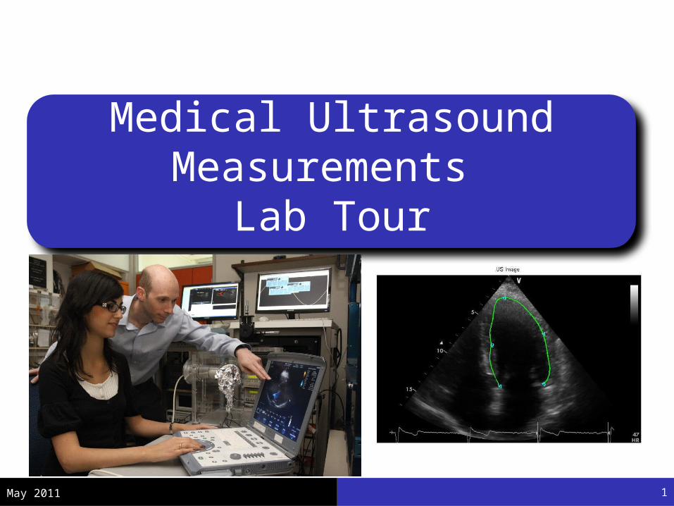
Medical Ultrasound Measurements
Lab Tour
May 2011 1
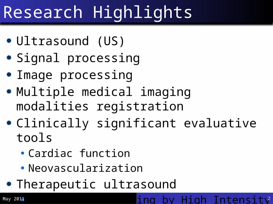
Ultrasound (US)Signal processingImage processingMultiple medical imaging modalities registrationClinically significant evaluative tools
Cardiac functionNeovascularization
Therapeutic ultrasoundAcute Cardiac Pacing by High Intensity Focused US
Research Highlights
May 2011 2
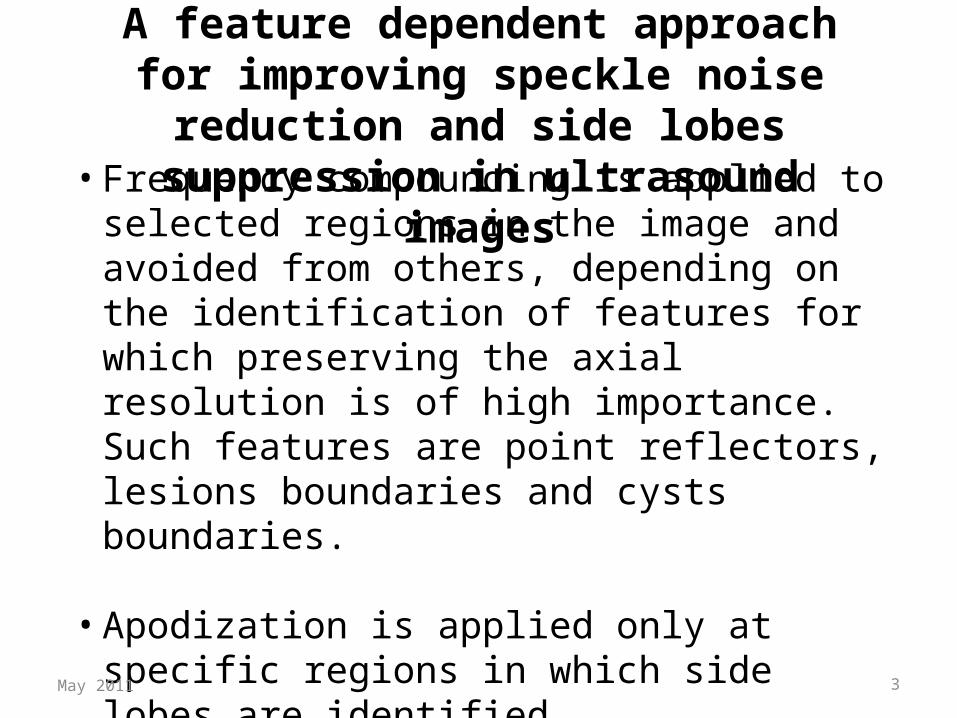
A feature dependent approach for improving speckle noise reduction and side lobes
suppression in ultrasound images
• Frequency compounding is applied to selected regions in the image and avoided from others, depending on the identification of features for which preserving the axial resolution is of high importance. Such features are point reflectors, lesions boundaries and cysts boundaries.
• Apodization is applied only at specific regions in which side lobes are identified.
May 2011 3

Feature dependent compounding
Reference image
Ordinary compounding Local optimum compounded image
Selective Local optimum compounded image
Scan image (2nd harmonic) - reference (no apodization
Scan image (2nd harmonic) - with ordinary Hanning apodization
Scan image (2nd harmonic) - side lobe suppressed by selective apodization
Ordinary apodization
Feature dependent apodization
May 2011 4

Speckle Tracking Echocardiography
May 2011 5
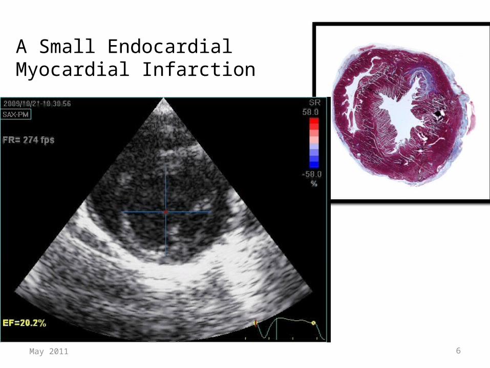
A Small Endocardial Myocardial Infarction
May 2011 6
0 0.05 0.1 0.15 0.2 0.25 0.3 0.35-20
0
20
Time [sec]Glo
ba
l Str
ain
[%]
X [cm]
Y [c
m]
0 1 1 2 2 3 3 4
0
1
1
1
1
2
2
2
2
-30
30
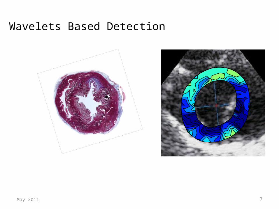
Wavelets Based Detection
May 2011 7
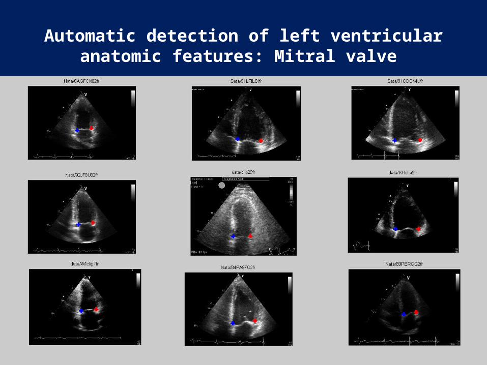
Automatic detection of left ventricular anatomic features: Mitral valve

Continues Valve Corner Tracking
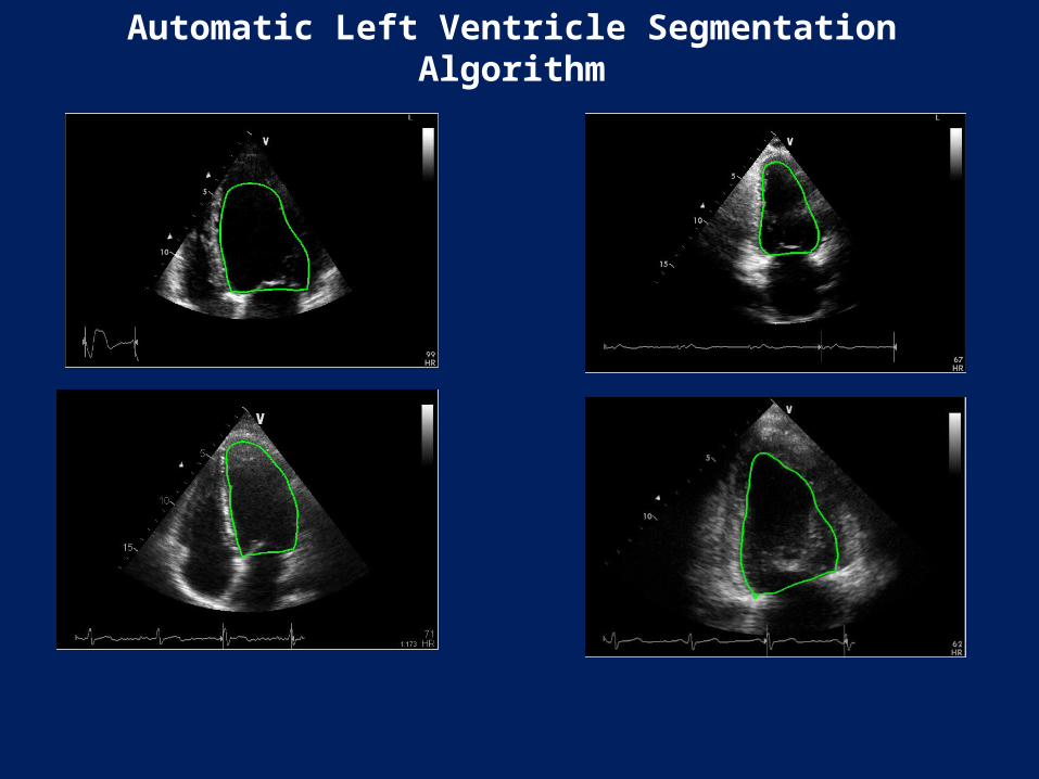
Automatic Left Ventricle Segmentation Algorithm

יד שמאל
יד ימין
מבט מכיוון כפות הרגליים
רקמת שומן -בטן
ריאה שמאל
ריאה ימין
חדר שמא
לחדר
ימין
עמוד שדרה
גב
עצם יד שמאל
עצם יד ימין
שריר הלב

זרימת דם לתוך החדר
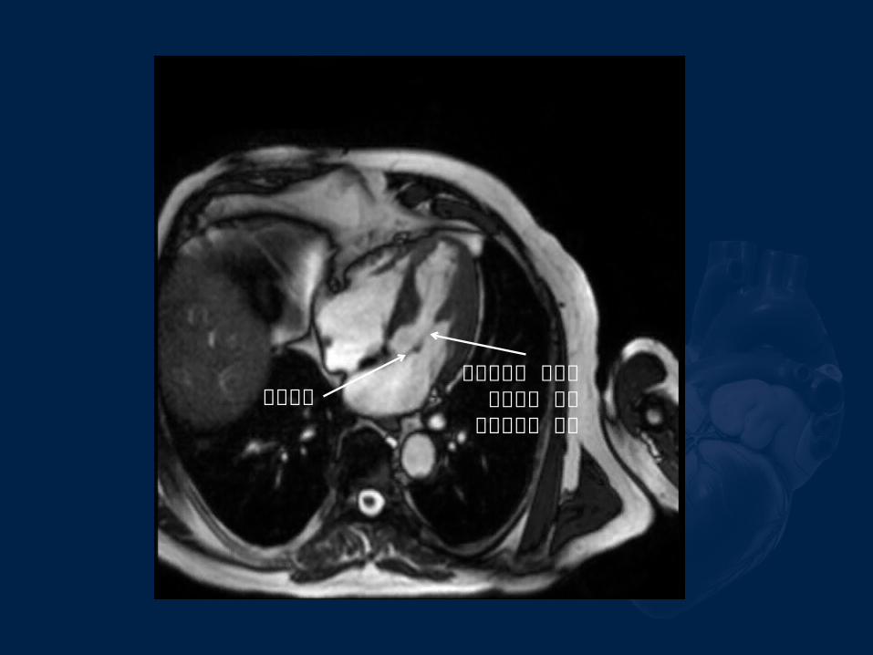
מסתםגיד שמושך
את העלה של המסתם
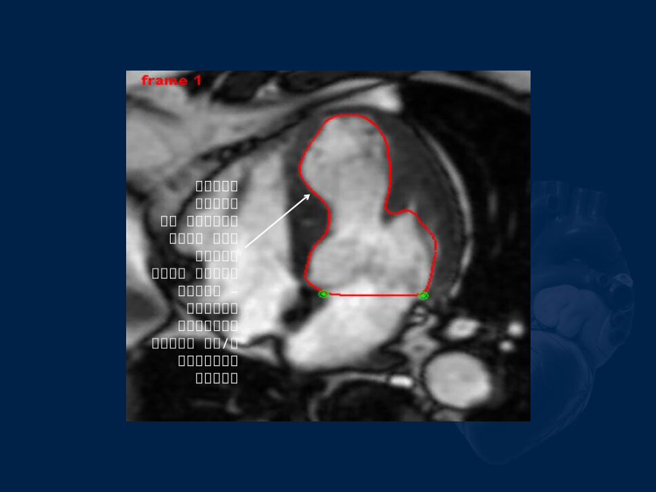
מציאת הגבול הפנימי של חדר
שמאל לאורך פעימה שלמה –
לצורך מדידות קליניות ו/או
התאמה להדמיות
אחרות

Registration of Ultrasound and MR Cardiac Images
• Given a collection of segmented ultrasound (US) and MR cardiac images, our challenge is to register or align them
• There are several factors that make this task difficult:– The heart is constantly in motion– The images are almost certainly not of the same slice– Segmentation of the US images is very difficult due
to poorer image quality
May 2011 15
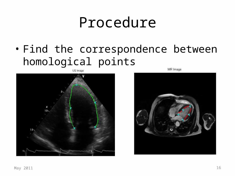
Procedure
• Find the correspondence between homological points
May 2011 16
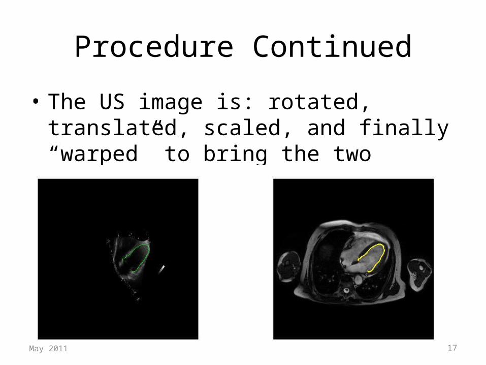
Procedure Continued
• The US image is: rotated, translated, scaled, and finally “warped” to bring the two contours into alignment
May 2011 17
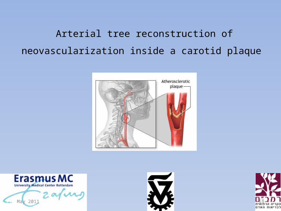
Arterial tree reconstruction of neovascularization
inside a carotid plaque
May 2011 18

Clinical Motivation
Atherosclerosis
Calcification Inflammation
Unstable Plaque Stable Plaque
Plaque rupture
Stroke and TIA
:The disease
:Atherosclerosis Progression
:Plaque’s content
Pathological result:
Clinical result:
:Plaque’s types
IMT ThickeningPlaque Generation
May 2011 19
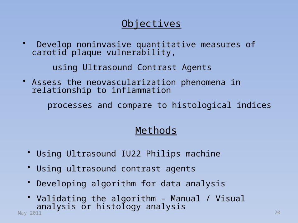
• Develop noninvasive quantitative measures of carotid plaque vulnerability,
using Ultrasound Contrast Agents
• Assess the neovascularization phenomena in relationship to inflammation
processes and compare to histological indices
Objectives
Methods
• Using Ultrasound IU22 Philips machine
• Using ultrasound contrast agents
• Developing algorithm for data analysis
• Validating the algorithm – Manual / Visual analysis or histology analysis
May 2011 20
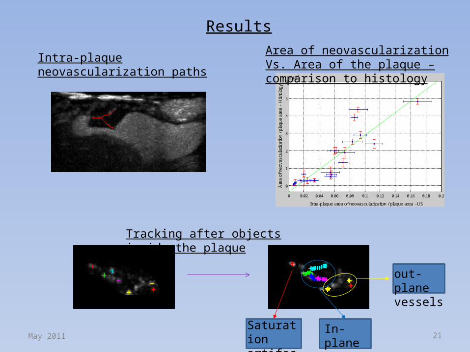
Results
Frame no.1 Frame no.30
Saturationartifacts
In-planevessels
out-planevessels
Intra-plaque neovascularization paths
0 0.02 0.04 0.06 0.08 0.1 0.12 0.14 0.16 0.18 0.2
0
1
2
3
4
5
6x 10
-3
Intra-plaque area of neovascularization / plaque area - US
Are
a of
neo
vasc
ular
izat
ion
/ pla
que
area
- H
isto
logy
Area of neovascularization Vs. Area of the plaque – comparison to histology
Tracking after objects inside the plaque
May 2011 21
Tumor Vascularization Quantification
May 2011 22

Tumor Vascularization Imaging - Current researchQualitative estimation of blood flow
Dynamic contrast-enhanced ultrasonography (DCE-US)
Contrast-Enhanced Gray-Scale Ultrasound
Zhou et al. , 2011 control treated
Lassau et al. , 2010
23May 2011
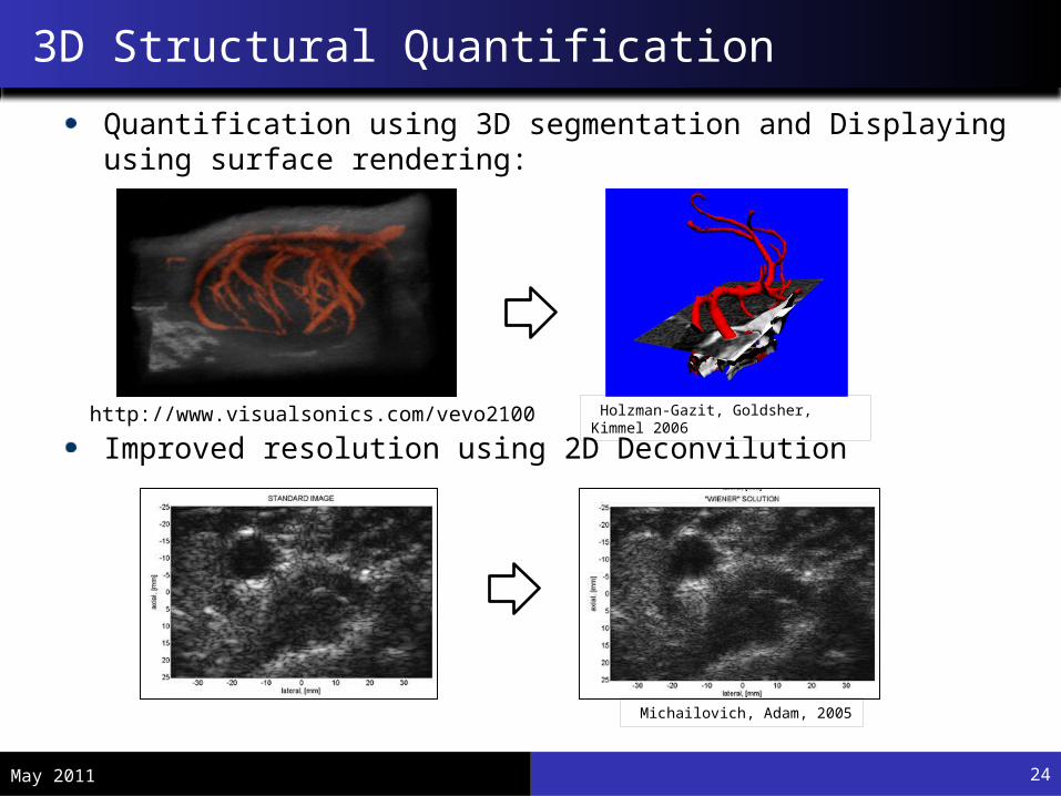
3D Structural Quantification
Quantification using 3D segmentation and Displaying using surface rendering:
Improved resolution using 2D Deconvilution
Holzman-Gazit, Goldsher, Kimmel 2006http://www.visualsonics.com/vevo2100
Michailovich, Adam, 2005
24May 2011

Extracorporeal Cardiac Pacing by High Intensity Focused Ultrasound Possible Application
Emergency Medicine Cardiac Arrest
Resynchronization Therapy Electrode Implantation Site Evaluation
May 2011 25
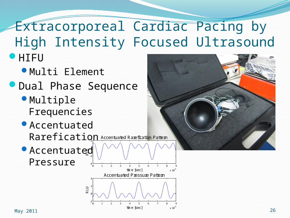
Extracorporeal Cardiac Pacing by High Intensity Focused Ultrasound
HIFUMulti Element
Dual Phase SequenceMultiple FrequenciesAccentuated
RareficationAccentuated Pressure
0 1 2 3 4 5 6 7 8 9
x 10-6
-4
-2
0
2Accentuated Rarefication Pattern
time [sec]
RL
U
0 1 2 3 4 5 6 7 8 9
x 10-6
-2
0
2
4Accentuated Pressure Pattern
time [sec]
RL
U
May 2011 26
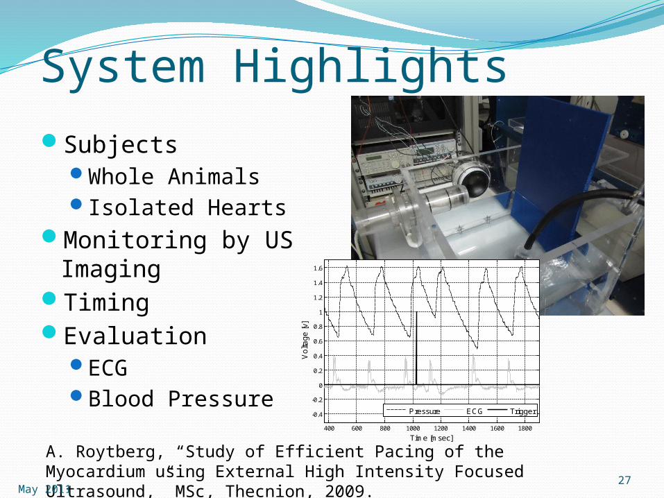
System HighlightsSubjects
Whole AnimalsIsolated Hearts
Monitoring by US Imaging
TimingEvaluation
ECGBlood Pressure
400 600 800 1000 1200 1400 1600 1800
-0.4
-0.2
0
0.2
0.4
0.6
0.8
1
1.2
1.4
1.6
Time [msec]
Vo
ltag
e [v
]
Pressure ECG Trigger
A. Roytberg, “Study of Efficient Pacing of the Myocardium using External High Intensity Focused Ultrasound,” MSc, Thecnion, 2009.May 2011
27
ThanksProf. Dan AdamNoa Bachner Ph.D.Yael Petrank Ph.D.Amit LivnehAssaf HoogiYossi TsadokAvinoam Bar ZionHanan KamisLiron ShlomoRan MaromTzvi Goldman
May 2011 28

QuestionsThank you.
May 2011 29