medically important flaviviruses hhs public access
TRANSCRIPT

Characterization of a novel insect-specific flavivirus from Brazil: potential for inhibition of infection of arthropod cells with medically important flaviviruses
Joan L. Kenney1, Owen D. Solberg2, Stanley A. Langevin2, and Aaron C. Brault1,*
1Division of Vector-Borne Diseases, Centers for Disease Control and Prevention, Fort Collins, CO, 80521
2Sandia National Labs, Livermore, California
Abstract
In the past decade there has been an upsurge in the number of newly described insect-specific
flaviviruses isolated pan-globally. We recently described the isolation of a novel flavivirus
(tentatively designated “Nhumirim virus”; NHUV) (Pauvolid-Correa et al., in review) that
represents an example of a unique subset of apparently insect-specific viruses that
phylogenetically affiliate with dual-host mosquito-borne flaviviruses despite appearing to be
limited to replication in mosquito cells. We characterized the in vitro growth potential, 3’
untranslated region (UTR) sequence homology with alternative flaviviruses, and evaluated the
virus’s capacity to suppress replication of representative Culex spp. vectored pathogenic
flaviviruses in mosquito cells. Only mosquito cell lines were found to support NHUV replication,
further reinforcing the insect-specific phenotype of this virus. Analysis of the sequence and
predicted RNA secondary structures of the 3’ UTR indicate NHUV to be most similar to viruses
within the yellow fever serogroup, Japanese encephalitis serogroup, and viruses in the tick-borne
flavivirus clade. NHUV was found to share the fewest conserved sequence elements when
compared to traditional insect-specific flaviviruses. This suggests that, despite being apparently
insect-specific, this virus likely diverged from an ancestral mosquito-borne flavivirus. Co-
infection experiments indicated that prior or concurrent infection of mosquito cells with NHUV
resulted in significant reduction in viral production of West Nile virus (WNV), St. Louis
*Address for Correspondence: Aaron C. Brault, Division of Vector-Borne Diseases, Centers for Disease Control and Prevention, Fort Collins, CO 80521; [email protected]; phone: (970) 266-3517.
Sequences and accession numbers: Aedes Flavivirus NC012932, Alfuy virus AY898809, Alkhurma virus NC004355, Apoi virus NC003676, Aroa virus NC009026, Bagaza virus NC012534, Barkedji virus EU078325 , Bouboui virus DQ859057, Cell fusing agent virus NC001564, Chaoyang virus NC017086, Culex flavivirus NC008604, Deer tick virus AF311056, Dengue virus 1 NC001477, Dengue virus 2 NC001474, Dengue virus 3 NC001475, Dengue virus 4 NC002640, Donggang virus NC016997, Edge Hill virus DQ859060, Entebbe bat virus NC008718, Gadgets Gully virus DQ235145, Greek goat encephalitis virus DQ235153, Iguape virus AY632538, Ilheus BrMS-MQ10 KC481679, Japanese encephalitis virus NC001437, Kadam virus DQ235146, Kamiti River virus NC005064, Karshi virus DQ462443, Kedougou virus NC012533, Kokobera virus NC009029, Kyasanur forest virus HM055369, Lammi virus FJ606789, Langat virus NC003690, Meaban virus DQ235144, Modoc virus NC003635, Murray Valley encephalitis virus NC000943, Nakiwogo virus GQ165809, Nhumirim virus NC024017, Nounane virus FJ711167, Omsk hemorrhagic fever virus NC005062, Potiskum virus DQ859067, Powassan virus NC003687, Quang Binh virus NC012671, Rio Bravo virus NC003675, Rocio virus SPH34675, Royal Farm virus DQ235149, Saumarez Reef virus DQ235150, Sepik virus DQ837642, Spondweni virus DQ859064, St. Louis encephalitis virus NC007580, Tembusu Shandong1 JX965381, Tick-borne encephalitis virus NC001672, Tyuleniy virus DQ235148, Uganda S virus DQ859065, Usutu virus NC006551, Wesselsbron virus NC012735, West Nile virus NC009942, Yellow fever virus NC002031, Yokose virus NC005039, Zika virus NC012532
HHS Public AccessAuthor manuscriptJ Gen Virol. Author manuscript; available in PMC 2015 December 01.
Published in final edited form as:J Gen Virol. 2014 December ; 95(0 12): 2796–2808. doi:10.1099/vir.0.068031-0.
Author M
anuscriptA
uthor Manuscript
Author M
anuscriptA
uthor Manuscript

encephalitis virus (SLEV) and Japanese encephalitis virus. The inhibitory effect was most
effective against WNV and SLEV with over a million-fold and 10,000-fold reduction in peak
titers, respectively.
INTRODUCTION
New sequencing technologies have drastically improved the capabilities for rapid genetic
characterization of novel viruses and microorganisms, for both emerging pathogens of
animals as well as non-pathogenic microflora and microfauna that could modulate the
receptivity of hosts to infection with pathogens of medical and veterinary importance. This
has been exemplified recently by the identification of numerous novel flaviviruses (Aliota et
al., 2012; Cook et al., 2009; Crabtree et al., 2009; Evangelista et al., 2013; Hobson-Peters et
al., 2013; Hoshino et al., 2009; Huhtamo et al., 2009; Junglen et al., 2009; Kolodziejek et
al., 2013; Lee et al., 2013b; Parreira et al., 2012; Roiz et al., 2009; Sang et al., 2003;
Vazquez et al., 2012) with an arthropod-restricted host range that, although not known to
directly illicit disease in vertebrates, could alter the capacity of arthropods to transmit
vector-borne pathogens. Studies demonstrating the role of mosquito microbiome in the
modulation of vector competence for arboviruses capable of eliciting disease in humans
underscores the potential that infection with insect-specific flaviviruses could similarly
modulate transmission of human arboviral pathogens. (Bolling et al., 2012; Cirimotich et al.,
2011; Gubler, 2002; Hobson-Peters et al., 2013; Kent et al., 2010a; Weiss & Aksoy, 2011).
Flaviviruses are enveloped viruses comprised of a single-stranded, positive-sense RNA
genome of approximately 11 kb consisting of a 5’ and 3’ untranslated regions and a
methylated cap that allows for direct translation of a single open reading frame (ORF)
resulting in a of a single polyprotein (Markoff, 2003; Wengler et al., 1978). The ORF
encodes three structural proteins including the capsid (C), premembrane/membrane (prM),
and envelope (E), and seven nonstructural proteins including NS1, NS2A, NS2B, NS3,
NS4A, NS4B, and NS5 (Castle et al., 1986; Rice et al., 1985) that are cleaved co- and post-
translationally by host and viral proteases. Analyses of flavivirus genomes have
demonstrated them to cluster phylogenetically by host preference range: insect specific
flaviviruses (ISFs), dual-host tick-borne flaviviruses (TBFVs), viruses with no known vector
(NKV), or mosquito-borne flaviviruses (MBFVs) (Gould et al., 2003; Kuno et al., 1998).
ISFs constitute a relatively novel group of flaviviruses characterized as single-host viruses
that replicate in insects and have demonstrated replication incompetence in vertebrate cells.
To date, the only arthropod source of insect specific flaviviruses (ISFs) has included
members of the order Diptera, predominantly mosquitoes and sandflies. It remains to be
determined if ISFs exist in alternative arthropod taxa such as ticks. TBFVs are categorized
into two groups: the seabird tick-borne group and the mammalian tick-borne group. The
NKV group can be sub-grouped into viruses associated with bats or rodents. MBFVs with
dual hosts such as Japanese encephalitis virus (JEV) and yellow fever virus (YFV) form
distinct phylogenetic clades that correlate with the mosquito genus primarily associated with
viral transmission. However, some viruses, such as Entebbe bat virus (EBV) and Yokose
virus (YOKV), with no apparent mosquito vector also fall into the MBFV phylogenetic
Kenney et al. Page 2
J Gen Virol. Author manuscript; available in PMC 2015 December 01.
Author M
anuscriptA
uthor Manuscript
Author M
anuscriptA
uthor Manuscript

grouping. It is unclear as to whether the vector for these viruses has yet to be detected or if
they have lost the ability for mosquito-borne transmission (Kuno et al., 1998).
Like dual-host MBFVs, classically recognized ISFs form two phylogenetic subgroups based
on whether they are vectored by Aedes or Culex mosquitoes. Aedes associated viruses such
as cell fusing agent virus (CFAV), Aedes flavivirus (AeFV), and Kamiti River (KRV) virus
have been isolated respectively from Puerto Rico (Cook et al., 2006), Japan (Hoshino et al.,
2009), and Kenya (Crabtree et al., 2003). However, homologous viral sequences have also
been identified in Spain (Aranda et al., 2009; Sanchez-Seco et al., 2010), Italy (Calzolari et
al., 2010; Roiz et al., 2009), and Canada (Pabbaraju et al., 2009) indicating a widespread
geographic distribution. ISFs identified to infect Culex mosquitoes include Quang Binh
virus (QBV) isolated from Vietnam (Crabtree et al., 2009), Calbertado virus (CLBOV)
isolated from North America (Bolling et al., 2011), and Culex flavivirus (CxFV), which has
been isolated from Trinidad-Tobago (Kim et al., 2009), Guatemala (Morales-Betoulle et al.,
2008), Mexico (Farfan-Ale et al., 2009; Farfan-Ale et al., 2010), Uganda (Cook et al.,
2009), USA (Blitvich et al., 2009; Crockett et al., 2012; Kim et al., 2009), and Japan
(Hoshino et al., 2009). Nakiwogo virus (NAKV), a monophyletic Culex specific ISF, was
isolated from Mansonia africana nigerrima (Cook et al., 2009). A number of studies have
highlighted the potential for ISFs to have an inhibitory effect on co-infecting flaviviruses of
medical importance. For example, CxFV has been shown to suppress the capacity for Culex
spp. to become infected and transmit WNV (Bolling et al., 2012; Kent et al., 2010a).
Similarly, a potential role of superinfection exclusion was indicated by reduced replication
of Kunjin and Murray Valley fever virus in the presence of the ISF Palm Creek virus in
C6/36 cells (Hobson-Peters et al., 2013).
Interestingly, there is a growing number of ISF-like isolates that appear to be phenotypically
insect-specific with no indication of replication in vertebrates, yet are phylogenetically
distinct from the ISF clade, as they group with other dual-host MBFVs. These isolates,
characterized for the purposes of this manuscript as unidentified vertebrate host (UVHs)
viruses in the MBFV group, are limited to replication in arthropod cells include: Nounané
virus (NOUV) (Junglen et al., 2009), Lammi virus (LAMV) (Huhtamo et al., 2009),
Chaoyang virus (CHAOV) (Lee et al., 2013a), Barkedji virus (BJV) (Kolodziejek et al.,
2013), and Nanay virus (NANV) (Evangelista et al., 2013). Like viruses from the ISF
phylogenetic cluster, these viruses have been isolated from a wide geographic range
including Israel, Peru, Finland, Côte d’Ivoire, the Republic of Korea, and China.
Continued isolation and characterization of these unique flaviviruses will provide key
insights into the evolution of vector/host adaptation and, potentially, flavivirus origins.
Herein, we describe the characterization of a novel mosquito-borne flavivirus, tentatively
designated Nhumirim virus (NHUV), isolated from the Pantanal region of Brazil (Pauvolid-
Correa et al., in review) that appears to be most closely related to other novel flaviviruses
that have insect-specific host replication capabilities but differ from their projected
phylogenetic relationships by grouping within dual-host MBFVs. We evaluated its
phylogenetic relationship to other flaviviruses, identified permissive cell lines in vitro,
analyzed the predicted secondary structure of the 3’ UTR, and demonstrated the virus’s
Kenney et al. Page 3
J Gen Virol. Author manuscript; available in PMC 2015 December 01.
Author M
anuscriptA
uthor Manuscript
Author M
anuscriptA
uthor Manuscript

ability to suppress replication of representative Culex spp. vectored pathogenic flaviviruses
in vitro.
RESULTS
Virus Isolation and in vitro characterization
A novel flavivirus tentatively designated Nhumirim virus (NHUV) was isolated from a
single pool of 43 non-engorged adult female Culex chidesteri collected in April 2010 in
Brazil (Pauvolid-Correa et al., in review). Evidence of cytopathic changes were definitively
identified six days following initial inoculation onto C6/36 cells, whereas no CPE was
observed from initial inoculation on Vero cells. Upon secondary passage in C6/36, NHUV
manifested CPE in the form of rounded cells still attached to the monolayer, observable
within 3 days post-infection (dpi), and limited syncytia development by 6 dpi (Fig. 1). The
C6/36 TCID50 of the stock isolated from the second passage of NHUV was 9.1 log10
TCID50/ml and was used to inoculate additional cell lines. While NHUV was able to
replicate in other mosquito cell lines including Ae. albopictus C7/10, C6/36, and Cx.
quinquefasciatus cells, the virus did not replicate in alternative invertebrate cells.
Inoculation of ISE6 tick cells failed to generate detectable infectious virus assayed on C6/36
cells as screened by IFA (Fig. 2). Furthermore, RNA extracted from culture supernatants of
the second passage were RT-PCR negative using pan-flavivirus primers. Attempted
culturing in vertebrate cell lines, including Vero, BHK21, DF-1, and Xenopus laevis, proved
unsuccessful as confirmed by negative RT-PCR amplification of the second passage and
lack of detectable antigen detection by IFA using a pan-flavivirus (4G2) monoclonal
antibody developed from a Dengue 2 (New Guinea C) strain (Gentry et al., 1982) (Table 1).
Sequence and phylogenetic analysis
The complete NHUV genome, including the 5’ and 3’ UTRs, was sequenced and identified
to be 10,891 nucleotides (nt) in length. The predicted open reading frame (ORF) was 10,338
nt, while the 5’ UTR was 102 nt, and the 3’ UTR 451 nt. Three flavivirus-type structural
proteins C, prM, E, and seven flaviviral non-structural proteins, NS1, NS2A, NS2B, NS3,
NS4A, NS4B, and NS5 were identified (Table 2) and polyprotein cleavage sites were
predicted (Table 3. The full viral sequence has been deposited in GenBank under accession
number KJ210048 (Pauvolid-Correa et al., in review). The coding region of NHUV had the
greatest nucleotide identity to BJV (65.9%) and NOUV (56.2%), both recent isolates that
have demonstrated the unique phenotypic characteristics of UVH viruses (Table 4).
Maximum likelihood phylogenetic analysis of the ORF regions of 59 flavivirus sequences
similarly indicated that NHUV was most closely related to a group of novel UVH viruses
that cluster within the dual-host mosquito borne flaviviruses with strong bootstrap support
(Fig. 3). Interestingly, viruses that have been shown to be restricted to growth in insect cells
within the MBFV clade cluster in two distinct branches with robust bootstrap support: one
consisting of DGV, LAMV, and CHAOV, with the other branch containing NHUV, BJV,
and NOUV (Fig. 3).
Kenney et al. Page 4
J Gen Virol. Author manuscript; available in PMC 2015 December 01.
Author M
anuscriptA
uthor Manuscript
Author M
anuscriptA
uthor Manuscript

3’ UTR characterization
Studies of the flavivirus 3’ UTR have identified a number of direct repeats that appear to
have evolved from a progenitor of six long repeated sequences (LRS) that are homologous
to extant TBFV 3’ UTR sequences (Gritsun & Gould, 2006c; 2007b). Alignments between
the flavivirus 3’ UTRs have indicated the evolutionary remnants of these LRSs can be
identified in MBFV, NKV, and ISFV and tend to show conservation within each group
(Gritsun & Gould, 2007a; b). Comparison of the NHUV 3’ UTR to representative viruses
for each flavivirus group MBFV, TBFV, NKV, and ISFV were made in order to identify
homologous regions of conservation (Fig. 4). NHUV was identified to have several
conserved structural elements in common with viruses in the MBFV group including a 3’
terminal long-stem loop (3’LSH) with conserved pentanucleotide sequence CACAG, a
conserved stem loop (SL2), and a conserved dumbbell shaped element (DB1) with an
internal conserved sequence element CS2 (Fig. 4). A third MBFV conserved sequence
element, previously identified as CS1, was found to be incorporated with a small stem loop
structure (SL3) (Markoff, 2003). The absence of a Y-shaped structure typically conserved
between TBFVs, NKVs, and ISFVs was noted (Fig. 4). The only common features that
NHUV had with ISFVs was the 3’ LSH structure and a pentanucleotide sequence although
only four out of five nucleotides were conserved. Comparisons of NHUV to other
representative UVH viruses were inconclusive as many of these have incomplete 3’ UTR
sequences available.
Codon usage frequencies
Studies of codon usage have indicated that certain codon dinucleotide pairings are utilized
preferentially and this codon bias can often be correlated to match an organism’s transfer
RNA pool (Akashi, 1994; Clarke, 1970; Ikemura, 1981). In particular, TA dinucleotides
have been found to be proportionally underutilized for encoding amino acids in both
vertebrate and invertebrate hosts in order to minimize targeting by endoribonucleases In
addition, vertebrate hosts underutilize CG dinucleotides to reduce DNA methylase targeting
and subsequent increased mutation rates at these sites (Zhao & Jiang, 2007). Examination of
RNA viruses have indicated that they have evolved optimized codon usage for facilitated
replication in hosts utilized (Lauring et al., 2012). In order to utilize these potential codon
usage biases for the assessment of the potential vertebrate host infectivity by NHUV, we
generated histograms of leucine (TA dinucleotide containing codons) and threonine, proline,
and arginine codon usage frequencies (CG dinucleotide containing codons) for NHUV,
WNV, CxFV, and MODV. As would be expected, all four viruses demonstrated an
underutilization of TA (CTA and TTA codons) dinucleotides (Fig. 5a; Leu panel). MODV
was found to highly underutilize CG dinucleotides with the mean codon frequency of these
codons encoding Arg (CGT, CGC, CGA and CGG) to be 4.5% ± 0.1. In contrast, CxFV
exhibited a usage of 19.3% ± 2.8 and 11.4% ± 6.3 for CG and non-CG codons. NHUV was
identified to mirror the codon usage frequencies of CG dinucleotide codons (threonine,
leucine, and proline) of WNV. Both WNV [11% ± 2.9 (non-CG) /28% ± 7.9 (CG)] and
NHUV [13.5% ± 3.7 (non-CG) /22.3% ± 21.4 (CG)] demonstrate a bias against CG
dinucleotide codons that was less significant that that of MODV (Fig 5).
Kenney et al. Page 5
J Gen Virol. Author manuscript; available in PMC 2015 December 01.
Author M
anuscriptA
uthor Manuscript
Author M
anuscriptA
uthor Manuscript

Inhibition of WNV, JEV, and SLEV growth
C6/36 cells were inoculated at an MOI of 5 with NHUV and challenged at day 0 (co-
infected), day 1, day 3, and day 5 post infection with WNV at an MOI of 0.1. Similar studies
were performed with JEV and SLEV infection at 0 and 3 days post NHUV infection to
determine if NHUV affects alternative representative MBFV flaviviruses isolated from
Culex spp. mosquitoes. A two-way repeated measures ANOVA indicated a significant
difference between NHUV co-infected groups and control viruses for WNV, JEV, and
SLEV (p < .0001). A secondary Dunett’s multiple comparison test with a corrected p-value,
found all control infections of WNV, JEV, or SLEV alone to have a significantly higher (p <
0.0001) average titer for each daily time point sample (day 2 post-infection through day 7
post-infection) as compared to groups pre- or co-infected with NHUV (Fig. 6). Comparison
of SLEV and SLEV+NHUV d0pi, also from the D1 time point, was the only comparison in
which a NHUV co-infected group did not show significantly reduced replication as
compared to the control. The control viruses WNV, JEV, and SLEV alone achieved a 6.2
log10 (PFU/ml), 1.2 log10 (PFU/ml), and 4.3 log10 (PFU/ml) higher mean peak titer than
matched groups coinfected with NHUV (Fig. 6). These differences in peak titer translate to
1.5 million-fold reduction for WNV, an 80-fold reduction for JEV, and a 15,000-fold
reduction for SLEV in the presence of NHUV in vitro.
DISCUSSION
We describe the characterization of a novel mosquito-borne virus, Nhumirim virus (NHUV),
from the Pantanal region of Brazil and establish with high degree of certainty that it
segregates with MBFVs within the genus Flavivirus. While the source mosquito, Culex
chidesteri, has not been shown to be a disease vector, WNV has been isolated from this
species (Kent et al., 2010b). Field studies have indicated this mosquito feeds on a range of
hosts including humans, chickens, rabbits, and turtles (Almiron & Brewer, 1995). The
NHUV isolate is part of a novel group of flaviviruses that we are tentative designating as
unidentified vertebrate host (UVH) viruses that have been isolated from a wide geographic
range including China (Wang et al., 2009), Republic of Korea (Lee et al., 2013a), Côte
d’Ivoire (Junglen et al., 2009), Finland (Huhtamo et al., 2009), Israel (Kolodziejek et al.,
2013) and Peru (Evangelista et al., 2013). Upon phylogenetic characterization, we found
that NHUV clustered most closely with these viruses and had the same apparent inability to
replicate in mammalian vertebrate cells, despite being grouped within the dual-host
mosquito vectored clade of flaviviruses. Interestingly, NHUV, NOUV, and BJV form a
distinct clade from the branch encompassing CHAOV, LAMV, and DGV, which may be
due to vector/host species differences. CHAOV, LAMV, and DGV viruses were all
reportedly isolated from Aedes spp. while NANV, NHUV, and BJV were isolated from
either Culex spp. or Uranotaenia spp. (Evangelista et al., 2013; Junglen et al., 2009;
Kolodziejek et al., 2013; Lee et al., 2013a; Wang et al., 2009). While there is a precedent
for vector/host preference being correlated with phylogenetic divergence throughout the
Flavivirus genus, research into the vector preference or transmission mechanism (i.e.
transovarial or oral infectious) of these viruses has yet to be performed.
Kenney et al. Page 6
J Gen Virol. Author manuscript; available in PMC 2015 December 01.
Author M
anuscriptA
uthor Manuscript
Author M
anuscriptA
uthor Manuscript

The phylogenetic branching pattern indicates that NHUV, NOUV, and BJV share a more
recent common ancestor with viruses from the MBFV group than with the other members of
the distinct clade UVH viruses including CHAOV, LAMV, and DGV in that phylogenetic
cluster. The phenotypic and phylogenetic contrast is what makes these viruses of particular
interest because historically, flaviviruses have been found to cluster by host/vector
preference. There are three possible explanations for this anomaly: 1) these viruses are a
distinct group of ISFs that never evolved the ability to replicate in vertebrate hosts, 2) these
viruses are part of the dual-host mosquito vectored clade and have lost the ability to replicate
in vertebrates, or 3) these viruses are part of the dual-host mosquito vectored clade and
replicate in an as of yet to identified non-insect secondary host. 3’ UTR analysis in concert
with phylogenetic findings indicate that NHUV has the most conserved structures and
sequences (present and absent) with viruses of the MBFV group. Specifically, the complete
conservation of the 3’ LSH pentanucleotide with other MBFVs, the presence of SL2 which
is conserved between TBFV, MBFV, and NKV, but not ISFV (Gritsun & Gould, 2007b),
and the absence of Y-1 which is conserved in NKV (Charlier et al., 2002), TBFV (Gritsun &
Gould, 2007b; Gritsun et al., 1997; Proutski et al., 1997), ISFV (Gritsun et al., 2014) but not
MBFV, supports the likelihood that NHUV is a member of the mosquito-borne flavivirus
group and has either lost its ability to replicate in vertebrates or has an as yet unidentified
vertebrate host. Similar observations were made upon informal analysis of codon usage
preferences in that NHUV codon usage by amino acid more closely resembled that of WNV
than the insect specific model utilized, CxFV. A study by Lobo et al. indicated that
Flaviviridae members which persist in a single host cycle have codon usage profiles more
similar to their hosts than to closely related Flaviviridae (Lobo et al., 2009). The dissimilar
codon usage profiles between the insect-specific virus CxFV and NHUV in concert with the
similarities between the codon profile of NHUV and WNV supports the theory that NHUV
is not a mosquito-specific virus, but either a dual-host virus with an as of yet undiscovered
vertebrate host or a virus that has recently lost its ability to replicate in vertebrates.
We compared the ability of NHUV to inhibit representative Culex spp. vectored MBFV
replication upon simultaneous co-infection and delayed secondary infection. We were able
to determine that NHUV had a significant inhibitory effect on the replication of WNV,
SLEV, and JEV in culture by decreasing peak titers anywhere from 6.2 log10 (PFU/ml) to
1.2 log10 (PFU/ml). This inhibitory effect was observed as early as one-day post-infection
for both WNV and JEV, and by day two post-infection for SLEV, which is not unexpected
as SLEV is a slower growing virus. Inhibition of WNV growth in vitro following co-
infection with an insect specific flavivirus, CxFV, has been demonstrated previously by
Bolling et al., in which ∼1.0 log10 reduction of WNV in co-infected C6/36 cells (Bolling et
al., 2012) and Hobson-Peters et al showed inhibition of up to 1.6 log10 for Palm Creek Virus
(PCV) inhibition of Murray Valley encephalitis virus and a 1.0 log10 reduction in WNV
replication (Hobson-Peters et al., 2013). Such inhibition upon dual-infection has often been
described by superinfection exclusion, the phenomenon in which a cell infected with one
virus cannot be secondarily infected with another, closely related virus. It has been
previously demonstrated in vitro and in vivo for both alphaviruses and flaviviruses (Bolling
et al., 2012; Eaton, 1979; 1981; Hobson-Peters et al., 2013; Karpf et al., 1997; Kent et al.,
2010a; Pepin et al., 2008; Pesko & Mores, 2009). However, while we did observe inhibition
Kenney et al. Page 7
J Gen Virol. Author manuscript; available in PMC 2015 December 01.
Author M
anuscriptA
uthor Manuscript
Author M
anuscriptA
uthor Manuscript

with secondary infection with WNV, SLEV, and JEV that would be consistent with
superinfection exclusion, we also saw equally marked MBFV inhibition when the viruses
were infected with NHUV simultaneously. This is of particular interest as studies of
superinfection have indicated the exclusion of secondary infection generally does not take
effect until at least one hour following infection with the initial virus (Eaton, 1979; Johnston
et al., 1974). Therefore, it is likely that NHUV has a distinct mechanism that interferes with
replication of these Culex spp. vectored MBFV as demonstrated in vitro. It is also of note
that NHUV was found to have a high TCID50 in C6/36 cells, which may also contribute to
the efficiency of the inhibition effect. Because the range of species, rate, or geographic
spread of NHUV infection remains unknown, we cannot draw conclusions regarding any
potential impact this inhibition phenomenon may have in the field. Further studies will need
to examine whether these same inhibitory effects translate into in vivo mosquito infection,
dissemination, and transmission blockage. Previous studies investigating the inhibition of
CxFV on transmission of WNV in Culex spp. have shown mixed results, indicating that
observed replication interference in vitro may not necessarily be indicative of in vivo
findings (Bolling et al., 2012; Kent et al., 2010a). However the increased phylogenetic
relatedness between MBFV and NHUV as compared to that of MBFV and CxFV could be
an important contributing variable to the degree of inhibition. The phylogenetic relatedness
between NHUV and MBFV also improves prospects for vaccine development, as
recombinant and chimeric viruses are more likely to be stable and viable. Ongoing studies
are focusing on examining chimeras between WNV and NHUV in order to evaluate regions
responsible for co-infection inhibition, as well as determine regions responsible for ablated
vertebrate replication. Identification of these regions could have implications for improved
attenuation strategies, which would allow for an additional safety factors as well as shed
light on fundamental genetic determinants that dictate host range differences of flaviviruses.
METHODS
Virus isolation and sequencing
Adult mosquitoes were collected between 2009 and 2010 in the Nhecolândia sub-region of
the Pantanal, within the State of Mato Grosso do Sul, Brazil as previously described in
Pauvolid-Correa et al. (Pauvolid-Correa et al., 2013). Pools of mosquitoes were
homogenized in 300 µl Dulbecco’s modified Eagle medium (DMEM) complete with
penicillin (100U/ml), streptomycin (100mg/ml), 10% fetal bovine serum (FBS), and
50µg/ml amphotericin B. Clarified supernatants from triturated mosquito pools were used to
inoculate both C6/36 (mosquito) and Vero (mammalian) cells in 24-well plates. Inoculated
cells were observed daily and harvested upon the appearance of cytopathic effect (CPE) or
following ten days incubation. Ae. albopictus C6/36 cells were maintained at 28°C with
complete DMEM supplemented with 10% FBS, and penicillin/streptomycin. Viral RNA was
extracted from 140ul of the harvested supernatant using QIAamp RNA mini kit (Qiagen,
Inc., Valencia, CA). RT-PCR was performed on the extracted RNA using flavivirus-specific
primers as previously described (Pauvolid-Correa et al., 2013; Pauvolid-Correa et al., in
review). The full coding sequence was acquired with second generation sequencing (SGS)
using a Mi-Seq system (Illumina Inc., San Diego, CA, USA) and the NHUV virus genome
was constructed via an automated computational pipeline as previously described (Langevin
Kenney et al. Page 8
J Gen Virol. Author manuscript; available in PMC 2015 December 01.
Author M
anuscriptA
uthor Manuscript
Author M
anuscriptA
uthor Manuscript

et al., 2013; Pauvolid-Correa et al., in review). The 5’ and 3’ untranslated regions (UTRs)
were confirmed using the corresponding kit for rapid amplification of cDNA ends (RACE)
(Invitrogen, Carlsbad, CA, USA).
Phylogenetic analysis/codon usage frequency calculations
The NHUV polyprotein open reading frame sequence was aligned with available flavivirus
sequences in the NCBI database using MUSCLE on the Cipres Science Gateway (Edgar,
2004; Miller et al., 2010). Maximum likelihood inference was performed using RAxML
7.06 on the Cipres Science Gateway (Stamatakis et al., 2008). 1000 replicates of
bootstrapping resampling were utilized to assess the accuracy of tree topologies. Output
trees were manipulated using Fig Tree v1.4. Codon frequency calculations were performed
using MacVector (v10.6) (MacVector, Inc, Cary, North NC, USA) software based on the
coding regions of the following arboviral strains; WNV (382-99; AF196835), MODV
(M544; AJ242984), CxFV (FJ663034) and NHUV (KJ210048) strain characterized herein.
IFA and TCID50
To confirm and quantify the growth of the non-plaque forming NHUV flavivirus isolate,
immunofluorescence assays (IFAs) were performed in conjunction with the Reed and
Muench method for titrating endpoints (Biacchesi et al., 2005; Reed & Muench, 1938).
C6/36 cells were inoculated with 10-fold serial dilutions in a 96-well format and fixed with
20% acetone 24 hours post-infection. Once fixed, cells were washed with phosphate
buffered saline (PBS), incubated with a pan-flavivirus monoclonal (Dengue 2, New Guinea
C; 4G2) antibody, washed with PBS, and incubated with a FITC-labeled secondary antibody
(goat anti-mouse IgG; Jackson ImmunoResearch Laboratories, West Grove, PA). After a
final wash, cells were examined for the presence of viral antigen with an inverted
fluorescent microscope.
In vitro characterization
In vitro propagation of the isolate was attempted in various cell lines including Aedes
albopictus mosquito (C6/36 and C7/10), Culex quinquefasciatus, Ixodes scapularis tick cells
(ISE6), African green monkey (Vero), and hamster (BHK21-clone 15), chicken (DF-1), and
Xenopus laevis (South African clawed toad) cells. Each cell monolayer was inoculated at a
multiplicity of infection of 10 TCID50 units from supernatant isolated from the original
passage of the triturated mosquito pool sample as determined by titration on C6/36 cells.
Cultures were observed for CPE for 7 days prior to harvest. Virus was serially blind
passaged three times each on Vero cells, ISE6 cells, and BHK21 clone15 cells as no initial
CPE was identified following a single passage. To confirm the presence or absence of viral
replication, RT-PCR was performed on supernatant taken from the third passage using pan-
flavivirus primers, FU1 and CFD3R, designed to amplify a ∼1085 nt portion of the NS5
gene region (Kuno et al., 1998). Negative RT-PCR samples were confirmed by IFA.
Inhibition of West Nile virus growth in vitro
West Nile virus utilized for co-infection studies was derived from an infectious clone of the
New York 1999 strain (Kinney et al., 2006). Twelve well plates of C6/36 cells all originally
Kenney et al. Page 9
J Gen Virol. Author manuscript; available in PMC 2015 December 01.
Author M
anuscriptA
uthor Manuscript
Author M
anuscriptA
uthor Manuscript

seeded at the same time and density, were inoculated at an MOI of 5 with NHUV. These
cultures were subsequently inoculated with WNV, JEV, or SLEV at an MOI of 0.1 on day 0
(simultaneous co-infection) and day 3 following initial NHUV infection. Additional pre-
inoculation of NHUV was performed at −1 and −5 dpi for WNV inhibition studies. All
infections were performed in duplicate with mock WNV, JEV, and SLEV infection controls
for each experimental time-point group. Additionally, a positive infection control for each
virus was inoculated at 0.1 MOI on C6/36 cells that were split at the same time as the
experimental dual infection replicate cultures. Supernatant samples were observed and
collected daily from triplicate cultures and subsequently titered by plaque assay. A two-way
ANOVA with an a posteriori Tukey’s multiple comparison was utilized to assess statistical
differences in viral titers between the control and dual-infection groups.
3’ UTR characterization
It has been previously proposed that an ancestral form of the flavivirus 3’ UTR has evolved
in such a way that divergence of the TBFV, MBFV, NKV, and ISF groups can be
distinguished by the presence and number of long repeated sequences (LRS) and shorter
direct repeats (DR), as well as the characterization of secondary structure RNA elements
that are found in the 3’ UTR (Grard et al., 2007; Gritsun & Gould, 2006a; b; c; 2007a; b;
Hahn et al., 1987). As such, the 3’ UTR of the NHUV isolate was compared to 3’ UTRs of
representative members from other flaviviruses representing the distinctive phylogenetic and
phenotypic grouping viruses in order to identify homologous secondary structures and repeat
elements that could associate with phylogenetic or phenotypic patterns. R-Coffee (Moretti et
al., 2008) was utilized to generate multiple alignments between available 3’ UTR regions of
flaviviruses for identification of conserved repeat regions and location of homologous
secondary structure RNA elements in concert with direct comparison to structural elements
and sequences identified from previous studies (Gritsun & Gould, 2006a; b; c; Markoff,
2003). Mfold web server was utilized to predict secondary structure formation with the
maximum distance between paired bases set to 80 as previously described by Gritsun et al.
2014 (Gritsun et al., 2014; Zuker, 2003).
Acknowledgements
We would like to thank Robert Tesh for providing the amphibian cell line, Nisha Duggal and Goro Kuno for reviewing the manuscript as well as Tamara Gritsun for advice on the 3’ UTR analysis. JLK was supported by an ASM/CDC postdoctoral fellowship. Sandia is a multi-program laboratory managed and operated by Sandia Corporation, a wholly owned subsidiary of Lockheed Martin Corporation, for the US Department of Energy’s National Nuclear Security Administration under contract DE-AC04-94AL85000.
References
Akashi H. Synonymous codon usage in Drosophila melanogaster: natural selection and translational accuracy. Genetics. 1994; 136:927–935. [PubMed: 8005445]
Aliota MT, Jones SA, Dupuis AP, Ciota AT 2nd, Hubalek Z, Kramer LD. Characterization of rabensburg virus, a flavivirus closely related to west nile virus of the Japanese encephalitis antigenic group. PLoS One. 2012; 7:e39387. [PubMed: 22724010]
Almiron WR, Brewer MM. [Host preference of Culicidae (Diptera) collected in central Argentina]. Revista de saude publica. 1995; 29:108–114. [PubMed: 8525319]
Aranda C, Sanchez-Seco MP, Caceres F, Escosa R, Galvez JC, Masia M, Marques E, Ruiz S, Alba A, Busquets N, Vazquez A, Castella J, Tenorio A. Detection and monitoring of mosquito flaviviruses
Kenney et al. Page 10
J Gen Virol. Author manuscript; available in PMC 2015 December 01.
Author M
anuscriptA
uthor Manuscript
Author M
anuscriptA
uthor Manuscript

in Spain between 2001 and 2005. Vector Borne Zoonotic Dis. 2009; 9:171–178. [PubMed: 18959502]
Biacchesi S, Skiadopoulos MH, Yang L, Murphy BR, Collins PL, Buchholz UJ. Rapid human metapneumovirus microneutralization assay based on green fluorescent protein expression. J Virol Methods. 2005; 128:192–197. [PubMed: 15955576]
Blitvich BJ, Lin M, Dorman KS, Soto V, Hovav E, Tucker BJ, Staley M, Platt KB, Bartholomay LC. Genomic sequence and phylogenetic analysis of Culex flavivirus, an insect-specific flavivirus, isolated from Culex pipiens (Diptera: Culicidae) in Iowa. J Med Entomol. 2009; 46:934–941. [PubMed: 19645300]
Bolling BG, Eisen L, Moore CG, Blair CD. Insect-specific flaviviruses from Culex mosquitoes in Colorado, with evidence of vertical transmission. Am J Trop Med Hyg. 2011; 85:169–177. [PubMed: 21734144]
Bolling BG, Olea-Popelka FJ, Eisen L, Moore CG, Blair CD. Transmission dynamics of an insect-specific flavivirus in a naturally infected Culex pipiens laboratory colony and effects of co-infection on vector competence for West Nile virus. Virology. 2012; 427:90–97. [PubMed: 22425062]
Calzolari M, Bonilauri P, Bellini R, Caimi M, Defilippo F, Maioli G, Albieri A, Medici A, Veronesi R, Pilani R, Gelati A, Angelini P, Parco V, Fabbi M, Barbieri I, Lelli D, Lavazza A, Cordioli P, Dottori M. Arboviral survey of mosquitoes in two northern Italian regions in 2007 and 2008. Vector Borne Zoonotic Dis. 2010; 10:875–884. [PubMed: 20370434]
Castle E, Leidner U, Nowak T, Wengler G, Wengler G. Primary structure of the West Nile flavivirus genome region coding for all nonstructural proteins. Virology. 1986; 149:10–26. [PubMed: 3753811]
Charlier N, Leyssen P, Pleij CW, Lemey P, Billoir F, Van Laethem K, Vandamme AM, De Clercq E, de Lamballerie X, Neyts J. Complete genome sequence of Montana Myotis leukoencephalitis virus, phylogenetic analysis and comparative study of the 3’ untranslated region of flaviviruses with no known vector. J Gen Virol. 2002; 83:1875–1885. [PubMed: 12124451]
Cirimotich CM, Ramirez JL, Dimopoulos G. Native microbiota shape insect vector competence for human pathogens. Cell host & microbe. 2011; 10:307–310. [PubMed: 22018231]
Clarke B. Darwinian evolution of proteins. Science (New York, NY). 1970; 168:1009–1011.
Cook S, Bennett SN, Holmes EC, De Chesse R, Moureau G, de Lamballerie X. Isolation of a new strain of the flavivirus cell fusing agent virus in a natural mosquito population from Puerto Rico. J Gen Virol. 2006; 87:735–748. [PubMed: 16528021]
Cook S, Moureau G, Harbach RE, Mukwaya L, Goodger K, Ssenfuka F, Gould E, Holmes EC, de Lamballerie X. Isolation of a novel species of flavivirus and a new strain of Culex flavivirus (Flaviviridae) from a natural mosquito population in Uganda. J Gen Virol. 2009; 90:2669–2678. [PubMed: 19656970]
Crabtree MB, Nga PT, Miller BR. Isolation and characterization of a new mosquito flavivirus, Quang Binh virus, from Vietnam. Arch Virol. 2009; 154:857–860. [PubMed: 19347244]
Crabtree MB, Sang RC, Stollar V, Dunster LM, Miller BR. Genetic and phenotypic characterization of the newly described insect flavivirus, Kamiti River virus. Arch Virol. 2003; 148:1095–1118. [PubMed: 12756617]
Crockett RK, Burkhalter K, Mead D, Kelly R, Brown J, Varnado W, Roy A, Horiuchi K, Biggerstaff BJ, Miller B, Nasci R. Culex flavivirus and West Nile virus in Culex quinquefasciatus populations in the southeastern United States. J Med Entomol. 2012; 49:165–174. [PubMed: 22308785]
Eaton BT. Heterologous interference in Aedes albopictus cells infected with alphaviruses. J Virol. 1979; 30:45–55. [PubMed: 480461]
Eaton BT. Viral interference and persistence in Sindbis virus infected Aedes albopictus cells. Canadian journal of microbiology. 1981; 27:563–567. [PubMed: 6266625]
Edgar RC. MUSCLE: multiple sequence alignment with high accuracy and high throughput. Nucleic acids research. 2004; 32:1792–1797. [PubMed: 15034147]
Evangelista J, Cruz C, Guevara C, Astete H, Carey C, Kochel TJ, Morrison AC, Williams M, Halsey ES, Forshey BM. Characterization of a novel flavivirus isolated from Culex (Melanoconion) ocossa mosquitoes from Iquitos, Peru. J Gen Virol. 2013; 94:1266–1272. [PubMed: 23515021]
Kenney et al. Page 11
J Gen Virol. Author manuscript; available in PMC 2015 December 01.
Author M
anuscriptA
uthor Manuscript
Author M
anuscriptA
uthor Manuscript

Farfan-Ale JA, Lorono-Pino MA, Garcia-Rejon JE, Hovav E, Powers AM, Lin M, Dorman KS, Platt KB, Bartholomay LC, Soto V, Beaty BJ, Lanciotti RS, Blitvich BJ. Detection of RNA from a novel West Nile-like virus and high prevalence of an insect-specific flavivirus in mosquitoes in the Yucatan Peninsula of Mexico. Am J Trop Med Hyg. 2009; 80:85–95. [PubMed: 19141845]
Farfan-Ale JA, Lorono-Pino MA, Garcia-Rejon JE, Soto V, Lin M, Staley M, Dorman KS, Bartholomay LC, Hovav E, Blitvich BJ. Detection of flaviviruses and orthobunyaviruses in mosquitoes in the Yucatan Peninsula of Mexico in 2008. Vector Borne Zoonotic Dis. 2010; 10:777–783. [PubMed: 20370430]
Gentry MK, Henchal EA, McCown JM, Brandt WE, Dalrymple JM. Identification of distinct antigenic determinants on dengue-2 virus using monoclonal antibodies. Am J Trop Med Hyg. 1982; 31:548–555. [PubMed: 6177259]
Gould EA, de Lamballerie X, Zanotto PM, Holmes EC. Origins, evolution, and vector/host coadaptations within the genus Flavivirus. Adv Virus Res. 2003; 59:277–314. [PubMed: 14696332]
Grard G, Moureau G, Charrel RN, Lemasson JJ, Gonzalez JP, Gallian P, Gritsun TS, Holmes EC, Gould EA, de Lamballerie X. Genetic characterization of tick-borne flaviviruses: new insights into evolution, pathogenetic determinants and taxonomy. Virology. 2007; 361:80–92. [PubMed: 17169393]
Gritsun DJ, Jones IM, Gould EA, Gritsun TS. Molecular Archaeology of Flaviviridae Untranslated Regions: Duplicated RNA Structures in the Replication Enhancer of Flaviviruses and Pestiviruses Emerged via Convergent Evolution. PLoS One. 2014; 9:e92056. [PubMed: 24647143]
Gritsun TS, Gould EA. The 3’ untranslated region of tick-borne flaviviruses originated by the duplication of long repeat sequences within the open reading frame. Virology. 2006a; 354:217–223. [PubMed: 17063566]
Gritsun TS, Gould EA. The 3’ untranslated regions of Kamiti River virus and Cell fusing agent virus originated by self-duplication. J Gen Virol. 2006b; 87:2615–2619. [PubMed: 16894200]
Gritsun TS, Gould EA. Direct repeats in the 3’ untranslated regions of mosquito-borne flaviviruses: possible implications for virus transmission. J Gen Virol. 2006c; 87:3297–3305. [PubMed: 17030864]
Gritsun TS, Gould EA. Direct repeats in the flavivirus 3’ untranslated region; a strategy for survival in the environment? Virology. 2007a; 358:258–265. [PubMed: 17067651]
Gritsun TS, Gould EA. Origin and evolution of 3’UTR of flaviviruses: long direct repeats as a basis for the formation of secondary structures and their significance for virus transmission. Adv Virus Res. 2007b; 69:203–248. [PubMed: 17222695]
Gritsun TS, Venugopal K, Zanotto PM, Mikhailov MV, Sall AA, Holmes EC, Polkinghorne I, Frolova TV, Pogodina VV, Lashkevich VA, Gould EA. Complete sequence of two tick-borne flaviviruses isolated from Siberia and the UK: analysis and significance of the 5’ and 3’-UTRs. Virus Res. 1997; 49:27–39. [PubMed: 9178494]
Gubler DJ. The global emergence/resurgence of arboviral diseases as public health problems. Archives of medical research. 2002; 33:330–342. [PubMed: 12234522]
Hahn CS, Hahn YS, Rice CM, Lee E, Dalgarno L, Strauss EG, Strauss JH. Conserved elements in the 3’ untranslated region of flavivirus RNAs and potential cyclization sequences. J Mol Biol. 1987; 198:33–41. [PubMed: 2828633]
Hobson-Peters J, Yam AW, Lu JW, Setoh YX, May FJ, Kurucz N, Walsh S, Prow NA, Davis SS, Weir R, Melville L, Hunt N, Webb RI, Blitvich BJ, Whelan P, Hall RA. A new insect-specific flavivirus from northern Australia suppresses replication of West Nile virus and Murray Valley encephalitis virus in co-infected mosquito cells. PLoS One. 2013; 8:e56534. [PubMed: 23460804]
Hoshino K, Isawa H, Tsuda Y, Sawabe K, Kobayashi M. Isolation and characterization of a new insect flavivirus from Aedes albopictus and Aedes flavopictus mosquitoes in Japan. Virology. 2009; 391:119–129. [PubMed: 19580982]
Huhtamo E, Putkuri N, Kurkela S, Manni T, Vaheri A, Vapalahti O, Uzcategui NY. Characterization of a novel flavivirus from mosquitoes in northern europe that is related to mosquito-borne flaviviruses of the tropics. J Virol. 2009; 83:9532–9540. [PubMed: 19570865]
Kenney et al. Page 12
J Gen Virol. Author manuscript; available in PMC 2015 December 01.
Author M
anuscriptA
uthor Manuscript
Author M
anuscriptA
uthor Manuscript

Ikemura T. Correlation between the abundance of Escherichia coli transfer RNAs and the occurrence of the respective codons in its protein genes: a proposal for a synonymous codon choice that is optimal for the E. coli translational system. J Mol Biol. 1981; 151:389–409. [PubMed: 6175758]
Johnston RE, Wan K, Bose HR. Homologous interference induced by Sindbis virus. J Virol. 1974; 14:1076–1082. [PubMed: 4473566]
Junglen S, Kopp A, Kurth A, Pauli G, Ellerbrok H, Leendertz FH. A new flavivirus and a new vector: characterization of a novel flavivirus isolated from uranotaenia mosquitoes from a tropical rain forest. J Virol. 2009; 83:4462–4468. [PubMed: 19224998]
Karpf AR, Lenches E, Strauss EG, Strauss JH, Brown DT. Superinfection exclusion of alphaviruses in three mosquito cell lines persistently infected with Sindbis virus. J Virol. 1997; 71:7119–7123. [PubMed: 9261447]
Kent RJ, Crabtree MB, Miller BR. Transmission of West Nile virus by Culex quinquefasciatus say infected with Culex Flavivirus Izabal. PLoS neglected tropical diseases. 2010a; 4:e671. [PubMed: 20454569]
Kent RJ, Deus S, Williams M, Savage HM. Development of a multiplexed polymerase chain reaction-restriction fragment length polymorphism (PCR-RFLP) assay to identify common members of the Subgenera Culex (Culex) and Culex (Phenacomyia) in Guatemala. Am J Trop Med Hyg. 2010b; 83:285–291. [PubMed: 20682869]
Kim DY, Guzman H, Bueno R Jr, Dennett JA, Auguste AJ, Carrington CV, Popov VL, Weaver SC, Beasley DW, Tesh RB. Characterization of Culex Flavivirus (Flaviviridae) strains isolated from mosquitoes in the United States and Trinidad. Virology. 2009; 386:154–159. [PubMed: 19193389]
Kinney RM, Huang CY, Whiteman MC, Bowen RA, Langevin SA, Miller BR, Brault AC. Avian virulence and thermostable replication of the North American strain of West Nile virus. J Gen Virol. 2006; 87:3611–3622. [PubMed: 17098976]
Kolodziejek J, Pachler K, Bin H, Mendelson E, Shulman L, Orshan L, Nowotny N. Barkedji virus, a novel mosquito-borne flavivirus identified in Culex perexiguus mosquitoes, Israel, 2011. J Gen Virol. 2013
Kuno G, Chang GJ, Tsuchiya KR, Karabatsos N, Cropp CB. Phylogeny of the genus Flavivirus. J Virol. 1998; 72:73–83. [PubMed: 9420202]
Langevin SA, Bent ZW, Solberg OD, Curtis DJ, Lane PD, Williams KP, Schoeniger JS, Sinha A, Lane TW, Branda SS. Peregrine: A rapid and unbiased method to produce strand-specific RNA-Seq libraries from small quantities of starting material. RNA biology. 2013; 10:502–515. [PubMed: 23558773]
Lauring AS, Acevedo A, Cooper SB, Andino R. Codon usage determines the mutational robustness, evolutionary capacity, and virulence of an RNA virus. Cell host & microbe. 2012; 12:623–632. [PubMed: 23159052]
Lee JS, Grubaugh ND, Kondig JP, Turell MJ, Kim HC, Klein TA, O’Guinn ML. Isolation and genomic characterization of Chaoyang virus strain ROK144 from Aedes vexans nipponii from the Republic of Korea. Virology. 2013a; 435:220–224. [PubMed: 23127596]
Lee RC, Hapuarachchi HC, Chen KC, Hussain KM, Chen H, Low SL, Ng LC, Lin R, Ng MM, Chu JJ. Mosquito cellular factors and functions in mediating the infectious entry of chikungunya virus. PLoS neglected tropical diseases. 2013b; 7:e2050. [PubMed: 23409203]
Lobo FP, Mota BE, Pena SD, Azevedo V, Macedo AM, Tauch A, Machado CR, Franco GR. Virus-host coevolution: common patterns of nucleotide motif usage in Flaviviridae and their hosts. PLoS One. 2009; 4:e6282. [PubMed: 19617912]
Markoff L. 5’- and 3’-noncoding regions in flavivirus RNA. Adv Virus Res. 2003; 59:177–228. [PubMed: 14696330]
Miller, MA.; Pfeiffer, W.; Schwartz, T. Proceedings of the Gateway Computing Environments Workshop (GCE). New Orleans, LA: 2010. Creating the CIPRES Science Gateway for inference of large phylogenetic trees.
Morales-Betoulle ME, Monzon Pineda ML, Sosa SM, Panella N, Lopez MR, Cordon-Rosales C, Komar N, Powers A, Johnson BW. Culex flavivirus isolates from mosquitoes in Guatemala. J Med Entomol. 2008; 45:1187–1190. [PubMed: 19058647]
Kenney et al. Page 13
J Gen Virol. Author manuscript; available in PMC 2015 December 01.
Author M
anuscriptA
uthor Manuscript
Author M
anuscriptA
uthor Manuscript

Moretti S, Wilm A, Higgins DG, Xenarios I, Notredame C. R-Coffee: a web server for accurately aligning noncoding RNA sequences. Nucleic acids research. 2008; 36:W10–W13. [PubMed: 18483080]
Pabbaraju K, Ho KC, Wong S, Fox JD, Kaplen B, Tyler S, Drebot M, Tilley PA. Surveillance of mosquito-borne viruses in Alberta using reverse transcription polymerase chain reaction with generic primers. J Med Entomol. 2009; 46:640–648. [PubMed: 19496438]
Parreira R, Cook S, Lopes A, de Matos AP, de Almeida AP, Piedade J, Esteves A. Genetic characterization of an insect-specific flavivirus isolated from Culex theileri mosquitoes collected in southern Portugal. Virus Res. 2012
Pauvolid-Correa A, Kenney JL, Couto-Lima D, Campos ZM, Schatzmayr HG, Nogueira RM, Brault AC, Komar N. Ilheus virus isolation in the pantanal, west-central Brazil. PLoS neglected tropical diseases. 2013; 7:e2318. [PubMed: 23875051]
Pauvolid-Correa A, Solberg OD, Couto-Lima D, Kenney JL, Serra-Freire NM, Brault AC, Nogueira JR, Langevin SA, Komar N. Nhumirim virus, a novel flavivirus isolated from mosquitoes from the Pantanal, Brazil. Arch Virol. in review
Pepin KM, Domsic J, McKenna R. Genomic evolution in a virus under specific selection for host recognition. Infection, genetics and evolution : journal of molecular epidemiology and evolutionary genetics in infectious diseases. 2008; 8:825–834.
Pesko K, Mores CN. Effect of sequential exposure on infection and dissemination rates for West Nile and St. Louis encephalitis viruses in Culex quinquefasciatus. Vector Borne Zoonotic Dis. 2009; 9:281–286. [PubMed: 19492941]
Proutski V, Gould EA, Holmes EC. Secondary structure of the 3’ untranslated region of flaviviruses: similarities and differences. Nucleic acids research. 1997; 25:1194–1202. [PubMed: 9092629]
Reed M, Muench H. A simple method of estimating fifty percent endpoints. Am J Hyg. 1938; 27:493–497.
Rice CM, Lenches EM, Eddy SR, Shin SJ, Sheets RL, Strauss JH. Nucleotide sequence of yellow fever virus: implications for flavivirus gene expression and evolution. Science. 1985; 229:726–733. [PubMed: 4023707]
Roiz D, Vazquez A, Seco MP, Tenorio A, Rizzoli A. Detection of novel insect flavivirus sequences integrated in Aedes albopictus (Diptera: Culicidae) in Northern Italy. Virology journal. 2009; 6:93. [PubMed: 19575816]
Sanchez-Seco MP, Vazquez A, Collao X, Hernandez L, Aranda C, Ruiz S, Escosa R, Marques E, Bustillo MA, Molero F, Tenorio A. Surveillance of arboviruses in Spanish wetlands: detection of new flavi- and phleboviruses. Vector Borne Zoonotic Dis. 2010; 10:203–206. [PubMed: 19485777]
Sang RC, Gichogo A, Gachoya J, Dunster MD, Ofula V, Hunt AR, Crabtree MB, Miller BR, Dunster LM. Isolation of a new flavivirus related to cell fusing agent virus (CFAV) from field-collected flood-water Aedes mosquitoes sampled from a dambo in central Kenya. Arch Virol. 2003; 148:1085–1093. [PubMed: 12756616]
Stamatakis A, Hoover P, Rougemont J. A rapid bootstrap algorithm for the RAxML Web servers. Systematic biology. 2008; 57:758–771. [PubMed: 18853362]
Vazquez A, Sanchez-Seco MP, Palacios G, Molero F, Reyes N, Ruiz S, Aranda C, Marques E, Escosa R, Moreno J, Figuerola J, Tenorio A. Novel flaviviruses detected in different species of mosquitoes in Spain. Vector Borne Zoonotic Dis. 2012; 12:223–229. [PubMed: 22022811]
Wang Z, An S, Wang Y, Han Y, Guo J. A new virus of flavivirus: Chaoyang virus isolated in Liaoning province. Chin Public Health. 2009; 25:769–772.
Weiss B, Aksoy S. Microbiome influences on insect host vector competence. Trends in parasitology. 2011; 27:514–522. [PubMed: 21697014]
Wengler G, Wengler G, Gross HJ. Studies on virus-specific nucleic acids synthesized in vertebrate and mosquito cells infected with flaviviruses. Virology. 1978; 89:423–437. [PubMed: 568848]
Zhao Z, Jiang C. Methylation-dependent transition rates are dependent on local sequence lengths and genomic regions. Molecular biology and evolution. 2007; 24:23–25. [PubMed: 17056644]
Zuker M. Mfold web server for nucleic acid folding and hybridization prediction. Nucleic acids research. 2003; 31:3406–3415. [PubMed: 12824337]
Kenney et al. Page 14
J Gen Virol. Author manuscript; available in PMC 2015 December 01.
Author M
anuscriptA
uthor Manuscript
Author M
anuscriptA
uthor Manuscript
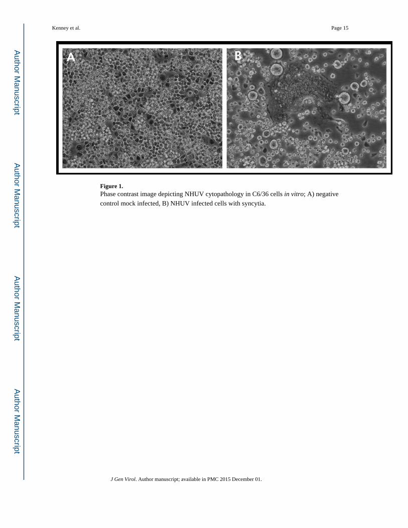
Figure 1. Phase contrast image depicting NHUV cytopathology in C6/36 cells in vitro; A) negative
control mock infected, B) NHUV infected cells with syncytia.
Kenney et al. Page 15
J Gen Virol. Author manuscript; available in PMC 2015 December 01.
Author M
anuscriptA
uthor Manuscript
Author M
anuscriptA
uthor Manuscript

Figure 2. Epifluorescent images of IFA tests in the various cell types examined
Kenney et al. Page 16
J Gen Virol. Author manuscript; available in PMC 2015 December 01.
Author M
anuscriptA
uthor Manuscript
Author M
anuscriptA
uthor Manuscript

Figure 3. Phylogenetic analysis based on nucleotide sequences of complete polyprotein coding
sequences. Phylogenies were constructed using the maximum likelihood method with
labeled bootstrap percentages as support. Labels include taxon name and accession number.
NHUV is highlighted in gray and clades are labeled by host association designations on the
far right of the figure.
Kenney et al. Page 17
J Gen Virol. Author manuscript; available in PMC 2015 December 01.
Author M
anuscriptA
uthor Manuscript
Author M
anuscriptA
uthor Manuscript

Figure 4. Mfold generated prediction and labels denoting conserved secondary structure and sequence
elements for CFAV (shown in alternating display for clarity), TBEV, MODV, WNV, and
NHUV. Nucleotides included in conserved MBFV sequences such the pentanucleotide,
conserved sequence 1 (CS1), and CS2 are highlighted with grey circles. A) Key structures
identified in CFAV include the 3’ LSH with an internal conserved pentanucleotide
(CACCG), a Y-shaped element, and a conserved hexanucleotide sequence element. B).
TBEV had the 3’LSH, pentanucleotide (CACAG), SL2, and Y-1 with an internal
Kenney et al. Page 18
J Gen Virol. Author manuscript; available in PMC 2015 December 01.
Author M
anuscriptA
uthor Manuscript
Author M
anuscriptA
uthor Manuscript

hexanucleotide sequence. C) MODV demonstrated the 3’ LSH, pentanucleotide (CUCAG),
and Y-1 with internal hexanucleotide sequence.multiple. D) WNV showed a 3’LSH, the
conserved pentanucleotide sequence (CACAG), SL2, conserved sequences CS1, CS2, and
CS3. E) NHUV was found to have a 3’ LSH, a conserved pentanucleotide (CACAG), SL2,
and only CS1 and CS2.
Kenney et al. Page 19
J Gen Virol. Author manuscript; available in PMC 2015 December 01.
Author M
anuscriptA
uthor Manuscript
Author M
anuscriptA
uthor Manuscript

Figure 5. Histograms demonstrating threonine, arginine, leucine, and proline codon usage frequencies
for NHUV, WNV, CxFV, and MODV.
Kenney et al. Page 20
J Gen Virol. Author manuscript; available in PMC 2015 December 01.
Author M
anuscriptA
uthor Manuscript
Author M
anuscriptA
uthor Manuscript
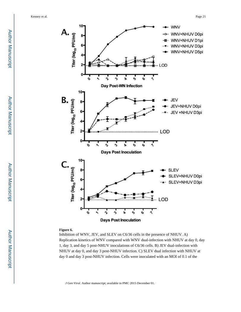
Figure 6. Inhibition of WNV, JEV, and SLEV on C6/36 cells in the presence of NHUV. A)
Replication kinetics of WNV compared with WNV dual-infection with NHUV at day 0, day
1, day 3, and day 5 post-NHUV inoculations of C6/36 cells. B) JEV dual-infection with
NHUV at day 0, and day 3 post-NHUV infection. C) SLEV dual infection with NHUV at
day 0 and day 3 post-NHUV infection. Cells were inoculated with an MOI of 0.1 of the
Kenney et al. Page 21
J Gen Virol. Author manuscript; available in PMC 2015 December 01.
Author M
anuscriptA
uthor Manuscript
Author M
anuscriptA
uthor Manuscript

representative MBFV and exposed to NHUV at an MOI of 5. Time points were collected
daily for seven days following infection with each MBFV.
Kenney et al. Page 22
J Gen Virol. Author manuscript; available in PMC 2015 December 01.
Author M
anuscriptA
uthor Manuscript
Author M
anuscriptA
uthor Manuscript

Author M
anuscriptA
uthor Manuscript
Author M
anuscriptA
uthor Manuscript
Kenney et al. Page 23
Table 1
Summary of NHUV in vitro infection of various cell types
IFA RT-PCRa CPE
Vero - - -
BHK21-15 - - -
DF-1 - - -
Xenopus laevis - - -
C6/36 + + +
C710 + + +
Cx. quinquefasciatus + + +
ISE6 - - -
aRT-PCR on supernatant from second passage
J Gen Virol. Author manuscript; available in PMC 2015 December 01.

Author M
anuscriptA
uthor Manuscript
Author M
anuscriptA
uthor Manuscript
Kenney et al. Page 24
Table 2
Genome organization of NHUV virus
Region Gene Position in genome (nt) Protein size (aa)a
5’ UTR 1-102
Structural
C 103–486 128
pr 487–768 94
M 769–993 75
E 994–2502 503
Non-structural
NS1 2503–3555 351
NS2A 3556–4251 232
NS2B 4252–4641 130
NS3 4642–6507 622
NS4A 6508–6954 149
NS4B 6955–7719 255
NS5 7720–10440 907
3’ UTR 10441–10891
aaa, amino acids
J Gen Virol. Author manuscript; available in PMC 2015 December 01.

Author M
anuscriptA
uthor Manuscript
Author M
anuscriptA
uthor Manuscript
Kenney et al. Page 25
Tab
le 3
Puta
tive
poly
prot
ein
clea
vage
site
s of
NH
UV
and
oth
er c
lose
ly r
elat
ed f
lavi
viru
ses
Cle
avag
eN
HU
VN
OU
VB
JVJE
VSL
EV
WN
V
Anc
hC/ v
irio
n C
NR
TR
R/A
RR
GM
VSK
RR
/GSA
SLK
TSK
R/G
LQ
QSF
RK
QN
KR
/GG
NE
GS
PSK
KR
/GG
TR
SSK
QK
KR
/GG
KT
GI
C/p
rMT
MV
AC
/VT
VG
TG
VA
SA/V
TFT
TW
TM
AA
C/A
TL
GM
FIA
YA
GA
/MK
LSN
FG
LA
SS/L
QL
STIA
SVG
A/V
TL
SN
pr/M
RR
SRR
/SV
AL
SPQ
RSR
R/S
VG
ISK
RR
SKR
/SV
AIA
SKR
SRR
/SV
SVQ
TR
RSR
R/S
ISV
QSR
RSR
R/S
LT
VQ
T
prM
/EV
APA
YS/
TH
CV
RIP
AY
S/M
KC
IGV
APA
YS/
LH
CR
SVV
APA
YS/
FNC
LG
MA
PAY
S/FN
CL
GV
APA
YS/
FNC
LG
M
E/N
S1T
SAH
A/E
VG
CS
TSV
SA/E
LG
CS
TT
VA
G/D
VG
CN
LT
NV
HA
/DT
GC
AI
TSV
QA
/DSG
CA
VN
VH
A/D
TG
CA
I
NS1
/NS2
AW
VT
AG
/QM
TG
IL
GV
LA
M/T
MM
FW
TT
AG
/NA
TG
IDQ
VD
AF/
NG
EM
VSR
VT
A/G
VA
GG
QV
NA
Y/N
AD
MID
NS2
A/N
S2B
KSG
KR
/SV
SMG
KT
TK
R/S
VPQ
SG
SGK
R/S
VSM
GE
PNK
KR
/GW
PAT
EPN
GK
R/S
WPA
SPN
RK
R/G
WPA
TE
NS2
B/N
S3SA
TQ
R/A
GA
MW
EN
RK
R/S
ND
TP
EK
GT
QK
/AG
AM
WD
LK
TT
KR
/GG
VFW
DK
HSK
R/G
GA
LD
LQ
YT
KR
/GG
VL
WD
NS3
/NS4
AA
EG
RR
/GA
MD
LA
GG
KR
/SA
VD
LA
EG
RR
/GA
SDIW
AA
GK
R/S
AIS
FIA
AG
KR
/SA
LG
MA
SGK
R/S
QIG
LI
NS4
A/N
S4B
TL
MIA
A/N
EK
GL
LG
AV
AA
/NE
YG
ML
AV
TA
/NE
KG
LG
VV
AA
/NE
YG
MG
VV
AA
/NE
MG
LSA
VA
A/N
EM
GW
NS4
B/N
S5K
SAR
R/G
TPG
GA
YK
KR
/GIW
EV
KSA
RK
/GT
PGG
PSL
KR
/GR
PGG
PKG
KR
/GG
GK
GPG
LK
R/G
GA
KG
J Gen Virol. Author manuscript; available in PMC 2015 December 01.

Author M
anuscriptA
uthor Manuscript
Author M
anuscriptA
uthor Manuscript
Kenney et al. Page 26
Tab
le 4
Perc
enta
ge o
f nu
cleo
tide
and
amin
o ac
id id
entit
y be
twee
n N
HU
V a
nd r
epre
sent
ativ
e fl
aviv
irus
OR
F se
quen
ces.
BJV
NH
UV
NO
UV
JEV
SLE
VW
NV
LA
MV
YF
VM
OD
VP
OW
V
% nt% AA
% nt% AA
% nt% AA
% nt% AA
% nt% AA
% nt% AA
% nt% AA
% nt% AA
% nt% AA
% nt% AA
NH
UV
65.9
70.7
--
--
--
-
NO
UV
56.1
53.0
56.2
53.0
--
--
-
JEV
53.4
49.2
53.9
49.2
52.8
47.2
--
-
SLE
V54
.049
.454
.149
.853
.548
.064
.266
.9
WN
V53
.849
.154
.349
.452
.546
.969
.176
.565
.167
.8-
LA
MV
53.2
48.0
53.2
47.7
52.6
46.5
54.6
49.7
55.1
50.6
54.5
49.5
YFV
51.2
44.4
51.1
44.2
50.8
43.8
51.9
45.3
52.0
45.9
51.6
45.1
51.7
45.6
MO
DV
44.5
36.1
44.5
35.8
44.4
35.2
45.0
36.5
45.8
36.2
45.0
36.6
45.4
35.6
47.0
38.2
POW
V47
.739
.347
.238
.946
.538
.348
.040
.547
.940
.648
.640
.647
.840
.649
.542
.248
.741
.6
CFA
V36
.724
.836
.524
.236
.024
.536
.624
.936
.624
.936
.924
.535
.824
.636
.124
.435
.524
.036
.024
.1
J Gen Virol. Author manuscript; available in PMC 2015 December 01.