medicine, heart, vascular, and thoracic institute
TRANSCRIPT

Stress testing and noninvasivecoronary imaging:What’s the best test for my patient?
C oronary artery disease (CAD) is the lead-ing cause of death in both men and women
in the United States.1 Its diagnosis and risk strat-ifi cation are an important aspect of medical care for all practitioners, regardless of specialty. Coronary catheterization has been the tech-nical standard for the diagnosis of CAD and is the recommended pathway for patients who are at high risk or who present with acute coronary syndrome.2,3 However, given that chest pain and anginal-equivalent symptoms are frequent in patients presenting to community clinics and emergency rooms and on inpatient wards, many practitioners need the skills and knowl-edge to conduct cardiac risk evaluation. Noninvasive testing is often used to cat-egorize patients as being at lower risk or having noncardiac chest pain vs those who are likely to have ischemia or obstructive CAD, which may require invasive coronary catheterization for further evaluation or intervention. Testing for CAD may be functional or anatomic (Table 1). In this article, we review what each test measures, its specifi c features, diagnostic and prognostic value, clinical util-ity, and limitations. These considerations help practitioners select the best test for a patient in a given setting or provide answers to a specifi c clinical question.
■ CORONARY ARTERY DISEASE
CAD is an infl ammatory pathologic process that starts as fatty streaks in the intimal layers of coronary arteries, then progresses to nonob-structive and then obstructive atherosclerotic
REVIEW
doi:10.3949/ccjm.88a.20068
ABSTRACTCoronary artery disease (CAD) causes signifi cant morbid-ity and mortality. Accurate noninvasive evaluation is im-portant to facilitate appropriate diagnosis and treatment. The ubiquitous nature of CAD requires all practitioners, regardless of their specialty, to be familiar with noninva-sive diagnostic modalities. This article reviews currently available tests, including specifi c features, diagnostic and prognostic value, strengths, and limitations.
KEY POINTSNoninvasive cardiac imaging (stress testing or anatomic evaluation) is warranted in patients who present with symptoms suspected to be cardiac, and in asymp tomatic patients in clinical scenarios in which possible CAD needs to be assessed or excluded.
Patients with symptoms and low to intermediate pretest probability for CAD are ideal candidates for electrocardi-ography exercise stress testing, stress echocardiography, or coronary computed tomography angiography.
Myocardial perfusion imaging is particularly useful in patients with known underlying CAD to determine if ischemia is present, and positron emission tomography stress imaging provides greater accuracy in those who are obese.
Milad Matta, MDDepartment of Internal Medicine, Cleveland Clinic, Cleveland, OH
Serge C. Harb, MDSection of Cardiovascular Imaging, Department of Cardiovascular Medicine, Heart, Vascular, and Thoracic Institute, Cleveland Clinic; Assistant Professor of Medicine, Cleveland Clinic Lerner College of Medicine of Case Western Reserve University, Cleveland, OH
Paul Cremer, MDSection of Cardiovascular Imaging, Department of Cardiovascular Medicine, Heart, Vascular, and Thoracic Institute, Cleveland Clinic, Cleveland, OH
Rory Hachamovitch, MD, MScSection of Cardiovascular Imaging, Department of Cardiovascular Medicine, Heart, Vascular, and Thoracic Institute, Cleveland Clinic, Cleveland, OH
Chadi Ayoub, MD, PhD, FACC, FASE, FSCCTSection of Cardiovascular Imaging, Department of Cardiovascular Medicine, Heart, Vascular, and Thoracic Institute, Cleveland Clinic; Assistant Professor of Medicine, Cleveland Clinic Lerner College of Medicine of Case Western Reserve University, Cleveland, OH
502 CLEVELAND CLINIC JOURNAL OF MEDICINE VOLUME 88 • NUMBER 9 SEPTEMBER 2021
on October 24, 2021. For personal use only. All other uses require permission.www.ccjm.orgDownloaded from

CLEVELAND CLINIC JOURNAL OF MEDICINE VOLUME 88 • NUMBER 9 SEPTEMBER 2021 503
MATTA AND COLLEAGUES
plaques. The process is driven by genetic and environmental cardiovascular risk factors.4 Ischemia occurs when coronary atherosclerotic plaque becomes severely stenotic or obstruc-tive (generally if stenosis is ≥ 50% in the left main coronary artery and ≥ 70% in the other epicardial coronary arteries), and it may be as-sociated with symptoms of angina or dyspnea.4 Moderate stenosis (50%–70%) may also cause ischemia and anginal-equivalent symp-toms due to lesion characteristics such as location and length of plaque, presence of endothelial dysfunction, and presence of mi-crovascular disease.5 Both obstructive and nonobstructive coronary stenosis may be com-plicated by acute plaque rupture and thrombo-sis, leading to acute loss of blood fl ow to the myocardium and myocardial infarction.4
Clinical scores incorporate variables such as age, sex, cardiovascular risk factors (hyper-tension, hyperlipidemia, diabetes, history of smoking, family history of CAD), changes on electrocardiography (ECG), cardiac enzyme levels, and symptoms. These scores help de-termine if patients have a low, intermediate, or high pretest probability of CAD.2,3,6 Chest pain may be classifi ed as noncardiac, atypical angina, and typical angina. Women and patients with diabetes may present without chest pain but with anginal-equivalent symp-toms such as shortness of breath on exertion or arm pain, or they may also have silent ischemia. Patients presenting with the acute coro-nary syndrome, high pretest probability of CAD, or concerning clinical features proceed straight to invasive coronary angiography. Noninvasive imaging (stress testing or ana-tomical evaluation) is warranted in those who present with symptoms that are suspected to be cardiac, particularly if the patient has an increased pretest probability of CAD. Table 2 lists the common indications for stress testing and coronary computed tomog-raphy (CT) angiography.3,7–10
■ ELECTROCARDIOGRAPHY EXERCISE STRESS TESTING
Test featuresECG exercise stress testing is the workhorse in community and hospital practices as an initial functional test to evaluate chest pain and sus-
pected CAD. Patients with low or intermedi-ate pretest probability for CAD are ideal can-didates for ECG exercise stress testing.11 The Bruce protocol is the most commonly used format. It starts the patient on a treadmill at a speed of 1.7 miles per hour and a 10% in-cline. Every 3 minutes, the speed and angle of incline are increased. Standard 12-lead ECG is used. If motion artifact occurs, moving the extremity elec-trodes to the torso and ensuring good elec-trode contact with the skin (eg, shaving if required) may help. Baseline ECG is taken before starting. The stress test continues until the patient is fatigued and asks to stop or develops cardiac symptoms, signifi cant ECG changes, or other high-risk features. An ECG stress test is considered diagnos-tic if the patient achieves at least 85% of the maximum age-predicted heart rate. If a test is terminated before achieving this threshold be-cause of positive fi ndings but the results meet the ECG criteria for ischemia, then the re-sults are still considered positive for ischemia. However, if a test is terminated before achiev-ing 85% of the predicted heart rate and there are no ECG changes, it is considered nondi-agnostic as it is not known whether ischemic changes would have occurred if the patient had continued to the required workload.
Diagnostic and prognostic featuresAn ECG stress test is considered positive for ischemia if there is at least a 1-mm horizontal or down-sloping ST-segment depression. Up-sloping ST-segment depression is not consid-ered a positive fi nding. An ST-segment eleva-tion greater than 1 mm is highly suggestive of signifi cant ischemia.
An ECG stress test is positive for ischemia if there is at least a 1-mm horizontal or down-sloping ST-segment depression
TABLE 1
Testing for coronary artery diseaseFunctional Electrocardiography exercise stress test
Stress echocardiographyNuclear medicine myocardial perfusion imaging techniques Single-photon emission computed tomography Positron emission tomographyCardiac magnetic resonance imaging with stress perfusion
Anatomic Coronary computed tomography angiographyInvasive coronary catheterization
on October 24, 2021. For personal use only. All other uses require permission.www.ccjm.orgDownloaded from

504 CLEVELAND CLINIC JOURNAL OF MEDICINE VOLUME 88 • NUMBER 9 SEPTEMBER 2021
STRESS TESTING
A meta-analysis of 24,074 patients in 147 studies found that ECG stress testing for de-tecting CAD has a sensitivity of 68% and specifi city of 77%.12 The Coronary Artery Surgery Study database suggested that the de-velopment of ST depression and functional capacity (duration of exercise) are the 2 most prognostic markers.13 Functional capacity is the strongest prog-nostic marker of an ECG stress test.7,14 It is estimated by metabolic equivalents (METs), which approximate oxygen uptake during ex-ercise, with 1 MET representing 3.5 mL/kg/min. Laboratories estimate functional capac-ity from exercise duration in a specifi c exer-
cise protocol based on published nomograms. Prognostic markers from an ECG stress test are shown in Table 3.7,13–19 The Duke treadmill score20 is reported by many laboratories and predicts 5-year mortal-ity risk in patients without known CAD. It incorporates variables that include degree of ST change and symptoms, with lower scores associated with higher mortality and increased likelihood of signifi cant CAD.20
LimitationsAn ECG stress test result positive for ischemia usually has ST-segment depressions mostly in the inferior and precordial leads, and it may
Functionalcapacity is the strongest prognostic marker of an ECG stress test
TABLE 2
Common indications for stress testingand coronary computed tomography angiography
Assessment for coronary artery disease
Angina or anginal equivalent symptoms and negative cardiac enzymes
Atypical symptoms in patients with diabetes or with high probability of diabetes
New diagnosis of cardiomyopathy (to defi ne whether the cause is ischemic or nonischemic)
New or increasing heart failure symptoms despite adherence to medical therapy
Re-evaluation of known heart failure (systolic or diastolic) in patients with a change in clinical statuswithout a clear precipitating change in medication or diet
Arrhythmias such as ventricular tachycardia or atrial fi brillation (to exclude ischemia as the cause) or new left bundle-branch block
To exclude severe ischemia prior to noncardiac surgery in those with increased coronary artery disease risks, angina symptoms, or poor exercise capacity (< 4 metabolic equivalents)
To defi ne presence or absence of ischemia in those with moderate coronary stenosis (stress test or fractional fl ow reserve-computed tomography)
Evaluation of anomalous coronary arteries
Stress testing for indications other than coronary artery disease assessment
Valve assessment Mitral valve stenosis or regurgitation severity (exercise stress echocardiography) Low-fl ow low gradient aortic stenosis (dobutamine stress echocardiography)
Exercise-induced pulmonary hypertension or diastolic dysfunction (exercise stress echocardiography)
Hypertrophic cardiomyopathy to demonstrate provocable left ventricular outfl ow tract obstruction (exercise stress echocardiography)
Exercise-induced arrhythmia or chronotropic incompetence (exercise stress echocardiography)
To defi ne cardiopulmonary disease and aerobic exercise capacity (metabolic stress test)
Data from references 3 and 7–10.
on October 24, 2021. For personal use only. All other uses require permission.www.ccjm.orgDownloaded from

CLEVELAND CLINIC JOURNAL OF MEDICINE VOLUME 88 • NUMBER 9 SEPTEMBER 2021 505
MATTA AND COLLEAGUES
not necessarily correspond to specifi c coronary artery territories. An ECG stress test should not be used if any of the following abnormalities are found on ECG: complete left bundle-branch block or paced ventricular rhythm (limits interpretation of the test), pre-excitation syndrome, or greater than 1 mm of resting ST-segment depression.21 Pronounced left ventricular (LV) hypertrophy or use of digoxin therapy can affect stress-related results. Patients with impaired mobility such as amputees or those with severe arthritis may not be able to safely exercise on the treadmill or go long enough to complete a diagnostic test. False-positive results may be more frequent in women. Many of these limitations can be overcome by adding an imaging component to the stress test or performing a pharmacologic stress test in those who cannot exercise.
■ STRESS ECHOCARDIOGRAPHY
Test featuresStress echocardiography uses imaging with echocardiography after exercise or pharma-
cologically induced stress to show coronary abnormalities. Ischemia is identifi ed if there is a new or worsening regional wall-motion abnormality, which generally correlates with stenosis in the corresponding coronary ter-ritory. Stress echocardiography is most often used to diagnose CAD in patients in whom an ECG stress test would be contraindicated, uninterpretable, or nondiagnostic, or if their CAD risk is suffi cient to warrant an imaging component to enhance the sensitivity and specifi city of the test. Images are typically obtained from stan-dard views including the parasternal long-axis and short-axis views, and apical long-axis and apical 2- and 4-chamber views to show the wall motion in each of the LV walls before and after stress (Figure 1). Results are ana-lyzed and scored using a 17-segment model of the LV (divided into apical, mid, and basal segments), with each segment graded on a 4-degree scale of regional wall-motion analy-sis (normokinesis, hypokinesis, dyskinesis, and akinesis).22
On stress echo-cardiography, ischemia isidentifi edif there is a newor worseningregionalwall-motionabnormality
TABLE 3
Prognostic indicators on electrocardiography stress testingIndicator Comments
Functional capacity Strongest prognostic indicator, reported as metabolic equivalents.7,14
ST-segment depressionor elevation
> 1-mm ST deviation is suggestive of ischemia.13
Exercise-inducedhypotension
Defi ned as systolic blood pressure that is lower during exercise than while standing at rest before exercise, refl ecting failure of cardiac output to increase during exercise.
Associated with severe coronary artery disease or left ventricular systolic dysfunction.15
Chronotropic incompetence Failure of heart rate to increase as expected during exercise, defi ned as achieving < 80% of predicted heart rate (or < 62% for patients takingbeta-blockers).16
Associated with increased all-cause and cardiovascular mortality.17
Impaired heart rate recovery
Heart rate fails to decrease normally after cessation of exercise.
Predicts all-cause mortality and cardiovascular events, including sudden death.18
Ventricular arrhythmia Sustained ventricular tachychardia or ventricular fi brillation.
Associated with signifi cant coronary artery disease or left ventriculardysfunction.19
on October 24, 2021. For personal use only. All other uses require permission.www.ccjm.orgDownloaded from

506 CLEVELAND CLINIC JOURNAL OF MEDICINE VOLUME 88 • NUMBER 9 SEPTEMBER 2021
STRESS TESTING
Intravenous (IV) echo contrast agents can be used to improve visualization of the endo-cardium if the image quality is suboptimal, particularly in patients with large body habitus or lung disease, or if more than 2 contiguous myocardial segments have poor endocardial defi nition. Echo contrast agents do not con-tain iodine and have been shown to improve the accuracy of the assessment of ventricular volume and ejection fraction, enhance recog-nition of wall-motion abnormalities, and im-prove reproducibility.23
Stress echocardiography can be exercise-based on a treadmill or bicycle, or pharma-cologically based with dobutamine infusion. Treadmill stress tests most often use the Bruce protocol. It is important to obtain postexercise im-aging as soon as possible after exercise stops, as regional wall-motion abnormalities that persist into recovery become less pronounced and resolve as the heart rate comes down.22 As such, the patient is moved immediately from the treadmill to the imaging bed in a left lat-eral decubitus position for poststress imaging. Stress echocardiography using a bicycle (supine or upright), although less frequent, is quieter, which permits sensitive precordial measurements with less motion artifact and allows imaging while the patient is exercising at different stages during the stress test. When used, it is often for valvular or hemodynamic assessment. Exercise stress echocardiography may al-low for hemodynamic evaluation in addition to that for ischemia. Doppler assessment may be helpful in patients with dyspnea and sus-pected exercise-induced diastolic dysfunction or pulmonary hypertension24 or in those with mitral valve stenosis or regurgitation that is clinically suspected to be more severe than a resting echocardiogram suggests. Stress echo-cardiography can also assess for dynamic LV outfl ow-tract obstruction in hypertrophic car-diomyopathy. Pharmacologic stress echocardiography can assess for ischemia in patients who cannot exercise or can help defi ne the severity of aor-tic stenosis, particularly when low-fl ow, low-gradient severe aortic stenosis is suspected. This is performed predominantly with dobuta-mine infusion, although it is possible to use di-
pyridamole or adenosine for ischemia testing. Dobutamine is a synthetic catecholamine that stimulates beta-1 adrenergic receptors causing a chronotropic effect (increase in heart rate) and an inotropic effect (increase in myocardi-al contractility), resulting in increased oxygen demand. The typical dobutamine stress proto-col consists of continuous IV infusion of dobu-tamine in 3-minute increments, starting with 5 mg/kg/min and increasing to a maximum of 40 mg/kg/min.25 Dobutamine may have an arrhythmogenic or hypertensive effect, and requires monitoring throughout. Patients with severe conduction disorders or advanced asthma or airway disease are not affected by dobutamine.
Diagnostic and prognostic featuresStress echocardiography results are reported by description of wall motion as normal, isch-emic, viable, or scarred myocardium. Normal myocardium has normal motion of segments at rest, and after stress, all segments demon-strate either normal motion or hyperkinesia, with overall increase in ejection fraction. When the myocardium is ischemic, contrac-tile function goes from normal to hypokinetic, akinetic, or dyskinetic after stress, in at least 2 adjacent segments for the test to be positive.26 When myocardium is scarred (due to previous MI), resting dysfunction (hypokinesis or aki-nesis) remains fi xed after stress. The myocardium is considered viable when segments with resting hypokinesis show either a maintained improvement with stress (indicating the presence of “stunning”) or im-provement during an early stress phase with subsequent deterioration in contractility at peak (ie, biphasic response), which portends potential improvement with revasculariza-tion.27 Other features that may suggest signifi cant ischemia are a decrease in LV ejection frac-tion after exercise (instead of an increase) or an increase in the LV cavity size after stress (expected to decrease as a result of increased contractility). A meta-analysis has demonstrated that stress echocardiography with exercise, dobuta-mine, dipyridamole, and adenosine has a sen-sitivity of 83%, 81%, 72%, and 79%, respec-tively, and a specifi city of 84%, 84%, 95%, and
On stress echo-cardiography,a decrease in LV ejection fractionafter exercise or an increase in LV cavity sizeafter stresscan suggestsignifi cantischemia
on October 24, 2021. For personal use only. All other uses require permission.www.ccjm.orgDownloaded from
CLEVELAND CLINIC JOURNAL OF MEDICINE VOLUME 88 • NUMBER 9 SEPTEMBER 2021 507
MATTA AND COLLEAGUES
MPI enables cliniciansto assessthe physiologic signifi canceof coronarystenosisby measuringheterogeneityin coronary fl ow
91%, respectively.28 Stress echocardiography is generally considered more specifi c than nucle-ar perfusion imaging, although nuclear perfu-sion imaging is considered more sensitive.29 A strength of stress echocardiography is improved diagnostic accuracy compared with stress ECG alone without ionizing radiation exposure. As such, it is often the preferred test for middle-aged women who may have symp-toms and intermediate cardiovascular risk. It may also be desirable in patients who have dyspnea, in whom other hemodynamic evalu-ation can be done in the same test. Stress echocardiography has prognostic value. A normal test with no regional wall-motion abnormalities confers a less than 1% per year cardiac event rate. Increasing sever-ity of regional wall-motion abnormalities af-ter peak stress corresponds to higher clinical event rates.8
LimitationsAs with all imaging, interpretation of a stress echocardiogram may be affected by subjectiv-ity. Thus, it is important to have good imaging protocols and quality acquisitions along with experienced practitioners to interpret the im-ages. Stress echocardiography may miss mild ischemia that is due to small, distal, or branch-vessel disease, and it is considered slightly less sensitive than nuclear imaging.30 Patients with obesity or emphysema may have poor acoustic windows, resulting in suboptimal images.
■ NUCLEAR MEDICINE MYOCARDIAL PERFUSION IMAGING
Test featuresNuclear myocardial perfusion imaging (MPI) may be performed by either single-photon emission CT (SPECT) or positron emission tomography (PET). As with stress echocar-diography, MPI stress testing may be exercise or pharmacologically induced. MPI involves IV administration of radioactive tracers. A gamma camera detects radio emissions from the tracer that perfuses the myocardium. Trac-er uptake depends on fl ow dynamics as well as myocyte membrane integrity. Color-coded images of myocardial perfusion pre- and post-stress are generated in different axes to allow assessment for each coronary distribution.31
The radioisotopes and cameras used in
PET and SPECT differ. PET generally uses rubidium or ammonia radionuclides for perfu-sion imaging, and fl uorodeoxyglucose (FDG) may be used to assess myocardial viability and infl ammation. SPECT scanners predominant-ly use technetium 99 (sestamibi) for perfusion imaging. Thallium has been phased out be-cause of associated high radiation. PET carries advantages over SPECT in-cluding superior image quality, due to more favorable tracer characteristics and count statistics. Positron-emitting radiotracers used in PET can produce higher-energy photons than those produced by SPECT radiotracers, resulting in less attenuation artifact. PET can also detect smaller and more subtle perfusion defects (typically 4–7 mm) owing to its high-er spatial resolution than SPECT (typically 12–15 mm).32 Other advantages of PET over SPECT include a lower radiation burden and shorter scan time. Stress MPI can be exercise-induced (using the treadmill Bruce protocol, which has the added value of providing functional capacity data that is prognostic) or pharmacologic for those unable to exercise. Vasodilators are the most frequently used stress agents, primarily regadenoson (which has a more favorable pro-fi le) or dipyridamole and adenosine. Vasodila-tors increase coronary blood fl ow through their effect on the adenosine A2A receptor, which increases blood velocity and fl ow rate in nor-mal vessels compared with a lesser response in stenotic vessels that are already maximally di-lated, thus decreasing subendocardial fl ow to regions supplied by diseased vessels. Dobuta-mine infusion may also be used, although it is rare in MPI practice.
Diagnostic and prognostic featuresMPI enables clinicians to assess the physiolog-ic signifi cance of coronary stenosis by measur-ing heterogeneity in coronary fl ow. The abil-ity to maintain the maximum fl ow (“coronary fl ow reserve”) is impaired when there is more than 50% coronary stenosis.33 Normal myo-cardial perfusion is shown by homogeneous radiotracer distribution on both stress and rest images. In general, perfusion defects in a coronary territory that occur after stress with normal fl ow at rest suggest inducible ischemia, whereas a fi xed defect in perfusion both at rest
on October 24, 2021. For personal use only. All other uses require permission.www.ccjm.orgDownloaded from

508 CLEVELAND CLINIC JOURNAL OF MEDICINE VOLUME 88 • NUMBER 9 SEPTEMBER 2021
STRESS TESTING
and after stress in a coronary distribution sug-gests either scarred myocardium (from prior myocardial infarction) or hibernating myo-cardium (which may improve in function if revascularized).34
MPI defects are generally reported with reference to the following:• Defect size or extent: small (< 10% of LV
myocardium affected), medium (10%–20% affected), or large (> 20% affected)
• Severity of perfusion defect (mild, moder-ate, severe)
• Extent of reversibility (reversible, irrevers-ible)
• Location (based on 17-segment LV model and coronary artery territory).
Tomograms are also produced on MPI studies that estimate LV ejection fraction. The presence of transient ischemic dilation is a sign of severe ischemia. It refers to the en-largement of the LV poststress instead of de-crease of cavity size as would be expected with increased contractility. Myocardial blood fl ow and myocardial fl ow reserve offer a quantitative assessment of myo-cardial perfusion35 and, in some cases, may help identify the presence of microvascular disease. Some centers routinely include these measures on clinical PET reports, and similar quantitative measures may also be available for SPECT in the future. A systematic review reported sensitivity and specifi city of SPECT for the diagnosis of CAD of 82% and 76%, respectively, and 91% and 89% for PET, with the difference accounted for by superior spatial resolution and attenuation correction of PET.36 SPECT is considered more sensitive than stress echo-cardiography but less specifi c. PET is generally accepted as the most accurate noninvasive functional test for ischemia. MPI provides clinically helpful prognostic information. For those with normal MPI results, the 2-year clinical event rate for cardiac death or myocardial infarction is less than 1%.37 The presence of perfusion defects is prognostic for clinical myocardial infarction and mortality. MPI is useful in symptomatic patients with suspected CAD to show the presence or absence of ischemia (Figure 2), as well as in those with known CAD to evaluate if ste-nosis is functionally signifi cant. In addition,
Positronemissiontomographyis generallyacceptedas the mostaccuratenoninvasive functional test for ischemia
Figure 1. Stress echocardiogram from a 54-year-old woman with chest pain shows ischemia in the left anterior descending artery and right coronary artery territories. Panels A and B show the 4-chamber view before and after the stress test. On the poststress image (B), the arrows point to hypokinesis at the apex and distal septum; enlargement of left ventricular cavitysuggests signifi cant ischemia. In the long-axis view (C, D), arrows point to hypo-kinesis at the apex and distal septum. In the 2-chamber view (E, F), arrows point to hypokinesis on the mid and apical inferior wall.
on October 24, 2021. For personal use only. All other uses require permission.www.ccjm.orgDownloaded from
CLEVELAND CLINIC JOURNAL OF MEDICINE VOLUME 88 • NUMBER 9 SEPTEMBER 2021 509
MATTA AND COLLEAGUES
those with impaired LV systolic function may also have viability concurrently assessed dur-ing MPI imaging, particularly with FDG-PET techniques, which may help guide revascu-larization decisions. Those with left bundle- branch block may be suitable candidates for regadenoson pharmacologically induced SPECT or PET (as an ECG exercise stress test would be nondiagnostic). For obese patients, PET MPI is the superior modality.
LimitationsMPI techniques involve radiation exposure, and there needs to be suffi cient clinical value to justify testing. SPECT may be more prone to artifact from diaphragmatic attenuation or gut scatter that may result in false-positive re-sults being identifi ed in the inferior LV wall, particularly in patients who are obese. This is less of an issue with PET. The main limitations for PET are higher cost and limited availability. Only healthcare facilities with an on-site cyclotron to produce isotopes daily (due to their short half-life) can offer PET imaging.32
Finally, a normal result on MPI suggests the absence of obstructive CAD. However, it does not exclude mild to moderate atheroscle-rosis that may not be contributing to symp-toms but nonetheless may warrant aggressive preventive measures. Identifying coronary calcium on CT scout images before an MPI may help fl ag for the presence of subclinical coronary atherosclerosis.
■ CORONARY CT ANGIOGRAPHY
Test featuresCoronary CT angiography (CCTA) is an ana-tomic noninvasive modality that can identify and assess the severity of CAD. It differs from stress testing in that it directly visualizes the coronary arteries and can quantify the degree of stenosis and assess plaque characteristics (Figure 3). In contrast, stress testing assesses LV wall-motion abnormalities or perfusion defects to de-termine if obstructive CAD is present. Adequate patient preparation is needed to enable high-quality image acquisition and improve accuracy. Ideally, the heart rate needs
Coronary CTangiographycan identifyand assess the severity of CADby quantifying the degreeof stenosisand assessingplaquecharacteristics
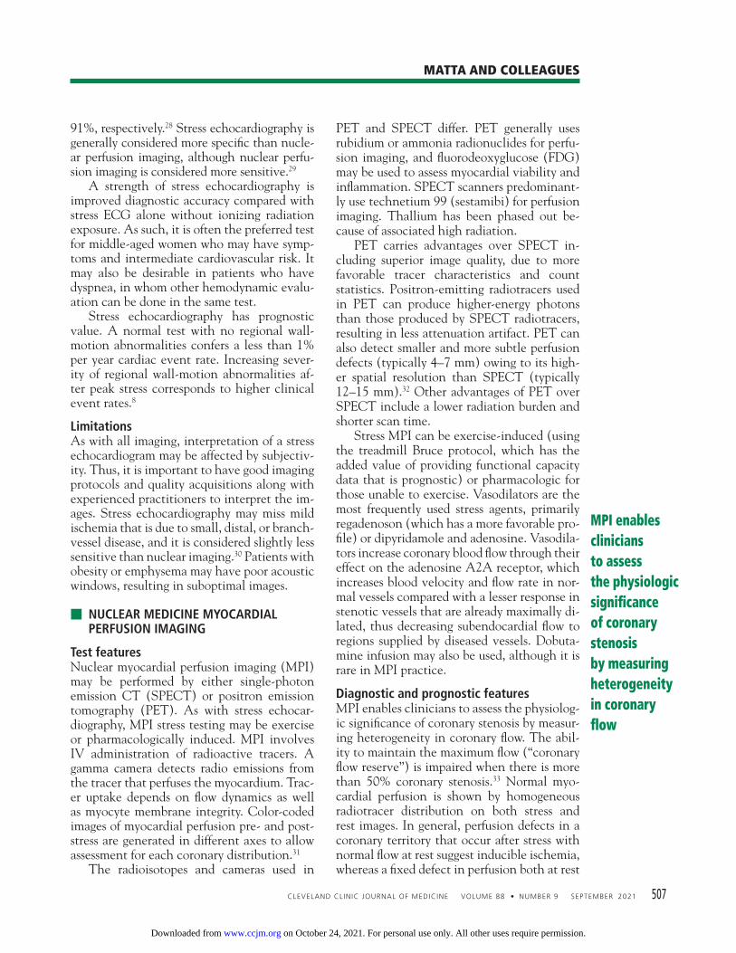
Figure 2. Single-photon emission CT myocardial perfusion imaging in a 62-year-old man with diabetes and a 2-month history of dyspnea shows moderate left anterior descending coronary artery ischemia. Panels A, C, and E are poststress images that show perfusion de-fects in the apex, apical septum, and apical anterior wall (arrows). Panels B, D, and F show relatively normal perfusion at rest at the corresponding levels.
on October 24, 2021. For personal use only. All other uses require permission.www.ccjm.orgDownloaded from

510 CLEVELAND CLINIC JOURNAL OF MEDICINE VOLUME 88 • NUMBER 9 SEPTEMBER 2021
STRESS TESTING
to be less than 60 beats per minute, although less than 70 beats per minute is acceptable on more advanced scanners. A beta-blocker or calcium channel blocker may be administered orally or intravenously to help achieve the target heart rate. Sublingual nitroglycerin is given just be-fore scanning to help dilate the coronary ar-teries and improve the image quality. Then an iodinated contrast agent is administered through an IV line in the cubital fossa, and CT images are acquired with ECG gating. To reduce radiation exposure, it is preferable to use a prospective acquisition protocol for CCTA scans in which images are obtained at a point in end diastole (or sometimes end systole) when cardiac and coronary motion is least, thus reducing motion artifact.
Diagnostic and prognostic featuresImages are reconstructed and analyzed for the presence, degree, and location of coronary ste-nosis. Plaque composition (whether calcifi ed, noncalcifi ed, or mixed) and high-risk plaque features, if present, are also reported. The So-
ciety of Cardiovascular CT recommends using the CAD reporting and data system to stan-dardize CCTA reports.38 It categorizes coro-nary segments as having no stenosis, mini-mal (0%–24%), mild (25%–49%), moderate (50%–69%), or severe (70%–99%) stenosis, or total occlusion (100%).38
High-risk plaque features include low Hounsfi eld unit attenuation (signifying more lipid-laden plaque), high plaque volume, positive remodeling (plaque extending out-wards from the vessel wall and not just into the lumen), or spotty calcifi cation within the plaque. These features suggest that plaque is more vulnerable to rupture and, thus, the pa-tient has a greater likelihood of clinical events such as myocardial infarction.39 These features are mandated for clinical reporting in the So-ciety of Cardiovascular CT guidelines.38
Numerous meta-analyses have confi rmed the diagnostic accuracy of CCTA, including reported sensitivity of 99% and specifi city of 89%.40 As such, it has excellent negative pre-dictive value and can accurately rule out CAD. The European Society of Cardiology 2019
Coronary CT angiographycan identifyplaque featuresthat suggest the plaqueis morevulnerableto rupture
Figure 3. Coronary computed tomography angiography in a 40-year-old man who smoked and had a family history of premature coronary artery disease. Panel A is a 3-D rendering showing proximal left anterior descending (LAD) coronary artery stenosis (arrow). Panel B is a multiplanar reconstruction showing proximal LAD coronary artery stenosis with pre-dominantly soft (lipid-laden) noncalcifi ed plaque (arrow). Panel C shows the corresponding LAD lesion (arrow) on coronary catheterization.
LCx = left circumfl ex artery; RCA = right coronary artery
on October 24, 2021. For personal use only. All other uses require permission.www.ccjm.orgDownloaded from

CLEVELAND CLINIC JOURNAL OF MEDICINE VOLUME 88 • NUMBER 9 SEPTEMBER 2021 511
MATTA AND COLLEAGUES
guidelines give CCTA a class 1 indication to assess for (or rule out) CAD in symptomatic patients with low to intermediate cardiovascu-lar risk, and a class IIa indication if functional testing is not diagnostic or is equivocal.3 UK guidelines recommend CCTA as fi rst-line test-ing for evaluating stable chest pain.41
CCTA results are prognostic. Patients with obstructive CAD identifi ed by CCTA have worse outcomes than those with nonob-structive CAD,42 who, in turn, have a higher clinical event rate than those without CAD. CCTA is useful clinically, as it may identify pa-tients with nonobstructive CAD (such as 50% stenosis), which a stress test would call normal, as nonobstructive lesions are not fl ow-limiting. In addition, identifi cation of nonobstruc-tive CAD by CCTA offers an opportunity for aggressive risk factor modifi cation, including statin therapy.43 CCTA can be also used for the assessment of coronary artery bypass graft patency, and is excellent in the assessment of suspected anomalous coronary arteries. Although CCTA is predominately used for anatomic coronary assessment, techniques such as fractional fl ow reserve CT (FFR-CT)and stress perfusion imaging by CT are now available to determine the functional sig-nifi cance of a moderate coronary lesion (eg, whether a 50% to 70% stenosis on CT is fl ow- limiting or nonobstructive). FFR-CT has ad-ditional costs, and the images are sent off-site for analysis. In addition, CT perfusion requires higher radiation and contrast doses and longer scan time, limiting its widespread adoption. Another advance is surrogate imaging markers for infl ammation such as attenuation in coronary perivascular fat on CCTA, which may be predictive of cardiac mortality and thus may play a clinical role in prevention.44 Anticipated developments in artifi cial intel-ligence and radiomic assessment are expected to enhance automated image evaluation and quantitative assessment of CCTA, with im-provements in workfl ow and diagnostic accu-racy.45 These are expected to have a signifi cant impact on clinical practice.
LimitationsCCTA involves exposure to ionizing radia-tion. It requires an iodinated contrast agent, which needs premedication in patients with
iodine allergy. And its use is limited in those with renal insuffi ciency. CCTA is generally less useful for evaluat-ing coronary stents because of blooming arti-fact from the metal struts, limiting its ability to assess for in-stent restenosis unless the stent is large in caliber. Arrhythmias, including atrial fi brillation and ectopy, make it more diffi cult to obtain a quality image, requiring adjustment of protocols. More rapid heart rate also reduces image quality. Heavy calcifi cation can result in segments being uninterpretable for stenosis, potentially limiting the utility of CCTA in elderly or di-alysis patients. Patients unable to adequately hold their breath would not be suited for CCTA.
■ CORONARY ARTERY CALCIUM SCORE
Test featuresA coronary artery calcium (CAC) score is widely accepted and used for CAD risk strati-fi cation in asymptomatic patients. It is a sur-rogate marker for the presence and the burden of CAD as it quantifi es coronary calcifi cation and, hence, the extent of atherosclerotic dis-ease. It involves rapid CT scan acquisition without contrast, with the fi eld of view fo-cused on the heart. Axial slices with 3-mm thickness are acquired prospectively with ECG gating in mid to late diastole. The CAC Agatston score takes into account the amount and density of calcium, with more than 130 Hounsfi eld units or at least 3 adjacent voxels needed to generate a numeric score.46
Diagnostic and prognostic featuresThere are strong data to support the prognos-tic value of CAC, and it enhances risk strati-fi cation incremental to traditional clinical cardiovascular risk factors.47 In absolute terms, a calcium score of 0 is associated with excel-lent prognosis; scores in categories of 1 to 99, 100 to 299, and 300 and above are associated with respective increased risks of mortality.47 However, risk prediction is often reported as a percentile with adjustment for age, sex, and ethnicity. The CAC score may be useful in the clini-cal decision-making process for patients who are asymptomatic with borderline (5%–7.5%) or intermediate (7.5%–20%) 10-year risk ac-
A coronary artery calcium score is used for CAD risk stratifi cation in asymptomatic patients
on October 24, 2021. For personal use only. All other uses require permission.www.ccjm.orgDownloaded from

512 CLEVELAND CLINIC JOURNAL OF MEDICINE VOLUME 88 • NUMBER 9 SEPTEMBER 2021
STRESS TESTING
cording to the atherosclerotic cardiovascu-lar disease (ASCVD) risk calculator, and in whom the benefi t of a statin is in question.6 The 2019 American College of Cardiology/American Heart Association guidelines rec-ommend initiating a statin in patients with diabetes or in those age 40 to 75 with an AS-CVD risk above 7.5% over 10 years.6 In this latter group, the CAC score may be used to restratify the risk either up or down and better guide statin initiation. For example, a patient with borderline or intermediate ASCVD risk and a CAC score of 0 would not be started on a statin. However, if the CAC score were above 100 (or ≥ 75th percentile for age/sex/race), then the risk would be restratifi ed up, clearly defi ning a patient who would benefi t from a statin.6
LimitationsIt must be stressed that although the CAC score has use in prognostication in asymptom-atic patients, if anginal-equivalent symptoms are being evaluated, then the CAC score has no role as it cannot determine whether a cal-cifi ed coronary plaque is stenotic, and other tests would need to be considered.
■ STRESS CARDIAC MAGNETIC RESONANCE IMAGING
Test features Stress cardiac magnetic resonance imaging (MRI) is a promising modality, with advan-tages such as good spatial and temporal resolu-
tion, wide fi eld of view, and ability to acquire images in different planes.48 It uses gadolinium contrast rather than iodinated contrast and does not use ionizing radiation. MRI perfusion images can be assessed for perfusion defects (Figure 4), just as is done with nuclear MPI before and after stress. In addition, cine imag-es from MRI can be assessed for regional wall-motion abnormalities as is done with stress echocardiography. MRIs also provide morpho-logic information including quantifi cation of ventricular and valvular function.49 However, current technology limits MRI anatomic as-sessment of the coronary arteries in adults to visualization of only the proximal portions.
Diagnostic and prognostic featuresStress cardiac MRI compares favorably with established noninvasive modalities in terms of accuracy for detecting CAD. Studies show stress-induced wall-motion abnormality im-aging by MRI has a sensitivity of 83% and specifi city of 86%.48 Perfusion imaging with MRI has a sensitivity of 91% and specifi city of 81%.48 Stress cardiac MRI that is negative for ischemia has prognostic value and is associ-ated with very low risk of cardiovascular death and myocardial infarction (less then 1% com-bined rate per annum).50
LimitationsStress cardiac MRI is relatively new and is the least frequently used compared with the other modalities discussed. Its availability and
Stress cardiac MRI compares favorably with established noninvasive tests in terms of accuracy for detecting CAD
Figure 4. Stress cardiac magnetic resonance imaging in a 67-year-old woman with diabe-tes and chest pain shows normal perfusion at rest (A). Panel B shows a poststress image with a perfusion defect in the inferior and inferoseptal segments (arrow), suggestive of ischemia in the right coronary artery territory. Panel C is a delayed gadolinium-enhanced image showing mild subendocardial enhancement (arrow) in the corresponding region, consistent with a small area of scar.
on October 24, 2021. For personal use only. All other uses require permission.www.ccjm.orgDownloaded from
CLEVELAND CLINIC JOURNAL OF MEDICINE VOLUME 88 • NUMBER 9 SEPTEMBER 2021 513
MATTA AND COLLEAGUES
access may be limited, with practical experi-ence still nascent and limited in most centers. Other potential limitations include cost and long duration of scanning, which may be in-tolerable for those with signifi cant claustro-phobia or inability to hold their breath. It may be contraindicated in those with metal devices or prostheses, or in those with severe renal dysfunction due to risk of nephrogenic systemic fi brosis.51
■ APPROPRIATE USE CRITERIA
Appropriate-use criteria (AUC) guidelines are available for each imaging modality. They summarize the evidence and provide broad recommendations for given clinical scenarios by way of categorization as appropriate, may be appropriate, inappropriate, or rarely appro-priate. In 2019, a group of healthcare societies re-leased consensus AUC guidelines for cardiac multimodality imaging including stress testing that address appropriateness of test selection in broad categories.9 The 2014 AUC guide-lines, however, are more focused on testing for CAD and give more of a detailed and ex-tensive list of scenarios for appropriate use,10
although additional evidence has accrued since then. Nevertheless, both guidelines are useful in improving understanding for appro-priate test selection. Ultimately, AUC guide-lines cannot determine a single best test, and a physician must take into account the whole clinical picture and test features when making a selection.
■ TAKE-HOME MESSAGE
Both stress testing with ECG, echocardiog-raphy, nuclear perfusion imaging, and MRI and anatomical evaluation with coronary CT provide details for evaluating CAD in at-risk patients. Results can help in risk stratifi cation and assist with prognostication. Appropriate test selection is based on the patient’s clinical picture, including the nature of symptoms, the risk profi le, the clinical question being asked, and the strengths and limitations of the test-ing modality. Other factors that may infl uence test selection include local expertise, avail-ability and access to a given modality, cost, and patient preference. ■
■ DISCLOSURESThe authors report no relevant fi nancial relationships which, in the context of their contributions, could be perceived as a potential confl ict of interest.
■ REFERENCES 1. US Centers for Disease Control and Prevention. Heart disease in the
United States. https://www.cdc.gov/heartdisease/facts.htm. Accessed August 10, 2021.
2. Amsterdam EA, Wenger NK, Brindis RG, et al. 2014 AHA/ACC guide-line for the management of patients with non-ST-elevation acute coronary syndromes: a report of the American College of Cardiol-ogy/American Heart Association Task Force on Practice Guidelines. J Am Coll Cardiol 2014; 64(24):e139–e228. doi:10.1016/j.jacc.2014.09.017
3. Knuuti J, Wijns W, Saraste A, et al. 2019 ESC guidelines for the diag-nosis and management of chronic coronary syndromes. Eur Heart J 2020; 41(3):407–477. doi:10.1093/eurheartj/ehz425
4. Falk E, Shah PK, Fuster V. Coronary plaque disruption. Circulation 1995; 92(3):657–671. doi:10.1161/01.cir.92.3.657
5. Crossman DC. The pathophysiology of myocardial ischaemia. Heart 2004; 90(5):576–580. doi:10.1136/hrt.2003.029017
6. Arnett DK, Blumenthal RS, Albert MA, et al. 2019 ACC/AHA guide-line on the primary prevention of cardiovascular disease: executive summary: a report of the American College of Cardiology/American Heart Association Task Force on Clinical Practice Guidelines. J Am Coll Cardiol 2019; 74(10):1376–1414. doi:10.1016/j.jacc.2019.03.009
7. Gibbons RJ, Balady GJ, Bricker JT, et al. ACC/AHA 2002 guideline update for exercise testing: summary article: a report of the Ameri-can College of Cardiology/American Heart Association Task Force on Practice Guidelines (Committee to Update the 1997 Exercise Testing Guidelines). Circulation 2002; 106(14):1883–1892. doi:10.1161/01.cir.0000034670.06526.15
8. Pellikka PA, Arruda-Olson A, Chaudhry FA, et al. Guidelines for
performance, interpretation, and application of stress echocar-diography in ischemic heart disease: from the American Society of Echocardiography. J Am Soc Echocardiogr 2020; 33(1):1–41.e8. doi:10.1016/j.echo.2019.07.001
9. Doherty JU, Kort S, Mehran R, et al. ACC/AATS/AHA/ASE/ASNC/HRS/SCAI/SCCT/SCMR/STS 2019 appropriate use criteria for multimodal-ity imaging in the assessment of cardiac structure and function in nonvalvular heart disease: a report of the American College of Car-diology Appropriate Use Criteria Task Force, American Association for Thoracic Surgery, American Heart Association, American Society of Echocardiography, American Society of Nuclear Cardiology, Heart Rhythm Society, Society for Cardiovascular Angiography and Inter-ventions, Society of Cardiovascular Computed Tomography, Society for Cardiovascular Magnetic Resonance, and the Society of Thoracic Surgeons. J Am Coll Cardiol 2019; 73(4):488–516. doi:10.1016/j.jacc.2018.10.038
10. Wolk MJ, Bailey SR, Doherty JU, et al. ACCF/AHA/ASE/ASNC/HFSA/HRS/SCAI/SCCT/SCMR/STS 2013 multimodality appropriate use cri-teria for the detection and risk assessment of stable ischemic heart disease: a report of the American College of Cardiology Foundation Appropriate Use Criteria Task Force, American Heart Association, American Society of Echocardiography, American Society of Nuclear Cardiology, Heart Failure Society of America, Heart Rhythm Society, Society for Cardiovascular Angiography and Interventions, Society of Cardiovascular Computed Tomography, Society for Cardiovascular Magnetic Resonance, and Society of Thoracic Surgeons. J Am Coll Cardiol 2014; 63(4):380–406. doi:10.1016/j.jacc.2013.11.009
11. Dowsley T, Al-Mallah M, Ananthasubramaniam K, Dwivedi G, McArdle B, Chow BJ. The role of noninvasive imaging in coronary artery disease detection, prognosis, and clinical decision making.
on October 24, 2021. For personal use only. All other uses require permission.www.ccjm.orgDownloaded from

514 CLEVELAND CLINIC JOURNAL OF MEDICINE VOLUME 88 • NUMBER 9 SEPTEMBER 2021
STRESS TESTING
Can J Cardiol 2013; 29(3):285–296. doi:10.1016/j.cjca.2012.10.022 12. Detrano R, Gianrossi R, Froelicher V. The diagnostic accuracy of the
exercise electrocardiogram: a meta-analysis of 22 years of research. Prog Cardiovasc Dis. 1989; 32(3):173–206. doi:10.1016/0033-0620(89)90025-x
13. Weiner DA, Ryan TJ, McCabe CH, et al. Prognostic importance of a clinical profi le and exercise test in medically treated patients with coronary artery disease. J Am Coll Cardiol 1984; 3(3):772–779. doi:10.1016/s0735-1097(84)80254-5
14. Arena R, Myers J, Williams MA, et al. Assessment of functional capacity in clinical and research settings: a scientifi c statement from the American Heart Association Committee on Exercise, Rehabilita-tion, and Prevention of the Council on Clinical Cardiology and the Council on Cardiovascular Nursing. Circulation 2007; 116(3):329–343. doi:10.1161/CIRCULATIONAHA.106.184461
15. Dubach P, Froelicher VF, Klein J, Oakes D, Grover-McKay M, Friis R. Exercise-induced hypotension in a male population. Criteria, causes, and prognosis. Circulation 1988; 78(6):1380–1387. doi:10.1161/01.cir.78.6.1380
16. Kligfi eld P, Lauer MS. Exercise electrocardiogram testing: beyond the ST segment. Circulation 2006; 114(19):2070–2082. doi:10.1161/CIRCULATIONAHA.105.561944
17. Myers J, Tan SY, Abella J, Aleti V, Froelicher VF. Comparison of the chronotropic response to exercise and heart rate recovery in predict-ing cardiovascular mortality. Eur J Cardiovasc Prev Rehabil 2007; 14(2):215–221. doi:10.1097/HJR.0b013e328088cb92
18. Jouven X, Empana JP, Schwartz PJ, Desnos M, Courbon D, Duci-metière P. Heart-rate profi le during exercise as a predictor of sud-den death. N Engl J Med 2005; 352(19):1951–1958. doi:10.1056/NEJMoa043012
19. Miller TD. Exercise treadmill test: estimating cardiovascular progno-sis. Cleve Clin J Med 2008; 75(6):424–430. doi:10.3949/ccjm.75.6.424
20. Mark DB, Shaw L, Harrell FE Jr, et al. Prognostic value of a treadmill exercise score in outpatients with suspected coronary artery disease. N Engl J Med 1991; 325(12):849–853. doi:10.1056/NEJM199109193251204
21. Fletcher GF, Ades PA, Kligfi eld P, et al. Exercise standards for testing and training: a scientifi c statement from the American Heart As-sociation. Circulation 2013; 128(8):873–934. doi:10.1161/CIR.0b013e31829b5b44
22. Cerqueira MD, Weissman NJ, Dilsizian V, et al. Standardized myo-cardial segmentation and nomenclature for tomographic imaging of the heart. A statement for healthcare professionals from the Cardiac Imaging Committee of the Council on Clinical Cardiology of the American Heart Association. Circulation 2002; 105(4):539–542. doi:10.1161/hc0402.102975
23. Mulvagh SL, Rakowski H, Vannan MA, et al. American Society of Echocardiography consensus statement on the clinical applications of ultrasonic contrast agents in echocardiography. J Am Soc Echocar-diogr 2008; 21(11):1179–1281. doi:10.1016/j.echo.2008.09.009
24. Ha JW, Andersen OS, Smiseth OA. Diastolic stress test: invasive and noninvasive testing. JACC Cardiovasc Imaging 2020; 13(1 pt 2):272–282. doi:10.1016/j.jcmg.2019.01.037
25. Geleijnse ML, Fioretti PM, Roelandt JR. Methodology, feasibility, safety and diagnostic accuracy of dobutamine stress echocardiogra-phy. J Am Coll Cardiol 1997; 30(3):595–606. doi:10.1016/s0735-1097(97)00206-4
26. Sicari R, Nihoyannopoulos P, Evangelista A, et al. Stress echocar-diography expert consensus statement: European Association of Echocardiography (EAE) (a registered branch of the ESC). Eur J Echocardiogr 2008; 9(4):415–437. doi:10.1093/ejechocard/jen175
27. Senior R, Lahiri A. Enhanced detection of myocardial ischemia by stress dobutamine echocardiography utilizing the “biphasic” response of wall thickening during low and high dose dobutamine infusion. J Am Coll Cardiol 1995; 26(1):26–32. doi:10.1016/0735-1097(95)00139-q
28. Heijenbrok-Kal MH, Fleischmann KE, Hunink MG. Stress echocar-diography, stress single-photon-emission computed tomography and electron beam computed tomography for the assessment of
coronary artery disease: a meta-analysis of diagnostic performance. Am Heart J 2007; 154(3):415–423. https://doi.org/10.1016/j.ahj.2007.04.061
29. Mairesse GH, Marwick TH, Arnese M, et al. Improved identifi cationof coronary artery disease in patients with left bundle branch block by use of dobutamine stress echocardiography and comparison with myocardial perfusion tomography. Am J Cardiol 1995; 76(5):321–325. doi:10.1016/s0002-9149(99)80093-9
30. O’Keefe JH Jr, Barnhart CS, Bateman TM. Comparison of stress echocardiography and stress myocardial perfusion scintigraphy for diagnosing coronary artery disease and assessing its severity. Am J Cardiol 1995; 75(11):25D–34D. pmid:7726110
31. Henzlova MJ, Duvall WL, Einstein AJ, Travin MI, Verberne HJ. ASNC imaging guidelines for SPECT nuclear cardiology procedures: stress, protocols, and tracers. J Nucl Cardiol 2016; 23(3):606–639. doi:10.1007/s12350-015-0387-x
32. Driessen RS, Raijmakers PG, Stuijfzand WJ, Knaapen P. Myocar-dial perfusion imaging with PET. Int J Cardiovasc Imaging 2017; 33(7):1021–1031. doi:10.1007/s10554-017-1084-4
33. Goodwill AG, Dick GM, Kiel AM, Tune JD. Regulation of coronary blood fl ow. Compr Physiol 2017; 7(2):321–382. doi:10.1002/cphy.c160016
34. Dvorak RA, Brown RK, Corbett JR. Interpretation of SPECT/CT myocardial perfusion images: common artifacts and quality control techniques. Radiographics 2011; 31(7):2041–2057. doi:10.1148/rg.317115090
35. Di Carli MF, Hachamovitch R. Quantitative coronary fl ow capacityfor risk stratifi cation and clinical decision making: is it ready for prime time? J Nucl Med 2019; 60(3):407–409. doi:10.2967/jnumed.118.219717
36. Al Moudi M, Sun Z, Lenzo N. Diagnostic value of SPECT, PET and PET/CT in the diagnosis of coronary artery disease: a systematic review. Biomed Imaging Interv J 2011; 7(2):e9. doi:10.2349/biij.7.2.e9
37. Hachamovitch R, Berman DS, Shaw LJ, et al. Incremental prognostic value of myocardial perfusion single photon emission computed to-mography for the prediction of cardiac death: differential stratifi ca-tion for risk of cardiac death and myocardial infarction. Circulation 1998; 97(6):535–543. doi:10.1161/01.cir.97.6.535
38. Moss AJ, Williams MC, Newby DE, Nicol ED. The updated NICE guidelines: cardiac CT as the fi rst-line test for coronary artery dis-ease. Curr Cardiovasc Imaging Rep 2017; 10(5):15. doi:10.1007/s12410-017-9412-6
39. Chow BJ, Small G, Yam Y, et al. Incremental prognostic value of cardiac computed tomography in coronary artery disease using CONFIRM: coronary computed tomography angiography evalua-tion for clinical outcomes: an international multicenter registry. Circ Cardiovasc Imaging 2011; 4(5):463–472. doi:10.1161/CIRCIMAGING.111.964155
40. Chow BJ, Small G, Yam Y, et al. Prognostic and therapeutic implica-tions of statin and aspirin therapy in individuals with nonobstruc-tive coronary artery disease: results from the CONFIRM (coronary CT angiography evaluation for clinical outcomes: an international multicenter registry) registry. arterioscler Thromb Vasc Biol 2015; 35(4):981–989. doi:10.1161/ATVBAHA.114.304351
41. Oikonomou EK, Marwan M, Desai MY, et al. Non-invasive detection of coronary infl ammation using computed tomography and predic-tion of residual cardiovascular risk (the CRISP CT study): a post-hoc analysis of prospective outcome data. Lancet 2018; 392(10151):929–939. doi:10.1016/S0140-6736(18)31114-0
42. Dey D, Slomka PJ, Leeson P, et al. Artifi cial intelligence in cardiovas-cular imaging: JACC state-of-the-art review. J Am Coll Cardiol 2019; 73(11):1317–1335. doi:10.1016/j.jacc.2018.12.054
43. Nandalur KR, Dwamena BA, Choudhri AF, Nandalur MR, Carlos RC. Diagnostic performance of stress cardiac magnetic resonance imag-ing in the detection of coronary artery disease: a meta-analysis. J Am Coll Cardiol 2007; 50(14):1343–1353. doi:10.1016/j.jacc.2007.06.030
44. Chalian H, O’Donnell JK, Bolen M, Rajiah P. Incremental value of PET and MRI in the evaluation of cardiovascular abnormalities.
on October 24, 2021. For personal use only. All other uses require permission.www.ccjm.orgDownloaded from
CLEVELAND CLINIC JOURNAL OF MEDICINE VOLUME 88 • NUMBER 9 SEPTEMBER 2021 515
MATTA AND COLLEAGUES
Insights Imaging 2016; 7(4):485–503. doi:10.1007/s13244-016-0494-5 45. American College of Cardiology Foundation Task Force on Expert
Consensus Documents; Hundley WG, Bluemke DA, Flamm SD, et al. ACCF/ACR/AHA/NASCI/SCMR 2010 expert consensus document on cardiovascular magnetic resonance: a report of the American College of Cardiology Foundation Task Force on Expert Consensus Documents. J Am Coll Cardiol 2010; 55(23):2614–2662. doi:10.1016/j.jacc.2009.11.011
46. Agatston AS, Janowitz WR, Hildner FJ, Zusmer NR, Viamonte M Jr, Detrano R. Quantifi cation of coronary artery calcium using ultrafast computed tomography. J Am Coll Cardiol 1990; 15(4):827–832. doi:10.1016/0735-1097(90)90282-t
47. Detrano R, Guerci AD, Carr JJ, et al. Coronary calcium as a predic-tor of coronary events in four racial or ethnic groups. N Engl J Med 2008; 358(13):1336–1345. doi:10.1056/NEJMoa072100
48. Cury RC, Abbara S, Achenbach S, et al. Coronary artery disease—re-porting and data system (CAD-RADS): an expert consensus docu-
ment of SCCT, ACR and NASCI: Endorsed by the ACC. JACC Cardio-vasc Imaging 2016; 9(9):1099–1113. doi:10.1016/j.jcmg.2016.05.005
49. Motoyama S, Ito H, Sarai M, et al. Plaque characterization by coronary computed tomography angiography and the likelihood of acute coronary events in mid-term follow-up. J Am Coll Cardiol 2015; 66(4):337–346. doi:10.1016/j.jacc.2015.05.069
50. Lipinski MJ, McVey CM, Berger JS, Kramer CM, Salerno M. Prognos-tic value of stress cardiac magnetic resonance imaging in patients with known or suspected coronary artery disease: a systematic review and meta-analysis. J Am Coll Cardiol 2013; 62(9):826-838. doi:10.1016/j.jacc.2013.03.080
51. Mowatt G, Cook JA, Hillis GS, et al. 64-Slice computed tomography angiography in the diagnosis and assessment of coronary artery dis-ease: systematic review and meta-analysis. Heart 2008; 94(11):1386–1393. doi:10.1136/hrt.2008.145292
Address: Milad Matta, MD, Department of Internal Medicine, G10, Cleve-land Clinic, 9500 Euclid Avenue, Cleveland, OH 44195; [email protected]
on October 24, 2021. For personal use only. All other uses require permission.www.ccjm.orgDownloaded from