meiotic chromosome abnormalities and spermatic fish...
TRANSCRIPT

6
Meiotic Chromosome Abnormalities and Spermatic FISH in Infertile Patients
with Normal Karyotype
Simón Marina, Susana Egozcue, David Marina, Ruth Alcolea and Fernando Marina
Instituto de Reproducción CEFER. ANACER member, Barcelona Spain
1. Introduction
It is generally accepted that infertility affects 15% of couples at reproductive ages. The causes of infertility are 38% female, 20% male, 27% mixed and 15% unknown (Ferlin et al., 2007). The male factor is the sole responsible party or a copartner in infertility in 50% of couples. About 7-8% of men have infertility problems or are the cause of miscarriages. Chromosomal causes rank high among the causes of infertility. 50% of first-trimester abortive eggs have aneuploidy (Hassold et al., 1980). In second and third-trimester miscarriages, the aneuploidy rate drops to 15% and 5%, respectively (Simpson, 2007). Among living newborns, 0.5% to 1% shows aneuploidy (Gardner and Sutherland, 2004). Among the infertile population, 15% of infertility is due to chromosomal or genetic reasons (Griffin and Finch, 2005).
The prevalence of chromosomal alterations that are only meiotic, with a normal mitotic karyotype, is unknown. Checking for sperm chromosomal alterations requires testicular biopsy, which is an invasive procedure because meiotic cells, spermatocytes I and II present in ejaculate does not tend to be valid, due to their scarcity and poor condition. Unlike mitosis, which can affect other organs and functions, chromosomal alterations of solely meiosis may have an impact on reproductive capacity. Meiotic chromosomal anomalies can cause alterations to one or more of the basic seminal parameters including sperm count, motility and morphology, even lead to the formation of aneuploid gametes. At a clinical level, men with meiotic anomalies will have primary or secondary infertility or produce gestations with miscarriages. Secondary infertility or difficulty in having another child can be explained by the coexistence of altered cell lines and other normal cell lines (mosaicism).
The meiotic chromosomal anomalies may be studied by directly observing meiotic cells obtained from testicular biopsies. Understanding impact on fertility requires: 1) ascertaining the patient’s reproductive history with his current and past partners, if there were any; 2) semen analysis, which can be normal with regard to its three basic parameters, have somewhat severe alterations in some or all sperm parameters, or even azoospermia; 3) studying the testicular histopathology that, at an optical level, can swing between complete blockage at the level of spermatocyte I or II, through apparently normal spermatogenesis and 4) studying aneuploidy present in sperm.
www.intechopen.com
Advances in Embryo Transfer
74
Genetic–not chromosomal– alterations that have an impact on fertility such as Kallmann Syndrome, cystic fibrosis, globozoospermia, 9+0 Syndrome, Y-chromosome microdeletions, etc. will not be the focus of this chapter. Nonetheless, the limits between genetic and chromosomal alterations are more academic than real, as genetic alteration of meiosis tends to be the grounds for chromosomal meiotic alteration.
This chapter will set forth the results of FISH on sperm and of the study on meiotic chromosomes in testicular biopsy for infertile patients. Among the patients on whom both studies were conducted -testicular biopsy and sperm-FISH- we present the findings for 60 with more precise clinical data on semenology for FISH and testicular meiosis.
The aims of this paper are two: a) to know the incidence and types of spermatic aneuploidies and testicular meiotic anomalies in general infertile men, and b) to correlate the results of both studies: spermatic aneuploidies with meiotic chromosomes.
2. Spermatogenesis
The spermatogenesis process lasts some 64 days (Heller and Clermont, 1964) and takes place inside the seminiferous tubules in adult testes. There are three different stages:
2.1 Spermatogonial stage
In this phase, the spermatogonia divide by mitosis. Some remain as cell reserves to divide again later and others enter meiosis. The spermatogonia are located in the basal compartment of the seminiferous tubule between the tubular wall, the Sertoli cells and the inter-Sertolian tight junctions. The Sertoli cells and the tight junctions form the hemato-testicular barrier and create an avascular space (the luminal compartment) in the centre of the seminiferous tubule. The spermatogonia that go into meiosis move from the basal to luminal compartment through the spaces between the Sertoli cells, thanks to the dissolution and reformation of the tight junctions (Byers et al., 1993).
2.2 Spermatocytal or meiotic stage
During this phase, meiosis has two cell divisions that take place in spermatocytes I and II. In the first meiotic division, the spermatocyte I gives rise to two spermatocytes II. The division of the spermocyte II, the second meiotic division or equational divisions, gives rise to two spermatids. This process usually lasts about two weeks (Heller and Clermont, 1964). Finally, each spermatid spawns one spermatozoon.
2.3 Spermiogenesis stage
There is no cell division in this stage, but cell differentiation of spermatid into spermatozoon.
2.4 Spermiation
The sperm detach from the Sertoli cells and are released into seminiferous tubule lumen.The meiotic and spermiogenic stages take place in the luminal compartment of the seminiferous tubule. There, specific hormonal conditions are created, among which the high concentration of testosterone must be pointed out. This chapter will only deal with the meiotic stage.
www.intechopen.com

Meiotic Chromosome Abnormalities and Spermatic FISH in Infertile Patients with Normal Karyotype
75
3. Meiosis
During the meiotic process, two essential and specific events occur: genetic recombination and reductional cell division. Genetic recombination is produced by the exchange of genes between homologous chromosomes that form a pair, with one inherited from the father and the other from the mother. This interchange of genes gives rise to an astronomical genetic variability of spermatozoa (and also the oocytes), on the order of 223 per gamete and 223 x 223 per embryo. Thus, the process facilitates the appearance of gene combinations that are different than the ones the man and woman have. Some of these new gene combinations can be advantageous for the individual and for the species. Others can be more or less pathological. Meiosis is the physical foundation of Mendelian genetic inheritance. The second crucial event that occurs during meiosis is reductional cell division. Meiosis means reduction in Greek. All of the body’s nucleated cells contain 46 chromosomes (23 pairs). Only the cells from meiosis I: spermatocytes II, spermatids and spermatozoa, have 23 chromosomes instead of 23 pairs. Spermatocyte II chromosomes contain two chromatids and spermatids and spermatozoa chromosomes contain a single chromatid. The opposite of meiosis is fertilization. In this process, the chromosomes of the haploid spermatozoon (n=23) join with those from the oocyte, also haploid (n=23) and through syngamy form a zygote, which is diploid, with 23 pairs of chromosomes (n=46).
Sexual reproduction is based on meiosis and fertilization. Meiosis assures that the number of chromosomes remains constant from one generation to the next, from parents to children, and that they have different gene combinations than their progenitors. Each chromosome, except for the sexual XY pair, has its homologous chromosome: one of paternal and the other of maternal origin. The X chromosome is always maternal and the Y is always of paternal origin. A chromosome is determined by a centromere and can have one or two chromatids –called sisters- depending on the phase of the cell cycle. There is no genetic recombination between them, as one is a copy of the other. Genetic recombination takes place between homologous chromatids, not sisters.
3.1 First meiotic division
Premeiotic or preleptotene phase: During premeiotic synthesis (phase S), two chromatids are produced in each chromosome via DNA replication with identical genetic content, called sister chromatids, which remain joined, bound, through the G2 phase of the cellular cycle. Premeiotic synthesis is particularly long, lasting some 24 hours. When the S phase ends, the DNA content of each homologous chromosome pair is a tetrad, namely, each pair has four chromatids. The two chromatids from the same chromosome are called sisters. They are termed homologous with respect to the chromatids in the homologous chromosome. The union of the sister chromatids is maintained by a ring-shaped structure that mediates cohesion between them (Gruber et al., 2003) and is formed of proteins from the cohesin complex. At a centromeric level, it is formed by the cohesins shugoshin and sororin. Shugoshin has been located in the pericentromeric region (Lee et al., 2008).
3.1.1 Prophase I
Attachment of chromosomes to the internal nuclear membrane
Dispersed throughout the nucleus, when meiosis begins the chromosomes start to move towards the nuclear membrane, attaching by their telomeres (Fig. 1A). The karyotheca is
www.intechopen.com

Advances in Embryo Transfer
76
denser at the sites where the telomeres attach. The telomeres move along the internal face of the karyotheca and congregate around the centrosome, shaping a bouquet (Fig. 1B) (Zickler and Kleckner, 1998; Scherthan, 2001; Bass, 2003). The formation of the bouquet requires actin (Trelles-Sticken et al., 2005). Each chromosome approaches its homologue, which it recognizes.
Fig. 1. Fixing the telomere to the nuclear envelope (A). Formation of the bouquet (B)
If the telomeres do not attach to the karyotheca and/or do not form a bouquet, pairing and
genetic recombination are altered (Trelles-Sticken et al., 2000). The bouquet shape is seen at
the end of the leptotene stage and during the zygotene stage and disappears in the
pachytene phase. The chromosomes, already paired and after genetic recombination has
taken place between homologous chromatids, disperse over the entire surface of the
karyotheca, but are still attached to it. The telomeres detach from the nuclear envelope and
the cells proceed to diakinesis (review: Alsheimer, 2009).
Leptotene: The alignment of homologous chromosomes
Almost in parallel to the grouping of the telomeres, the homologous chromosomes align, which is conditioned by the formation of long thin strands along the chromosomes during the first stage of prophase I, or leptotene. These are the lateral elements (LE). Each LE is associated with a pair of sister chromatids. The LEs expand when attaching to the internal nuclear membrane.
Zygotene: The pairing of homologous chromosomes, or synapsis
The leptotene stage is followed by the zygotene, during which the chromosomes thicken and each one pairs up with its homologue. The XY sex pair forms the sex body.
www.intechopen.com

Meiotic Chromosome Abnormalities and Spermatic FISH in Infertile Patients with Normal Karyotype
77
Pairing, or synapsis, requires the formation of the synaptonemal complex (SC) described by Fawcett with the electronic transmission microscope (Fawcett, 1956). The SC (Fig. 2) is a protein structure with three longitudinal elements, two lateral elements (LE), already seen in the previous leptotene stage, and a central element (CE), which provides stability to the SC (Hamer et al., 2006; Bolcun-Filas et al., 2007). The LEs are parallel and equidistant from the CE. The LEs stick to the nuclear envelope. The SC’s structure is completed by fine transverse filaments (TFs), which connect the LEs and are perpendicular to the LEs and CE. Each LE is associated with a pair of sister chromatids. The homologous chromosomes are intimately associated at a distance of 100 nm (Zickler, 2006).
Fig. 2. Diagram of the synaptonemal complex (SC)
LEs are made up of the proteins SYCP2 and SYCP3 and cohesin complexes, which include SMC1 beta, REC8 and STAG3, specific to meiosis (Revenkova and Jessberger, 2006). The specific proteins SYCE1, SYCE2 and TEX12 have been identified in the CE (Costa et al., 2005; Hamer et al., 2006). The protein SYCP1 has been identified in the TFs (Meuwissen et al., 1997). The chromatin is attached to the LEs, forming a series of loops.
Pachytene: Genetic exchange or recombination
Genetic exchange or recombination starts in the DNA double-strand break (DSB) that initiates in the preleptotene and leptotene phases, generated by the enzyme topoisomerase II (Lichten, 2001; Keeney and Neale, 2006). Genetic recombination takes place between homologous chromatids, not sisters. This process is independent in each spermatocyte, which explains the differences between siblings. It takes place predominantly in genomic loci, termed hotspots, close to the telomeres (Lynn et al., 2004). The DSB is an indicator of high genetic exchange activity in the hotspots and is not distributed either randomly or uniformly (Petes, 2001; Nishant and Rao, 2006; Buard and de Massy, 2007).
www.intechopen.com

Advances in Embryo Transfer
78
There is proof of epigenetic control in genetic recombination, as hotspot activity is not
determined by the local DNA sequence (Neumann and Jeffreys, 2006). The factors and
mechanisms that determine the location of hotspots in the genome are unknown. The
hotspot is believed to be the minimum functional unit of recombination. To detect hotspots,
a resolution power between 100-200 kb is needed. Crossovers are more frequent close to the
telomeres (Lynn et al., 2004). They are the basis of chiasmata (crossings) observed with the
optical microscope (Nishant and Rao, 2006). Only two homologous chromatids intersect in
each chiasma. The other two do not participate. The number of chiasmata observed in the
pachytene and diplotene is similar. This number oscillates from 50 to 53, with an inter- and
intra-individual variation of 3-10% (Codina-Pascual et al., 2006). During the pachytene
stage, homologous chromosomes are attached only by the crossovers or chiasmata. A single
chiasma is observed in the XY pair in the pseudoautosomal region. This pair of sexual
chromosomes has a condensed chromatin and forms a corpuscle, termed the sex body,
glued to the internal face of the spermatocyte’s karyotheca (Solari, 1974). The breaking of the
DNA chain needed for genetic exchange must be repaired. The broken DNA chain that ends
in 3’ remains free. It associates with recombinases and starts searching for complementary
base sequences in another DNA molecule corresponding to the homologous chromatid.
Diplotene: Desynapsis
The SC is dismantled in the following diplotene stage. The homologous chromosomes desynapse, separate, but remain connected by chiasmata and the sister chromatids continue to be attached.
3.1.2 Metaphase I
The nuclear membrane disappears in this phase and the homologous chromosome pairs (bivalent) align along the equator of the meiotic spindle. A bivalent chromosome has two centromeres and four chromatids. Each homologous chromosome has at least one chiasma required for correct segregation.
3.1.3 Anaphase I
During anaphase I, the arms of the homologous chromosomes lose cohesion, with the
chiasmata disappearing and segregating. The centromeres attach to the spindle’s
microtubules, which pull each homologue towards an opposite pole. Half of the
chromosomes are attracted towards each opposite pole by the spindle fibres. Homologous
chromosomes separate completely, without separation of the sister chromatids. Each
chromosome has two chromatids. Centromeres do not duplicate or divide. The segregation
of homologous chromosomes in anaphase I requires the release of the REC8 cohesin by the
separase enzyme (Kudo et al., 2009). Cohesion between the arms of the sister chromatids is
lost, although cohesion is maintained at a centromeric level.
3.1.4 Telophase I
Each spermatocyte II has a haploid number of chromosomes (n=23), but each chromosome
is formed of two chromatids. All the homologous chromosomes are pulled towards opposite
poles and the nuclear membrane is formed.
www.intechopen.com

Meiotic Chromosome Abnormalities and Spermatic FISH in Infertile Patients with Normal Karyotype
79
3.1.5 Interphase or interkinesis
Interkinesis, or interphase, between the first meiotic cell division and the second is very
short. The second meiotic division is not preceded by DNA synthesis. It is a quick phase,
similar to mitotic cell division. The division of the cytoplasm is incomplete and the two cells
remain in communication via intercellular bridges.
3.2 Second meiotic division
During anaphase II, the shugoshin cohesin becomes inactive and the centromeres' cohesion
is lost (Marston and Amon, 2004). The two kinetochores, elliptical disks on each side of the
centromere, separate. The kinetochores have an anphitelic orientation (biorientation,
namely, each one is pulled towards an opposite pole). They attach to the spindle
microtubules and the chromatids segregate by action of the separase enzyme, like during
anaphase I (Kudo et al., 2009; review: Barbero, 2011).
The resulting cells –spermatids- only contain one set of haploid chromosomes (n=23), where each chromosome has one chromatid. Through cell differentiation, spermatids give rise to spermatozoa.
Of the approximate two weeks that meiosis lasts, prophase I takes some 12 days, and the other phases of meiosis happen in one to two days.
During the two weeks of meiosis, two cell divisions have taken place and four haploid cells have been generated from a cell with tetrad DNA, each one with a different genetic content. Both genetic recombination and haploidization have occurred.
4. Causes of meiotic chromosome abnormalities
Meiotic alterations can be due to different causes and different mechanisms. To summarise, we can group them into the sections below.
4.1 Mitotic alterations of spermatogonia that have an impact on meiosis
In Klinefelter Syndrome with a 47, XXY karyotype, XY pairing is altered. At least part of the spermatogonia in patients with euploid spermatozoa seems to be euploid (Bergère et al., 2002). Robertsonian and reciprocal translocations produce trivalents and tetravalents at a meiotic level, respectively.
4.2 Alterations of genes involved in meiosis
More than 200 genes are expressed in meiosis. If there is gene expression in other organs and tissues as well, man will exhibit other pathologies. These may be revealed in alterations of the cohesins (cohesinopathies) that intervene in meiosis, in DNA repair (Watrin and Peters, 2006), and in gene expression (Dorsett, 2007). Roberts’ Syndrome is due to a cohesinopathy (Gerkes et al., 2010). The alteration of SC proteins can lead to infertility. Patients with heterozygosis mutation in the SYCP3 protein gene exhibit azoospermia (Miyamoto et al., 2003). The absence of the REC8 cohesin also causes infertility by altering synapsis (Bannister et al., 2004; Xu et al., 2005). Shugoshin and sororin are proteins from the cohesin group needed to maintain the cohesion of the centromere. The inactivation of shugoshin in meiosis II allows the separation of
www.intechopen.com

Advances in Embryo Transfer
80
the sister chromatids. If there is no inactivation, then there is no disjunction of these chromatids and aneuploid gametes are produced. Infertility in mice has been described owing to the lack of the cohesin SMC1 beta that causes blockage during pachytene (Revenkova et al., 2004). The lack of the SYCP1 protein on TFs lets the chromosomes align, but they do not create pairs (de Vries et al., 2005).
4.3 Epigenetics
Epigenetic entails temporary inheritable changes in gene expression without changes to the DNA base sequence. Gene expression is influenced by the degree of DNA methylation. Methylation can be affected by cadmium chloride, arsenic and nickel compounds. There are claims that incorrect DNA methylation can induce aneuploidy (review: Pacchierotti and Eichenlaub-Ritter, 2011).
4.4 Organophosphate pesticides
Organophosphate pesticides pass through the hemato-testicular barrier, interfere with chromosome segregation and affect fertility (Perry, 2008). This effect depends on exposure time and pesticide concentration (Härkonen, 2005).
4.5 Folate deficiency
This acid provides methyl groups for DNA methylation. Deficiency causes an alteration to chromosome segregation (Pacchierotti and Eichenlaub-Ritter, 2011).
5. Diagnosis of meiotic chromosome alterations
Diagnosing meiotic alterations with normal mitotic karyotype requires a direct study of the testicle or spermatic aneuploidy as a consequence of meiotic anomalies, but testicular biopsy is invasive and has some limitations. The fragment of testicular parenchyma may not contain meiotic cells, may have a reduced number of them, reveal only cells in prophase I but not in metaphase I or particularly, in metaphase II, given the brevity of the second meiotic division (Hultén et al., 1992). (Fig. 3). The most frequent result is to observe cells in prophase I, which is the meiotic stage that lasts longest. Another limitation of the meiotic study is that the specific chromosomes are not identified.
The study of meiotic chromosomes only reveals if the affected chromosomes are large, medium or small sized. It is not uncommon to see two cell lines, one with normal meiosis and another with altered meiosis, and then assess the percentage from each of them.
The study of spermatic aneuploidy using FISH is not possible in cases of azoospermia. The value drops in patients with cryptozoospermia, as a minimum of 500 to 1000 spermatozoa must be studied in order for the results to have statistical value.
FISH with the used probes only provides information on the studied chromosomes, normally five: 13, 18, 21, X and Y. If aneuploidy affects any of the 19 remaining chromosomes, it is not detected. It is possible to study all chromosomes with FISH or array CGH (comparative genomic hibrization) but the cost is very expensive. Despite these limitations, this test is the one most often employed to diagnose meiotic alterations,
www.intechopen.com

Meiotic Chromosome Abnormalities and Spermatic FISH in Infertile Patients with Normal Karyotype
81
as it is non-invasive. If the FISH is altered, a testicular biopsy does not need to be done. Reproductive history can let meiotic anomalies be ruled out. For example, if the patient has had healthy children that he wanted with a past partner.
Fig. 3. Normal spermatocytes I and II
Physical examination does not provide representative data on meiotic alterations. Standard semen analysis does not reveal specific alterations of meiotic anomalies, although their frequency is higher with low total spermatic counts (TSC) and in patients with severe teratozoospermia.
6. Repercussions of chromosome alterations limited to meisois
The complex meiotic process requires the precise function and coordination of a large
number of genes and their proteins. Some genes are expressed only in the testicles and are
specific to meiosis and these gene alterations could impact reproductive ability. Another
some genes are expressed not only in testis, but also in other tissues and organs, which is
the case of the genes in the cohesin complex. These gene alterations may affect
reproduction and also exhibit other pathologies. All men, including fertile ones, have
meiotic anomalies. Depending on the type of anomaly and the quantity of affected meiotic
cells, the impact on reproductive capacity will range from insignificant to different
degrees of severity. Meiotic chromosome alterations can have an impact on testicular
histology, blocking meiosis in different phases of the process and preventing it from
finishing. Using an optical microscope, maturation blocks can be identified at a
spermatocyte I and spermatocyte II level, albeit much less frequently. In parallel, the
blocking of meiotic maturation can be complete or incomplete (Fig. 4). In this case, meiosis
progresses but is quantitatively reduced.
There may be meiotic alterations without meiotic blockage. In these cases, the testicular
histology at an optical level is normal. At a semen level, the repercussions of meiotic
alterations may be: 1) azoospermia if meiotic blocking is complete, 2) greater or lesser
reduction of total spermatic count (TSC) related to the severity of the maturation block, 3)
normal or even high TSC (polyzoospermia) or 4) more or less severe teratozoospermia.
www.intechopen.com

Advances in Embryo Transfer
82
Fig. 4. Seminiferous tubule H&E stain (X200).
In meiotic chromosome studies during testicular biopsies, the alterations observed are
primarily in prophase I (pairing anomalies); in metaphase I (desynapsis) (Fig. 5). In
metaphase II (diploidy, hyperploidy), they are less frequent. The absence of cells in
metaphase II is not considered a meiotic alteration. The meiotic chromosomal anomalies can
produce gametes with aneuploidies, chromosomal translocation, deletion, inversion, etc....
We will focus on aneupliodies.
Fig. 5. Desynapsis of bivalent (left) and general desynapsis (right)
At a clinical level, men with complete meiotic blockage and azoospermia are sterile. Patients
with incomplete meiotic blocks and reduced TSCs will have reduced reproductive capacity
depending on the TSC and the number of aneuploid gametes. They can lead to a lack of
gestation; gestation and subsequent miscarriage; or having a healthy child and difficulty
having another (including having a sick child).
The quantification of these situations is complex, given the multitude of factors that
intervene in attaining gestation, as well as female fertility.
- 32-year-old patient seeking treatment forinfertility.
- Anamnesis without pathological data of note. - Physical examination: Testicles - 25 ml. Rest of
the andrological examination normal. - Ejaculate: 4.6ml. Azoospermia. - Normal mitotic karyotype. - Normal micro-deletion of Y. - FSH: normal (5.2mUI/ml). - TESE: No sperm were observed in the right or
left testicle. - Biopsy of the testicle. Pathological anatomy:
Complete blockage at spermatocyte I. Generaldesynapsis
www.intechopen.com

Meiotic Chromosome Abnormalities and Spermatic FISH in Infertile Patients with Normal Karyotype
83
IVF-ICSI treatments are useful in patients with low TSC if they have euploid spermatozoa. IVF-ICSIs do not resolve the problem if gametes are aneuploid. This is why it is crucial to know if the patients’ spermatozoa that we treat are aneuploid or not, and what percentage of them.
7. Our experience
The data given here correspond to studies carried out on patients treated in the Barcelona
CEFER Reproduction Institute. The sperm FISH study and/or biopsy of testicles with
meiotic chromosome were not indicated for all patients. Not all the patients in whom the
FISH technique and/or biopsy was indicated actually went through with it.
The sperm FISH and/or testicular biopsy indication criteria were not the same for the entire
trial. At the beginning, the FISH study was not indicated if the patient had a healthy son or
if the seminogram was normal. It was indicated more frequently in patients with
oligoasthenoteratozoospermia (OAT). The patients were divided into three groups.
7.1 Groups of patients
Group 1. Patients with FISH (n= 1813; 100%)
All infertile patients had a normal mitotic karyotype and sufficient sperm in the semen for
FISH analysis.
Group 2. Patients with testicular biopsy (n= 216; 100%)
This group included all patients with the meiotic biopsy study in the last three years. All of
these sought consultation due to infertility or miscarriage and had normal mitotic
karyotype. The seminogram varied from azoospermia to normal semen.
Group 3. Patients with FISH and testicular biopsy (n=60; 100%)
This group includes sterile patients or patients prone to miscarriage with the sperm FISH
and meiotic study on the testicular biopsy.
However, the following patients were excluded: i) patients with healthy children from their
current partner or a previous one; ii) patients whose karyotype showed morphological
variations not considered to be of pathological significance, such as pericentric inversion of
chromosome 9. All the patients included in our study had normal mitotic karyotypes; iii)
patients with a total sperm count below 0.10 x 10⁶; these patients were not included because
of the difficulty of evaluating the FISH results, given the reduced number of sperm
available; and iv) patients whose semen contained many non-gamete cells; these patients
were rejected due to the difficulty of interpreting the FISH results.
7.2 Semen analysis
Semen analysis was carried out following WHO recommendations (WHO, 1999). In some
cases, analysis was done using the sample provided closest to the date of the sample used
for FISH; in the majority of patients, semen analysis and FISH were performed on the same
semen sample.
www.intechopen.com
Advances in Embryo Transfer
84
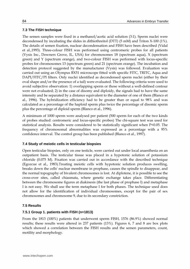
7.3 The FISH technique
The semen samples were fixed in a methanol/acetic acid solution (3:1). Sperm nuclei were
decondensed by incubating the slides in dithiothreitol (DTT) (5 mM) and Triton X-100 (1%).
The details of semen fixation, nuclear decondensation and FISH have been described (Vidal
et al.,1993). Three-colour FISH was performed using centromeric probes for all patients
(Vysis Inc., Downers Grove, IL, USA) for chromosomes 18 (spectrum aqua), X (spectrum
green) and Y (spectrum orange), and two-colour FISH was performed with locus-specific
probes for chromosomes 13 (spectrum green) and 21 (spectrum orange). The incubation and
detection protocol suggested by the manufacturer (Vysis) was followed. Evaluation was
carried out using an Olympus BX51 microscope fitted with specific FITC, TRITC, Aqua and
DAPI/FITC/PI filters. Only nuclei identified as decondensed sperm nuclei (either by their
oval shape and/or the presence of a tail) were evaluated. The following criteria were used to
avoid subjective observation: 1) overlapping sperm or those without a well-defined contour
were not evaluated; 2) in the case of disomy and diploidy, the signals had to have the same
intensity and be separated by a distance equivalent to the diameter of one of them (Blanco et
al., 1996). The hybridization efficiency had to be greater than or equal to 98% and was
calculated as a percentage of the haploid sperm plus twice the percentage of disomic sperm
plus the percentage of diploid sperm (Blanco et al., 1996).
A minimum of 1000 sperm were analysed per patient (500 sperm for each of the two kinds
of probes studied: centromeric and locus-specific probes) The chi-square test was used for
statistical analysis. Results were considered to be statistically significant when P<0.05. The
frequency of chromosomal abnormalities was expressed as a percentage with a 95%
confidence interval. The control group has been published (Blanco et al., 1997).
7.4 Study of meiotic cells in testicular biopsies
Open testicular biopsies, only on one testicle, were carried out under local anaesthesia on an
outpatient basis. The testicular tissue was placed in a hypotonic solution of potassium
chloride (0.075 M). Fixation was carried out in accordance with the described technique
(Egozcue et al., 1983).Treating meiotic cells with hypotonic solution produces swelling,
breaks down the cells' nuclear membrane in prophase, causes the spindle to disappear, and
the normal topography of bivalent chromosomes is lost. At diplotene, it is possible to see the
cross-over sites, called chiasmata, where genetic exchange takes place. Differentiating
between the chromosome figures at diakinesis (the last phase of prophase I) and metaphase
I is not easy. We shall use the term metaphase I for both phases. The technique used does
not allow for the identification of individual chromosomes, except for the pair of sex
chromosomes and chromosome 9, due to its secondary constriction.
7.5 Results
7.5.1 Group 1. patients with FISH (n=1813)
From the 1813 (100%) patients that underwent sperm FISH, 1576 (86.9%) showed normal
results; these results were altered in 237 patients (13%). Figures 6, 7 and 8 are box plots
which showed a correlation between the FISH results and the semen parameters, count,
motility and morphology.
www.intechopen.com

Meiotic Chromosome Abnormalities and Spermatic FISH in Infertile Patients with Normal Karyotype
85
Fig. 6. Relationship between the FISH results and the overall spermatic count.
Fig. 7. Relationship between the FISH results and spermatic motility.
www.intechopen.com

Advances in Embryo Transfer
86
Fig. 8. Relationship between the FISH results and spermatic morphology.
7.5.2 Group 2. patients with testicular biopsy (n=216)
All patients sought consultation due to infertility or miscarriage and had normal mitotic karyotype. The seminogram varied from azoospermia to normal semen in the basic sperm count, motility and morphology parameters. All cases of spermatocytes in prophase I and metaphase I without alterations were considered normal. The absence of spermatocytes II was not considered pathological. The coexistence of normal and altered spermatocytes were diagnosed as mosaicism. Any cases with a variety of abnormalities were included in the group with the most frequent abnormalities. Table I gives the results for this group of patients.
DIAGNOSIS No. %
Normal meiosis 67 31
Only Sertoli cells 16 7.4
Only cells in prophase I 31 14.3
Altered meiosis 42 19.4
Mosaicism 60 27.7
Total 216 100
Table I. Results of the meiotic study on the testicular biopsy.
The types of meiotic abnormality observed in 42 patients (100%) are given in table II.
www.intechopen.com

Meiotic Chromosome Abnormalities and Spermatic FISH in Infertile Patients with Normal Karyotype
87
ABNORMALITIES No % Abnormal mating in PI 6 14.2 Desynapsis of the bivalents 14a 33.3 Reduction in the number of chiasmata 3 7.1 Presence of univalents in MI 11b 26.2 XY separation 2 4.7 MI hyperploids 5 11.9 MII diploids 1 2.3 Total 42 100
a)we observed spermatocyte II diploids in 4 cases and 2 cases with general desynapsis, b) there were also 2 cases with separation of the XY pair.
Table II. Meiotic abnormalities. PI, MI and MII: Cells in prophase I, metaphase I and II.
7.5.3 Group 3. patients with FISH and testicular biopsy (n=60; 100%)
The study of FISH and meiosis in the testicular biopsies did not reveal any abnormalities in 18 of the 60 cases studied (30%) (table III). The partners of 13 patients in this group had experienced miscarriages (21.6%): six of 18 in the group with normal meiosis (33.3%); and seven of 42 in the two groups with altered meiosis (16.6%)(tables IV and V). These figures were low, but significant differences in the miscarriage rate were observed in both groups, with normal meiosis and with altered meiosis. An absence of figures in metaphase II in the fragment of testicular tissue studied was observed in 13 of the 18 cases with normal meiosis (72.2%). Of those 13 cases, nine had a normal total sperm count, which would seem to be incompatible with the total absence of figures in metaphase II. The shortness of this phase explains the frequent absence of figures observed in metaphase II in testicular-biopsy samples (Hultén et al., 1992).
The sperm FISH in 25 patients was normal and the testicular meiosis showed alterations. The seminological data and testicular meiosis are presented in table IV.
The data collected from the 17 patients with sperm FISH and testicular meiosis altered are contained in table V.
The abnormalities found in the FISH for this group of patients are given in table VI.
Of the 17 patients with altered FISH results and altered meiosis, the partner of only one had a history of miscarriages. Total sperm count was normal in nine cases (52.9%) Sperm motility was normal or moderately low (≥30%) in nine cases (52.9%); and sperm morphology was normal or moderately low (≥20%) in 11 patients (64.7%). Individual data with meiotic results are shown in table V.
The meiotic abnormalities observed (n=42; 70%) and the individual data for each case are summarized in tables IV and V. All these patients (n=42; 100%) had meiotic figures at prophase I and all of them had sex body. Pairing of homologous chromosomes at pachytene was normal in 24 cases (57.1%). At metaphase I the most common abnormality was incomplete desynapsis observed as bivalents with some but not all of their chiasmata, small univalents or an association of both abnormalities. It affected some metaphase figures in 29 out of 38 cases in which figures at metaphase I were observed (76.3%). Complete desynapsis was observed in four cases (10,5%).
www.intechopen.com

Advances in Embryo Transfer
88
MISCARRIAGES SEMEN ANALYSIS STUDY OF MEIOSIS
Case n TSC
(x106) Motility
(%) Morphology
(%) Prophase
I Metaphase I Metaphase II
1 No 738 20 8 Normal Normal Normal
2 No 205 10 3 Normal Normal Normal
3 No 203 25 6 Normal Normal Normal
4 No 157 50 17 Normal Normal Normal
5a No 121 15 3 Normal Normal No
6 No 116 30 40 Normal Normal No
7b No 104 5 18 Normal Normal No
8 No 85 55 17 Normal Normal No
9 No 57 25 18 Normal Normal No
10 No 1.1 40 38 Normal Normal No
11c No 0.7 0 4 Normal Normal No
12 No 0.13 0 8 Normal Normal No
13 6 564 60 47 Normal Normal No
14 2 495 50 16 Normal Normal Normal
15 4d 161 50 18 Normal Normal No
16 2e 154 40 23 Normal Normal No
17 2 90 30 16 Normal Normal No
18 3f 0.5 30 9 Normal Normal No
aleft varicocele; bunilateral microorchidism; cunilateral cryptorchidism,done with trisomy 13 and one with 46, XX; eone with trisomy 16; fone with 46, XX
TSC = total sperm count
Table III. Patients with normal FISH results and normal meiosis (18 out of 60; 30%):miscarriages, semen analysis and study of meiosis
www.intechopen.com

Meiotic Chromosome Abnormalities and Spermatic FISH in Infertile Patients with Normal Karyotype
89
MISCARR
IAGES SEMEN ANALYSIS STUDY OF MEIOSIS
Case Nº TSC
(x106) Motility
(%) Morphology
(%) Prophase I Metaphase I
Metaphase II
1 No 1721 45 19 PA (some) 2-3 desynaptic bivalentsbc No
2 No 670 45 17 Normal 2-4 univalentsa No
3 No 363 40 4 PA (some) 2-3 desynaptic bivalentsb No
4 No 330 40 9 Normal 2-4 desynaptic bivalentsb No
5 No 287 11 33 Normal 2-6 univalentsa and 2-3 desynaptic bivalentsb
No
6 No 265 3 3 Normal
(70%); PA (30%)
Normal (70%), and complete desynapsis (30%)d
No
7 No 192 35 27 Normal 2 desynaptic bivalentsb No
8 No 159 40 7 Normal 23 bivalentsd No
9 No 88 15 5 PA (some) 1 desynaptic bivalentbe No
10 No 77 50 21 Normal Normal (75%), and complete desynapsis (25%)d
No
11 No 42 30 5 Normal 1 extra bivalent: hyperploidy
No
12 No 28 35 4 PA 2 desynaptic bivalentsace No
13 No 17 15 11 PA No No
14 No 10 25 34 Normal No No
15 No 9.6 60 15 Normal 2-3 desynaptic bivalentsbcd
No
16 No 9 65 23 PA (some) 2-4 univalents a No
17 No 4 16 2 Normal 2-3 desynaptic bivalentsce No
18 No 0.2 8 10 Normal 23 bivalentsd No
19 No 0.18 50 8 PA and
tetraploidsf Complete desynapsis Diploids
20 3 1716 40 54 Normal 2-3 desynaptic bivalentsb No
21 5 489 40 25 Normal 2-4 univalentsa No
22 3 218 30 11 Normal 23 bivalentsd and 23 normal bivalents
No
23 4 208 50 35 Tetraploids
(12%) Tetraploids (some) 1 diploid
24 5 198 15 19 PA (some) 2-4 univalentsa and 2 desynaptic bivalentsabe
No
25 4 73 55 20 PA (some) 2-3 desynaptic bivalentsc No
asmall; bmedium-sized; cbig; ddegenerative in appearance; eearly separation of XY pair; fsome tetraploid figures with two sex vesicles
TSC = total sperm count; PA = pairing anomalies
Table IV. Patients with normal FISH results and altered meiosis (25 out of 60; 41.6%): miscarriages, semen analysis and study of meiosis
www.intechopen.com

Advances in Embryo Transfer
90
TSC: total sperm count; PA: pairing anomalies; asmall; bmedium-sized; clarge; dearly separation of XY pair; eone with 92,XX,YY
Table V. Patients with altered FISH results and altered meiosis (17 out of 60; 28.3%):
www.intechopen.com

Meiotic Chromosome Abnormalities and Spermatic FISH in Infertile Patients with Normal Karyotype
91
FISH ABNORMALITIES No. (%)
Diploidies 7 41,1
Diploidies and gonosomic disomies 3 17,6
Diploidies and autosomic disomies a 3 17,6
Gonosomic disomies 2 11,7
Autosomic disomies b 2 11,7
TOTAL 17 100,0
a Chromosome 13 and 21 were affected b Chromosome 13 was affected in both cases
Table VI. The anomalies observed with the FISH technique (n=17; 100%)
Incomplete desynapsis and/or the presence of small univalents was observed in patients with polyzoospermia and those with normal, low or very low total sperm counts: normal, low or very low sperm motility; and normal, low or very low sperm morphology (tables IV and V). No relationship was observed between incomplete desynapsis and more or less severe alterations of basic semen parameters. No relationship was detected between the type of meiotic abnormalities observed in the testes and the kind of gamete aneuploidy (diploidies, disomies). The synaptic process is controlled individually for each chromosome and not at cell level (Templado et al., 1981). The cases of complete desynapsis of all bivalents and in all cells had very low total sperm counts: 0.18 and 1.2 x 10⁶ (Case 19 in table IV and Case 14 in table V). Cases six and ten in table IV, which had complete desynapsis but not in all cells (only in 30% and 25%, respectively) presented with normal total sperm counts: 265 and 77 million, respectively. No figures at metaphase II were observed in 37 of 42 cases (88%).
The meiotic report from the testicular biopsy cannot simply be extrapolated to the entire
testicular parenchyma.
8. Remarks
When the first successful birth was achieved using the IVF-ICSI technique (Palermo et al., 1992) and one year later when this technique was used successfully on sperm extracted from the testicle (TESE) (Schoysman et al., 1993), andrology entered a new era. It simply became a matter of obtaining a dozen or so sperm from the semen or the testicle in order to have a child. These 20 years of experience in IVF-ICSI have come to show that the reality is not quite as straightforward as this. The few sperm used have to be euploids. IVF-ICSI cannot resolve the problem of infertility if the sperm introduced into the ooplasm is an aneuploid.
If the sperm selected for microinjection into the mature ovocyte is aneuploid, the oocyte may not be fertilized (Lee et al., 2002) or an aneuploid embryo may be produced but stops developing in the first few days after fertilization. Such embryos have higher levels of chromosomal abnormalities (Almeida and Bolton, 1998); another possibility is that the
www.intechopen.com
Advances in Embryo Transfer
92
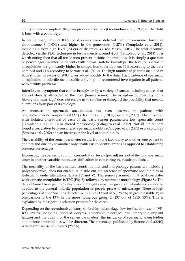
embryo does not implant; they can produce abortions (Giorlandino et al., 1998) or the child is born with a pathology.
In fertile men, around 0.1% of disomies were detected per chromosome; lower in
chromosome 8 (0.03%) and higher in the gonosomes (0.27%) (Templado et al.,2011),
including a very high level (0.43%) of disomies XY (de Massy, 2003). The total disomies
detected via the FISH technique in fertile men is around 4.5% (Templado et al., 2011). It is
worth noting then that all fertile men present meiotic abnormalities. It is simply a question
of percentages. In infertile patients with normal mitotic karyotype, the level of spermatic
aneuploidies is significantly higher in comparison to fertile men: 13% according to the data
obtained and 14% according to Sarrate et al., (2010). The high number of patients included in
both studies, in excess of 2000, gives added solidity to the data. The incidence of spermatic
aneuploidies in infertile men is sufficiently high to recommend investigation in all patients
with fertility problems.
Infertility is a symptom that can be brought on by a variety of causes, including causes that
are not directly attributed to the man (female issues). The symptom of infertility (or a
history of miscarriage) does not enable us to confirm or disregard the possibility that meiotic
alterations form part of its etiology.
An increase in spermatic aneuploidies has been observed in patients with
oligoasthenoteratozoospermia (OAT) (Devillard et al., 2002; Lee et al., 2002). Also in semen
with isolated alterations of each of the basic semen parameters: low spermatic count
(Templado et al., 2011); or altered morphology (Calogero et al., 2002). Not all the authors
found a correlation between altered spermatic motility (Calogero et al., 2001) or morphology
(Sbracia et al., 2002) and an increase in the level of aneuploidies.
The variability of the semen parameter results from one laboratory to another, one patient to
another and one day to another only enables us to identify trends as opposed to establishing
concrete percentages.
Expressing the spermatic count in concentration levels (per ml) instead of the total spermatic
count is another variable that causes difficulties in comparing the results published.
The normality of the basic semen, count, motility and morphology parameters including
polyzoospermia, does not enable us to rule out the presence of spermatic aneuploidies or
testicular meiotic alterations (tables IV and V). The semen parameter that best correlates
with gametic aneuploidies is TSC (Fig. 6); followed by spermatic morphology (Figure 8). The
data obtained from group 3 refer to a small highly selective group of patients and cannot be
applied to the general infertile population or people prone to miscarriage. There is high
percentages of abnormalities detected with FISH (17 out of 60; 28.3%) in group 3 (table V) in
comparison to the 13% in the more numerous group 2 (237 out of 1813; 13%). This is
explained by the rigorous selection process for the cases.
Depending on the reproductive history (infertility, miscarriage, low fertilisation rate in IVF-
ICSI cycles, including donated oocytes, embryonic blockages and embryonic implant
failure) and the quality of the semen parameters, the incidence of spermatic aneuploidies
and meiotic abnormalities will be different. The percentage published by Sarrate et al.,(2010)
is very similar (26.5%) to ours (28.3%).
www.intechopen.com

Meiotic Chromosome Abnormalities and Spermatic FISH in Infertile Patients with Normal Karyotype
93
The FISH study of five chromosomes enabled us to detect a higher percentage of aneuploidies, specifically 11.7% of chromosome 13 in the data provided (table VI), than if only three probes were used, typically chromosomes 18, X and Y in this case.
Patients with altered FISH (100%) in this study presented meiotic abnormalities in the testicular biopsy. Sarrate et al., (2010) encountered the situation in 91.7% of patients. If the sperm FISH is altered, it is not necessary to carry out a meiotic study on the testicular biopsy as any gametic aneuploidy is considered to be caused by meiotic alteration. In a small percentage of patients, between 5 and 8%, according to both our not-published data and that published by Sarrate et al., (2010), the FISH is altered and testicular meiosis is normal. We did not observe any cells in metaphase II in the biopsy in our patients. Meiotic alteration causing gametic and aneuploidy could occur in the second meiotic division. Another possible explanation is the presence of mosaicism.
In patients with normal FISH, the meiosis is altered in 41.6% of cases (25 out of 60) in this study and reaches 73.6% in the Sarrate et al., (2010) publication. The data indicate that a normal FISH does not rule out the existence of meiotic abnormalities. This situation may be explained by the existence of altered meiotic cells that do not produce spermatozoa, along with normal meiotic cells that produce euploid gametes. Another possible explanation is that the spermatozoa are aneuploids but for other chromosomes than those studied with the probes used. If the FISH is normal then it will be necessary to consider whether to indicate the meiotic study or not on the testicular biopsy depending on the reproductive history and the semen parameters.
We did not find a correlation between the meiotic anomalies observed in the testicle and the type of FISH alteration: diploidies or disomies of one or other chromosome. We also did not observe an increase in gonosomic disomies in comparison to autosomic disomies. The number of cases presented is small (n=25) and not conclusive.
The detection of meiotic chromosomal abnormalities both with the FISH and by means of testicular biopsy, together with the reproductive history of the patients, has led the andrologist to indicate a study of the chromosomes in the embryos in order to avoid transferring aneuploids. Another option is to propose the use of a sperm bank. Repeating and re-repeating IVF cycles without studying possible meiotic alteration is not the most reliable option.
The meiotic study on the testicle biopsy was normal in 31% of cases and was not informative in 7.4%. The blockage in prophase I (14.3% of cases) may be due to genetic alterations with no translation of mating abnormalities at an optic level. Out of the alterations observed, the most frequent is desynapsis 33,3% (table II).
It is worth noting that 27.7% (60 out of 216) of patients presented mosaicism. This is the group that may benefit from IVF-ICSI. The use of the IMSI technique (Intracytoplasmic Morphologically Selected Spermatozoon Injection) will enable a better selection of spermatozoon for micro-injection. The selection of spermatozoon for introduction into the oocyte can be 16000x instead of 400x the standard ICSI. It has been correlated the presence of vacuoles in the spermatic nucleus with aneuploid spermatozoon. (Garolla et al., 2008)
The PGD technique, particularly the array comparative genomic hibridization (aCGH) technique that studies all chromosomes enables us to select euploid embryos that are suitable for transferring to the uterus.
www.intechopen.com

Advances in Embryo Transfer
94
9. Conclusions
We can draw the following conclusions from the data presented and quoted in the bibliography.
1. The incidence of spermatic aneuploidies is three times greater in the infertile population (13% - 14%) than the fertile population (4.5%).
2. None of the clinical or seminological data enable us to confirm or rule out the possibility that an infertile patient with normal mitotic karyotype may or may not produce aneuploid gametes. We have observed an inverted relationship in particular between the sperm count and the aneuploidy level.
3. The concordance of the results of the altered FISH and testicular meiosis is almost 100% and in this case there is no need to carry out a testicular biopsy.
4. In case of normal FSIH may be necessary to do testicular biopsy because in more than 40% of this patients we observed testicular meiotic abnormalities. It depends on reproductive history.
5. The FISH study must be carried out together with the seminogram for all infertile patients. The information is significant and cannot be obtained in a more straightforward fashion.
10. References
Almeida PA & Bolton VN. (1998). Cytogenetic analysis of human preimplantation embryos
following developmental arrest in vitro. Reprod Fertil Dev 10:505-513.
Alsheimer M. (2009). The Dance Floor of Meiosis: Evolutionary Conservation of Nuclear
Envelope Attachment and Dynamics of Meiotic Telomeres. In: Meiosis, Benavente R,
Volff JN (Eds.), pp.81-93, S. Karger, Basel, Switzerland.
Bannister LA, Reinholdt LG, Munroe RJ & Schimenti JC. (2004). Positional cloning and
characterization of mouse mei8, a disrupted allele of the meiotic cohesin Rec8. Genesis
40:184-194.
Barbero JL. (2011). Sister Chromatid Cohesion Control and Aneuploidy. In: Aneuploidy,
Delhanty J D A, Pellestor F (Eds.), pp. 223-233, S. Karger, Basel, Switzerland.
Bass HW. (2003). Telomere dynamics unique to meiotic prophase: formation and significance of
the bouquet. Cell Mol Life Sci 60:2319-2324.
Bergère M, Wainer R, Nataf V, Bailly M, Gombault M, Ville Y & Selva J. (2002). Biopsied
testis cells of four 47,XXY patients: fluorescence in-situ hybridization and ICSI results.
Hum Reprod 17:32-37.
Blanco J, Egozcue J & Vidal F. (1996). Incidence of chromosome 21 disomy in human spermatozoa
as determined by fluorescent in-situ hybridization. Hum Reprod 11: 722-726.
Blanco J, Rubio C. Simón C, Egozcue J & Vidal F. (1997). Increased incidence of disomic sperm
nuclei in a 47, XYY male assessed by fluorescent in situ hybridization (FISH). Hum
Genet 99: 413-416.
Bolcun-Filas E, Costa Y, Speed R, Taggart M, Benavente R, De Rooij DG & Cooke HJ.
(2007). SYCE2 is required for synaptonemal complex assembly, double strand break
repair, and homologous recombination. J Cell Biol 176:741-747.
www.intechopen.com

Meiotic Chromosome Abnormalities and Spermatic FISH in Infertile Patients with Normal Karyotype
95
Buard J & de Massy B. (2007). Playing hide and seek with mammalian meiotic crossover
hotspots. Trends Genet 23:301-309.
Byers S, Pelletier R-M. & Suárez-Quian C. (1993). Sertoli cell junctions and the Seminiferous
Epithelium Barrier. In: The Sertoli Cell; L.D. Russell and M.D. Griswold (Eds.), pp.
431-446; Cache River Press. Clearwater F.L. U.S.A.
Calogero AE, De Palma A, Grazioso C, Barone N, Romeo R, Rappazzo G & D'Agata R.
(2001). Aneuploidy rate in spermatozoa of selected men with abnormal semen
parameters. Hum Reprod 16:1172-1179.
Calogero AE, Vicari E, De Palma A, Burrello N, Barone N, Grazioso C, Zahi M & D’Agata
R. (2002). Elevated sperm aneuploidy rate in patients with absolute polymorphic
teratozoospermia. Hum Reprod 17 (Abstract book 1), pp. 95–96.
Codina-Pascual M, Campillo M, Kraus J, Speicher M, Egozcue J, Navarro J & Benet .J
(2006). Crossover frequency and synaptonemal complex length: their variability and
effects on human male meiosis. Mol Hum Reprod 12:123-133.
Costa Y, Speed R, Ollinger R, Alsheimer M, Semple CA, et al. (2005). Two novel proteins
recruited by synaptonemal complex protein 1 (SYCP1) are at the centre of meiosis. J Cell
Sci 118:2755-2762.
de Massy B. (2003). Distribution of meiotic recombination sites. Trends Genet 19:514-521.
de Vries FA, de Boer E, van den Bosch M, Baarends WM, Ooms M, et al. (2005). Mouse
Sycp1 functions in synaptonemal complex assembly, meiotic recombination, and XY
body formation. Genes Dev 19:1376-1389.
Devillard F, Metzler-Guillemain C, Pelletier R, DeRobertis C, Bergues U, Hennebicq S,
Guichaoua M, Sele B & Rousseaux S. (2002). Polyploidy in large-headed sperm: FISH
study of three cases. Hum Reprod 17:1292-1298.
Dorsett D. (2007). Roles of the sister chromatid cohesion apparatus in gene expression,
development, and human syndromes. Chromosoma 116:1-13.
Egozcue J, Templado C, Vidal F, Navarro J, Morer-Fargas F& Marina S. (1983). Meiotic
studies in a series of 1100 infertile and sterile males. Hum Genet 65:185-188.
Fawcett DW. (1956). The fine structure of chromosomes in the meiotic prophase of vertebrate
spermatocytes. J Biophys Biochem Cytol 2:403-406.
Ferlin A, Raicu F, Gatta V, Zuccarello D, Palka G & Foresta C. (2007). Male infertility: role of
genetic background. Reprod Biomed Online 14:734-745.
Gardner RJ & Sutherland GR. (2004). Chromosome Abnormalities and Genetic Counseling, 3rd
ed. (Oxford University Press, New York).
Garolla A, Fortini D, Menegazzo M et al. (2008). High-power microscopy for selecting
spermatozoa for ICSI by physiological status. Reproductive BioMedicine Online
17:610-616.
Gerkes EH, van der Kevie-Kersemaekers AM, Yakin M, Smeets DF & van Ravensswaaij-
Arts CM. (2010). The importance of chromosome studies in Roberts syndrome/SC
phocomelia and other cohesinopathies. Eur J Med Genet 53:40-44.
Giorlandino C, Calugi G, Iaconianni L, Santoro ML & Lippa A. (1998). Spermatozoa with
chromosomal abnormalities may result in a higher rate of recurrent abortion. Fertil Steril
70:576-577.
www.intechopen.com

Advances in Embryo Transfer
96
Griffin DK & Finch KA. (2005). The genetic and cytogenetic basis of male infertility. Hum
Fertil (Camb) 8:19-26.
Gruber S, Haering CH & Nasmyth K. (2003). Chromosomal cohesin forms a ring. Cell
112:765-777.
Hamer G, Gell K, Kouznetsova A, Novak I, Benavente R & Höög C. (2006). Characterization
of a novel meiosis-specific protein within the central element of the synaptonemal
complex. J Cell Sci 119:4025-4032.
Härkönen K. (2005). Pesticides and the induction of aneuploidy in human sperm. Cytogenet
Genome Res 111:378-383.
Hassold T, Chen N, Funkhouser J, Jooss T, Manuel B, et al. (1980). Scytogenetic study of
1000 spontaneous abortions. Ann Hum Genet 44:151-178.
Heller CG & Clermont Y. (1964). Kinetics of the germinal epithelium in man. Rec Progr
Hormone Res 20:545-575.
Hultén MA, Goldman ASH, Saadallah N, Wallace BMN & Creasy MR. (1992). Meiotic
studies in man. In: De Rooney and BH Czepulkowski, Hum Cytogenet. A practical
approach., pp 193-221, Oxford University Press, Oxford, UK.
Keeney S & Neale MJ. (2006). Initiation of meiotic recombination by formation of DNA double-
stranded breaks: mechanism and regulation. Biochem Soc Trans 34:523-525.
Kudo NR, Anger M, Peters AH, Stemmann O, Theussl HC, et al. (2009). Role of cleavage by
separase of the Rec8 kleisin subunit of cohesion during mammalian meiosis I. J Cell Sci
122:2686-2698.
Lee MS, Tsao HM, Wu HM, Huang CC, Chen CI and Lin David PC (2002). Correlations
between sperm apoptosis and aneuploidy. Hum Reprod 17 (Abstract book 1), pp. 112 –
113.
Lee J, Kitajima TS, Tanno Y, Yoshida K, Morita T, et al. (2008). Unified mode of centromeric
protection by shugoshin in mammalian oocytes and somatic cells. Nat Cell Biol 10:42-52.
Lichten M. (2001). Meiotic recombination: Breaking the genome to save it. Curr Biol 11:R253-
R256.
Lynn A, Ashley T & Hassold T. (2004). Variation in human meiotic recombination. Annu Rev
Genomics Hum Genet 5:317-349.
Marston AL & Amon A. (2004). Meiosis: cell-cycle controls shuffle and deal. Nat Rev Mol Cell
Biol 5:983-997.
Meuwissen RL, Meerts I, Hoovers JM, Leschot N &, Heyting C. (1997). Human
synaptonemal complex protein1 (SCP1): isolation and characterization of the DNA and
chromosomal localization of the gene. Genomics 39:337-384.
Miyamoto T, Hasuike S, Yogev L, Maduro MR, Ishikawa M, Westphal H & Lamb DJ.
(2003). Azoospermia in patients heterozygous for a mutation in SYCP3. Lancet
362:1714-1719.
Neumann R & Jeffreys AJ. (2006). Polymorphism in the activity of human crossover hotspots
independent of local DNA sequence variation. Hum Mol Genet 15:1401-1411.
Nishant KT & Rao MR. (2006). Molecular features of meiotic recombination hotspots. Bioessays
28:45-56.
www.intechopen.com

Meiotic Chromosome Abnormalities and Spermatic FISH in Infertile Patients with Normal Karyotype
97
Pacchierotti & Eichenlaub-Ritter. (2011). Envioronmental Hazard in the Aetiology of Somatic
and Germ Cell Aneuploigy. In: Aneuploidy, Delhanty JDA and Pellestor F (Eds.),
pp. 254-268,S. Karger, Basel, Switzerland.
Palermo G, Joris H, Devroey P & Van Steirteghem AC. (1992). Pregnancies after
intracytoplasmic injection of single spermatozoon into an oocyte. Lancet 340:17-18.
Perry MJ. (2008). Effects of environmental and occupational pesticide exposure on human sperm:
a systematic review. Hum Reprod Update 14:233-242.
Petes TD. (2001). Meiotic recombination hot spots and cold spots. Nat Rev Genet 2:360-369.
Revenkova E, Eijpe M, Heyting C, Hodges CA, Hunt PA, et al. (2004). Cohesin SCM1 beta is
required for meiotic chromosome dynamics, sister chromatid cohesion and DNA
recombination. Nat Cell Biol 6:555-562.
Revenkova E & Jessberger R. (2006). Shaping meiotic prophase chromosomes: Cohesins and
synaptonemal complex proteins. Chromosoma 115:235-240.
Sarrate Z, Vidal F & Blanco J. (2010). Role of sperm fluorescent in situ hybridization studies in
infertile patients: indications, study approach, and clinical relevance. Fertil Steril
93:1892-1902.
Sbracia M, Baldi M, Cao D, Sandrelli A, Chiandetti A, Poverini R & Aragona C. (2002).
Preferential location of sex chromosomes, their aneuploidy in human sperm, and their
role in determining sex chromosome aneuploidy in embryos after ICSI. Hum Reprod
17:320-324.
Scherthan H. (2001). A bouquet makes ends meet. Nat Rev Mol Cell Biol 2:621-627.
Schoysman R, Vanderzwalmen P, Nijs M, Segal L, Segal-Bertin G, Geerts L, van
Roosendaal E & Schoysman D. (1993). Pregnancy after fertilisation with human
testicular spermatozoa. Lancet 342:1237-1238
Simpson JL. (2007). Genetics of spontaneous abortions. In: Recurrent Pregnancy Loss.
Howard J.A. Carp (Ed), pp.23-34, Informa Healthcare. London, U.K.
Solari AJ. (1974). The behaviour of the XY pair in mammals. Int Rev Cytol 38:273-317.
Templado C, Vidal F, Marina S, Pomerol JM & Egozcue J. (1981.) A new meiotic mutation:
Desynapsis of individual bivalents. Hum Genet 59:345-348.
Templado C, Vidal F & Estop A. (2011). Aneuploidy in human Spermatozoa. In: Aneuploidy,
Delhanty J D A, Pellestor F, (Eds.),pp.91-99, S. Karger, Basel, Switzerland.
Trelles-Sticken E, Dresser ME & Scherthan H. (2000). Meiotic telomere protein Ndj1p is
required for meiosis specific telomere distribution, bouquet formation and efficient
homologue pairing. J Cell Biol 151:95-106.
Trelles-Sticken E, Adelfalk C, Loidl J & Scherthan H. (2005). Meiotic telomere clustering
requires actin for its formation and cohesion for its resolution. J Cell Biol 170:213-223.
Vidal F, Moragas M, Català V, Torelló MJ, Santaló J, Calderon G, Gimenez C, Barri P N,
Egozcue J & Veiga A. (1993). Sephadex filtration and human serum albumin gradients
do not select spermatozoa by sex chromosome: a fluorescent in-situ hybridization study.
Hum Reprod 8:1740-1743.
Watrin E & Peters JM. (2006). Cohesin and DNA damage repair. Exp Cell Res 312:2687-2693.
WHO. (1999). Laboratory manual for the examination of human semen and sperm-cervical mucus
interaction. 4th ed. Cambridge University Press, Cambridge, UK.
www.intechopen.com

Advances in Embryo Transfer
98
Xu H, Beasley MD, Warren WD, van der Horst GT & McKay MJ. (2005). Absence of mouse
REC8 cohesin promotes synapsis of sister chromatids in meiosis. Dev Cell 8:949-961.
Zickler D. (2006). From early homologue recognition to synaptonemal complex formation.
Chromosoma 115:158-174.
Zickler D & Kleckner N. (1998). The leptotene-zygotene transition of meiosis. Annu Rev Genet
32:619-697.
www.intechopen.com
Advances in Embryo TransferEdited by Dr. Bin Wu
ISBN 978-953-51-0318-9Hard cover, 248 pagesPublisher InTechPublished online 14, March, 2012Published in print edition March, 2012
InTech EuropeUniversity Campus STeP Ri Slavka Krautzeka 83/A 51000 Rijeka, Croatia Phone: +385 (51) 770 447 Fax: +385 (51) 686 166www.intechopen.com
InTech ChinaUnit 405, Office Block, Hotel Equatorial Shanghai No.65, Yan An Road (West), Shanghai, 200040, China
Phone: +86-21-62489820 Fax: +86-21-62489821
Embryo transfer has become one of the prominent high businesses worldwide. This book updates and reviewssome new developed theories and technologies in the human embryo transfer and mainly focus on discussingsome encountered problems during embryo transfer, which gives some examples how to improve pregnancyrate by innovated techniques so that readers, especially embryologists and physicians for human IVFprograms, may acquire some new and usable information as well as some key practice techniques. Majorcontents include the optimal stimulation scheme for ovaries, advance in insemination technology, improvedembryo transfer technology and endometrial receptivity and embryo implantation mechanism. Thus, this bookwill greatly add new information for readers to improve human embryo transfer pregnancy rate.
How to referenceIn order to correctly reference this scholarly work, feel free to copy and paste the following:
Simón Marina, Susana Egozcue, David Marina, Ruth Alcolea and Fernando Marina (2012). MeioticChromosome Abnormalities and Spermatic FISH in Infertile Patients with Normal Karyotype, Advances inEmbryo Transfer, Dr. Bin Wu (Ed.), ISBN: 978-953-51-0318-9, InTech, Available from:http://www.intechopen.com/books/advances-in-embryo-transfer/meiotic-chromosome-abnormalities-and-spermatic-fish-in-infertile-patients-with-normal-karyotype