melatonin administration aids bone healing in diabetic rats
TRANSCRIPT
Israel Journal of Veterinary Medicine Vol. 68 (3) September 2013158 Kaya, G.S.
Melatonin Administration Aids Bone Healing in Diabetic RatsKaya, G.S.,1 Kaya, M.,2* Gursan, N.,3 Kiziltunc, A.,4 Alp, H.H.,4 Balta, H.,3 Hayirli, A.5
1 Department of Oral and Maxillofacial Surgery, Faculty of Dentistry, Atatürk University, Erzurum, Turkey.2 Department of Surgery, Faculty of Veterinary Medicine, Atatürk University, Erzurum, Turkey.3 Department of Pathology, Faculty of Medicine, Atatürk University, Erzurum, Turkey.4 Department of Biochemistry, Faculty of Medicine, Atatürk University, Erzurum, Turkey.5 Department of Animal Nutrition and Nutritional Disorders, Faculty of Veterinary Medicine, Atatürk University, Erzurum, Turkey.
* Corresponding author: Dr. Mahir Kaya, Department of Surgery, Faculty of Veterinary Medicine, Atatürk University, Erzurum, Turkey. Tel: +904422315527, Fax: +904422360881. E-mail: [email protected]
ABST RACTThis randomized experimental study was conducted to determine the effect of melatonin on bone healing processes in diabetic rats with bone defects. Adult male Sprague Dawley rats (n = 64) were divided into dia-betic and control groups. The diabetic group was administered with streptozotocin (60 mg/kg BW, (intraperi-toneally) I.P.), whereas the control group was administered with saline containing 6% ethanol intraperito-neally. Defects were created in the left tibias, and rats were then administered with either saline or melatonin (10 mg/kg BW, I.P.) for 21 or 42 days. Overall, the diabetic rats had significantly higher serum glucose con-centrations than the control rats (462 vs. 118 mg/dl; P<0.0001). Melatonin administration increased serum glucose by 5% in the control groups and decreased it by 43% in the diabetic groups (P<0.0001). Serum mela-tonin concentrations in the control rats were lower than in the diabetic rats (666 vs. 745 pmol/L; P<0.006). Melatonin administration increased serum melatonin concentrations to a similar extent in both groups (by 15%). Diabetes was associated with pathologies (degranulation, vacuolization, degeneration, and necrosis) in pancreatic β cells and aggravated inflammation indicators (P<0.0001 for all). Melatonin administration considerably decreased the number of rats with pancreatic β cell pathologies (63%) and depressed inflam-mation indicators (from P<0.004 to P<0.0008). Melatonin administration increased osteogenesis indicators (collagen, cartilage, and osteocyte intensities) (2.0 vs. 1.0; P<0.04) and numerically decreased inflammation indicators (macrophage, lymphocyte, and polymorph nuclear leukocyte) (1.0 vs. 2.0) at a greater extent in the diabetic rats than in the control rats. In conclusion, postoperative administration of melatonin supports bone healing in diabetic rats apparently through alleviating pancreatic cytopathology and hyperglycemia.
Keywords: Melatonin, bone healing, diabetes mellitus, rat.
INTRODUCTIONDiabetes mellitus is a common and serious metabolic disor-der associated with many functional and structural complica-tions. It is one of the most frequently diagnosed endocrinop-athies in both humans (1) and household pets (cats and dogs) (2-5). Obesity and physical inactivity are major predisposing factors in both humans (1) and pet animals (6, 7). Diabetes increases the risk of fracture (8) and interferes with healing processes (9, 10) by slowing bone formation.
The pineal gland hormone, melatonin (N-acetyl-5-methoxytryptamine) has a variety of physiological, immuno-logical and biochemical functions. It is a direct endogenous free-radical scavenger and indirectly exerts chemoprotec-tive, immunostimluatory and myelostimulatory effects (11), which are important in the pathogenesis and treatment of diabetes complications (12). Moreover, melatonin interacts with insulin (13), and consequently alleviates the adverse ef-fects of diabetes by lowering blood glucose levels (14) and
Israel Journal of Veterinary Medicine Vol. 68 (3) September 2013 159 Melatonin in Diabetic Rats with Bone Defects
enhancing antioxidant defense capacities (15) to detoxify harmful reactive oxygen species (16). As a strong scaven-ger, melatonin may diminish the negative effects of oxida-tive stress, which is an important cause of diabetes-related complications. Melatonin injection has been shown to de-crease the amount of superoxide radicals in rat plasma and to increase total antioxidative capacity as well as the activity of antioxidative enzymes, including superoxide dismutase and glutathione peroxidase (17).
Melatonin is considered an important mediator in osteo-genesis (18, 19) through shortening the osteoblast differen-tiation period (20). Studies of osteoporosis and menopause confirm the influence of melatonin on bone metabolism; in-deed, the low melatonin level in menopausal women may-be one of the reasons for their susceptibility to orthopedic problems (21). Despite the well-known effects of melatonin on bone tissue in non-diabetic subjects (22), the effects of melatonin on bone healing in diabetic subjects has not been studied. Therefore, this study was conducted to examine the effects of melatonin administration on the healing process in diabetic rats with tibial defects.
MATERIALS AND METHODS
Animals and ManagementSixty-four male Sprague Dawley rats weighing an average of 260 g (220-340 g) at 8 weeks of age were obtained from The Atatürk University Experimental Medical Application and Research Center and used in full compliance with the 1975 Helsinki Declaration of Animal Rights (revised in 2000) after the approval of the Atatürk University Faculty of Veterinary Sciences Ethical Committee on Animal Care and Use. Rats were kept in pairs in cages in a room with a 12-hour daylight/darkness cycle and an ambient temperature of 23±2°C and humidity of 55±10% throughout the study (23). Standard rat chow (Bayramoglu Feed Co., Erzurum, Turkey) and water were provided ad libitum.
Experimental GroupsRats were distributed into 4 groups in a 2×2 factorial arrange-ment with 2 health statuses (control-healthy and diabetic) and 2 agent administrations (saline and melatonin). Each group was further divided according to time of agent admin-istered (21 and 42 days), yielding 8 replications per group.
Pineal indole melatonin (Sigma, St. Louis, MO, USA)
was freshly dissolved in saline containing 6% ethanol (total volume of 1 ml/kg). Daily i.p. injections of 10 mg/kg BW were administered postoperatively using a 1-mL syringe with a 25-gauge needle, with 6% ethanol saline as a vehicle; ex-perimental rats received the melatonin injection, whereas control rats received 6% ethanol saline only. To avoid inter-fering with the daily circadian rhythm, melatonin and saline were administered at 23:00-24:00 hrs under dim light by il-lumination from an adjacent room.
Diabetes InductionPrior to experimentation, rats were grouped by their live weights to avoid variability in body weight. After over-night fasting, 32 rats were injected intraperitoneally (i.p.) with streptozotocin (STZ; Sigma, St. Louis, MO, USA), at a dose of 60 mg/kg BW in 0.1 M cold citrate buffer (pH 4.5) to induce Type I diabetes mellitus (24). In order to prevent the possibility of fatality due to STZ-induced hypoglycemia caused by massive insulin release, rats were administered a 5% dextrose solution for 24 hrs beginning 6 hrs after STZ administration. Diabetes induction was verified 72 hrs af-ter STZ administration using a glucometer (Optium Xceed Glucometer; Abbott, Chicago, IL, USA). The rats were mon-itored for 14 d to ensure that blood glucose levels had stabi-lized. Rats with fasting blood glucose levels above 220 mg/dl were considered diabetic.
Tibial Defect CreationUnder general anesthesia with xylazine (5 mg/kg, Bayer, Istanbul, Turkey) and ketamine (75 mg/kg, Eczacıbaşı, Lüleburgaz, Turkey), the left hind leg of each rat was shaved, the skin was disinfected using povidone iodine, and an incision (1 cm) was made in the anteriomedial aspect of the tibia. A full-thickness flap was reflected in order to expose the bone surface of the left tibia. Under profuse saline solution irrigation, a unicortical circumfer-ential critical-size defect (3.0×3.0 mm) was created on the methaphyseal area of the tibia with a 3.0 mm wide tre-phine burr at a rotary speed not exceeding 1,500 rpm (25). The soft tissue was repositioned and fascia and skin lay-ers were surgically closed. Animals were closely monitored after surgery, but their functional activity was not restrict-ed. Antibiotic (amoxicillin, 150 mg/kg intramuscularly for 3 d, Bremer Pharma, Warburg, Germany) and analgesic (buprenorphine 0.05 mg/kg subcutaneously every 12 hrs
Research Articles

Israel Journal of Veterinary Medicine Vol. 68 (3) September 2013Kaya, G.S.160
for 3 d, Reckitt and Colman, Richmond, VA, USA) were administrated following surgery to prevent infection and minimize pain. No complications were noted during the postsurgical period.
MeasurementsHalf of the rats in each group were sacrificed with thiopen-tal (200 mg/kg, i.p., Abbott, Campoverde di Aprilia, Italy) administered 21 days after the tibial defect creation and the other half at 42 days. Blood samples were collected from the heart under anesthesia prior to sacrifice, and pancreases and left tibias were collected post-sacrifice for biochemical and histopathological examination.
Biochemical AnalysisBlood glucose levels were measured using a glucometer (Optium Xceed Glucometer; Abbott, Chicago, IL, USA). Blood samples were then centrifuged at 3,000 g for 15 min, and serum melatonin concentrations were measured using ELISA (USCN Life Science Inc., Wuhan, China) in a mi-croplate reader (BioTek, PowerWave XS, California, CA, USA) at 450 nm optical density and a standard range of 0-2000 ρmol/L.
Histopathological AnalysisPancreatic tissue samples were fixed in 10% buffered neutral formaldehyde and Bouin’s fixatives for light microscopic ex-amination, passed through a standard alcohol dehydration-xylene sequence and embedded in paraffin. General pan-creatic structure was evaluated by applying Mallory’s triple stain to 5-6 µm-thick sections cut from tissue blocks and by examining them under a light microscope (Olympus BX51; Olympus Corp., Tokyo, Japan) at x40 magnification. Samples were evaluated by two pathologists blinded to the sample groups and scored on a graded scale, as follows: 0 = no necro-sis; 1 = slight degenerative changes (≤ 25% of islets affected); 2 = mild necrosis (≤ 50% of islets affected); 3 = severe necrosis (≤ 75% of islets affected).
Bone tissue samples were fixed in 10% buffered forma-lin and decalcified in hydrochloric acid/formic acid solu-tion 20 times their own volume, with fresh solution pro-vided daily until decalcification was complete (about 96 hrs). Specimens were routinely embedded in paraffin and then thoroughly washed under running tap water. The sections (5 µm) were placed on polylysine-coated slides,
stained with hematoxyline and eosin (H&E), randomized and examined under a light microscope (Olympus BX51; Olympus Corp., Tokyo, Japan) at x40 magnification by two pathologists blinded to the specimen groups. Polymorph nuclear leukocyte, lymphocyte and macrophage infiltra-tions as well as osteocyte density, cartilage formation and collagen accumulation were evaluated semi-quantitatively on a graded scale based on degree of healing, as follows: 0 = absent; 1 = mild; 2 = moderate; 3 = remarkable.
Statistical AnalysisTo achieve 10% increase in serum melatonin concentration and to consider this elevation significant at α error of 0.05 and power (1 – β error) of 0.90, power analysis yielded 7 rats per group. Blood and bone metabolites (continuous variables) were analyzed using two-way ANOVA as repeated measures. The linear model included the main effect of health status (healthy vs. diabetic) and agent administration (saline vs. melatonin) and their interaction as whole-plot terms and time, and time by whole-plot term interactions as sub-plot terms (26). Moreover, correlation between blood glucose lev-el and inflammation and osteogenesis markers was attained. In the analyses of bone histopathological parameters (dis-crete variables), Kruskal-Walis test was employed. Statistical significance was set at P<0.05.
RESULTS
Serum MetabolitesBlood glucose concentrations of diabetic rats on post-induction day 21 and 42 were, respectively, 4.4 and 3.5 times greater than those of controls (P<0.0001; Table 1). Melatonin administration resulted in 27% and 44% re-ductions in blood glucose concentrations on day 21 and 42, respectively as compared to saline administration (P<0.0001; Table 1). Following melatonin administra-tion, there were slight increases in blood glucose concen-trations in the control rats (4.2 and 6.1%), whereas there were considerable decreases in blood glucose concentra-tions in the diabetic rats (-33 and -54%) on days 21 and 42 (P<0.0001; Table 1).
Serum melatonin concentrations of the control rats were lower than those of the diabetic rats (664 vs. 713 ρmol/L on d 21 and 667 vs. 778 ρmol/L on day 42, P<0.006; Table 1). As compared to saline administration,
Research Articles
Israel Journal of Veterinary Medicine Vol. 68 (3) September 2013 161 Melatonin in Diabetic Rats with Bone Defects
melatonin administration resulted in 19% and 10% in-creases in serum melatonin concentrations on days 21 and 42, respectively (P<0.002; Table 1). However, the extent of increase in serum melatonin concentration upon melato-nin administration to the control (28 and 14%) and dia-betic (11 and 7%) rats did not differ both on days 21 and 42 (P<0.33; Table 1).
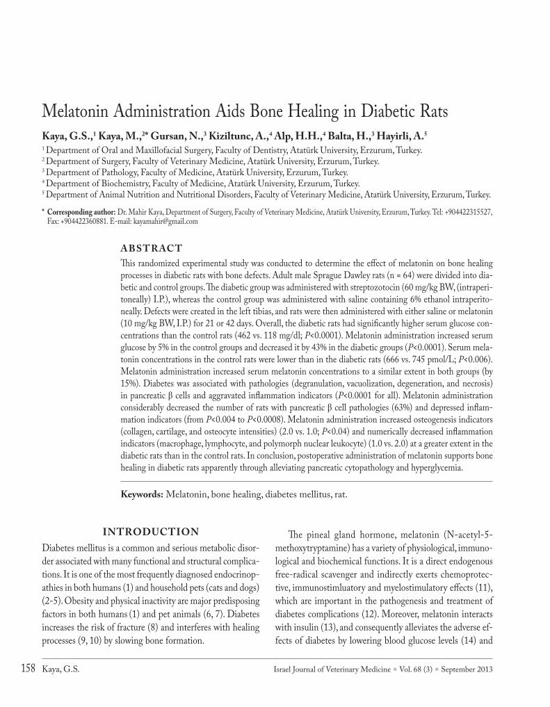
Pancreas HistopathologyIslets of Langerhans in the control groups injected with sa-line and melatonin appeared histologically normal (Figures 1a-b). In the diabetic groups, islets of Langerhans showed degranulation, vacuolization, degeneration and necrosis of β cells (Figure 1c); however, notably fewer histological chang-es were observed in the diabetic rats administered melato-nin when compared to the diabetic rats administered saline (Figure 1d). Severe necrotic and vacuolized pancreatic β cells were observed in more diabetic rats than controls (Table 2), and the number of diabetic rats administered melatonin with severe necrotic and vacuolized pancreatic β cells was consid-erably lower when compared to the diabetic rats adminis-tered saline.
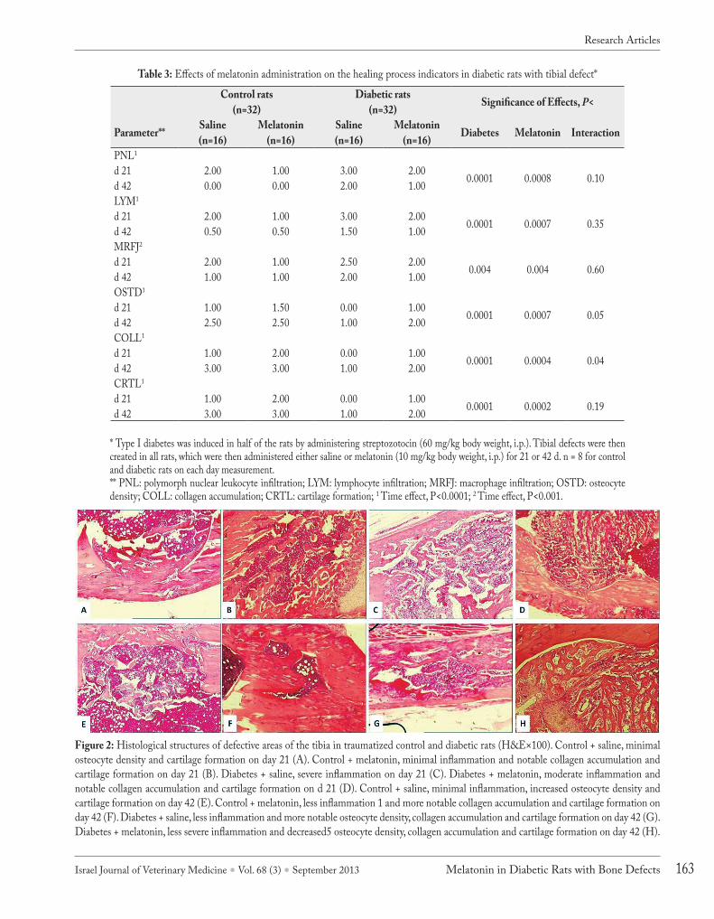
Bone HistopathologyInflammation indicators, i.e. polymorph nuclear leuko-cyte (P<0.0001), lymphocyte (P<0.0001) and macrophage (P<0.004) infiltrations, were more severe in the diabetic rats than the control rats (Table 3). The degree of infil-
tration decreased with melatonin administration (Table 3) at a similar extent in both groups. Cellular infiltration decreased considerably over time independently of health status.
Osteogenesis indicators improved significantly in the diabetic rats following melatonin administration. Osteocyte density decreased following diabetes induction (P<0.0001) and increased following melatonin adminis-tration (P<0.0007; Table 3), with more notable improve-ments in diabetic rats when compared to the control rats (P<0.05; Table 3). Diabetes induction was also accompa-nied by compromised collagen accumulation and cartilage
Table 1: Effects of melatonin administration on blood glucose (mg/dl), and serum melatonin (ρmol/L) concentrations in diabetic rats with tibial defect*
Control rats (n=32) Diabetic rats (n=32) Significance of Effects, P<Parameter Saline
(n=16)Melatonin
(n=16)Saline(n=16)
Melatonin(n=16)
SEM Diabetes Melatonin Interaction
Glucoseday 21 111.7 116.4 598.1 402.3 31.4 0.0001 0.0001 0.0001day 42 118.4 125.6 578.3 268.0 29.4Melatoninday 21 582 746 677 749 40 0.006 0.002 0.330day 42 628 706 750 805 38
* Type I diabetes was induced in half of the rats by administering streptozotocin (60 mg/kg body weight, i.p). Tibial defects were then created in all rats, which were then administered either saline or melatonin (10 mg/kg body weight, i.p.) for 21 or 42 d. n = 8 for control and diabetic rats on each day measurement.1 Time effect, P<0.0001.
Table 2: Semi-quantitative analysis of the pancreatic β cells in diabetic rats with tibial defect (n=8)*
Islet necrosis and vacuolization1
Groups 0 1 2 3Control + saline 8 0 0 0Control + melatonin 7 1 0 0Diabetes + saline 0 1 3 4Diabetes + melatonin 5 2 1 0
* Type I diabetes was induced in half of the rats by administering streptozotocin (60 mg/kg body weight, i.p.). Tibial defects were then created in all rats, which were then administered either saline or melatonin (10 mg/kg body weight, i.p.) for 42 d.1 Values represent numbers of rats. β cell necrosis and vacuolization were graded using light microscopy, as follows: 0 = no necrosis; 1 = slight degenerative changes (≤ 25% islets affected); 2 = moderate necrosis and vacuolization (≤ 50% islets affected); 3 = extensive and marked necrosis and vacuolization in central zone of the islets (≤ 75% islets affected).
Research Articles
Israel Journal of Veterinary Medicine Vol. 68 (3) September 2013Kaya, G.S.162
formation (P<0.0001 for both; Table 2). Melatonin admin-istration enhanced both collagen accumulation (P<0.0004) and cartilage formation (P<0.0002). The increase in collagen accumulation was significantly greater in the diabetic rats when compared to the control rats, whereas the degree of cartilage formation was unaffected by health status (P<0.04; Figure 2, Table 2).
Overall, blood glucose levels showed a positive correla-tion with polymorph nuclear leukocyte (r = 0.56; P<0.0001), lymphocyte (r = 0.48; P<0.0002), and macrophage (r = 0.39; P<0.003) infiltrations and a negative correlation with osteo-cyte density (r = -0.47; P<0.0002), collagen accumulation (r = -0.56; P<0.0001), and cartilage formation (r = -0.52; P<0.0001).
DISCUSSIONThe low antioxidative capacity of pancreatic β-cells makes them highly susceptible to oxidative changes due to hyper-glycemia (27). Cell damage by free radicals has been shown to lead to β-cell degranulation or necrosis in STZ-induced diabetic rats, resulting in a decrease in insulin secretion and an increase in blood glucose (28). In previous studies, mela-tonin (50 mg/kg) treatment has been shown to cause a sharp decrease in serum glucose, a slight increase in serum insulin concentrations and partial regeneration/proliferation of islet β-cells (29, 30). Similar findings were observed in the current study using 10mg/kg melatonin (Figure 1, Table 2). Other studies showed that melatonin exerted similar protective ef-fects on the liver (31) and kidneys (32) of diabetic rats. This
Figure 1: Histological structures of Langerhans islets in traumatized control and diabetic rats on day 42 post-tibial defect creation (H&E×100). Control + saline (a), control + melatonin (b), diabetes + saline (c), diabetes + melatonin (d). Arrows indicate numerous degenerative and vacuolized
β cells in diabetic rats administered with saline and arrow heads indicate a few degenerative cell in diabetic rats treated with melatonin.
Research Articles
Israel Journal of Veterinary Medicine Vol. 68 (3) September 2013 163 Melatonin in Diabetic Rats with Bone Defects
Table 3: Effects of melatonin administration on the healing process indicators in diabetic rats with tibial defect*
Control rats(n=32)
Diabetic rats(n=32) Significance of Effects, P<
Parameter** Saline(n=16)
Melatonin(n=16)
Saline(n=16)
Melatonin(n=16) Diabetes Melatonin Interaction
PNL1
d 21 2.00 1.00 3.00 2.00 0.0001 0.0008 0.10d 42 0.00 0.00 2.00 1.00LYM1
d 21 2.00 1.00 3.00 2.00 0.0001 0.0007 0.35d 42 0.50 0.50 1.50 1.00MRFJ2
d 21 2.00 1.00 2.50 2.00 0.004 0.004 0.60d 42 1.00 1.00 2.00 1.00OSTD1
d 21 1.00 1.50 0.00 1.00 0.0001 0.0007 0.05d 42 2.50 2.50 1.00 2.00COLL1
d 21 1.00 2.00 0.00 1.00 0.0001 0.0004 0.04d 42 3.00 3.00 1.00 2.00CRTL1
d 21 1.00 2.00 0.00 1.00 0.0001 0.0002 0.19d 42 3.00 3.00 1.00 2.00
* Type I diabetes was induced in half of the rats by administering streptozotocin (60 mg/kg body weight, i.p.). Tibial defects were then created in all rats, which were then administered either saline or melatonin (10 mg/kg body weight, i.p.) for 21 or 42 d. n = 8 for control and diabetic rats on each day measurement.** PNL: polymorph nuclear leukocyte infiltration; LYM: lymphocyte infiltration; MRFJ: macrophage infiltration; OSTD: osteocyte density; COLL: collagen accumulation; CRTL: cartilage formation; 1 Time effect, P<0.0001; 2 Time effect, P<0.001.
Figure 2: Histological structures of defective areas of the tibia in traumatized control and diabetic rats (H&E×100). Control + saline, minimal osteocyte density and cartilage formation on day 21 (A). Control + melatonin, minimal inflammation and notable collagen accumulation and cartilage formation on day 21 (B). Diabetes + saline, severe inflammation on day 21 (C). Diabetes + melatonin, moderate inflammation and notable collagen accumulation and cartilage formation on d 21 (D). Control + saline, minimal inflammation, increased osteocyte density and cartilage formation on day 42 (E). Control + melatonin, less inflammation 1 and more notable collagen accumulation and cartilage formation on day 42 (F). Diabetes + saline, less inflammation and more notable osteocyte density, collagen accumulation and cartilage formation on day 42 (G). Diabetes + melatonin, less severe inflammation and decreased5 osteocyte density, collagen accumulation and cartilage formation on day 42 (H).
Research Articles
Israel Journal of Veterinary Medicine Vol. 68 (3) September 2013Kaya, G.S.164
is attributed to the immunostimulatory effect of melatonin, which stimulates the secretion of opioid peptides and cyto-kines from lymphocytes and prevents the translocation of nuclear factor-kB to the nucleus and its binding with DNA (33). Indeed, the reduction of hyperglycemia severity with melatonin treatment (34) also helps to reduce the severity of inflammation in diabetic subjects (14, 35).
Endocrine pancreas β-cells and pineal gland β-cells have interrelated functions (36). Both melatonin receptors 1A and 1B are expressed in the pancreatic islets, and these are upregu-lated in diabetes (30). Melatonin enhances insulin-receptor kinase and insulin-receptor substrate-1 phosphorylation, sug-gesting possible communication between melatonin and insu-lin pathways. Through its influence on insulin secretion, mela-tonin is capable of decreasing the severity of hyperglycemia. As in the present study (Table 1), previous rat studies showed that melatonin level was lower in Type 2 diabetic subjects (37) and higher in Type 1 diabetic subjects and STZ-injected ani-mals (30) when compared to normal controls.
Bone remodeling is a constant and dynamic process in which osteoclasts resorb old bone and osteoblasts form new bone (22, 38). Melatonin influences the release of growth hormone and promotes bone formation (39, 40) by suppress-ing osteoclast activity; stimulating the formation and pro-liferation of a mineralized matrix and Type I collagen; in-creasing osteoblast alkaline phosphatase activity through the increased genetic expression of Type I collagen, osteopontin, bone sialoprotein and osteocalcin; and acting as an autacoid (a biological factor exerting its effects like a local hormone for a short period near the site of synthesis) to stimulate bone formation. In ovariectomized rats, decreased serum mela-tonin levels were accompanied by elevated bone resorption markers (41), suggesting that low melatonin during the post-menopause and post-andropausal periods may involve bone degradation in elderly subjects (42). In rats exposed to irra-diation, melatonin exerted protective effects against cellular oxidation (43). The development of scoliosis and decreased vertebral stiffness in pinealectomized fish are indications of the role of melatonin in skeletal development (44); further evidence is apparent in the reductions in bone mineral den-sity and osteocyte cell numbers reported in pinealectomized chickens (45).
As a result of hyperglycemia, diabetes mellitus compro-mises bone-tissue maintenance and osseous healing by in-creasing bone resorption, decreasing bone turnover, suppress-
ing cell differentiation and delaying revascularization (10, 46, 47). Melatonin has been shown to speed the regeneration of cortical bone width and length around implants in rabbit (48) and rat tibias (49). When compared to the control rats, the diabetic rats exhibited poor osteointegration between bone marrow and implants, but this was improved with hy-perglycemia treatment and the establishment of normogly-cemia (50). In another study (9), a 50% reduction in bone formation on the contact surface between bone and tibial implants was found in diabetic rats when compared to the healthy rats, whereas insulin injections improved the ultra-structural characteristics of the bone-implant interface of the diabetic rats to a level similar to non-diabetic rats.
Based on these findings, the authors suggested metabolic control to be essential for osseointegration, as constant hy-perglycemia delays the healing of bone surrounding implants. Moreover, osteoclasts generate free radicals, and melatonin administration enhances antioxidant status, thereby contrib-uting to improved fracture healing (51). Collagen also plays an important role in every stage of wound healing, demon-strating a regulatory and stabilizing role in tissue formation (52). In human bone cells and osteoblastic cell lines, melato-nin increases collagen accumulation (53, 54). In the present study, the control rats administered with melatonin for 21 and 42 days had reduced inflammatory markers and enhanced os-teogenesis indicators (Figure 2, Table 3). These effects were more notable in the diabetic rats (Figure 2, Table 3).
This study demonstrated that hyperglycemia was associ-ated with pathologies in pancreatic β cells and slowed down recovery process of tibial defect in diabetic rats. Melatonin administration caused a partial recovery in pancreas histopa-thology associated with lowered blood glucose levels, reduced the severity of inflammatory markers (polymorph-nuclear-leukocyte, lymphocyte and macrophage infiltration) and en-hanced osteogenesis indicators (osteocyte density, collagen accumulation and cartilage formation).
In conclusion, bone healing can be enhanced by the ad-ministration of melatonin through alleviating pancreatic cy-topathologies and indirectly controlling hyperglycemia in streptozotocin-induced diabetic rats with tibial defects.
ACKNOWLEDGEMENTS
This study was supported by the Atatürk University Scientific Research Projects Fund (project #: 2009/147).
Research Articles

Israel Journal of Veterinary Medicine Vol. 68 (3) September 2013 165 Melatonin in Diabetic Rats with Bone Defects
CONFLICT OF INTERESTNone of the authors of this paper has a financial or personal rela-tionship with other people or organizations that could inappropri-ately influence or bias the content of the paper.
REFERENCES1. Wild, S., Roglic, G., Green, A., Sicree, R. and King, H.: Glob-
al prevalence of diabetes: estimates for the year 2000 and projec-tions for 2030. Diabetes Care. 27:1047-1053, 2004.
2. Panciera, D.L., Thomas, C.B., Eicker, S.W. and Atkins, C.E.: Epizootiological patterns of diabetes mellitus in cats: 333 cas-es (1980-1986). J. Am. Vet. Med. Assoc. 197:1504-1508, 1990.
3. Baral, R., Rand, J.S., Catt, M. and Farrow, H.A.: Prevalence of feline diabetes mellitus in a feline private practice. J. Vet. Intern. Med. 17:434-437, 2003.
4. Guptill, L., Glickman, L. and Glickman, N.: Time trends and risk factors for diabetes mellitus in dogs: analysis of veterinary medical database records (1970–1999). Vet. J. 165:240-247, 2003.
5. Catchpole, B., Ristic, J.M., Fleeman, L.M. and Davison, L.J.: Ca-nine diabetes mellitus: can old dogs teach us new tricks? Diabeto-logia. 48:1948-1956, 2005.
6. Rand, J.S. and Martin, G.J.W.: Management of feline diabetes. Vet. Clin. North Am. Small. Anim. Pract. 31:881-913, 2001.
7. Prahl, A., Guptill, L., Glickman, N.W., Tetrick, M. and Glick-man, L.T.: Time trends and risk factors for diabetes mellitus in cats presented to veterinary teaching hospitals. J. Feline Med. Surg. 9:351-358, 2007.
8. Siqueira, J.T., Cavalher-Machado, S.C., Arana-Chavez, V.E. and Sannomiya, P.: Bone formation around titanium implants in the rat tibia: role of insulin. Implant. Dent. 12:242-251, 2003.
9. Esteves, J.C., Aranega, A.M., Borrasca, A.G., Fattah, C.M. and Garcia-Junior, I.R.: Repair process of surgical defects filled with autogenous bone grafts in tibiae of diabetic rats. J. Appl. Oral. Sci. 16:316-320, 2008.
10. Mariano, R., Messora, M., de Morais, A., Nagata, M., Furlaneto, F., Avelino, C., Paula, F., Ferreira, S., Pinheiro, M. and de Sene, J.P.: Bone healing in critical-size defects treated with platelet-rich plasma: a histologic and histometric study in the calvaria of diabetic rat. Oral. Surg. Oral. Med. Oral. Pathol. Oral. Radiol. Endod. 109:72-78, 2010.
11. Winiarska, K., Fraczyk, T., Malinska, D., Drozak, J. and Bryla, J.: Melatonin attenuates diabetes-induced oxidative stress in rabbits. J. Pineal Res. 40:168-176, 2006.
12. Sudnikovich, E.J., Maksirachik, Y.Z., Zabrodskaya, S.V., Ku-byshin, V.L., Lapshina, E.A., Bryszewska, M., Reiter, R.J. and Zavodnik, I.B.: Melatonin attenuates metabolic disorders due to streptozotocin-induced diabetes in rats. Eur. J. Pharmacol. 569:180-187, 2007.
13. Robeva, R., Kirilov, G., Tomova, A. and Kumanov, P.: Melatonin-insulin interactions in patients with metabolic syndrome. J. Pin-eal Res. 44:52-56, 2008.
14. Montilla, P.L., Vargas, J.F., Tunez, I.F., Munoz de Aqueda, M.C., Valdelvira, M.E. and Cabrera, E.S.: Oxidative stress in diabetic rats induced by streptozotocin: Protective effects of melatonin. J. Pineal Res. 25:94-100, 1998.
15. Cam, M., Yavuz, O., Guven, A., Ercan, F., Bukan, N. and Ustund-ag, N.: Protective effects of chronic melatonin treatment against renal injury in streptozotocin-induced diabetic rats. J. Pineal Res. 35: 212-220, 2003.
16. Maritim, A.C., Moore, B.H., Sanders, R.A. and Watkins, J.B.: Ef-fects of melatonin on oxidative stress in streptozotocin-induced diabetic rats. Int. J. Toxicol. 18:161-166, 1999.
17. Klepac, N., Rudes, Z. and Klepac, R.: Effects of melatonin on plasma oxidative stress in rats with streptozotocin induced diabe-tes. Biomed. Pharmacother. 60:32-35, 2006.
18. Satomura, K., Tobiume, S., Tokuyama, R., Yamasaki, Y., Kudoh, K. and Nagayama, M.: Melatonin at pharmacological doses en-hances human osteoblastic differentiation in vitro and promotes mouse cortical bone formation in vivo. J. Pineal Res. 42:231-239. 2007.
19. Suzuki, N., Somei, M., Seki, A., Reiter, R.J. and Hattori, A.: Nov-el bromomelatonin derivatives as potentially effective drugs to treat bone diseases. J. Pineal Res. 45:229-234, 2008.
20. Maldonado, M.D., Murillo-Cabezas, F., Terron, M.P., Flores, L.J., Tan, D.X., Manchester, L.C. and Reiter, R.J.: The potential of melatonin in reducing morbidity-mortality after craniocere-bral trauma. J. Pineal Res. 42:1-11, 2007.
21. Sack, R.L., Lewy, A.J., Erb, D.L., Vollmer, W.M. and Singer, CM. Human melatonin production decreases with age. J. Pineal Res. 3:379-388, 1986.
22. Cardinali, D.P., Ladizesky, M.G., Boggio, V., Cutrera, R.A. and Mautalen, C.: Melatonin effects on bone: experimental facts and clinical perspectives. J. Pineal Res.34:81-87, 2003.
23. CCAC. 1993. Guide to the Care and Use of Experimental Ani-mals, Vol. I, 2nd ed. Canadian Council on Animal Care, Bradda Printing Services Inc., Ottawa, ON, Canada.
24. Singab, A.N., El-Beshbishy, H.A., Yonekawa, M., Nomura, T. and Fukai, T.: Hypoglycemic effect of Egyptian Morus alba root bark extract: effect on diabetes and lipid peroxidation of strepto-zotocin-induced diabetic rats. J. Ethnopharmacol. 100:333-338, 2005.
25. Lewandrowski, K.U., Cattaneo, M.V., Gresser, J.D., Wise, D.L., White, R.L., Bonassar, L. and Trantola, D.J.: Effect of a poly (propylene fumarate) foaming cement on the healing of bone de-fects. Tissue Eng. 5:305-316, 1999.
26. SAS. User’s Guide. Statistics, Version 9th. Statistical Analysis System. SAS Inst., Inc., Cary, NC, USA 2002.
27. Pi, J., Bai, Y., Danie,l K.W., Liu, D., Lyght, O., Edelstein, D., Brownlee, M., Corkey, B.E. and Collins, S.: Persistent oxidative stress due to absence of uncoupling protein 2 associated with impaired pancreatic beta-cell function. Endocrinol. 150: 3040-3048, 2009.
28. Chen, H., Carlson, E.C., Pellet, L., Moritz, J.T. and Epstein, P.N.: Overexpression of Metallothionein in Pancreatic β-Cells Reduces Streptozotocin-Induced DNA Damage and Diabetes. Diabetes. 50:2040-2046, 2001.
29. Kanter, M., Uysal, H., Karaca, T. and Sagmanligil, H.O.: Depres-sion of glucose levels and partial restoration of pancreatic beta-cell damage by melatonin in streptozotocin-induced diabetic rats. Arch. Toxicol. 80: 362-369, 2006.
30. Peschke, E., Wolgast, S., Bazwinsky, I., Pönicke, K. and Muh-lbauer, E.: Increased melatonin synthesis in pineal glands of rats
Research Articles
Israel Journal of Veterinary Medicine Vol. 68 (3) September 2013Kaya, G.S.166
in streptozotocin induced type 1 diabetes. J. Pineal Res. 45:439-448, 2008.
31. Guven, A., Yavuz, O., Cam, M., Ercan, F., Bukan, N., Comunoglu, C. and Gokce F.: Effects of melatonin on streptozotocin-induced diabetic liver injury in rats. Acta Histochem. 108:85-93, 2006.
32. Derlacz, R.A., Sliwinska, M., Piekutowska, A., Winiarska, K., Drozak, J. and Bryla, J.: Melatonin is more effective than taurine and 5-hydroxytryptophan against hyperglycemia-induced kid-ney-cortex tubules injury. J. Pineal Res. 42:203-209, 2007.
33. Garcia-Maurino, S., Pozo, D., Calvo, J.R. and Guerrero, J.M.: Correlation between nuclear melatonin receptor expression and enhanced cytokine production in human lymphocytic and mono-cytic cell lines. J. Pineal Res.29:129-137, 2000.
34. Shieh, J.M., Wu, H.T., Cheng, K.C. and Cheng, J.T.: Melatonin ameliorates high fat diet-induced diabetes and stimulates glyco-gen synthesis via a PKC zeta-Akt-GSK3 beta pathway in hepatic cells. J. Pineal Res. 47:339-344, 2009.
35. Andersson, A.K. and Sandler, S.: Melatonin protects against streptozotocin, but not interleukin-1β-induced damage of rodent pancreatic β-cell. J. Pineal Res.30:157-165, 2001.
36. Kedziora-Kornatowska, K., Szewczyk-Golec, K., Kozakiewicz, M., Pawluk, H., Czuczejko, J., Kornatowski, T., Bartosz G. and Kedziora, J.: Melatonin improves oxidative stress parameters measured in the blood of elderly type 2 diabetic patients. J. Pineal Res. 46:333-337, 2009.
37. Peschke, E., Frese, T., Chankiewitz, E., Peschke, D., Preiss, U., Schneyer, R., Spessert, R. and Mühlbauer, E.: Diabetic Goto Kakizaki rats as well as type 2 diabetic patients show a decreased diurnal serum melatonin level and an increased pancreatic mela-tonin-receptor status. J. Pineal Res. 40:135-143, 2006.
38. Ostrowska, Z.: Menopause, obesity, and bone status. Postepy. Hig. Med. Dosw. 63:39-46, 2009.
39. Ladizesky, M.G., Boggio, V., Cutrera, R.A., Mondelo, N., Mast-aglia, S., Somoza, J. and Cardinali, D.P.: Melatonin effect on bone metabolism in rats treated with methylprednisolone. J. Pin-eal Res. 40:297-304, 2006.
40. Roth, J.A., Kim, B.G., Lin, W.L. and Cho, M.I.: Melatonin pro-motes osteoblast differentiation and bone formation. J. Biol. Chem. 274:22041-22047, 1999.
41. Ostrowska, Z., Kos-Kudla, B., Swietochowska, E., Marek, B., Ka-jdaniuk, D. and Gorski, J.: Assessment of the relationship be-tween dynamic pattern of nighttime levels of melatonin and cho-sen biochemical markers of bone metabolism in a rat model of postmenopausal osteoporosis. Neuro. Endorinol. Lett. 22:129-136, 2001.
42. Ladizesky, M.G., Cutrera, R.A., Boggio, V., Somoza, J., Centrella, J.M., Mautalen, C. and Cardinali, D.P.: Effect of melatonin on bone
metabolism in ovariectomized rats. Life Sci. 70:557-565, 2001.43. Yilmaz, S. and Yilmaz, E.: Effects of melatonin and vitamin E on
oxidative-antioxidative status in rats exposed to irradiation. Toxi-col. 222:1-7, 2006.
44. Fjelldal, P.G., Grotmol, S., Kryvi, H., Gjerdet, N.R., Taranger, G.L., Hansen, T., Porter M.J. and Totland, G.K.: Pinealectomy induces malformation of the spine and reduces the mechanical strength of the vertebrae in Atlantic salmon, Salmo salar. J. Pineal Res. 36:132-139, 2004.
45. Turgut, M., Kaplan, S., Turgut, A.T., Aslan, H., Güvenç, T., Cul-lu, E. and Erdogan, S.: Morphological, stereological and radio-logical changes in pinealectomized chicken cervical vertebrae. J. Pineal Res. 39:392-399, 2005.
46. Ogasawara, A., Nakajima, A., Nakajima, F., Goto, K. and Yamaza-ki, M.: Molecular basis for affected cartilage formation and bone union in fracture healing of the streptozotocin-induced diabetic rat. Bone. 43:832-839, 2008.
47. Musumecia, G., Loretoa, C., Clementib, G., Fiore, C.E. and Martinez, G.: An in vivo experimental study on osteopenia in di-abetic rats. Acta Histochem. 113:619-625, 2011.
48. Calvo-Guirado, J.L., Ramirez-Fernandez, M.P., Gomez-More-no, G., Mate-Sanchez, J.E., Delgado-Ruiz, R., Guardia, J., Lopez-Mari, L, Barone, A., Ortiz-Ruiz, A.J., Martinez-Gonzalez J.M. and Bravo L.A.: Melatonin stimulates the growth of new bone around implants in the tibia of rabbits. J. Pineal Res.49:356-363, 2010.
49. Takechi, M., Tatehara, S., Satomura, K., Fujisawa, K. and Na-gayama, M.: Effect of FGF-2 and melatonin on implant bone healing: a histomorphometric study. J. Mater. Sci. Mater. Med. 19:2949-2952, 2008.
50. Kopman, J.A., Kim, D.M., Rahman, S.S., Arandia, J.A., Karim-bux, N.Y. and Fiorellini, J.P.: Modulating the effects of diabetes on osseointegration with aminoguanidine and doxycycline. J. Per-iodontol.76:614-620, 2005.
51. Halici, M., Oner, M., Guney, A., Canoz, O., Narin, F. and Halici, C.: Melatonin promotes fracture healing in the rat model. Joint Diseases Related Surg.21:172-177, 2010.
52. Erol, F.S., Kavakli, A., Ilhan, N., Ozercan, I.H. and Sarsilmaz, M.: Effects of melatonin and octreotide on peridural fibrosis in an animal model of laminectomy. Turk. Neurosurg. 20:50-56, 2010.
53. Nakade, O., Koyama, H., Ariji, H., Yajima, A. and Kaku, T.: Me-latonin stimulates proliferation and type I collagen synthesis in human bone cells in vitro. J. Pineal Res. 27:106-10, 1999.
54. Drobnik, J., Karbownik-Lewinska, M., Szczepanowska, A., Slor-winska, D., Olczak, S., Jakubowski, L. and Dabrowski, R.: Regu-latory influence of melatonin on collagen accumulation in the in-farcted heart scar. J. Pineal Res. 45:285-290, 2008.
Research Articles