membrane interaction of the glycosyltransferase murg:...
TRANSCRIPT

JOURNAL OF BACTERIOLOGY, July 2003, p. 3773–3779 Vol. 185, No. 130021-9193/03/$08.00�0 DOI: 10.1128/JB.185.13.3773–3779.2003Copyright © 2003, American Society for Microbiology. All Rights Reserved.
Membrane Interaction of the Glycosyltransferase MurG: a SpecialRole for Cardiolipin
Els van den Brink-van der Laan,* Jan-Willem P. Boots,† Robin E. J. Spelbrink,Gerda M. Kool, Eefjan Breukink, J. Antoinette Killian, and Ben de Kruijff
Department Biochemistry of Membranes, Centre for Biomembranes and Lipid Enzymology, Institute ofBiomembranes, Utrecht University, 3584 CH, Utrecht, The Netherlands
Received 20 December 2002/Accepted 26 March 2003
MurG is a peripheral membrane protein that is one of the key enzymes in peptidoglycan biosynthesis. Thecrystal structure of Escherichia coli MurG (S. Ha, D. Walker, Y. Shi, and S. Walker, Protein Sci. 9:1045-1052,2000) contains a hydrophobic patch surrounded by basic residues that may represent a membrane associationsite. To allow investigation of the membrane interaction of MurG on a molecular level, we expressed andpurified MurG from E. coli in the absence of detergent. Surprisingly, we found that lipid vesicles copurify withMurG. Freeze fracture electron microscopy of whole cells and lysates suggested that these vesicles are derivedfrom vesicular intracellular membranes that are formed during overexpression. This is the first study whichshows that overexpression of a peripheral membrane protein results in formation of additional membraneswithin the cell. The cardiolipin content of cells overexpressing MurG was increased from 1 � 1 to 7 � 1 mol%compared to nonoverexpressing cells. The lipids that copurify with MurG were even further enriched incardiolipin (13 � 4 mol%). MurG activity measurements of lipid I, its natural substrate, incorporated in purelipid vesicles showed that the MurG activity is higher for vesicles containing cardiolipin than for vesicles withphosphatidylglycerol. These findings support the suggestion that MurG interacts with phospholipids of thebacterial membrane. In addition, the results show a special role for cardiolipin in the MurG-membraneinteraction.
Bacterial cell membranes are surrounded by peptidoglycan,a heteropolymer consisting of glycan strands cross-linked bypeptides. An enzyme involved in peptidoglycan biosynthesis isthe glycosyltransferase MurG. As schematically shown in Fig.1, the substrate of MurG is lipid I, which consists of an unde-caprenyl-pyrophosphate anchored within the membrane andlinked to a muramyl pentapeptide. MurG catalyzes the transferof N-acetylglucosamine (GlcNAc) from UDP-GlcNAc to lipidI to form lipid II (19). This process takes place on the cyto-plasmic surface of the bacterial membrane prior to the trans-location of lipid II to the extracellular face of the cell mem-brane (10). Subsequently, the disaccharide is polymerized andcross-linked to the preexisting peptidoglycan strands of the cellwall (15). This peptidoglycan biosynthesis pathway has beenshown to be a good target for antibiotics, for example, for thelantibiotic nisin Z, which uses lipid II as a receptor (8, 9).
MurG is a moderately hydrophobic protein without anymembrane-spanning domain and with a molecular mass of37,684 Da (19). Based on cell fractionation experiments, it wassuggested that the protein is associated with membranes (19).Bupp and Van Heijenoort concluded that MurG is associatedwith the cytoplasmic face of the inner membrane of Escherichiacoli, based on the finding that MurG in spheroplasts was in-
sensitive to trypsin (10). The crystal structure of E. coli MurG(14) showed that the protein consists of a C-terminal domain,which probably contains the UDP-GlcNAc-binding site, and anN-terminal domain, which probably contains the lipid I bindingsite. Additionally, a hydrophobic patch in the N domain, sur-rounded by basic residues, was found. It was proposed that thisis the membrane association site and that association involvesboth hydrophobic and electrostatic interactions with the mem-brane (14).
To study the membrane interaction of MurG, we expressedthe His-tagged protein in E. coli and purified it in the absenceof detergent. Surprisingly, we found that lipids in the form ofvesicles copurify with MurG. These MurG-lipid vesicles wereenriched in the negatively charged lipid cardiolipin (CL). Us-ing freeze fracture electron microscopy (EM), we showed thatoverexpression of MurG in E. coli results in the formation ofvesicular intracellular membranes which contain almost nointegral membrane proteins. This is the first time that suchintracellular membranes have been observed for overexpres-sion of a peripheral membrane protein. Activity measurementswith lipid I-containing vesicles showed that MurG activity isenhanced by the presence of CL. The results suggest thatMurG binds to membranes via a direct interaction with thelipids and that it preferentially interacts with CL.
MATERIALS AND METHODS
Materials. E. coli BL21(DE3)/plysS was obtained from The Netherlands Cul-ture Collection of Bacteria. E. coli SD12 (pyrD34 his68 galK35 phoA8 glpD3 glpR2glpK glpKp) and SD11 (SD12 cls) were first described by Shibuya et al. (26). LipidI was produced and purified according to the method of Breukink et al. (7) usingMicrococcus flavus membrane vesicles, UDP-MurNAc-pentapeptide purifiedfrom Staphylococcus simulans, and undecaprenylphosphate purified from Laurus
* Corresponding author. Mailing address: Department of Biochem-istry of Membranes, Centre for Biomembranes and Lipid Enzymology,Institute of Biomembranes, Utrecht University, Padualaan 8, 3584 CH,Utrecht, The Netherlands. Phone: (31)(30)2533553. Fax: (31)(30)2522478. E-mail: [email protected].
† Present address: Division of R&D R&A, DMV International,5460 BA, Veghel, The Netherlands.
3773
on June 29, 2018 by guesthttp://jb.asm
.org/D
ownloaded from

nobilus, which was a kind gift of E. Swiezewska. The plasmid pET21b carrying theMurG gene containing a C-terminal LEHHHHHH sequence was a kind gift ofS. Walker. MurG without a His tag but with an additional leucine at the Cterminus was cloned from MurG containing a C-terminal LEHHHHHH se-quence according to the QuikChange site-directed mutagenesis method (Strat-agene), which was confirmed by DNA sequencing. Ni-nitrilotriacetic acid (NTA)agarose was from Qiagen. His tag monoclonal antibody was obtained fromNovagen. 1,2-Dioleoyl-sn-glycero-3-phosphocholine (DOPC), 1,2-dioleoyl-sn-glycero-3-phosphoethanolamine (DOPE), 1,2-dioleoyl-sn-glycero-3-phospho-glycerol (DOPG), and 1,1�,2,2�-tetraoleoyl CL (TOCL) were purchased fromAvanti Polar Lipids Inc. IPTG (isopropylthiogalactoside), UDP-GlcNAc, andn-octyl-�-D-glucopyranoside (octylglucoside) were obtained from Sigma. Uridinediphospho-N-acetyl-D-[U-14C]glucosamine (14C-UDP-GlcNAc; 9.84 GBq/mmol)was purchased from Amersham Pharmacia. DNase and RNase were from Sigma.Lysozyme was obtained from Roche. All other chemicals were of the highestpurity commercially available.
Expression of MurG. BL21(DE3)/plysS cells overexpressing MurG (with orwithout a His tag) from a pET21b vector were grown at 37°C in Luria-Bertanimedium supplemented with 100 �g of ampicillin/ml and 25 �g of chloramphen-icol/ml. When the optical density at 600 nm reached a value of 0.6, IPTG wasadded to a final concentration of 1 mM. The induced cell culture was grown foranother 3 h. The cell cultures were then incubated on ice for 15 min andharvested by centrifugation (Sorvall RC3Bplus; H-6000A rotor; 4°C; 30 min;7,300 � g). The cell pellets were frozen at �20°C.
Purification of His-tagged MurG. For the purification of His-tagged MurG inthe absence of detergent, we modified the protocol described by Ha et al. (14).The cell pellets were thawed at 4°C, and the cells were resuspended in 5 ml of 5mM imidazole– 20 mM Tris-HCl, pH 8.0 (buffer A), per g of cells and disruptedby forcing them through a French press three times at 12,000 lb/in2. The celllysate was centrifuged (Sorvall RC5Bplus; Sorvall SS34 rotor; 4°C; 30 min; 4,300� g), and the supernatant was applied to a Ni-NTA agarose column equilibratedwith buffer A. After the column was washed with 25 column volumes of buffer A,the MurG protein was eluted with a stepwise imidazole gradient in buffer A,starting with 15 column volumes of 25 mM imidazole in buffer A, followed by 15column volumes of 50 mM imidazole in buffer A and 15 column volumes of 100mM imidazole in buffer A at a flow rate of 1 ml/min. The eluted fractions wereconcentrated with Centricon Plus-20 concentrators (nominal molecular weightlimit [NMWL], 10,000; Millipore). The purified MurG was stored at �20°C inthe presence of 20% glycerol. The protein concentration was determined by theBradford assay (6).
Preparation of SD11 and SD12 cell lysates. SD11 and SD12 cells were grownat 37°C in Luria-Bertani medium till the optical density at 600 nm reached avalue of 0.8. Subsequently, the cells were harvested by centrifugation (SorvallRC3Bplus; H-6000A rotor; 4°C; 30 min; 7,300 � g) and washed once withphysiological salt solution. The cells were resuspended in 5 ml of buffer per g ofcells, containing 50 mM Tris-HCl, pH 8.0, 0.2 mM phenylmethylsulfonyl fluoride,3% Triton X-100, 4 mM dithiothreitol, 5 mM MgCl2, 40 �g of lysozyme/ml, 20�g of DNase/ml, and 20 �g of RNase/ml. The cell suspension was incubated onice for 45 min and subsequently at 37°C for 15 min. The cell lysate was clearedby centrifugation (Sorvall RC5Bplus; SS34 rotor; 4°C; 30 min; 48,000 � g).Finally, the protein content was determined using a bicinchoninic acid proteindetermination kit (Pierce). The most concentrated lysate was diluted to obtainthe same protein content for both lysates (dilution factor, 1.4), using a buffercontaining 50 mM Tris-HCl, pH 8.0, 3% Triton X-100, 4 mM dithiothreitol, and5 mM MgCl2.
Enzymatic activity assay. To determine the enzymatic activity of MurG, anassay was developed, based on a recently developed protocol for the synthesisand purification of lipid II (7). The assay was performed as follows: 50 or 100 ngof MurG was incubated with 3 nmol of lipid I, 10 nmol of UDP-GlcNAc, and 35pmol of 14C-UDP-GlcNAc in 0.1 M Tris-HCl, pH 8.0, containing 6.7 mM MgCl2and 1% octylglucoside or Triton X-100 in a total volume of 30 �l at roomtemperature for 1 to 15 min. Lipids were extracted in 60 �l of butanol containing6.25% pyridine-acetic acid, pH 4.2, which was washed once with 60 �l of water.The amount of lipid II in the butanol-pyridine mixture was determined by liquidscintillation counting. The extraction procedure was validated by thin-layer chro-matography on silica plates (Silica gel 60; Merck) using chloroform-methanol-water-ammonia (88:48:10:1, by volume) as eluents, showing that after extractionno detectable amount of lipid II was present in the water phase while no free14C-UDP-GlcNAc was present in the butanol phase (data not shown).
To test the activity of MurG on lipid vesicles, large unilamellar vesicles(200-nm diameter) were prepared as follows. A dry lipid film containing 1% lipidI was hydrated in 50 mM NaCl– 20 mM Tris-HCl, pH 8.0. The dispersion wasfrozen and thawed 10 times and extruded 6 times through 200-nm-pore-sizemembrane filters (Anatop 10; Whatman), essentially as described previously(16). The assay was performed as described above for 100 ng of MurG withoutthe addition of octylglucoside or Triton X-100.
Light scattering. The size distribution of vesicles was determined by using lightscattering by means of a Zetasizer 3000 (Malvern Instruments) using a mono-modal analysis mode. As a reference, large unilamellar vesicles with a defineddiameter of 100 nm were used, which were prepared as follows. A dry lipid film
FIG. 1. Schematic representation of the role of MurG within the peptidoglycan biosynthesis pathway. The substrate for MurG is lipid I, whichconsists of undecaprenylpyrophosphate and the sugar N-acetylmuramic acid (MurNAc) pentapeptide (L-Ala-�D-Glu-L-Lys-D-Ala-D-Ala). MurGadds the sugar GlcNAc to lipid I to form lipid II. Subsequently, lipid II is translocated across the cell membrane, after which the disaccharide-pentapeptide is transferred to the peptidoglycan network.
3774 VAN DEN BRINK-VAN DER LAAN ET AL. J. BACTERIOL.
on June 29, 2018 by guesthttp://jb.asm
.org/D
ownloaded from

of DOPE-DOPG-CL (75:20:5) was hydrated in 50 mM NaCl– 20 mM Tris-HCl,pH 8.0. The dispersion was frozen and thawed 10 times and extruded throughtwo stacked 100-nm-pore-size polycarbonate filters (Millipore) as described pre-viously (16).
Freeze fracture EM. Samples of whole cells were taken from the cell pelletdirectly after harvesting. Samples of cell lysate and purified MurG were concen-trated by Centricon Plus-20 concentrators (10,000 NMWL; Millipore) or ultra-centrifugation (Beckman L-60 ultracentrifuge; 60 Ti rotor; 4°C; 90 min; 45,000 �g).
These samples were sandwiched between two hat-shaped copper holders. Nocryoprotectants were added. The samples were fast frozen by plunging them intoliquid propane, cooled to its melting point with liquid nitrogen, using a KF80plunge-freezing device (Reichert Jung). The samples were fractured and subse-quently replicated with platinum according to standard procedures, using aBAF400 freeze fracture device (Bal-tec AG). The replicas were stripped fromthe copper holders and cleaned with chromic acid followed by distilled wateraccording to the method of Costello et al. (12). A CM10 electron microscope(Philips) operated at 80 kV was used for examining the replicas.
Phospholipid analysis. Lipids were extracted according to the method of Blighand Dyer (5). The phosphorus concentration was determined according to themethod of Rouser et al. (24). The phospholipid composition of the lipid extractswas analyzed by two-dimensional thin-layer chromatography on silica plates,using chloroform-methanol-water (65:25:4, by volume) for the first dimensionand chloroform-methanol-acetic acid (65:25:10, by volume) for the second di-mension.
RESULTS
Expression and purification of His-tagged MurG. His-tagged MurG was expressed from a pet21b vector in an E. coliBL21(DE3)/plysS host. As shown in Fig. 2 (compare lanes 1and 2), induction with IPTG resulted in the appearance of anintense band on sodium dodecyl sulfate-polyacrylamide gelelectrophoresis at a molecular mass of �38 kDa, which is themolecular mass of MurG. This protein contained a His tag, aswas shown by Western blotting using anti-His tag antibodies(data not shown). Because our aim was to study the membraneinteraction of MurG, we wanted to purify the protein in theabsence of detergent. Therefore, we set up a new purificationprocedure, using a Ni-NTA column with an imidazole gradi-ent. The result of the purification is shown in Fig. 2, lane 3. The
yield of purified protein was �5 mg per liter of cells. Theenzymatic activity of His-tagged MurG in 1% octylglucosidewas determined to be 10.2 2.7 �mol/min/mg of protein. Thisis much higher than the value of 2.8 nmol/min/mg of proteinpublished earlier by Crouvoisier et al. (13). This difference isprobably due to different assay conditions, since we used purelipid I while Crouvoisier et al. used E. coli membranes (13).Addition of 20% glycerol before storage at �20°C was foundto be necessary to keep MurG enzymatically active (data notshown).
Phospholipids copurify with His-tagged MurG. Based on aphosphate determination after Bligh and Dyer extraction, wefound that the solution of purified His-tagged MurG still con-tained phospholipids. The lipid composition was determinedand is shown in Table 1. We found that the lipids that copurifywith His-tagged MurG are enriched in CL compared to thetotal lipid extract of nonoverexpressing cells: the CL content isincreased from between 1 and 2 to 13 mol%. Furthermore, thetotal lipid extract of His-tagged MurG-overproducing cells alsoshows enrichment of CL, but to a lesser extent: the CL contentwas found to be 7 mol%.
The question then arose as to the form in which the lipids inassociation with MurG are present in the solution: as vesiclesor as molecular complexes with His-tagged MurG. To get afirst indication about the aggregation form of the lipids, weused dynamic light scattering. We observed that particles werepresent in a solution of purified His-tagged MurG. We foundthat the particles had an average diameter of 130 nm, with apolydispersity index of 0.36 (data not shown). This large sizesuggests that the particles represent lipids that are present aslipid vesicles, but the possibility that they represent large pro-tein aggregates cannot be excluded.
To further investigate the nature of these particles, we there-fore used freeze fracture EM. The result is shown in Fig. 3.This EM picture shows the presence of spherical particles withsmooth fracture faces, typical of lipid vesicles. The size of thesevesicles, which have an average diameter of 148 87 nm (n
FIG. 2. Expression and purification of His-tagged MurG. Lane 1,E. coli BL21(DE3)/plysS cells containing the pet21b�MurG vectorbefore IPTG induction; lane 2, E. coli BL21(DE3)/plysS cells contain-ing the pet21b�MurG vector 3 h after IPTG induction, just beforeharvesting; lane 3, purified MurG. A protein size marker is shown onthe left.
TABLE 1. Phospholipid composition of phospholipid extracts ofpurified MurG and E. coli cells with and without MurG
overexpressiona
Phospholipid extractPhospholipid composition (mol%)
PEf PG CL
Purified MurGb 66 7 21 4 13 4Nonoverexpressing cells
(MurG vector; no IPTG)c75 4 24 4 1 1
Nonoverexpressing cells(empty vector; � IPTG)d
69 2 29 1 2 1
MurG-overexpressing cells(MurG vector; � IPTG)e
75 2 18 2 7 1
a See Materials and Methods for the exact description of MurG expression andpurification, lipid extraction, and determination of the phospholipid composi-tion. Standard deviations are based on at least three independent measurements.
b Lipid extract of MurG.c Total lipid extract of cells containing the plasmid pet21b�MurGhistag with-
out induction by IPTG.d Total lipid extract of cells containing the plasmid pet21b with induction by
IPTG.e Total lipid extract of cells containing the plasmid pet21b�MurGhistag with
induction by IPTG.f PE, phosphatidylethanolamine.
VOL. 185, 2003 MurG MEMBRANE INTERACTION 3775
on June 29, 2018 by guesthttp://jb.asm
.org/D
ownloaded from
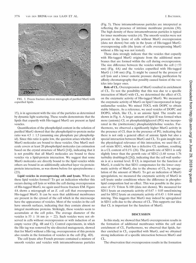
15), is in agreement with the size of the particles as determinedby dynamic light scattering. These results demonstrate that thelipids that copurify with His-tagged MurG are present as lipidvesicles.
Quantification of the phospholipid content in the solution ofpurified MurG showed that the phospholipid-to-protein molarratio was 4.5 1.5 (assuming one phosphate per phospholip-id). Since this ratio is quite low, the question arises whether allMurG molecules are bound to these vesicles. One MurG mol-ecule covers at least 20 phospholipid molecules (an estimationbased on the crystal structure of MurG [14]), indicating that itis not possible that all MurG molecules are bound to thesevesicles via a lipid-protein interaction. We suggest that someMurG molecules are directly bound to the lipid vesicles whileothers are bound in an additionally adsorbed layer via protein-protein interactions, as was shown before for apocytochrome c(23).
Lipid vesicles in overexpressing cells and lysate. When arethese lipid vesicles formed? To get an indication whether thisoccurs during cell lysis or within the cell during overexpressionof His-tagged MurG, we again used freeze fracture EM. Figure4A shows a micrograph of an E. coli cell that overexpressesHis-tagged MurG. It can be seen that additional membranesare present in the cytosol of the cell which in the microscopehave the appearance of vesicles. Most of the vesicles in the cellhave smooth surfaces, indicating that they contain almost nointegral membrane proteins. Strikingly, these vesicles seem toaccumulate at the cell poles. The average diameter of thevesicles is 53 16 nm (n 22). Such vesicles were not ob-served in cells without overexpression or with induction of anempty vector (Fig. 4B and C). Control experiments, in whichthe His tag was removed by site-directed mutagenesis, showedthat for MurG without a His tag, overexpression of this proteinalso results in the formation of vesicles in the cell (Fig. 4D).
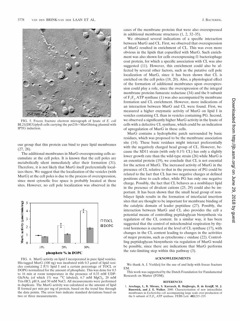
The cell lysate after French pressure contained a mixture ofsmooth vesicles and vesicles with intramembranous particles
(Fig. 5). These intramembranous particles are interpreted asreflecting the presence of intrinsic membrane proteins (30).The high density of these intramembranous particles is typicalfor inner membrane vesicles (4). The smooth vesicles were notpresent in the lysate of cells without MurG overexpression(data not shown) but only in the lysate of His-tagged MurG-overexpressing cells (the lysate of cells overexpressing MurGwithout a His tag was not tested).
These data strongly indicate that the vesicles that copurifywith His-tagged MurG originate from the additional mem-branes that are formed within the cell during overexpression.The size difference between the vesicles within the cell (53nm) (Fig. 4A) and the vesicles copurified with His-taggedMurG (148 nm) (Fig. 3) might be caused by the process ofcell lysis and a lower osmotic pressure during purification byaffinity chromatography that possibly caused fusion of the ves-icles into larger ones.
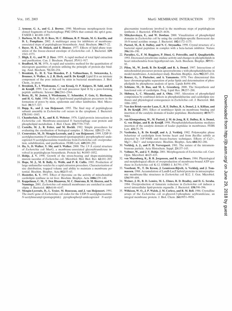
Role of CL. Overexpression of MurG resulted in enrichmentof CL. To test the possibility that this was due to a specificinteraction of MurG with CL, we determined the effect of CLon the enzymatic activity of His-tagged MurG. We measuredthe enzymatic activity of MurG on lipid I incorporated in largeunilamellar vesicles. We mixed TOCL with DOPC to obtainstable bilayers. As a reference, we used vesicles of DOPC andDOPG, which, like CL, is an anionic lipid. The results areshown in Fig. 6. A larger amount of lipid II was formed whenmore (anionic) CL or phosphatidylglycerol (PG) was incorpo-rated in the (zwitterionic) phosphatidylcholine vesicles. Fur-thermore, we observed a significantly higher MurG activity inthe presence of CL than in the presence of PG, indicating thatthere is not only a general effect of anionic lipids but also aspecial interaction between MurG and CL. To gain insight intothe physiological relevance of this interaction, we used the E.coli strain SD11, which has a defective CL synthase, resultingin a CL content of 0.1% (26). The growth rate of SD11 is onlyslightly lower than the growth rate of SD12 (1.68 versus 1.76turbidity doublings/h [26]), indicating that the cell wall synthe-sis is at a normal level. If CL is important for the function ofMurG, it could be that SD11 compensates for the lower enzy-matic activity of MurG, due to the absence of CL, by upregu-lation of the amount of MurG. To get an indication of MurGupregulation, we measured the enzymatic activity of MurG incell lysate under conditions where the difference in phospho-lipid composition had no effect. This was possible in the pres-ence of 1% Triton X-100 (data not shown). We measured forSD11 lysate an enzymatic activity of 0.67 0.05 nmol/min/mgand for SD12 lysate an enzymatic activity of 0.43 0.04 nmol/min/mg. This suggests that MurG may indeed be upregulatedin SD11 cells due to the absence of CL. This supports our ideathat CL is important for the function of MurG.
DISCUSSION
In this study, we showed that MurG overexpression results inthe formation of additional membranes within the cell andenrichment of CL. Furthermore, we observed that lipids, fur-ther enriched in CL, copurified with MurG, and we obtainedstrong indications of a specific interaction between MurG andCL.
The observation that overexpression results in the formation
FIG. 3. Freeze fracture electron micrograph of purified MurG withcopurified lipids.
3776 VAN DEN BRINK-VAN DER LAAN ET AL. J. BACTERIOL.
on June 29, 2018 by guesthttp://jb.asm
.org/D
ownloaded from

of additional membrane structures within the cell is ratherstriking. Similar observations were done previously for variousother proteins, such as fumarate reductase (34), membrane-bound ATP synthase (32), sn-glycerol-3-phosphate acyltrans-ferase (35), LamB-LacZ hybrid proteins (33), sp6.6 (2), andthe b subunit of F1Fo ATP synthase (1). However, these pro-teins are all integral membrane proteins, which need a mem-brane to be expressed in a functional form. This is the first timethat such an effect has been shown for a peripheral membraneprotein. This suggests that for overexpression of MurG as well,the membrane is important. Because of that, we conclude thatin E. coli most of the overexpressed MurG is probably boundto the vesicles.
The membrane structures in MurG-overexpressing cells
have smooth surfaces. This is also the case for the vesicles thatcopurified with MurG, indicating that they do not contain anyintegral membrane protein. Therefore, we suggest that theoverexpressed MurG is bound to these vesicles via a directinteraction with the lipids and not via a (specific) interactionwith an integral membrane protein. This is supported by thefact that MurG is able to use lipid I as a substrate, even whenit is incorporated in vesicles composed of only lipids (Fig. 6). Itis also in agreement with the suggestion of Ha and coworkers(14) that MurG is bound to the membrane via a hydrophobicpatch surrounded by basic residues. Moreover, this proteinshows a certain similarity with the catalytic domain of leaderpeptidase, which contains a large exposed hydrophobic patchas a membrane association surface (21). It has been shown by
FIG. 4. Freeze fracture electron micrographs of cells. (A) E. coli BL21(DE3)/plysS cells carrying the pet21b�MurGhistag plasmid with IPTGinduction. (B) E. coli BL21(DE3)/plysS cells carrying the pet21b�MurGhistag plasmid without IPTG induction. (C) E. coli BL21(DE3)/plysS cellscarrying the empty pet21b plasmid with IPTG induction. (D) E. coli BL21(DE3)/plysS cells carrying the pet21b�MurG (without His tag) plasmidwith IPTG induction.
VOL. 185, 2003 MurG MEMBRANE INTERACTION 3777
on June 29, 2018 by guesthttp://jb.asm
.org/D
ownloaded from
our group that this protein can bind to pure lipid membranes(27, 28).
The additional membranes in MurG-overexpressing cells ac-cumulate at the cell poles. It is known that the cell poles aremetabolically silent immediately after their formation (31).Therefore, it is not likely that MurG itself preferentially local-izes there. We suggest that the localization of the vesicles (withMurG) at the cell poles is due to the process of overexpression,since most cytosolic free space is probably located at thesesites. However, no cell pole localization was observed in the
cases of the membrane proteins that were also overexpressedin additional membrane structures (1, 2, 32–35).
We obtained several indications of a specific interactionbetween MurG and CL. First, we observed that overexpressionof MurG resulted in enrichment of CL. This was even moreobvious in the lipids that copurified with MurG. Such enrich-ment was also shown for cells overexpressing f1 bacteriophagecoat protein, for which a specific association with CL was alsosuggested (11). However, this enrichment could also be af-fected by several other factors, such as the putative cell polelocalization of MurG, since it has been shown that CL isenriched on the cell poles (18, 20). Also, a physiological effectof the formation of additional membranes upon overexpres-sion could play a role, since the overexpression of the integralmembrane proteins fumarate reductase (34) and the b subunitof F1Fo ATP synthase (1) was also accompanied by membraneformation and CL enrichment. However, more indications ofan interaction between MurG and CL were found. First, wemeasured a higher enzymatic activity of MurG on lipid I invesicles containing CL than in vesicles containing PG. Second,we observed a significantly higher MurG activity in the lysate ofcells with a defective CL synthase, which could be an indicationof upregulation of MurG in these cells.
MurG contains a hydrophobic patch surrounded by basicresidues, which was proposed to be the membrane associationsite (14). These basic residues might interact preferentiallywith the negatively charged head group of CL. However, be-cause the SD11 strain (with only 0.1% CL) has only a slightlylower growth rate than the wild-type strain (26) while MurG isan essential protein (19), we conclude that CL is not essentialfor the action of MurG. The increased activity of MurG in thepresence of CL relative to that in the presence of PG could berelated to the fact that CL has two negative charges at definedpositions close to each other, while PG has only one negativecharge. Possibly, the fact that CL behaves as a nonbilayer lipidin the presence of divalent cations (25, 29) could also be im-portant. It has been shown that the small head group of non-bilayer lipids results in the formation of interfacial insertionsites that are thought to be important for membrane binding ofthe catalytic domain of leader peptidase (27). Possibly, theinteraction between MurG and CL also provides the cell apotential means of controlling peptidoglycan biosynthesis viaregulation of the CL content. In a similar way, it has beensuggested that the control of mitochondrial respiration by thy-roid hormones is exerted at the level of CL synthase (17), withchanges in the CL content leading to changes in the activitiesof major proteins, such as cytochrome c oxidase (22). Control-ling peptidoglycan biosynthesis via regulation of MurG wouldbe possible, since there are indications that MurG performsthe rate-limiting step within this pathway (3).
ACKNOWLEDGMENTS
We thank A. J. Verkleij for the use of and help with freeze fractureEM.
This work was supported by the Dutch Foundation for FundamentalResearch on Matter (FOM).
REFERENCES
1. Arechaga, I., B. Miroux, S. Karrasch, R. Huijbregts, B. de Kruijff, M. J.Runswick, and J. E. Walker. 2000. Characterisation of new intracellularmembranes in Escherichia coli accompanying large scale over-production ofthe b subunit of F1Fo ATP synthase. FEBS Lett. 482:215–219.
FIG. 5. Freeze fracture electron micrograph of lysate of E. coliBL21(DE3)/plysS cells carrying the pet21b�MurGhistag plasmid withIPTG induction.
FIG. 6. MurG activity on lipid I incorporated in pure lipid vesicles.His-tagged MurG (100 ng) was incubated with 0.3 �mol of lipid vesi-cles containing 2 Pi% lipid I and a certain percentage of TOCL orDOPG normalized for the amount of phosphate. This was done for 0.5to 16 min at room temperature in the presence of 0.33 mM UDP-GlcNAc (of which 1% was 14C labeled), 6.7 mM MgCl2, 20 mMTris-HCl, pH 8, and 50 mM NaCl. All measurements were performedin duplicate. The MurG activity was calculated as the amount of lipidII formed per min per mg of protein, based on the trend line throughthe data points. The error bars indicate standard deviations based ontwo or three measurements.
3778 VAN DEN BRINK-VAN DER LAAN ET AL. J. BACTERIOL.
on June 29, 2018 by guesthttp://jb.asm
.org/D
ownloaded from
2. Armour, G. A., and G. J. Brewer. 1990. Membrane morphogenesis fromcloned fragments of bacteriophage PM2 DNA that contain the sp6.6 gene.FASEB J. 4:1488–1493.
3. Barbosa, M. D., H. O. Ross, M. C. Hillman, R. P. Meade, M. G. Kurilla, andD. L. Pompliano. 2002. A multitarget assay for inhibitors of membrane-associated steps of peptidoglycan biosynthesis. Anal. Biochem. 306:17–22.
4. Bayer, M. E., M. Dolack, and E. Houser. 1977. Effects of lipid phase tran-sition of the freeze-cleaved envelope of Escherichia coli. J. Bacteriol. 129:1563–1573.
5. Bligh, E. G., and W. J. Dyer. 1959. A rapid method of total lipid extractionand purification. Can. J. Biochem. Physiol. 37:911–917.
6. Bradford, M. M. 1976. A rapid and sensitive method for the quantitation ofmicrogram quantities of protein utilizing the principle of protein-dye bind-ing. Anal. Biochem. 72:248–254.
7. Breukink, E., H. E. Van Heusden, P. J. Vollmerhaus, E. Swiezewska, L.Brunner, S. Walker, A. J. R. Heck, and B. De Kruijff. Lipid II is an intrinsiccomponent of the pore induced by nisin in bacterial membranes. J. Biol.Chem., in press.
8. Breukink, E., I. Wiedemann, C. van Kraaij, O. P. Kuipers, H. Sahl, and B.de Kruijff. 1999. Use of the cell wall precursor lipid II by a pore-formingpeptide antibiotic. Science 286:2361–2364.
9. Brotz, H., M. Josten, I. Wiedemann, U. Schneider, F. Gotz, G. Bierbaum,and H. G. Sahl. 1998. Role of lipid-bound peptidoglycan precursors in theformation of pores by nisin, epidermin and other lantibiotics. Mol. Micro-biol. 30:317–327.
10. Bupp, K., and J. van Heijenoort. 1993. The final step of peptidoglycansubunit assembly in Escherichia coli occurs in the cytoplasm. J. Bacteriol.175:1841–1843.
11. Chamberlain, B. K., and R. E. Webster. 1976. Lipid-protein interactions inEscherichia coli. Membrane-associated f1 bacteriophage coat protein andphospholipid metabolism. J. Biol. Chem. 251:7739–7745.
12. Costello, M. J., R. Fetter, and M. Hochli. 1982. Simple procedures forevaluating the cryofixation of biological samples. J. Microsc. 125:125–136.
13. Crouvoisier, M., D. Mengin-Lecreulx, and J. van Heijenoort. 1999. UDP-N-acetylglucosamine:N-acetylmuramoyl-(pentapeptide) pyrophosphoryl unde-caprenol N-acetylglucosamine transferase from Escherichia coli: overproduc-tion, solubilization, and purification. FEBS Lett. 449:289–292.
14. Ha, S., D. Walker, Y. Shi, and S. Walker. 2000. The 1.9 A crystal structureof Escherichia coli MurG, a membrane-associated glycosyltransferase in-volved in peptidoglycan biosynthesis. Protein Sci. 9:1045–1052.
15. Holtje, J. V. 1998. Growth of the stress-bearing and shape-maintainingmurein sacculus of Escherichia coli. Microbiol. Mol. Biol. Rev. 62:181–203.
16. Hope, M. J., M. B. Bally, G. Webb, and P. R. Cullis. 1985. Production oflarge unilamellar vesicles by a rapid extrusion procedure. Characterization ofsize distribution, trapped volume and ability to maintain a membrane po-tential. Biochim. Biophys. Acta 812:55–65.
17. Hostetler, K. Y. 1991. Effect of thyroxine on the activity of mitochondrialcardiolipin synthase in rat liver. Biochim. Biophys. Acta 1086:139–140.
18. Koppelman, C. M., T. Den Blaauwen, M. C. Duursma, R. M. Heeren, and N.Nanninga. 2001. Escherichia coli minicell membranes are enriched in cardi-olipin. J. Bacteriol. 183:6144–6147.
19. Mengin-Lecreulx, D., L. Texier, M. Rousseau, and J. van Heijenoort. 1991.The murG gene of Escherichia coli codes for the UDP-N-acetylglucosamine:N-acetylmuramyl-(pentapeptide) pyrophosphoryl-undecaprenol N-acetyl-
glucosamine transferase involved in the membrane steps of peptidoglycansynthesis. J. Bacteriol. 173:4625–4636.
20. Mileykovskaya, E., and W. Dowhan. 2000. Visualization of phospholipiddomains in Escherichia coli by using the cardiolipin-specific fluorescent dye10-N-nonyl acridine orange. J. Bacteriol. 182:1172–1175.
21. Paetzel, M., R. E. Dalbey, and N. C. Strynadka. 1998. Crystal structure of abacterial signal peptidase in complex with a beta-lactam inhibitor. Nature396:186–190.
22. Paradies, G., F. M. Ruggiero, P. Dinoi, G. Petrosillo, and E. Quagliariello.1993. Decreased cytochrome oxidase activity and changes in phospholipids inheart mitochondria from hypothyroid rats. Arch. Biochem. Biophys. 307:91–95.
23. Pilon, M., W. Jordi, B. De Kruijff, and R. A. Demel. 1987. Interactions ofmitochondrial precursor protein apocytochrome c with phosphatidylserine inmodel membranes. A monolayer study. Biochim. Biophys. Acta 902:207–216.
24. Rouser, G., S. Fkeischer, and A. Yamamoto. 1970. Two dimensional thinlayer chromatographic separation of polar lipids and determination of phos-pholipids by phosphorus analysis of spots. Lipids 5:494–496.
25. Schlame, M., D. Rua, and M. L. Greenberg. 2000. The biosynthesis andfunctional role of cardiolipin. Prog. Lipid Res. 39:257–288.
26. Shibuya, I., C. Miyazaki, and A. Ohta. 1985. Alteration of phospholipidcomposition by combined defects in phosphatidylserine and cardiolipin syn-thases and physiological consequences in Escherichia coli. J. Bacteriol. 161:1086–1092.
27. Van den Brink-van der Laan, E., R. E. Dalbey, R. A. Demel, J. A. Killian, andB. De Kruijff. 2001. Effect of nonbilayer lipids on membrane binding andinsertion of the catalytic domain of leader peptidase. Biochemistry 40:9677–9684.
28. van Klompenburg, W., M. Paetzel, J. M. de Jong, R. E. Dalbey, R. A. Demel,G. von Heijne, and B. de Kruijff. 1998. Phosphatidylethanolamine mediatesinsertion of the catalytic domain of leader peptidase in membranes. FEBSLett. 431:75–79.
29. Vasilenko, I., B. De Kruijff, and A. J. Verkleij. 1982. Polymorphic phasebehaviour of cardiolipin from bovine heart and from Bacillus subtilis asdetected by 31P-NMR and freeze-fracture techniques. Effects of Ca2�,Mg2�, Ba2� and temperature. Biochim. Biophys. Acta 684:282–286.
30. Verkleij, A. J., and P. H. Ververgaert. 1981. The nature of the intramem-branous particle. Acta Histochem. Suppl. 23:137–143.
31. Vollmer, W., and J. V. Holtje. 2001. Morphogenesis of Escherichia coli. Curr.Opin. Microbiol. 4:625–633.
32. von Meyenburg, K., B. B. Jorgensen, and B. van Deurs. 1984. Physiologicaland morphological effects of overproduction of membrane-bound ATP syn-thase in Escherichia coli K-12. EMBO J. 3:1791–1797.
33. Voorhout, W., T. De Kroon, J. Leunissen-Bijvelt, A. Verkleij, and J. Tom-massen. 1988. Accumulation of LamB-LacZ hybrid proteins in intracytoplas-mic membrane-like structures in Escherichia coli K12. J. Gen. Microbiol.134:599–604.
34. Weiner, J. H., B. D. Lemire, M. L. Elmes, R. D. Bradley, and D. G. Scraba.1984. Overproduction of fumarate reductase in Escherichia coli induces anovel intracellular lipid-protein organelle. J. Bacteriol. 158:590–596.
35. Wilkison, W. O., J. P. Walsh, J. M. Corless, and R. M. Bell. 1986. Crystallinearrays of the Escherichia coli sn-glycerol-3-phosphate acyltransferase, anintegral membrane protein. J. Biol. Chem. 261:9951–9958.
VOL. 185, 2003 MurG MEMBRANE INTERACTION 3779
on June 29, 2018 by guesthttp://jb.asm
.org/D
ownloaded from