membrane lipid modifications and therapeutic effects mediated by hydroxydocosahexaenoic acid on...
TRANSCRIPT

Biochimica et Biophysica Acta 1838 (2014) 1680–1692
Contents lists available at ScienceDirect
Biochimica et Biophysica Acta
j ourna l homepage: www.e lsev ie r .com/ locate /bbamem
Membrane lipid modifications and therapeutic effects mediated byhydroxydocosahexaenoic acid on Alzheimer's disease☆
Manuel Torres a,⁎, Samantha L. Price b, Maria A. Fiol-deRoque a, Amaia Marcilla-Etxenike a, Hasna Ahyayauch c,Gwendolyn Barceló-Coblijn a, Silvia Terés a, Loukia Katsouri b, Margarita Ordinas a, David J. López a,Maitane Ibarguren a, Félix M. Goñi c, Xavier Busquets a, Javier Vitorica d,Magdalena Sastre b,⁎⁎, Pablo V. Escribá a,⁎a Laboratory of Molecular Cell Biomedicine, University of the Balearic Islands, Palma de Mallorca, Spainb Division of Brain Sciences, Imperial College London, London, United Kingdomc Biophysics Unit (CSIC, UPV/EHU) and Department of Biochemistry and Molecular Biology, University of the Basque Country, Bilbao, Spaind IBIS Seville Biomedical Research Institute, Virgen del Rocio University Hospital, CSIC— University of Seville, and CIBERNED, Seville, Spain
Abbreviations: AD, Alzheimer's disease; APP, amyloid pr1; Aβ, amyloid-β peptide; GFP, green fluorescent protein;Wmatography/mass spectrometry; PC, phosphatidylcholine;PS, phosphatidylserine; PI, phosphatidylinositol; Cho, chPUFA(s), polyunsaturated fatty acid(s); DHA, docosahexaeacid; DPA, docosapentaenoic acid; OHDHA, 2-hydroxydoco☆ This Article is Part of a Special Issue Entitled: MemRelevance in the Cell's Physiology, Pathology and Therapy⁎ Corresponding authors at: Laboratory of Molecular C
Biology, University of the Balearic Islands, Crta. ValldemMallorca, Spain. Tel.: +34 97117 3331; fax: +34 97117 3⁎⁎ Correspondence to: Division of Brain ScienceHammersmith Hospital, Du Cane Road, W12 0NN Londo2075946673; fax: +44 2075946548.
E-mail addresses:[email protected] (M. T(M. Sastre), [email protected] (P.V. Escribá).
0005-2736/$ – see front matter © 2013 Elsevier B.V. All rhttp://dx.doi.org/10.1016/j.bbamem.2013.12.016
a b s t r a c t
a r t i c l e i n f oArticle history:Received 26 September 2013Received in revised form 16 December 2013Accepted 18 December 2013Available online 27 December 2013
Keywords:LipidMembraneAlzheimer's diseaseDHALipid raftsAmyloid-βTau phosphorylation
Alzheimer's disease (AD) is a neurodegenerative pathologywith relevant unmet therapeutic needs. Both naturalaging and AD have been associated with a significant decline in the omega-3 polyunsaturated fatty aciddocosahexaenoic acid (DHA), and accordingly, administration of DHA has been proposed as a possible treatmentfor this pathology. However, recent clinical trials inmild-to-moderately affected patients have been inconclusiveregarding the real efficacy of DHA in halting this disease. Here, we show that the novel hydroxyl-derivative ofDHA (2-hydroxydocosahexaenoic acid — OHDHA) has a strong therapeutic potential to treat AD. We demon-strate that OHDHA administration increases DHA levels in the brain of a transgenic mouse model of AD(5xFAD), as well as those of phosphatidylethanolamine (PE) species that carry long polyunsaturated fatty acids(PUFAs). In 5xFADmice, administration of OHDHA induced lipidmodifications thatwere paralleledwith a reduc-tion in amyloid-β (Αβ) accumulation and full recovery of cognitive scores. OHDHA administration also reducedAβ levels in cellular models of AD, in association with alterations in the subcellular distribution of secretasesand reduced Aβ-induced tau protein phosphorylation as well. Furthermore, OHDHA enhanced the survival ofneuron-like differentiated cells exposed to different insults, such as oligomeric Aβ and NMDA-mediated neuro-toxicity. These results were supported by model membrane studies in which incorporation of OHDHA intolipid-raft-like vesicles was shown to reduce the binding affinity of oligomeric and fibrillar Aβ to membranes.Finally, the OHDHA concentrations used here did not produce relevant toxicity in zebrafish embryos in vivo. Inconclusion, we demonstrate the pleitropic effects of OHDHA that might prove beneficial to treat AD, whichsuggests that an upstream event, probably the modulation of the membrane lipid composition and structure,influences cellular homeostasis reversing the neurodegenerative process. This Article is Part of a Special IssueEntitled: Membrane Structure and Function: Relevance in the Cell's Physiology, Pathology and Therapy.
© 2013 Elsevier B.V. All rights reserved.
ecursor protein; PS1, presenilin-T, wild type; LC/MS, liquid chro-PE, phosphatidylethanolamine;olesterol; SM, sphingomyelin;noic acid; EPA, eicosapentaenoicsahexaenoic acidbrane Structure and Function:.ell Biomedicine, Department ofossa km. 7.5, 07122 Palma de184.s, Imperial College London,n, United Kingdom. Tel.: +44
orres), [email protected]
ights reserved.
1. Introduction
Alzheimer's disease is a neurodegenerative disorder that produces se-vere cognitive impairment as it progresses. This pathology is the mainneurological cause of dementia and it is suffered by 36 million peopleworldwide, elderly adults in most cases (World Alzheimer Report2011). Unfortunately, there are still no effective treatments that mitigatethe neurological deficits associated with AD. Currently, these patientsmay be treated with two classes of approved drugs that ameliorate thesymptoms of AD, acetylcholinesterase inhibitors and NMDA receptor an-tagonists, although their clinical efficacy is considered to be very limited[1]. Other promising therapeutic approaches have been proposed forAD, such as statins and non-steroidal anti-inflammatory drugs, althoughthey have yet to offer conclusive results in clinical trials [2–4].

1681M. Torres et al. / Biochimica et Biophysica Acta 1838 (2014) 1680–1692
DHA(22:6 n-3) is themost abundant omega-3 PUFA in the brain andit is tightly involved in the functioning of the central nervous system(CNS) [5], particularly in neurogenesis, synaptogenesis and synaptictransmission [6,7]. This fatty acid is obtained through the diet and its de-ficiency is associatedwith age-related cognitive decline andwith neuro-degenerative diseases, such as AD [8,9]. In recent years, PUFAs like DHAhave gained much attention due to promising results that suggest theymay be useful to treat AD. In this sense, several studies have demon-strated that oral intake of DHA or fish oil reduces AD-associated brainpathology, for instance, improving cognitive deficits, protecting againstsynaptic degeneration and lowering Aβ levels in transgenic AD mousemodels [10–13]. Moreover, these results are supported by epidemiolog-ical studies indicating an inverse relationship between DHA intake andAD incidence, which correlate high DHA levels with reduced risk of cog-nitive dysfunction [14,15]. However, direct administration of DHA inclinical trials only showed improved cognition of a small subgroup ofpatients with verymild cognitive dysfunction and therewas no clear ef-fect inmost patients [16,17], even thoughDHAadministration improvesthe physiological, but not pathological, age-related cognitive decline[18].
In this context, there would appear to be a link between AD andlipid alterations in neuronal membranes, especially diminished DHAlevels. Therefore, molecules that are effective in restoring DHA andnormalizing the membrane lipid composition could constitutetherapeutic tools to treat AD. In the present work, we show thatOHDHA regulatesmembrane lipid composition and structure, cell signal-ing and, additionally, it improves cognitive scores in animalmodels of AD[19], thereby representing a novel therapeutic candidate for the treat-ment of AD. This DHA derivative bears a hydroxyl group on the α-carbon that impedes its β-oxidation and increases its half-life in lipidmembranes. Interestingly, natural DHA hydroxyl derivates are also pro-duced in the brain, such as neuroprotectin D1 (NPD1), and like DHA,NPD1 is also strongly diminished in the brain of AD patients [20]. The bi-ological function of this molecule has been related tomultiple neuropro-tective effects, such as antioxidant, anti-inflammatory and anti-apoptoticroles. NPD1 also downregulates Aβ peptide production bymodulating β-andα-secretase activities, and it favors neuronal survival against Aβ tox-icity [20,21]. In the present work we found that OHDHA administrationleads to enrichment of membranes in long PUFAs, which is associatedwith neuronal survival and neuroregeneration, so that OHDHA mimicsthe effects of NPD1. These changes in membrane lipid compositioncould facilitate the maintenance of a functional cell membrane structurethat, in the case of AD, may reverse neurons from a pathological to ahealthy condition [22]. Specifically, we show that by downregulatingAβ generation and Aβ-induced tau protein hyperphosphorylation,OHDHA promotes neuroprotection, and cell survival against differentknownAD-associated insults. In addition, this compound can also induceneuron stem cell proliferation via molecular and cellular mechanismsthat remain largely unknown [19] and patients. Thus, OHDHA-inducedneuron survival and proliferation should lead to improved cognition inAD models [19] and patients. In conclusion, OHDHA is presented hereas a novel therapeutic candidate for the treatment of the AD-relatedneurodegeneration.
2. Materials and methods
2.1. Transgenic mice and treatments
A double transgenic PS1/APP mouse model was used in this work(5xFAD; line Tg6799) that harbors five human mutations associated tofamilial AD: the Swedish (K670N/M671L), Florida (I716V) and London(V717I) mutations in APP (amyloid precursor protein); and a humanmutated PS1 (presenilin 1) harboring theM146L and L286V clinicalmu-tations. Both these transgenes are expressed under control of the Thy-1promoter. These mice display cognitive decline from 4 months of age[23]. These transgenic 5xFAD and wild type (WT)mice were purchased
from Jackson Laboratories (USA), and they weremaintained on a B6/SJLhybrid genetic background (C57BL/6 x SJL) by crossing heterozygoustransgenic micewith B6/SJLWT (F1) breeders. All animals were housedat a controlled temperature and humidity (22 ± 2 °C; 70% humidity)on a 12 h–12 h light–dark cycle, and theywere provided a standard lab-oratory diet ad libitum (Panlab A03; Barcelona, Spain).
WT and transgenic 5xFAD male mice were orally administeredOHDHA (Lipopharma Therapeutics; Palma de Mallorca, Spain) dis-solved in 5% ethanol at a dose of 15 mg/kg·day or the vehicle solutionalone (5% ethanol; 15 ml/kg·day). These treatments started when themice reached 3 months of age (dosed 5 days/week) and theywere con-tinued until themice reached 7 months of age. During the last month oftreatment, all the animals were submitted to a hypocaloric diet neces-sary to perform the selected behavioral spatial learning and memorytest (food craving test in a radial armmaze) [24]. The results concerningthe radial armmaze test have been reported previously [19], and a sum-mary table (containing more relevant findings and total number of an-imals that were used for the test) has been also included in thediscussion section of the present work (Table 4). Following the behav-ioral test, themicewere kept on normal diet (and treatment) for an ad-ditional week, after which they were euthanized, and their brain wasremoved immediately and dissected down the midline on a cold sur-face. Having removed the cerebellum, each cerebellum-free hemibrainwas frozen in liquid nitrogen and stored at −80 °C. A total number of9 animals were used in this work: 3 vehicle-treated WT, 3 vehicle-treated 5xFAD and 3 OHDHA-treated 5xFAD mice. All the protocolsemployedwere approved by the Bioethical Committee of the Universityof the Balearic Islands, and they are in agreement with national and in-ternational regulations on animal welfare.
2.2. Lipid extraction and determination of cholesterol content
One hemi-brain from each animal was homogenized in a guanidine-salt buffer (RLTBuffer; Qiagen) at 1:20 (w:v) using a blade homogenizer(Polytron PT3100). The samples were then incubated at room tempera-ture for 10 min and centrifuged for 5 min (10,000 g, 4 °C). The resultingsupernatant was recovered, aliquoted and stored at −80 °C. Lipidswere extracted using Bligh and Dyer's method [25]. The recoveredorganic phasewas stored under aN2 atmosphere at−80 °C. The choles-terol (Cho) content was determined as described previously [26]. Brief-ly, lipid extracts were evaporated under argon flow for at least 2 h andthen resuspended in isopropanol. Total cho was measured in an aliquot(10 μl) using an enzymatic colorimetric kit (Biosystems; Barcelona,Spain) and determined through the appearance of a product absorbingat 500 nm.
2.3. Sphingolipid and phospholipid lipidomic analysis
Lipidomic studies were performed as described previously [27,28].Liquid cromatography/mass spectrometry (LC/MS) analysis was per-formed on aWaters Aquity UPLC system connected to aWaters LCT Pre-mier orthogonal accelerated time of flight mass spectrometer (Waters),operated in positive electrospray ionization mode. Full scan spectrafrom50 to 1500 Dawere acquired and individual spectrawere summedto provide data points each 0.2 s. Mass accuracy and reproducibilitywere maintained by using an independent reference spray viaLockSpray interference. The analytical column was a 100 × 2.1 mminner diameter, 1.7 μm C8 Acquity UPLC bridged ethylene hybrid(Waters), thermostated at 30 °C. The two mobile phases bothcontained 5 mM ammonium formate: phase A, MeOH/H2O/HCOOH(74:25:1, v/v); and phase B: MeOH/HCOOH (99:1, v/v). Quantificationwas carried out on 50mDawindows using the extracted ion chromato-gram of each compound, and the linear dynamic range was determinedby injecting standard mixtures. Positive identification of compoundswas based on accurate mass measurement, with an error of b5 ppm,and LC retention times compared to that of standards (±2%).

1682 M. Torres et al. / Biochimica et Biophysica Acta 1838 (2014) 1680–1692
For lysosphingolipid quantification, extracts were analyzed byLC/MS/MS with a system consisting of a Waters Alliance 2690 LC pumpequipped with an autosampler and connected to a Quattro LC triple-quadrupolemass spectrometer (Micromass,Manchester, UK). Separationwas achieved on a Purospher STAR-RP-18 column (125 × 2 mm, 5 μm;Merck) using the same mobile phases as described above with a flowrate of 0.3 ml min−1. The gradient used was: 0.0 min, 50% B; 2 min,50% B; 7 min, 100% B; 17 min, 100% B; 19 min, 50% B; and 26 min, 50%B.MS/MSdetectionwasperformedwith an electrospray interface operat-ing in the positive ion mode acquiring the following selected reactionmonitoring transitions: C17 d-erythro-dihydrosphingosine-1-phosphate,368–252 Da, collision energy 18 eV; and S1P, 380–264 Da, collisionenergy 16 eV.
2.4. Cell culture and treatments
2.4.1. Cell linesMouse neuroblastoma N2a cells stably transfected with human
Swedish-mutated APP, also called N2aSw (kindly provided by GopalThinakaran; University of Chicago, USA), were maintained in a 1:1(v:v) mixture of Dulbecco's Modified Eagle Medium (DMEM) andOPTI-MEM (Invitrogen) supplemented with 5% fetal bovine serum(FBS; Sigma), penicillin/streptomycin (PAA) and G-418 (final concen-tration, 0.2 mg/ml). N2aSw cellswere transfectedwith PS1-GFP (kindlyprovided by Dr Christoph Kaether; Leibniz Institute for Age Research;Germany) or BACE1 (kindly provided by Jochen Walter; University ofBonn; Germany) cDNAs using the calcium phosphate method [29].
Human neuroblastoma SH-SY5Y cells were maintained in DMEMHams F12 (Invitrogen) supplemented with 10% FBS (Sigma), penicillin/streptomycin (PAA), non-essential aminoacids (Sigma) and 2 mML-glutamine (Sigma). Differentiation of these cells to a neuron-like phenotype was carried out as described previously [30]. Briefly,cells were plated in poly-L-Lysine pre-coated dishes and 24 h later,the medium was replaced with fresh medium supplemented with10 μM retinoic acid (Sigma). The cells were then incubated in the darkfor 5 days and the medium was replaced with medium withoutserum and supplemented with 50 ng/ml of human brain-derivedneurotrophic factor (hBDNF; Alomone Labs; Tel Aviv, Israel). Finally,the cells were incubated for 6 days to complete differentiation.
Embryonalmultipotent stem P19 cells were cultured inα-MinimumEssential Medium (αMEM; Sigma) containing 10% FBS (Sigma) andpenicillin/streptomycin (PAA). Differentiation of P19 cells to a neuron-like phenotype was carried out as described previously [31]. Briefly,differentiation was induced by 300 nM retinoic acid for 48 h, followedby sub-culturing 1:4 in the presence of 300 nM retinoic acid for another48 h. Incubation with retinoic acid was performed in the dark. Cellclusters were then seeded 1:4 in 6-well plates for an additional 24 hto complete the differentiation process.
MDCK cells, stably expressing the human APP-GFP fusion proteinwere cultured in DMEM supplemented with 5% FBS (Sigma) andpenicillin/streptomycin (PAA). All cell lines were incubated in a 5%CO2 atmosphere at 37 °C.
2.4.2. Hydroxylated lipids, Aβ and NMDA treatmentsStock solutions containing 100 mMOHDHA in DMSO were used for
cell treatments. DMSO was diluted to be below 0.1% in the medium.N2aSw cells were incubated in the presence or absence of 5, 10 or50 μM OHDHA for 24 h. Neuron-like differentiated SH-SY5Y cells wereexposed to oligomeric Aβ (5 μM) for 24 h in the presence or absenceof OHDHA (5 and 10 μM). Neuron-like differentiated P19 cells weretreated for 24 h with OHDHA (10 μM), and then for 30 min withNMDA (10 mM, Sigma) in fresh media containing glycine (530 μM,Sigma) and calcium (10 mM, Sigma). Finally, APP-GFP MDCK cellswere treated for 72 h with OHDHA at 1, 3, 10, 15, 20, 25, 30, 40, 50, 60and 70 μM.
2.5. Protein isolation and Western blotting
Hemibrains from WT and 5xFAD mice were homogenized in aguanidine-thiocyanate-based buffer (RLT buffer, Qiagen) as describedabove (Section 2.2) and the protein was isolated using the All PrepDNA/RNA/protein isolation kit (Qiagen) following the manufacturer'sinstructions. The protein pellet was resuspended in Laemmli'sSDS-PAGE loadingbuffer and incubated overnight at room temperatureprior to use. Alternatively, the proteinwas extracted from SH-SY5Y cellsas described previously [32].
In the case of N2aSw cells, soluble αAPP and Aβ peptide levelswere determined in harvested cell media [33]. The Aβ peptide wasimmunoprecipitated from the medium overnight at 4 °C using the2964 antibody [34] and Protein-A sepharose beads (Invitrogen). Thebeads were then recovered by centrifugation (500 g, 5 min, 4 °C),washed three times and diluted in Laemmli's buffer.
Protein samples (5–20 μg)were resolved on 16% (for brain samples)or 4–12% (for cell culture samples) polyacrylamide gels, using Tris-tricine or Tris-glycine electrophoresis buffer. The proteins were thentransferred to nitrocellulose membranes (GE, Amersharm) that weresubsequently blocked with 5% (w:v) non-fat dry milk in 0.1% (v:v)Tween-20 PBS. These membranes were then probed overnight at 4 °Cwith the corresponding primary antibody: anti-Aβ1-16 (clone 6E10,1:4000; Signet Labs.), anti-Ser202/Thr205-PHF-tau (clone AT8,1:1000; Pierce), anti-phospho-Ser202-tau (clone CP13; 1:1000;kindly provided by Dr. Peter Davies, Albert Einstein College of Med-icine, USA), anti-total-tau (Tau46; 1:2000; Pierce), anti-α-tubulin(1:5000; Sigma). Antibody binding was detected with the IRDye-800CW-labeled anti-mouse IgG for brain samples (1:5000; LI-CORInc.) or horseradish peroxidase-conjugated anti-mouse IgG for cell sam-ples (1:2000; GE Healthcare). Membrane fluorescence was detectedusing anOdyssey Infrared Imaging System (LI-COR Inc.) andmembranechemiluminescence was detected by ECL (GE, Amersham) in an auto-mated SRX 101A developer (Konica). The intensity of the bands wasquantified by densitometry using the Image J software (NationalInstitutes of Health) and normalized to α-tubulin.
2.6. RNA isolation, reverse transcription and real time RT-PCR
RNA isolation, reverse transcription (RT) and quantitative real-timePCR were performed according to the Minimum Information for publi-cation of Quantitative real-time PCR Experiments (MIQE) guidelines[35]. After homogenization in RLT-buffer (see Section 2.2), total RNAwas isolated from the brain using the All Prep DNA/RNA/proteinisolation kit (Qiagen) according to the manufacturer's instructions.Residual DNA was removed by incubation with DNase (Qiagen) priorto performing reverse transcription. RT was performed with the HighCapacity cDNA Archive kit (Applied Biosystems) on 4 μg of the totalRNA template, following the manufacturer's recommendations [36].
Human and mouse APP gene products were amplified from5xFAD brain cDNA samples using specific primers: human/mouseAPP695 forward (5′-CATCATGGTGTGGTGGAG-3′), human APP695reverse (5′-GCGATAATGAGTAAATCATAAAAC-3′) and mouse APP695reverse (5′-GGGTGAGTAAATAAACGGAA-3′). For each assay, a standardcurve was first defined using increasing amounts of cDNA and in allcases, the slope of these curves indicated the optimal amplification effi-ciency (slope 3.2-3.4). GAPDHwas used as a control housekeeping geneand this amplification was detected by using commercial Taqmanprobes (Mm99999915_g1; Applied Biosystems) according to themanufacturer's instructions. GAPDH was amplified in parallel with thetarget gene. The results were expressed using the comparative Ctmeth-od, as described elsewhere [36,37]. As a control condition, we selectedmouse APP expression in WT mice. In consequence, the expression ofbothmouse and humanAPP genes, for allmouse strains and treatments,was referenced to mouse APP expression levels observed in WT mice.

1683M. Torres et al. / Biochimica et Biophysica Acta 1838 (2014) 1680–1692
2.7. Immunocytochemistry
Mouse neuroblastoma N2aSw cells were fixed and permeabilized in100%methanol at−20 °C for 20 min, the cells were then rehydrated inPBS for 10 min and blocked in 1% BSA in PBS for 20 min at roomtemperature. Subsequently, the cells were incubated with the primaryantibody diluted in 1% BSA in PBS overnight: mouse anti-APP (clone6E10, 1:200; Signet Labs.), rabbit anti-BACE-1 (1:50; Cell Signaling),rabbit anti-APP-C-terminal (R1(57); kindly provided by Dr P. Mehta,New York State Institute for Basic Research in Developmental Disabil-ities, USA); mouse anti-Lamp-1 (1:200; Abcam) and/or rabbit anti-LC3(1:200; Abcam). The cells were then washed 3 times with PBS, and in-cubated with Alexa 488 and 594-conjugated secondary antibodies(Invitrogen) at room temperature in the dark. After 3 washes withPBS, the coverslips weremounted onto slides using Vectashield mount-ing solution containing DAPI (Vector Labs) and images were capturedon a Leica LAS AF SP5 confocal microscope. N2aSw cells transfectedwith PS1-GFP were also permeabilized, rehydrated and analyzed asabove.
MDCK cells expressing APP-GFP were fixed with 4% paraformalde-hyde (Sigma) for 30 min and mounted onto slides using Vectashieldmounting solution (Vector Labs). Images were captured by confocalmicroscopy (Becton Dickinson) and the number of fluorescent APP-containing vesicles per cell was calculated with AttoVision software(Becton Dickinson).
2.8. Amyloid-β42 peptide preparation
Monomeric, oligomeric and fibrillar Aβ were prepared as describedpreviously [38]. An Αβ42 stock solution was prepared by dissolvingthe peptide at 1 mg/ml in hexafluoro-2-propanol (HFIP; Sigma) to ren-der the Aβmonomeric. HFIPwas removed under vacuum in a SpeedVac(Millipore) and the peptide film was stored desiccated at −20 °C. Forthe Aβ42 monomer preparation, the peptide film was resuspended inDMSO just prior to use at a concentration of 5 mM and it was sonicatedfor 10 min in a bath-type ultrasound device. Oligomeric Aβwas gener-ated bydiluting themonomeric preparation to 100 μMin 150 mMNaCl,1 mMEDTA, 10 mMTris–HCl [pH 7.4], whichwas incubated for 24 h at4 °C. Finally, fibrillar Aβ was generated by adding 10 mM HCl to theinitial solution of monomeric Aβ to obtain 100 μM Aβ [pH 2.0], andthis solution was then incubated for 48 h at 37 °C.
2.9. Aβ binding assays to lipid vesicles
2.9.1. Preparation of lipid vesiclesLarge unilamellar vesicles (LUVs) were generated as described pre-
viously [39]. LUVs composed of sphingomyelin (SM) and Cho (1:1 molratio) or SM:Cho:OHDHA (1:1:0.1 mol ratio) were extruded in 10 mMHepes, 1 mM EDTA, 100 mM NaCl [pH 7.4] using an extruder with200 nm filters. The lipid composition of LUVs was quantified as de-scribed previously [40], showing that the final composition did not dif-fer significantly from the initial lipid mixture.
2.9.2. Isothermal Titration Calorimetry (ITC)ITC measurements were performed in a VP-ITC Micro-calorimeter
(MicroCal, Inc., Northampton, USA) as described previously [41,42].Briefly, the experiments were set-up with 23 μM of Aβ peptide (mono-mer, oligomer or fibrils) in the cell at 37 °C (cell volume: 1.4 ml) and35 mM of LUV in the syringe. Thirty injections of 10 μl were adminis-tered at an interval between injections of 10 min. All the thermodynam-ic parameters were calculated using MicroCal Origin software. Thebinding constant Ka (Kd = 1 / Ka) and enthalpy (ΔH) were obtainedfrom the fitting of ITC isotherms, and the Gibbs free energy (ΔG) and
entropy (ΔS) of binding were determined from the expression:
ΔG ¼ ΔH−ΤΔS ¼ −RT ln Ka
where R is the gas constant and T is temperature.
2.10. Cell viability (MTT)
Cell viability was determined using the MTT (methyl-thiazolyldiphenyl tetrazolium bromide) method, as described previously [32].The MTT reagent (Sigma) was diluted to a final concentration of0.5 mg/ml in PBS and added to the cell culture for 2 h. Mitochondrialdehydrogenases in viable cells reduced the tetrazolium salt, yieldingwater insoluble colored formazan crystals. The MTT reagent was thenremoved and the formazan crystals solubilized by adding one volumeof DMSO for 5 min, and after gentle shaking, the absorbance of thesolution was determined at 570 nm.
2.11. Zebrafish embryo toxicity
Evaluation of OHDHA toxicity in zebrafish embryos was performedas described previously [43] and following the OECD (Organization forEconomic Co-operation and Development) Draft Guidelines. Briefly,five concentrations of OHDHA were added to different tanks of water:1, 3, 10, 30 and 100 mg/l. The OHDHA was primarily dissolved inDMSO to obtain the working stocks, keeping the final concentration ofDMSO always below 0.1% in the toxicity assays. Each concentration ofOHDHA was tested in 20 individual embryos for 24, 48 and 72 h at25 °C. After incubation, the percentage of embryos showing embryomortality, sub-lethal effects, teratogenic effects and viability wasdetermined.
2.12. Statistical analysis
Datawere expressed as themean ± SEM. Comparison between twogroups of data was performed using a two-tailed t-test. For comparisonbetween several groups, we used one-way ANOVA followed by Tukey'spost hoc multiple comparisons (Statgraphics plus 3.1). The level ofsignificance was set at 95% of confidence (p b 0.05).
3. Results
3.1. Effects of OHDHA on membrane lipid composition in the brains of5xFAD mice
Themembrane lipid species in the brains ofWT, 5xFAD andOHDHA-treated 5xFADmice were analyzed by lipidomic analysis (Fig. 1, Table 1and Fig. S1). The most abundant lipid species detected were the differ-ent classes of PE (see Fig. S1), with all PE species together constitutingapproximately half of the total membrane lipids in the brain of WTmice (47.77 ± 4.26%). A significant increase in total PE levels in thebrain of 5xFAD mice was observed after treatment with OHDHA(47.26 ± 6.11% increase with respect to untreated 5xFAD mice), andthese OHDHA-treated mice also showed higher PE levels compared toWT mice (43.80 ± 5.96%; Fig. 1A). In addition, the elevated PE levelsin the brain of OHDHA-treated mice were due to a general increase inall analyzed PE species (Fig. S1).
Treatment with OHDHA also increased the concentration of PUFA-containing phospholipids (containing 5 or more double bonds, e.g.DHA or EPA-containing phospholipids) compared to WT and 5xFADuntreated mice, whereas it had a smaller impact on saturated fattyacid-containing phospholipids (Figs. 1B and C). The higher level ofPUFA -containing phospholipids in the brain of 5xFADmice wasmainlydue to the increase in PE species provoked by OHDHA (see Table 1). Inthis sense, the most important increase among diacyl-PE subspecieswas observed in PE 40:6 (an 85.2 ± 8.7% increase with respect to the

Fig. 1. Effect of OHDHA onmembrane lipid composition in the brain of 5xFADmice. Bar diagrams show the relative change in lipids in OHDHA-treated (dark gray bars) and control 5xFADmice (light gray bars) compared to the WT (considered as 100%, filled bars). LC/MS was used to analyze the levels of: (A) phosphatidylethanolamine (diacyl-PE, lyso-PE and theirplasmalogens), (B) polyunsaturated phospholipids (containing 5 or more double bonds), (C) saturated phospholipids, (D) phosphatidylcholine (diacyl-PC, lyso-PC and theirplasmalogens), (E) phosphatidylinositol (diacyl-PI), (F) phosphatidylserine (diacyl-PS and lyso-PS), (H) sphingolipids (sphingomyelin and dihydrosphingomyelin) and (I) ceramides (cer-amide, lactosyl-ceramide and hexosyl-ceramides); and (G) cholesterol levelswere determinedby an enzymatic colorimetricmethod. These data showed a significant increase in PE, PI andPUFA-containing phospholipids in OHDHA-treated 5xFADmice compared with 5xFAD andWT control mice (panels A, B and E, respectively). No remarkable differences between 5xFADandWT samples were observed for any of the lipids studied. Bars show themean value from 3 animals ± SEM and the statistical analysiswas performed by one-way ANOVA and Tukey'spost hoc multiple comparison: *, p b 0.05.
1684 M. Torres et al. / Biochimica et Biophysica Acta 1838 (2014) 1680–1692
WT), while other polyunsaturated diacyl-PE subspecies also showedimportant elevations in the brain of 5xFADmice after OHDHA treatment(40:5, 38:5, 38:4, 36:5 and 36:4; Table 1). This result is compatible withthe elevated levels of DHA in lipid membranes after OHDHA treatmentsince diacyl-PE 40:6 may be constituted principally by 22:6 (DHA) and18:0 fatty acids. Indeed, our lipidomic analysis also revealed that lyso-PE 18:0 is the most abundant subspecie among all the lyso-PE lipids(not shown), such that the diacyl-PE 40:6 detected must carry DHA atposition C2 of glycerol.
In the same context, an analysis of the hydrocarbon chain length ofTable 1-showed diacyl-PE subspecies revealed that long (40C) and me-dium (36–38C)-chain-fatty acid-containing PEs increased more strong-ly in the brain of 5xFAD mice after OHDHA treatment than short-chain(32–34C) fatty-acid-containing PEs (a 50.59 ± 8.04% and 40.47 ±7.04% increase, respectively, compared to theWT). Again, no significantdifferences were observed when comparing WT and 5xFAD controlmice (Table 1). All these data demonstrate that OHDHA produces anelevation in the PE phospholipids in the brain of 5xFAD mice thatpreferentially contain long/middle polyunsaturated acyl chains (40:6,40:5, 38:5, 38:4, 36:5 and 36:4).
Finally, a significant increase in the levels of phosphatidylinositol(PI) in the brain of 5xFAD mice was also associated with OHDHA treat-ment (Fig. 1E). By contrast, no significant changes were observed inother phospholipid species, such as phosphatidylcholine (PC) orphosphatidylserine (PS: Figs. 1D and F), nor were there significant
differences in other lipid classes, such as cholesterol, sphingolipids(sphingomyelin and dihydrosphingomyelin) and ceramides (ceramide,hexosyl-ceramides and lactosyl-ceramide) (Figs. 1G, H and I, respective-ly). However, the higher levels of PE in the brain of OHDHA-treatedmice induced modest general decreases in other lipids, such as Cho,diacyl-PC and SM (see Fig. S1). In conclusion, changes in lipid mem-brane composition induced by OHDHA may influence the lipid mem-brane structure, provoking the formation of liquid disordered pronestructures [44].
3.2. OHDHA treatments induce a reduction in Aβ levels and tau proteinphosphorylation in transgenic 5xFAD mice and cellular models of AD
The production and accumulation of Aβ is one of the most studiedneuropathological features of AD. The accumulation of Aβ correlatesstrongly with synaptic degeneration, and with memory and learningdeficits in the 5xFADmodel [23,45,46]. Indeed, serious cognitive deficitswere reported previously in thesemicewhen cognitive impairmentwasanalyzed using the radial arm maze test, as reflected by the increase inthe time spent in completing the test and the number of errors com-pared to WT mice [19]. On the other hand, enzymes implicated in theamyloidogenic route (β- and γ-secretases) are both integral membraneproteins that are highly regulated by the membrane lipid environment[47,48]. In this context, we hypothesized that OHDHA-mediated mem-brane lipid modifications might influence Aβ generation/accumulation

Table 1LC/MS characterization of diacyl-PE acyl chains in the brain of OHDHA-treated 5xFAD mice.
Diacyl-PE subspecies WT 5xFAD 5xFAD + OHDHA
Meana SEMa Meana SEMa % changeb Meana SEMa % changeb
Short acyl chains 32:0 112 3 105 3 −6.3 ± 3.0 NS 134 3 19.6 ± 3.0 **32:1 168 4 205 39 22.2 ± 22.9 NS 240 13 42.5 ± 7.7 NS34:1 2197 148 1817 71 −17.3 ± 3.2 NS 3030 188 37.9 ± 8.5 **34:2 326 25 372 71 14.4 ± 21.7 NS 402 17 23.5 ± 5.4 NSSubtotal 2803 154 2500 85 −10.7 ± 3.0 NS 3805 216 35.7 ± 7.7 **
Medium acyl chains 36:1 3862 477 3332 185 −13.7 ± 4.8 NS 5426 204 40.5 ± 5.3 **36:2 3108 195 2584 98 −16.9 ± 3.2 NS 4319 210 39.0 ± 6.8 **36:3 274 30 254 27 −7.2 ± 9.9 NS 303 22 10.4 ± 8.2 NS36:4 3184 454 3416 465 7.3 ± 14.6 NS 5390 352 69.3 ± 11.0 *36:5 201 16 318 69 58.8 ± 34.6 NS 295 11 46.9 ± 5.5 *38:1 588 12 441 50 −24.9 ± 8.5 NS 713 53 21.4 ± 9.0 *38:2 547 18 389 6 −28.8 ± 1.0 * 593 57 8.5 ± 10.4 NS38:4 16,804 2171 16,670 1969 −0.8 ± 11.7 NS 24,716 1072 47.1 ± 6.4 *38:5 2799 135 2903 143 3.7 ± 5.1 NS 4579 90 63.6 ± 3.2 ***38:6 13,444 2330 12,244 821 −8.9 ± 6.1 NS 16,532 1412 23.0 ± 10.5 NS38:7 244 30 340 100 38.9 ± 40.8 NS 425 19 73.7 ± 7.6 NSSubtotal 45,054 5749 42,891 3716 −4.8 ± 8.2 NS 63,290 3171 40.4 ± 7.0 *
Long acyl chains 40:1 174 13 106 17 −38.9 ± 9.7 NS 160 23 −8.0 ± 13.0 NS40:3 2768 369 2764 315 −0.2 ± 11.4 NS 4121 257 48.9 ± 9.3 NS40:5 26,517 3064 26,194 2062 −1.2 ± 7.8 NS 38,945 2170 46.9 ± 8.2 *40:6 3289 896 3901 785 18.6 ± 23.9 NS 6089 286 85.2 ± 8.7 *Subtotal 32,747 4196 32,964 3137 0.7 ± 9.6 NS 49,314 2633 50.5 ± 8.0 *
Total 80,605 10,036 78,356 6833 −2.8 ± 8.5 NS 116,411 10,334 44.4 ± 7.4 *
Multiple statistical analysis was performed with one-way ANOVA & Tukey's post hoc test. NS: not significant, * p b 0.05, ** p b 0.01 and *** p b 0.001.a The measure of diacyl-PE subspecies (mean ± SEM) shown as pmol of lipid/mg of protein.b The relative change in 5xFAD or OHDHA-treated 5xFAD brains relative to the mean WT value is shown as the mean ± SEM.
1685M. Torres et al. / Biochimica et Biophysica Acta 1838 (2014) 1680–1692
in the brain of 5xFAD mice. Thus, we investigated the effect of OHDHAon Aβ production in 5xFAD brain samples. As expected, we only identi-fied monomeric Aβ peptide in 5xFAD brain samples (Fig. 2A, upperpanel), in which there was a modest, yet significant downregulation ofAβ levels following OHDHA treatment (Fig. 2A, middle panel). In addi-tion, human APP transgene expression remained unmodified byOHDHA in these samples (see Fig. S2), demonstrating that Aβ levelsare not affected by changes in human APP gene expression. In parallelwith the reductions in Aβ, OHDHA-treated 5xFAD mice also displayeda significant recovery of learning and memory capabilities comparedto the 5xFAD control mice, evident through the reduction in the timespent and working/reference memory errors committed when com-pleting the behavioral test (previously reported by [19], see discussionbelow). Thus, these results suggest that the modifications of lipids inbrain membranes are associated with a decrease in Aβ levels and im-proved cognitive scores in this AD model.
The effect of DHA (and probably OHDHA) on Aβ production isstrongly influenced by dietary lipid intake. Indeed, this effect might beunderestimated due to the presence of specific lipids in the diet thatstimulate amyloidogenesis [49]. Therefore, we further studied thisissue in a cellular model of neuroblastoma cells stably expressing theSwedish-mutant form of APP (N2aSw). In this context, the Aβ producedand secreted into the cell culturemediumwas immunoprecipitated andanalyzed by immunoblotting. Cell cultures were treated with 5 and10 μM of OHDHA, which led to a drastic reduction in Aβ in the mediumover 24 h (Fig. 2B). We also determined the soluble APPα (sAPPα)levels in the culture medium (Fig. 2B) and the full length APP (flAPP)in cell extracts (not shown) using the 6E10 antibody. Unlike previousreports [12], we found no remarkable changes in either sAPPα orflAPP, suggesting that the non-amyloidogenic route is apparently unaf-fected by OHDHA and that the changes observed in Aβ are not due tothe downregulation of human APP expression. These data confirmedour previous results in the brain of OHDHA-treated 5xFAD mice.
On the other hand, tau phosphorylation was tested in the brain of5xFAD mice at Ser202/Thr205 (AT8 epitope) and Ser202 (CP13 epi-tope). Interestingly, AT8 antibody retrieved none or very low signalwhen using these brain samples (not shown). However, when using
CP13 antibody our experiments showed a clear band of ca. 64 kDa,especially in 5xFADmice (Fig. 2A, upper panel). Indeed, tau phosphory-lation in 5xFAD brain samples was statistically increased compared toWT controls, whereas this increase was prevented in OHDHA-treated5xFAD mice (Fig. 2A, lower panel). Furthermore, total tau levels weredetermined with Tau46 antibody in these samples. Labeling with thisantibody showed several specific bands ranging from 49 to 73 kDa(probably corresponding to different phosphorylated forms of tauprotein) (Fig. 2A, upper panel). Minimal differences in total tau werefound and, consequently, phospho/total tau ratio showed similardifferences among WT, 5xFAD and OHDHA-treated 5xFAD miceto those shown by phospho-Ser202-tau (Fig. 2A, lower panel). Wealso tested the efficacy of OHDHA in reducing the Aβ-mediatedhyperphosphorylation of tau in a cellularmodel of neuron-like differen-tiation of SH-SY5Y cells [30]. For these assays, differentiated neuron-likecells were stimulated for 24 h with oligomeric Aβ (5 μM) to induce tauhyperphosphorylation in the presence or absence of OHDHA (5 and10 μM). The addition of Aβ strongly induced tau phosphorylation atSer202/Thr205 (AT8epitope), although thepresence ofOHDHAmarkedlyand significantly reduced the effect of Aβ on tau phosphorylation, whichwas similar to that found in unstimulated controls (Fig. 2C). Consequent-ly, OHDHA appears to inhibit Aβ-induced tau hyperphosphorylation.
Although there is strong evidence that membrane lipids participatein the amyloidogenic pathway, the mechanism by which DHA orOHDHA regulates Aβ generation still remains largely unknown. To de-termine whether OHDHA affects the levels and distribution of flAPP,as well as the secretases responsible for amyloidogenic APP cleavage,we evaluated the subcellular localization of APP, PS1 (the proteincontaining the catalytic site of γ-secretase complex [50]) and BACE-1(the principal β-secretase enzyme in the brain [51]) in N2aSw cells. Inthese assays, N2aSw cells were treated with OHDHA (10 μM) for 24 h,and the subcellular distribution of APP and BACE-1 was analyzed byimmunofluorescence with the 6E10 and anti-BACE1 antibodies, respec-tively (Fig. 3, panels A1 and A2 or A5 and A6, respectively). The distribu-tion of PS1 was visualized in N2aSw cells transfected with PS1-GFPcDNA (Fig. 3, panels A3 and A4). A diffuse distribution of APP, PS1 andBACE was observed in the cytoplasm and Golgi apparatus of N2aSw

Fig. 2.OHDHA diminishes the β-amyloid brain pathology and tau hyperphosphorylation. (A) Representativewestern blots showingmonomeric Aβ, phospho-tau (Ser-202; CP13 epitope)and total tau (Tau46) in 5xFAD brain samples (samemice used in Fig. 1)with tubulin-α used as a loading control (upper panel).Western blot quantification showed a significant decreasein Aβ in the brain of 5xFAD mice that received OHDHA (middle panel). In the same way, quantification of phospho and total tau revealed a significant increase in phospho-tau levels in5xFAD thatwasmajorly prevented inOHDHA-treated 5xFADmice, as comparedwithWT,whereas total-tau levels did not showany significant differences. Consequently, the phosho/totaltau ratio was showed significantly increased in 5xFAD but not in OHHDA-treated 5xFAD mice, as compared with WT (lower panel) (bars show the mean value from 3 animals ± SEM).(B) Representative western blot of Aβ and sAPPα in N2aSw cell culture media (upper panel). Western blot quantification revealed that exposing N2aSw cells to OHDHA (10 μM) for 24 hdrastically reduced Aβ production without altering sAPPα levels (n = 3) (lower panel). (C) Representative western blot of tau hyperphosphorylation in neuron-like differen-tiated SH-SY5Y cells (upper panel). Western blot quantification showed a strong induction of tau phosphorylation at the AT8 epitope after Aβ stimulation, whereas addition of OHDHA(5 μM)partly reduced Aβ-mediated tau phosphorylation and it was completely abolished in the presence of OHDHA (10 μM; n = 3) (lower panel). #, p b 0.05 according to two-tailed t-test between two groups; *, p b 0.05 according to multiple statistical analysis by one-way ANOVA and Tukey's post hoc test.
1686 M. Torres et al. / Biochimica et Biophysica Acta 1838 (2014) 1680–1692
control cells (exposed to the DMSO vehicle alone: Fig. 3, panels A1, A3and A5, respectively). Following exposure to OHDHA these proteins lo-calized to defined vesicle-like punctae (Fig. 3, panels A2, A4 and A6, re-spectively), suggesting that OHDHA provokes the clustering of APP, PS1and BACE-1 into defined cell vesicles/organelles. These results indicatethat the recruitment of these proteins into cell vesicles actively partici-pates in the OHDHA-mediated inhibition of amyloidogenesis (Fig. 2B).In order to elucidate the possible implication of the endolysosomal sys-tem on APP/PS1/BACE-1 vesicle recruitment and Aβ level downregula-tion in OHDHA-treated N2aSw cells, we further studied the nature ofthese vesicles by confocal immunofluorescence co-localization of APPand LC3 (autophagic vesicle marker), or APP and Lamp1 (lysosomemarker). Our results clearly showed no co-localization between APPand LC3, whereas APP and Lamp1 showed some weak co-localizationin OHDHA-treated cells (Fig. S3), suggesting that the origin of theseAPP-positive vesicles is not the autophagic or lysosomal system. Onthe other hand, these results were confirmed in MDCK cells stablyexpressing the APP-GFP fusion protein, where the clustering of APPalone was shown to be induced by OHDHA over a wider range of con-centrations (Fig. 3B, testing exposure to 1, 3, 10, 15, 20, 25 and 30 μMOHDHA for 72 h). These results confirmed the effect previously
observed in N2aSw cells, as APP clustering in vesicles first appeared inthe presence of 10 μM OHDHA, and it was more prominent at 15,20 and 25 μM (Fig. 3, panels B1, B2, B3 and B4, respectively). Thus,GFP-positive vesicles rapidly accumulated and this effect became satu-rated between 20 and 30 μM, indicating a reduced amyloidogenicsecretase activity in OHDHA-treated MDCK cells. Indeed, quantifyingthe fluorescent vesicles allowed the half maximal effective concentra-tion (EC50) to be determined as 17.8 μMunder these experimental con-ditions (Fig. 3, panel B5).
3.3. Neuroprotective effect of OHDHA on neuron-like differentiated cellsexposed to an Aβ or a NMDA-mediated toxic insult
Our results demonstrate that OHDHA regulates the lipid composi-tion ofmembranes, aswell as Aβ accumulation and tauphosphorylationin the brain of 5xFAD mice, suggesting a possible neuroprotective rolefor OHDHA. Therefore, we studied whether OHDHA might promoteneuron survival by examining the response of human neuroblastomaSH-SY5Y cells and embryonic stem P19 cells differentiated to aneuron-like phenotype when confronted by known neurotoxic com-pounds. First, neuron-like cells differentiated from SH-SY5Y cells were

Fig. 3. Subcellular distribution of APP, PS1 and BACE1 inN2aSw (APPsw) andMDCK (APP-GFP) cell cultures treatedwith OHDHA. (A) Confocal images showingN2a cells stably expressingSwedish APP. Cells were immunolabeled with the 6E10 antibody to detect APP (panels A1–A2), with an anti-BACE1 antibody (panels A5–A6) or transfectedwith a PS1-GFP vector (panelsA3–A4). Immunofluorescence assays showedAPP, BACE-1 and PS1 cluster in cell vesicles/organelles following OHDHA treatment (panels A2, A4 and A6, respectively) as compared to con-trol cells (panels A1, A3 and A5, respectively). (B) Confocal images of MDCK cells stably expressing APP-GFP and exposed to increasing concentrations of OHDHA (B1: 10 μM; B2: 15 μM;B3: 20 μM;B4: 25 μM). Vesicular accumulation of APP-GFP confirmedprevious resultswithN2aSw cells (panel A2). APP clusteringwas evident in thepresence of OHDHA (10 μM)and thenumber of GFP-positive vesicles rapidly increased at higher concentrations until saturation. Fluorescent vesicle quantification showed an EC50 value of 17.8 μM (B5). Scale bars: 10 μm(A1–A6) and 20 μm (B1–B4).
1687M. Torres et al. / Biochimica et Biophysica Acta 1838 (2014) 1680–1692
incubated with oligomeric Aβ for 24 h [52], in the presence or absenceof OHDHA, and cell viability was determined with the MTT assay.Exposure to Aβ42 reduced cell viability by 20.48 ± 1.18% and whileneuronal death caused by Aβ was inhibited in the presence of 5 μMOHDHA, in the presence of 10 μM OHDHA the number of neuron-likecells was even greater than that of the control cells not exposed to Aβ(37.85 ± 1.55% over untreated control cells: Fig. 4A). Similarly,NMDA-induced Ca2+ excitotoxicity caused a reduction in the numberof P19-derived cells (a 39.42 ± 6.24% loss compared to untreated con-trol cells: Fig. 4B). In this context, exposure to OHDHA (10 μM, 24 h)induced an increase in the number of viable cells with respect toNMDA-treated and untreated control cells (133.58 ± 32.26% and94.36 ± 32.26%, respectively: Fig. 4B). These results demonstratedthe neuroprotective effect of OHDHA against oligomeric Aβ andNMDA-mediated excitotoxicity, and suggest that OHDHA may eveninduce neuronal proliferation [19].
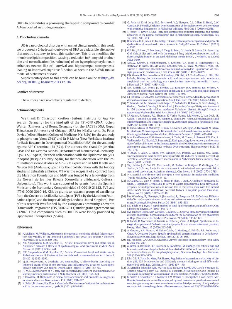
To further investigate the mechanism by which OHDHA offers neu-roprotection against oligomeric-Aβ-mediated toxicity, we carried outmodel membrane-based assays to analyze the binding of different ag-gregated forms of Aβ to lipid-raft-like vesicles composed of Cho andSM. We chose this type of vesicles because lipid-raft membrane do-mainshave beendescribed as the loci of Aβ generation and binding,me-diating Aβ cellular toxicity [42,53,54]. We observed that the binding ofmonomeric Aβ to raft-like bilayers only occurred in the presence ofOHDHA (Table 2A), while the affinity of Aβ oligomers for raft-likemem-branes was lower in the presence of OHDHA (ca. 8 fold reduction:Table 2B). Moreover, the affinity of Aβ fibrils for raft-like membraneswas reduced by ca. 160 fold in the presence of OHDHA (an increase inthe Kd value from 4.76 to 775 μM: Table 2C). These results clearly indi-cate that OHDHA favors the interaction of monomeric Aβwith raft-like
membranes and that OHDHA impairs the interaction of oligomeric/fi-brillar Aβ with membranes. These effects might explain the neuropro-tection provided by OHDHA against oligomeric-Aβ-mediated toxicityin neuron-like cells (see Fig. 4A).
3.4. Evaluation of OHDHA toxicity in zebrafish embryos
The data presented here suggests that OHDHA is a therapeutic can-didate for AD. For this reason, we tested the safety of OHDHA as adrug for human administration by evaluating its lethal, sub-lethal andteratogenic effects in zebrafish embryos. We tested zebrafish embryomortality and viability on exposure to OHDHA at 1, 3, 10, 30 and100 mg/l (2.7, 8.1, 27.3, 81.8 and 273 μM, respectively) for 24, 48 and72 h, as well as measuring its sub-lethal and teratogenic effects. Sub-lethal effects were considered to include the lack of spontaneous move-ment, pigmentation deficits and edema or clots in organs or otherinternal structures, while teratogenic effects included deformation oforgans or internal structures, scoliosis and general growth delay (seeTable 3). OHDHA had only a mild effect on embryo mortality at thehighest concentrations tested (100 mg/l), producing 20% mortality upto 48 h and 35% after a 72-hour exposure (Table 3A). Accordingly,embryo viability was tested at 72 h and negative effects were only pro-duced on exposure to 100 mg/l OHDHA, whereas lower concentrations(1 to 30 mg/l) were correlated with acceptable levels of embryo viabil-ity (between 80 and 100%: Table 3B). Similarly, sub-lethal effects wereonly observed at high concentrations of OHDHA (100 mg/l), and in atime-dependent manner (from 56 to 77%, between 48 and 72 h),whereas no remarkable effects were observed at lower concentrations(Table 3C). It should be noted that in all cell culture experiments per-formed in the present work, OHDHA was used at concentrations
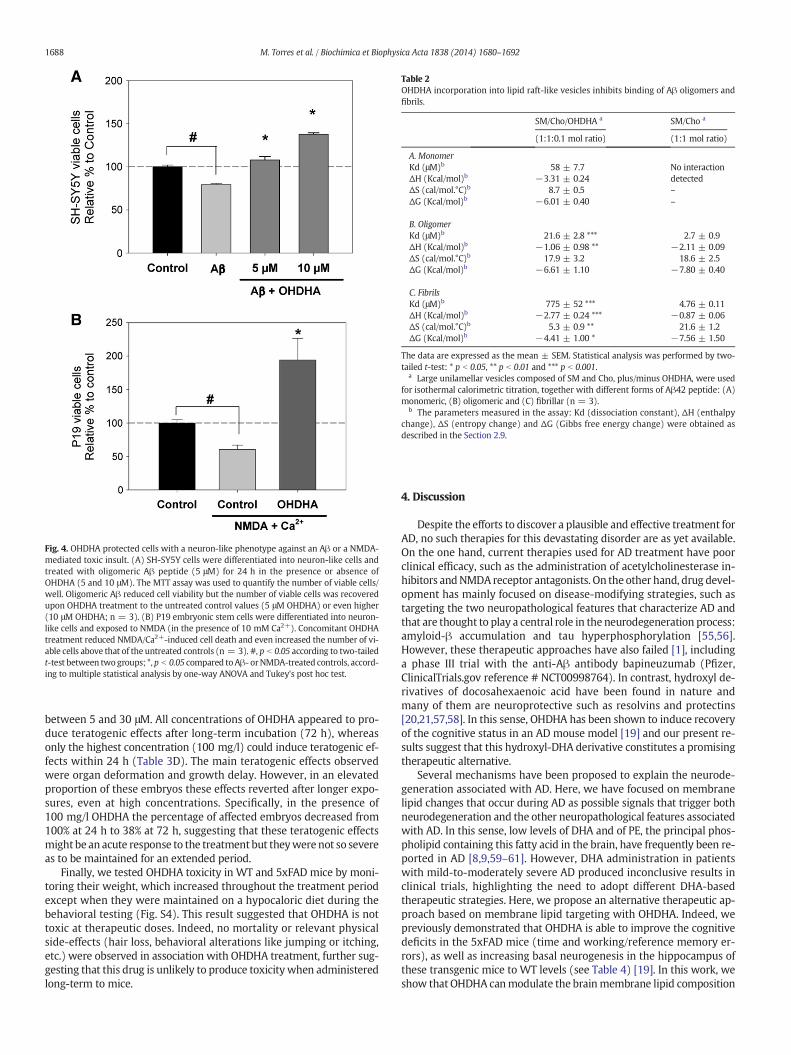
Fig. 4. OHDHA protected cells with a neuron-like phenotype against an Aβ or a NMDA-mediated toxic insult. (A) SH-SY5Y cells were differentiated into neuron-like cells andtreated with oligomeric Aβ peptide (5 μM) for 24 h in the presence or absence ofOHDHA (5 and 10 μM). The MTT assay was used to quantify the number of viable cells/well. Oligomeric Aβ reduced cell viability but the number of viable cells was recoveredupon OHDHA treatment to the untreated control values (5 μM OHDHA) or even higher(10 μM OHDHA; n = 3). (B) P19 embryonic stem cells were differentiated into neuron-like cells and exposed to NMDA (in the presence of 10 mM Ca2+). Concomitant OHDHAtreatment reduced NMDA/Ca2+-induced cell death and even increased the number of vi-able cells above that of the untreated controls (n = 3). #, p b 0.05 according to two-tailedt-test between twogroups; *,p b 0.05 compared toAβ- orNMDA-treated controls, accord-ing to multiple statistical analysis by one-way ANOVA and Tukey's post hoc test.
Table 2OHDHA incorporation into lipid raft-like vesicles inhibits binding of Aβ oligomers andfibrils.
SM/Cho/OHDHA a SM/Cho a
(1:1:0.1 mol ratio) (1:1 mol ratio)
A. MonomerKd (μM)b 58 ± 7.7 No interactionΔH (Kcal/mol)b −3.31 ± 0.24 detectedΔS (cal/mol.°C)b 8.7 ± 0.5 –
ΔG (Kcal/mol)b −6.01 ± 0.40 –
B. OligomerKd (μM)b 21.6 ± 2.8 *** 2.7 ± 0.9ΔH (Kcal/mol)b −1.06 ± 0.98 ** −2.11 ± 0.09ΔS (cal/mol.°C)b 17.9 ± 3.2 18.6 ± 2.5ΔG (Kcal/mol)b −6.61 ± 1.10 −7.80 ± 0.40
C. FibrilsKd (μM)b 775 ± 52 *** 4.76 ± 0.11ΔH (Kcal/mol)b −2.77 ± 0.24 *** −0.87 ± 0.06ΔS (cal/mol.°C)b 5.3 ± 0.9 ** 21.6 ± 1.2ΔG (Kcal/mol)b −4.41 ± 1.00 * −7.56 ± 1.50
The data are expressed as the mean ± SEM. Statistical analysis was performed by two-tailed t-test: * p b 0.05, ** p b 0.01 and *** p b 0.001.
a Large unilamellar vesicles composed of SM and Cho, plus/minus OHDHA, were usedfor isothermal calorimetric titration, together with different forms of Aβ42 peptide: (A)monomeric, (B) oligomeric and (C) fibrillar (n = 3).
b The parameters measured in the assay: Kd (dissociation constant), ΔH (enthalpychange), ΔS (entropy change) and ΔG (Gibbs free energy change) were obtained asdescribed in the Section 2.9.
1688 M. Torres et al. / Biochimica et Biophysica Acta 1838 (2014) 1680–1692
between 5 and 30 μM. All concentrations of OHDHA appeared to pro-duce teratogenic effects after long-term incubation (72 h), whereasonly the highest concentration (100 mg/l) could induce teratogenic ef-fects within 24 h (Table 3D). The main teratogenic effects observedwere organ deformation and growth delay. However, in an elevatedproportion of these embryos these effects reverted after longer expo-sures, even at high concentrations. Specifically, in the presence of100 mg/l OHDHA the percentage of affected embryos decreased from100% at 24 h to 38% at 72 h, suggesting that these teratogenic effectsmight be an acute response to the treatment but theywere not so severeas to be maintained for an extended period.
Finally, we tested OHDHA toxicity in WT and 5xFAD mice by moni-toring their weight, which increased throughout the treatment periodexcept when they were maintained on a hypocaloric diet during thebehavioral testing (Fig. S4). This result suggested that OHDHA is nottoxic at therapeutic doses. Indeed, no mortality or relevant physicalside-effects (hair loss, behavioral alterations like jumping or itching,etc.) were observed in association with OHDHA treatment, further sug-gesting that this drug is unlikely to produce toxicity when administeredlong-term to mice.
4. Discussion
Despite the efforts to discover a plausible and effective treatment forAD, no such therapies for this devastating disorder are as yet available.On the one hand, current therapies used for AD treatment have poorclinical efficacy, such as the administration of acetylcholinesterase in-hibitors andNMDA receptor antagonists. On the other hand, drug devel-opment has mainly focused on disease-modifying strategies, such astargeting the two neuropathological features that characterize AD andthat are thought to play a central role in the neurodegeneration process:amyloid-β accumulation and tau hyperphosphorylation [55,56].However, these therapeutic approaches have also failed [1], includinga phase III trial with the anti-Aβ antibody bapineuzumab (Pfizer,ClinicalTrials.gov reference # NCT00998764). In contrast, hydroxyl de-rivatives of docosahexaenoic acid have been found in nature andmany of them are neuroprotective such as resolvins and protectins[20,21,57,58]. In this sense, OHDHA has been shown to induce recoveryof the cognitive status in an AD mouse model [19] and our present re-sults suggest that this hydroxyl-DHA derivative constitutes a promisingtherapeutic alternative.
Several mechanisms have been proposed to explain the neurode-generation associated with AD. Here, we have focused on membranelipid changes that occur during AD as possible signals that trigger bothneurodegeneration and the other neuropathological features associatedwith AD. In this sense, low levels of DHA and of PE, the principal phos-pholipid containing this fatty acid in the brain, have frequently been re-ported in AD [8,9,59–61]. However, DHA administration in patientswith mild-to-moderately severe AD produced inconclusive results inclinical trials, highlighting the need to adopt different DHA-basedtherapeutic strategies. Here, we propose an alternative therapeutic ap-proach based on membrane lipid targeting with OHDHA. Indeed, wepreviously demonstrated that OHDHA is able to improve the cognitivedeficits in the 5xFAD mice (time and working/reference memory er-rors), as well as increasing basal neurogenesis in the hippocampus ofthese transgenic mice to WT levels (see Table 4) [19]. In this work, weshow that OHDHA canmodulate the brainmembrane lipid composition

Table 3Evaluation of OHDHA toxicity in zebrafish embryos.
OHDHA(mg/ml)
Incubation (h)
24 48 72
A. Embryo mortality1 0 0 03 0 0 010 0 0 030 0 0 10100 20 20 35
B. Embryo viability1 – – 1003 – – 8010 – – 8030 – – 94100 – – 46
C. Sublethal effects1 0 0 03 0 0 010 0 5 530 0 10 0100 0 56 77
D. Teratogenic effects1 0 0 53 0 0 7010 5 5 5030 10 25 100100 100 66 38
The data are expressed as the percentage of embryos that showed any of the parametersstudied: (A) embryomortality, (B) embryo viability, (C) sub-lethal effects: lack of sponta-neous movements, pigmentation deficits and apparition of edema or clots in internalstructures, and (D) teratogenic effects: deformation of internal structures, scoliosis andgeneral growth delay.
1689M. Torres et al. / Biochimica et Biophysica Acta 1838 (2014) 1680–1692
by increasing the concentration of long chain PUFA-containing PE spe-cies, aswell as diminishingAβ generation in N2aSw cells and Aβ burdenin the brain of 5xFAD mice. Likewise, oral administration of OHDHA in-hibits tau protein phosphorylation in the brain of 5xFAD mice and inAβ-treated neuron-like cells, and it enhances neuron-like cell viabilityagainst oligomeric-Aβ and NMDA/Ca2+-mediated toxicity. Additional-ly, OHDHA exhibited little pharmacological toxicity in vivo, andzebrafish embryos showed no remarkable alterations in terms of mor-tality and sublethal effects at the doses used here.
OHDHA induced relevant lipid modifications in the brain of 5xFADmice that were the opposite of those reported for AD. Accordingly, ourlipidomic analysis showed an increase in all PE species (includingdiacyl-PE, lyso-PE and their plasmalogens) and in polyunsaturatedacyl chains (phospholipids containing 5 or more double bonds), similarto those produced by DHA in the mouse brain [62] or in human plasma[63]. Specifically, increases in all diacyl-PE subclasses were observed,
Table 4Improved cognition and neurogenesis in OHDHA-treated-5xFAD mice.
WT 5xFAD
mean ± SEM mean ± SEM
Time (s) c 75.69 ± 4.77 124.4 ± 12.3WME (number of errors) c 2.32 ± 0.32 3.95 ± 0.47RME (number of errors) c 5.88 ± 0.39 8.31 ± 0.64Phospho histone-H3 positive neurons (cells/mm3)d 7431 ± 1918 1970 ± 767.
The statistical analysis was performedwith a two-tailed t-test: * p b 0.05 and ** p b 0.01 after cotreated-5xFAD and 5xFAD control mice. Summary of data extracted from [19].
a Percentage change in 5xFAD control mice was expressed as the mean ± SEM relative to thb The percentage change in 5xFAD + OHDHA was expressed as the mean ± SEM relative tc Time (s), and the number of working and referencememory errors (WME andRME, respect
5xFAD + OHDHA, n = 12).d Neuronal proliferation in the dentate gyrus (hippocampus) was determined by quantif
immunolabeled brain sections (WT, n = 4; 5xFAD n = 7 and 5xFAD + OHDHA, n = 7).
although species containing long (40C) and medium length (36–38C)acyl chains increased markedly with respect to shorter acyl chains(32–34C). It is noteworthy that OHDHA caused the highest increase indiacyl-PE 40:6 in the brain of 5xFAD mice, mainly formed by 22:6(DHA) and 18:0 fatty acids. In addition, significant increases in otherdiacyl-PE subspecies were also evident in OHDHA-treated mice, suchas 40:5 and 38:5, which contain the omega-3 fatty acids 22:5 (DPA) or20:5 (EPA) and that can be generated enzymatically from the 22:6acyl chain [62,64]. In this context, the OHDHA-induced lipid modifica-tions should counteract the lipid deficits associatedwith AD. Indeed, bi-opsies from AD brains were characterized by the loss of diacyl-PEsubspecies (especially, 40:6, 38:6 and 38:4) and low levels of lipids con-taining long acyl chains, along with an increase in species containingshort acyl chains in the human prefrontal cortex, among other lipidalterations [65]. Fatty acid length and their degree of saturation are de-terminant factors influencing membrane structure and functionality[66]. Indeed, by adequately regulating membrane lipid compositioncell membranes may reverse the pathological changes in neurons, asmay be the case in AD [22]. On the other hand, no remarkable differ-ences in lipids were found in the brain of the 5xFAD ADmice comparedto WT controls. However, other brain membrane lipid alterations havebeen detected in different AD models [65,67] and there have beenmany discrepancies between post-mortem AD tissue and AD modelbrains, implying that AD models are likely not to faithfully reproduceall the lipid alterations associated with AD [65,67].
We also demonstrated that OHDHA affects Aβ production in cul-tured cells stably expressing Swedish APP and in the brain of 5xFADmice, consistent with previous observations for DHA in cultured cellsand in ADmodels [11,12,68]. In this context, membrane lipids canmod-ulate the amyloidogenic route by acting on BACE-1 and the γ-secretasecomplex [47,48,69]. The activity of these enzymes increases in raft-likedomains that are enriched in Cho and SM. PE, OHDHA and PUFAs canchange the membrane from a liquid ordered to a liquid disorderedstate, which does not support amyloidogenesis [44,70,71], therefore,cell membranes in the mouse brain or cultured cells that are exposedto OHDHA should undergo such changes, dampening the activity ofthe secretases implicated in Aβ generation. In addition, we demonstrat-ed that OHDHA induces strong clustering of the APP, PS1 and BACE1proteins into cellular vesicles/organelles. Although we have yet todefine the nature of these vesicles, these results suggest that OHDHA-induced membrane lipid changes might regulate endolysosomalproteolysis [72], and this mechanism of action may also contribute tothe downregulation of Aβ after OHDHA treatment. Interestingly, au-tophagy/lysosomal proteolysis is impaired in AD and AD models, andits stimulation has been proposed as a potentially effective therapeuticstrategy to combat AD [37,73–75]. However, our confocal microscopyassays showed weak co-localization between APP and LC3, or APP andLamp1, in the vesicles observed after treatment with OHDHA inN2aSw cells. Consequently, these results did not allow concluding the
5xFAD + OHDHA
% changea mean ± SEM % changeb
8 64.35 ± 9.95 ** 105.4 ± 10.34 −15.27 ± 9.81 NS70.25 ± 11.89 * 2.21 ± 0.40 −44.05 ± 18.09 §41.32 ± 7.70 * 6.07 ± 0.73 −26.94 ± 12.02 §
1 −73.48 ± 38.93 * 5374 ± 939.7 272.79 ± 17.47 §
mparison betweenWT and 5xFAD; NS: not significant and § p b 0.05 comparing OHDHA-
e WT as a reference.o 5xFAD mice.ively)were determined during the radial armmaze test (WT, n = 10; 5xFAD, n = 11 and
ying the number of phospho-histone H3-positive neurons in the granullar cell layer of

1690 M. Torres et al. / Biochimica et Biophysica Acta 1838 (2014) 1680–1692
possible implication of the autophagy/lysosomal route as a molecularmechanism mediating downregulation of Aβ levels in OHDHA-treatedN2a cells. Therefore, further analysis would be necessary to confirmthis hypothesis and to identify the origin of these vesicles.
In addition, we demonstrated that OHDHA treatment protectsneuron-like cells against the AD-related toxicitymediated by oligomericAβ or NMDA, and it prevented Aβ-induced tau hyperphosphorylation.We provided here evidence that these phenomena may be explainedby the modification of membrane lipid composition and structure byOHDHA, for example the weaker affinity of oligomeric and fibrillar Aβfor raft-like vesicles containing OHDHA observed in the ITC assays.Moreover, OHDHA increases the relative abundance of polyunsaturatedlipid species, which might inhibit the interaction of Aβ oligomers withlipid raft-associated protein receptors, thereby inhibiting their cytotoxiccell signaling [76]. In this context, ITC assays also demonstrated thatmonomeric Aβ exclusively interacted with raft-like vesicles in thepresence of OHDHA, suggesting that monomeric Aβ preferentiallybinds to liquid disordered-prone structures [42]. Interestingly, thepersistence of monomeric Aβ is associated with neuroprotection,supporting neuronal survival under conditions of trophic deprivationand combating excitotoxicity. Furthermore, monomeric Aβ enhancesglycogen synthase kinase-3β (GSK-3β) phosphorylation, suggestingthat monomeric Aβ may downregulate tau phosphorylation [77].Therefore, these results suggest that OHDHApromotes neuronal surviv-al by favoring the membrane interactions of monomeric Aβ but notthose of oligomeric or fibrillar Aβ (the presence of OHDHA induces ca.170-fold reductions in the affinity of fibrils for lipid rafts). Nevertheless,it cannot be ruled out that OHDHA may regulate APP processing by al-tering themembrane lipid composition, limiting the ensuing alterationsoriginated by Aβ generation, such as Aβ oligomerization and tauhyperphosphorylation. Moreover, NMDA-mediated Ca2+ excitotoxicityalso leads to neurodegeneration in AD [78,79] and this may also be re-versed by OHDHA. However, the molecular mechanism connecting
Fig. 5. Postulated mechanism of action for OHDHA. Summary of the proposed molecular mebrain membranes in PE, especially those carrying DHA and other long PUFAs. These lipid chandisordered-prone structures, and they might reverse the cellular signaling associated withhyperphosphorylation; and (ii) decreasing neuron vulnerability to extracellular toxic agents suduced proliferation of both neuron-like cells in vitro and neuron stem cells in the mouse brain.that may be associated with the improved cognitive capabilities seen in the 5xFAD mouse mod
OHDHA-mediated membrane lipid changes and NMDA-receptortrafficking at synaptic terminals remains unknown.
Interestingly, exposingdifferentiatedneuron-like cells toOHDHA in-creased the proportion of viable cells in culture in the presence of a toxicinsult (oligomeric Aβ or NMDA) even above that of control neuron-likecells (not submitted to toxic insult), suggesting that OHDHA mightinduce mitotic division of neuronal stem cells. Indeed, OHDHA in-duced neuronal stem cell proliferation in the hippocampus of5xFAD mice, as reflected by a remarkable increase in the number ofphospho-histone-H3-positive neurons (see Table 4) [19], althoughthe molecular mechanisms associated with this phenomenon remainunknown. In summary, OHDHAnot only has a neuroprotective influenceon neuron-like cells but also, it exerts an apparent neuroregenerative ef-fect in neuron-like cells in culture, and in the brain of 5xFADmice, both ofwhichmight explain the enhanced cell survival, and the improved learn-ing and memory capabilities of 5xFAD mice administered OHDHA.
AD is a complex neurodegenerative process that has been related tolipid diet and to metabolic diseases. Indeed, this pathology is probablydriven by lipid alterations in neuronal membranes that lead to defectiveAPP processing, altered cell signaling and the ensuing events character-istic of this pathology. In line with this, alterations in membrane lipids(e.g., PE and DHA) of AD patients have been described previously [65],although this fact has still not been taken into consideration whendesigning other therapeutic strategies. In the present work, we haveinvestigated the molecular basis underlying the effects of OHDHA onAD-associated cognitive loss. This “membrane lipid therapy”-baseddrug is a molecule designed to target lipid membranes and regulatesthe membrane-associated protein activity, which when altered maycause neurodegeneration and eventually, AD. The beneficial effects ofOHDHA include the restoration of cognitive scores in the 5xFADmodel of AD, the protection of cells against external AD-associatedtoxic agents, such as oligomeric Aβ, and a reduction in the produc-tion of Aβ and tau hyperphosphorylation (Fig. 5). In conclusion,
chanisms underlying the effects of OHDHA on neuronal membranes. OHDHA enrichesges may influence the structure of the cell membrane, leading to the formation of liquid-AD by: (i) downregulating amyloidogenic processing and Aβ-dependent tau protein
ch as oligomeric Aβ and NMDA/Ca2+-mediated excitotoxicity. In addition, OHDHA also in-Together, this evidence supports a neuroprotective and neuroregenerative role of OHDHAel.
1691M. Torres et al. / Biochimica et Biophysica Acta 1838 (2014) 1680–1692
OHDHA constitutes a promising therapeutic compound to combatAD-associated neurodegeneration.
5. Concluding remarks
AD is a neurological disorder with unmet clinical needs. In this work,we proposed a 2-hydroxyl-derivative of DHA as a plausible alternativetherapeutic strategy to treat this pathology. This drug modifies themembrane lipid composition, causing a reduction in β-amyloid produc-tion and normalization (i.e. reduction) of tau hyperphosphorylation, itenhances neuron-like cell survival and hippocampal neurogenesis,leading to improved cognitive function, as seen in the 5xFAD mousemodel of Alzheimer's disease.
Supplementary data to this article can be found online at http://dx.doi.org/10.1016/j.bbamem.2013.12.016.
Conflict of interest
The authors have no conflicts of interest to declare.
Acknowledgments
We thank Dr Christoph Kaether (Leibniz Institute for Age Re-search; Germany) for the kind gift of the PS1-GFP cDNA, JochenWalter (University of Bonn; Germany) for the BACE1 cDNA, GopalThinakaran (University of Chicago; USA) for N2aSw cells, Dr. PeterDavies (Albert Einstein College of Medicine, NY; USA) for the antibodyto phospho-tau (clone CP13) and Dr P. Mehta (New York State Institutefor Basic Research in Developmental Disabilities; USA) for the antibodyagainst APP C-terminal (R1(57)). The authors also thank Dr. JosefinaCasas and Dr. Gemma Fabriàs (Department of Biomedicinal Chemistry,IQAC, CSIC; Spain) for their collaboration with the lipidomic analysis,Innoprot (Basque Country; Spain) for their collaboration with the im-munofluorescence studies of APP-GFP expression in MDCK cells andNeuron BPh (Andalusia; Spain) for their collaboration with the toxicitystudies in zebrafish embryos. MT was the recipient of a contract fromthe Marathon Foundation and MAF was funded by a fellowship fromthe Govern de les Illes Balears (Conselleria d'Educació, Cultura iUniversitats). This work was supported by grants from the SpanishMinisterio de Economía y Competitividad (BIO2010-21132, PVE andIPT-010000-2010-16, XB), by grants to research groups of excellencefrom theGovern de les Illes Balears, Spain (PVE), by theMarathon Foun-dation (Spain) and the Imperial College London (United Kingdom). Partof this research was funded by the European Community's SeventhFramework Programme (FP7/2007-2013) under grant agreement No.212043. Lipid compounds such as OHDHA were kindly provided byLipopharma Therapeutics (Spain).
References
[1] K. Mullane, M. Williams, Alzheimer's therapeutics: continued clinical failures ques-tion the validity of the amyloid hypothesis-but what lies beyond? Biochem.Pharmacol. 85 (2013) 289–305.
[2] N.E. Shepardson, G.M. Shankar, D.J. Selkoe, Cholesterol level and statin use inAlzheimer disease: I. Review of epidemiological and preclinical studies, Arch.Neurol. 68 (2011) 1239–1244.
[3] N.E. Shepardson, G.M. Shankar, D.J. Selkoe, Cholesterol level and statin use inAlzheimer disease: II. Review of human trials and recommendations, Arch. Neurol.68 (2011) 1385–1392.
[4] J.J.M. Hoozemans, R. Veerhuis, J.M. Rozemuller, P. Eikelenboom, Soothing theinflamed brain: effect of non-steroidal anti-inflammatory drugs on Alzheimer'sdisease pathology, CNS Neurol. Disord. Drug Targets 10 (2011) 57–67.
[5] H.-M. Su, Mechanisms of n-3 fatty acid-mediated development and maintenance oflearning memory performance, J. Nutr. Biochem. 21 (2010) 364–373.
[6] E. Kawakita, M. Hashimoto, O. Shido, Docosahexaenoic acid promotes neurogenesisin vitro and in vivo, Neuroscience 139 (2006) 991–997.
[7] N. Salem, B. Litman, H.Y. Kim, K. Gawrisch,Mechanisms of action of docosahexaenoicacid in the nervous system, Lipids 36 (2001) 945–959.
[8] G. Astarita, K.-M. Jung, N.C. Berchtold, V.Q. Nguyen, D.L. Gillen, E. Head, C.W.Cotman, D. Piomelli, Deficient liver biosynthesis of docosahexaenoic acid correlateswith cognitive impairment in Alzheimer's disease, PLoS One 5 (2010) e12538.
[9] T. Fraser, H. Tayler, S. Love, Fatty acid composition of frontal, temporal and parietalneocortex in the normal human brain and in Alzheimer's disease, Neurochem. Res.35 (2010) 503–513.
[10] D. Arsenault, C. Julien, C. Tremblay, F. Calon, DHA improves cognition and preventsdysfunction of entorhinal cortex neurons in 3xTg-AD mice, PLoS One 6 (2011)e17397.
[11] G.P. Lim, F. Calon, T. Morihara, F. Yang, B. Teter, O. Ubeda, N. Salem, S.A. Frautschy,G.M. Cole, A diet enriched with the omega-3 fatty acid docosahexaenoic acid re-duces amyloid burden in an aged Alzheimer mouse model, J. Neurosci. 25 (2005)3032–3040.
[12] M.O.W. Grimm, J. Kuchenbecker, S. Grösgen, V.K. Burg, B. Hundsdörfer, T.L.Rothhaar, P. Friess, M.C. de Wilde, L.M. Broersen, B. Penke, M. Péter, L. Vígh, H.S.Grimm, T. Hartmann, Docosahexaenoic acid reduces amyloid β production via mul-tiple pleiotropic mechanisms, J. Biol. Chem. 286 (2011) 14028–14039.
[13] K.N. Green, H. Martinez-Coria, H. Khashwji, E.B. Hall, K.A. Yurko-Mauro, L. Ellis, F.M.LaFerla, Dietary docosahexaenoic acid and docosapentaenoic acid ameliorateamyloid-β and tau pathology via a mechanism involving presenilin 1 levels,J. Neurosci. 27 (2007) 4385–4395.
[14] M.C. Morris, D.A. Evans, J.L. Bienias, C.C. Tangney, D.A. Bennett, R.S. Wilson, N.Aggarwal, J. Schneider, Consumption of fish and n-3 fatty acids and risk of incidentAlzheimer disease, Arch. Neurol. 60 (2003) 940–946.
[15] E.J. Johnson, E.J. Schaefer, Potential role of dietary n-3 fatty acids in the prevention ofdementia and macular degeneration, Am. J. Clin. Nutr. 83 (2006) 1494S–1498S.
[16] Y. Freund-Levi, M. Eriksdotter-Jönhagen, T. Cederholm, H. Basun, G. Faxén-Irving, A.Garlind, I. Vedin, B. Vessby, L.O. Wahlund, J. Palmblad, Omega-3 fatty acid treatmentin 174 patients with mild to moderate Alzheimer disease: OmegAD study: arandomized double-blind trial, Arch. Neurol. 63 (2006) 1402–1408.
[17] J.F. Quinn, R. Raman, R.G. Thomas, K. Yurko-Mauro, E.B. Nelson, C. Van Dyck, J.E.Galvin, J. Emond, C.R. Jack, M. Weiner, L. Shinto, P.S. Aisen, Docosahexaenoic acidsupplementation and cognitive decline in Alzheimer disease: a randomized trial,JAMA 304 (2010) 1903–1911.
[18] K. Yurko-Mauro, D. McCarthy, D. Rom, E.B. Nelson, A.S. Ryan, A. Blackwell, N. Salem,M. Stedman, M. Investigators, Beneficial effects of docosahexaenoic acid on cogni-tion in age-related cognitive decline, Alzheimers Dement. 6 (2010) 456–464.
[19] M.A. Fiol-Deroque, R. Gutierrez-Lanza, S. Terés, M. Torres, P. Barceló, R.V. Rial, A.Verkhratsky, P.V. Escribá, X. Busquets, J.J. Rodríguez, Cognitive recovery and restora-tion of cell proliferation in the dentate gyrus in the 5XFAD transgenic mice model ofAlzheimer's disease following 2-hydroxy-DHA treatment, Biogerontology 14 (2013)763–775.
[20] Y. Zhao, F. Calon, C. Julien, J.W. Winkler, N.A. Petasis, W.J. Lukiw, N.G. Bazan,Docosahexaenoic acid-derived neuroprotectin D1 induces neuronal survival viasecretase- and PPARγ-mediated mechanisms in Alzheimer's disease models, PLoSOne 6 (2011) e15816.
[21] W.J. Lukiw, J.-G. Cui, V.L. Marcheselli, M. Bodker, A. Botkjaer, K. Gotlinger, C.N.Serhan, N.G. Bazan, A role for docosahexaenoic acid-derived neuroprotectin D1 inneural cell survival and Alzheimer disease, J. Clin. Invest. 115 (2005) 2774–2783.
[22] P.V. Escribá, Membrane-lipid therapy: a new approach in molecular medicine,Trends Mol. Med. 12 (2006) 34–43.
[23] H. Oakley, S.L. Cole, S. Logan, E. Maus, P. Shao, J. Craft, A. Guillozet-Bongaarts, M.Ohno, J. Disterhoft, L. Van Eldik, R. Berry, R. Vassar, Intraneuronal beta-amyloid ag-gregates, neurodegeneration, and neuron loss in transgenic mice with five familialAlzheimer's disease mutations: potential factors in amyloid plaque formation,J. Neurosci. 26 (2006) 10129–10140.
[24] B.A. Wirsching, R.J. Beninger, K. Jhamandas, R.J. Boegman, S.R. El-Defrawy, Differen-tial effects of scopolamine on working and reference memory of rats in the radialmaze, Pharmacol. Biochem. Behav. 20 (1984) 659–662.
[25] E.G. Bligh, W.J. Dyer, A rapid method of total lipid extraction and purification, Can.J. Biochem. Physiol. 37 (1959) 911–917.
[26] J.M. Jiménez-López, M.P. Carrasco, C. Marco, J.L. Segovia, Hexadecylphosphocholinedisrupts cholesterol homeostasis and induces the accumulation of free cholesterolin HepG2 tumour cells, Biochem. Pharmacol. 71 (2006) 1114–1121.
[27] D. Canals, D. Mormeneo, G. Fabriàs, A. Llebaria, J. Casas, A. Delgado, Synthesis and bi-ological properties of Pachastrissamine (jaspine B) and diastereoisomeric jaspines,Bioorg. Med. Chem. 17 (2009) 235–241.
[28] A. Garanto, N.A. Mandal, M. Egido-Gabás, G. Marfany, G. Fabriàs, R.E. Anderson, J.Casas, R. Gonzàlez-Duarte, Specific sphingolipid content decrease in Cerkl knock-down mouse retinas, Exp. Eye Res. 110 (2013) 96–106.
[29] R.E. Kingston, C.A. Chen, H. Okayama, Current Protocols in Immunology, John Wiley& Sons, Inc., 2001
[30] A. Jämsä, K. Hasslund, R.F. Cowburn, A. Bäckström, M. Vasänge, The retinoic acid andbrain-derived neurotrophic factor differentiated SH-SY5Y cell line as a model forAlzheimer's disease-like tau phosphorylation, Biochem. Biophys. Res. Commun.319 (2004) 993–1000.
[31] R.M. Gill, R. Slack, M. Kiess, P.A. Hamel, Regulation of expression and activity of dis-tinct pRB, E2F, D-type cyclin, and CKI family members during terminal differentia-tion of P19 cells, Exp. Cell Res. 244 (1998) 157–170.
[32] A. Marcilla-Etxenike, M.L. Martín, M.A. Noguera-Salvà, J.M. García-Verdugo, M.Soriano-Navarro, I. Dey, P.V. Escribá, X. Busquets, 2-Hydroxyoleic acid induces ERstress and autophagy in various human glioma cell lines, PLoS One 7 (2012) e48235.
[33] M. Sastre, I. Dewachter, G.E. Landreth, T.M.Willson, T. Klockgether, F. van Leuven,M.T.Heneka, Nonsteroidal anti-inflammatory drugs and peroxisome proliferator-activatedreceptor-gamma agonists modulate immunostimulated processing of amyloid pre-cursor protein through regulation of beta-secretase, J. Neurosci. 23 (2003) 9796–9804.

1692 M. Torres et al. / Biochimica et Biophysica Acta 1838 (2014) 1680–1692
[34] L. Katsouri, C. Parr, N. Bogdanovic, M. Willem, M. Sastre, PPARγ co-activator-1α(PGC-1α) reduces amyloid-β generation through a PPARγ-dependent mechanism,J. Alzheimers Dis. 25 (2011) 151–162.
[35] S. Taylor, M. Wakem, G. Dijkman, M. Alsarraj, M. Nguyen, A practical approach toRT-qPCR-Publishing data that conform to the MIQE guidelines, Methods 50(2010) S1–S5.
[36] B. Ramos, D. Baglietto-Vargas, J.C. del Rio, I. Moreno-Gonzalez, C. Santa-Maria, S.Jimenez, C. Caballero, J.F. Lopez-Tellez, Z.U. Khan, D. Ruano, A. Gutierrez, J.Vitorica, Early neuropathology of somatostatin/NPY GABAergic cells in the hippo-campus of a PS1xAPP transgenic model of Alzheimer's disease, Neurobiol. Aging27 (2006) 1658–1672.
[37] M. Torres, S. Jiménez, R. Sánchez-Varo, V. Navarro, L. Trujillo-Estrada, E.Sánchez-Mejias, I. Carmona, J.C. Davila, M. Vizuete, A. Gutiérrez, J. Vitorica, Defectivelysosomal proteolysis and axonal transport are early pathogenic events that worsenwith age leading to increased APP metabolism and synaptic Abeta in transgenicAPP/PS1 hippocampus, Mol. Neurodegener. 7 (2012) 59.
[38] M. Gobbi, F. Re, M. Canovi, M. Beeg, M. Gregori, S. Sesana, S. Sonnino, D. Brogioli, C.Musicanti, P. Gasco, M. Salmona, M.E. Masserini, Lipid-based nanoparticles with highbinding affinity for amyloid-beta1-42 peptide, Biomaterials 31 (2010) 6519–6529.
[39] L.D. Mayer, M.J. Hope, P.R. Cullis, Vesicles of variable sizes produced by a rapidextrusion procedure, Biochim. Biophys. Acta 858 (1986) 161–168.
[40] M.B. Ruiz-Argüello, G. Basáñez, F.M. Goñi, A. Alonso, Different effects ofenzyme-generated ceramides and diacylglycerols in phospholipid membrane fu-sion and leakage, J. Biol. Chem. 271 (1996) 26616–26621.
[41] C. Arnulphi, J. Sot,M. García-Pacios, J.-L.R. Arrondo, A. Alonso, F.M.Goñi, TritonX-100partitioning into sphingomyelin bilayers at subsolubilizing detergent concentra-tions: effect of lipid phase and a comparison with dipalmitoylphosphatidylcholine,Biophys. J. 93 (2007) 3504–3514.
[42] H. Ahyayauch, M. Raab, J.V. Busto, N. Andraka, J.-L.R. Arrondo, M. Masserini, I.Tvaroska, F.M. Goñi, Binding of β-amyloid (1–42) peptide to negatively chargedphospholipid membranes in the liquid-ordered state: modeling and experimentalstudies, Biophys. J. 103 (2012) 453–463.
[43] L. Yang, N.Y. Ho, R. Alshut, J. Legradi, C. Weiss, M. Reischl, R. Mikut, U. Liebel, F.Müller, U. Strähle, Zebrafish embryos as models for embryotoxic and teratologicaleffects of chemicals, Reprod. Toxicol. 28 (2009) 245–253.
[44] M. Ibarguren, D.J. López, J.A. Encinar, J.M. González-Ros, X. Busquets, P.V. Escribá,Partitioning of liquid-ordered/liquid-disordered membrane microdomains inducedby the fluidifying effect of 2-hydroxylated fatty acid derivatives, Biochim. Biophys.Acta 1828 (2013) 2553–2563.
[45] C.Y. Shao, S.S.Mirra, H.B.R. Sait, T.C. Sacktor, E.M. Sigurdsson, Postsynaptic degenerationas revealed by PSD-95 reduction occurs after advanced Aβ and tau pathology in trans-genic mouse models of Alzheimer's disease, Acta Neuropathol. 122 (2011) 285–292.
[46] S. Jawhar, A. Trawicka, C. Jenneckens, T.A. Bayer, O. Wirths, Motor deficits, neuronloss, and reduced anxiety coinciding with axonal degeneration and intraneuronalAβ aggregation in the 5XFAD mouse model of Alzheimer's disease, Neurobiol.Aging 33 (2012) e129–140(196).
[47] P. Osenkowski, W. Ye, R. Wang, M.S. Wolfe, D.J. Selkoe, Direct and potent regulationof gamma-secretase by its lipid microenvironment, J. Biol. Chem. 283 (2008)22529–22540.
[48] L. Kalvodova, N. Kahya, P. Schwille, R. Ehehalt, P. Verkade, D. Drechsel, K. Simons,Lipids as modulators of proteolytic activity of BACE: involvement of cholesterol,glycosphingolipids, and anionic phospholipids in vitro, J. Biol. Chem. 280 (2005)36815–36823.
[49] Z. Amtul, M. Keet, L. Wang, P. Merrifield, D. Westaway, R.F. Rozmahel, DHA supple-mented in peptamen diet offers no advantage in pathways to amyloidosis: is it timeto evaluate composite lipid diet? PLoS One 6 (2011) e24094.
[50] B. De Strooper, Aph-1, Pen-2, and Nicastrin with Presenilin generate an activegamma-Secretase complex, Neuron 38 (2003) 9–12.
[51] S.L. Cole, R. Vassar, BACE1 structure and function in health and Alzheimer's disease,Curr. Alzheimer Res. 5 (2008) 100–120.
[52] C. Haass, D.J. Selkoe, Soluble protein oligomers in neurodegeneration: lessons fromthe Alzheimer's amyloid beta-peptide, Nat. Rev. Mol. Cell Biol. 8 (2007) 101–112.
[53] R. Williamson, A. Usardi, D.P. Hanger, B.H. Anderton, Membrane-bound β-amyloidoligomers are recruited into lipid rafts by a fyn-dependent mechanism, FASEB J.22 (2008) 1552–1559.
[54] T. Kawarabayashi, M. Shoji, L.H. Younkin, L. Wen-Lang, D.W. Dickson, T. Murakami,E. Matsubara, K. Abe, K.H. Ashe, S.G. Younkin, Dimeric amyloid β protein rapidlyaccumulates in lipid rafts followed by apolipoprotein E and phosphorylated tau ac-cumulation in the Tg2576 mouse model of Alzheimer's disease, J. Neurosci. 24(2004) 3801–3809.
[55] B. De Strooper, R. Vassar, T. Golde, The secretases: enzymes with therapeutic poten-tial in Alzheimer disease, Nat. Rev. Neurol. 6 (2010) 99–107.
[56] C. Gao, C. Hölscher, Y. Liu, L. Li, GSK3: a key target for the development of noveltreatments for type 2 diabetes mellitus and Alzheimer disease, Rev. Neurosci. 23(2012) 1–11.
[57] B.D. Levy, Resolvins and protectins: natural pharmacophores for resolution biology,Prostaglandins Leukot. Essent. Fatty Acids 82 (2010) 327–332.
[58] K.H. Weylandt, C.-Y. Chiu, B. Gomolka, S.F. Waechter, B. Wiedenmann,Omega-3 fatty acids and their lipid mediators: towards an understanding ofresolvin and protectin formation, Prostaglandins Other Lipid Mediat. 97(2012) 73–82.
[59] M. Söderberg, C. Edlund, K. Kristensson, G. Dallner, Fatty acid composition of brainphospholipids in aging and in Alzheimer's disease, Lipids 26 (1991) 421–425.
[60] Z. Guan, Y. Wang, N.J. Cairns, P.L. Lantos, G. Dallner, P.J. Sindelar, Decreaseand structural modifications of phosphatidylethanolamine plasmalogen inthe brain with Alzheimer disease, J. Neuropathol. Exp. Neurol. 58 (1999)740–747.
[61] M.R. Prasad, M.A. Lovell, M. Yatin, H. Dhillon, W.R. Markesbery, Regional mem-brane phospholipid alterations in Alzheimer's disease, Neurochem. Res. 23(1998) 81–88.
[62] J.T. Wood, J.S. Williams, L. Pandarinathan, D.R. Janero, C.J. Lammi-Keefe, A.Makriyannis, Dietary docosahexaenoic acid supplementation alters select physio-logical endocannabinoid-system metabolites in brain and plasma, J. Lipid Res. 51(2010) 1416–1423.
[63] I. Ottestad, S. Hassani, G.I. Borge, A. Kohler, G. Vogt, T. Hyötyläinen, M. Orešič, K.W.Brønner, K.B. Holven, S.M. Ulven, M.C.W. Myhrstad, Fish oil supplementation altersthe plasma lipidomic profile and increases long-chain PUFAs of phospholipids andtriglycerides in healthy subjects, PLoS One 7 (2012) e42550.
[64] M. Grønn, E. Christensen, T.A. Hagve, B.O. Christophersen, Peroxisomalretroconversion of docosahexaenoic acid (22:6(n-3)) to eicosapentaenoic acid(20:5(n-3)) studied in isolated rat liver cells, Biochim. Biophys. Acta 1081 (1991)85–91.
[65] R.B. Chan, T.G. Oliveira, E.P. Cortes, L.S. Honig, K.E. Duff, S.A. Small, M.R. Wenk, G.Shui, G. Di Paolo, Comparative lipidomic analysis of mouse and human brain withAlzheimer disease, J. Biol. Chem. 287 (2012) 2678–2688.
[66] A.A. Spector, M.A. Yorek, Membrane lipid composition and cellular function, J. LipidRes. 26 (1985) 1015–1035.
[67] Y. Tajima, M. Ishikawa, K. Maekawa, M. Murayama, Y. Senoo, T. Nishimaki-Mogami, H.Nakanishi, K. Ikeda, M. Arita, R. Taguchi, A. Okuno, R. Mikawa, S. Niida, O. Takikawa, Y.Saito, Lipidomic analysis of brain tissues and plasma in amousemodel expressingmu-tated human amyloid precursor protein/tau for Alzheimer's disease, Lipids Health Dis.12 (2013) 68.
[68] C. Sahlin, F.E. Pettersson, L.N.G. Nilsson, L. Lannfelt, A.-S. Johansson, Docosahexaenoicacid stimulates non-amyloidogenic APP processing resulting in reduced Abeta levelsin cellular models of Alzheimer's disease, Eur. J. Neurosci. 26 (2007) 882–889.
[69] O. Holmes, S. Paturi, W. Ye, M.S. Wolfe, D.J. Selkoe, Effects of membrane lipids onthe activity and processivity of purified γ-secretase, Biochemistry 51 (2012)3565–3575.
[70] S.R. Shaikh, Biophysical and biochemical mechanisms by which dietary N-3 polyun-saturated fatty acids from fish oil disrupt membrane lipid rafts, J. Nutr. Biochem. 23(2012) 101–105.
[71] K.S. Vetrivel, G. Thinakaran, Membrane rafts in Alzheimer's disease beta-amyloidproduction, Biochim. Biophys. Acta 1801 (2010) 860–867.
[72] H. Schulze, T. Kolter, K. Sandhoff, Principles of lysosomal membrane degradation:cellular topology and biochemistry of lysosomal lipid degradation, Biochim.Biophys. Acta 1793 (2009) 674–683.
[73] R.A. Nixon, J. Wegiel, A. Kumar,W.H. Yu, C. Peterhoff, A. Cataldo, A.M. Cuervo, Exten-sive involvement of autophagy in Alzheimer disease: an immuno-electron micros-copy study, J. Neuropathol. Exp. Neurol. 64 (2005) 113–122.
[74] R. Sanchez-Varo, L. Trujillo-Estrada, E. Sanchez-Mejias, M. Torres, D.Baglietto-Vargas, I. Moreno-Gonzalez, V. De Castro, S. Jimenez, D. Ruano, M.Vizuete, J.C. Davila, J.M. Garcia-Verdugo, A.J. Jimenez, J. Vitorica, A. Gutierrez,Abnormal accumulation of autophagic vesicles correlates with axonal and syn-aptic pathology in young Alzheimer's mice hippocampus, Acta Neuropathol.123 (2012) 53–70.
[75] V. Schaeffer, I. Lavenir, S. Ozcelik, M. Tolnay, D.T. Winkler, M. Goedert, Stimulation ofautophagy reduces neurodegeneration in a mouse model of human tauopathy,Brain 135 (2012) 2169–2177.
[76] J.V. Rushworth, H.H. Griffiths, N.T. Watt, N.M. Hooper, Prion protein-mediated toxic-ity of amyloid-β oligomers requires lipid rafts and the transmembrane LRP1, J. Biol.Chem. 288 (2013) 8935–8951.
[77] M.L. Giuffrida, F. Caraci, B. Pignataro, S. Cataldo, P. De Bona, V. Bruno, G. Molinaro, G.Pappalardo, A. Messina, A. Palmigiano, D. Garozzo, F. Nicoletti, E. Rizzarelli, A.Copani, Beta-amyloid monomers are neuroprotective, J. Neurosci. 29 (2009)10582–10587.
[78] R. Resende, C. Pereira, P. Agostinho, A.P. Vieira, J.O. Malva, C.R. Oliveira, Susceptibilityof hippocampal neurons to Abeta peptide toxicity is associated with perturbation ofCa2+ homeostasis, Brain Res. 1143 (2007) 11–21.
[79] L. Texidó, M. Martín-Satué, E. Alberdi, C. Solsona, C. Matute, Amyloid β peptideoligomers directly activate NMDA receptors, Cell Calcium 49 (2011) 184–190.