membranes - 1 file download
TRANSCRIPT

MEMBRANES:STRUCTURE & TRANSPORT
V.S.RAVIKIRAN, MSc.

V.S.RAVIKIRAN, MSc.,
Department of Biochemistry,
ASRAM Medical college,
Eluru-534005.AP, India.

MEMBRANES:STRUCTURE & TRANSPORT

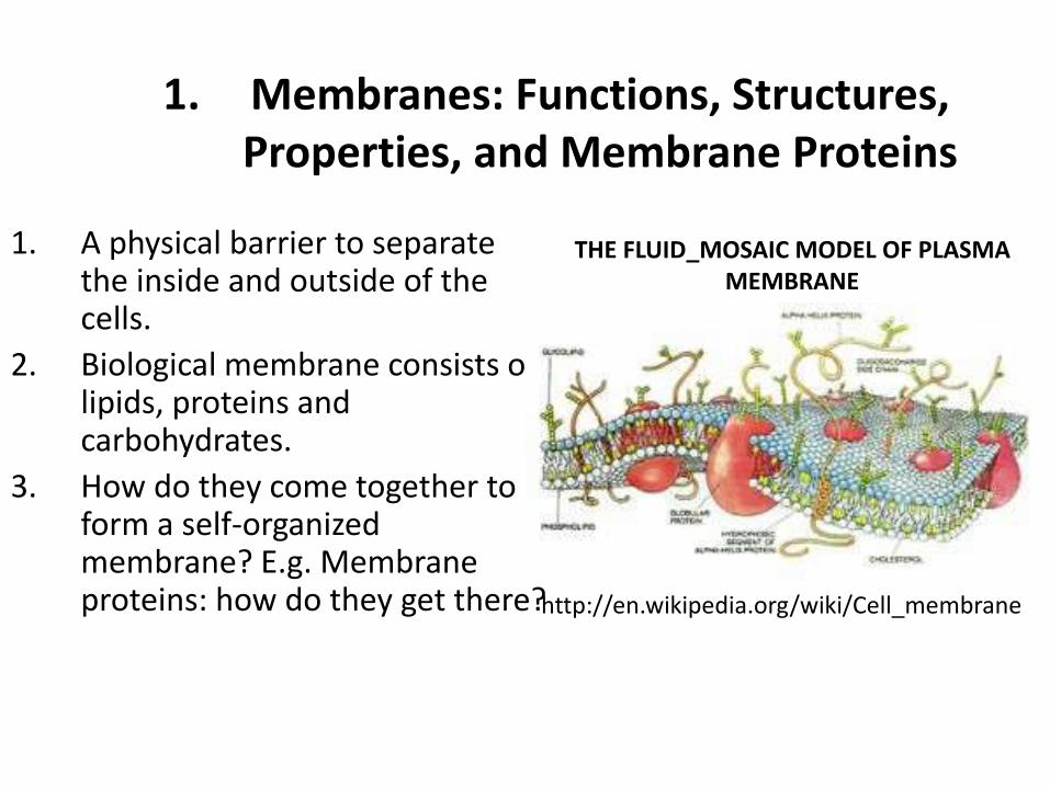
1. A physical barrier to separate the inside and outside of the cells.
2. Biological membrane consists of lipids, proteins and carbohydrates.
3. How do they come together to form a self-organized membrane? E.g. Membrane proteins: how do they get there?
1. Membranes: Functions, Structures, Properties, and Membrane Proteins
http://en.wikipedia.org/wiki/Cell_membrane
THE FLUID_MOSAIC MODEL OF PLASMA MEMBRANE
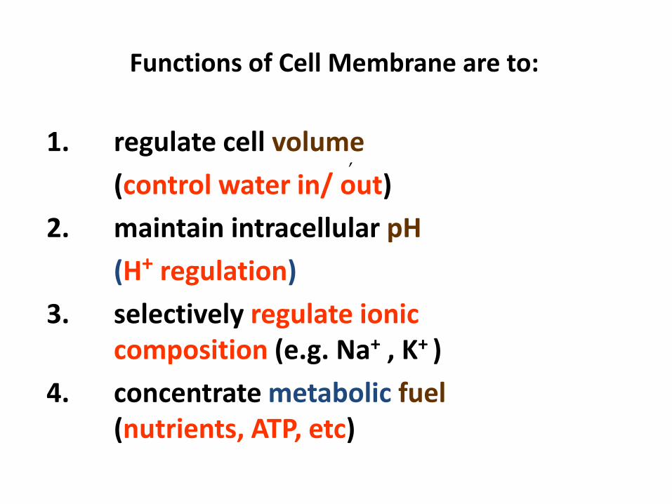
Functions of Cell Membrane are to:
1. regulate cell volume
(control water in/ out)
2. maintain intracellular pH
(H+ regulation)
3. selectively regulate ionic composition (e.g. Na+ , K+ )
4. concentrate metabolic fuel (nutrients, ATP, etc)
,
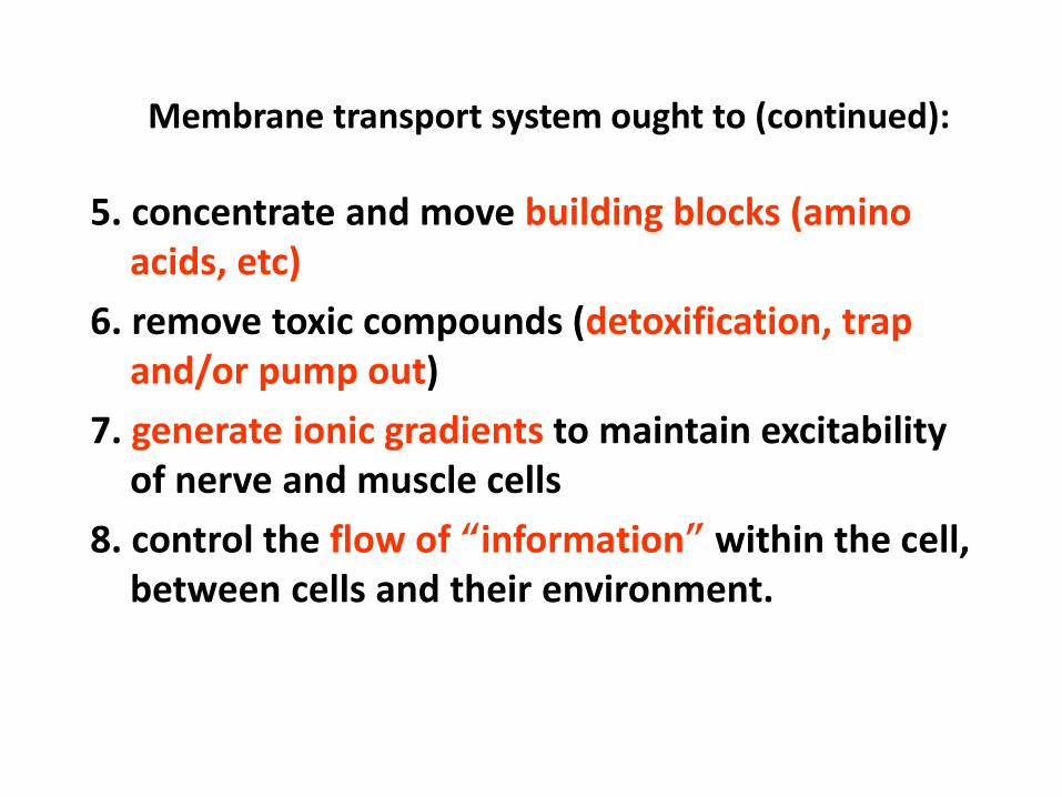
Membrane transport system ought to (continued):
5. concentrate and move building blocks (amino acids, etc)
6. remove toxic compounds (detoxification, trap and/or pump out)
7. generate ionic gradients to maintain excitability of nerve and muscle cells
8. control the flow of “information” within the cell, between cells and their environment.

Functions and properties of membrane
• Membrane is a selectively permeable barrierbetween the cell and the external environment.
• Its function is to maintain Homeostasis. Its selective permeability allows the cell to maintain a constant internal environment.
• Plasma membranes form compartments (compartmentation) within cells.

Fig 7.3 Fluid Mosaic Model for Membrane
• 1972 SJ Singer and G Nicolson
Copyright © 2005 Pearson Education, Inc., publishing as Benjamin Cummings
membrane proteins are
dispersed and individually
inserted into the
membrane

Cell Membrane

Fig 7.7 Animal Cell Plasma Membrane

1.5 The Fluid-Mosaic Model
(Singer and Nicolson, 1972):
1. Amphipathic lipids stabilized by the hydrophobic interaction form the lipid bilayer .
2. Asymmetric property. The components of membranes with lipids and proteins are asymmetrically oriented: the two faces are different.

4. It is a fluid-like structure, with fluidity regulated by the
number of double bonds in the fatty acids (increasing
unsaturation increases fluidity) and cholesterol content
(increasing cholesterol decreases fluidity).
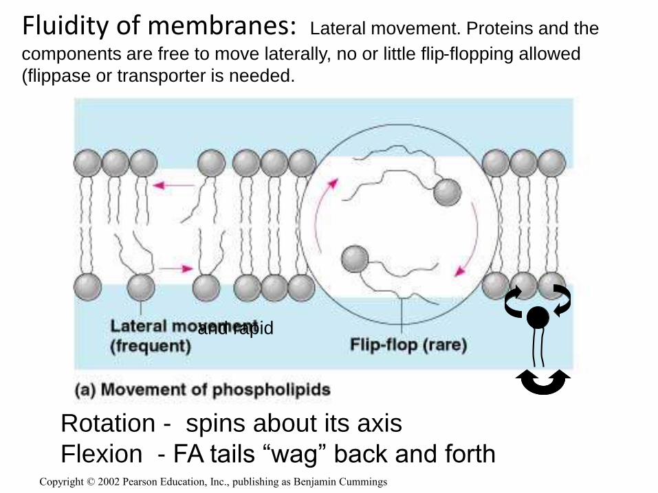
Rotation - spins about its axis
Flexion - FA tails “wag” back and forthCopyright © 2002 Pearson Education, Inc., publishing as Benjamin Cummings
Fluidity of membranes: Lateral movement. Proteins and the
components are free to move laterally, no or little flip-flopping allowed
(flippase or transporter is needed.
and rapid

Fatty acids and phospholipids
• Membranes have 3 kinds of lipids:
phospholipids, glycolipids, & cholesterol
• Fatty acids (FAs) form the basic structures of
phospholipids & glycolipids
• Saturated FAs VS Unsaturated FAs
• Strong van der waals interaction between the
non-polar hydrocarbon regions of the molecules

Three views of the same molecule

Fatty acids (FAs) form the basic structures of
phospholipids
1.2.1 Simplified structure of lipid bilayer and phospholipids
Polar group
Phosphate
Glycerol
Fatty acid-(unsaturated)
Phospholipid molecule
Fatty acid (saturated)
Hydrophilic head
Hydrophobic fatty acid tails
Hyd
rop
ho
bic
co
re
of
lip
id b
ila
ye
r
Hydrophilic head to cytoplasm
Hydrophilic head on surface

1.2.2 Basic structure of fatty acid chains: saturated and unsaturated.
Hydrophilic carboxyl end
Hydrophobic hydrocarbon (alipathic) tail
Forming an amphipathic molecule
Adapted from Alberts et al., 1998. Essential Cell Biology. An Introduction to Molecular Biology of the Cell. Garland Pub.Inc.
Saturated Fatty Acids
CH2
CH2
CH2
CH2
CH2
CH2
CH2
CH2
CH2
CH2
CH2
CH3
COHO
CH2
CH2

Characteristics of Plasma membranes
• Membranes are held up with van der Waal’s force with no covalent interactions among molecules, therefore they can fuse together.
• Hydrophobic interaction also help stabilization membrane bilayer.
• Heat promotes lipid bilayer from gel (crystalline) state to fluid state. http://en.wikipedia.org/wiki/Cell_membrane
Explain why and how a detergent can kill germs?? Why bleach and alcohol can kill bacteria and viruses??

• Membrane processes like budding, exo- and endo-cytosis are common events.
• By membrane fusion, Golgi complex and move proteins to vesicles and surface membrane. Secretary proteins are stored in the vesicles, their membranes can fuse to the plasma membranes to excrete the secretory proteins with exocytosis.
• The mitochondrion has 2 layers of membrane, the inner is similar to their descendant of prokaryotes, the outer from eukaryotes; indicating its symbiotic origin.

Asymmetry of plasma membranes
1. Membrane separates interior and exterior side of the
cell and the 2 faces of lipid bilayer are different in
composition and structure, with different proteins and
phospholipids.
2. The plasma membrane core contain 1/3 cholesterol
and 2/3 phospholipids and sphingolipids, the outer
leaflet contains 5% glycolipids.
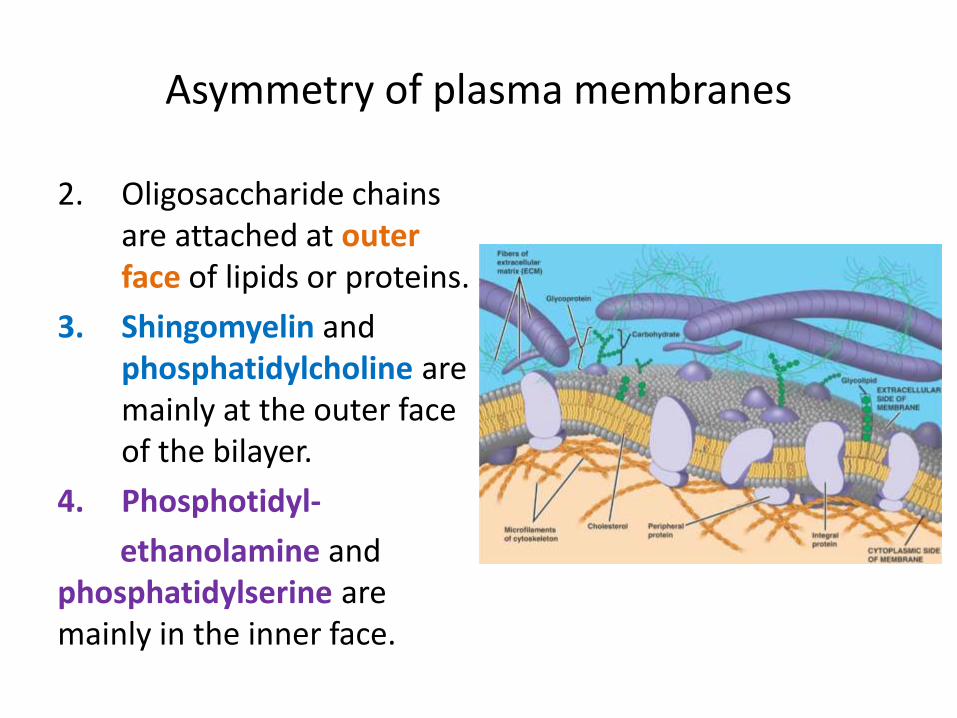
Asymmetry of plasma membranes
2. Oligosaccharide chains are attached at outer face of lipids or proteins.
3. Shingomyelin and phosphatidylcholine are mainly at the outer face of the bilayer.
4. Phosphotidyl-
ethanolamine and phosphatidylserine are mainly in the inner face.
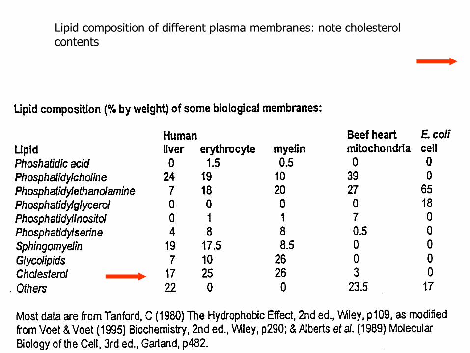
Lipid composition of different plasma membranes: note cholesterol contents

Cholesterol stays in between fatty acids with its rigid planar steroid ring
Phosphate
Glycerol
Fatty acid-(unsaturated)
Hydrophobic fatty acid
tails
Polar Head
Rigid Planar Steroid Ring
Polar Head
Non polar hydrocarbon Tail
Cholesterol is a metabolic precursor of steroid hormones; it is a member of steroids or lipids and major component of animal plasma membrane.

Cholesterol contents
• Plasma membrane= 25-30%
• Nuclear membrane= 10%
• Golgi/R.E.R.= 6 - 7%
• Mitochondria: outer= 4 -5%, inner= 2 -5%

Cholesterol contents
• Bacterial cells do not have cholesterol, whereas in
animal cells, cholesterol is a key regulator of
membrane fluidity.
• It makes bilayer less fluid (reduce fluidity), but it also
prevent hydrocarbon chains come together to
crystallize. It inhibits phase transitions.
4-27
Fluid-mosaic model

• Proteins - may be peripheral proteins or integral proteins.

• Ion channel
• Transporter
• Receptor
• Enzyme
• Cell Identity marker
• Linker
30
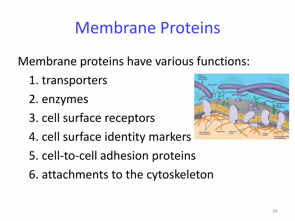
Membrane Proteins
Membrane proteins have various functions:
1. transporters
2. enzymes
3. cell surface receptors
4. cell surface identity markers
5. cell-to-cell adhesion proteins
6. attachments to the cytoskeleton

31

4-32
• Other Components
• 1) Proteins - may be peripheral proteins or integral proteins.
• 2) Glycolipids - phospholipids with carbohydrate chains attached
• 3) Glycoproteins -proteins have carbohydrate chains attached.
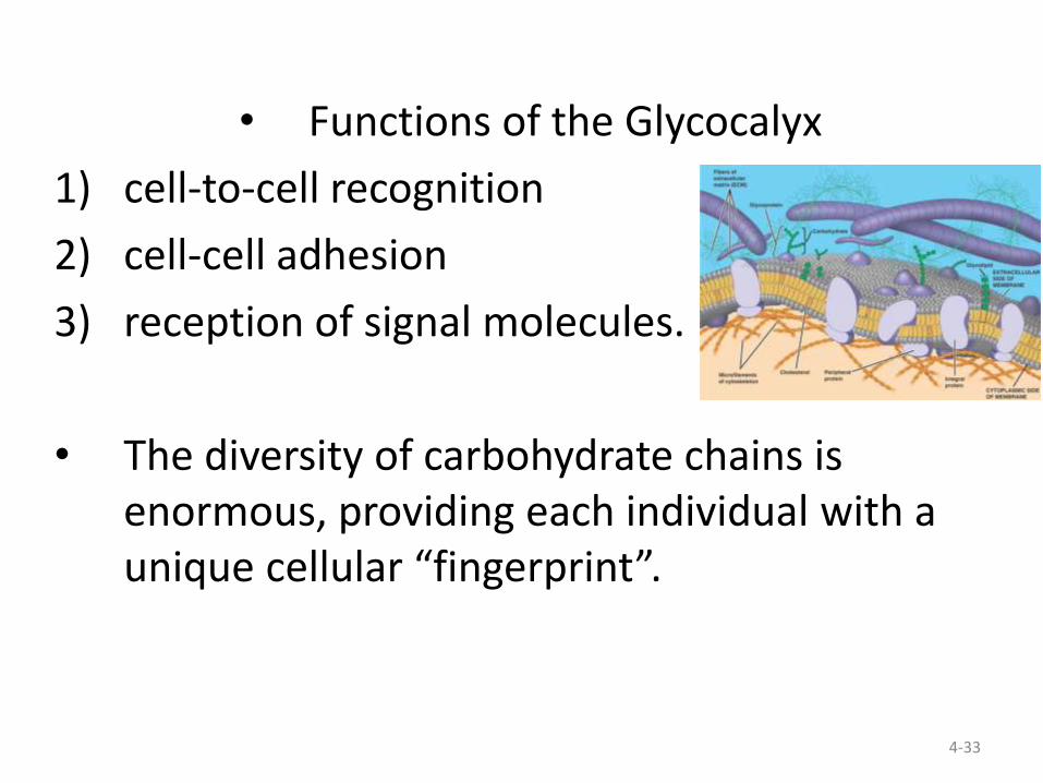
• The carbohydrate chains of 2) and 3) form the glycocalyx
4-33
• Functions of the Glycocalyx
1) cell-to-cell recognition
2) cell-cell adhesion
3) reception of signal molecules.
• The diversity of carbohydrate chains is enormous, providing each individual with a unique cellular “fingerprint”.

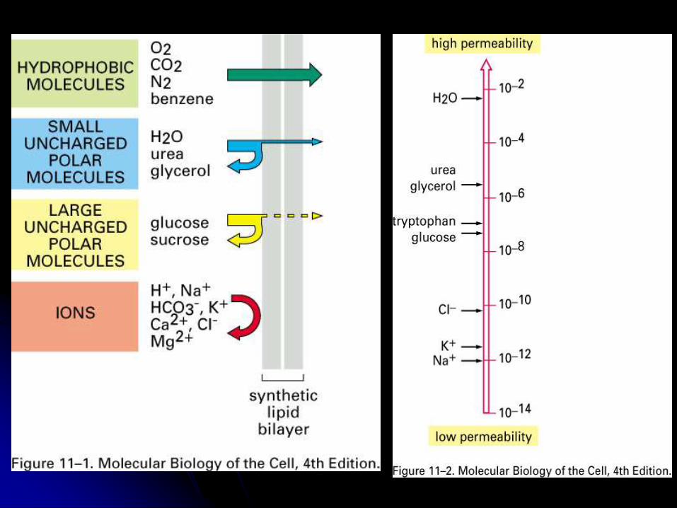
The Modes of Membrane Transport
Lipid bilayer
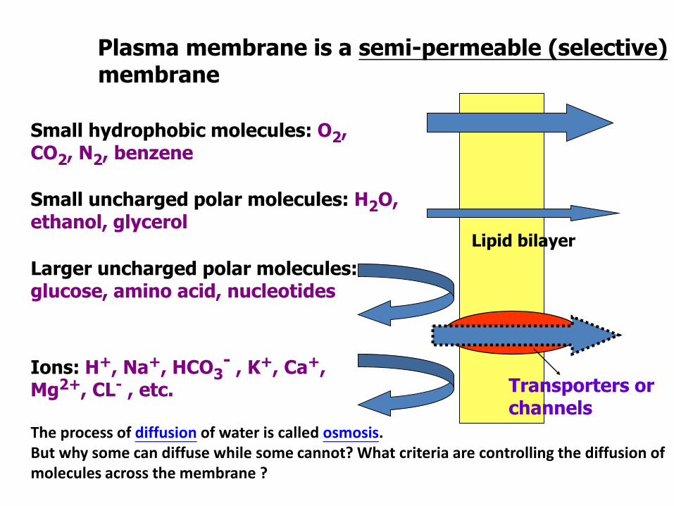
Small hydrophobic molecules: O2, CO2, N2, benzene
Small uncharged polar molecules: H2O, ethanol, glycerol
Larger uncharged polar molecules: glucose, amino acid, nucleotides
Plasma membrane is a semi-permeable (selective)membrane
Ions: H+, Na+, HCO3- , K+, Ca+,
Mg2+, CL- , etc. Transporters or channels
The process of diffusion of water is called osmosis. But why some can diffuse while some cannot? What criteria are controlling the diffusion of molecules across the membrane ?

Passive transport: facilitated diffusion for all channel protein
and part of carrier (concentration or electrochemical gradient)
Active transport: against electrochemical gradient; mediated
by carrier (pump); use ATP or ion gradient as energy source
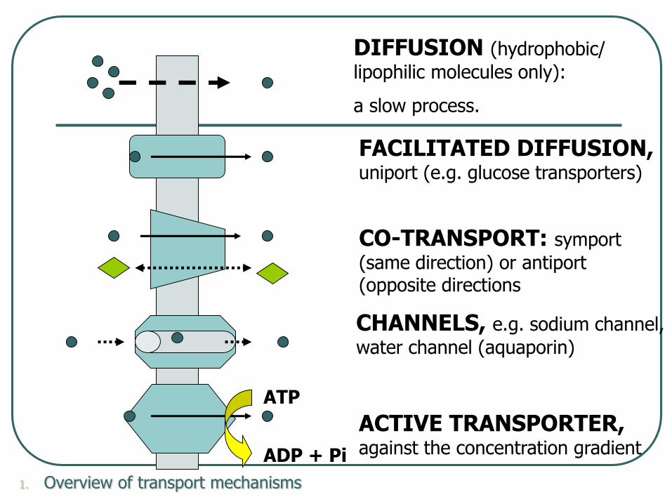
DIFFUSION (hydrophobic/
lipophilic molecules only):
a slow process.
FACILITATED DIFFUSION, uniport (e.g. glucose transporters)
CO-TRANSPORT: symport
(same direction) or antiport(opposite directions
ACTIVE TRANSPORTER, against the concentration gradient
CHANNELS, e.g. sodium channel,
water channel (aquaporin)
1. Overview of transport mechanisms
ATP
ADP + Pi

Passive Transport
Diffusion Simple (Passive)Diffusion
no carriers is involved
tendency of molecules to spread out into the
available space
driven by the kinetic energy (heat) of the
molecules
random motion of individual molecules, but
movement of a population may be directional

High concentration to low concentration until conc. is equal
Movement down a concentration gradient.
Diffusion of solutes across
membranes
High LowHigh Low
Copyright © 2005 Pearson Education, Inc., publishing as Benjamin Cummings

High concentration to low concentration until conc. is equal
Movement down a concentration gradient
Fig 7.11b Diffusion of two
solutes
- each diffuses independently
Copyright © 2005 Pearson Education, Inc., publishing as Benjamin Cummings
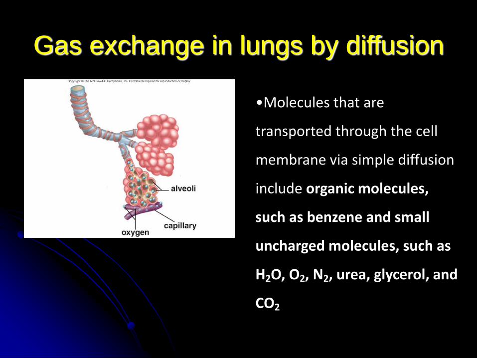
•Molecules that are
transported through the cell
membrane via simple diffusion
include organic molecules,
such as benzene and small
uncharged molecules, such as
H2O, O2, N2, urea, glycerol, and
CO2
Gas exchange in lungs by diffusion

4-43
Facilitated diffusion Passive transport (facilitated
diffusion)energy independent, down the concentration gradient
1) follows concentration gradient
2) requires carrier protein
3) often for large or charged molecules
4) can be regulated
4-44
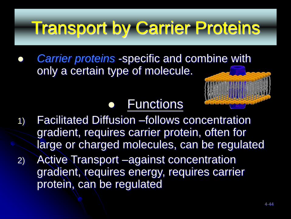
Transport by Carrier Proteins
Carrier proteins -specific and combine with only a certain type of molecule.
Functions
1) Facilitated Diffusion –follows concentration gradient, requires carrier protein, often for large or charged molecules, can be regulated
2) Active Transport –against concentration gradient, requires energy, requires carrier protein, can be regulated

Two classes of transfer protein:
(1) Carrier protein (permease, transporter,
pump) : for specific molecule; usually
coupled with energy source
(2) Channel protein: inorganic ions; down to its
concentration gradient; fast
overall, transfer proteins create electrical
(because of membrane potential) and
concentration gradient, in turn, used as a
driving force (electrochemical gradient) to
facilitate the transport process

Electrochemical gradient combines with
membrane potential as a driving force
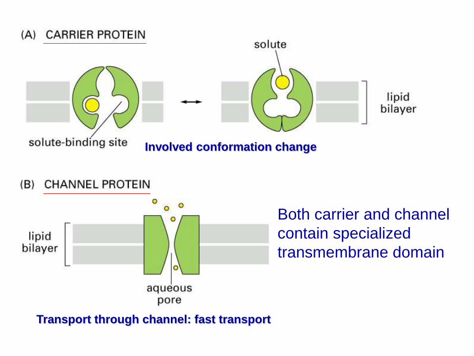
Involved conformation change
Both carrier and channel
contain specialized
transmembrane domain
Transport through channel: fast transport
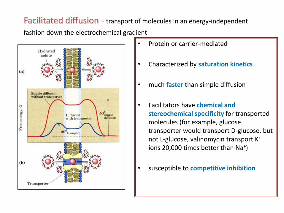
• Protein or carrier-mediated
• Characterized by saturation kinetics
• much faster than simple diffusion
• Facilitators have chemical and stereochemical specificity for transported molecules (for example, glucose transporter would transport D-glucose, but not L-glucose, valinomycin transport K+
ions 20,000 times better than Na+)
• susceptible to competitive inhibition
Facilitated diffusion - transport of molecules in an energy-independent
fashion down the electrochemical gradient

Kinetics of simple diffusion and carrier-mediated
diffusion (expressed as Vmax/Km or Bmax/Kd)

Model of Carrier protein: passive transport
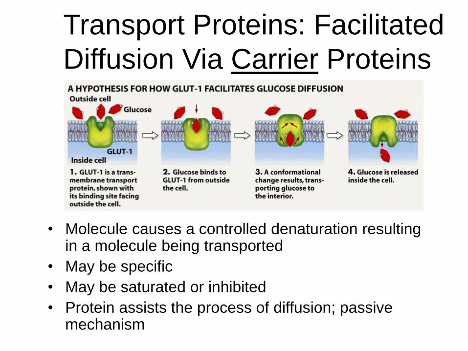
Transport Proteins: Facilitated
Diffusion Via Carrier Proteins
• Molecule causes a controlled denaturation resulting in a molecule being transported
• May be specific
• May be saturated or inhibited
• Protein assists the process of diffusion; passive mechanism

Passive-Mediated Glucose Transportfacilitates glucose uptake about 50,000 fold
• Erythrocytes glucose transporter is a 55 kDa glycoprotein with 12 transmembrane segments
• The transporter is believed to function through “alternating conformation” mechanism
• Transport can occur in either direction and serves mainly to equilibrate glucose concentration

Three types of carrier-mediated transport: uniport, symport
( kidney/GI epitghelial cells ) and antiport (determined by its
path direction)

Classes of
carrier
proteins
Uniport (facilitated diffusion) carriers mediate
transport of a single solute.
An example is the GLUT1 glucose carrier.
The ionophore valinomycin is also a uniport carrier.
Uniport Symport Antiport
A A B A
B

A gradient of one substrate, usually an ion, may drive uphill
(against the gradient) transport of a co-substrate.
It is sometimes referred to as secondary active transport.
E.g: glucose-Na+ symport, in plasma membranes
of some epithelial cells
bacterial lactose permease, a H+ symport carrier.
Uniport Symport Antiport
A A B A
B
Symport (cotransport) carriers
bind two dissimilar solutes
(substrates) & transport them
together across a membrane.
Transport of the two solutes is
obligatorily coupled.
A substrate binds & is transported.
Then another substrate binds & is transported in the
other direction.
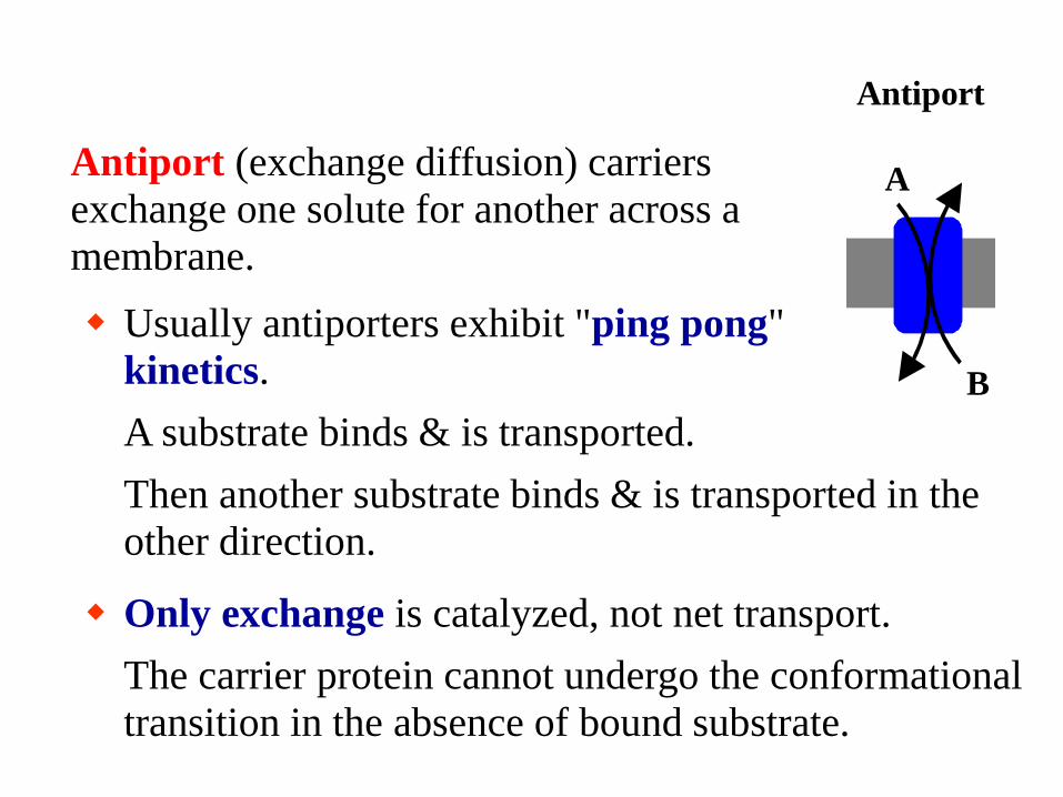
Only exchange is catalyzed, not net transport.
The carrier protein cannot undergo the conformational
transition in the absence of bound substrate.
Antiport (exchange diffusion) carriers
exchange one solute for another across a
membrane.
Usually antiporters exhibit "ping pong"
kinetics.
Uniport Symport Antiport
A A B A
B


Example of an antiport carrier:
Adenine nucleotide translocase (ADP/ATP exchanger)
catalyzes 1:1 exchange of ADP for ATP across the inner
mitochondrial membrane.
ATP 4
ADP 3
mitochondrial matrix
adenine nucleotide translocase

Ionophores: small hydrophobic molecules (originally
formed by microorganism) in membrane to transport
specific ions; not coupled to energy source (down to
concentration gradient)
1. Valinomycin: potassium ion (mobile)
2. FCCP: hydrogen ion (mobile)
3. A23187: calcium and magnesium ion (mobile)
4. Gramicidin A: monovalent cation (channel former)

Osmosis
• Direction of osmosis is determined only by a difference in total solute concentration– kinds of solutes in the solutions do not matter
• What happens to cells in hypertonic, hypotonic, and isotonic solutions?– cytoplasm vs. solution cells are in

Passive Transport of Water -Osmosis
• Terms to define comparisons of solute concentrations in solutions
– Hypertonic• the solution with the higher concentration of solutes
– Hypotonic• the solution with the lower concentration of solutes
– Isotonic• solutions with equal solute concentrations
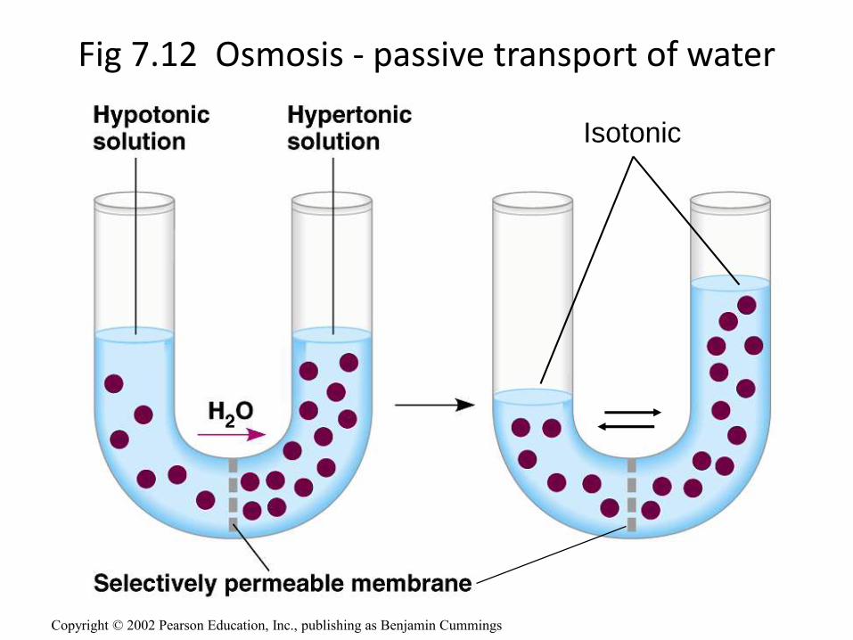
Isotonic
Fig 7.12 Osmosis - passive transport of water
Copyright © 2002 Pearson Education, Inc., publishing as Benjamin Cummings

Fig 7.12 Osmosis
Copyright © 2005 Pearson Education, Inc.,
publishing as Benjamin Cummings

Fig 7.13 The water balance of living cells
(wilts) (lethal)
Copyright © 2005 Pearson Education, Inc., publishing as Benjamin Cummings

Ion channels: channels mediate inorganic ion transport
1. Narrow, highly selective pores that can open and close
2. Approximately 100 million ions can pass through / second (105 times greater as compared to any carrier)
3. Can not couple to energy source to perform active transport (always passive – downhill)
4. They are gated (open and close status); prolong stimulation would desensitized or inactivated ( closed; usually through phorphorylation)
5. All animal cells contain ion channels, not limited to neuron; each neuron might have more than 10 types of channel

Ion channels open in response to special stimulus

Ligand-gated ion channels
• Mediate rapid action of neurotransmitters at synapse by changing the potential of the membrane in response to neurotransmitter (ligand) binding
• selectively activated by specific ligand
• discriminate between negatively and positively charged ions, but otherwise are not strongly selective
Cation-conducting channels - acetylcholine-,
serotonin- and glutamate receptors
Anion-conducting channels - glycine and g-
aminobutiric (GABA) acid -gated receptors

Active transport
- energy-dependent, against concentration gradient
4-72
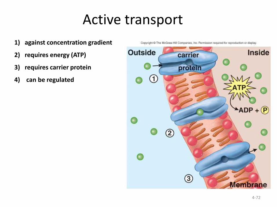
Active transport
1) against concentration gradient
2) requires energy (ATP)
3) requires carrier protein
4) can be regulated
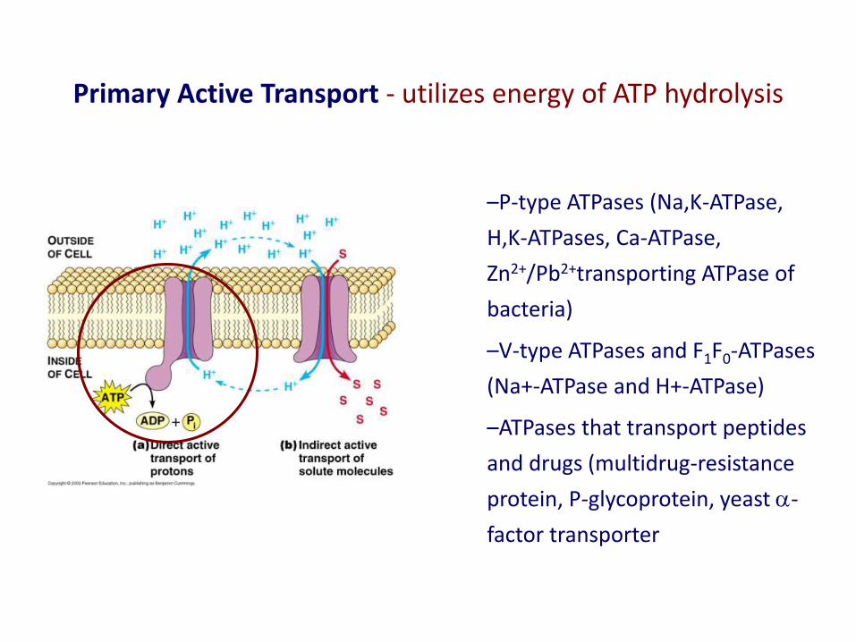
Primary Active Transport - utilizes energy of ATP hydrolysis
–P-type ATPases (Na,K-ATPase,
H,K-ATPases, Ca-ATPase,
Zn2+/Pb2+transporting ATPase of
bacteria)
–V-type ATPases and F1F0-ATPases
(Na+-ATPase and H+-ATPase)
–ATPases that transport peptides
and drugs (multidrug-resistance
protein, P-glycoprotein, yeast a-
factor transporter

Greatest consumer cellular energy
Sets up concentration & electrical gradients
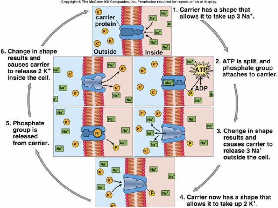
Hydrolysis of 1 ATP moves 2K+ in and 3Na+ out against
their concentration gradients
Na+/ K+ ATPase
Na,K-ATPase is a receptor of digitalis and related cardiac
glycosides used to strengthen the heartbeat
Mader: Biology 8th Ed.

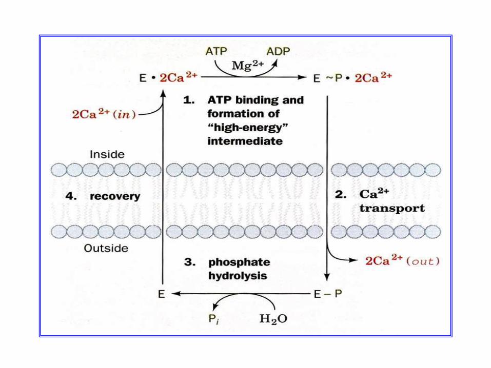
Ca2+-ATPase of Sarcoplasmic Reticulum
• Plays a major role in muscle relaxation by transporting released Ca back into SR
• A single subunit protein with 10 transmembrane fragments
• Is highly homologous to Na,K-ATPase
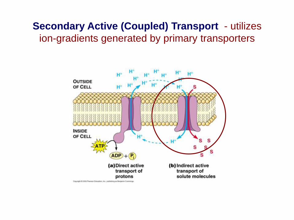
Secondary Active (Coupled) Transport - utilizes
ion-gradients generated by primary transporters

Types of Secondary Transporters
– Symporters (two solutes
move in same direction) Lac- permease,
Na+/glucose transporter)
– Antiporters (two solutes
move in opposite directions
Na+/Ca2+ exchanger)
– Uniporters (mitochondrial
Ca2+ uniporter and NH+4-
transporter in plants require
H+ gradient)
4-80
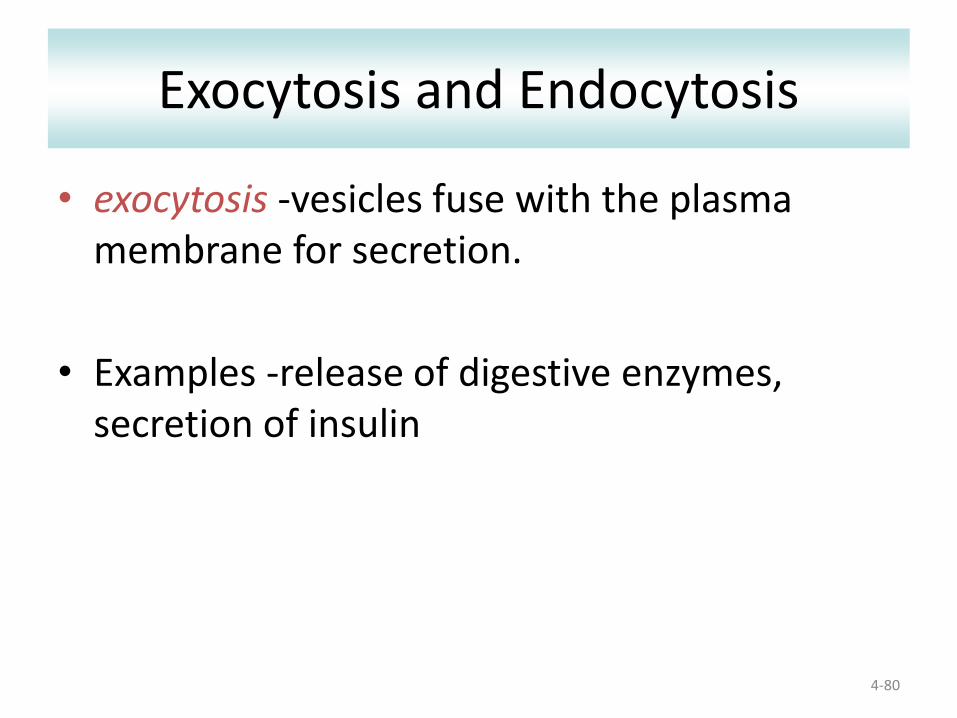
Exocytosis and Endocytosis
• exocytosis -vesicles fuse with the plasma membrane for secretion.
• Examples -release of digestive enzymes, secretion of insulin

4-81
Exocytosis

AS Biology, Cell membranes and Transport 82
Vesicle-mediated transport Vesicles and vacuoles that fuse with the cell membrane may be utilized to
release or transport chemicals out of the cell or to allow them to enter a cell. Exocytosis is the term applied when transport is out of the cell.

4-83
Endocytosis
• Endocytosis -cells fold membrane around substances and bring them into the cytoplasm (form a vesicle)
• Endocytosis occurs as:
• Phagocytosis – large particles
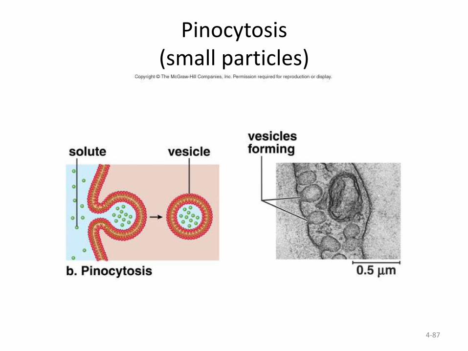
• Pinocytosis – small particles
• Receptor-mediated endocytosis – specific particles

Endocytosis

Phagocytosis
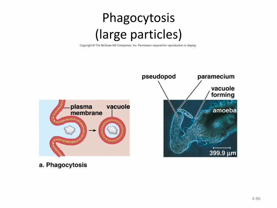
4-86
Phagocytosis(large particles)
4-87
Pinocytosis(small particles)

4-88
Receptor-mediated endocytosis(specific molecules)

AS Biology, Cell membranes and Transport 89
Cell Membrane - Function - EndocytosisThe cell membrane can also engulf structures that are much too large to fit through the pores in
the membrane proteins this process is known as endocytosis. In this process the membrane itself wraps around the particle and pinches off a vesicle inside the cell. In this animation an ameba
engulfs a food particle.

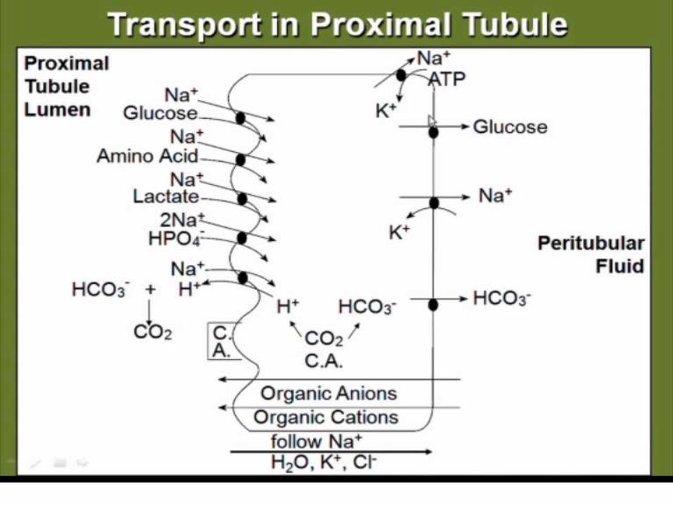
Transcellular transporter: apical to basolateral transport
nutrients (intestinal epithelial cells: microvilli)
Ex., - Ig A, transferrin, insulin

CLINICAL APPLICATIONS
Membrane Transport and Human
Disease
Membrane Transport and Human Disease
Hartnup’s disease
Cystinuria
Vitamin D resistant rickets
Nephrogenic diabetes insipidus (NDI) [renal AQP2],
acquired hypokalemia and hypercalcemia

Clinical applications of channels
Liddle’s disease, the sodium channels in
the renal epithelium are mutated, resulting
in excessive sodium reabsorption, water
retention and elevated blood pressure.

“Long QT syndrome”
Potassium channel mutations in “Long
QT syndrome” leads to inherited cardiac
arrhythmia, where repolarization of the
ventricle is delayed, resulting in prolonged
QT intervals in ECG.
“Long QT syndrome”
Potassium channel blockers are used in
cardiac arrhythmias.
Potassium channel openers as smooth
muscle dilators.

Chloride channel
The role of GABA and glycine as inhibitory
neurotransmitters is attributed to their
ability to open the chloride channels at the
postsynaptic membranes.
Bartter syndrome
Bartter syndrome is due to mutations in
potassium and chloride channels in the
renal tubules, especially the ascending
limb.
The condition is characterized by
hypokalemia and alkalosis and loss of
chloride and potassium in urine.

Calcium channel
Calcium channel blockers are used in the
treatment of hypertension.
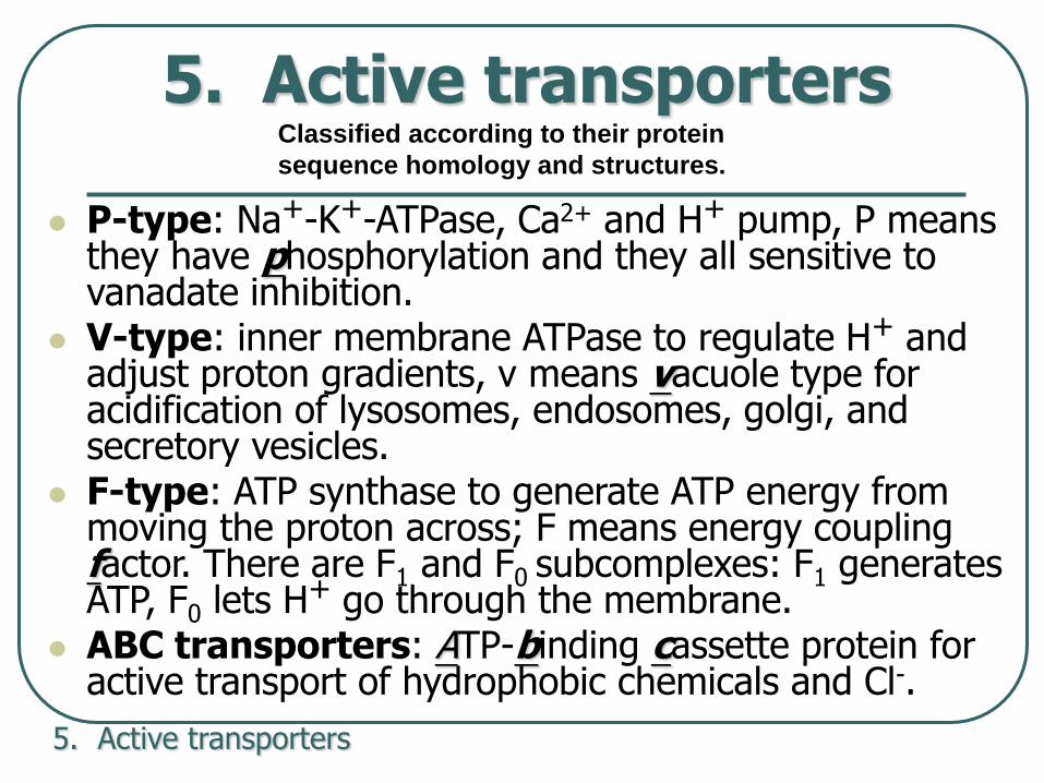
5. Active transporters
P-type: Na+-K+-ATPase, Ca2+ and H+ pump, P means they have phosphorylation and they all sensitive to vanadate inhibition.
V-type: inner membrane ATPase to regulate H+ and adjust proton gradients, v means vacuole type for acidification of lysosomes, endosomes, golgi, and secretory vesicles.
F-type: ATP synthase to generate ATP energy from moving the proton across; F means energy coupling factor. There are F1 and F0 subcomplexes: F1 generates ATP, F0 lets H+ go through the membrane.
ABC transporters: ATP-binding cassette protein for active transport of hydrophobic chemicals and Cl-.
5. Active transporters
Classified according to their protein
sequence homology and structures.

ATP-dependent ion pumps are grouped into classes
based on transport mechanism, as well as genetic &
structural homology.
Examples include:
P-class pumps Na+,K+-ATPase, (H+, K+)-ATPase,
Ca++-ATPases
F-class (e.g., F1Fo-ATPase)
& related V-class pumps.
ABC (ATP binding cassette) transporters, which
catalyze transmembrane movements of various organic
compounds including amphipathic lipids and drugs.

Menkes and Wilson Diseases are caused by mutated copper ion transporters
Copper ion transporters (ATP 7A and 7B) are essential to homeostasis of copper contents in our body.
Menkes disease: copper gets into the intestine but cannot transport further (mutated ATP7A), leading to copper deficiency. Copper histidine is needed for infiltration treatment.
Wilsons disease: copper in the liver cannot get into ceruloplasmin to excrete (mutated ATP7B), leading to copper accumulation in kidney, brain, and cornea. Penicillamine is needed for treatment to remove excessive copper, with zinc supplement.
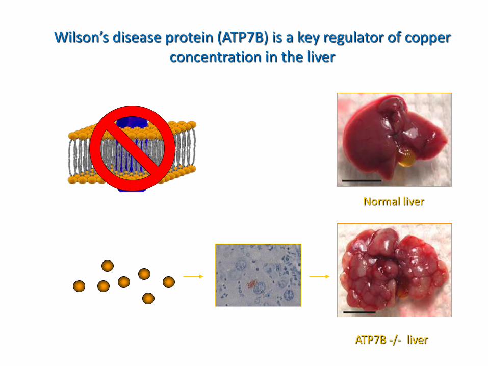
Wilson’s disease protein (ATP7B) is a key regulator of copper concentration in the liver
Normal liver
ATP7B -/- liver
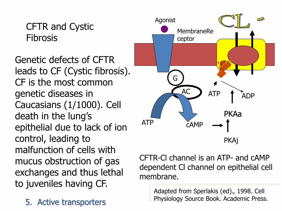
Genetic defects of CFTR leads to CF (Cystic fibrosis). CF is the most common genetic diseases in Caucasians (1/1000). Cell death in the lung’s epithelial due to lack of ion control, leading to malfunction of cells with mucus obstruction of gas exchanges and thus lethal to juveniles having CF.
Adapted from Sperlakis (ed)., 1998. Cell Physiology Source Book. Academic Press.
G
AC
ATP
ATP
cAMP
ADP
Agonist
MembraneReceptor
PKAa
PKAj
CFTR-Cl channel is an ATP- and cAMP dependent Cl channel on epithelial cell membrane.
CFTR and Cystic Fibrosis
5. Active transporters

• Cystic Fibrosis and CFTR (the most common fatal childhood disease in Caucasian populations). Inadequate secretion of pancreatic enzymes leading to nutritional deficiencies, bacterial infections of the lung and respiratory failure, male infertility.
• Bile Salt Transport Disorders Several ABC transporters, specifically expressed in the liver, have a role in the secretion of components of the bile, and are responsible for several forms of progressive familial intrahepatic cholestasis, that leads to liver cirrhosis and failure.
Membrane Transport and Human Disease

Membrane Transport and Human Disease
• Retinal Degeneration The ABCA4 gene produts transports retinol (vitamin A) derivatives from the photoreceptor outer segment disks into the cytoplasm. A loss of ABCA4 function leads to retinitis pigmentosa and to macular dystrophy with the loss of central vision.
• Mitochondrial Iron Homeostasis ABCB7 has been implicated in mitochondrial iron homeostasis. Two distinct missense mutations in ABCB7 are associated with the X-linked sideroblastic anemia and ataxia
• Multidrug Resistance ABC genes have an important role in MDR and at least six different ABC transporters are associated with drug transport

Ouabains for treatments of angina pectoris and myocardial infarction
• Ouabain blocks Na + -K + -ATPase. By blocking the Na+-K+-ATPase, Intracellular Na+ remains high
• Hence, the Na+- Ca2+ antiport cannot remove Ca2+ ions out from the cardiac muscle cells.
• Eventually, the Ca2+ ion level is restored to maintain the contraction power of cardiac muscle.

Anion antiport in parietal cells of stomach with H+- K+ -ATPase to produce stomach acid.
Basolateral membrane
Apical membrane
CO2
CO2
HCO3
Carbonic anhydrase
Cl - Cl -
Cl -
HCO3
K+
K+
K+ channel
Anion antiport
Cl -channel
H+-K+-ATPase
ATP
ADP + Pi
K+
H2O
+ OH -
Omeprazole inhibits the proton pump.
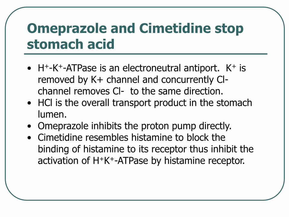
Omeprazole and Cimetidine stop stomach acid
• H+-K+-ATPase is an electroneutral antiport. K+ is removed by K+ channel and concurrently Cl-channel removes Cl- to the same direction.
• HCl is the overall transport product in the stomach lumen.
• Omeprazole inhibits the proton pump directly.• Cimetidine resembles histamine to block the
binding of histamine to its receptor thus inhibit the activation of H+K+-ATPase by histamine receptor.

THE END
THANKS FOR YOUR
ATTENTION