memory and executive function in older adults ...vanpettc/reprints/vanpetten04a.pdf · memory and...
TRANSCRIPT

Neuropsychologia 42 (2004) 1313–1335
Memory and executive function in older adults: relationships withtemporal and prefrontal gray matter volumes and
white matter hyperintensities
Cyma Van Pettena,∗, Elena Planteb, Patrick S. R. Davidsona, Trudy Y. Kuoa,Leslie Bajuscakb, Elizabeth L. Gliskya
a Department of Psychology, University of Arizona, Tucson, AZ 85721, USAb Department of Speech and Hearing Sciences, University of Arizona, Tucson, AZ 85721, USA
Received 1 December 2003; received in revised form 1 December 2003; accepted 25 February 2004
Abstract
Forty-eight healthy adults aged 65–85 were recruited for structural magnetic resonance scans after an extensive neuropsychologicalbattery that ensured a high degree of variability across the sample in performance on long-term memory tests, and on tests traditionallythought to rely on prefrontal cortex. Gray matter volumes were measured for three gyri in the frontal lobe (superior, middle, inferior), sixgyri in the temporal lobe (superior, middle, inferior, fusiform, parahippocampal, and hippocampus), and the occipital lobe. Gray mattervolumes declined across the age range evaluated, but with substantial regional variation—greatest in the inferior frontal, superior temporal,and middle temporal gyri but negligible in the occipital lobe. Both memory performance and executive function declined as the numberof hyperintense regions in the subcortical white matter increased. Memory performance was also significantly correlated with gray mattervolumes of the middle frontal gyrus (MFG), and several regions of temporal neocortex. However, the correlations were all in the negativedirection; better memory performance was associated with smaller volumes. Several previous reports of significant negative correlationsbetween gray matter volumes and memory performance are described, so that the possible reasons for this surprising finding are discussed.© 2004 Elsevier Ltd. All rights reserved.
Keywords: Gray matter; Magnetic resonance (MR); Elderly
1. Introduction
Numerous aspects of brain anatomy change with ad-vancing age. Postmortem studies show that decreased brainweight and volume (largely attributed to shrinkage of neu-rons, especially the dendritic arbor), decreased density ofnoradrenergic, dopaminergic, serotonergic and muscarinicreceptors in the cortex, and increased accumulation of lipo-fuscin are all associated with age at time of death (Esiri,1994; Kemper, 1994; Peters, Morrison, Rosene, &Hyman, 1998; Powers, 2000). In vivo measurementsof whole brain volumes by computerized tomographyand magnetic resonance (MR) likewise show declines incross-sectional studies comparing subjects of different ages.Age-related volume decrements are observed in both cere-bral gray matter and white matter, although white-matterdecline may begin later in life (Coffey, 2000; Courchesne
∗ Corresponding author. Tel.:+1-520-6218830; fax:+1-621-9306.E-mail address: [email protected] (C. Van Petten).
et al., 2000; Jernigan et al., 2001; Raz, 2000). In late life,white matter shows additional changes such as prolonga-tion of T1 and T2 relaxation time constants (indicative ofincreases in free water), and increases in the prevalence ofhyperintense spots on T2-weighted images. The regionaldistribution of age-related changes has not been fully char-acterized, but there is fairly widespread agreement thatprefrontal cortex shows the largest volume loss with age,temporal cortex generally shows losses, and the occipitallobe is relatively spared (Coffey, 2000; Raz, 2000; Razet al., 1997).
Cognitive abilities in elderly as compared to young adultshave also received a great deal of research attention, withevidence of relative sparing and loss across different aspectsof perception and cognition. Episodic memory is one do-main in which healthy older adults clearly perform worsethan younger adults, in contrast to smaller age effects for re-trieval from semantic memory (general knowledge) and forperceptual priming of recently experienced stimuli (Albert,1994; Ardila & Rosselli, 1989; Bäckman, Small, Wahlin, &
0028-3932/$ – see front matter © 2004 Elsevier Ltd. All rights reserved.doi:10.1016/j.neuropsychologia.2004.02.009

1314 C. Van Petten et al. / Neuropsychologia 42 (2004) 1313–1335
Table 1Frontal lobe volumes and cognitive tests in healthy participants
Study (notes) Sample Sample Normalize Cognitive tests Correlations
Raz et al., 1993(a) Twenty-nine adults(age 18–78)
Dorsolateralprefrontal gray,prefrontal white
Residual aftercranial vault andvault and
Fluid intelligence (CFIT),vocabulary
ns
Hanninen et al.,1997
Forty-seven olderadults (meanage 71.1)
Frontal lobe None WCST, categories nsWCST, preservation ns left,−0.28 rightVerbal fluency, letter nsVerbal fluency, category 0.25 left, ns rightBenton visual recognition nsWMS paired associates ns
Gur et al., 1998 Seventeen mid-ageadults (mean age)
Frontal lobe Change involume over2 years
Attention/vigilance composite nsWCST nsVerbal intelligence composite(including verbal fluency)
0.53*
Spatial organization composite nsVerbal memory composite nsVisual memory composite nsSpeed-processing (includingWAIS digit-symbol, trails A andB, Stroop)
ns
Fine manual/motor functions ns
Raz et al., 1998 Ninety-five adults(age 18–77)
Dorsolateralprefrontal gray
Residual aftersubject height
WCST, perseveration −0.42Verbal memory nsNonverbal memory 0.20Verbal priming nsVerbal working memory 0.27Nonverbal working memory 0.21
Baare et al., 1999 Fourteen youngadults (meanage 26.9)
Prefrontal gray Divide by brainvolume
CVLT, long-delay cued-recall nsVerbal fluency, letter nsVerbal fluency, semantic 0.68 (Spearmanr)WMS-R visual reproduction 2
Schretlen et al.,2000
One hundred andninety-seven adults(age 20–92)
Frontal lobe None +WCST, categories 0.21
Sowell et al., 2001(b)
Thirty-five children(age 7–16)
Frontal gray Residual afterbrain volume
CVLT, delayed free recall −0.56Rey-Osterrieth, delayed recall −0.50
MacLullich et al.,2002 (c)
Ninety-five oldermen (age 65–70)
Frontal lobe None Verbal fluency nsRaven’s matrices 0.22 left, 0.25 rightNART 0.23 left, ns rightWAIS, digit-symbol 0.26 left, ns rightBenton vis. Retention 0.23 left, 0.22 rightWMS-R, visual reproduction nsRey AVLT nsWMS-R, logical memory ns
Salat et al., 2002 Thirty-one olderadults (meanage 84.0)
IFG grey Residual aftercranial vault
Conditional association errors(SFG only)
−0.47
MFG grey Working memory (orbital only) −0.46SFG grey, orbitalgrey
Object alternation ns
Sanfilipo et al.,2002
Twenty-sevenmid-age adults(mean age 35.7)
Prefrontal gray Residual aftercranial vault andage
WCST composite nsPrefrontal white WAIS, digit-symbol 0.44 (gray)
Buschke selective reminding nsVerbal fluency composite nsWMS-R, visual reproduction nsVerbal intelligence composite ns

C. Van Petten et al. / Neuropsychologia 42 (2004) 1313–1335 1315
Table 1 (Continued )
Study (notes) Sample Sample Normalize Cognitive tests Correlations
Gunning-Dixon &Raz, 2003(d)
One-hundred andthirty-nine older adults(mean age 63.7)
Prefrontal gray Residual afterheight
WCST perseveration −0.30
Working memory composite 0.30
Correlations indicating associations between larger volumes and better performance in standard font; correlations indicating associations between smallervolumes and better performance in bold; * correlation value estimated from published sample size and probability level. Correlations are Pearson exceptwhere Spearman rank-order correlation is indicated. (a) InRaz et al. (1993), correlations between fluid intelligence and volumes of prefrontal gray andwhite matter were significant before removing variance associated with head size and age. (b) The delayed memory measures inSowell et al. (2001)wereresiduals after adjusting for immediate recall. (c) The significant correlations between raw volumes and performance inMacLullich et al. (2002)becamenon-significant when normalized for cranial vault size. (d) The relationship between prefrontal gray volume and working memory became non-significantafter accounting for the effect of age on working memory. CFIT: Cattell culture-fair intelligence test; WCST: Wisconsin card sorting test, perseverationis an error score, categories is number of categories achieved; NART: national adult reading test; Rey AVLT: Rey auditory verbal learning test; CVLT:California verbal learning test; WMS-R: Wechsler memory scale-revised; WAIS: Wechsler adult intelligence scale. See original articles for details aboutcognitive testing procedures.
Larsson, 2000; Light, 1991; Nyberg, Bäckman, Erngrund,Olofsson, & Nilsson, 1996; Schmand et al., 1997). Morerecently it has been suggested that an assortment of abili-ties thought to be dependent on prefrontal cortex—workingmemory, task-switching, speeded verbal fluency, directingattention away from irrelevant stimuli, setting response crite-ria, and monitoring performance—are particularly suscepti-ble to advancing age (Chao & Knight, 1997; Craik & Grady,2002; MacPherson, Phillips, & Della Sala, 2002; West, 1996,2000; Zacks & Hasher, 1991; but see alsoBand, Ridderinkof,& Segalowitz, 2002; Greenwood, 2000for skepticism abouta unidimensional “frontal theory of aging”). Many recentstudies have evaluated relationships between cognitive abil-ities and brain volumes assessed in MR scans. In samplesof patients with neurological diagnoses, smaller volumesare generally associated with worse performance. In sam-ples with Alzheimer’s disease, dementia with Lewy bodies,Down syndrome, traumatic brain injury, subcortical cere-brovascular injury, and organic amnesia of mixed etiology,volumes of the hippocampi (and sometimes other regionsof the temporal lobe) are positively correlated with memoryperformance in a variety of tests (Barber, McKeith, Ballard,Gholkar, & O’Brien, 2001; Bigler et al., 1997, 2004; Deweeret al., 1995; Jernigan, Ostergaard, & Fennema-Notestine,2001; Köhler et al., 1998; Kopelman et al., 2001; Krasuski,Alexander, Horwitz, Rapoport, & Schapiro, 2002; Mungaset al., 2002; Petersen et al., 2000; Wilson et al., 1996).
However, results of volume/cognition correlations forgroups of healthy individuals have been much less consis-tent. The hippocampus has received a great deal of attentionin structural MR studies, but as the accompanying reviewpaper (Van Petten, in press) notes, correlations betweenhippocampal volume and memory performance have beenpositive, negative, and null in different reports. Fewer stud-ies have evaluated the relationship between memory andother regions of the medial temporal lobe; we are awareof three reports of a null relationship between volume ofthe parahippocamapal gyrus in healthy subjects (Köhleret al., 1998; Lupien et al., 1998; Petersen et al., 2000), andone significant positive correlation (Tisserand, Visser, VanBoxtel, & Jolles, 2000). Relationships between prefrontal
cortical volumes and cognition have also received lessattention than the hippocampus;Table 1 summarizes alleleven studies of which we are aware. The cognitive testsadministered in the prefrontal studies are too various toallow a formal meta-analysis, but the table does not show ahigh degree of consistency across studies and includes sig-nificant correlations that are both positive (larger volumesassociated with better performance), and negative (smallervolumes associated with better performance).
The extant literature provides rather weak support for thesimplest account of regional structure/function relationshipsin healthy subjects, namely that “bigger is better”. Perhapsa more reasonable account of volume/cognition relation-ships is that one should expect no relationship in a neuro-logically intact adult, because even gray matter volume is acrude measure that reveals little about relative proportionsof neurons and astrocytes, synaptic densities, ratios of exci-tatory to inhibitory synapses, patterns of synaptic connectiv-ity, numbers of receptors for various transmitter substances,and other factors that contribute to neural efficiency. How-ever, the fact remains that below-normal volumes have beenclearly associated with cognitive deficits in samples withneuropathology. Because apparently normal aging is alsoassociated with reductions in brain volume, it is of some in-terest to know whether this source of tissue loss also resultsin cognitive decline. Below, we describe some motivationsfor undertaking another volumetry study, then the methodsand the results of the present examination of cognitive per-formance, temporal and prefrontal volumes in a group ofcognitively intact older adults.
2. The present study
2.1. Region selection
One rationale for a new volumetry study is that finer sub-division of the cortex might reveal volume/cognition rela-tions not evident in previous studies. Although age-relatedvolume loss has uniformly been reported in measurementsof the whole frontal lobe, only one recent study has ex-

1316 C. Van Petten et al. / Neuropsychologia 42 (2004) 1313–1335
amined individual frontal gyri and cognitive measures inolder adults, and reported regionally specific relationshipsbetween superior and orbital gray matter and working mem-ory (Salat, Kaye, & Janowsky, 2001). In the present study,gray matter volumes of the inferior, middle, and superiorfrontal gyri are measured separately.
As regards long-term memory across the lifespan, thehippocampus has received a great deal of attention (VanPetten, in press), but the rest of the temporal lobe has beenconsidered less often, and rarely subdivided. The hippocam-pus is a small structure, and many older studies measuredless than its full volume because it is difficult to visualizethe entire anterior–posterior extent in standard coronal slices(Sullivan, Marsh, Mathalon, Lim, & Pfefferbaum, 1995).When very small volumes are evaluated, small measure-ment uncertainties in both the MR and the memory vari-ables may be sufficient to preclude finding subtle but realvolume/cognition associations. In the present study, coronalslices were aligned perpendicular to the long axis of the hip-pocampus, and covered its full length, permitting more com-plete volume estimation of this structure. Moreover, regionsof the temporal lobe outside the hippocampus are importantfor normal memory function as well. Although differentialroles of the hippocampus and other medial temporal regionsare a topic of some debate, the parahippocampal gyrus is fre-quently damaged in cases of human amnesia. Animal stud-ies, intracranial ERP recordings, and hemodynamic imagingall suggest an important role for the parahippocampal gyrusin memory (Brown & Aggleton, 2001). The same sorts ofevidence indicate modulations of activity in the inferior tem-poral gyri (ITG) and fusiform gyri related to both encodingand retrieval of visual stimuli (both objects and, in humans,words (Baker, Sanders, Maccotta, & Buckner, 2001; Fahy,Riches, & Brown, 1993; Fernandez et al., 2001; Moscovitch,Kapur, Kohler, & Houle, 1995; Otten & Rugg, 2001;Sobotka & Ringo, 1993)). We thus measured six regions ofthe temporal lobe: superior, middle, inferior, fusiform, andparahippocampal gyri as well as the hippocampus.
2.2. Subject selection and cognitive assessment
In volumetry/cognition studies, there is a tradeoff betweenthe power to detect small effects offered by very large num-bers of subjects, and a thorough characterization of eachsubject’s cognitive status which may not be feasible for verylarge samples. For the evaluation of normal aging, intensiveneuropsychological examination is desirable because it of-fers some assurance that subjects with incipient dementia areexcluded. However, the likelihood of detecting relationshipsbetween volumes and cognitive status will also increase asthe range of cognitive performance increases, so that an ex-tremely homogeneous sample may also be undesirable. Thecompromise reached in the present study was to scan a mod-erate size sample of 48 older adults who were selected froma larger pool of approximately 100 individuals who hadundergone about 4 h (two sessions) of neuropsychological
evaluation. Subjects were invited for MR scans when theyscored within the normal range of all the standardized testsused, but showed above- or below-average performance ontests of memory, or tests thought to depend on prefrontalcortex. This approach combines the benefits of assuring thecognitive intactness of the sample, but maximizing range ofperformance.
Because understanding the relationships among a vari-ety of cognitive abilities and a variety of brain regions isone goal of volumetry/cognition studies, measuring a largenumber of regions and a large number of cognitive abili-ties is desirable. However, such a “wide net” approach mayalso increase the likelihood of obtaining spuriously signif-icant correlations. At the same time, applying a statisticalcorrection for multiple tests may be too stringent, given thatsmall magnitude effects might reflect genuine relationships.Our compromise approach was to combine a large numberof neuropsychological tests into two composite measuresbased on a factor analysis showing shared variance betweena set of five memory tests (memory factor or M-factor), andshared variance between five other tests traditionally associ-ated with frontal lobe function (frontal factor or F-factor, seeSection 3for detail). The factor score approach allows incor-poration of a large number of neuropsychological measuresof cognitive ability, while reducing the number of statisticaltests employed to detect volume/cognition relationships.
We have previously utilized the two composite neuropsy-chological factors to study individual differences in thememory performance of older adults. After removing vari-ance due to age alone, high scores on the M-factor have beenassociated with better performance in discriminating stud-ied from unstudied words in experimental recognition tests(both two-alternative forced-choice and old/new formats(Davidson & Glisky, 2002; Glisky, Polster, & Routhieaux,1995)). Although the individual measures which contributethe F-factor score do not include any tests of long-termmemory, the F-factor has also proved influential for perfor-mance in memory tests, but in a different manner than theM-factor. High scores on the F-factor have been associatedwith better performance on source memory tests that requiredecisions about studied conjunctions of stimulus attributes(e.g. whether a word is spoken in the same or a differentvoice as when initially studied (Davidson & Glisky, 2002;Glisky, Rubin, & Davidson, 2001)), better performance onexclusion memory tests that require decisions about whichstudy list a word belonged to (Davidson & Glisky, 2002),and lower false alarm rates in old/new recognition tests(without an impact on hit rates (Rubin, Van Petten, Glisky,& Newberg, 1999)). Event-related potential (ERP) studiesin similar paradigms in young adults have identified focalprefrontal potentials during source memory tests as com-pared to item memory tests, and have additionally linkedthe amplitude of prefrontal potentials to the ability to resistmaking false alarms in an associative memory test (Senkfor,2002; Senkfor & Van Petten, 1998; Van Petten, Luka,Rubin, & Ryan, 2002; Van Petten, Senkfor, & Newberg,
C. Van Petten et al. / Neuropsychologia 42 (2004) 1313–1335 1317
2000). Our previous work is thus consistent with the viewthat the M-factor indexes basic memory ability, while theF-factor indexes other abilities dependent on prefrontal cor-tex that contribute to performance in some memory tests,such as selecting an encoding strategy, guiding memorysearch during the retrieval phase of memory tests, and eval-uating the correspondence between stimuli and responsecriteria.
Our a priori predictions were thus that the M-factor scorewould be associated with gray matter volumes of medialand perhaps inferior regions of the temporal lobe, while theF-factor score would be associated with prefrontal volumes.Gray matter volume of the occipital lobes was measured asa control structure expected to have no relationship to eithercomposite neuropsychological measure.
2.3. Normalization for head size and age
Most structural MR studies devoted to aging and/or cog-nition perform some sort of normalization for head or bodysize prior to analysis of volume measures. The (usually un-stated) motivation for normalization is that brain size willbe driven, in part, by the size of the sensory receptive sur-faces (skin, retina, etc.) and the number of muscle fibers inthe body, and that these are noise factors for understand-ing aging or cognition. However, the appropriate methodof normalization is not completely obvious, and may dif-fer depending on whether aging per se or cognition/volumerelationships are of greater interest. If the primary interestis in age-related tissue loss, brain volumes corrected for in-ternal volume of the skull (cranial vault) have face-valuevalidity—a widening gap between intracranial volume andbrain volume with increasing age can only indicate tissueloss. In a cross-sectional study, normalizing volumes by cra-nial vault also helps to distinguish aging from cohort effectscreated by the general increase in body and brain size overthe last century (Miller & Corsellis, 1977).
In contrast, normalizing by skull size may not be appro-priate when the focus is on cognitive abilities. Because braingrowth during development drives skull growth, removingvariance associated with cranial vault size may be akin tothrowing out the baby with the bath water—attenuating gen-uine relationships between the size of brain structures andcognitive abilities. Some researchers thus normalize brainvolumes for body height rather than intracranial vault size(see (Raz et al., 1997) for discussion of normalization strate-gies). However, this would also seem to have a potentialdrawback in an elderly sample, because the presence of os-teoporosis in some individuals can lead to an old-age heightthat is some inches shorter than in young adulthood. Giventhe lack of a perfect solution to this issue, we report corre-lations for both vault-corrected and raw volumes when ex-amining the impact of age on brain volumes.
A similar question arises about the treatment of agewhen analyzing relationships between cortical volumesand cognitive measures. Given the typical observation of
age-related brain shrinkage, some published studies ex-amine partial correlations between regional volumes andcognitive measures after variance due to age has been re-moved. If one assumes the causal chain that aging causesbrain shrinkage, and this shrinkage in turn causes cognitivedecline, then partialling out the effect of age will tend toeliminate volume/cognition relationships. In fact, signifi-cant volume/cognition correlations before age-correctionwhich approach zero after age-correction might be taken asa strong sign that age-related shrinkage influences cognitiveperformance (seeTisserand et al., 2000; for an example ofthis pattern of results). However, correcting for age beforeexamining volume/cognition relationships is appropriateunder other views of the causal relationships. An analysisthat removes the average effect of age on a given regionwill serve to mark individuals as having large/small gyralvolumes for their age, and to detect whether deviations fromthe average are associated with stronger/weaker cognitiveperformance. Such an analysis thus focuses on individualdifferences rather than age per se. A volume/cognition rela-tionship that survives age-correction (as inGur et al., 2000)indicates that cognitive heterogeneity in the sample is notprimarily due to chronological age, but remains compatiblewith two rather different interpretations. On the one hand,a sample in which some subjects have pathological atrophyand low cognitive performance will yield such a result. Onthe other hand, such a result could also arise from an intrin-sic (non-pathological) relationship between the volumetricand cognitive measures if there is sufficient variability inboth, and relatively little of that variability is correlatedwith the age of the subjects in the sample. We do not knowwhich of these causal perspectives is likely to be correct,and so examine correlations between volumes and cognitiveperformance using both raw volumes and residual volumesafter removing variance due to cranial vault size and age.
2.4. White matter hyperintensities (WMHs)
The temporal and frontal lobe gyral measurements re-ported here are for gray matter, which could be more se-curely assigned to individual gyri than the underlying whitematter. However, increased prevalence of bright spots in thewhite matter of healthy individuals has been a robust find-ing in MR aging studies. Severity of WMH has sometimesbeen linked with risk factors for vascular disease such as hy-pertension, ischemic heart disease, and diabetes (but with-out agreement as to individual factors or how severe theymust be). The physiological genesis and functional impactof such MR signal changes may vary according to their loca-tion and visual appearance (Coffey, 2000; Gunning-Dixon &Raz, 2000; Pantoni & Garcia, 1997). Some authors suggestthat those located along the borders of the ventricles reflectbreakdown of the ependymal lining, and are both more com-mon and less reflective of pathology than hyperintensities inthe white matter underlying the cortex (Coffey, 2000). Wethus quantified WMHs underlying the cortical mantle but

1318 C. Van Petten et al. / Neuropsychologia 42 (2004) 1313–1335
not periventricular hyperintensities. Small round (punctate)hyperintensities are likely to reflect shrinkage of tissue sur-rounding a blood vessel, leaving a fluid-filled space. Largerirregularly shaped patchy hyperintensities may reflect moresevere (although still subclinical) ischemic damage to myelinand/or axons (Fazekas, Schmidt, Kleinert, Kapeller, & Roob,1998). We thus quantified punctate and patchy WMHs sep-arately.
Some studies of WMHs in healthy older adults have re-ported no correlation with cognitive status, but a recentmeta-analysis of 23 such studies concluded that there is in-deed a modest correlation between WMH burden and poorperformance in tests of executive function (related to theF-factor used here) and memory (Gunning-Dixon & Raz,2000). We thus examine the joint impact of WMH numberand gray matter volumes on cognitive performance.
3. Methods
3.1. Subjects
Forty-eight individuals aged 65.5–85.8 years of age(mean 73.2) participated in an MR scan and a battery ofcognitive tests. All were community-dwelling and in gen-erally good health as assessed by screening interviewsand questionnaires prior to participation. Thirty-three werewomen (mean age 74.0) and 15 were men (mean age 71.5;not statistically different from female age). Mean years offormal education was 15 (range of 10–20). Exclusionarycriteria were current or past diagnosis of neurological orpsychiatric disorder (excepting peripheral neuropathy suchas carpal tunnel syndrome or nerve injury due to trauma), apast head injury that resulted in skull fracture or loss of con-sciousness for more than 5 min, heart attack, major coronaryartery disease leading to bypass or angioplasty, depres-sion as indicated by the mood assessment scale (Spreen &Strauss, 1998), prior or current treatment for alcohol abuse,and current consumption of more than 10 alcoholic drinksper week. Other exclusionary criteria were those that mightmake an MR scan risky: metal in the body, or claustropho-bia. Three men and 13 women who reported hypertensioncontrolled by medication were included. Six men and sevenwomen with mild ischemic heart disease participated (fourof these also had hypertension). One man and one womanwith non-insulin-dependent diabetes were also included.
Participants were recruited from a larger pool of olderadults who volunteer to participate in memory studies atthe University of Arizona. The roughly two-to-one ratioof women to men in the current MR sample reflects theover-representation of women among the more than 400individuals participating in our aging and memory studiesover the last 9 years; US census data from 1990 indicate afemale/male ratio of 1.48/1 in the over-65 population nation-ally. In the larger pool, all individuals take standardized neu-
ropsychological tests including the Wechsler memory scaleIII (WMS-3; Wechsler, 1997), and the Wechsler adult intelli-gence scale revised (WAIS-R;Wechsler, 1987) or Wechslerabbreviated scale of intelligence (WASI, 1999). Those scor-ing within the normal range according to published normsfor the standardized tests (within two standard deviationsof the mean for their age group) may then choose to partic-ipate in various memory experiments; those scoring outsidethe normal range are referred to neurological clinics andexcluded from the research pool. Individuals were recruitedto undergo MR scans from a group of approximately 100currently active research participants. Average time betweenthe neuropsychological battery and MR scan was 6 months.
3.2. Cognitive measures
For the current sample who underwent MR scans, meanperformance IQ was 116 (S.D. 13), and mean verbal IQ was120 (S.D. 9). On the WMS-3, mean index score for Immedi-ate Memory was 117 (S.D. 14, 87th percentile for adults inthe same age range); mean index score for general memory(i.e. delayed memory) was 118 (S.D. 15, 88th percentile).Despite this generally high level of cognitive ability, wesought to maximize variability in specific cognitive domainsby recruiting only participants who scored above or belowthe mean of the larger sample (by at least 0.2 standard de-viations) on tests of memory, and/or tests thought to rely onprefrontal cortex.
The two orthogonal measures used for subject selectionare based on factor analyses of a larger set of neuropsycho-logical tests (Glisky et al., 1995). The M-factor score re-flects performance on tests of recognition and recall fromepisodic memory, including three tests from the WMS-3(logical memory I, verbal paired associates I, face recogni-tion I), one from the Wechsler memory scale revised (visualpaired associates II), and the long-delay cued-recall mea-sure of the California verbal learning test (CVLT;Delis,Kramer, Kaplan, & Ober, 1987). The F-factor score reflectsperformance on five tests traditionally considered sensitiveto frontal lobe damage: number of categories achieved inthe modified Wisconsin card sorting test, number of wordsgenerated in response to initial letters F, A, and S in thecontrolled oral word association test, mental arithmetic testfrom the WAIS-R, and the backward digit span and mentalcontrol tests of the WMS-3 (Benton & Hamsher, 1976; Hart,Kwentus, Wade, & Taylor, 1988; Wechsler, 1987; Wechsler,1997). Table 2shows performance levels on the 10 individ-ual cognitive tests for the 48 current subjects. “M” and “F”scores were assigned to each participant by comparing theirperformance to a normative group of 100 older adults testedat the University of Arizona, and averaging their z-scores(standard deviations from the mean) for the two sets of tests.In the current MR sample, F-factor scores range from nega-tive 1.50 to positive 1.27 (mean−0.20), and M-factor scoresrange from negative 1.73 to positive 0.73 (mean−0.05),ensuring substantial cognitive variability within the sample.

C. Van Petten et al. / Neuropsychologia 42 (2004) 1313–1335 1319
Table 2Neuropsychological tests
Mean S.E. Range
Tests contributing to M-factorVerbal paired associates I (WMS-3) 21.6 0.9 3–31Visual paired associates II (WMS-R) 5.6 0.1 2–6Logical memory I (WMS-3) 23.5 0.9 5–33Face recognition I (WMS-3) 36.0 0.6 25–46CVLT, long-delay cued-recall 11.3 0.4 6–16
Tests contributing to F-factorWisconsin card sort cued recall 4.9 0.2 0–6Controlled oral word association 45.1 1.7 25–86Mental arithmetic (WAIS-R) 12.7 0.5 5–19Mental control (WMS-3) 29.4 1.0 15–40Backward digit span (WMS-3) 7.0 0.3 2–12
Ten subjects had positive scores (above the mean of thelarger sample) for both the M and the F-factors, 12 had neg-ative scores on both factors, 12 had negative F-factors butpositive M-factors, and 14 had positive F-factors but nega-tive M-factors.
In addition to the “M” and “F” factor score measures, re-lationships between the MR measures, verbal IQ (VIQ), andperformance IQ (PIQ) are also analyzed. In follow-up anal-yses examining the individual tests contributing to the factorscores, three memory tests from the WMS-3 or WMS-R notincluded in the M-factor are also included: logical memory2, verbal paired associates 2, and visual paired associates1. The eight memory tests include four that test immedi-ate memory (those with a “1” in their names) and four thattest memory after a delay of about 20 min (those with a “2”in their names, plus the long-delay cued-recall test of theCVLT). A different way to categorize the memory tests isthat two are free recall (logical memory 1 and 2), five arecued-recall (verbal paired associates 1 and 2, visual pairedassociates 1 and 2, the CVLT measure), and one is recog-nition (face recognition 1). Individual tests contributing tothe F-factor were also analyzed, together with the number ofperseverative errors on the WCST (mean 4.75, S.D. 6.44).
3.3. MR scan protocol
Three sets of MR scans were acquired on a GE Signa 1.5 Tmagnet. The first consisted of a sagittal T1-weighted local-izer scan, used to identify the hippocampal formation. Thisimage was used to position 3 mm thick coronal slices ac-quired perpendicular to the long axis of the left hippocampalbody. Thirty-two T2-weighted coronal images were acquiredfrom just anterior to the amygdala to the trigones of the lat-eral ventricles, using a fast spin echo sequence (TR= 400,TE = 96, FOV= 22× 16, matrix= 256× 192, NEX= 2,scan time= 4.48 min). These images served the dual pur-pose of providing high resolution images of temporal lobestructures, and highlighting possible white matter pathol-ogy. Finally, a set of 1 mm T1-weighted axial images wereobtained using a 3D SPGR sequence (TR= 26, TE = 5,FOV = 22×16, matrix= 265×192, NEX= 2, scan time=
16.02 min). The volume was positioned so that the entirecerebrum fell within the 124 slices of the acquisition volume.Because these images were obtained with isovolume voxelsof 1 mm, they could be flipped from the axial to sagittalplane without significant error (Plante, Boliek, Binkiewicz,& Erly, 1996).1 This last set of images was used to obtainvolumes of the entire cerebrum and frontal gyri in the axialplane, and cranial vault measures at the sagittal midline.
3.4. MR measurements
A team of operators were trained in the computerized seg-mentation of the regions of interest (ROIs) employed in thisstudy; all were blind to the age and cognitive status of thesubjects, and each specialized in one brain region (e.g. alloccipital lobes were measured by a single operator). Generalsegmentation procedures involved identifying relevant neu-roanatomic features that defined each ROI. The ROIs werehand-traced by computer mouse on a slice-by-slice basis foreach subject, so that individual variations in neuroanatomywere captured by the segmentation. Once the ROI was re-moved from the surrounding tissue, operators segmentedgray matter from white matter, cerebral spinal fluid (CSF),and other tissue (e.g. meninges, large vessels). Segmenta-tion of tissue types was done on a slice-by-slice basis sothat operators could assess local areas of signal change inwhite matter to accurately separate these from gray matter.The volume of the relevant tissue types was then calculatedfor the summed slices of the ROI.
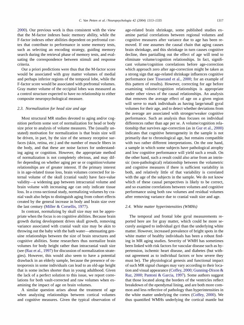
The cranial vault measurement at the sagittal midline in-cluded all supratentorial gray and white matter plus CSF. To-tal cerebral volume consisted of gray and white matter. Vol-umes within smaller cortical ROIs consisted of gray matteronly. In the frontal lobe, these were the superior, middle, andinferior gyri. In the temporal lobe, these were the hippocam-pal formation and the superior, middle, inferior, fusiform,and parahippocampal gyri. Finally, gray matter within theoccipital lobe was measured as a control structure expectedto show little relationship to either age or the cognitive abil-ities assessed.Figs. 1 and 2illustrate the frontal, temporal,and occipital ROIs. Detailed methods for segmentation andidentification of each ROI are provided in theAppendix A.
Signal intensity changes within the white matter were alsoevaluated and ranked. To ensure that these were sampledover a standard region across brains of different sizes, a sub-set of coronal slices was evaluated, starting anteriorally in
1 The choice of scan protocols represented a solution to a problemwith multiple constraints: (1) T2-weighted images are best for viewingwhite-matter hyperintensities; (2) images oriented perpendicular to thelong axis of the hippocampus are superior for measuring the volume ofthat structure, but do not include the prefrontal ROIs; (3) T2-weightedscans are considerably longer in duration than other scan protocols;and (4) even optimally healthy elderly subjects are unlikely to generatemovement-free MRI data with sessions exceeding an hour. We thus usedfaster SPGRs in a standard plane of section to capture the frontal lobes,but T2s aligned to the hippocampus for both the temporal lobe measuresand the WMH counts.

1320 C. Van Petten et al. / Neuropsychologia 42 (2004) 1313–1335
Fig. 1. Regions of interest in the frontal and occipital lobes, illustratingsegmentation of gray matter from a T1-weighted MRI scan. SFG: superiorfrontal gyrus; MFG: middle frontal gyrus; IFG: inferior frontal gyrus;OCC: occipital lobe.
the first slice in which the putamen could be seen, and endingposteriorly at the first slice in which the temporal-occipitalsulcus was visible. Within this range, adjacent pairs of con-secutive 3 mm slices were averaged to produce a series of6 mm slices, effectively filtering the data in thez-direction.This served to decrease random signal variation that couldbe mistakenly identified as patchy or punctate signalchanges.
Two types of white matter features were assessed. Thefirst consisted of diffuse intensity changes, apparent on theT2-weighted images as patches of white matter with inten-sities that appeared to fall between those typical of whitematter and those typical of gray matter. The severity ofthese patchy abnormalities was assessed by the number ofgyri showing such changes in each coronal slice, summingacross slices, and then dividing by total number of slices.The second type of white matter anomaly was visually dis-tinct from the first, and consisted of hyperintense spots onthe T2-weighted scans that were symmetrical in shape andhad a relatively distinct border. This class certainly included
Fig. 2. Regions of interest in the temporal lobe, illustrating segmentationof gray matter and hippocampus (Hi) from a T2-weighted MRI scan.STG: superior temporal gyrus; MTG: middle temporal gyrus; ITG: inferiortemporal gyrus; FG: fusiform gyrus; PHG: parahippocampal gyrus.
Fig. 3. Reverse T2 image showing patchy (solid arrow heads) and punctate(open arrow heads) abnormalities in the white matter of the frontal andtemporal lobe. Contrast and brightness of the image have been enhanced.Note that similar abnormalities within the cortical gray and sulci were notcounted in the white matter abnormality count. Furthermore, abnormalitieswithin or around the basal ganglia and external capsule were not counteddue to the difficulty of distinguishing between the narrow border of whitematter and other gray matter structures in this region.
blood vessels (typically punctate, but sometimes dilated inappearance), but probably also included other types of phe-nomena that met the visual criteria. These punctate fea-tures were counted, regardless of size, so that higher countscorresponded to greater numbers of these hyperintensities.Both sorts of changes within the white matter were eval-uated on the T2 images, which highlighted the patchy in-tensity changes, and on intensity-inverted versions of theseimages, which tended to highlight the punctate hyperinten-sities.Fig. 3 shows examples of both patchy and punctatehyperintensities.
3.5. Reliability
After segmentation of all scans was completed, opera-tors each randomly selected three brains (six hemispheres)for reanalysis of each ROI (different random samplesacross ROIs). The ROIs corresponding to each slice werere-extracted and the areas for each ROI at that slice recalcu-lated. This resulted in six re-measurements of each structure(60 total). The left column ofTable 3shows the correlationsbetween original and retest volumes for each structure,which averaged 0.93. The second column shows the aver-age percent discrepancy between original and re-measuredvolumes. There is little uniformity in the correspondencebetween these two metrics of measurement error. Note,for instance, that the occipital lobe yielded a substantially

C. Van Petten et al. / Neuropsychologia 42 (2004) 1313–1335 1321
Table 3Test/retest and cross-subject variability of volumetric measures
Region of interest Test/retestcorrelation
Percent differencebetween test and retest
Standard deviation aspercent of mean
Range of variationacross brains
Inferior frontal gyrus 0.94 2.6 45.7 156Middle frontal gyrus 0.92 2.4 24.6 257Superior frontal gyrus 0.97 3.3 17.3 330Hippocampus 0.78 3.5 16.4 118Parahippocampal gyrus 0.97 4.8 15.5 92Fusiform gyrus 0.99 4.5 18.7 122Inferior temporal gyrus 0.95 8.0 13.9 97Middle temporal gyrus 0.87 8.0 18.2 390Superior temporal gyrus 0.99 6.2 14.2 88Occipital lobe 0.95 3.9 23.3 295Mean 0.93 4.7 20.8 195
Note: the first column shows Pearsonr for the correlation between initial and re-measured volumes of each ROI. The second column shows the percentdiscrepancy between original and repeated measures. The third column shows the average absolute difference between volumes from an individual brain(n = 48) and the mean volume of that gyrus (or lobe), as a percent of the mean volume. The right column shows the range of cross-subject variabilityin volume: largest individual volume minus the smallest, divided by the smallest.
higher correlation between test and retest than did the hip-pocampus, although the percent discrepancy between thetwo measurements is slightly worse for the occipital lobe.This is because correlational measures are strongly influ-enced by the numerical range of values analyzed. Becausethe hippocampus is a small structure, the numerical rangeof volumes across brains was restricted to 2.92 cm3, whilethe range across occipital lobes was 52.0 cm3. Percent dis-crepancy scores, in contrast, are standardized to the size ofthe ROI evaluated. We believe that the percent discrepancymetric allows a more transparent evaluation of the impactof measurement error for a dataset. Discrepancy betweentest and retest is clearly noise. For correlational analysesof MR and cognitive measures, variation among subjectsis desirable, and can be regarded as signal. The third andfourth columns ofTable 3 show summary measures ofcross-subject variability: the standard deviation as a percentof the mean of each ROI, and the range of variation acrossbrains. Comparison across the columns ofTable 3suggests agenerally adequate signal-to-noise ratio (SNR), but also re-gional variability in SNR. The potential impact of measure-ment error on statistical outcomes is addressed inSection 4.
4. Results
Results are organized according to: (1) the impact of ageand educational level on the various MR measures, and thenrelationships between the MR measures and (2) IQ; (3) theF-factor; and (4) the M-factor. For the latter three sections,we begin with a regression analysis attempting to predictcognitive performance from the MR and demographic vari-ables. When a significant regression is obtained, simplecorrelations between each MR variable and the cognitivemeasures are examined, as well as partial correlations forthe volumetric measures after normalizing for cranial vaultsize and age.
4.1. Age
4.1.1. Cerebral volumesTable 4 shows mean values for hemispheric volumes,
frontal and temporal lobe gyri. Visual inspection of the
Table 4Volumetric measures and age
Mean (se) Age correlation
Cerebral hemispheres(gray and white)
1092 (22) −0.47∗∗∗ (−0.52∗∗∗)
Occipital lobes 40.3 (1.4) −0.02 (−0.08)
Superior temporal gyrus 11.2 (0.2) −0.30∗ (−0.38∗∗)Middle temporal gyrus 8.1 (0.7) −0.37∗∗ (−0.44∗∗)Inferior temporal gyrus 6.6 (0.4) 0.09 (0.01)Fusiform gyrus 6.5 (0.8) −0.19 (−0.25)Hippocampus 3.6 (0.9) −0.24 (−0.30∗)Parahippocampal gyrus 3.6 (0.8) −0.10 (−0.05)Left neocortical temporal 32.4 (0.6) −0.30∗ (−0.38∗∗)Left medial temporal 7.2 (0.1) −0.25 (−0.28)Right neocortical temporal 32.8 (0.6) −0.18 (−0.28)Right medial temporal 7.3 (0.2) −0.14 (−0.15)
Temporal 39.9 (0.7) −0.27 (−0.35∗)Superior frontal gyrus 30.4 (0.8) −0.19 (−0.26)Middle frontal gyrus 21.6 (0.8) −0.28 (−0.33∗)Inferior frontal gyrus 4.5 (0.3) −0.35∗ (−0.33∗)Left prefrontal 57.4 (1.5) −0.36∗ (−0.42∗∗)Right prefrontal 57.9 (1.4) −0.30∗ (−0.37∗∗)Prefrontal 57.6 (2.7) −0.35∗ (−0.42∗∗)
All volumes in cm3. Except where noted, volumes reflect gray matter onlyand are the average of left and right hemispheres. “Prefrontal” is the sumof the superior, middle and inferior frontal gyri; “temporal” is the sumof the six temporal lobe gyri listed; “neocortical temporal” is the sum ofthe superior, middle, and inferior temporal gyri plus the fusiform gyrus;“medial temporal” is the sum of hippocampus and the parahippocampalgyrus. Correlations between age and volumes are partial correlations aftervariance due to cranial vault size is removed; simple correlations withraw volumes in parentheses.
∗ P < 0.05.∗∗ P < 0.01.∗∗∗ P < 0.001.

1322 C. Van Petten et al. / Neuropsychologia 42 (2004) 1313–1335
Fig. 4. Top panel: relationship between volume of the cerebral hemispheres(gray plus white matter) and age. Bottom panel: residual cerebral volume(after removing variance due to cranial vault size) is plotted againstparticipant age in years.
scans indicated larger ventricles and widened cortical sulciin all or nearly all of the subjects as compared to our ex-perience with young brains (Clark & Plante, 1998; Jackson& Plante, 1996). Fig. 4(top) shows a decline in raw volumeof the cerebrum across the sampled age range of 65–85years (r = −0.52, P < 0.0005). Because capacity of thecranial vault (head size) is correlated with cerebrum size(r = 0.42, P < 0.005), we also examined partial corre-lations between age and cerebral volume after removingvariance due to volume of the cranial vault. Normalizingfor head size may be more appropriate than raw volumesgiven that the sample contains both men and women, andthe male subjects had significantly larger cranial vaults(F(1, 46) = 34.2, P < 0.0001) and larger hemispheres thanthe female (F(1, 46) = 8.08, P < 0.01). Sex was no longera significant determinant of cerebral volume when cranial
vault was used as a covariate (F(1, 46) = 2.83, P = 0.10).In addition to reducing the impact of sex, cerebral volumeresidualized on cranial vault provides a more transparentmeasure of age-related tissue loss.Fig. 4 (bottom) showsthat advancing age continued to be associated with decline incerebral volume after normalizing for volume of the supra-tentorial vault (r = −0.47, P < 0.001). Educational levelwas not significantly correlated with raw or residual cerebralvolume.
4.1.2. White matter signal changesThe number of patchy and punctate abnormalities within
the white matter were correlated with each other (r = 0.32,P < 0.05). Neither variety of signal change increased withage in the elderly sample (r = −0.12 and−0.06), norwas there a significant association with educational level.The subjects reporting ischemic heart disease had somewhatmore patchy abnormalities than those without heart disease(F(1, 46) = 4.50, P < 0.05), but the two groups did notdiffer in prevalence of punctate abnormalities. The subjectsreporting hypertension controlled by medication did not dif-fer from those with normal blood pressure in prevalence ofeither sort of abnormality.
4.1.3. Occipital lobesAs seen inTable 4, the gray matter volume of the oc-
cipital lobes was unrelated to age in our elderly sample.Nor were there any significant correlations with educationallevel.
4.1.4. Temporal lobesVolumes of most gyri in the temporal lobes showed
negative correlations with age after removing variance as-sociated with cranial vault size (Table 4), but these reachedstatistical significance for only the superior and middle tem-poral gyri. When gyral measures were combined into moreglobal neocortical (superior, middle and inferior temporalgyri plus fusiform gyrus) and medial cortical (hippocampusplus parahippocampal gyrus) regions in each hemisphere,only the left temporal neocortex showed a significant de-cline in volume across the 65–85 year age range. None ofthe temporal lobe measures showed significant associationswith educational level.
4.1.5. Frontal lobesTable 4shows that only the inferior frontal gyrus showed
a significant volume loss across age. However, the sum ofsuperior, middle and inferior gyri also showed negative cor-relations with age for both the right and left hemispheres,suggesting moderate age-related declines throughout theprefrontal region measured (which did not include the or-bital gyrus, cingulate gyus, or gyrus rectus). Volume of theinferior frontal gyrus also showed a positive correlationwith years of formal education (r = 0.32,P < 0.05 for rawvolume, partialr = 0.34,P < 0.05 after cranial vault size);other regions showed no significant association.

C. Van Petten et al. / Neuropsychologia 42 (2004) 1313–1335 1323
4.1.6. Cognitive measuresThe present study was not designed to evaluate cognitive
changes with age per se, but instead relationships betweenbrain morphometry and cognition in old age. The partici-pants were thus not selected randomly, but rather on the basisof high or low performance on neuropsychological tests ofmemory and/or tests thought to depend on prefrontal cortex.We thus had no predictions about age and cognitive perfor-mance in the selected sample. Indeed, none of the summarycognitive measures were significantly related to subject age:VIQ (r = −0.06), PIQ (r = 0.22), the factor score forneuropsychological tests thought to depend on the frontallobe (F-factor,r = −0.12), factor score for memory tests(M-factor, r = −0.13).
4.2. Predicting IQ from the MR measures
In an initial analysis, all of the raw gyral volumes, hemi-sphere and cranial vault size, occipital lobe volume, numberof WMHs (patchy and punctate summed), age, and educa-tional level were allowed to serve as potential predictors ofverbal IQ score in a stepwise regression. Only the volumeof the middle temporal gyrus met the minimumF-value of4.0 to enter the equation, with a simple correlation ofr =−0.31,P < 0.05. No other significant correlations were ob-served. For performance IQ, there was no significant regres-sion equation nor any significant correlations with individualvariables.
4.3. Predicting executive function from the MR measures
The same set of potential predictor variables were enteredinto an analysis with the F-factor as the dependent variable.Cranial vault size (positive coefficient) and WMHs (nega-tive coefficient) together accounted for 22% of the variancein the F-factor (F(2, 45) = 6.24, P < 0.005 for the re-gression,R2 for vault = 0.13, R2 for WMH = 0.09).2 Re-membering that the male subjects had larger cranial vaultsthan the female, we wondered if the influence of vault sizewas a sex difference in disguise. However, repeating theanalysis on the female brains alone (n = 33) produced thesame two significant predictors of the F-factor (R2 = 0.26,F(2, 30) = 5.29, P < 0.02; R2 for WMH = 0.15, R2 forvault size= 0.11). There was no significant regression forthe male subjects alone, given the small sample (n = 15). Fi-nally, the regression was essentially unchanged when the 13subjects reporting mild heart disease were excluded (R2 =0.33,F(2, 32) = 7.59,P < 0.005 for the regression;R2 forWMH = 0.17; R2 for vault = 0.15).
2 Because the distribution of WMH counts across brains shows a positiveskew from the normal distribution, this analysis was redone using a naturallog transform of WMH count. The regression results were essentiallyunchanged:F(2, 45) = 6.38, R2 = 0.22, P < 0.005; R2 for vault = 0.22,R2 for WMH = 0.09).
Table 5Best-fit regression analysis for the memory factor score
Variable R2 F-ratio P-value
ITG volume 0.18 10.04 0.01MFG volume 0.11 6.79 0.05Age 0.06 4.09 0.05White matter hyperintensities 0.06 4.63 0.05
Total 0.41 7.50 0.0005
No significant simple correlations between the frontalgray measures (raw or normalized) and the F-factor scorewere observed, nor any of the individual tests in the F-factor.The simple correlations for cranial vault size were signifi-cant for the overall F-factor (r = 0.36, P < 0.01), and fortwo of the tests contributing to the factor (mental arithmetic,r = 0.33, P < 0.05; mental control,r = 0.31, P < 0.05).
4.4. Predicting memory performance from the MRmeasures
Table 5shows that four of the predictor variables jointlyaccounted for 41% of the variance in the M-factor score.Smaller gray volumes of the inferior temporal and mid-dle frontal gyri (MFG), older age, and greater number ofwhite matter abnormalities all predicted better memoryperformance.3 Note that age was not significantly corre-lated with memory performance in the simple correlationsreported above, but contributed additional predictive valueafter volumes of the ITG and MFG entered the equation.
4.4.1. White matter hyperintensitiesExamination of the simple correlations showed that the
memory factor score declined as the number of WMHs in-creased (patchy:r = −0.32,P < 0.05; punctate:r = −0.35,P < 0.05; sum of patchy and punctate:r = −0.37, P <
0.01).4 Four of the eight individual memory tests showed
3 The impact of the estimated measurement error (test–retest discrep-ancy scores inTable 3) on this result was evaluated by creating four“noise-added” datasets: (a) add 2.4% to the measured volume of the MFGand 8.0% to the ITG for odd-numbered scans, subtract from both foreven-numbered scans; (b) subtract from both gyri for odd-numbered scans,add for even-numbered scans; (c) add to the MFG and subtract from theITG for even-numbered, subtract from the MFG and add to the ITG forodd-numbered; and (d) the converse of (c). Simultaneous regressions onthe M-factor then assessed the replicability of the original result, usingage, WMH count, and the “noisy” gray matter volumes from the twogyri. All four regressions were significant (F(4, 43) > 6.34, P < 0.0005,R2 > 0.37); coefficients for each predictor variable were uniformly nega-tive. In each of the four regressions, WMH count and MFG volume madeindependently significant contributions to the equation. In versions (a)and (d), age showed only a statistical trend toward significance (P = 0.10and 0.12, respectively). ITG volume made an independently significantcontribution in three of the analyses; in version (c) it showed only a trend(P = 0.10). These analyses suggest that differential measurement erroracross gyri do influence the results of regression analyses, but also that theobserved relationships were fairly resistant to the noise level of the data.
4 Correlation between natural log of WMH count and M-factor,r =−0.34, P < 0.02.

1324 C. Van Petten et al. / Neuropsychologia 42 (2004) 1313–1335
negative correlations with the total number of WMHs (vi-sual paired associates I,r = −0.38,P < 0.01; visual pairedassociates II,r = −0.38, P < 0.01; long-delay cued-recallmeasure of the CVLT,r = −0.31,P < 0.05; logical memoryII, r = −0.31, P < 0.05). The correlation between poorermemory and more white matter abnormalities remained sig-nificant after excluding the 13 subjects with mild heart dis-ease (r = −0.37, P < 0.05).
4.4.2. Temporal lobe gray volumesThe composite memory score (M-factor) was nega-
tively correlated with volumes of the inferior temporal andfusiform gyri (both r = −0.34, P < 0.05). When gy-ral measures were combined into more global neocorticaland medial cortical regions, both left and right temporalneocortex showed negative partial correlations with theM-factor (r of −0.34 and−0.30 respectively (P < 0.05);sum of left and right,r = −0.33, P < 0.05), as shown inFig. 5.
Fig. 5. Top: relationship between volume of left temporal neocortex (sum of superior, middle and inferior temporal gyri plus the fusiform gyrus) andthe factor score reflecting performance on five neuropsychological tests of memory. Bottom: residual volume of left temporal neocortex (after removingvariance due to cranial vault size and age) is plotted against the memory factor score.
Correlations between individual gyri and individual mem-ory tests are shown inTable 6, both as partial correlationsafter removing variance accounted by cranial vault size andage, and as simple correlations with raw volumes. This pro-cedure yielded a large number of statistical tests (96 for 12gyri by eight memory tests), so that a handful of significantcorrelations–both positive and negative—would be expectedby chance alone (some five at an alpha level ofP < 0.05).Nonetheless,Table 6shows a large number of significantcorrelations between individual gyri and individual memorytests (18 partial, 22 simple), and all were negative. The sig-nificant correlations spanned seven of the eight memory tests(excepting delayed cued-recall of visual paired associates),and seven of the eight neocortical gyri in the temporal lobe(excepting the left fusiform). There was no obvious differ-entiation between the immediate and delayed memory tests,nor between those using free- versus cued-recall (note thatthere was only one recognition test, but it appeared to fol-low the same general pattern). Within the medial temporal

C.
VanPetten
etal./N
europsychologia42
(2004)1313–1335
1325
Table 6Correlations between temporal lobe gyri and memory tests
Gyrus CVLT Logicalmemory I
Logicalmemory II
Verbal pairedassociation I
Visual pairedassociation II
Visual pairedassociation 1
Visual pairedassociation II
Facerecognition I
L. superior −0.05 −0.17 −0.35 −0.34 −0.33 −0.36 0.04 0.06 −0.03 −0.02 −0.06 −0.29 −0.22 −0.21 −0.04 −0.10R. superior −0.35 −0.40 −0.21 −0.23 −0.27 −0.30 0.02 0.05 −0.19 −0.14 −0.30 −0.35 −0.01 −0.19 −0.13 −0.18L. middle −0.24 −0.29 −0.06 −0.08 −0.18 −0.11 −0.20 −0.14 −0.30 −0.23 −0.22 −0.26 0.08 −0.08 −0.20 −0.20R. middle −0.02 −0.09 −0.18 −0.17 −0.23 −0.24 −0.15 −0.11 −0.10 −0.07 −0.12 −0.17 0.14 0.01 −0.06 −0.09L. inferior −0.27 −0.32 0.02 −0.03 −0.03 −0.09 −0.31 −0.31 −0.46 −0.47 −0.12 −0.17 0.07 0.01 −0.40 −0.42R. inferior −0.14 −0.26 −0.35 −0.38 −0.26 −0.34 −0.35 −0.32 −0.41 −0.41 −0.30 −0.37 0.25 0.08 −0.1 0.16L. fusiform −0.14 −0.18 −0.21 −0.24 −0.23 −0.23 −0.21 −0.16 −0.25 −0.20 −0.14 −0.18 0.17 0.05 −0.25 −0.25R. fusiform −0.48 −0.52 −0.33 −0.35 −0.26 −0.30 −0.07 −0.06 −0.20 −0.21 −0.34 −0.38 −0.05 −0.13 −0.10 −0.14L. hippocampal −0.19 −0.22 −0.20 −0.18 −0.16 −0.18 −0.17 −0.12 −0.21 −0.17 −0.27 −0.29 −0.09 −0.18 0.19 0.14R. hippocampal −0.27 −0.30 −0.21 −0.21 −0.26 −0.19 −0.04 −0.02 −0.18 −0.16 −0.23 −0.26 −0.16 −0.24 0.19 0.13L. parahippocampal −0.22 −0.16 −0.11 −0.07 −0.07 −0.03 0.07 0.08 −0.01 0.01 −0.14 −0.10 −0.01 −0.01 −0.08 −0.08R. parahippocampal −0.30 −0.15 0.02 0.10 −0.1 0.01 −0.16 −0.16 −0.31 −0.27 −0.37 −0.25 −0.09 −0.08 −0.23 −0.16
Correlations between gyral gray matter volume and cognitive test score: left columns are partial correlations after removing variance due to cranial vault size and subject age, right columns (italics) arecorrelations with raw volumes. Boldr are P < 0.05, r > 0.37 areP < 0.01, r > 0.41 areP < 0.005, andr > 0.46 areP < 0.001. Note that the “M” factor score is a composite measure based onperformance in the CVLT, logical memory I, verbal paired associates I, visual paired associates II, and face recognition I tests.

1326 C. Van Petten et al. / Neuropsychologia 42 (2004) 1313–1335
lobe, the right parahippocampal gyrus demonstrated signif-icant relationships to three of the eight memory tests, butother medial temporal regions yielded no significantr. (Thecombined volumes of left and right hippocampi were nega-
Fig. 6. Correlations between memory tests and gray matter volumes in thetemporal lobe. Solid lines are partial correlations after removing variancein volume associated with cranial vault size and subject age. Dashed linesare simple correlations with raw volumes. Test 1 is long-delay cued-recallmeasure of the California verbal learning test (Delis et al., 1987); Test 2is logical memory I; Test 3 is logical memory II; Test 4 is verbal pairedassociates I; Test 5 is verbal paired associates II; Test 6 is visual pairedassociates I; Test 7 is visual paired associates II; Test 8 is face recognitionI (tests 2–8 from the Wechsler memory scale III). “M” is the factor scorecomposite measure of memory performance.
tively correlated with visual paired associates 1,r = −0.29,P < 0.05, and marginally with the CVLT,r = −0.28, P =0.07, but these correlations did not survive correction forvault and age.)Fig. 6shows correlations between individualmemory tests and neocortical temporal volumes. Overall,the individual correlations support the more global analy-ses in indicating that smaller temporal lobe volumes wereassociated with better memory performance in this sample.
4.4.3. Frontal lobe gray volumesLike some of the temporal lobe gyri, bilateral volume of
the middle frontal gyrus showed a negative partial correla-tion with the M-factor (r = −0.42, P < 0.005), shown inFig. 7. Relationships between individual memory tests andthe middle frontal gyri are shown inTable 7.
4.4.4. Occipital lobe gray volumeOccipital volume showed no significant correlations with
the M-factor, nor with the F-factor or IQ measures.
Fig. 7. Top: relationship between volume of middle frontal gyrus andthe factor score reflecting performance on five neuropsychological testsof memory (M-factor). Bottom: residual volume after removing variancedue to cranial vault size and age.

C. Van Petten et al. / Neuropsychologia 42 (2004) 1313–1335 1327
Table 7Correlations between individual memory tests and middle frontal gyri
Left MFG Right MFG
CVLT −0.33∗ (−0.34∗) −0.31∗ (−0.36∗)Logical memory I −0.53∗∗∗∗ (−0.47∗∗∗∗) −0.51∗∗∗∗ (−0.47∗∗∗∗)Logical memory II −0.39∗∗ (−0.37∗) −0.40∗∗ (−0.40∗∗)Verbal paired
associates I0.07 (0.09) −0.10 (−0.06)
Verbal pairedassociates II
0.06 (0.07) −0.05 (−0.04)
Visual pairedassociates I
−0.10 (−0.14) −0.15 (−0.21)
Visual pairedassociates II
−0.24 (−0.30∗) −0.18 (−0.28)
Face recognition I −0.02 (−0.05) −0.16 (−0.19)M-factor −0.36∗ (−0.35∗) −0.44∗∗∗ (−0.45∗∗∗)
Partial correlations between gyral volume and cognitive test score, afterremoving variance due to cranial vault size and subject age; correlationswith raw volumes in parentheses. CVLT is the long delay cued-recalltest of the California verbal learning test (Delis et al., 1987), other testsfrom the Wechsler memory scale III (Wechsler, 1997). Note that the “M”factor score is a composite measure based on performance in the CVLT,logical memory I, verbal paired associates I, visual paired associates II,and face recognition I tests.
∗ P < 0.05.∗∗ P < 0.01.∗∗∗ P < 0.005.∗∗∗∗ P < 0.001.
5. Discussion
5.1. Age effects on frontal and temporal gray matter
Although the subjects were drawn from a restricted agerange of 65–85 years, older age was associated with smallergray matter volumes in the temporal and frontal lobes. Noage effect was observed for occipital gray volume. This dif-ferential pattern of loss across lobes is generally consis-tent with previous reports from subjects across a broad agespan (Bigler et al., 1997; Coffey, 2000). For the summedvolumes of left and right temporal neocortex, the slopesof the regression lines estimate 7.8 and 7.0% gray mattervolume loss per decade, respectively. For the frontal gyri(summed), the slope of the age/volume functions estimategray matter loss at 8.9% per decade. The magnitudes ofthese age effects are somewhat larger than previously re-ported:Courchesne et al. (2000)estimate 5% reduction perdecade for whole-brain gray matter,Jernigan et al. (2001)estimate 2.3% for whole-cortex gray, andRaz et al. (1997)estimate 4.9% per decade for prefrontal gray. Our estimatesmay reflect the participation of only adults over the age of65, rather than a mix of young, middle-aged and elderlyadults as in the previous reports.
Within the frontal lobe, age-related loss of gray matterfollowed a dorsal/ventral gradient, with the largest effect inthe inferior frontal gyrus, similar toTisserand et al.’s (2002)recent report of a larger age effect in the IFG than MFG.Within the temporal lobe, the largest age effects were ob-
served for the superior and middle gyri, while age differencesin the inferior regions were small and non-significant. Theage effect for the hippocampus was moderate, and signifi-cant only when no correction for cranial vault size was ap-plied. No previous MR studies have evaluated age effects inboth superior and inferior regions of the temporal lobe in thesame sample, so that the differential impact of age observedhere is a novel finding. In studies that have evaluated morethan one temporal lobe region, a more typical approach is tocontrast age effects in the hippocampus to a single neocorti-cal gyrus, or to the temporal lobe as a whole. These resultshave been extremely mixed, from reports that hippocampalvolumes decline precipitiously across age while neocorticalregions experience less volume loss (Golomb et al., 1994;Jernigan et al., 2001), to mild losses in both neocortical andmedial regions (Raz et al., 1997), to observations of neocor-tical but not hippocampal volume loss in healthy subjects(Good et al., 2001; Gur et al., 2000; Sullivan et al., 1995).The present results fall into the category of greater neocor-tical than hippocampal tissue loss. It remains possible thataccelerated atrophy in the medial temporal lobe relative tothe rest of the temporal lobe is a sign of incipient dementia,but this issue is hotly debated (Kaye et al., 1997; Ohnishi,Matsuda, Tabira, Asada, & Uno, 2001).
5.2. MR measures and “frontal” cognitive tests
A more central goal of the present study was to relate re-gional gray matter volumes to cognitive abilities in late life.Our working hypothesis was that age-related decrements ingray matter would lead to decrements in the cognitive abili-ties served by specific regions. One aspect of the results wasdisappointing in that no relationships were observed betweengyral volumes in the prefrontal cortex and performance inneuropsychological tests thought to be dependent on pre-frontal cortex. This may reflect limitations in the regionsmeasured, which did not cover the orbital gyrus, gyrus rec-tus, or cingulate gyrus. It is also true that the neuropsycho-logical tests comprising the “F-factor” score are diverse, sothat high performance is likely to depend on numerous dis-crete cognitive abilities. Although the individual tests sharesufficient variance to be identified as a cohesive factor in astatistical factor analysis (Glisky et al., 1995), their neuralsubstrates may be too widely distributed for performanceto correlate with specific frontal gyri. To a lesser degree,this argument also extends to individual tests: the notion offrontal executive function is that prefrontal cortex exerts amodulatory influence on basic processes subserved by poste-rior cortical regions (Knight, Staines, Swick, & Chao, 1999),so that successful performance depends on the integrity ofboth frontal and posterior regions and their successful co-ordination (seeGunning-Dixon & Raz, 2003; for a similarsuggestion about working memory).
In the final regression analysis, two MR variables didhave predictive value for the F-factor: cranial vault size andprevalence of white matter hyperintensities (R2 = 0.22).

1328 C. Van Petten et al. / Neuropsychologia 42 (2004) 1313–1335
The latter finding is in agreement withGunning-Dixon andRaz’s (2000)recent meta-analysis of WMHs and executivefunction. If long-distance projections from prefrontal cortexto more posterior regions are the anatomical substrate ofexecutive function, deterioration of fiber tracts would beexpected to have a deleterious effect. One limitation of thepresent study is that WMH counts were global rather thanregional; a targeted count of hyperintensities in the whitematter underlying the frontal lobe may yield a yet strongerrelationship with executive function.
The meaning of the association between cranial vault sizeand the F-factor is much less clear. Mature cranial capacityis attained at age 12–15 and shows little to no change there-after (Courchesne et al., 2000; Pfefferbaum et al., 1994).Cranial size in old age thus reflects both genetic factors andconditions during embryonic, neonatal and childhood devel-opment, rather than aging. Protein malnutrition early in life,for instance, results in smaller head and brain size in child-hood and early adulthood (Oyedeji et al., 1997; Portman,Neuringer, & Alexander, 1987). Two recent studies reportnegative associations between cranial vault size and physi-cal abuse in childhood (De Bellis, Keshavan, & Clark, 1999;Fennema-Notestine, Stein, Kennedy, Archibald, & Jernigan,2002). Some researchers have suggested that large head orcranial vault size is an index of “neural reserve”, such thatlarge head sizes are associated with better cognitive functionin the healthy elderly (MacLullich et al., 2002; Reynolds,Johnston, Dodge, DeKosky, & Ganguli, 1999), and with re-duced risk of meeting the criteria for Alzheimer’s disease(Graves et al., 2001; Miller & Corsellis, 1977; Schofield,Logroscino, Andrews, Albert, & Stern, 1997, but see ref.(Edland et al., 2002) for conflicting findings). The simplestversion of this theory is that larger intracranial capacitytranslates to larger remaining brain size and cognitive abilityeven after aging or AD pathology have taken their toll. Bythis theory, current brain size would appear to be the criticalfactor. In the present results however, cerebral volume had aweaker and non-significant relationship with the F-factor ascompared to cranial vault size (simpler for cerebral volumeand F-factor= 0.19, ns; simpler for vault size and F-factor= 0.36,P < 0.02). We are aware of no other studies thatcontrast current total brain size and intracranial capacity asdeterminants of intellectual function in old age, althoughMacLullich et al. (2002)concluded that intracranial capac-ity was more important than the current sizes of the frontalor temporal lobes. A more sophisticated version of neuralreserve theory might stipulate that instead of merely servingas a proxy for current brain size, large head size reflects fa-vorable conditions during early development that also leadto optimal neural function at the microanatomic level (seeStern, 2002; for discussion of different definitions of “neu-ral reserve”). This idea may be difficult to test in living hu-man subjects.5 In the present study, the influence of cranial
5 In non-human primates, manipulations that influence cortical structureat the microscopic level (such as neonatal nutrition) may be associated
vault size was restricted to the F-factor, and not apparent formemory ability, verbal IQ, or performance IQ, so that sup-port for head size as an indication of “neural reserve” wasmodest.
5.3. MR measures and memory ability
Significant relationships between memory performanceand gray matter volumes of the middle frontal gyrus andmost temporal lobe gyri were observed. Within the tem-poral lobe, memory correlations were more robust for theneocortical gyri than the hippocampus or parahippocampalgyrus, although a small number of correlations between in-dividual memory tests and medial structures were observed.Inferior temporal and middle frontal gray volumes, age,and prevalence of white matter abnormalities jointly ac-counted for 41% of the variance in memory performance.Although memory encoding and retrieval have more tradi-tionally been associated with temporal cortex, patients withdamage to prefrontal cortex show mild memory deficits(Wheeler, Stuss, & Tulving, 1995), and hemodynamic imag-ing studies frequently report activation of dorsolateral pre-frontal cortex (largely composed of the MFG) during mem-ory tasks (Cabeza & Nyberg, 2000). The present correla-tions linking both temporal and prefrontal cortex to individ-ual memory ability are consistent with the view that normalmemory performance is dependent on both temporal andfrontal regions.
Because we had adopted the view that gray matter vol-umes in old age would largely reflect age-related tissueloss, and further that age-related loss is a mild versionof the loss that occurs during neurodegenerative diseaseslike Alzheimer’s, we were surprised by the direction ofthe correlations–that smaller gray matter volumes wereassociated with better memory performance across a largebattery of tests. We initially considered artifactual sourcesof the negative correlations. Inspections of the scatter plotsin Figs. 5 and 7indicate that the correlations do not reflectundue influence of outlier subjects.Tables 6 and 7, andFig. 6, indicate that the correlations are not peculiar to onememory measure. The negative correlations did not hingeon the exact statistical procedures: although normalizingfor intracranial vault size and age influenced individual cor-relations, the general pattern of results was much the samefor the raw and partial correlations.
Finally, we considered a potential MR artifact specificto the elderly population, namely the possibility that vox-els occupied by deteriorating white matter were misclassi-fied as gray matter. The present results, like those of pre-vious studies in the elderly population, showed hyperin-tense spots that clearly lay within the white matter under-lying the cortex and were coded as such by one of the
or dissociated with changes in overall brain size (seePalackal, Kujawa,Moretz, Neuringer, & Sturman, 1991; Palackal, Neuringer, & Sturman,1993) for examples of a dissociation).

C. Van Petten et al. / Neuropsychologia 42 (2004) 1313–1335 1329
authors. The concern is that additional patches of whitematter hyperintensity were coded as gray matter.Jerniganet al. (2001, p. 592) describes this as a potential problemfaced by all structural MR studies of older adults: “All tissuesegmentation schemes that classify voxels as gray, white, orCSF will misclassify such voxels, since these changes repre-sent the shift of white matter signal values toward, into, andultimately beyond, the range of signal values characteristicof gray matter”. Despite the inherent difficulty of the tissueclassification problem, there are several reasons to think thatit did not contribute substantial error to the present measure-ments. First, the segmentation methods incorporated inputfrom human operators who were well aware of the presenceof white matter hyperintensities, rather than the more au-tomated methods whichJernigan et al. (2001)pick out asparticularly susceptible to gray/white misclassification in el-derly brains. By employing the more labor intense methodof tissue segmentation based on local (gyri-specific) ratherthan global (lobe or brain-wide) values, operators were ableto make fine adjustments in the segmentation criteria toavoid grouping white matter hyerintensities with gray mat-ter. Second, asJernigan et al. (2001)also note, the net ef-fect of misclassifying deteriorating white matter as gray isto reduce the estimate of age-related decrements in graymatter volume. The present observation of substantial grayvolume decrements across the 65–85 year age range sug-gests relatively little contribution from misclassified vox-els. Finally, the regression equation predicting memory per-formance from the MR measures (Table 5) indicates thatgray matter volumes and the prevalence of identified whitematter hyperintensities accounted for independent variancein memory performance. These considerations suggest thatthe observed correlations between smaller gray matter vol-umes and better memory performance are unlikely to beartifactual.
The accompanying review paper (Van Petten, in press)indicates that negative correlations between MR volumetricand memory measures are not especially rare across pub-lished studies, so that it is worth revisiting our initial assump-tion that gray matter volumes in the elderly largely reflecttissue loss in late life. A more complete view is that the brainof an adult in late life will reflect developmental processes,any brain changes that occur in early and mid-adulthood,and finally any processes that are specific to late life, whichmay include pathology. MR morphometry studies of chil-dren and adolescents indicate that cortical gray matter vol-ume (relative to brain or cranial vault size) increases frombirth through early childhood (6–9 years of age), then de-clines across later childhood and teenage years (Courchesneet al., 2000; Pfefferbaum et al., 1994; Sowell, Trauner,Gamst, & Jernigan, 2002). This developmental decline ingray matter volume is likely to reflect regressive events incortical development described in both monkey and humanstudies, such as the pruning of ineffective synapses (Cowan,Fawcett, O’Leary, & Stanfield, 1984; Huttenlocher, 1993;Rakic, Bourgeois, Eckenhoff, Zecevic, & Goldman-Rakic,
1986), and occurs during a period of obvious expansion incognitive abilities. A recent MR study makes this connectionmore directly. Sowell and colleagues (Sowell, Delis, Stiles,& Jernigan, 2001) observed a direct relationship betweensmaller gray matter volumes and better performance in bothverbal and nonverbal memory tests in subjects aged 7–16years. Their nonverbal memory test additionally yieldeda negative correlation with medial temporal gray mattervolume.
Studies of healthy young adults (18–31 years) have simi-larly reported negative correlations between memory perfor-mance and MR volumes of medial temporal lobe structures(Chant̂ome et al., 1999; Foster et al., 1999; these studies didnot measure the neocortical gyri additionally reported here).Foster et al. (1999, p. 715) interpreted the adult volumesas the residue of developmental processes, specifically that“insufficient pruning of the hippocampus during childhoodand adolescence (following adequate growth) may lead toreduced mnemonic efficiency”. The pruning part of this hy-pothesis is speculative; one can imagine other factors thatmight influence both volumetric measures and neural effi-ciency, such as neuron/glia ratios. But the results from youngadults are of interest because they reflect intrinsic individualdifferences in mnemonic ability, uncontaminated by declinesthat might occur in late life. As a thought experiment, wecan wonder what would happen if Foster’s (or Chantome’s)subjects were rescanned in 50 years. Would smaller volumesstill be associated with better memory? Or would cerebralatrophy in late life cause a reversal of the relationship?
This particular experiment is unlikely to take place, butconsidering the alternative outcomes forces considerationof the fact that gray matter volumes in late life reflect anindividual’s entire lifespan. During development, gray mat-ter reduction appears to have a positive influence on memoryability (see also; Van Petten, in press), while later reductionsmay be deleterious. Which of these determinants of graymatter volume proves dominant is likely to be highly variableacross elderly individuals. Participants with Alzheimer’sdisease are one extreme, for whom small volumes of tem-poral lobe structures have been clearly linked to impairedmemory (Deweer et al., 1995; Köhler et al., 1998; Petersenet al., 2000; Wilson et al., 1996). Similar observations havebeen made for elderly subjects diagnosed with mild cog-nitive impairment or age-associated memory impairment(Golomb et al., 1996; Soininen et al., 1994). In contrast, theparticipants in the present experiment can be described as“successfully aging”; mean verbal IQ, performance IQ, andindex scores from the Wechsler memory battery (WMS-3)were more than a standard deviation above those of nor-mative age groups. The same description can be offered forthe elderly samples in two other recent reports of negativecorrelations between cortical volumes and memory ability.Salat et al. (Salat, Kaye, & Janowsky, 2002) observed neg-ative correlations between working memory measures andprefrontal gray matter volumes in older adults (aged 72–94)who scored substantially above the age norms for multiple

1330 C. Van Petten et al. / Neuropsychologia 42 (2004) 1313–1335
tests from the Wechsler memory and intelligence batteries.6
Köhler et al. (1998)observed negative correlations be-tween hippocampal volumes and both immediate and de-layed verbal recall in an elderly sample with mini-mentalstate scores that also appear to be above the normativemean.
Our supposition is thus that, in cognitively superior el-derly samples, variation in memory ability across individualsmay be largely determined by pre-existing differences ratherthan differential degrees of age-related decline. When suchintrinsic individual differences dominate measures of mem-ory ability, the relationships between gray matter volumesand memory measures will tend to resemble those observedin young adults, which have frequently consisted of negativecorrelations (Chant̂ome et al., 1999; Foster et al., 1999; VanPetten, in press). When age-related decline or neuropathol-ogy dominate tests of current memory ability, positive corre-lations will be observed. The hypothesis that developmentalversus late-life changes in gray matter volumes exert oppos-ing influences on memory function is speculative, but offersa unified account for the variable outcomes observed acrosspublished studies.
In addition to the relative dominance of pre-existing ver-sus age-related influences on current memory ability, an-other source of variability in the relationship between graymatter volumes and memory performance will be the de-gree of age-related atrophy across cortical regions. In thepresent cross-section of the 65–85 year age range, regionalestimates of gray matter loss ranged from essentially zero inoccipital cortex to over eight percent per decade for the mid-dle temporal and inferior frontal gyri. The gray matter vol-umes which showed the strongest negative correlations withmemory performance were in regions with non-significantage effects (inferior temporal, fusiform, and middle frontalgyri), consistent with the view that these regional measureswere more strongly influenced by developmental influencesthan by late-life atrophy. Similarly, Salat and colleagues(Salat et al., 2001; Salat et al., 2002) report negative corre-lations between working memory and a region of prefrontalcortex (orbital) that showed the least age-related change involume.
In contrast to what we have suggested is an inherent am-biguity in the interpretation of gray matter volumes in cog-nitively intact older adults, white matter changes may be amore specific reflection of the negative aspects of aging. Incontrast to the apparently continuous decline in gray volumebeginning in late childhood, white volume is reported to in-
6 Differences among elderly samples may also be relevant for relation-ships between prefrontal gray volumes and cognitive measures. In a recentreport (Gunning-Dixon & Raz, 2003) of a negative correlation betweenprefrontal gray (superior, middle, inferior and orbital) and perseverativeerrors in the WCST, the number of such errors appears to be high forthe age range tested (29.4, as compared to mean of 12, S.D. of 11 in theHeaton (2004)norm). In contrast, the number of perseverative errors forthe current sample is about average for the version of the WCST usedhere (4.75 as compared to 4.1, S.D. 4.8 in theHart et al. (1988)norm).
crease across early adulthood and plateau in midlife; whendeclines are observed, these are reported to begin only afterthe age of 60 (Courchesne et al., 2000; Jernigan et al., 2001;Paus et al., 2001; Pfefferbaum et al., 1994). The presentstudy did not quantify white matter volumes, but instead theprevalence of hyperintense spots in the WM underlying thecortex. These are rarely observed in the brains of healthyindividuals prior to age 50 (Jernigan et al., 2001), and wereassociated with poor performance in both of the cognitivedomains examined here. If, as generally thought, WMHs inMR scans indicate degenerating myelin (and possibly theunderlying axons), these may be early signs of a loss of cor-tical connectivity. Like executive function, episodic memorymay be especially reliant on coordinated activity among cor-tical regions, and thus vulnerable to even subtle disruptionsof connectivity in cognitively intact older adults.
Acknowledgements
Financial support was provided by the National Insti-tute of Aging (AG 14792). We are grateful to Jesse Winer,Heather Rist, and Andrea Soulé for neuropsychological test-ing, and to Mary Pankratz and Andrea Morrison for techni-cal assistance in the MR analyses.
Appendix A. Definition of regions-of-interest
A.1. Cerebrum
Cerebral volumes were extracted from axial SPGRimages. The original images included one hundred andtwenty-four 1 mm thick slices. This number was reducedby summing every 10 consecutive slices, starting with thelowest slice on which the temporal lobes were visible. Thecerebral volumes were estimated from the resulting com-posite slices. Previous work (Plante, Uecker, Senkfor, &Gmitro, 1998) indicated that sampling as few as 1 in 10MRI slices produced accurate volume estimates of objectswith regular shapes.
An operator traced between the cerebrum and skull, andbetween the cerebrum and cerebellum to extract the cere-brum from other parts of the image.
The operator identified “seed” voxels within the extractedcomposite slice that corresponded to gray matter, white mat-ter, and cerebral spinal fluid. A Matlab-based program (Qi,1999) was used to classify the remaining pixels into thesetissue types based on the values of the seed voxels previ-ously identified.
Voxels classified as either gray matter or white matterwere summed over consecutive composite slices to producethe area of brain tissue within the cerebrum. This was con-verted into a volume by multiplying by the slice thicknessof the composite slices measured.

C. Van Petten et al. / Neuropsychologia 42 (2004) 1313–1335 1331
A.2. Cerebral vault
The cerebral vault measure was extracted from the axialSPGR images. The original 1 mm slices were rotated or-thogonally into the sagittal plane. The midline sagittal imagewas selected for segmentation.
An operator traced along the inner edge of the skull, be-tween the cerebellum and cerebrum along the tentorium, andacross the brainstem in a straight line from the most supe-rior edge of the tentorium to the base of the frontal lobe.This area was extracted from the remaining portions of theimage.
Numerical values for voxels corresponding to brain tissueand CSF within the extracted region were displayed and avalue that accurately separated these two types of voxels wasselected. The image was then thresholded above this valueand the number of voxels corresponding to brain tissue andCSF were summed to produce an area value for the midlinecerebral vault.
A.3. Frontal gyri
Frontal measurements were derived from the axial SPGRimages. The original 124 axial images were reduced by sum-ming every four consecutive slices, starting with the lowestslice on which the frontal lobes were visible. ROIs were ob-tained from the resulting composite slices. This samplingrate has been shown previously to result in acceptable ac-curacy for volume estimates of irregularly shaped objects(Plante et al., 1998).
Frontal ROIs included the superior frontal gyrus, the mid-dle frontal gyrus and the inferior frontal gyrus. These wereextracted together from the composite slices. The medialborder of the SFG was the cingulate sulcus. When this sul-cus included two branches (common anteriorly), the mostanterior branch of this sulcus was used. The lateral borderof the SFG was the superior frontal sulcus. This sulcus wasalso the anterior border of the MFG. Its posterior border wasthe inferior frontal sulcus, which also formed the anteriorborder of the IFG. The posterior border of the IFG was theprecentral sulcus.
Extraction of each ROI began at the most inferior com-posite slice on which the orbital gyrus was no longer visibleand continued superiorly until the gray matter could not bedistinguished from the meningies on the most superior slice,or until the gyrus was no longer visible (in the case of theIFG).
Operators extracted each frontal ROI from the remainingimage by tracing along the midline of each anterior or medialdefining sulcus and around the outer edge of the cortex, andalong the midline of each posterior defining sulcus. Tracingsincluded some supra-gyral CSF and white matter betweenthe sulci that formed the borders of the ROIs.
The operator identified “seed” voxels within the extractedcomposite slice that corresponded to gray matter, white mat-ter, and cerebral spinal fluid. A Matlab-based program (Qi,
1999) was used to classify the remaining pixels into thesetissue types based on the values of the seed voxels previ-ously identified.
Voxels classified as gray matter were summed over con-secutive composite slices to produce the slice area of braintissue within each ROI. This was converted into a volumeby multiplying by the slice thickness of the composite slicesmeasured.
A.4. Temporal gyri
Temporal lobe measurements were made on coronalobliqued Spin Echo images. These T2-weighted imagesincluded 32 contiguous 3 mm slices from which the ROIswere obtained. The slice angle was standardized to fallperpendicular to the long axis of the left hippocampus.This standardization minimized measurement error due tointersubject variations in slice angle. This method of sliceplacement was preferred to rotating the axial SPGR images,because non-orthogonal rotations introduce interpolationerror (Plante et al., 1998), the degree of which would varyfrom subject-to-subject. We wished to minimize any suchsources of error variance that might reduce statistical power.T2-weighting was used for this set of images to facilitateidentification of white-matter anomalies (see below) with-out having to obtain additional images which may havestressed older subject’s capabilities to comply with scanningprocedures.
Temporal ROIs included the superior temporal gyrus,the medial temporal gyrus, the inferior temporal gyrus, thefusiform gyrus, the parahippocampal gyrus (PHG), and thehippocampus (Hi). The superior border of the STG was thelateral sulcus. The superior temporal sulcus was the inferiorborder of the STG, as well as the superior border of theMTG. The middle temporal sulcus formed the inferior bor-der of the MTG and the superior border of the ITG. Whena bifurcated branch was visible between the MTG and ITG,the superior portion was included with the MTG and theinferior portion included with the ITG. The inferior tempo-ral sulcus delineated the boundary between the ITG and thefusiform gyrus. The medial border of the fusiform gyruswas the collateral sulcus. This sulcus was also the inferiorborder of the PHG. The lateral superior borders of the Hiwere defined by the inferior horn of the lateral ventricle.The inferior border of the Hi was the superior edge of thewhite matter of the PHG.
Extraction of the PHG and Hi began at the most anteriorslice on which both the elongated inferior horn of the lat-eral ventricle and the characteristic coiled shape of the Hiwere visible. The parahippocampal gyrus merges with thetemporal pole and this general region contains several corti-cal fields by cytoarchitectonic criteria (not available in MR);the PHG measure used here can be assumed to include bothentorhinal and perirhinal cortex as well as portions of theposterior parahippocampal gyrus, but not temporopolar cor-tex (seeInsausti et al., 1998a,b). Extraction of the remaining

1332 C. Van Petten et al. / Neuropsychologia 42 (2004) 1313–1335
ROIs began at the most anterior slice on which the inferiorhorn of the lateral ventricle and amygdala were visible. Ex-traction for all ROIs continued posteriorly until the trigonesof the lateral ventricles were clearly visible.
With the exception of the Hi, an operator extracted eachtemporal ROI from the remaining image by tracing along themidline of each superior or lateral defining sulcus and aroundthe outer edge of the cortex, and along the midline of eachinferior or medial defining sulcus. The Hi was extracted bytracing along the midline of the elongated inferior horn of thelateral ventricle and along the superior border of the whitematter of the PHG. Tracings included some blood vessels,supra-gyral cerebral spinal fluid, and white matter betweenthe sulci that formed the border of the ROIs. Where possible,tracings excluded areas of apparent demyelinization.
Operators identified numerical values for voxels withinthe extracted slice that corresponded to gray matter, whitematter and CSF. A value that accurately separated theCSF and gray matter was selected, and a value that ac-curately separated the gray matter and white matter wasselected. These values were used to quantify the voxelscorresponding to gray matter. Note that this threshold-ing procedure was used because the range of values forthe T2-images, the narrow gyri within the temporal lobe,and the presence of white matter abnormalities combinedto make the procedure used with other ROIs less stableand accurate with temporal lobe ROIs. Thresholding wascompleted using a customized program that used Khorossoftware.
Voxels quantified as gray matter were summed over con-secutive slices to produce the slice area of brain tissue withineach ROI. This was converted into a volume by multiplyingby the slice thickness of the slices measured.
A.5. Occipital lobes
Occipital lobes were extracted from the axial SPGR im-ages. The original 124 axial images were reduced by sum-ming every four consecutive slices, starting with the lowestslice on which the occipital lobes were visible. ROIs wereobtained from the resulting composite slices.
An operator extracted the occipital ROI from the remain-der of the image by tracing around the occipital lobes atlower slice levels, where they appear detached from the restof the brain in the axial view, or between the occipital lobesand cerebellum at slightly higher levels.
At higher levels, the occipital lobes are contiguous withthe temporal lobes. These two lobes were separated by iden-tifying the occipitotemporal sulcus (at or near the preoccip-ital incisura). This sulcus constituted the anterio-lateral bor-der of the occipital ROI. This sulcus was followed superiorlyover consecutive composite slices. The anterio-medial bor-der was the most anterior edge of the gray matter adjacentto the posterior edge of the brainstem.
Extraction was discontinued at the level of the sple-nium of the corpus callosum because of the loss of the
occipito-temporal sulcus as the anterior landmark aroundthis level.
The operator identified “seed” voxels within the extractedcomposite slice that corresponded to gray matter, white mat-ter, and cerebral spinal fluid. A Matlab-based program (Qi,1999) was used to classify the remaining pixels into thesetissue types based on the values of the seed voxels previ-ously identified.
Voxels classified as gray matter were summed over con-secutive composite slices to produce the slice area of braintissue within the cerebrum. This was converted into a vol-ume by multiplying by the slice thickness of the compositeslices measured.
References
Albert, M. S. (1994). Age-related changes in cognitive function. In M. L.Albert, & J. E. Knoefel (Eds.),Clinical neurology of aging (2nd ed.)(pp. 314–328). New York: Oxford University Press.
Ardila, A., & Rosselli, M. (1989). Neuropsychological characteristics ofnormal aging.Developmental Neuropsychology, 5, 307–320.
Baare, W. M. C., Pol, H. E. H., Hijman, R., Mali, W. P. T., Viergever, M.A., & Kahn, R. S. (1999). Volumetric analysis of frontal lobe regionsin schizophrenia: Relation to cognitive functions and symptomatology.Biological Psychiatry, 45, 1597–1605.
Bäckman, L., Small, B. J., Wahlin, A., & Larsson, M. (2000). Cognitivefunctioning in very old age. In F. I. M Craik, & T. A. Salthouse (Eds.),The handbook of cognitive aging (pp. 449–558). Mahwah NJ: Erlbaum.
Baker, J. T., Sanders, A. L., Maccotta, L., & Buckner, R. L. (2001). Neuralcorrelates of verbal memory encoding during semantic and structuralprocessing tasks.NeuroReport, 12, 1251–1256.
Band, G. P. H., Ridderinkhof, K. R., & Segalowitz, S. (2002). Explainingneurocognitive aging: Is one factor enough?Brain and Cognition, 49,259–267.
Barber, R., McKeith, I. G., Ballard, C., Gholkar, A., & O’Brien, J. T.(2001). A comparison of medial and lateral temporal lobe atrophyin dementia with Lewy bodies and Alzheimer’s disease: Magneticresonance imaging volumetric study.Dementia and Geriatric CognitiveDisorders, 12, 198–205.
Benton, A. L., & Hamsher, KdeS. (1976).Multilingual aphasia exami-nation manual. Iowa City: University of Iowa.
Bigler, E. D., Blatter, D. D., Anderson, C. V., Johnson, S. C., Gale, S.D., & Hopkins, R. O. et al., (1997). Hippocampal volume in normalaging and traumatic brain injury.American Journal of Neuroradiology,18, 11–23.
Bigler, E. D., Tate, D. F., Miller, M. J., Rice, S. A., Hessel, C. D.,& Earl, H. D. et al., (2004). Dementia, asymmetry of temporallobe structures, and Apolipoprotein E genotype: Relationships tocerebral atrophy and neuropsychological impairment.Journal of theInternational Neuropsychological Society, 8, 925–933.
Brown, M. W., & Aggleton, J. P. (2001). Recognition memory: What arethe roles of the perirhinal cortex and hippocampus?Nature ReviewsNeuroscience, 2, 51–61.
Cabeza, R., & Nyberg, L. (2000). Imaging cognition II: An empiricalreview of 275 pet and fMRI studies.Journal of Cognitive Neuroscience,12, 1–47.
Chant̂ome, M., Perruchet, P., Hasboun, D., Dormont, D., Sahel, M., &Sourour, N. et al., (1999). Is there a negative correlation betweenexplicit memory and hippocampal volume?NeuroImage, 10, 589–595.
Chao, L. L., & Knight, R. T. (1997). Prefrontal deficits in attention andinhibitory control with aging.Cerebral Cortex, 7, 63–69.
Clark, M. M., & Plante, E. (1998). Morphology of the inferior frontal gyrusin developmentally language-disordered adults.Brain & Language, 61,288–303.

C. Van Petten et al. / Neuropsychologia 42 (2004) 1313–1335 1333
Coffey, C. E. (2000). Anatomic imaging of the aging human brain. In C. E.Coffey, & J. L. Cummings (Eds.),Textbook of geriatric neuropsychiatry(2nd ed.) (pp. 181–238). Washington, DC: American Psychiatric Press.
Courchesne, E., Chisum, H. J., Townsend, J., Cowles, A., Covington,J., & Egaas, B. et al., (2000). Normal brain development and aging:Quantitative analysis at in vivo MR imaging in healthy volunteers.Radiology, 216, 672–682.
Cowan, W. M., Fawcett, J. W., O’Leary, D. D., & Stanfield, B. B. (1984).Regressive events in neurogenesis.Science, 225, 1258–1265.
Craik, F. I. M., & Grady, C. L. (2002). Aging, memory, and frontal lobefunctioning. In: D. T. Stuss, R. T. Knight (Eds.),Principles of frontallobe function (pp. 528–540). London: Oxford University Press.
Davidson, P. S. R., & Glisky, E. L. (2002). Neuropsychological correlatesof recollection and familiarity in normal aging.Cognitive Affective &Behavioral Neuroscience, 2, 174–186.
De Bellis, M. D., Keshavan, M. S., & Clark, D. B. (1999). A.E.Bennett Research Award. Developmental traumatology. Part II: Braindevelopment.Biological Psychiatry, 45, 1271–1284.
Delis, D. C., Kramer, J., Kaplan, E., & Ober, B. A. (1987).The Californiaverbal learning test. San Antonio, TX: Psychological Corporation.
Deweer, B., Lehéricy, S., Pillon, B., Baulac, M., Chiras, J., & Marsault,C. et al., (1995). Memory disorders in probable Alzheimer’s disease:The role of hippocampal atrophy as shown with MRI.Journal ofNeurology, Neurosurgery and Psychiatry, 58, 590–597.
Edland, S., Xu, Y., Plevak, M., O’Brien, P., Tangalos, E. G., Peterson,R. C., & Jack Jr., C. R. (2002). Total intracranial volume: Normativevalues and lack of association with Alzheimer’s disease.Neurology,59, 272–274.
Esiri, M. M. (1994). Dementia and normal aging: Neuropathology. InF. A. Huppert, C. Brayne, & D. W. O’Connor (Eds.),Dementia andnormal aging (pp. 385–436). Cambridge: Cambridge University Press.
Fahy, F. L., Riches, I. P., & Brown, M. W. (1993). Neuronal activityrelated to visual recognition memory and the encoding of recency andfamiliarity in the primate anterior and medial inferior temporal andrhinal cortex.Experimental Brain Research, 96, 457–472.
Fazekas, F., Schmidt, R., Kleinert, R., Kapeller, P., & Roob, G. E.F. (1998). The spectrum of age-associated brain abnormalites: Theirmeasurement and histopathological correlates.Journal of NeuralTransmission, Suppl 53, 31–39.
Fennema-Notestine, C., Stein, M. B., Kennedy, C. M., Archibald, S. L.,& Jernigan, T. L. (2002). Brain morphometry in female victims ofintimate partner violence with and without posttraumatic stress disorder.Biological Psychiatry, 51, 1089–1101.
Fernandez, G., Heitkemper, P., Grunwald, T., Van Roost, D., Urbach, H.,& Pezer, N. et al., (2001). Inferior temporal stream for word processingwith integrated mnemonic function.Human Brain Mapping, 14, 251–260.
Foster, J. K., Meikle, A., Goodson, G., Mayes, A. R., Howard, M., &Sünram, S. I. et al., (1999). The hippocampus and delayed recall:Bigger is not necessarily better?Memory, 7, 715–732.
Glisky, E. L., Polster, M. R., & Routhieaux, B. C. (1995). Doubledissociation between item and source memory.Neuropsychology, 9,229–235.
Glisky, E. L., Rubin, S. R., & Davidson, P. S. R. (2001). Sourcememory in older adults: An encoding or retrieval problem?Journalof Experimental Psychology: Learning, Memory and Cognition, 27,1131–1146.
Golomb, J., Kluger, A., DeLeon, M. J., Ferris, S. H., Convit, A., &Mittelman, M. S. et al., (1994). Hippocampal formation size in normalhuman aging: A correlate of delayed secondary memory performance.Learning & Memory, 1, 45–54.
Golomb, J., Kluger, A., de Leon, M. J., Ferris, S. H., Mittelman, M., &Cohen, J. et al., (1996). Hippocampal formation size predicts decliningmemory performance in normal aging.Neurology, 47, 810–813.
Good, C. D., Johnsrude, I. S., Ashburner, J., Henson, R. N. A., Friston,K. J., & Frackowiak, R. S. J. (2001). A voxel-based morphometricstudy of ageing in 465 adult human brains.Neuroimage, 14, 21–36.
Graves, A. B., Mortimer, J. A., Bowen, J. D., McCormick, W. C., McCurry,S. M., & Schellenberg, G. D. et al., (2001). Head circumferenceand incident Alzheimer’s disease. Modification by apolipoprotein E.Neurology, 51, 1453–1460.
Greenwood, P. M. (2000). The frontal aging hypothesis evaluated.Journalof the International Neuropsychological Society, 6, 705–726.
Gunning-Dixon, F. M., & Raz, N. (2000). The cognitive correlates ofwhite matter abnormalities in normal aging: A quantitative review.Neuropsychology, 14, 224–232.
Gunning-Dixon, F. M., & Raz, N. (2003). Neuroanatomical correlatesof selected executive functions in middle-aged and older adults: Aprospective MRI study.Neuropsychologia, 41, 1929–1941.
Gur, R. E., Cowell, P., Turetsky, B. I., Gallacher, F., Cannon, T., & Bilker,W. et al., (1998). A follow-up magnetic resonance imaging study ofschizophrenia.Archives of General Psychiatry, 55, 145–152.
Gur, R. E., Turetsky, B. I., Cowell, P. E., Finkelman, C., Maany, V., &Grossman, R. I. et al., (2000). Temporolimbic volume reductions inschizophrenia.Archives of General Psychiatry, 57, 769–775.
Hanninen, T., Hallikainen, M., Koivisto, K., Partanen, K., Laakso, M.P., & Riekkinen, P. J. et al., (1997). Decline of frontal lobe volumesin subjects with age-associated memory impairment.Neurology, 48,148–153.
Hart, R. P., Kwentus, J. A., Wade, J. B., & Taylor, J. R. (1988).Modified Wisconsin card sorting test in elderly normal.ClinicalNeuropsychologist, 2, 49–56.
Heaton, R. K. (2004).Wisconsin card sorting test manual. Odessa FL:Psychological Assessment Resources
Huttenlocher, P. R. (1993). Morphometric study of human cerebral cortexdevelopment. In M. H. Johnson (Ed.),Brain development and cognition(pp. 112–124). Oxford: Blackwell.
Insausti, R., Juottonen, K., Soininen, H., Insausti, A. M., Partanen, K.,& Vainio, P. et al., (1998a). MR volumetric analysis of the humanentorhinal, perirhinal, and temporopolar cortices.American Journal ofNeuroradiology, 19, 659–671.
Insausti, R., Insausti, A. M., Sobreviela, M. T., Salina, A., &Mart́ınez-Peñuela, J. M. (1998b). Human medial temporal lobe inaging: Anatomical basis of memory preservation.Microscopy Researchand Technique, 43, 8–15.
Jackson, T., & Plante, E. (1996). Gyral morphology in the posteriorSylvian region in families affected by developmental language disorder.Neuropsychology Review, 6, 81–94.
Jernigan, T. L., Archibald, S. L., Fennema-Notestine, C., Gamst, A. C.,Stout, J. C., & Bonner, J. et al., (2001). Effect of age on tissues andregions of the cerebrum and cerebellum.Neurobiology of Aging, 22,581–594.
Jernigan, T. L., Ostergaard, A. L., & Fennema-Notestine, C. (2001).Mesial temporal, diencephalic, and striatal contributions to deficits insingle word reading, word priming, and recognition memory.Journalof the International Neuropsychological Society, 7, 63–78.
Kaye, J. A., Swihart, T., Howieson, D., Dame, A., Moore, M. M., Karnos,T., Camicioli, R., Ball, M., Oken, B., & Sexton, G. (1997). Volumeloss of the hippocampus and temporal lobe in healthy elderly personsdestined to develop dementia.Neurology, 48, 1297–1304.
Kemper T. L. (1994). Neuroanatomical and neuropathological changesduring aging and dementia. In M. L. Albert, J. E. Knoefel (Eds.),Clinical neurology of aging (2nd ed.) (pp. 3–67). New York: OxfordUniversity Press.
Knight, R. T., Staines, W. R., Swick, D., & Chao, L. L. (1999). Prefrontalcortex regulates inhibition and excitation in distributed neural networks.Acta Psychologica, 101, 159–178.
Köhler, S., Black, S. E., Sinden, M., Szekely, C., Kidron, D., & Parker,J. L. et al., (1998). Memory impairments associated with hippocampalversus parahippocampal-gyrus atrophy: An MR volumetry study inAlzheimer’s disease.Neuropsychologia, 36, 901–914.
Kopelman, M. D., Lasserson, D., Kingsley, D., Bello, F., Rush, C., &Stanhope, N. et al., (2001). Structural MRI volumetric analysis innpatients with organic amnesia. 2. Correlations with anterograde memory

1334 C. Van Petten et al. / Neuropsychologia 42 (2004) 1313–1335
and executive tests in 40 patients.Journal of Neurology Neurosurgeryand Psychiatry, 71, 23–28.
Krasuski, J. S., Alexander, G. E., Horwitz, B., Rapoport, S. I., & Schapiro,M. B. (2002). Relation of medial temporal lobe volumes to ageand memory function in nondemented adults with Down’s syndrome:Implications for the prodromal phase of Alzheimer’s disease.AmericanJournal of Psychiatry, 159, 74–81.
Light, L. L. (1991). Memory and aging: Four hypotheses in search ofdata.Annual Review of Psychology, 42, 333–376.
Lupien, S. J., DeLeon, M., DeSanti, S., Convit, A., Tarshish, C., & Nair,N. P. V. et al., (1998). Cortisol levels during human aging predicthippocampal atrophy and memory deficits.Nature Neuroscience, 1,69–73.
MacLullich, A. M. J., Ferguson, K. J., Deary, I. J., Seckl, J. R., Starr, J.M., & Wardlaw, J. M. (2002). Intracranial capacity and brain volumesare associated with cognition in healthy elderly men.Neurology, 59,169–174.
MacPherson, S. E., Phillips, L. H., & Della Sala, S. (2002). Age, executivefunction and social decision making: A dorsolateral prefrontal theoryof cognitive aging.Psychology & Aging, 17, 598–609.
Miller, A. K. H., & Corsellis, J. A. N. (1977). Evidence for a secularincrease in human brain weight during the last century.Annals ofHuman Biology, 4, 253–257.
Moscovitch, C., Kapur, S., Kohler, S., & Houle, S. (1995). Distinct neuralcorrelates of long-term memory for spatial location and object identity:A positron emission tomography study in humans.Proceedings of theNational Academy of Sciences of the United States of America, 92,3721–3725.
Mungas, D., Reed, B. R., Jagust, W. J., DeCarli, C., Mack, W. J., &Kramer, J. H. et al., (2002). Volumetric MRI predicts rate of cognitivedecline related to AD and cerebrovascular disease.Neurology, 59,867–873.
Nyberg, L., Backman, L., Erngrund, K., Olofsson, U., & Nilsson, L.G. (1996). Age differences in episodic memory, semantic memory,and priming: Relationships to demographic, intellectual, and biologicalfactors. Journals of Gerontology Series B-Psychological Sciences &Social Sciences, 51, P234–P240.
Ohnishi, T., Matsuda, H., Tabira, T., Asada, T., & Uno, M. (2001).Changes in brain morphology in Alzheimer disease and normal aging:Is Alzheimer disease an exaggerated aging process?American Journalof Neuroradiology, 22, 1680–1685.
Otten, L. J., & Rugg, M. D. (2001). Task-dependency of the neuralcorrelates of episodic encoding as measured by fMRI.Cerebral Cortex,11, 1150–1160.
Oyedeji, G. A., Olamijulo, S. K., Osinaike, A. I., Esimai, V. C., Odunusi,E. O., & Aladekomo, T. A. (1997). Head circumference of ruralNigerian children: The effect of malnutrition on brain growth.CentralAfrican Journal of Medicine, 43, 264–268.
Palackal, T., Kujawa, M., Moretz, R., Neuringer, M., & Sturman,J. (1991). Laminar analysis of the number of neurons, astrocytes,oligodendrocytes and microglia in the visual cortex (area 17) of3-month-old rhesus monkeys fed a human infant soy-protein formulawith or without taurine supplementation from birth.DevelopmentalNeuroscience, 13, 20–33.
Palackal, T., Neuringer, M., & Sturman, J. (1993). Laminar analysis ofthe number of neurons, astrocytes, oligodendrocytes and microglia inthe visual cortex (area 17) of 6- and 12-month-old rhesus monkeysfed a human infant soy-protein formula with or without taurinesupplementation from birth.Developmental Neuroscience, 15, 54–67.
Pantoni, L., & Garcia, J. H. (1997). Pathogenesis of leukoaraiosis.Stroke,28, 652–659.
Paus, T., Collins, D. L., Evans, A. C., Leonard, G., Pike, B., & Zijdenbos,A. (2001). Maturation of white matter in the human brain: A reviewof magnetic resonance studies.Brain Research Bulletin, 54, 255–266.
Peters, A., Morrison, J. H., Rosene, D. L., & Hyman, B. T. (1998). Areneurons really lost from the primate cerebral cortex during normalaging?Cerebral Cortex, 8, 295–300.
Petersen, R. C., Jack, C. R., Xu, Y.-C., Waring, S. C., O’Brien, P. C.,& Smith, G. E. et al., (2000). Memory and MRI-based hippocampalvolumes in aging and AD.Neurology, 54, 581–587.
Pfefferbaum, A., Mathalon, D. H., Sullivan, E. V., Rawles, J. M., Zipursky,R. B., & Lim, K. O. (1994). A quantitative magnetic resonance imagingstudy of changes in brain morphology from infancy to late adulthood.Archives of Neurology, 51, 874–887.
Plante, E., Boliek, C., Binkiewicz, A., & Erly, W. K. (1996). Elevatedandrogen, brain development and language/learning disabilities inchildren with congenital adrenal hyperplasia.Developmental Medicine& Child Neurology, 38, 423–437.
Plante, E., Uecker, A., Senkfor, A., & Gmitro, A. (1998). Imagemanipulation and error estimation for MRI analysis.Journal ofNeuroscience Methods, 85, 175–180.
Portman, O. W., Neuringer, M., & Alexander, M. (1987). Effects ofmaternal and long-term postnatal protein malnutrition on brain size andcomposition in rhesus monkeys.Journal of Nutrition, 117, 1844–1851.
Powers, R. E. (2000). Neurobiology of aging. In: C. E. Coffey, & J. L.Cummings (Eds.),Textbook of geriatric neuropsychiatry (2nd ed.) (pp.33–79). Washington DC: American Psychiatric Press.
Qi, Y. (1999). Image analysis software. Tucson, AZ.Rakic, P., Bourgeois, J.-P., Eckenhoff, M. F., Zecevic, N., &
Goldman-Rakic, P. S. (1986). Concurrent overproduction of synapsesin diverse regions of the primate cerebral cortex.Science, 232, 232–234.
Raz, N., Torres, I. J., Spencer, W. D., Millman, D., Baertschi, J. C.,& Sarpel, G. (1993). Neuroanatomical correlates of age-sensitiveand age-invariant cognitive abilities. An in vivo MRI investigation.Intelligence, 17, 407–422.
Raz, N., Gunning, F. M., Head, D., Dupuis, J. H., McQuain, J., &Briggs, S. D. et al., (1997). Selective aging of the human cerebralcortex observed in vivo: Differential vulnerability of the prefrontalgray matter.Cerebral Cortex, 7, 268–282.
Raz, N. (2000). Aging of the brain and its impact on cognitiveperformance: Integration of structural and functional findings. In F.I. M. Craik, & T. A. Salthouse (Eds.),The handbook of aging andcognition (2nd ed.) (pp. 1–90). Mahwah, NJ: Erlbaum.
Raz, N., Gunning-Dixon, F. M., Head, D., Dupuis, J. H., & Acker, J. D.(1998). Neuroanatomical correlates of cognitive aging: Evidence fromstructural magnetic resonance imaging.Neuropsychology, 12, 95–114.
Reynolds, M. D., Johnston, J. M., Dodge, H. H., DeKosky, S. T., &Ganguli, M. (1999). Small head size is related to low mini-mentalstate examination scores in a community sample of nondemented olderadults.Neurology, 53, 228–229.
Rubin, S. R., Van Petten, C., Glisky, E. L., & Newberg, W. M. (1999).Memory conjunction errors in younger and older adults: Event-relatedpotential and neuropsychological data.Cognitive Neuropsychology, 16,459–488.
Salat, D. H., Kaye, J. A., & Janowsky, J. S. (2001). Selective preservationand degeneration within the prefrontal cortex in aging and Alzheimerdisease.Archives of Neurology, 58, 1403–1408.
Salat, D. H., Kaye, J. A., & Janowsky, J. S. (2002). Greaterorbital prefrontal volume selectively predicts worse working memoryperformance in older adults.Cerebral Cortex, 12, 494–505.
Schmand, B., Smit, J., Lindeboom, J., Smits, C., Hooijer, C., & Jonker,C. et al., (1997). Low education is a genuine risk factor for acceleratedmemory decline and dementia.Journal of Clinical Epidemiology, 9,1025–1033.
Schofield, P. W., Logroscino, G., Andrews, H., Albert, S., & Stern, Y.(1997). An association between head circumference and Alzheimer’sdisease in a population-based study of aging.Neurology, 49, 30–37.
Pearlson, G. D., Anthony, J. C., Aylward, E. H., Augustine, A. M.,Davis, A., & Barta, P. (2000). Elucidating the contributions ofprocessing speed, executive ability, and frontal lobe volume to normalage-related differences in fluid cognition.Journal of the InternationalNeuropsychological Society, 6, 52–61.

C. Van Petten et al. / Neuropsychologia 42 (2004) 1313–1335 1335
Senkfor, A. J., & Van Petten, C. (1998). Who said what? An event-relatedpotential investigation of source and item memory.Journal ofExperimental Psychology: Learning, Memory and Cognition, 24, 1005–1025.
Senkfor, A. J. (2002). Episodic action memory: Characterization of timecourse and neural circuitry. In M. I. Stamenov, & V. Gallese (Eds.),Mirror neurons and the evolution of brain and language (87–100).Philadelphia: John Benjamins.
Sobotka, S., & Ringo, J. L. (1993). Investigation of long-term recognitionand association memory in unit responses from inferotemporal cortex.Experimental Brain Research, 96, 28–38.
Soininen, H. S., Partanen, K., Pitkänen, A., Vainio, P., Hänninen, T.,& Hallikainen, M. et al., (1994). Volumetric MRI analysis of theamygdala and the hippocampus in subjects with age-associated memoryimpairment.Neurology, 44, 1660–1668.
Sowell, E. R., Delis, D., Stiles, J., & Jernigan, T. L. (2001). Improvedmemory functioning and frontal lobe maturation between childhoodand adolescence: A structural MRI study.Journal of the InternationalNeuropsychological Society, 7, 312–322.
Sowell, E. R., Trauner, D. A., Gamst, A., & Jernigan, T. L. (2002).Development of cortical and subcortical brain structures in childhoodand adolscence: A structural MRI study.Developmental Medicine andChild Neurology, 44, 4–16.
Spreen O., Strauss E. (1998).A compendium of neuropsychological tests.New York: Oxford University Press.
Stern, Y. (2002). What is cognitive reserve? Theory and researchapplication of the reserve concept.Journal of the InternationalNeuropsychological Society, 8, 448–460.
Sullivan, E. V., Marsh, L., Mathalon, D. H., Lim, K. O., & Pfefferbaum,A. (1995). Age-related decline in MRI volumes of temporal lobe graymatter but not hippocampus.Neurobiology of Aging, 16, 591–606.
Sullivan, E. V., Marsh, L., Mathalon, D. H., Lim, K. O., & Pfefferbaum,A. (1995). Age-related decline in MRI volumes of temporal lobegray matter but not hippocampus.Neurobiology of Aging, 16, 591–606.
Tisserand, D. J., Pruessner, J. C., Argita, E. J. S., van Boxtel, M. P. J.,Evans, A. C., & Jolles, J. et al., (2002). Regional frontal cortical
volumes decrease differentially in aging: An MRI study to comparevolumetric approaches and voxel-based morphometry.Neuroimage, 17,657–669.
Tisserand, D. J., Visser, P. J., Van Boxtel, M. P. J., & Jolles, J. (2000).The relation between global and limbic brain volumes on MRI andcognitive performance in healthy individuals across the age range.Neurobiology of Aging, 21, 569–576.
Van Petten, C., Luka, B. J., Rubin, S. R., & Ryan, J. P. (2002). Frontalbrain activity predicts individual performance in an associative memoryexclusion task.Cerebral Cortex, 12, 1180–1192.
Van Petten, C., Senkfor, A. J., & Newberg, W. M. (2000). Memoryfor drawings in locations: Spatial source memory and event-relatedpotentials.Psychophysiology, 37, 551–564.
Van Petten, C. Hippocampal volumes and memory performance in healthyparticipants: Review and meta-analysis.Neuropsychologia, in press.
Wechsler Abbreviated Scale of Intelligence (1999). San Antonio, TX:Psychological Corporation, Harcourt Brace.
Wechsler D. (1987).Wechsler adult intelligence scale—revised. New York:Psychological Corporation.
Wechsler, D. (1997).Wechsler memory scale-III. San Antonio, TX:Psychological Corporation.
West, R. L. (1996). An application of prefrontal cortex function theoryto cognitive aging.Psychological Bulletin, 120, 272–292.
West, R. L. (2000). In defense of the frontal lobe hypothesis of cognitiveaging.Journal of the International Neuropsychological Society, 6, 727–729.
Wheeler, M. A., Stuss, D. T., & Tulving, E. (1995). Frontal lobe damageproduces episodic memory impairment.Journal of the InternationalNeuropsychological Society, 1, 525–536.
Wilson, R. S., Sullivan, M., deToledo-Morrell, L., Stebbins, G. T., Bennett,D. A., & Morrell, F. (1996). Association of memory and cognitionin Alzheimer’s disease with volumetric estimates of temporal lobestructures.Neuropsychology, 10, 459–463.
Zacks R. T., & Hasher L. (1991). Directed ignoring. Inhibitory regulationof working memory. In D. Dagenbach, & T. H. Carr (Eds.),Inhibitoryprocesses in attention, memory, and language (pp. 241–264). SanDiego: Academic Press.