memory and learning - uchcmeds371s.uchc.edu/antic-learning-memory.pdf · memory and learning srdjan...
TRANSCRIPT

Memory and LearningSrdjan Antic, M.D.
Department of Neuroscience
Meds 371, Systems Neuroscience
University of Connecticut Health Center
Sources:
Purves Textbook: Chapter 31, Memory
Review article: Lisman and Grace (2005) Neuron

Understanding Memory Formation and Storage
1. Is essential for understanding the brain function.
2. May improve preventive and therapeutic strategies
for treatment of memory disorders in clinical practice.
Why study learning + memory?

Memory disorders in clinical practiceThe Korsakoff syndrome
is the result of nutritional depletion, i.e. thiamine deficiency, alcohol abuse.
Herpes encephalitis
There is characteristically a fairly abrupt onset of acute fever, headache and nausea. Neck
rigidity, vomiting, and motor and sensory deficits may take several days to emerge
Epilepsy (transient epileptic amnesia)
This refers to the minority of transient global amnesia cases in whom epilepsy appears to be
the underlying cause.
Alzheimer’s Disease
Neurodegenerative, 5-10% population above 65, 45 % population above 85. Intracelular –
neurofibrilary tangles, Extracellular – amyloid plaques, loss of neurons. Limbic system.
Severe hypoxia
Severe hypoxia can give rise to an amnesic syndrome following carbon monoxide poisoning,
cardiac and respiratory arrests, or suicide attempts by hanging or poisoning with the exhaust
pipe from a car.
Vascular disorders (cerebrovascular insult - stroke)(intracranial hemorrhage)
Vascular disorders can particularly affect memory, as opposed to general cognitive
functioning, in (i) thalamic and medial temporal infarction, and (ii) sub arachnoid
haemorrhage.
Head injury (Commotion, Contusion)
(Head injury can give rise to either transient or persisting amnesia.

Box 31D Alzheimer’s DiseaseBox 31D Alzheimer’s Disease
Alzheimer’s Disease
Neurodegenerative, 5-10% population above 65, 45 %
population above 85. Intracelular – neurofibrilary tangles,
Extracellular – amyloid plaques, loss of neurons.

Cognitive impairment (thinking
and judgment).
Emotional behavior.
Personality changes and loss of
social skills.
Poor Memory (getting lost on
familiar routes).
Poor perception.
Language impairment.
Flat mood. Losing interest in
things previously enjoyed.
Alzheimer’s Disease

H. Okano et al (Proc. Nat. Acad. Sci. 2000 97:12403)
Memory
is a behavioral change caused by an experience.
Learning
is a process of acquiring memory.
Temporal categories of memory
Qualitative categories of memory

Temporal Categories of Memory
• Immediate
• Short-term
• Long-term
fraction of a second
several seconds
minutes - hours
days - years
Persistent Electrical Activity(working memory)
seq
uen
ce

Explicit
• Highly Flexible
• Involves association
of multiple bits and
pieces of information
• Rigid
• Tightly connected to
the original stimulus
conditions under
which learning
occurred
Implicitnon-declarative
procedural
declarative
Semantic Episodic

Examples of
Explicit (Declarative)
Memory
Name
Phone number
Address
Sequence of events
Distribution
of objects

Examples of
Implicit Memory
Ball juggling; Bicycle riding, Musical instrument
(violin); Tennis (serve, forehand, backhand, volley).
Puzzle solving. Poker game.
Non-declarative

Memory loss (based on the clinical case)
H.M. grew up outside of Hartford, Connecticut, and
was by all accounts an amiable young man with above
average intelligence. He liked to go ice skating and to
listen to mystery shows on the radio.
On his sixteenth birthday, Henry had his first grand mal
seizure. After that point, the paralyzing seizures arrived
with increasing frequency, until by the summer of 1953,
he was experiencing as many as eleven episodes per
week. He was unable to hold a steady job, and his
prospects for independent living seemed dim. There
were not many effective treatments available for
epilepsy in 1953, so it was with a mixture of hope and
trepidation that Henry's family turned to Dr. William
Scoville and his experimental surgery.
Patient H.M.

Hartford Courant: H.M. died in early Dec, 2008 of respiratory failure.
Patient H.M.
Henry G. Molaison

"He is known in the medical and scientific literatures as "the
amnesic patient, H.M." He was born in Manchester, CT and
graduated from East Hartford High School. In 1953, he
underwent an experimental brain operation at the Hartford
Hospital to relieve his seizure disorder. Immediately after the
operation, Mr. Molaison showed a profound amnesia, which
became the topic of intense scientific study for more than five
decades. From age 27 on, he was unable to establish new
memories for events in his everyday life and to acquire general
information about the world in which he lived. His memory
impairment was "pure" and not accompanied by intellectual or
personality disorders. For this reason, and because the
operation has not been repeated, he is the most widely
studied and famous case in the neuroscience literature of the
20th and 21st centuries. Mr. Molaison's contributions to
knowledge about memory have been groundbreaking, and
researchers worldwide are in his debt. Burial will be private.
Henry G. Molaison, 82, of Windsor Locks, CT died on Tuesday. Hartford Hospital
East Hartford
Patient H.M.

Patient H.M.
1. Temporal
cortex
2. Perirhinal
cortex
3. Entorhinal
cortex
4. Amygdala
REMOVED
BILATERALY
Dr. Scoville removed a large chunk of Henry's right and left temporal lobes,
which was a crucial decision because the brain is symmetrical and thus
most important structures are duplicated. Altogether, Henry lost about a fist-
sized portion of his brain, which encompassed (on both sides) the
hippocampus, the amygdala, and the entorhinal and perirhinal cortices.
Dr. Scoville
1906 - 1984

HIPPOCAMPUS - in the temporal lobe

Summary Diagram of the major Intrinsic Connections of the Rat Hippocampus Formation and Cortical Inputs
Cortical Inputs to Hippo
EC – entorhinal ctx.
PR – perirhinal ctx.
POR – postrhinal ctx.
(parahipocampal gyrus in
human)
RSP – retrosplenial ctx.
Par/Oc – pariet. Occip.
Prefrontal Cortex
lateral entorhinal cortex (LEC)


Unidirection progression of Excitatory Synaptic Inputs
Trisynaptic Circuit
In the trisynaptic pathway, information flows from layer II of the entorhinal cortex to the dentate gyrus
through the perforant path. Mossy fiber axons from dentate gyrus granule cells synapse on neurons in
CA3, which project to CA1 pyramidal cells through the Schaffer collaterals. Finally, CA1 neurons relay
information back to layer V of the entorhinal cortex.
Perforant Path
Mossy Fiber
Schaffer Collat

Patient H.M. (four points)
When he lost his hippocampi, Henry became frozen in 1953, remembering very well
the events before his operation but unable to create any new memories.
Henry had good recall of facts learned before his operation, meaning that his
long-termmemory was unharmed.
Also, Henry was able to hold information in storage for very short periods of
time. Most people can retain about seven pieces of information (a telephone
number, for example) in memory for about thirty seconds, and Henry scored
normally on these kinds of tasks. Thus, his working memory (or scratch-pad
memory) seemed unaffected by the loss of his hippocampus.
Henry shows the same kind of improvement on the star-tracing task, even
though each time he tries it, he claims to have never attempted it before. Thus,
skill learning appears to be a special kind of long-memory that does not require
the hippocampus.
The main problem for Henry was converting short-term memories into
permanent storage, a process called consolidation.
2. Working Memory
3. Implicit Memory
1. Explicit memories stored long time ago
4. Explicit memories cannot be stored

1) Short-term memories are biologically different from long-term memories
because they do not require the hippocampus for formation.
2) Long-term memories are stored throughout the brain, but the
hippocampus is necessary for the information to reach long-term storage.
Once the memory is permanently stored, however, the hippocampus is no
longer required. Said another way: the hippocampus is important for long-
term memory formation, but not for memory maintenance or retrieval.
Patient H.M. (conclusions)
Sensory Perception Working Memory Consolidation
StorageRetrieval
Based on the patterns of Henry's memory loss, researchers formed the
following hypotheses about memory formation:
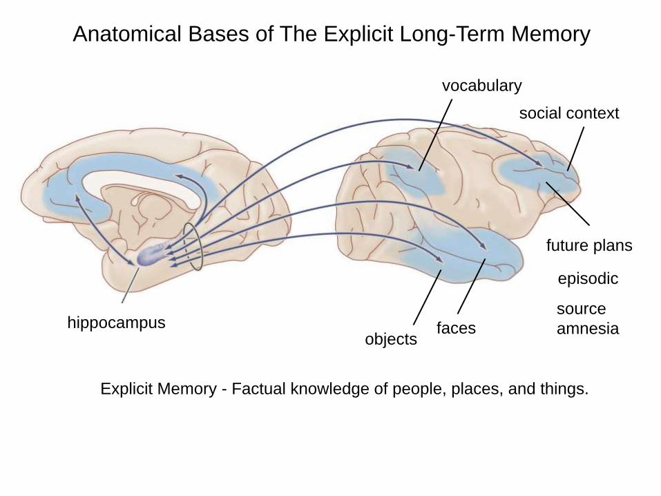
Anatomical Bases of The Explicit Long-Term Memory
future plans
social context
objectsfaces
vocabulary
hippocampus
Explicit Memory - Factual knowledge of people, places, and things.
episodic
source
amnesia

RAT HIPPOCAMPUS – vs. HUMAN HIPPOCAMPUS
RAT
HUMAN
cortex cortex
cortex
cortex
cortex
cortex
cortex
cortex
cortex
cortex
cortex
cortex
cortex
cortex
cortexhippocampus
hippocampus

Box 31D Alzheimer’s DiseaseBox 31D Alzheimer’s Disease – Patient HM
Dr. Scoville’s
Effort

This concludes the material presented in the textbook.
In the reminder of the session we will learn about:
1. Cellular bases of learning.
2. Dopamine role in learning and memory.

Electrical signal
Synaptic
potential
Second
Messengers
Calcium
Gene transcription
Protein synthesis
growth
Electrical signalChange in
Cellular / Molecular Bases of Learning

Cellular and Molecular Bases of
Learning
I. Changing the wiring between neurons (formation of new synapses).
II. Changing the strength of existing synapses (transient or long-lasting modification of neurotransmission)

Current thinking about long-term memory in neocortex is focused on changes in the strengths of
connections between neurons. But ongoing structural plasticity in the adult brain, including synapse
formation/elimination and remodeling of axons and dendrites, suggests that memory could also depend
on learning-induced changes in the cortical 'wiring diagram'. Given that the cortex is sparsely connected,
wiring plasticity could provide a substantial boost in storage capacity, although at a cost of more
elaborate biological machinery and slower learning.
Trachtenberg et al., (2002)
Changing the wiring between neurons

Hebb’s Postulate
When an axon of cell A
is near enough to excite cell B,
or repeatedly or consistently
takes part in firing it,
some growth process or metabolic
change take place
in one or both cells
such that A’s efficiency
is increased.
AB
synapseaxon

Hebb’s Postulate
A B
C D
What Fires together, Wires together.
A B
C D
A B
C D
Example: Pruning of synaptic contacts in brain development
Abundance of synapses Pruning Mature circuit

Changing the strength of the synapse
traditional mechanism for adult plasticity
Long-term Synaptic Potentiation syn.
stim.
after
before
Path 1
Path 2

It has been reported that the size of mEPSCs recorded at
the soma after glutamate activation of single spines is
positively correlated with the number of dendritic spines, or
simply the size of the spine head, that is, large spines
produce large synaptic responses.
LTP
induction
dendritic shaft dendritic shaft
Spontaneous and Evoked Synaptic Currents
EPSC
Excitatory Post-Synaptic Current (EPSC)
50 pA
Change in EPSC
Change in Size

Changing the strength of the synapse traditional
mechanism for
adult plasticity
homosynaptic
mechanism
PRESYNAPTIC
POSTSYNAPTIC
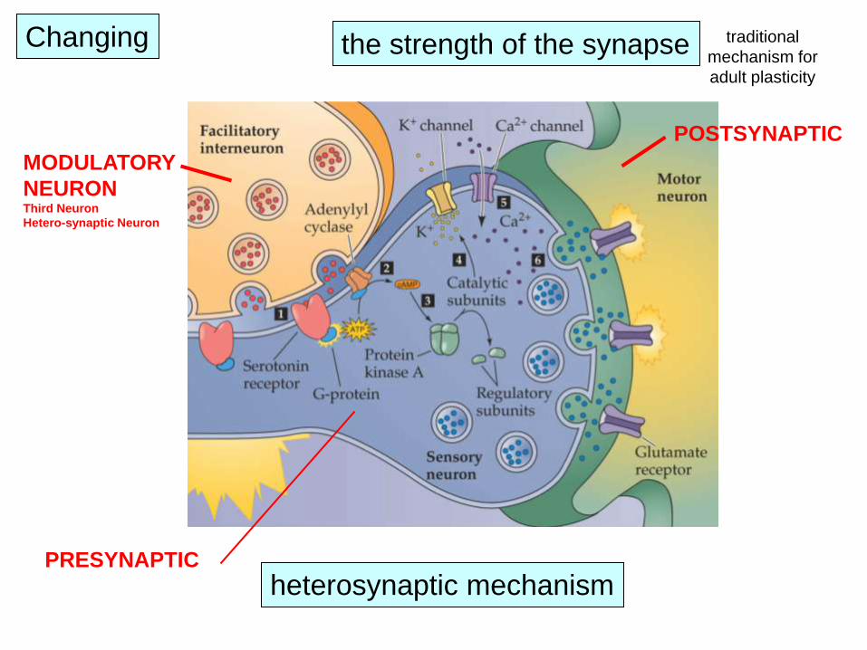
Changing traditional
mechanism for
adult plasticity
heterosynaptic mechanism
the strength of the synapse
MODULATORY
NEURONThird Neuron
Hetero-synaptic Neuron
POSTSYNAPTIC
PRESYNAPTIC

Heterosynaptic potentiation
Neuromodulators such as serotonin
and dopamine might be integral
parts of memory formation circuits
Example 1.

Dopaminergic Neurons Live in VTA
and Project to PFC.

Example 2.

The Role of Dopamine in Learning and Memory
based on the Review article by
John Lisman and Anthony Grace (2005) Neuron
There has been considerable investigation of the
modulation produced by noradrenaline and acetylcholine,
but the role of dopamine (DA) has been less extensively
studied, because of the early view that the hippocampus
did not receive a significant dopaminergic innervation.
It is now clear that the hippocampus does receive such
innervation from Ventral Tegmental Area (VTA).
Ventral
Tegmental
Area
DA
Dopamine (DA)
Hippocampus
Example 3.
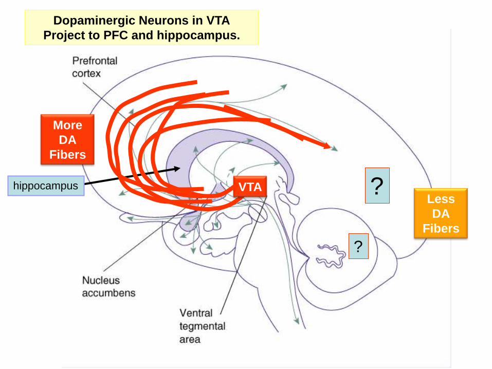
Dopaminergic Neurons in VTA
Project to PFC and hippocampus.
hippocampus ?
?
VTA
More
DA
Fibers
Less
DA
Fibers

The computation of novelty is the comparison of incoming information with stored
memories. If incoming information is different from stored memories, then it is
considered a novelty.
Dentate gyrus
CA1
CA3
Hippocampus
The hippocampus is a temporal lobe structure that is vital for the encoding
and recall of episodic memory.CA1

Several lines of experimental evidence indicate that the
computation of novelty is taking place in the hippocampus.
(1) Single-unit recordings and (2) imaging studies using PET
and fMRI as well as (3) c-Fos expression all indicate that
presentation of a novel stimulus produces a robust increase
in hippocampal activity
(4) interfering with hippocampal function inhibits the orienting
of rats to novel stimulus configurations.
(5) humans with hippocampal lesions are less responsive to
novel stimuli.
Measurements of hippocampal evoked responses in CA1
indicate that a form of novelty (expected versus unexpected
conditioned stimuli) can be detected in less than 100 ms. The
rapidity of this detection suggests that the hippocampus could
be part of the circuit that initiates the short-latency novelty-
dependent firing of the VTA and is not simply responding to it.
The hippocampus and VTA form a functional loop designed to detect novelty
and to use this novelty signal to control the entry of behaviorally significant
information into the hippocampal store of long-term memory.
Ventral
Tegmental
Area
Amygdala
novelty
dopamine
DA
DA
DA
Hippocampus
LATENCY ARGUMENT

Ihalainen et al., 1999.
Exposure of the rat to a novel environment
(such as new cage)
evokes hippocampal DA release as
measured by microdialysis and HPLC.

(A1) Putting a rat in a novel cage, but not a familiar cage evokes DA release in the accumbens. (A2) The
novelty-dependent release of DA is blocked by TTX injection into the subiculum (hippocampus).
Legault and Wise, 2001
Scientists were able to generate a behaviorally significant novelty event by allowing rats to
enter a part of their cage from which they were previously restricted. This event led to
substantial activation of the VTA, as evidenced by the DA released in a VTA target, the
nucleus accumbens.
To test whether this release was dependent on the hippocampus, TTX was injected into the
ventral subiculum, an output structure of the hippocampus that receives direct excitatory
input from CA1. TTX caused a nearly complete block of the novelty-induced DA release

Li et al., 2003.
(Top) A weak tetanus to the Schaffer
collateral input to CA1 pyramidal
cells fails to evoke LTP when the
animal is placed in a familiar cage.
(Second down). After the animal is
placed is a novel cage, the same
stimulus evokes LTP.
(Third down) This LTP can be
blocked by a systemic D1 antagonist.
(Bottom) Conversely, systemic
application of a D1 agonist allows
the stimulus to evoke LTP even in
the familiar cage.
Li, S., Cullen, W.K., Anwyl, R., and Rowan, M.J. (2003). Dopamine-dependent facilitation of LTP
induction in hippocampal CA1 by exposure to spatial novelty. Nat. Neurosci. 6, 526–531.

(A) Experimental design – brain slice – whole-cell patch clamp recording – extracellular stimulation
electrodes S1 and S2. (B) LTP (closed circles) in the CA1 region of a slice preparation is induced by
three tetani (100 Hz); no LTP occurred in the control pathway (open circles). (C) The same stimulation
given in the presence of the selective D1 dopamine receptor antagonist SCH23390 produced only early
LTP. Note that late phase of LTP is missing (arrow).
Morris et al., 2003.
Dopamine release in the hippocampus is important for maintenance of the long-
term synaptic plasticity (late phase of LTP).

SUMMARY Dopamine Role in LTP Formation
The MAIN finding of Li et al., 2003 is that:
novelty-mediated dopamine release into the hippocampus
facilitates the formation of LTP
in the striatum radiatum of CA1 pyramidal neurons
(CA3-to-CA1 pathway, also known as Schaeffer collaterals).