memory cd4+ t cells induce innate responses independently of pathogen
TRANSCRIPT

A r t i c l e s
558 VOLUME 16 | NUMBER 5 | MAY 2010 nAture medicine
Recognition of pathogen-associated molecular patterns (PAMPs) by their receptors results in the production of inflammatory mediators that act to control initial infection and mobilize elements of the innate immune sys-tem1,2. PAMP recognition also facilitates optimal development of adaptive immune responses by activating antigen-presenting cells (APCs) while ensuring that enhanced antigen-specific responses occur only when a path-ogen is present3,4. However, although the role of innate immune recognition in shaping adaptive immune responses is established, a role for adaptive immune cells in regulation of innate inflammation is largely unexplored.
Here we investigate the ability of memory CD4+ T cells to regu-late innate IIC production after influenza infection. Memory CD4+ T cells are crucial for optimal heterosubtypic immunity against influ-enza5, but how they contribute to protection is not well understood6. Whereas virus-specific T cell responses peak about 1 week after heterosubtypic challenge, the distinguishing characteristics of memory as compared to naive T cells, including less stringent require-ments for antigen density and co-stimulation and rapid production of a broader range of cytokines, suggest that memory cells could have key functions at earlier stages of infection7–9.
We show that memory, but not naive, CD4+ T cells act to markedly enhance early expression of IICs and enhance viral control. Induction of IIC requires that TH1- or TH17-polarized memory cells recognize antigen presented by CD11c+ major histocompatibility complex (MHC)-II+ cells in the lung. The protective response is coincident with activation of CD11c+ cells but independent of the IFN-γ, TNF-α and PAMP-recognition pathways. Similar IIC induction occurs when protein-specific memory cells recognize antigen in the absence of infec-tion. These results show that memory CD4+ T cells responding at the site of infection provide enhanced protection via a unique, pathogen-independent pathway for inducing inflammatory mediators.
RESULTSMemory CD4+ T cells enhance production of IICsTo investigate whether memory CD4+ T cells affect innate inflamma-tory responses upon challenge, we measured a panel of IICs (TNF-α, interleukin-1α (IL-1α), IL-1β, IL-6, IL-12, IFN-γ, CXCL9, CXCL10, CCL2, and CXCL1 (KC)) after influenza isolate A/PuertoRico/8/34 (A/PR8) challenge of naive mice compared to mice primed with a heterobsubtypic strain (A/Philippines). At 40 h after infection, we detected increased amounts of IICs in lung homogenates from primed mice (Fig. 1a), and some were enhanced systemically in serum (data not shown). Concentrations of most of the IICs detected remained elevated for several days in primed mice (Supplementary Fig. 1). To determine whether memory CD4+ T cells are responsible for enhanced IIC production, we depleted primed mice of CD4+ or Thy1.2+ cells by CD4-specific or Thyl.1-specific antibody treatment before rechal-lenge (Supplementary Fig. 2). Both treatments similarly lowered the amounts of most IICs in primed mice (Fig. 1a), suggesting that memory CD4+ T cells enhance a broad range of innate inflammatory responses at early time points after influenza challenge.
Because APC populations can also be altered by priming10,11, and because antibody treatment may not deplete memory T cells completely12, we next transferred total CD4+ T cells from influenza-primed mice to unimmunized hosts. Despite the small fraction of influ-enza-specific memory cells in the bulk population, transfer of primed cells significantly upregulated IICs at 40 h after infection (Fig. 1b) but did not result in global increases in inflammation, as levels of IL-4, IL-5, IL-10 and IL-13 were unchanged (data not shown). To compare equal numbers of antigen-specific naive and memory cells, and to facilitate mechanistic analysis, we used T cell receptor (TCR)-transgenic CD4+ T cells recognizing the A/PR8 hemagglutinin protein
Memory CD4+ T cells induce innate responses independently of pathogenTara M Strutt1,2, K Kai McKinstry1,2, John P Dibble1, Caylin Winchell1, Yi Kuang1, Jonathan D Curtis1, Gail Huston1, Richard W Dutton1 & Susan L Swain1
Inflammation induced by recognition of pathogen-associated molecular patterns markedly affects subsequent adaptive responses. We asked whether the adaptive immune system can also affect the character and magnitude of innate inflammatory responses. We found that the response of memory, but not naive, CD4+ T cells enhances production of multiple innate inflammatory cytokines and chemokines (IICs) in the lung and that, during influenza infection, this leads to early control of virus. Memory CD4+ T cell–induced IICs and viral control require cognate antigen recognition and are optimal when memory cells are either T helper type 1 (TH1) or TH17 polarized but are independent of interferon- (IFN-) and tumor necrosis factor- (TNF-) production and do not require activation of conserved pathogen recognition pathways. This represents a previously undescribed mechanism by which memory CD4+ T cells induce an early innate response that enhances immune protection against pathogens.
1Trudeau Institute, Saranac Lake, New York, USA. 2These authors contributed equally to this work. Correspondence should be addressed to T.M.S. ([email protected]).
Received 9 September 2009; accepted 19 March 2010; published online 2 May 2010; doi:10.1038/nm.2142
© 2
010
Nat
ure
Am
eric
a, In
c. A
ll ri
gh
ts r
eser
ved
.

A r t i c l e s
nAture medicine VOLUME 16 | NUMBER 5 | MAY 2010 559
(HNT cells)13. We generated memory cells in vivo by transferring naive HNT cells to host mice and then infected the mice with a sub-lethal dose of influenza A/PR8. We allowed the virus to clear and memory cells to develop for at least 40 d before reisolation. As influenza-specific CD4+ T cell responses are largely TH1 in nature6, we also generated memory cells in vitro by resting TH1-polarized effectors for 3 d in the absence of cytokine and antigen. We showed previously that in vitro–generated memory cells are virtually identical to long-term in vivo memory cells, as assessed by broad criteria14. We found that transfer of either in vivo– or in vitro–generated memory cells induced similar increases in IICs (Fig. 1b), and the expression remained elevated for several days in the lung and serum (Supplementary Fig. 3). These results demonstrate that mem-ory CD4+ T cells present in otherwise naive hosts drive enhanced IIC responses with comparable kinetics to those observed after influenza challenge of primed mice.
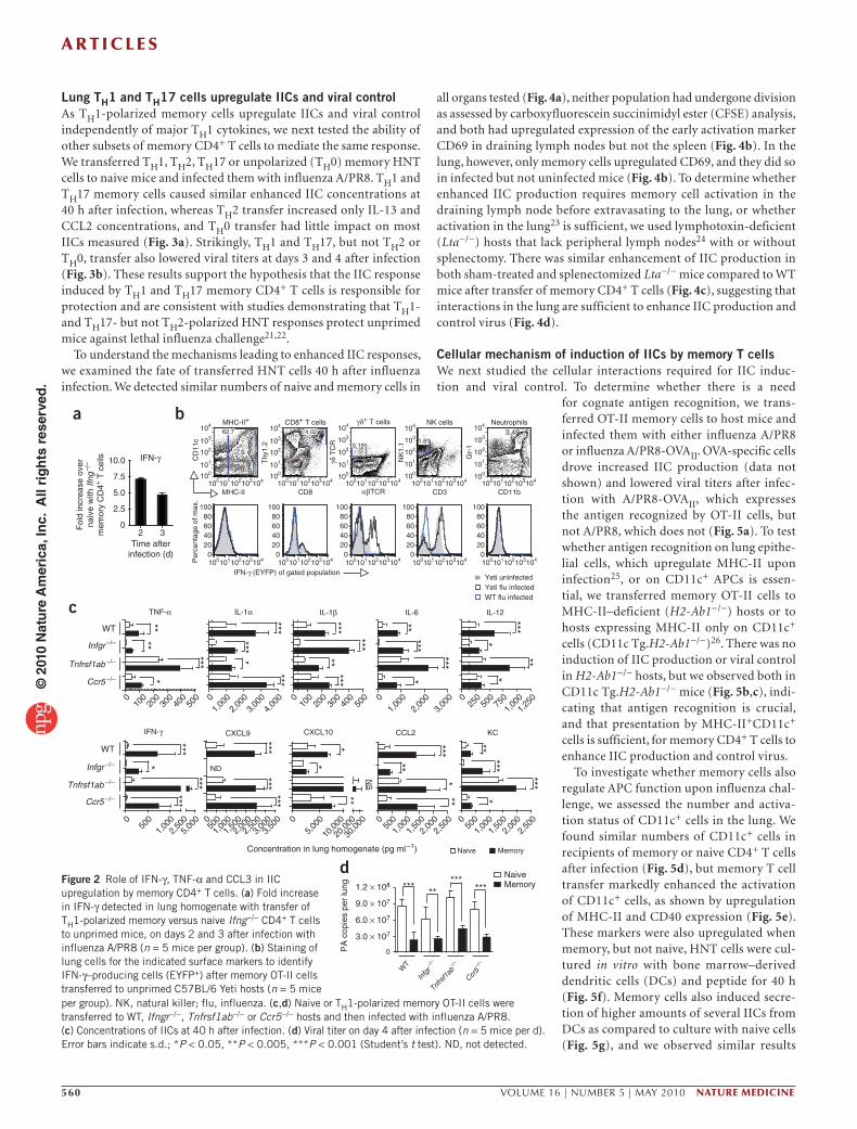
Memory cells upregulate IICs independently of TH1 cytokinesTH1 but not other subsets of memory CD4+ T cells produce abun-dant IFN-γ upon stimulation (Supplementary Fig. 4) and could thus account for the increased IFN-γ amounts observed upon influenza challenge. To determine whether memory cells stimulate IFN-γ production from other sources, we generated TH1-polarized memory cells recognizing ovalbumin (OVA) from IFN-γ–deficient, TCR-transgenic OT-II cells, transferred them to C57BL/6 hosts and infected the mice with virus expressing OVA (A/PR8-OVAII)
15. Memory cells induced elevated IFN-γ on days 2 and 3 after infection (Fig. 2a), indicating IFN-γ production from sources other than donor cells. Employing IFN-γ reporter (Yeti) mice as hosts16, we observed increased IFN-γ (enhanced yellow fluorescence protein (EYFP)) signal from natural killer cells and γδ+ T cells but not from MHC-II+, CD8+ or Gr-1+ cells, indicating contributions from multiple innate populations to the enhanced IIC response (Fig. 2b). In support of this conclusion, memory cell transfer to host mice carrying
the severe combined immune deficiency (SCID) mutation drove an elevated IIC response comparable to that in wild-type (WT) hosts upon influenza challenge (Supplementary Fig. 5).
Because IFN-γ is a potent regulator of inflammation17,18, we evaluated its role in driving enhanced IIC response. We transferred memory cells to IFN-γ receptor–deficient (Infgr−/−) or WT hosts and observed a simi-lar IIC response, with the exception of CXCL9 and CXCL10 (Fig. 2c). As TH1 memory cells also produce TNF-α and CCL3 (Supplementary Fig. 4), we used TNF-α receptor–deficient (Tnfrsf1ab−/−) and Ccr5−/− hosts and again observed a similar upregulation of the IIC response (Fig. 2c). These results indicate that memory CD4+ T cells enhance a broad spectrum of IICs in an IFN-γ–, TNF-α– and CCL3-independent manner, but they are consistent with a requirement for IFN-γ for optimal CXCL9 and CXCL10 induction19,20.
To investigate how enhanced IIC responses affect the course of influenza infection, we measured viral titers. Compared to transfer of naive OT-II cells, memory transfer resulted in significantly lower titers in WT, Infgr−/−, Tnfrsf1ab−/− and Ccr5−/− mice on day 3 (data not shown) and day 4 after infection with A/PR8-OVAII (Fig. 2d). These results demonstrate that enhanced IIC responses mediated by memory CD4+ T cells correlate with viral control after influenza infection.
80
60
40
20
0
150
100
50
0
90
TNF-α IL-1α IL-1β IL-6 IL-12
NaiveMemory: polyclonal
Memory: in vivoMemory: in vitro
Unprimed
Memory: isotype
Memory: CD4+ depleted
Memory: Thy1.2+ depleted
IFN-γ CXCL9 CXCL10 CCL2 KC
TNF-α
*
** *
**
** ** ** ** ******
* ** *
**
** **** ** **
*
*
**
*
**
*
** *
* * ** * *
*
* * * * * * * ** * *
IL-1α IL-1β IL-6 IL-12
IFN-γ
Con
cent
ratio
n in
lung
hom
ogen
ate
(pg
ml–1
)C
once
ntra
tion
in lu
ng h
omog
enat
e (p
g m
l–1)
CXCL9 CXCL10 CCL2 KC
60
30
0
0
100
200
7501,750
0 0 0 0250500750
1,0001,2501,5001,750
100
200
300600
700
3,000
6,000
9,000
500
1,000
1,5002,5003,500
6,000
4,000
5,000
2,000
1,000
3,000
0
650
150250450
100
50
0 0 0
75
150
225
250
375
450
500
1,000
1,500
2,000
750
500
250
0
500
400
200
300
0
100
8,000
6,000
4,000
2,000
0
6,000
4,000
2,000
0
400
300
200
100
0
400
300
200
100
0
900
600
300
0
6,000
4,000
2,000
0
a
b
***
Figure 1 Memory CD4+ T cells induce an acute increase in IICs upon influenza infection. (a) IIC concentrations 40 h after challenge in naive C57BL/6 mice, or mice primed with influenza A/Phil 60 d before treatment, treated with isotype, CD4- or Thy1.2-depleting antibody before influenza A/PR8 challenge (n = 5 mice per group). (b) IIC concentrations after bulk CD4+ T cells were isolated from naive or influenza A/PR8–primed mice (polyclonal memory) and equal numbers transferred to naive hosts or, alternatively, naive or in vivo– or in vitro–generated HNT memory cells were adoptively transferred to naive BALB/c hosts. All recipients were challenged with influenza A/PR8 and lung homogenates assessed for IIC after 40 h (n = 5 mice per group). Dotted lines in all figures represent levels of IICs in the absence of infection. Error bars indicate s.d.; *P < 0.05, **P < 0.005 (one-way analysis of variance (ANOVA) followed by Bonferroni’s post hoc test).
© 2
010
Nat
ure
Am
eric
a, In
c. A
ll ri
gh
ts r
eser
ved
.
A r t i c l e s
560 VOLUME 16 | NUMBER 5 | MAY 2010 nAture medicine
Lung TH1 and TH17 cells upregulate IICs and viral controlAs TH1-polarized memory cells upregulate IICs and viral control independently of major TH1 cytokines, we next tested the ability of other subsets of memory CD4+ T cells to mediate the same response. We transferred TH1, TH2, TH17 or unpolarized (TH0) memory HNT cells to naive mice and infected them with influenza A/PR8. TH1 and TH17 memory cells caused similar enhanced IIC concentrations at 40 h after infection, whereas TH2 transfer increased only IL-13 and CCL2 concentrations, and TH0 transfer had little impact on most IICs measured (Fig. 3a). Strikingly, TH1 and TH17, but not TH2 or TH0, transfer also lowered viral titers at days 3 and 4 after infection (Fig. 3b). These results support the hypothesis that the IIC response induced by TH1 and TH17 memory CD4+ T cells is responsible for protection and are consistent with studies demonstrating that TH1- and TH17- but not TH2-polarized HNT responses protect unprimed mice against lethal influenza challenge21,22.
To understand the mechanisms leading to enhanced IIC responses, we examined the fate of transferred HNT cells 40 h after influenza infection. We detected similar numbers of naive and memory cells in
all organs tested (Fig. 4a), neither population had undergone division as assessed by carboxyfluorescein succinimidyl ester (CFSE) analysis, and both had upregulated expression of the early activation marker CD69 in draining lymph nodes but not the spleen (Fig. 4b). In the lung, however, only memory cells upregulated CD69, and they did so in infected but not uninfected mice (Fig. 4b). To determine whether enhanced IIC production requires memory cell activation in the draining lymph node before extravasating to the lung, or whether activation in the lung23 is sufficient, we used lymphotoxin-deficient (Lta−/−) hosts that lack peripheral lymph nodes24 with or without splenectomy. There was similar enhancement of IIC production in both sham-treated and splenectomized Lta−/− mice compared to WT mice after transfer of memory CD4+ T cells (Fig. 4c), suggesting that interactions in the lung are sufficient to enhance IIC production and control virus (Fig. 4d).
Cellular mechanism of induction of IICs by memory T cellsWe next studied the cellular interactions required for IIC induc-tion and viral control. To determine whether there is a need
Fol
d in
crea
se o
ver
naiv
e w
ith If
ng–/
–
mem
ory
CD
4+ T
cel
ls 10.0
0
0
0 0 0 0
04,
000
3,00
02,
000
1,00
03,
000
3,00
0
2,00
0
2,00
02,
000
1,00
050
0
500
500
500
1,00
01,
000
1,00
03,
500
2,50
02,
500
1,50
01,
500 0
2,00
050
01,
000
2,50
01,
500
2,50
05,
000
5,00
0
30,0
00
20,0
00
10,0
00
400
300
200
100 0 0 0
1,25
01,
000
750
500
250
500
400
300
200
100
WT
****
***
***
***
***
***
***
***
***
*****
**
**
****
*
**
*
*
*
*
***
*****
**
***
******
***
******** ***
***
***
******
**
*
Infgr –/–
Ccr5 –/–
Tnfrsf1ab –/–
Infgr –/–
Ccr5 –/–
Tnfrsf1ab –/–
WT
Naive
Concentration in lung homogenate (pg ml–1)
Memory
32
IFN-γ
c
d
ba
IFN-γ
ND
NS
TNF-α IL-1α IL-1β
CXCL9 KCCCL2CXCL10
IL-12IL-6
MHC-II+
MHC-II
IFN-γ (EYFP) of gated population
CD8 αβTCR CD3 CD11b
Yeti uninfected
WT flu infectedYeti flu infected
CD
11c
Per
cent
age
of m
ax.
Thy
1.2
CD8+ T cells γδ+ T cells
γδ T
CR
Time afterinfection (d)
2.5
5.0
7.5
104
104
103
103100
62.7 4.02
0.191.81
3.49NK cells Neutrophils
NK
1.1
Gr-
1100
101
101
102
104
103
100
101
102
104
103
100
101
102
104
103
100
101
102
104
103
100
101
102
102 104103100101102 104103100101102 104103100101102 104103100101102
80100
60
02040
80100
60
02040
80100
60
02040
80100
60
02040
80100
60
02040
104103100101102104103100101102104103100101102 104103100101102 104103100101102
3.0 × 10⁷
6.0 × 10⁷
9.0 × 10⁷
1.2 × 10⁸
0PA
cop
ies
per
lung
Infg
r–/
–
Ccr5–/
–
Tnfrs
f1ab
–/–
WT
Naive Memory
Figure 2 Role of IFN-γ, TNF-α and CCL3 in IIC upregulation by memory CD4+ T cells. (a) Fold increase in IFN-γ detected in lung homogenate with transfer of TH1-polarized memory versus naive Ifng−/− CD4+ T cells to unprimed mice, on days 2 and 3 after infection with influenza A/PR8 (n = 5 mice per group). (b) Staining of lung cells for the indicated surface markers to identify IFN-γ–producing cells (EYFP+) after memory OT-II cells transferred to unprimed C57BL/6 Yeti hosts (n = 5 mice per group). NK, natural killer; flu, influenza. (c,d) Naive or TH1-polarized memory OT-II cells were transferred to WT, Ifngr−/−, Tnfrsf1ab−/− or Ccr5−/− hosts and then infected with influenza A/PR8. (c) Concentrations of IICs at 40 h after infection. (d) Viral titer on day 4 after infection (n = 5 mice per d). Error bars indicate s.d.; *P < 0.05, **P < 0.005, ***P < 0.001 (Student’s t test). ND, not detected.
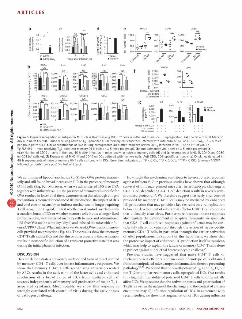
for cognate antigen recognition, we trans-ferred OT-II memory cells to host mice and infected them with either influenza A/PR8 or influenza A/PR8-OVAII. OVA-specific cells drove increased IIC production (data not shown) and lowered viral titers after infec-tion with A/PR8-OVAII, which expresses the antigen recognized by OT-II cells, but not A/PR8, which does not (Fig. 5a). To test whether antigen recognition on lung epithe-lial cells, which upregulate MHC-II upon infection25, or on CD11c+ APCs is essen-tial, we transferred memory OT-II cells to MHC-II–deficient (H2-Ab1−/−) hosts or to hosts expressing MHC-II only on CD11c+ cells (CD11c Tg.H2-Ab1−/−)26. There was no induction of IIC production or viral control in H2-Ab1−/− hosts, but we observed both in CD11c Tg.H2-Ab1−/− mice (Fig. 5b,c), indi-cating that antigen recognition is crucial, and that presentation by MHC-II+CD11c+ cells is sufficient, for memory CD4+ T cells to enhance IIC production and control virus.
To investigate whether memory cells also regulate APC function upon influenza chal-lenge, we assessed the number and activa-tion status of CD11c+ cells in the lung. We found similar numbers of CD11c+ cells in recipients of memory or naive CD4+ T cells after infection (Fig. 5d), but memory T cell transfer markedly enhanced the activation of CD11c+ cells, as shown by upregulation of MHC-II and CD40 expression (Fig. 5e). These markers were also upregulated when memory, but not naive, HNT cells were cul-tured in vitro with bone marrow–derived dendritic cells (DCs) and peptide for 40 h (Fig. 5f). Memory cells also induced secre-tion of higher amounts of several IICs from DCs as compared to culture with naive cells (Fig. 5g), and we observed similar results
© 2
010
Nat
ure
Am
eric
a, In
c. A
ll ri
gh
ts r
eser
ved
.

A r t i c l e s
nAture medicine VOLUME 16 | NUMBER 5 | MAY 2010 561
with the alveolar macrophage line MH-S (data not shown). To deter-mine whether direct cell contact was required for DC activation, we separated memory cells from DCs in a transwell system and stimu-lated them with CD3-specific antibody to induce cytokine produc-tion. DCs separated by the transwell did not upregulate MHC-II or CD40 (Fig. 5f), and we did not observe elevated levels of IICs (Fig. 5g). These results show that memory CD4+ T cells deliver cell contact–dependent signals that enhance the activation of the APCs and drive their production of several IICs.
Memory CD4+ T cells amplify IICs independently of PAMPsFlu viruses trigger Toll-like receptors27,28 and drive type I IFN produc-tion through retinoid-inducible gene-1 (ref. 29). Influenza-induced
effects through these pathways might synergize with or facilitate those mediated by memory CD4+ T cells, leading to enhanced IIC produc-tion and viral control. We transferred memory OT-II cells to IFN-α/β receptor-deficient (Ifnar2−/−), myeloid differentiation factor-88 (MyD88)- and Toll–IL-1 receptor domain–containing adaptor inducing interferon-β (TRIF, encoded by Ticam1)-deficient (Myd88−/−;Ticam1−/−) or WT hosts and infected the mice with influenza A/PR8-OVAII. We found similarly enhanced IIC production and decreased viral tit-ers in all hosts 40 h after challenge (Fig. 6a,b), suggesting that these major PAMP receptor pathways are not involved in memory CD4+ T cell–mediated enhancement.
Given this independence, we asked whether the enhanced IIC produc-tion could be induced by protein antigen in the absence of infection.
200
TNF-α IL-1α IL-1β IL-12 IL-13IL-6
800
200200
400
400
600
600
500200
400
600
800
1,000
1,500
2,000
2,000
4,000
6,000
150
100
50
0 0 0 0 0 0
* **
**
****
****** ***
Naive
Memory: TH0 unpolarized
Memory: TH17 polarized
Memory: TH2 polarized
Memory: TH1 polarized
Con
cent
ratio
n in
lung
hom
ogen
ate
(pg
ml–1
)
IFN-γ IL-17KCCCL2CXCL10CXCL9
300
600
900
500250
500
750
4,000
2,000
0
500
1,000
1,500
2,000
6,000
3,000
6,000
9,000
1,000
1,500
2,000
0000 0
*
*
*
* ******
**** ******
******
PA c
opie
s pe
r lu
ng (
log 10
)
437
8
9
10
Time after infection (d)
***
******
***
a b
Figure 3 TH1- or TH17-polarization is required for enhanced IIC response and viral control. Naive, TH1-, TH17- or TH2-polarized or TH0 unpolarized memory HNT cells were transferred to BALB/c hosts, which were then infected with influenza A/PR8. (a) Concentrations of IICs in lungs 40 h after infection (n = 5 mice per group). (b) Pulmonary viral titers (n = 5 mice per group). Error bars indicate s.d.; *P < 0.05, **P < 0.005, ***P < 0.001 (one-way ANOVA followed by Bonferroni’s post hoc test).
Day 2
d
cba
Eve
nts
NS
6
0
4,000
IL-1α IL-6 IL-12IL-1βTNF-α
IFN-γ CXCL10 CCL2 KCCXCL9
0
** *
* * ** *
* * ***
**
** ***
******
0 0
0
PA c
opie
s pe
r lu
ng (
log 10
)
000
Con
cent
ratio
n in
lung
hom
ogen
ate
(pg
ml–1
)
0
8
9
743
Time after infection (d)
Naive: splenectomy
Memory: shamNaive: sham
Memory: splenectomy
Memory: splenectomy
***
0
3,500
700
1,400
2,100
2,8001,000
1,500
500
100200300400500600700
120140
100806040200
15,000
10,000
5,0001,000
2,000
3,000
4,000 2,500
1,500
500
900
600
300
2,000
1,000
500
1,000
1,500
2,000
2,500
1,000
2,000
3,000
1
2
3
4
5 NS
NS
Num
ber
of d
onor
CD
4+
T c
ells
(lo
g 10)
Spleen
Lung
CFSE CD69
Memory
Spleen
DLNLu
ng
Naive
dLN
Day 2Day 6 Day 6
Naive: uninfected
Memory: fluMemory: uninfectedNaive: flu
Figure 4 Recognition of antigen in the lung is sufficient for IIC upregulation. CFSE-labeled Thy-disparate naive or TH1-polarized memory HNT cells were transferred to separate hosts, which were then infected with influenza A/PR8 (n = 5 per group). (a) Numbers of donor cells in spleen, draining lymph node (DLN) and lung 40 h after infection. (b) Representative CFSE and CD69 expression of donor cells 2 and 6 d after infection. Naive or memory OT-II cells were transferred to sham-treated or splenectomized Lta−/− hosts and infected with influenza A/PR8-OVAII. (c) Pulmonary IIC concentrations at 40 h after infection (n = 5 mice per group). (d) Pulmonary viral titers (n = 5 per day). Error bars indicate s.d.; *P < 0.05, **P < 0.005, ***P < 0.001 (one-way ANOVA followed by Bonferroni’s post hoc test or t test).
© 2
010
Nat
ure
Am
eric
a, In
c. A
ll ri
gh
ts r
eser
ved
.
A r t i c l e s
562 VOLUME 16 | NUMBER 5 | MAY 2010 nAture medicine
We administered lipopolysaccharide (LPS)-free OVA protein intrana-sally and still found broad increases in IICs in the presence of memory OT-II cells (Fig. 6c). Moreover, when we administered LPS-free OVA together with influenza A/PR8, the presence of memory cells specific for OVA resulted in lower viral titers, demonstrating that although antigen recognition is required for enhanced IIC production, the impact of IICs and viral control occurs by an indirect mechanism no longer requiring T cell recognition (Fig. 6d). To test whether viral control correlates with a transient burst of IICs or whether memory cells induce a longer-lived protective state, we transferred memory cells to mice and administered LPS-free OVA on the same day (day 0) and infected the mice with influ-enza A/PR8 7 d later. When infection was delayed, OVA-specific memory cells provided no protection (Fig. 6d). These results show that memory CD4+ T cells induce IICs and that this or other aspects of their activation results in nonspecific induction of a transient protective state that acts during the initial phases of infection.
DISCUSSIONHere we demonstrate a previously undescribed form of direct control by memory CD4+ T cells over innate inflammatory responses. We show that memory CD4+ T cells recognizing antigen presented by APCs results in the activation of the latter cells and enhanced production of a broad range of IICs from multiple cellular sources independently of memory cell production of major TH1-associated cytokines. Most notably, we show this response is strongly correlated with control of virus during the early phases of pathogen challenge.
How might this mechanism contribute to heterosubtypic responses against influenza? Our previous studies have shown that although survival of influenza-primed mice after heterosubtypic challenge is CD8+ T cell dependent, CD4+ T cell depletion results in severely com-promised protection5. We therefore suggest that early viral control provided by memory CD4+ T cells may be mediated by enhanced IIC production that may provide a key restraint on viral replication before the development of substantial effector CD8+ T cell responses that ultimately clear virus. Furthermore, because innate responses also regulate the development of adaptive immunity, we speculate that CD8+ T cell and B cell responses against influenza may be con-siderably altered or enhanced through the action of virus-specific memory CD4+ T cells, in particular through the earlier activation of APC populations. In support of this hypothesis, we show that the protective impact of enhanced IIC production itself is transient, which may help to explain the failure of memory CD4+ T cells alone to protect against supralethal heterosubtypic challenge5.
Previous studies have suggested that naive CD4+ T cells or uncharacterized effectors and memory phenotype cells obtained from unmanipulated mice dampen inflammation, thereby preventing pathology30,31. We found that only well-polarized TH1 and TH17, but not TH2 or unpolarized memory cells, upregulated IICs. Our results thus highlight the ability of polarized CD4+ T cells to differentially affect IICs. We speculate that the activation status and polarization of T cells, as well as the nature of the challenge and the context of antigen encounter, may all influence regulation of IICs. In agreement with recent studies, we show that augmentation of IICs during influenza
TNF-α
NSNS
NSNSNSNS
Vira
l tite
r ra
tiom
emor
y/na
ive
Vira
l tite
r ra
tiom
emor
y/na
ive
Num
ber
of C
D11
c+
cells
per
lung
(lo
g 10)
Con
cent
ratio
n in
lung
hom
ogen
ate
(pg
ml–1
)NS
NS
NS
* * * * * *
**
***
*
* * *
*
******
******
***** *** *** **
**NS
NS
a
f
g
edc
b140
0
250
500
750
1.25
1.00
0.75
0.50
3 4
0.25
1,000
1,250 4,000
5,000
10,000
15,000
1,0001,000
2,0002,000
3,000
3,000
300
600
900
1,200
300
600
900
1,200
1,500
0500
1,0001,5002,0002,5003,0003,500
0.3
0.6
0.9
1.2
100
200
300
400
500
1,000
2,000
3,000
4,000
0
0
0
51,100
500
300
3002,500
100
100
200
200
500
500
1,000
1,000
1,500
1,500
2,000
2,000
2,500
3,000
Uninfec
ted
Flu
infec
ted
Flu
infec
ted
700
500
900
1,300
1,700
MHC-II
MHC-II
CD40
CD40
CD80NS
400
500
600
700
900
0
0
DC: bottomDC: transwell
Peptide + anti-CD3
0 0 0
+ + + + + ++ +++
+ +++
+
++++ –––
–+
+–+
+
–+–
–
–––
––– – –
–+
++ +
255075
100125150175
1
2
3
4
0
WT
Naive
MF
I (C
D11
c+ g
ate)
MF
I (C
D11
c+ g
ate)
Con
cent
ratio
n (p
g m
l–1)
MemoryTime after infection (d)A/PR8A/PR8-OVAII
CD11c Tg.H2-Ab1–/–H2-Ab1–/–
0 00
0
0 020406080
100120
IL-1α IL-1β IL-6 IL-12
KCCXCL10IFN-γ CXCL9 CCL2
NaiveMemory
IL-6 CXCL10IL-1β
NaiveMemory
DC: bottomDC: transwell
Peptide + anti-CD3
Uninfec
ted
Flu
infec
ted
Uninfec
ted
Figure 5 Cognate recognition of antigen on MHC class II–expressing CD11c+ cells is sufficient to induce IIC upregulation. (a) The ratio of viral titers on day 4 in naive C57/BL6 mice receiving naive or TH1-polarized OT-II memory cells and then infected with influenza A/PR8 or A/PR8-OVAII. (n = 5 mice per group per virus.) (b,c) Concentrations of IICs in lung homogenates 40 h after influenza A/PR8-OVAII infection in WT, H2-Ab1−/− or CD11c Tg.H2-Ab1−/− mice receiving TH1-polarized memory OT-II cells (n = 5 mice per group). (b) and pulmonary viral titers (n = 5 mice per group) (c). (d,e) Number of CD11c+ cells in the lung 40 h after infection in mice receiving naive or memory cells (d) and (e) expression of MHC-II, CD40 and CD80 on CD11c+ cells (e). (f) Expression of MHC-II and CD40 on DCs cultured with memory cells. Anti-CD3, CD3-specific antibody. (g) Cytokines detected in 48-h supernatants of naive or memory HNT cells cultured with DCs. Error bars indicate s.d.; *P < 0.05, **P < 0.005, ***P < 0.001 (one-way ANOVA followed by Bonferroni’s post hoc test or t test).
© 2
010
Nat
ure
Am
eric
a, In
c. A
ll ri
gh
ts r
eser
ved
.

A r t i c l e s
nAture medicine VOLUME 16 | NUMBER 5 | MAY 2010 563
infection can be a key element of a protec-tive response32,33, and, because TH2 and TH0 cells neither protect nor induce IICs, we sug-gest that tempering the upregulation of acute inflammation IICs may be deleterious.
We envisage several teleological benefits for memory CD4+ T cell induction of IICs. First, this mechanism may represent a major failsafe for eliciting optimal inflammation and rapid containment of infection in situations where pathogens evade PAMP recognition34. Second, because memory cells can be induced by low concentrations of antigen and co-stimulation, they may activate innate effector mechanisms before pathogen levels are sufficient to trigger robust responses through PAMP recognition. Independent recruitment of IICs by memory cells may also counteract strategies to dampen innate responses used by pathogens, including influenza35. Finally, cytokines such as IL-12 and IL-6 are key in the activation of CD8+ T cells and B cells36,37, and memory CD4+ T cell upregulation of IICs may thus facilitate optimal development of adap-tive responses at later stages after infection.
The protective versus detrimental roles of individual IICs during influ-enza infection is not well understood38. Our results suggest that IFN-γ, TNF-α, CCL3, CXCL-9 and CXCL-10 are not required for early viral control. In contrast, protective roles for IL-1, IL-6 and IL-12 have been previously reported39–41, and these factors were markedly upregulated by protective memory CD4+ T cells. Thus, we suggest that it is most likely that the protective impact of memory CD4+ T cells is not dependent on any one element of IICs alone, but a combination of many. Further studies will be required to determine the degree to which these factors, and others, alone or in combination contribute to viral control.
Our results support the hypothesis that cognate recognition of anti-gen on CD11c+ APC by memory CD4+ T cells is the initiating step in driving IIC production from various innate cells. Most likely, several of the IICs assayed are produced by multiple cellular populations. For example, alveolar macrophages produce robust amounts IL-1, IL-6 and TNF-α; alveolar epithelial cells can produce IL-1, TNF-α and CCL2; natural killer cells produce IFN-γ, TNF-α and CCL3; and activated DCs are sources IL-6, IL-12, TNF-α, CXCL10, and type I IFN36,42–45. Our observations of enhanced levels of many IICs from
DCs and alveolar macrophages stimulated in vitro, combined with observations of multiple cellular sources of IFN-γ in vivo, support the hypothesis that the initial activation of APCs is sufficient to initi-ate a cytokine and chemokine cascade that ultimately results in viral control46. Defining the crucial signals involved in this process will require further study, although our preliminary experiments have ruled out roles for the co-stimulatory molecules CD28, CD40 ligand, OX40 and inducible co-stimulator (data not shown).
Of note, very few donor memory T cells (thousands) are seen in the lung in these studies, yet these small numbers are sufficient to induce IIC production even when induced by low levels of nonreplicating antigen. We speculate that this unique mechanism of IIC upregulation could help explain why memory CD4+ T cells have been implicated in several models of autoimmunity including diabetes, experimental autoimmune encephalomyelitis and collagen-induced arthritis47–49. Further studies will need to determine the full risks and benefits of IICs induced by memory CD4+ T cells in the absence of PAMP recognition.
METHODSMethods and any associated references are available in the online version of the paper at http://www.nature.com/naturemedicine/.
Note: Supplementary information is available on the Nature Medicine website.
ACKnoWLeDGMenTSThis work was supported by the US National Institutes of Health (P01AI04630 to and P01AI04566 to S.L.S.), the US Department of Defense (HR#3222) and the Trudeau Institute. We thank J. Kohlmeier and D. Woodland (Trudeau Institute)
TNF-α
80
a
300
200
100
500
500
40060
40
20
0 0 0 0 0
300
600
900
1,200
1,000
1,000
1,500
2,000
3,000
4,000
IL-1βIL-1α IL-12
P < 0.05P < 0.05P < 0.05P < 0.05
P < 0.05IL-6
300
200
100
500
1,000
2,000
3,000
4,000
5,000
0 0
100
100 10
50
100
150
200
250
3,000
6,000
9,000
2,5000
300
600
900
250
500
750
1,000
1,250
5,0007,500
10,00012,50015,00017,500
0 0
Concentration of LPS-free OVA i.n. (µg)
Con
cent
ratio
n in
lung
hom
ogen
ate
(pg
ml–1
)C
once
ntra
tion
in lu
ng h
omog
enat
e (p
g m
l–1)
0 0
250
500
750
400
0
P < 0.05
P < 0.05P < 0.05
0
1,000
2,000
3,000
0
1,000
2,000
3,000
4,000
5,000
IFN-γ CXCL9 CXCL10 CCL2 KC
P < 0.05 P < 0.05
300
200
100
500
0
0
0OVA day 0
A/PR8 day 7
––
–– ++ +
+ +A/PR8 day 0
NS
NS
5.0 × 10⁷
5.0 × 10⁷
1.0 × 10⁸
2.5 × 10⁸
1.0 × 10⁸
1.5 × 10⁸
1.5 × 10⁸
2.0 × 10⁸
2.0 × 10⁸
NS
Day 3
Day 4
Vira
l tite
r ra
tiom
emor
y/na
ive
0.25
0.50
0.75
1.00
400
00
0 0 0
1,000
250
500
750
Ifnar2–/–
Myd88–/–;Ticam1
WT
TNF-α IL-1α IL-1β IL-12IL-6
100
100 10 10
010
0 10 100
100 10 10
010
0 10
300200100
500
500
2,500
2,000
1,500
1,000
400
600
200
400
700600
100
100 1010
010
0 10100
100 1010
010
0 10100
100 10
IFN-γ CXCL9 CXCL10 CCL2 KC
PA c
opie
s pe
r lu
ng
b
c dMemoryNaive
Naive: WT Memory: Ifnar2–/– Memory: Myd88–/–;Ticam1Memory: WT
*** *** ******
***
*********
*************** ***
*****
**
*
**
****** ***
Figure 6 Memory CD4+ T cells induce IIC responses independently of PAMP recognition. (a,b) Naive or TH1-polarized OT-II memory cells were transferred to naive WT, Ifnar2−/− or Myd88−/−;Ticam1 hosts, which were then infected with influenza A/PR8-OVAII. (a) Concentrations of IICs detected in lungs 40 h after infection (n = 5 mice per group). (b) Viral titers. (c) Concentrations of IICs 40 h after naive or TH1-polarized memory OT-II cells were transferred to C57BL/6 hosts, which were then administered 100 or 10 µg of soluble LPS-free OVA intranasally (i.n.) (n = 5 mice per group). (d) Memory OT-II cells were transferred to C57/BL6 hosts administered LPS-free OVA as in c and infected with A/PR8 on the same day or 7 d later, or only infected with A/PR8, and viral titers determined (n = 5 per group). Error bars indicate s.d.; *P < 0.05, **P < 0.005, ***P < 0.001 (one-way ANOVA followed by Bonferroni’s post hoc test or t test).
© 2
010
Nat
ure
Am
eric
a, In
c. A
ll ri
gh
ts r
eser
ved
.

A r t i c l e s
564 VOLUME 16 | NUMBER 5 | MAY 2010 nAture medicine
for Ccr5–/– and Ifnar2–/– mice and M. Mohrs (Trudeau Institute) for C57BL/6-Tg (IFN-γ–EYFP) mice. Influenza A/Philippines was obtained from S. Epstein (CBER FDA), and engineered virus A/PR8-OVAII was obtained from P. Doherty (University of Melbourne). LPS-free whole OVA protein was a generous gift from T. Moran (Mount Sinai School of Medicine).
AUTHoR ConTRIBUTIonST.M.S. and K.K.M. contributed equally to the design, processing, collection and analysis of data and, together with S.L.S., wrote the paper. S.L.S. and R.W.D. contributed to study design. J.P.D., C.W., Y.K., J.D.C. and G.H. processed and collected data. All authors discussed results and commented on the manuscript.
CoMPeTInG FInAnCIAL InTeReSTSThe authors declare no competing financial interests.
Published online at http://www.nature.com/naturemedicine/. Reprints and permissions information is available online at http://npg.nature.com/reprintsandpermissions/.
1. Janeway, C.A. Jr. & Medzhitov, R. Innate immune recognition. Annu. Rev. Immunol. 20, 197–216 (2002).
2. Iwasaki, A. & Medzhitov, R. Toll-like receptor control of the adaptive immune responses. Nat. Immunol. 5, 987–995 (2004).
3. Pulendran, B. Modulating vaccine responses with dendritic cells and Toll-like receptors. Immunol. Rev. 199, 227–250 (2004).
4. Pulendran, B., Palucka, K. & Banchereau, J. Sensing pathogens and tuning immune responses. Science 293, 253–256 (2001).
5. Powell, T.J. et al. Priming with cold-adapted influenza A does not prevent infection but elicits long-lived protection against supralethal challenge with heterosubtypic virus. J. Immunol. 178, 1030–1038 (2007).
6. Swain, S.L. et al. CD4+ T-cell memory: generation and multi-faceted roles for CD4+ T cells in protective immunity to influenza. Immunol. Rev. 211, 8–22 (2006).
7. Rogers, P.R., Dubey, C. & Swain, S.L. Qualitative changes accompany memory T cell generation: faster, more effective responses at lower doses of antigen. J. Immunol. 164, 2338–2346 (2000).
8. London, C.A., Lodge, M.P. & Abbas, A.K. Functional responses and costimulator dependence of memory CD4+ T cells. J. Immunol. 164, 265–272 (2000).
9. Bradley, L.M., Duncan, D.D., Yoshimoto, K. & Swain, S.L. Memory effectors: a potent, IL-4–secreting helper T cell population that develops in vivo after restimulation with antigen. J. Immunol. 150, 3119–3130 (1993).
10. Dahl, M.E., Dabbagh, K., Liggitt, D., Kim, S. & Lewis, D.B. Viral-induced T helper type 1 responses enhance allergic disease by effects on lung dendritic cells. Nat. Immunol. 5, 337–343 (2004).
11. Didierlaurent, A. et al. Sustained desensitization to bacterial Toll-like receptor ligands after resolution of respiratory influenza infection. J. Exp. Med. 205, 323–329 (2008).
12. Chace, J.H., Cowdery, J.S. & Field, E.H. Effect of anti-CD4 on CD4 subsets. I. Anti-CD4 preferentially deletes resting, naive CD4 cells and spares activated CD4 cells. J. Immunol. 152, 405–412 (1994).
13. Scott, B. et al. A role for non-MHC genetic polymorphism in susceptibility to spontaneous autoimmunity. Immunity 1, 73–83 (1994).
14. McKinstry, K.K. et al. Rapid default transition of CD4 T cell effectors to functional memory cells. J. Exp. Med. 204, 2199–2211 (2007).
15. Thomas, P.G. et al. An unexpected antibody response to an engineered influenza virus modifies CD8+ T cell responses. Proc. Natl. Acad. Sci. USA 103, 2764–2769 (2006).
16. Mayer, K.D. et al. The functional heterogeneity of type 1 effector T cells in response to infection is related to the potential for IFN-γ production. J. Immunol. 174, 7732–7739 (2005).
17. Boehm, U., Klamp, T., Groot, M. & Howard, J.C. Cellular responses to interferon-gamma. Annu. Rev. Immunol. 15, 749–795 (1997).
18. Farber, J.M. Mig and IP-10: CXC chemokines that target lymphocytes. J. Leukoc. Biol. 61, 246–257 (1997).
19. Amichay, D. et al. Genes for chemokines MuMig and Crg-2 are induced in protozoan and viral infections in response to IFN-γ with patterns of tissue expression that suggest nonredundant roles in vivo. J. Immunol. 157, 4511–4520 (1996).
20. Nakanishi, Y., Lu, B., Gerard, C. & Iwasaki, A. CD8+ T lymphocyte mobilization to virus-infected tissue requires CD4+ T-cell help. Nature 462, 510–513 (2009).
21. Brown, D.M., Dilzer, A.M., Meents, D.L. & Swain, S.L. CD4 T cell–mediated protection from lethal influenza: perforin and antibody-mediated mechanisms give a one-two punch. J. Immunol. 177, 2888–2898 (2006).
22. McKinstry, K.K. et al. IL-10 deficiency unleashes an influenza-specific TH17 response and enhances survival against high-dose challenge. J. Immunol. 182, 7353–7363 (2009).
23. Wakim, L.M., Waithman, J., van Rooijen, N., Heath, W.R. & Carbone, F.R. Dendritic cell-induced memory T cell activation in nonlymphoid tissues. Science 319, 198–202 (2008).
24. De Togni, P. et al. Abnormal development of peripheral lymphoid organs in mice deficient in lymphotoxin. Science 264, 703–707 (1994).
25. Debbabi, H. et al. Primary type II alveolar epithelial cells present microbial antigens to antigen-specific CD4+ T cells. Am. J. Physiol. Lung Cell Mol. Physiol. 289, L274–L279 (2005).
26. Lemos, M.P., Fan, L., Lo, D. & Laufer, T.M. CD8α+ and CD11b+ dendritic cell–restricted MHC class II controls TH1 CD4+ T cell immunity. J. Immunol. 171, 5077–5084 (2003).
27. Diebold, S.S., Kaisho, T., Hemmi, H., Akira, S. & Reis e Sousa, C. Innate antiviral responses by means of TLR7-mediated recognition of single-stranded RNA. Science 303, 1529–1531 (2004).
28. Imai, Y. et al. Identification of oxidative stress and Toll-like receptor 4 signaling as a key pathway of acute lung injury. Cell 133, 235–249 (2008).
29. Pichlmair, A. et al. RIG-I–mediated antiviral responses to single-stranded RNA bearing 5′-phosphates. Science 314, 997–1001 (2006).
30. Guarda, G. et al. T cells dampen innate immune responses through inhibition of NLRP1 and NLRP3 inflammasomes. Nature 460, 269–273 (2009).
31. Kim, K.D. et al. Adaptive immune cells temper initial innate responses. Nat. Med. 13, 1248–1252 (2007).
32. Salomon, R., Hoffmann, E. & Webster, R.G. Inhibition of the cytokine response does not protect against lethal H5N1 influenza infection. Proc. Natl. Acad. Sci. USA 104, 12479–12481 (2007).
33. Tuvim, M.J., Evans, S.E., Clement, C.G., Dickey, B.F. & Gilbert, B.E. Augmented lung inflammation protects against influenza A pneumonia. PLoS One 4, e4176 (2009).
34. Kalinski, P. & Moser, M. Consensual immunity: success-driven development of T-helper-1 and T-helper-2 responses. Nat. Rev. Immunol. 5, 251–260 (2005).
35. Hale, B.G., Randall, R.E., Ortin, J. & Jackson, D. The multifunctional NS1 protein of influenza A viruses. J. Gen. Virol. 89, 2359–2376 (2008).
36. Joffre, O., Nolte, M.A., Sporri, R. & Reis e Sousa, C. Inflammatory signals in dendritic cell activation and the induction of adaptive immunity. Immunol. Rev. 227, 234–247 (2009).
37. Dienz, O. et al. The induction of antibody production by IL-6 is indirectly mediated by IL-21 produced by CD4+ T cells. J. Exp. Med. 206, 69–78 (2009).
38. Szretter, K.J. et al. Role of host cytokine responses in the pathogenesis of avian H5N1 influenza viruses in mice. J. Virol. 81, 2736–2744 (2007).
39. Schmitz, N., Kurrer, M., Bachmann, M.F. & Kopf, M. Interleukin-1 is responsible for acute lung immunopathology but increases survival of respiratory influenza virus infection. J. Virol. 79, 6441–6448 (2005).
40. Lee, S.W., Youn, J.W., Seong, B.L. & Sung, Y.C. IL-6 induces long-term protective immunity against a lethal challenge of influenza virus. Vaccine 17, 490–496 (1999).
41. Hama, Y. et al. Interleukin 12 is a primary cytokine responding to influenza virus infection in the respiratory tract of mice. Acta Virol. 53, 233–240 (2009).
42. GeurtsvanKessel, C.H. & Lambrecht, B.N. Division of labor between dendritic cell subsets of the lung. Mucosal Immunol. 1, 442–450 (2008).
43. McGill, J., Heusel, J.W. & Legge, K.L. Innate immune control and regulation of influenza virus infections. J. Leukoc. Biol. 86, 803–812 (2009).
44. Monteiro, J.M., Harvey, C. & Trinchieri, G. Role of interleukin-12 in primary influenza virus infection. J. Virol. 72, 4825–4831 (1998).
45. Zhao, M.Q. et al. Alveolar epithelial cell chemokine expression triggered by antigen-specific cytolytic CD8+ T cell recognition. J. Clin. Invest. 106, R49–R58 (2000).
46. Guidotti, L.G. & Chisari, F.V. Noncytolytic control of viral infections by the innate and adaptive immune response. Annu. Rev. Immunol. 19, 65–91 (2001).
47. Le Saout, C., Mennechet, S., Taylor, N. & Hernandez, J. Memory-like CD8+ and CD4+ T cells cooperate to break peripheral tolerance under lymphopenic conditions. Proc. Natl. Acad. Sci. USA 105, 19414–19419 (2008).
48. Elyaman, W. et al. Distinct functions of autoreactive memory and effector CD4+ T cells in experimental autoimmune encephalomyelitis. Am. J. Pathol. 173, 411–422 (2008).
49. Latham, K.A., Whittington, K.B., Zhou, R., Qian, Z. & Rosloniec, E.F. Ex vivo characterization of the autoimmune T cell response in the HLA-DR1 mouse model of collagen-induced arthritis reveals long-term activation of type II collagen-specific cells and their presence in arthritic joints. J. Immunol. 174, 3978–3985 (2005).
© 2
010
Nat
ure
Am
eric
a, In
c. A
ll ri
gh
ts r
eser
ved
.
nAture medicinedoi:10.1038/nm.2142
ONLINE METHODSMice. Male BALB/c, C57BL/6, MHC-II–deficient (H2-Ab1−/−), CD11c Tg.H2-Ab1−/−, Lta−/−, Ifng−/−, Ifngr−/−, Ifnar2−/−, Ccr5−/−, Myd88−/−;Ticam1−/−, C57BL/6-Ig (IFN-γ– EYFP) (Yeti), and HNT (Vα15, Vβ8.3) and OT-II (Vα2, Vβ5) TCR-transgenic mice were obtained from Trudeau Institute. Male SCID, Tnfrsf1ab−/− and nude mice were purchased from Jackson Laboratories. All infected mice were at least 8 weeks old. All experimental procedures in mice were conducted in accordance with the Trudeau Institute Animal Care and Use Committee guidelines.
Naive and memory CD4 T cell isolation. Naive CD4+ T cells were obtained from pooled spleen and lymph nodes of unimmunized mice ,as previously described14. TH0, TH1, TH2 and TH17 effectors were generated from naive TCR-transgenic CD4+ T cells as previously desribed14,22, and in vitro–generated memory cells were generated from effectors as previously described14. In some experiments, CD4+ T cells were labeled with CFSE.
Polyclonal influenza-specific memory CD4 T cells were enriched by MACS purification (Miltenyi) of bulk CD4+ T cells from mice primed 60 d previously with influenza A/PR8. In vivo–generated memory HNT cells were obtained by first transferring naive HNT cells to nude hosts, which were then infected with a sublethal dose of influenza A/PR8. We isolated donor HNT cells after 40–60 d via CD4+ MACS, after host mice had completely cleared virus. The purity of Vβ8.3+Thy1.1+CD4+ cells was determined by flow cytometry.
Adoptive T cell transfers and T cell depletion. Naive or memory cells were adoptively transferred in 200 µl PBS by intravenous injection to naive hosts. In all experiments, mice received equal numbers of naive and memory CD4+ T cells (5 × 10⁶ TCR-transgenic or 1 × 10⁷ polyclonal cells).
In some experiments, mice were given 500 µg of either CD4-depleting (GK1.5) or Thy1.2-depleting (30H12) antibody via intraperitoneal injection (Bio x Cell).
Virus stocks, quantification of viral titer, ovalbumin protein and infections. Influenza A/PR8 (H1N1) virus was produced in the allantoic cavity of embryo-nated hen eggs from virus stocks originating at St. Jude Children’s Hospital and characterized by a core facility at the Trudeau Institute. A/Philippines (H3N2), obtained from S. Epstein, and engineered virus A/PR8-OVAII obtained from P. Doherty, were prepared similarly.
Mice were infected intranasally under light isoflurane anesthesia (Webster Veterinary Supply) with 50 µl of virus in PBS. In some experiments, mice first received a 500 half-maximal embryonic infectious dose (EID50 (about 0.1half-maximal lethal dose (LD50))) of priming virus 30–60 d before heterosubtypic chal-lenge. All challenge doses of virus were 10,000 EID50 (about 2 LD50). Viral titers were determined by quantification of viral RNA as previously described22, and the number of copies of the acidic polymerase mRNA per lung was calculated.
LPS-free whole OVA protein was a generous gift from T. Moran.
Cell culture. Naive or memory HNT CD4+ T cells, 5 × 10⁴ per well, were cultured in vitro in Costar 24-well plates (Corning) in the absence or pres-ence of HNT peptide with 1 × 10⁵ bone marrow–derived DCs, prepared as previously described50. Alternatively, naive and memory CD4+ T cells were stimulated with CD3-specific antibody and were cultured with bone marrow–derived DCs present either in the bottom of the well or within tran-swell inserts (0.4 µm).
Flow cytometry. Mice were killed followed by exsanguination achieved by per-foration of the abdominal aorta, and lungs were perfused by injecting 10 ml of PBS in the left ventricle. Single-cell suspensions were prepared from lungs, spleen and mediastinal lymph nodes by mechanical disruption and passage through a nylon membrane.
Flow cytometry was performed as previously described14 with allophycocyanin-labeled Thy1.1-specific (OX-7) antibody, allophycocyanin-Thy1.2-specific (53-2.1) antibody (both eBiosciences), PerCP-labeled CD4-specific (RM4.5) antibody or phycoerythrin-labeled Vβ8.3-specific (1B3.3) antibody (both Pharmingen) to identify donor T cells. Phycoerythrin-labeled CD69-specific (H12F3) antibody (Pharmingen) and isotype controls were used for phenotype analysis. Analysis was performed with Becton Dickson FACS Scan (BD Biosciences) and FlowJo (Tree Star) software.
Detection of inflammatory cytokines and chemokines. Lungs were collected and homogenized in RPMI 1640 medium supplemented with 2mM l-glutamine (Invitrogen), 100 international units penicillin, 100 µg ml−1 streptomycin (MediaTech), 10 mM HEPES (Research Organics), 50 µM 2-mercaptoethanol (Sigma-Aldrich) and 7.5% FBS (Hyclone).
Concentrations of cytokines and chemokines in lung homogenates, serum and culture supernatants were determined with mouse multiplex luminex kits (Invitrogen) read on a Luminex 100 reader (Luminex). Concentrations of cytokines and chemokines in unprimed mice are represented by dotted lines in all figures, as we generally observed no significant differences in levels of protein in unprimed mice of different strains, WT values are depicted.
Statistical analyses. Unpaired, two-tailed, Student’s t tests, ∝ = 0.05, were used to assess whether the means of two normally distributed groups differed significantly. The Welch-correction was applied when variances were found to differ. One-way ANOVA analysis with Bonferroni’s multiple comparison post-test was used to compare multiple means. Significance is indicated as *P < 0.05, **P < 0.005 and ***P < 0.001. All error bars represent the s.d.
50. Strutt, T.M., Uzonna, J., McKinstry, K.K. & Bretscher, P.A. Activation of thymic T cells by MHC alloantigen requires syngeneic, activated CD4+ T cells and B cells as APC. Int. Immunol. 18, 719–728 (2006).
© 2
010
Nat
ure
Am
eric
a, In
c. A
ll ri
gh
ts r
eser
ved
.