memory subsets
DESCRIPTION
Memory SubsetsTRANSCRIPT
12 Feb 2004 16:10 AR AR210-IY22-25.tex AR210-IY22-25.sgm LaTeX2e(2002/01/18)P1: IKH10.1146/annurev.immunol.22.012703.104702
Annu. Rev. Immunol. 2004. 22:745–63doi: 10.1146/annurev.immunol.22.012703.104702
Copyright c© 2004 by Annual Reviews. All rights reservedFirst published online as a Review in Advance on December 12, 2003
CENTRAL MEMORY AND EFFECTOR MEMORY
T CELL SUBSETS: Function, Generation, andMaintenance
Federica Sallusto, Jens Geginat, and Antonio LanzavecchiaInstitute for Research in Biomedicine, CH-6500 Bellinzona, Switzerland;email: [email protected]; [email protected];[email protected]
Key Words TH1/TH2, T lymphocyte activation, chemokine receptors, lymphocytemigration, effector function
■ Abstract The memory T cell pool functions as a dynamic repository of antigen-experienced T lymphocytes that accumulate over the lifetime of the individual. Recentstudies indicate that memory T lymphocytes contain distinct populations of centralmemory (TCM) and effector memory (TEM) cells characterized by distinct homingcapacity and effector function. This review addresses the heterogeneity of TCM and TEM,their differentiation stages, and the current models for their generation and maintenancein humans and mice.
THE CELLULAR BASIS OF IMMUNOLOGICAL MEMORY
Memory is the hallmark of the acquired immune system. It results from the clonalexpansion and differentiation of antigen-specific lymphocytes that ultimately per-sist for a lifetime. Memory lymphocytes confer immediate protection in peripheraltissues and mount recall responses to antigens in secondary lymphoid organs. Inthe B cell system these functions are carried out by distinct cell types. Protec-tive memory is mediated by plasma cells that secrete antibodies, whereas reactivememory is mediated by memory B cells that proliferate and differentiate to plasmacells in response to secondary antigenic stimulation (1–3). A similar division oflabor has recently emerged in the T cell system (4–6). According to the model pro-posed (7), protective memory is mediated by effector memory T cells (TEM) thatmigrate to inflamed peripheral tissues and display immediate effector function,whereas reactive memory is mediated by central memory T cells (TCM) that hometo T cell areas of secondary lymphoid organs, have little or no effector function, butreadily proliferate and differentiate to effector cells in response to antigenic stim-ulation. In this review we first describe the properties of TCM and TEM in terms oftheir heterogeneity, effector functions, and responsiveness to antigen or cytokines.
0732-0582/04/0423-0745$14.00 745
Ann
u. R
ev. I
mm
unol
. 200
4.22
:745
-763
. Dow
nloa
ded
from
ww
w.a
nnua
lrev
iew
s.or
gby
Har
vard
Uni
vers
ity o
n 10
/16/
13. F
or p
erso
nal u
se o
nly.

12 Feb 2004 16:10 AR AR210-IY22-25.tex AR210-IY22-25.sgm LaTeX2e(2002/01/18)P1: IKH
746 SALLUSTO ¥ GEGINAT ¥ LANZAVECCHIA
We then discuss a pathway of progressive T cell differentiation along which TCM
are arrested at an intermediate stage preceding that of TEM. Finally, we considercurrent models for generation and maintenance of memory T cell subsets.
PHENOTYPIC AND FUNCTIONAL PROPERTIESOF TCM AND TEM
Definition of Human TCM and TEM
TCM and TEM were initially defined in the human system based on two distinctcriteria: (a) the absence or presence of immediate effector function and (b) theexpression of homing receptors that allow cells to migrate to secondary lymphoidorgans versus nonlymphoid tissues (4). Human TCM are CD45R0+memory cellsthat constitutively express CCR7 and CD62L, two receptors that are also char-acteristic of na¨ıve T cells, which are required for cell extravasation through highendothelial venules (HEV) and migration to T cell areas of secondary lymphoidorgans (8, 9). When compared with na¨ıve T cells, TCM have higher sensitivity toantigenic stimulation, are less dependent on costimulation, and upregulate CD40Lto a greater extent, thus providing more effective stimulatory feedback to dendriticcells (DC) and B cells. Following TCR triggering, TCM produce mainly IL-2, butafter proliferation they efficiently differentiate to effector cells and produce largeamounts of IFN-γ or IL-4.
Human TEM are memory cells that have lost the constitutive expression ofCCR7, are heterogeneous for CD62L expression, and display characteristic setsof chemokine receptors and adhesion molecules that are required for homing toinflamed tissues. When compared with TCM, TEM are characterized by rapid effectorfunction. CD8 TEM carry large amounts of perforin, and both CD4 and CD8 produceIFN-γ , IL-4, and IL-5 within hours following antigenic stimulation. Some CD8TEM express CD45RA (here defined as TEMRA) and carry the largest amount ofperforin. Thus in humans, the TEM pool contains bona fide TH1, TH2, and CTL.
The relative proportions of TCM and TEM in blood vary in the CD4 and CD8compartments; TCM is predominant in CD4 and TEM in CD8. Within the tissues,however, TCM and TEM show characteristic patterns of distributions. TCM are en-riched in lymph nodes and tonsils, whereas lung, liver, and gut contain greaterproportions of TEM (10). In antigen-primed individuals, tetanus toxoid-specificCD4 T cells can be detected in circulating TCM and TEM up to 10 years after anti-genic stimulation, and their frequencies increase in both subsets following boosterimmunization (4). The same is true for the CD8 compartment: Antigen-specific Tcells, detected by HLA/peptide multimer staining, can be found in TCM and TEM
subsets, although the relative proportion can be highly variable. For example, HIV-specific T cells are largely TEM (CD45RA–CCR7−), whereas CMV-specific T cellsare predominantly TEMRA (CCR7–CD45RA+) (11). In some melanoma patients,melan-A tetramer+ T cells are mainly CCR7+CD45RA+ and do not respond to
Ann
u. R
ev. I
mm
unol
. 200
4.22
:745
-763
. Dow
nloa
ded
from
ww
w.a
nnua
lrev
iew
s.or
gby
Har
vard
Uni
vers
ity o
n 10
/16/
13. F
or p
erso
nal u
se o
nly.

12 Feb 2004 16:10 AR AR210-IY22-25.tex AR210-IY22-25.sgm LaTeX2e(2002/01/18)P1: IKH
MEMORY T CELL SUBSETS 747
antigenic stimulation ex vivo, whereas in other patients melan-A tetramer+ cellshave a TEM or TEMRA phenotype (CCR7− CD45R0+ or CCR7−CD45RA+) andresponses to melan-A peptide can be readily demonstrated (12, 13). Interestingly,a longitudinal study of EBV-infected patients revealed differences in lytic versuslatent epitope-specific composition of T cell populations in the chronic carrierstage of infection (14). Thus while tetramer-positive cells in the infectious stagehave a CCR7−CD45RA− phenotype, cells specific for lytic but not latent epitopesacquire CD45RA in the chronic carrier stage.
A detailed TCR repertoire analysis was performed by spectra-typing inCD45R0+ memory CD8 T cells using CD62L to discriminate between TCM andTEM (15). The analysis of six influenza-specific T cell clones showed that twoclonotypes were shared between TCM and TEM, whereas four were detected onlyin TCM. A similar picture was obtained after a nine-month period. These resultsindicate that the same expanded clone can be present in both TCM and TEM subsetsand that, within the influenza-specific CD8 memory T cell pool, the clonotypedistribution is remarkably stable with no evidence of conversion from CD62L+and CD62L− memory subsets or vice versa.
The presence of memory T cells with different migratory capacity and effec-tor function was also documented in mice. Two populations of memory CD4 Tcells survive for months after immunization with antigen in adjuvant: One, foundprimarily in the lymph nodes, produces IL-2; the other larger population found innonlymphoid tissues produces IFN-γ (5). Similarly, two populations of antigen-specific memory CD8 T cells are present following viral or bacterial infection (6).Whereas CD8 memory T cells isolated from nonlymphoid tissues exhibit lyticactivity directly ex vivo, their splenic counterparts do not. These results extendedthe TCM/TEM paradigms to the mouse system.
In summary, there is now convincing evidence that antigen-specific CD4 andCD8 memory T cells persist as TCM and TEM populations. However, since the firstdescription of TCM and TEM, it became evident that these two broad subsets wereheterogeneous in expression of chemokine receptors, adhesion, and costimulatorymolecules (4).
Heterogeneity of TCM and TEM
Subsets of TCM and TEM with distinct functional programs can be identified accord-ing to the expression of surface molecules. Costimulatory molecules have beenthe first markers used to dissect the heterogeneity of memory T cells. CD27 andCD28, which are expressed on na¨ıve T cells, are also expressed on some memoryT cells but are absent in a subset of CD8 memory T cells characterized by high ef-fector function and expression of CD45RA (16, 17). The CD27−CD45RA+CD8T cell population largely overlaps with TEMRA. However, some cells within TEMRA
express CD27 and display phenotypic and functional features that are intermediatebetween na¨ıve and effector T cells (18).
A sizable proportion of circulating TCM expresses CXCR5, the receptor forCXCL13, a chemokine produced in B cell follicles (19, 20). These CXCR5+ T
Ann
u. R
ev. I
mm
unol
. 200
4.22
:745
-763
. Dow
nloa
ded
from
ww
w.a
nnua
lrev
iew
s.or
gby
Har
vard
Uni
vers
ity o
n 10
/16/
13. F
or p
erso
nal u
se o
nly.

12 Feb 2004 16:10 AR AR210-IY22-25.tex AR210-IY22-25.sgm LaTeX2e(2002/01/18)P1: IKH
748 SALLUSTO ¥ GEGINAT ¥ LANZAVECCHIA
cells, which have been defined as follicular helper T cells (TFH), are nonpolarizedand upon activation produce IL-2 and some IL-10. They are also present in tonsilswhere they express CD40L and ICOS and provide spontaneous help to B cells(21–23).
The TEM pool can be subdivided according to the expression of chemokinereceptors characteristic of TH1 or TH2 cells (24). Thus within TEM, CCR5 andCXCR6 discriminate TH1 and CTL, whereas CCR3 and CRTh2 identify TH2 cells(25–28). CXCR3 and CCR4 are expressed on TH1 and TH2 (29, 30), respectively,but are also expressed on distinct subsets of TCM. As is discussed below, CXCR3+and CCR4+ TCM subsets represent pre-effector cells (pre-TH1 and pre-TH2, re-spectively).
The combinatorial expression of adhesion molecules and chemokine receptorsallows tissue-specific targeting of T cell and leukocyte subsets (31, 32). Thus thesimultaneous expression of CLA and CCR4 identifies skin homing T cells (33),whereas the expression ofα4β7 and CCR9 is characteristic of gut-homing T cells(34). Some skin-homing and gut-homing T cells express CCR7 (10), suggestingthat they may be capable of homing to lymphoid as well as nonlymphoid tissues.In addition, while most TCM simultaneously express CCR7 and CD62L, there areseveral TEM, especially within CD4, that lack CCR7 but express CD62L (4). Thisfinding is consistent with the ability of CD62L+TEM to enter lymph nodes throughHEV using other receptors that bind to chemokines capable of mediating arrestunder flow (35). This may be particularly relevant in inflammatory conditions whenchemokines produced in peripheral tissues may be transported and displayed onthe luminal face of endothelial cells (36, 37).
In summary, functional subsets can be discriminated within TCM and TEM usingchemokine receptors and other markers (Figure 1). It should be noted, however, thatsome of these markers are rapidly and transiently modulated upon cell activation.For instance, following antigenic stimulation TEM transiently upregulate CCR7and CXCR5 while downregulating CCR5 (38). Furthermore, CD62L is rapidlyshed after TCR triggering or following lymph node immigration (39, 40). Thusthe phenotypic characterization of TCM and TEM applies to only resting cells, i.e.,those that are not engaged in an antigen-driven response.
Response of TCM and TEM to Antigenic Stimulation
When stimulated in vitro, memory T cells show low-activation threshold and vig-orous proliferation. Although both TCM and TEM have a high responsiveness toantigenic stimulation, the expansion potential decreases from TCM to TEM and isvery low in CD8 TEMRA (4, 41). The reduced proliferative capacity correlates witha decrease in telomere length and with an increased propensity to undergo apopto-sis. These intrinsic constraints can be overridden by costimulation, which inducestelomerase activity and upregulation of antiapoptotic molecules (42, 43). Indeed,whereas memory T cells (especially TEM) have short telomeres (4), a small subset
Ann
u. R
ev. I
mm
unol
. 200
4.22
:745
-763
. Dow
nloa
ded
from
ww
w.a
nnua
lrev
iew
s.or
gby
Har
vard
Uni
vers
ity o
n 10
/16/
13. F
or p
erso
nal u
se o
nly.

12 Feb 2004 16:10 AR AR210-IY22-25.tex AR210-IY22-25.sgm LaTeX2e(2002/01/18)P1: IKH
MEMORY T CELL SUBSETS 749
Figure 1 Phenotypic heterogeneity of human memory T cells. Note that the percentagesare only indicative of those found in a healthy human adult because there is considerableindividual variability.
of CD8 memory T cells, which have undergone several rounds of cell divisionin vivo (as detected by low levels of T cell receptor excision circles), has longtelomeres, possibly owing to induction of telomerase activity (18).
When analyzed at the clonal level, TCM stimulated in vitro appear to be het-erogeneous in their ability to differentiate (44). Some cells can be propagated ina noneffector state by stimulation under neutral conditions and can be inducedto differentiate to TH1 and TH2 upon stimulation in the presence of IL-12 orIL-4, respectively. Others, however, spontaneously differentiate to IFN-γ - or IL-4-producing cells, even if stimulated in the absence of polarizing cytokines. There-fore, these cells represent pre-TH1 and pre-TH2 and can be identified accordingto the expression of CXCR3 and CCR4, respectively (41; J. Geginat, unpublisheddata).
When stimulated under neutral conditions, TEM retain their TH1 or TH2 pheno-type, demonstrating that the pattern of cytokine gene expression that is imprinted atpriming can be stably maintained (44). However, when stimulated under oppositepolarizing conditions, most TEM can acquire the ability to express the alternative cy-tokine. Thus TH1 maintain the IFN-γ -producing ability but are also able to produceIL-4. Similarly, most TH2 maintain IL-4 production and acquire IFN-γ -producingability. An exception is represented by CRTh2+ T cells, which are irreversiblycommitted to the TH2-lineage and are unable to acquire IFN-γ -producing abilitywhen stimulated with IL-12. The ability of most human TEM to undergo divergent
Ann
u. R
ev. I
mm
unol
. 200
4.22
:745
-763
. Dow
nloa
ded
from
ww
w.a
nnua
lrev
iew
s.or
gby
Har
vard
Uni
vers
ity o
n 10
/16/
13. F
or p
erso
nal u
se o
nly.
12 Feb 2004 16:10 AR AR210-IY22-25.tex AR210-IY22-25.sgm LaTeX2e(2002/01/18)P1: IKH
750 SALLUSTO ¥ GEGINAT ¥ LANZAVECCHIA
TH1 and TH2 differentiation is a property that is not shared by mouse T cells, whichbecome rapidly committed following stimulation (45). Considering that in the hu-man system a substantial number of TCM are uncommitted and most memory Tcells maintain cytokine flexibility, it is possible that expression of the opposite cy-tokine can be enforced by delivering recall antigens together with the appropriatepolarizing signals.
In summary, TCM are heterogeneous and contain both uncommitted and pre-committed noneffector cells. Importantly, the CCR7+ phenotype is rapidly lostupon antigenic stimulation concomitant with the differentiation of TCM to effectorcells. In addition, with the exception of a transient CCR7 upregulation induced byTCR stimulation, TEM remain CCR7− and maintain both memory and flexibil-ity of cytokine gene expression. These results are consistent with the notion thatCCR7 expression is irreversibly lost upon differentiation and marks the TCM toTEM transition.
How do TCM and TEM achieve antigenic stimulation in vivo? TCM, with accessto secondary lymphoid organs, can be stimulated by antigen presented by matureDC, whereas TEM, which are largely excluded from these areas, can be stimulatedby antigen presented by nonprofessional APC in a milieu that does not favor stablecell-cell interactions (46, 47). How then can the quality of the primary responsebe perpetuated in the secondary response? We consider three possibilities. First,the T cell stimulatory conditions provided by the innate immunity through DCactivation may be similar in both primary and secondary responses and thereforegenerate the same type of effector cells from uncommitted precursors. Second,precommitted T cells may have been generated in the primary response, and uponsubsequent antigen exposure, the cells rapidly differentiate to TH1 or TH2. A thirdpossibility is that TEM may condition DC in the peripheral tissues or may reachthe antigen-stimulated lymph nodes by upregulating CCR7 or through alternativepathways. In both of these cases the local production of IFN-γ and IL-4 by TEM
may further direct a TH1 or TH2 response.It is interesting to consider the differentiation stage of memory T cells within
the framework of the hygiene hypothesis (48). Individuals that have undergonemultiple TH1 responses may be protected from allergic diseases not only by mem-ory TH1 cells present in nonlymphoid tissues but also by the large repertoire ofpotentially cross-reactive pre-TH1 cells present in secondary lymphoid organs.
Response of TCM and TEM to Homeostatic Cytokines
Proliferation of memory T cells can be driven not only by antigenic stimulation butalso by cytokines. Indeed, under steady-state conditions, memory T cells slowlyturn over in the absence of antigen (49, 50). In vivo studies using gene-targetedmice demonstrated that IL-7 and IL-15, which are constitutively produced by avariety of cells, play an essential role for maintenance of both CD4 (51) andCD8 T cells (52–54). Early studies in the human system showed that T cellsproliferate in response toγ -common dependent cytokines in a TCR-independent
Ann
u. R
ev. I
mm
unol
. 200
4.22
:745
-763
. Dow
nloa
ded
from
ww
w.a
nnua
lrev
iew
s.or
gby
Har
vard
Uni
vers
ity o
n 10
/16/
13. F
or p
erso
nal u
se o
nly.

12 Feb 2004 16:10 AR AR210-IY22-25.tex AR210-IY22-25.sgm LaTeX2e(2002/01/18)P1: IKH
MEMORY T CELL SUBSETS 751
fashion (55). More recent studies demonstrated that TCM not only proliferate inresponse to homeostatic cytokines but also differentiate to effector cells expressingreceptors for inflammatory chemokines and producing large amounts of effectorcytokines (41, 56). In response to IL-7 and IL-15, CXCR3+ TCM differentiate toTH1, whereas most CCR4+ TCM differentiate to TH2, consistent with the notionthat these subsets are precommitted. Under the same culture conditions, CD8 TCM
generate a variety of effector cells that include CD45RA+ and CD45RA− CTL(41, 57). The findings that TEMRA appear only late during the immune response (14)and that CD45RA+ can be upregulated on memory T cells following cytokine-driven but not antigen-driven proliferation (41) suggest that the TEMRA subset isgenerated primarily through homeostatic rather than antigen-dependent pathways.
T CELL DIFFERENTIATION: EFFECTOR CELLS ANDINTERMEDIATES
The molecular definition of TH1 and TH2 differentiation provides a powerfulparadigm to understand where, along the T cell differentiation pathway, TCM andTEM are positioned and to identify the signals involved in their generation. Theevidence that we discuss below indicates that TCM are cells arrested at intermedi-ate stages of differentiation preceding TEM and suggests that TCM are generated bysubthreshold stimulation.
Imprinting of TH1 and TH2 Differentiation
Upon antigenic stimulation, na¨ıve T cells can enter a TH1 or TH2 differentiationprogram, which involves the coordinated expression of genes controlling tissuehoming and effector function. Signals emanating from the TCR and from theIL-12 or the IL-4 receptor act in synergy to induce specific transcription factors(58, 59) that mediate chromatin remodeling events at target genes (60–64). Inparticular in differentiating TH1 and TH2 cells, T-bet and GATA-3 induce histonemodifications and DNA methylation of the IFN-γ and IL-4 genes, respectively,increasing their accessibility to the transcriptional machinery (65). Other genescharacteristic of TEM, such as receptors for inflammatory chemokines and perforin,appear to be controlled by similar chromatin remodeling events (66, 67). Histoneacetylation at a locus can be inherited through mitosis and can therefore contributeto the maintenance of specific states of gene activity from one generation to thenext. Circulating CCR5+ and CRTh2+ TEM show patterns and levels of histoneacetylation at cytokine genes characteristic of in vitro cultured TH1 and TH2 cells,whereas TCM have an acetylation state similar to that of na¨ıve T cells (44).
The cytokine gene flexibility characteristic of human TEM described above canbe explained by the capacity of these cells to upregulate the relevant transcrip-tion factor. Indeed, upon stimulation under TH2 conditions, CCR5+ TH1 cellsupregulate GATA-3 and acquireIl4 acetylation and expression. In contrast, when
Ann
u. R
ev. I
mm
unol
. 200
4.22
:745
-763
. Dow
nloa
ded
from
ww
w.a
nnua
lrev
iew
s.or
gby
Har
vard
Uni
vers
ity o
n 10
/16/
13. F
or p
erso
nal u
se o
nly.

12 Feb 2004 16:10 AR AR210-IY22-25.tex AR210-IY22-25.sgm LaTeX2e(2002/01/18)P1: IKH
752 SALLUSTO ¥ GEGINAT ¥ LANZAVECCHIA
stimulated in the presence of IL-12, CRTh2+ TH2 cells are unable to upregulate T-bet and therefore fail to acquire IFN-γ -producing capacity (44). These results arein agreement with transfection experiments demonstrating that T-bet and GATA-3are necessary and sufficient to induce IFN-γ and IL-4 gene expression (58, 68).Recent data on the expression of T-bet and GATA-3 in TCM and TEM suggest aprogressive loss of flexibility as T helper cells proceed from na¨ıve to the effectormemory stages of differentiation (69).
Chromatin remodeling at theIfng and Il4 loci is acquired progressively asa function of TCR and cytokine stimulation (70). Regulation of cytokine geneaccessibility allows for a wide range of expression levels in response to antigenicstimulation. For instance, IFN-γ and IL-4 are produced in very small amountsby naı̈ve T cells (45) and at increased levels in some TCM, whereas a rapid andgreater production is characteristic of highly differentiated TH1 and TH2 presentwithin TEM (44). The levels and kinetics of cytokine gene expression are obviouslyrelevant parameters of effector function and, in this context, it is worth consideringthat in vitro and in vivo read outs can differ significantly and that the nature of thestimulus (i.e., antigenic versus pharmacological stimulation) may lead to divergentresults (68, 71).
T cell differentiation to TH1 or TH2 can be inhibited either by neutralizing IL-12 and IL-4 or by addition of TGF-β (30, 72). This results in the generation ofnonpolarized T cells that retain constitutive CCR7 expression, have low levels ofGATA-3 and T-bet, and carry hypoacetylated IFN-γ and IL-4 genes (44, 73, 74).These cells have all the characteristics of TCM, i.e., being either uncommitted orprecommitted to TH1 or TH2.
Signal Strength and Progressive T Cell Differentiation
In addition to the role cytokines play, it is worth emphasizing that of TCR signalstrength as a major factor in determining T cell differentiation. The strength of sig-naling that T cells receive can vary widely depending not only on the concentrationof antigen and costimulatory molecules (determining the rate of TCR triggering andsignal amplification) (75, 76) but also on the duration of the interaction between Tcells and APC (determining the duration of signaling) (77). Recent studies revealedthat DC/T cell interactions are highly dynamic and can last from a few minutes toseveral hours (46, 78). Importantly, a prolonged stimulation can compensate for alower level of antigen or costimulatory molecules, whereas an increase in antigenor costimulatory molecules can compensate for a shorter stimulation.
With use of an in vitro priming system in which the strength of antigenicstimulation is tightly controlled, na¨ıve CD4 T cells primed by a weak stimulusproliferate but do not develop effector function (79). Upon in vivo transfer thesecells migrate to lymph nodes where they rapidly proliferate and differentiate inresponse to antigen. In contrast, T cells primed by a strong stimulus in the presenceof IL-12 or IL-4 differentiate to TH1 or TH2 that, upon in vivo transfer, are excludedfrom lymph nodes and migrate to inflamed peripheral tissues. After an initial
Ann
u. R
ev. I
mm
unol
. 200
4.22
:745
-763
. Dow
nloa
ded
from
ww
w.a
nnua
lrev
iew
s.or
gby
Har
vard
Uni
vers
ity o
n 10
/16/
13. F
or p
erso
nal u
se o
nly.
12 Feb 2004 16:10 AR AR210-IY22-25.tex AR210-IY22-25.sgm LaTeX2e(2002/01/18)P1: IKH
MEMORY T CELL SUBSETS 753
stimulation, CD8 T cell differentiation to peripheral tissue homing effector cellsis promoted by IL-2, whereas IL-15 expands nonpolarized T cells that, upon invivo transfer, home to both lymphoid and nonlymphoid tissues (80, 81). Signalstrength can also be modulated in vitro by the number and type of DC used (82).Whereas low DC/T cell ratio preferentially generate TCM-like cells, higher ratiosfavor priming of effector cells and eventually lead to T cell deletion by activation-induced cell death. The expression of costimulatory molecules, such as ICOS-Land 4-1BBL, plays a role in reinforcing the signals by APC and is required foreffective T cell responses (83–86).
Initial studies on the requirements for CD8 T cell priming suggested that theirdifferentiation follows an autonomous program after brief stimulation with highlystimulatory engineered APC (87). However, further studies showed that signalstrength also plays a key role in CD8 T cell differentiation (88, 89). Priming bya signal of suboptimal strength, such as that provided by immature DC, inducesCD8 T cell proliferation, but the cells fail to upregulate antiapoptotic moleculesand receptors for homeostatic cytokines (88). Upon in vivo transfer these cells,defined as unfit, die by neglect. Thus both CD4 and CD8 T cells show a signalstrength–dependent differentiation despite the more readily inducible proliferationand differentiation of CD8 T cells.
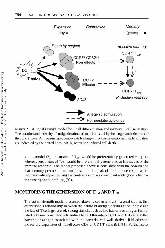
Taken together the available data are consistent with the concept that the strengthof the signals delivered by TCR and cytokine receptors drives T cells through hier-archical thresholds of differentiation (90). The progression follows a sequence ofproliferation, preceding the acquisition of fitness, effector function and, eventually,death (Figure 2). Because TCR and cytokine stimulation are stochastic events, notall proliferating T cells receive the same strength of signal. Consequently, primedT cells reach a variety of differentiation stages that contain effector cells as well ascells that have been arrested at intermediate levels of differentiation. These interme-diates retain lymph node homing receptors and have initiated, but not completed,the remodeling events of genes involved in effector functions. This spectrum canbe simply resolved into distinct subsets of TCM and TEM surviving after antigenclearance.
The question remains—how can different levels of signal strength be deliveredin the course of the same immune response? We can envision two possibilities.The first is that in highly stimulatory conditions some activated T cells may pre-maturely detach from DC and fail to receive further stimulation consistent withobservations of dynamic DC/T cell interactions in vivo (78). Second, differentlevels of stimulation may be delivered at different times. At early stages, largenumbers of mature DC carrying high doses of antigen and secreting large amountsof polarizing cytokines would promote a massive proliferation of antigen-specificT cells and drive them into effector cells, some of which will persist as TEM. Atlater time points, the stimulatory conditions may change substantially with arrivalof fewer DC, which furthermore carry small amounts of antigen and have ex-hausted their cytokine-producing capacity (91). Such conditions may lead to theexpansion of noneffector cells, of which the fit ones will persist as TCM. According
Ann
u. R
ev. I
mm
unol
. 200
4.22
:745
-763
. Dow
nloa
ded
from
ww
w.a
nnua
lrev
iew
s.or
gby
Har
vard
Uni
vers
ity o
n 10
/16/
13. F
or p
erso
nal u
se o
nly.
12 Feb 2004 16:10 AR AR210-IY22-25.tex AR210-IY22-25.sgm LaTeX2e(2002/01/18)P1: IKH
754 SALLUSTO ¥ GEGINAT ¥ LANZAVECCHIA
Figure 2 A signal strength model for T cell differentiation and memory T cell generation.The duration and intensity of antigenic stimulation is indicated by the length and thickness ofthe solid arrows. Antigen-independent events leading to T cell proliferation and differentiationare indicated by the dotted lines. AICD, activation-induced cell death.
to this model (7), precursors of TEM would be preferentially generated early on,whereas precursors of TCM would be preferentially generated at late stages of theimmune response. The model proposed above is consistent with the observationthat memory precursors are not present at the peak of the immune response butprogressively appear during the contraction phase coincident with global changesin transcriptional profiling (92).
MONITORING THE GENERATION OF TCM AND TEM
The signal strength model discussed above is consistent with several studies thatestablished a relationship between the nature of antigenic stimulation in vivo andthe fate of T cells generated. Strong stimuli, such as live bacteria or antigen formu-lated with microbial products, induce fully differentiated CTL and TH1 cells; killedbacteria or antigen associated with the bacterial cell wall–derived Ribi adjuvantinduce the expansion of noneffector CD8 or CD4 T cells (93, 94). Furthermore,
Ann
u. R
ev. I
mm
unol
. 200
4.22
:745
-763
. Dow
nloa
ded
from
ww
w.a
nnua
lrev
iew
s.or
gby
Har
vard
Uni
vers
ity o
n 10
/16/
13. F
or p
erso
nal u
se o
nly.

12 Feb 2004 16:10 AR AR210-IY22-25.tex AR210-IY22-25.sgm LaTeX2e(2002/01/18)P1: IKH
MEMORY T CELL SUBSETS 755
antigens cross-presented by immature DC induce abortive proliferation or expan-sion of noneffector cells depending on the level of antigen expression (95–99). Inthese conditions, in vivo administration of IL-12 or T cell help increases the gen-eration of effector cells. Reintroduction of antigen, but not inflammatory stimuli,during primary infection reinforces the generation of memory T cells, indicat-ing that a sustained antigen presentation is critical for effective memory T cellgeneration (100).
Several studies examined the heterogeneity of responding T cells in the courseof antigen-driven immune responses. Following immunization with a protein anti-gen, two types of responding T cells can be identified as early as day five: B helperT cells expressing the follicular homing receptor CXCR5+ and tissue homing in-flammatory T cells expressing CXCR3 (101). CD4 T cells responding to influenzavirus were found to be a heterogeneous continuum of cells in terms of number ofcell divisions, phenotype, and function (102). Interestingly, those migrating to thelung are characterized by loss of CD62L, downregulation of CCR7, secretion oflarge amounts of IFN-γ , and reduced IL-2 levels relative to those in the secondarylymphoid organs. While the response declines with viral clearance, a range of rest-ing cell subsets reflecting the pattern at the peak of response is retained, suggestingthat heterogeneous effector populations may give rise to corresponding memorypopulations (102). Furthermore, the same CD8 T cell clonotypes responding toinfluenza are present in the lymphoid tissue and in the virus-infected lung (103),demonstrating that the heterogeneity in tissue localization can be generated withinthe same T cell clone.
In the mouse system it has been possible to directly address the relationshipbetween population of primed T cells and memory subsets. A study on CD4 T cellsshowed that cytokine-secreting cells fail to generate memory, whereas nonpolar-ized cells, with the characteristic of pre-TH1, have the ability to transfer strongsecondary TH1 responses (104). In a recent study the precursor-product relation-ship between TCM and TEM has been analyzed in the CD8 system by adoptivetransfer experiments (105). The results point to three main conclusions. First, TEM
are present only transiently, and, upon transfer, they all convert to TCM by reac-quiring CCR7 and CD62L expression. Second, the rate of conversion from TEM
to TCM is inversely proportional to the strength of stimulation. Third, TCM convertto TEM upon antigenic restimulation but not under steady-state conditions. Thesefindings led to the proposition that in mice TCM and TEM do not necessarily rep-resent distinct subsets, but are part of a continuum in a linear nave→ effector→TEM→ TCM differentiation pathway (106). These results are in apparent contrastwith the persistence and stability of both TCM and TEM subsets in humans (15)and with the failure of TEM to reacquire the constitutive CCR7 expression in vitro(107), suggesting a relevant difference between these two species.
Recent findings indicate that T cell help is an absolute requirement for gener-ation of functional CD8 T cell memory. Indeed, animals primed in the absenceof T cell help mount normal primary CTL responses but fail to mount secondaryresponses to the same antigen in spite of normal numbers of memory T cells
Ann
u. R
ev. I
mm
unol
. 200
4.22
:745
-763
. Dow
nloa
ded
from
ww
w.a
nnua
lrev
iew
s.or
gby
Har
vard
Uni
vers
ity o
n 10
/16/
13. F
or p
erso
nal u
se o
nly.
12 Feb 2004 16:10 AR AR210-IY22-25.tex AR210-IY22-25.sgm LaTeX2e(2002/01/18)P1: IKH
756 SALLUSTO ¥ GEGINAT ¥ LANZAVECCHIA
(108–110). The nature of the T cell defect it is not clear, but it is tempting tospeculate that it may be due to a qualitative or quantitative defect of TCM. Themechanism of T cell help is also unclear. Helper T cells can boost CTL responsesnot only through DC activation (111–113) and provision of IL-2 but also throughdirect T-T interactions mediated by CD40L (114). Positive signals controlling thegeneration of memory T cells are provided by other members of the TNF receptorfamily such as OX40 and CD27 (115–117).
OPEN QUESTIONS AND PERSPECTIVES
TCM and memory B cells represent memory stem cells because they respond tochronic or repeated antigenic stimulation by self-renewing and generation of effec-tor T cells and plasma cells (118). Memory cells divide constitutively, even in theabsence of antigen (50), and in response to cytokines proliferate and differentiateto effector T cells. We therefore propose that TCM and memory B cells may alsofunction as stem cells under homeostatic conditions. Accordingly, TCM may divideasymmetrically in response to cytokines, maintaining their number and, at the sametime, differentiating and replenishing TEM that are lost in peripheral tissues, suchas lung or gut. A similar mechanism was recently proposed for human memory Bcells, which maintain serological memory through continuous polyclonal activa-tion (119). Cell tracking experiments and detailed kinetics and repertoire analysisof memory T cell subsets should be performed in order to address this importantissue in humans.
Mouse experiments showed that TCM have much higher capacity to reconstitutethe memory T cell pool than effector cells (104, 105). These observations arehighly relevant for immunotherapy. Injection of large numbers of effector cells mayprovide immediate protection but fail to reconstitute long-term memory (120). Itwould be important to find out the optimal conditions to prime and expand TCM-likecells in humans.
Although the existence of TCM and TEM is now well documented, there are sig-nificant differences between mouse and human that need to be clarified. One aspectrelates to the extent of differentiation of the TCM subset and the mechanisms respon-sible for cytokine memory and flexibility in both CD4 and CD8 compartments. Inaddition, human TCM and TEM populations appear to be stable with no evidence ofinterconversion, whereas they appear to be dynamic and plastic in the mouse sys-tem (15, 105). This difference may be related to the experimental systems or mayreflect differences between the species. A particularly striking difference betweenexperimental animals and humans relates to number of specificities that need tobe accomodated and maintained in the memory pool. Since the total pool does notsubstantially increase with aging, new specificities that progressively accumulatelead to dilution of pre-existing memory T cells. The same mechanism will favor theexpansion of cross-reacting T cells, which become enriched in the memory pool.Both attrition and cross-reactivity have been documented in mice (121) and may
Ann
u. R
ev. I
mm
unol
. 200
4.22
:745
-763
. Dow
nloa
ded
from
ww
w.a
nnua
lrev
iew
s.or
gby
Har
vard
Uni
vers
ity o
n 10
/16/
13. F
or p
erso
nal u
se o
nly.

12 Feb 2004 16:10 AR AR210-IY22-25.tex AR210-IY22-25.sgm LaTeX2e(2002/01/18)P1: IKH
MEMORY T CELL SUBSETS 757
play an even more important role in humans because of the much longer lifespanand antigenic exposure. The prevalence of cross-reactive memory T cells in elderlyshould be tested experimentally. Solving any of these questions will have not onlytheoretical but also practical implications for vaccination and immunotherapy.
ACKNOWLEDGMENTS
We thank Amanda Gett for critical reading and comments. The Institute for Re-search in Biomedicine is supported by the Helmut Horten Foundation. The workon immunological memory in the authors’ laboratories is supported by grants fromthe European Community (QLK2-CT-2001–01250) and the Swiss National Funds(3100–63885 and 3100–101962).
TheAnnual Review of Immunologyis online at http://immunol.annualreviews.org
LITERATURE CITED
1. Manz RA, Thiel A, Radbruch A. 1997.Lifetime of plasma cells in the bone mar-row. Nature388:133–34
2. Slifka MK, Antia R, Whitmire JK, AhmedR. 1998. Humoral immunity due to long-lived plasma cells.Immunity8:363–72
3. Ochsenbein AF, Pinschewer DD, SierroS, Horvath E, Hengartner H, Zinker-nagel RM. 2000. Protective long-term an-tibody memory by antigen-driven and Thelp-dependent differentiation of long-lived memory B cells to short-livedplasma cells independent of secondarylymphoid organs.Proc. Natl. Acad. Sci.USA97:13263–68
4. Sallusto F, Lenig D, Forster R, LippM, Lanzavecchia A. 1999. Two subsetsof memory T lymphocytes with distincthoming potentials and effector functions.Nature401:708–12
5. Reinhardt RL, Khoruts A, Merica R, ZellT, Jenkins MK. 2001. Visualizing the gen-eration of memory CD4 T cells in thewhole body.Nature410:101–5
6. Masopust D, Vezys V, Marzo AL, Lefran-cois L. 2001. Preferential localization ofeffector memory cells in nonlymphoid tis-sue.Science291:2413–17
7. Lanzavecchia A, Sallusto F. 2000. Dy-
namics of T lymphocyte responses: in-termediates, effectors, and memory cells.Science290:92–97
8. Campbell JJ, Bowman EP, Murphy K,Youngman KR, Siani MA, et al. 1998.6-C-kine (SLC), a lymphocyte adhesion-triggering chemokine expressed by highendothelium, is an agonist for the MIP-3beta receptor CCR7.J. Cell Biol.141:1053–59
9. Forster R, Schubel A, Breitfeld D, Krem-mer E, Renner-Muller I, et al. 1999. CCR7coordinates the primary immune responseby establishing functional microenviron-ments in secondary lymphoid organs.Cell99:23–33
10. Campbell JJ, Murphy KE, Kunkel EJ,Brightling CE, Soler D, et al. 2001. CCR7expression and memory T cell diversity inhumans.J. Immunol.166:877–84
11. Champagne P, Ogg GS, King AS, Kna-benhans C, Ellefsen K, et al. 2001.Skewed maturation of memory HIV-specific CD8 T lymphocytes.Nature410:106–11
12. Dunbar PR, Smith CL, Chao D, Salio M,Shepherd D, et al. 2000. A shift in thephenotype of melan-A-specific CTL iden-tifies melanoma patients with an active
Ann
u. R
ev. I
mm
unol
. 200
4.22
:745
-763
. Dow
nloa
ded
from
ww
w.a
nnua
lrev
iew
s.or
gby
Har
vard
Uni
vers
ity o
n 10
/16/
13. F
or p
erso
nal u
se o
nly.
12 Feb 2004 16:10 AR AR210-IY22-25.tex AR210-IY22-25.sgm LaTeX2e(2002/01/18)P1: IKH
758 SALLUSTO ¥ GEGINAT ¥ LANZAVECCHIA
tumor-specific immune response.J. Im-munol.165:6644–52
13. Valmori D, Scheibenbogen C, DutoitV, Nagorsen D, Asemissen AM, et al.2002. Circulating tumor-reactive CD8+T cells in melanoma patients contain aCD45RA+CCR7− effector subset exert-ing ex vivo tumor-specific cytolytic activ-ity. Cancer Res.62:1743–50
14. Hislop AD, Annels NE, Gudgeon NH,Leese AM, Rickinson AB. 2002. Epitope-specific evolution of human CD8+ Tcell responses from primary to persistentphases of Epstein-Barr virus infection.J.Exp. Med.195:893–905
15. Baron V, Bouneaud C, Cumano A, Lim A,Arstila TP, et al. 2003. The repertoires ofcirculating human CD8+ central and ef-fector memory T cell subsets are largelydistinct.Immunity18:193–204
16. Hamann D, Baars PA, Rep MH, Hooib-rink B, Kerkhof-Garde SR, et al. 1997.Phenotypic and functional separation ofmemory and effector human CD8+ Tcells.J. Exp. Med.186:1407–18
17. Appay V, Dunbar PR, Callan M, Klener-man P, Gillespie GM, et al. 2002. Mem-ory CD8+ T cells vary in differentiationphenotype in different persistent virus in-fections.Nat. Med.8:379–85
18. Rufer N, Zippelius A, Batard P, Pittet MJ,Kurth I, et al. 2003. Ex vivo character-ization of human CD8+ T subsets withdistinct replicative history and partial ef-fector functions.Blood102:1779–87
19. Gunn MD, Ngo VN, Ansel KM, EklandEH, Cyster JG, Williams LT. 1998. AB-cell-homing chemokine made in lym-phoid follicles activates Burkitt’s lym-phoma receptor-1.Nature391:799–803
20. Legler DF, Loetscher M, Roos RS, Clark-Lewis I, Baggiolini M, Moser B. 1998.B cell-attracting chemokine 1, a humanCXC chemokine expressed in lymphoidtissues, selectively attracts B lymphocytesvia BLR1/CXCR5.J. Exp. Med.187:655–60
21. Breitfeld D, Ohl L, Kremmer E, Ellwart J,
Sallusto F, et al. 2000. Follicular B helperT cells express CXC chemokine receptor5, localize to B cell follicles, and supportimmunoglobulin production.J. Exp. Med.192:1545–52
22. Schaerli P, Willimann K, Lang AB, LippM, Loetscher P, Moser B. 2000. CXCchemokine receptor 5 expression definesfollicular homing T cells with B cellhelper function.J. Exp. Med.192:1553–62
23. Kim CH, Rott LS, Clark-Lewis I, Camp-bell DJ, Wu L, Butcher EC. 2001. Subspe-cialization of CXCR5+ T cells: B helperactivity is focused in a germinal center-localized subset of CXCR5+ T cells. J.Exp. Med.193:1373–81
24. Sallusto F, Lanzavecchia A, Mackay CR.1998. Chemokines and chemokine recep-tors in T-cell priming and Th1/Th2- medi-ated responses.Immunol. Today19:568–74
25. Loetscher P, Uguccioni M, Bordoli L,Baggiolini M, Moser B, et al. 1998. CCR5is characteristic of Th1 lymphocytes.Na-ture391:344–45
26. Kim CH, Kunkel EJ, Boisvert J, John-ston B, Campbell JJ, et al. 2001.Bonzo/CXCR6 expression defines type 1-polarized T-cell subsets with extralym-phoid tissue homing potential.J. Clin. In-vest.107:595–601
27. Sallusto F, Mackay CR, Lanzavecchia A.1997. Selective expression of the eotaxinreceptor CCR3 by human T helper 2 cells.Science277:2005–7
28. Nagata K, Tanaka K, Ogawa K, Kem-motsu K, Imai T, et al. 1999. Selectiveexpression of a novel surface moleculeby human Th2 cells in vivo.J. Immunol.162:1278–86
29. Bonecchi R, Bianchi G, Bordignon PP,D’Ambrosio D, Lang R, et al. 1998. Dif-ferential expression of chemokine recep-tors and chemotactic responsiveness oftype 1 T helper cells (Th1s) and Th2s.J.Exp. Med.187:129–34
30. Sallusto F, Lenig D, Mackay CR,
Ann
u. R
ev. I
mm
unol
. 200
4.22
:745
-763
. Dow
nloa
ded
from
ww
w.a
nnua
lrev
iew
s.or
gby
Har
vard
Uni
vers
ity o
n 10
/16/
13. F
or p
erso
nal u
se o
nly.

12 Feb 2004 16:10 AR AR210-IY22-25.tex AR210-IY22-25.sgm LaTeX2e(2002/01/18)P1: IKH
MEMORY T CELL SUBSETS 759
Lanzavecchia A. 1998. Flexible programsof chemokine receptor expression on hu-man polarized T helper 1 and 2 lympho-cytes.J. Exp. Med.187:875–83
31. Butcher EC, Picker LJ. 1996. Lympho-cyte homing and homeostasis.Science272:60–66
32. von Andrian UH, Mackay CR. 2000. T-cell function and migration. Two sides ofthe same coin.N. Engl. J. Med.343:1020–34
33. Campbell JJ, Haraldsen G, Pan J, RottmanJ, Qin S, et al. 1999. The chemokine recep-tor CCR4 in vascular recognition by cuta-neous but not intestinal memory T cells.Nature400:776–80
34. Zabel BA, Agace WW, Campbell JJ,Heath HM, Parent D, et al. 1999. Hu-man G protein-coupled receptor GPR-9-6/CC chemokine receptor 9 is selec-tively expressed on intestinal homing Tlymphocytes, mucosal lymphocytes, andthymocytes and is required for thymus-expressed chemokine-mediated chemo-taxis.J. Exp. Med.190:1241–56
35. Campbell JJ, Hedrick J, Zlotnik A, SianiMA, Thompson DA, Butcher EC. 1998.Chemokines and the arrest of lympho-cytes rolling under flow conditions.Sci-ence279:381–84
36. Middleton J, Neil S, Wintle J, Clark-LewisI, Moore H, et al. 1997. Transcytosis andsurface presentation of IL-8 by venular en-dothelial cells.Cell 91:385–95
37. Stein JV, Rot A, Luo Y, NarasimhaswamyM, Nakano H, et al. 2000. The CCchemokine thymus-derived chemotacticagent 4 (TCA-4, secondary lymphoidtissue chemokine, 6Ckine, exodus-2) trig-gers lymphocyte function-associated anti-gen 1-mediated arrest of rolling T lym-phocytes in peripheral lymph node highendothelial venules.J. Exp. Med.191:61–76
38. Sallusto F, Kremmer E, Palermo B, HoyA, Ponath P, et al. 1999. Switch inchemokine receptor expression upon TCRstimulation reveals novel homing poten-
tial for recently activated T cells.Eur. J.Immunol.29:2037–45
39. Kishimoto TK, Jutila MA, Butcher EC.1990. Identification of a human peripherallymph node homing receptor: a rapidlydown-regulated adhesion molecule.Proc.Natl. Acad. Sci. USA87:2244–48
40. Chao CC, Jensen R, Dailey MO. 1997.Mechanisms of L-selectin regulation byactivated T cells.J. Immunol.159:1686–94
41. Geginat J, Lanzavecchia A, Sallusto F.2003. Proliferation and differentiation po-tential of human CD8+ memory T-cellsubsets in response to antigen or home-ostatic cytokines.Blood101:4260–66
42. Son NH, Murray S, Yanovski J, HodesRJ, Weng N. 2000. Lineage-specific telo-mere shortening and unaltered capacityfor telomerase expression in human Tand B lymphocytes with age.J. Immunol.165:1191–96
43. Boise LH, Minn AJ, Noel PJ, June CH,Accavitti MA, et al. 1995. CD28 costim-ulation can promote T cell survival by en-hancing the expression of Bcl-XL.Immu-nity 3:87–98
44. Messi M, Giacchetto I, Nagata K, Lan-zavecchia A, Natoli G, Sallusto F. 2003.Memory and flexibility of cytokine geneexpression as separable properties of hu-man TH1 and TH2 lymphocytes.Nat. Im-munol.4:78–86
45. Grogan JL, Mohrs M, Harmon B, LacyDA, Sedat JW, Locksley RM. 2001. Earlytranscription and silencing of cytokinegenes underlie polarization of T helpercell subsets.Immunity14:205–15
46. Gunzer M, Schafer A, Borgmann S,Grabbe S, Zanker KS, et al. 2000. Anti-gen presentation in extracellular matrix:interactions of T cells with dendritic cellsare dynamic, short lived, and sequential.Immunity13:323–32
47. Harris NL, Watt V, Ronchese F, Le GrosG. 2002. Differential T cell function andfate in lymph node and nonlymphoid tis-sues.J. Exp. Med.195:317–26
Ann
u. R
ev. I
mm
unol
. 200
4.22
:745
-763
. Dow
nloa
ded
from
ww
w.a
nnua
lrev
iew
s.or
gby
Har
vard
Uni
vers
ity o
n 10
/16/
13. F
or p
erso
nal u
se o
nly.

12 Feb 2004 16:10 AR AR210-IY22-25.tex AR210-IY22-25.sgm LaTeX2e(2002/01/18)P1: IKH
760 SALLUSTO ¥ GEGINAT ¥ LANZAVECCHIA
48. Yazdanbakhsh M, Kremsner PG, van ReeR. 2002. Allergy, parasites, and the hy-giene hypothesis.Science296:490–94
49. Michie CA, McLean A, Alcock C, Bever-ley PC. 1992. Lifespan of human lympho-cyte subsets defined by CD45 isoforms.Nature360:264–65
50. Tough DF, Sprent J. 1994. Turnover ofnaive- and memory-phenotype T cells.J.Exp. Med.179:1127–35
51. Seddon B, Tomlinson P, Zamoyska R.2003. Interleukin 7 and T cell receptor sig-nals regulate homeostasis of CD4 memorycells.Nat. Immunol.4:680–86
52. Zhang X, Sun S, Hwang I, Tough DF,Sprent J. 1998. Potent and selective stim-ulation of memory-phenotype CD8+ Tcells in vivo by IL-15. Immunity8:591–99
53. Lodolce JP, Boone DL, Chai S, Swain RE,Dassopoulos T, et al. 1998. IL-15 receptormaintains lymphoid homeostasis by sup-porting lymphocyte homing and prolifer-ation.Immunity9:669–76
54. Schluns KS, Kieper WC, Jameson SC,Lefrancois L. 2000. Interleukin-7 medi-ates the homeostasis of naive and mem-ory CD8 T cells in vivo.Nat. Immunol.1:426–32
55. Unutmaz D, Pileri P, Abrignani S. 1994.Antigen-independent activation of naiveand memory resting T cells by a cytokinecombination. J. Exp. Med.180:1159–64
56. Geginat J, Sallusto F, Lanzavecchia A.2001. Cytokine-driven proliferation anddifferentiation of human naive, centralmemory, and effector memory CD4+ Tcells.J. Exp. Med.194:1711–19
57. Alves NL, Hooibrink B, Arosa FA, VanLier RA. 2003. IL-15 induces antigen-independent expansion and differentia-tion of human naive CD8+T cells in vitro.Blood102:2541–46
58. Zheng W, Flavell RA. 1997. The tran-scription factor GATA-3 is necessary andsufficient for Th2 cytokine gene expres-sion in CD4 T cells.Cell 89:587–96
59. Szabo SJ, Kim ST, Costa GL, ZhangX, Fathman CG, Glimcher LH. 2000. Anovel transcription factor, T-bet, directsTh1 lineage commitment.Cell 100:655–69
60. Mullen AC, High FA, Hutchins AS, LeeHW, Villarino AV, et al. 2001. Role ofT-bet in commitment of TH1 cells be-fore IL-12-dependent selection.Science292:1907–10
61. Agarwal S, Rao A. 1998. Modulationof chromatin structure regulates cytokinegene expression during T cell differentia-tion. Immunity9:765–75
62. Takemoto N, Kamogawa Y, Jun-Lee H,Kurata H, Arai KI, et al. 2000. Cuttingedge: chromatin remodeling at the IL-4/IL-13 intergenic regulatory region forTh2-specific cytokine gene cluster.J. Im-munol.165:6687–91
63. Lee GR, Fields PE, Flavell RA. 2001.Regulation of IL-4 gene expression by dis-tal regulatory elements and GATA-3 at thechromatin level.Immunity14:447–59
64. Iezzi G, Scotet E, Scheidegger D, Lanza-vecchia A. 1999. The interplay betweenthe duration of TCR and cytokine signal-ing determines T cell polarization.Eur. J.Immunol.29:4092–101
65. Ansel KM, Lee DU, Rao A. 2003. An epi-genetic view of helper T cell differentia-tion. Nat. Immunol.4:616–23
66. Scotet E, Schroeder S, LanzavecchiaA. 2001. Molecular regulation of CC-chemokine receptor 3 expression in hu-man T helper 2 cells.Blood98:2568–70
67. Lu Q, Wu A, Ray D, Deng C, Attwood J, etal. 2003. DNA methylation and chromatinstructure regulate T cell perforin gene ex-pression.J. Immunol.170:5124–32
68. Afkarian M, Sedy JR, Yang J, Jacob-son NG, Cereb N, et al. 2002. T-bet is aSTAT1-induced regulator of IL-12R ex-pression in naive CD4+ T cells.Nat. Im-munol.3:549–57
69. Sundrud MS, Grill SM, Ni D, Nagata K,Alkan SS, et al. 2003. Genetic reprogram-ming of primary human T cells reveals
Ann
u. R
ev. I
mm
unol
. 200
4.22
:745
-763
. Dow
nloa
ded
from
ww
w.a
nnua
lrev
iew
s.or
gby
Har
vard
Uni
vers
ity o
n 10
/16/
13. F
or p
erso
nal u
se o
nly.
12 Feb 2004 16:10 AR AR210-IY22-25.tex AR210-IY22-25.sgm LaTeX2e(2002/01/18)P1: IKH
MEMORY T CELL SUBSETS 761
functional plasticity in Th cell differenti-ation.J. Immunol.171:3542–49
70. Lee DU, Agarwal S, Rao A. 2002. Th2 lin-eage commitment and efficient IL-4 pro-duction involves extended demethylationof the IL-4 gene.Immunity16:649–60
71. Barber DL, Wherry EJ, Ahmed R.2003. Cutting edge: rapid in vivo killingby memory CD8 T cells.J. Immunol.171:27–31
72. Sad S, Mosmann TR. 1994. Single IL-2-secreting precursor CD4 T cell candevelop into either Th1 or Th2 cy-tokine secretion phenotype.J. Immunol.153:3514–22
73. Gorelik L, Fields PE, Flavell RA. 2000.Cutting edge: TGF-beta inhibits Th type 2development through inhibition of GATA-3 expression.J. Immunol.165:4773–77
74. Gorelik L, Constant S, Flavell RA. 2002.Mechanism of transforming growth factorbeta-induced inhibition of T helper type1 differentiation.J. Exp. Med.195:1499–505
75. Valitutti S, Muller S, Cella M, Padovan E,Lanzavecchia A. 1995. Serial triggeringof many T-cell receptors by a few peptide-MHC complexes.Nature375:148–51
76. Viola A, Schroeder S, Sakakibara Y,Lanzavecchia A. 1999. T lymphocytecostimulation mediated by reorganiza-tion of membrane microdomains.Science283:680–82
77. Iezzi G, Karjalainen K, Lanzavecchia A.1998. The duration of antigenic stimula-tion determines the fate of naive and ef-fector T cells.Immunity8:89–95
78. Stoll S, Delon J, Brotz TM, GermainRN. 2002. Dynamic imaging of T cell-dendritic cell interactions in lymph nodes.Science296:1873–76
79. Iezzi G, Scheidegger D, Lanzavecchia A.2001. Migration and function of antigen-primed nonpolarized T lymphocytes invivo. J. Exp. Med.193:987–93
80. Manjunath N, Shankar P, Wan J, WeningerW, Crowley MA, et al. 2001. Effector dif-ferentiation is not prerequisite for genera-
tion of memory cytotoxic T lymphocytes.J. Clin. Invest.108:871–78
81. Weninger W, Crowley MA, Manjunath N,von Andrian UH. 2001. Migratory proper-ties of naive, effector, and memory CD8+T cells.J. Exp. Med.194:953–66
82. Langenkamp A, Casorati G, Garavaglia C,Dellabona P, Lanzavecchia A, Sallusto F.2002. T cell priming by dendritic cells:thresholds for proliferation, differentia-tion and death and intraclonal functionaldiversification.Eur. J. Immunol.32:2046–54
83. Hutloff A, Dittrich AM, Beier KC, El-jaschewitsch B, Kraft R, et al. 1999. ICOSis an inducible T-cell co-stimulator struc-turally and functionally related to CD28.Nature397:263–66
84. Coyle AJ, Lehar S, Lloyd C, Tian J, De-laney T, et al. 2000. The CD28-relatedmolecule ICOS is required for effectiveT cell-dependent immune responses.Im-munity13:95–105
85. Kopf M, Coyle AJ, Schmitz N, Barner M,Oxenius A, et al. 2000. Inducible costimu-lator protein (ICOS) controls T helper cellsubset polarization after virus and parasiteinfection.J. Exp. Med.192:53–61
86. Diehl L, van Mierlo GJ, den Boer AT,van der Voort E, Fransen M, et al. 2002.In vivo triggering through 4-1BB enablesTh-independent priming of CTL in thepresence of an intact CD28 costimulatorypathway.J. Immunol.168:3755–62
87. van Stipdonk MJ, Lemmens EE, Schoen-berger SP. 2001. Naive CTLs require a sin-gle brief period of antigenic stimulationfor clonal expansion and differentiation.Nat. Immunol.2:423–29
88. Gett AV, Sallusto F, Lanzavecchia A,Geginat J. 2003. T cell fitness determinedby signal strength.Nat. Immunol.4:355–60
89. van Stipdonk MJ, Hardenberg G, BijkerMS, Lemmens EE, Droin NM, et al. 2003.Dynamic programming of CD8+ T lym-phocyte responses.Nat. Immunol.4:361–65
Ann
u. R
ev. I
mm
unol
. 200
4.22
:745
-763
. Dow
nloa
ded
from
ww
w.a
nnua
lrev
iew
s.or
gby
Har
vard
Uni
vers
ity o
n 10
/16/
13. F
or p
erso
nal u
se o
nly.

12 Feb 2004 16:10 AR AR210-IY22-25.tex AR210-IY22-25.sgm LaTeX2e(2002/01/18)P1: IKH
762 SALLUSTO ¥ GEGINAT ¥ LANZAVECCHIA
90. Lanzavecchia A, Sallusto F. 2002.Opinion-decision making in the immunesystem: progressive differentiation andselection of the fittest in the immune re-sponse.Nat. Rev. Immunol.2:982–87
91. Langenkamp A, Messi M, LanzavecchiaA, Sallusto F. 2000. Kinetics of dendriticcell activation: impact on priming of TH1,TH2 and nonpolarized T cells.Nat. Im-munol.1:311–16
92. Kaech SM, Hemby S, Kersh E, AhmedR. 2002. Molecular and functional profil-ing of memory CD8 T cell differentiation.Cell 111:837–51
93. Lauvau G, Vijh S, Kong P, Horng T, Kerk-siek K, et al. 2001. Priming of memory butnot effector CD8 T cells by a killed bac-terial vaccine.Science294:1735–39
94. Wang X, Mosmann T. 2001. In vivo prim-ing of CD4 T cells that produce interleukin(IL)-2 but not IL-4 or interferon (IFN)-gamma, and can subsequently differen-tiate into IL-4- or IFN-gamma-secretingcells.J. Exp. Med.194:1069–80
95. Kurts C, Kosaka H, Carbone FR, MillerJF, Heath WR. 1997. Class I-restrictedcross-presentation of exogenous self-antigens leads to deletion of autoreactiveCD8+ T cells. J. Exp. Med.186:239–45
96. Hawiger D, Inaba K, Dorsett Y, Guo M,Mahnke K, et al. 2001. Dendritic cells in-duce peripheral T cell unresponsivenessunder steady state conditions in vivo.J.Exp. Med.194:769–79
97. Hernandez J, Aung S, Redmond WL,Sherman LA. 2001. Phenotypic and func-tional analysis of CD8+ T cells under-going peripheral deletion in response tocross-presentation of self-antigen.J. Exp.Med.194:707–17
98. Bonifaz L, Bonnyay D, Mahnke K, RiveraM, Nussenzweig MC, Steinman RM.2002. Efficient targeting of protein anti-gen to the dendritic cell receptor DEC-205in the steady state leads to antigen presen-tation on major histocompatibility com-plex class I products and peripheral CD8+
T cell tolerance.J. Exp. Med.196:1627–38
99. Hernandez J, Aung S, Marquardt K, Sher-man LA. 2002. Uncoupling of prolifera-tive potential and gain of effector func-tion by CD8+ T cells responding toself-antigens.J. Exp. Med.196:323–33
100. Busch DH, Kerksiek KM, Pamer EG.2000. Differing roles of inflammation andantigen in T cell proliferation and memorygeneration.J. Immunol.164:4063–70
101. Campbell DJ, Kim CH, Butcher EC. 2001.Separable effector T cell populations spe-cialized for B cell help or tissue inflam-mation.Nat. Immunol.2:876–81
102. Roman E, Miller E, Harmsen A, Wiley J,Von Andrian UH, et al. 2002. CD4 effectorT cell subsets in the response to influenza:heterogeneity, migration, and function.J.Exp. Med.196:957–68
103. Turner SJ, Diaz G, Cross R, Doherty PC.2003. Analysis of clonotype distributionand persistence for an influenza virus-specific CD8+ T cell response.Immunity18:549–59
104. Wu CY, Kirman JR, Rotte MJ, Davey DF,Perfetto SP, et al. 2002. Distinct lineagesof TH1 cells have differential capacitiesfor memory cell generation in vivo.Nat.Immunol.3:852–58
105. Wherry EJ, Teichgraber V, Becker TC,Masopust D, Kaech SM, et al. 2003. Lin-eage relationship and protective immunityof memory CD8 T cell subsets.Nat. Im-munol.4:225–34
106. Seder RA, Ahmed R. 2003. Similaritiesand differences in CD4+ and CD8+ ef-fector and memory T cell generation.Nat.Immunol.4:835–42
107. Langenkamp A, Nagata K, Murphy K,Wu L, Lanzavecchia A, Sallusto F.2003. Kinetics and expression patterns ofchemokine receptors in human CD4+ Tlymphocytes primed by myeloid or plas-macytoid dendritic cells.Eur. J. Immunol.33:474–82
108. Janssen EM, Lemmens EE, WolfeT, Christen U, von Herrath MG,
Ann
u. R
ev. I
mm
unol
. 200
4.22
:745
-763
. Dow
nloa
ded
from
ww
w.a
nnua
lrev
iew
s.or
gby
Har
vard
Uni
vers
ity o
n 10
/16/
13. F
or p
erso
nal u
se o
nly.

12 Feb 2004 16:10 AR AR210-IY22-25.tex AR210-IY22-25.sgm LaTeX2e(2002/01/18)P1: IKH
MEMORY T CELL SUBSETS 763
Schoenberger SP. 2003. CD4+ T cellsare required for secondary expansion andmemory in CD8+ T lymphocytes.Nature421:852–56
109. Sun JC, Bevan MJ. 2003. Defective CD8T cell memory following acute infec-tion without CD4 T cell help.Science300:339–42
110. Shedlock DJ, Shen H. 2003. Require-ment for CD4 T cell help in generatingfunctional CD8 T cell memory.Science300:337–39
111. Ridge JP, Di Rosa F, Matzinger P. 1998. Aconditioned dendritic cell can be a tempo-ral bridge between a CD4+ T-helper anda T-killer cell.Nature393:474–78
112. Schoenberger SP, Toes RE, van der VoortEI, Offringa R, Melief CJ. 1998. T-cellhelp for cytotoxic T lymphocytes is me-diated by CD40-CD40L interactions.Na-ture393:480–83
113. Bennett SR, Carbone FR, Karamalis F,Flavell RA, Miller JF, Heath WR. 1998.Help for cytotoxic-T-cell responses ismediated by CD40 signalling.Nature393:478–80
114. Bourgeois C, Rocha B, Tanchot C. 2002.A role for CD40 expression on CD8+ Tcells in the generation of CD8+ T cellmemory.Science297:2060–63
115. Maxwell JR, Campbell JD, Kim CH, Vella
AT. 1999. CD40 activation boosts T cellimmunity in vivo by enhancing T cellclonal expansion and delaying peripheralT cell deletion.J. Immunol.162:2024–34
116. Maxwell JR, Weinberg A, Prell RA, VellaAT. 2000. Danger and OX40 receptor sig-naling synergize to enhance memory Tcell survival by inhibiting peripheral dele-tion. J. Immunol.164:107–12
117. Hendriks J, Gravestein LA, Tesselaar K,van Lier RA, Schumacher TN, Borst J.2000. CD27 is required for generation andlong-term maintenance of T cell immu-nity. Nat. Immunol.1:433–40
118. Fearon DT, Manders P, Wagner SD.2001. Arrested differentiation, the self-renewing memory lymphocyte, and vac-cination.Science293:248–50
119. Bernasconi NL, Traggiai E, Lanzavec-chia A. 2002. Maintenance of serologicalmemory by polyclonal activation of hu-man memory B cells.Science298:2199–202
120. Dudley ME, Rosenberg SA. 2003.Adoptive-cell-transfer therapy for thetreatment of patients with cancer.Nat.Rev. Cancer3:666–75
121. Welsh RM, Selin LK. 2002. No one isnaive: the significance of heterologous T-cell immunity.Nat. Rev. Immunol.2:417–26
Ann
u. R
ev. I
mm
unol
. 200
4.22
:745
-763
. Dow
nloa
ded
from
ww
w.a
nnua
lrev
iew
s.or
gby
Har
vard
Uni
vers
ity o
n 10
/16/
13. F
or p
erso
nal u
se o
nly.

P1: FRK
February 27, 2004 22:25 Annual Reviews AR210-FM
Annual Review of ImmunologyVolume 22, 2004
CONTENTS
FRONTISPIECE—Leonard A. Herzenberg and Leonore A. Herzenberg x
GENETICS, FACS, IMMUNOLOGY, AND REDOX: A TALE OF TWO LIVESINTERTWINED, Leonard A. Herzenberg and Leonore A. Herzenberg 1
SELF- AND NONSELF-RECOGNITION BY C-TYPE LECTINS ONDENDRITIC CELLS, Teunis B.H. Geijtenbeek, Sandra J. van Vliet,Anneke Engering, Bert A. ’t Hart, and Yvette van Kooyk 33
TRANSCRIPTIONAL CONTROL OF EARLY B CELL DEVELOPMENT,Meinrad Busslinger 55
UBIQUITIN LIGASES AND THE IMMUNE RESPONSE, Yun-Cai Liu 81
LIGANDS FOR L-SELECTIN: HOMING, INFLAMMATION, AND BEYOND,Steven D. Rosen 129
INTEGRINS AND T CELL–MEDIATED IMMUNITY, Jonathan T. Pribila,Angie C. Quale, Kristen L. Mueller, and Yoji Shimizu 157
MULTIPLE ROLES OF ANTIMICROBIAL DEFENSINS, CATHELICIDINS,AND EOSINOPHIL-DERIVED NEUROTOXIN IN HOST DEFENSE,De Yang, Arya Biragyn, David M. Hoover, Jacek Lubkowski,and Joost J. Oppenheim 181
STARTING AT THE BEGINNING: NEW PERSPECTIVES ON THE BIOLOGYOF MUCOSAL T CELLS, Hilde Cheroutre 217
THE BCR-ABL STORY: BENCH TO BEDSIDE AND BACK,Stephane Wong and Owen N. Witte 247
CD40/CD154 INTERACTIONS AT THE INTERFACE OF TOLERANCEAND IMMUNITY, Sergio A. Quezada, Lamis Z. Jarvinen, Evan F. Lind,and Randolph J. Noelle 307
THE THREE ES OF CANCER IMMUNOEDITING, Gavin P. Dunn,Lloyd J. Old, and Robert D. Schreiber 329
AUTOIMMUNE AND INFLAMMATORY MECHANISMS INATHEROSCLEROSIS, Georg Wick, Michael Knoflach, and Qingbo Xu 361
THE DYNAMIC LIFE OF NATURAL KILLER CELLS, Wayne M. Yokoyama,Sungjin Kim, and Anthony R. French 405
v
Ann
u. R
ev. I
mm
unol
. 200
4.22
:745
-763
. Dow
nloa
ded
from
ww
w.a
nnua
lrev
iew
s.or
gby
Har
vard
Uni
vers
ity o
n 10
/16/
13. F
or p
erso
nal u
se o
nly.

P1: FRK
February 27, 2004 22:25 Annual Reviews AR210-FM
vi CONTENTS
THE ROLE OF COMPLEMENT IN THE DEVELOPMENT OF SYSTEMICLUPUS ERYTHEMATOSUS, Anthony P. Manderson, Marina Botto,and Mark J. Walport 431
DROSOPHILA: THE GENETICS OF INNATE IMMUNE RECOGNITION ANDRESPONSE, Catherine A. Brennan and Kathryn V. Anderson 457
RAGS AND REGULATION OF AUTOANTIBODIES, Mila Jankovic,Rafael Casellas, Nikos Yannoutsos, Hedda Wardemann,and Michel C. Nussenzweig 485
THE ROLE OF SUPPRESSORS OF CYTOKINE SIGNALING (SOCS)PROTEINS IN REGULATION OF THE IMMUNE RESPONSE,Warren S. Alexander and Douglas J. Hilton 503
NATURALLY ARISING CD4+ REGULATORY T CELLS FORIMMUNOLOGIC SELF-TOLERANCE AND NEGATIVE CONTROLOF IMMUNE RESPONSES, Shimon Sakaguchi 531
PHOSPHOINOSITIDE 3-KINASE: DIVERSE ROLES IN IMMUNE CELLACTIVATION, Jonathan A. Deane and David A. Fruman 563
IMMUNITY TO TUBERCULOSIS, Robert J. North and Yu-Jin Jung 599
MOLECULAR DEFECTS IN HUMAN SEVERE COMBINEDIMMUNODEFICIENCY AND APPROACHES TO IMMUNERECONSTITUTION, Rebecca H. Buckley 625
PHYSIOLOGICAL CONTROL OF IMMUNE RESPONSE ANDINFLAMMATORY TISSUE DAMAGE BY HYPOXIA-INDUCIBLEFACTORS AND ADENOSINE A2A RECEPTORS, Michail V. Sitkovsky,Dmitriy Lukashev, Sergey Apasov, Hidefumi Kojima, Masahiro Koshiba,Charles Caldwell, Akio Ohta, and Manfred Thiel 657
T LYMPHOCYTE–ENDOTHELIAL CELL INTERACTIONS, Jaehyuk Choi,David R. Enis, Kian Peng Koh, Stephen L. Shiao, and Jordan S. Pober 683
IMMUNOLOGICAL MEMORY TO VIRAL INFECTIONS,Raymond M. Welsh, Liisa K. Selin, and Eva Szomolanyi-Tsuda 711
CENTRAL MEMORY AND EFFECTOR MEMORY T CELL SUBSETS:FUNCTION, GENERATION, AND MAINTENANCE,Federica Sallusto, Jens Geginat, and Antonio Lanzavecchia 745
CONTROL OF T CELL VIABILITY, Philippa Marrack and John Kappler 765
ASTHMA: MECHANISMS OF DISEASE PERSISTENCE AND PROGRESSION,Lauren Cohn, Jack A. Elias, and Geoffrey L. Chupp 789
CD1: ANTIGEN PRESENTATION AND T CELL FUNCTION,Manfred Brigl and Michael B. Brenner 817
CHEMOKINES IN INNATE AND ADAPTIVE HOST DEFENSE: BASICCHEMOKINESE GRAMMAR FOR IMMUNE CELLS, Antal Rotand Ulrich H. von Andrian 891
Ann
u. R
ev. I
mm
unol
. 200
4.22
:745
-763
. Dow
nloa
ded
from
ww
w.a
nnua
lrev
iew
s.or
gby
Har
vard
Uni
vers
ity o
n 10
/16/
13. F
or p
erso
nal u
se o
nly.
P1: FRK
February 27, 2004 22:25 Annual Reviews AR210-FM
CONTENTS vii
INTERLEUKIN-10 AND RELATED CYTOKINES AND RECEPTORS,Sidney Pestka, Christopher D. Krause, Devanand Sarkar, Mark R. Walter,Yufang Shi, and Paul B. Fisher 929
INDEXESSubject Index 981Cumulative Index of Contributing Authors, Volumes 12–22 1011Cumulative Index of Chapter Titles, Volumes 12–22 1018
ERRATAAn online log of corrections to Annual Review of Immunology chaptersmay be found at http://immunol.annualreviews.org/errata.shtml
Ann
u. R
ev. I
mm
unol
. 200
4.22
:745
-763
. Dow
nloa
ded
from
ww
w.a
nnua
lrev
iew
s.or
gby
Har
vard
Uni
vers
ity o
n 10
/16/
13. F
or p
erso
nal u
se o
nly.