mems electrochemical patency sensor for detection...
TRANSCRIPT

MEMS ELECTROCHEMICAL PATENCY SENSOR FOR DETECTION OF HYDROCEPHALUS SHUNT OBSTRUCTION
Brian J. Kim, Willa Jin, Lawrence Yu, and Ellis Meng
Department of Biomedical Engineering, University of Southern California, Los Angeles, CA, USA ABSTRACT
Currently there are no practical methods to definitively diagnose the obstruction failure of hydrocephalus shunts without surgical intervention, resulting in suboptimal treatment and needless patient suffering. We present the first MEMS electrochemical patency sensor for direct and quantitative tracking of shunt patency and obstruction. The impact of electrode size, temperature, flow, and hydrogen peroxide (H2O2) plasma sterilization on sensor function was evaluated and sensor performance in the presence of static and dynamic obstruction was demonstrated. Electrode size was found to have a minimal effect on sensor performance and increased temperature and flow resulted in a slight decrease in the baseline impedance (~8.5%) due to an increase in ionic mobility. H2O2 plasma sterilization also had no effect on sensor performance. This low power and simple format sensor was developed with the intention of future integration into shunts for wireless monitoring of shunt state and more importantly, a more accurate and timely diagnosis of shunt failure. INTRODUCTION
Hydrocephalus is a chronic, incurable condition characterized by the accumulation of excess cerebrospinal fluid (CSF) within the ventricles of the brain, resulting in elevated intracranial pressure. Currently, hydrocephalus is treated by implanting a shunt, a multi-holed silicone catheter and valve system, into the ventricles to drain the excess CSF. Though effective, shunts fail at an alarming rate of 40% within the first year [1]. There are many causes of shunt failure, including mechanical issues [2] and infection [3], but the most common is blood and tissue obstruction of the drainage ports [2].
Though these failure rates are high, there are currently no reliable and convenient methods to detect shunt obstruction failure. Obstruction can present itself in only vague symptoms, such as headaches and nausea, which can be confused with other medical conditions. There is a need for a non-invasive method to periodically assess shunt patency so that treatment can be improved through the timely and accurate diagnoses of shunt failure.
We developed a Parylene C-based electrochemical patency sensor capable of providing periodic measurements of shunt obstruction. This sensor utilizes a very simple transduction scheme to assess the degree of shunt patency by measuring changes in electrochemical impedance in the conductive path through the drainage ports in the shunt catheter. As the sensor is constructed on a flexible, thin film Parylene C substrate, it is easily integrated with current shunt systems either as a modular add-on or by direct integration into the shunt.
DESIGN AND OPERATION The sensor consists of two electrodes, one positioned on each internal and external surface of the catheter, such that the catheter ports establish an ionic conductive path between them. When measuring the electrochemical impedance at a sufficiently high frequency (fm) to isolate the solution resistance, any disturbances in the volumetric conduction path between the two electrodes (i.e. port blockage) will register as impedance changes. Thus, upon partial or complete blockage of the catheter ports by tissue, the fluidic conduction pathway between the internal and external electrodes is perturbed and an increase in the electrochemical impedance is measured, similar to the mechanism of the Coulter counter (Figure 1).
Figure 1: (a) Conceptual cartoon of impedance sensing mechanism of patency sensor. Two electrodes on the internal and external surfaces of the catheter are fluidically connected via the drainage ports. Obstruction of these ports impedes the ionic conduction path between the electrodes, and (b) the electrochemical impedance between the electrodes increases for measurements above a certain frequency (fm). The patency sensor was constructed using standard surface micromachining processes for Parylene MEMS devices. Platinum electrodes (2000 Å) were deposited and patterned using the liftoff method on a Parylene C substrate (15 µm) supported by a silicon carrier wafer. Platinum was chosen as it has shown outstanding inert performance for in vivo and electrochemical sensing applications [4]. The electrodes were insulated using Parylene C (2 µm) and electrode sites were exposed via oxygen plasma etching. In the current design, four electrodes having different surface areas were fabricated on a single device to evaluate effects of electrode size on sensor performance (Figure 2a). Devices were released and electrically packaged using a zero insertion force (ZIF) connector and flat flexible cable (FFC) used for Parylene devices [5] before further assembly into the module (Figure 2b).
978-1-4799-7955-4/15/$31.00 ©2015 IEEE 662 MEMS 2015, Estoril, PORTUGAL, 18 - 22 January, 2015
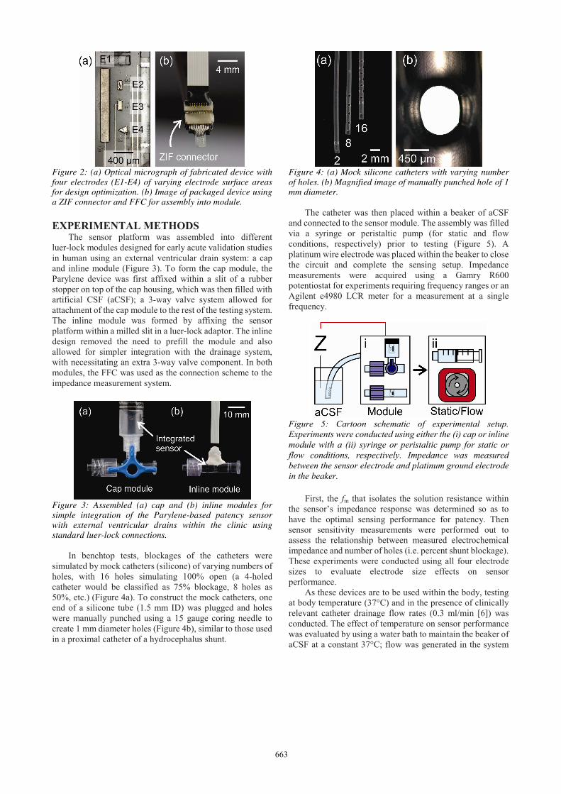
Figure 2: (a) Optical micrograph of fabricated device with four electrodes (E1-E4) of varying electrode surface areas for design optimization. (b) Image of packaged device using a ZIF connector and FFC for assembly into module. EXPERIMENTAL METHODS The sensor platform was assembled into different luer-lock modules designed for early acute validation studies in human using an external ventricular drain system: a cap and inline module (Figure 3). To form the cap module, the Parylene device was first affixed within a slit of a rubber stopper on top of the cap housing, which was then filled with artificial CSF (aCSF); a 3-way valve system allowed for attachment of the cap module to the rest of the testing system. The inline module was formed by affixing the sensor platform within a milled slit in a luer-lock adaptor. The inline design removed the need to prefill the module and also allowed for simpler integration with the drainage system, with necessitating an extra 3-way valve component. In both modules, the FFC was used as the connection scheme to the impedance measurement system.
Figure 3: Assembled (a) cap and (b) inline modules for simple integration of the Parylene-based patency sensor with external ventricular drains within the clinic using standard luer-lock connections. In benchtop tests, blockages of the catheters were simulated by mock catheters (silicone) of varying numbers of holes, with 16 holes simulating 100% open (a 4-holed catheter would be classified as 75% blockage, 8 holes as 50%, etc.) (Figure 4a). To construct the mock catheters, one end of a silicone tube (1.5 mm ID) was plugged and holes were manually punched using a 15 gauge coring needle to create 1 mm diameter holes (Figure 4b), similar to those used in a proximal catheter of a hydrocephalus shunt.
Figure 4: (a) Mock silicone catheters with varying number of holes. (b) Magnified image of manually punched hole of 1 mm diameter. The catheter was then placed within a beaker of aCSF and connected to the sensor module. The assembly was filled via a syringe or peristaltic pump (for static and flow conditions, respectively) prior to testing (Figure 5). A platinum wire electrode was placed within the beaker to close the circuit and complete the sensing setup. Impedance measurements were acquired using a Gamry R600 potentiostat for experiments requiring frequency ranges or an Agilent e4980 LCR meter for a measurement at a single frequency.
Figure 5: Cartoon schematic of experimental setup. Experiments were conducted using either the (i) cap or inline module with a (ii) syringe or peristaltic pump for static or flow conditions, respectively. Impedance was measured between the sensor electrode and platinum ground electrode in the beaker.
First, the fm that isolates the solution resistance within the sensor’s impedance response was determined so as to have the optimal sensing performance for patency. Then sensor sensitivity measurements were performed out to assess the relationship between measured electrochemical impedance and number of holes (i.e. percent shunt blockage). These experiments were conducted using all four electrode sizes to evaluate electrode size effects on sensor performance.
As these devices are to be used within the body, testing at body temperature (37°C) and in the presence of clinically relevant catheter drainage flow rates (0.3 ml/min [6]) was conducted. The effect of temperature on sensor performance was evaluated by using a water bath to maintain the beaker of aCSF at a constant 37°C; flow was generated in the system
663

using a peristaltic pump. Also, for use of this device in clinical applications, it is vital that the sensor modules are sterilized to eliminate any source of infection. Considering this, the functionality of the devices following hydrogen plasma (H2O2) sterilization, a commonly used sterilization technique used by hospitals, performed with a Sterrad 100 NX system was also assessed. RESULTS AND DISCUSSION Sensor Characterization
Initial experiments were conducted at fm between 0.1 Hz – 1 MHz and the measured impedance over certain frequency ranges (>fm, corresponding to where the solution resistance dominates the impedance response) correlated well with catheter blockage over all electrode sizes and types (data not shown). An optimal measured fm was determined for each sensor size (Table 1).
Table 1: Obtained optimal impedance measurement frequencies and sensitivities for electrodes of the Parylene-based EC-MEMS patency sensor. Electrode design
Surface area (μm2)
Measurement frequency (kHz)
Sensitivity (% Z / %Blockage)
E1 300,000 10 0.183E2 20,000 30 0.157E3 20,000 30 0.168E4 17,320 30 0.161
By analyzing the data for each electrode at its
corresponding optimal measurement frequency, calibration curves were formed and results indicated that the impedance varied inversely with the percent blockage of the catheter (Figure 6). The inverse relationship follows from the equation for solution resistance (Rs), where ρ is the conductivity of the solution, l, the distance between the electrodes, and A, the cross-sectional area between the electrodes:
RsρlA
(1)
The results suggest that hole blockages alter the area term of equation 1. Though all electrode sizes were similar in performance, the sensitivity of the largest electrode (E1) was slightly higher than the others (0.183 % Δ impedance/% blockage), and thus was chosen for further sensor development.
Figure 6: Representative calibration curve for E1 sensor electrode indicated that the response of the sensor was inversely proportional (R2 = 0.89967) to the number of holes. Temperature and Flow Characterization Both an increase in fluidic temperature from room temperature (20°C) to body temperature (37°C) and the addition of flow in the system decreased the baseline impedance measured (~8.5% for both cases), but still retained a similar inverse relationship and sensitivity (Figure 7). The decrease in baseline impedance is due to an increase in ionic mobility that occurs at elevated temperatures and increased fluidic flow as observed within literature [7, 8].
Figure 7: Sensor performance at (a) elevated temperature and (b) with flow conditions demonstrated a decrease in the baseline impedance due to increased ion mobility, but the functionality of the sensor was maintained.
664

Sterilization Characterization H2O2 sterilization of inline module packaged devices
did not alter sensor performance (Figure 8). Electrode characterization using electrochemical impedance spectroscopy and cyclic voltammetry also indicated no changes in electrode surface area or properties following sterilization (data not shown). Further studies are underway to assess the efficacy of the method in sterilizing the modules.
Figure 8: Plots of sensitivity pre and post sterilization, indicating no change to devices following hydrogen peroxide sterilization. Transient Patency Measurement The ability of the sensor to measure dynamic blockages in the benchtop system was also assessed. A 16-holed catheter was used, and transient blockages were simulated by sheathing/unsheathing the drainage ports using larger diameter tubing. Results indicated that the sensor was capable of repeatedly measuring blockage events over time (Figure 9).
Figure 9: Transient blockage experiments of sheathing/unsheathing a 16-holed catheter to simulate dynamic blockage illustrated the real-time measurement capabilities of the device. Obstruction events are labeled with an X. CONCLUSION
We designed, fabricated, packaged, and characterized a MEMS electrochemical patency sensor to detect obstruction of hydrocephalus shunts. A cap and inline module for
integration of the sensor with current catheter systems was developed for testing, and percent shunt blockage was found to correlate inversely with electrochemical impedance measured at an optimal measurement frequency varying with electrode size. The integration of patency sensors into shunts will enable quantitative monitoring of shunt performance and more importantly, provide accurate and timely diagnosis of impending failure to improve treatment for hydrocephalus patients. ACKNOWLEDGEMENTS
The authors thank Dr. Eisha Christian of Children’s Hospital Los Angeles for clinical consultation during device design, Dr. Donghai Zhu of the Keck Photonics Laboratory for help with fabrication, and members of the Biomedical Microsystems Laboratory of USC for their assistance. An OAI model 30 light source was used for processing of Parylene devices.
This work was funded in part by the NSF under award number EFRI-1332394 and the University of Southern California Provost Ph.D. Fellowship (BK). REFERENCES [1] J. Drake, et al., "CSF shunts 50 years on–past, present and
future," Child's Nervous System, vol. 16, pp. 800-804, 2000.
[2] S. R. Browd, et al., "Failure of cerebrospinal fluid shunts: part I: obstruction and mechanical failure," Pediatric neurology, vol. 34, pp. 83-92, 2006.
[3] A. V. Kulkarni, et al., "Cerebrospinal fluid shunt infection: a prospective study of risk factors," Journal of Neurosurgery, vol. 94, pp. 195-201, 2001.
[4] Y. Nam, "Material considerations for in vitro neural interface technology," MRS bulletin, vol. 37, pp. 566-572, 2012.
[5] C. A. Gutierrez, et al., "Epoxy-less packaging methods for electrical contact to parylene-based flat flexible cables," in Solid-State Sensors, Actuators and Microsystems Conference (TRANSDUCERS), 2011 16th International, 2011, pp. 2299-2302.
[6] M. E. Wagshul, et al., "Amplitude and phase of cerebrospinal fluid pulsations: experimental studies and review of the literature," Journal of Neurosurgery, vol. 104, pp. 810-819, 2006.
[7] H. E. Ayliffe and R. Rabbitt, "An electric impedance based microelectromechanical system flow sensor for ionic solutions," Measurement science and Technology, vol. 14, p. 1321, 2003.
[8] C. A. Gutierrez, "Development of Flexible Polymer-Based MEMS Technologies for Integrated Mechanical Sensing in Neuroprosthetic Systems," Ph.D. Dissertation, Biomedical Engineering, University of Southern California, Los Angeles, 2011.
CONTACT
*E. Meng, tel: +1-213-8213949; [email protected]
665