meredith l. seamon, md february 11, 2016 -...
TRANSCRIPT


DISCLOSURE
I have no relevant financial relationships to disclose in regard to the content of this
presentation

Objectives
• Hematuria– List some common causes of urinary discoloration that are
not hematuria and the definition of microscopic hematuria
– Differentiate between glomerular and non-glomerular etiologies of hematuria
– Know the common causes as well as the initial management approach to both microscopic and gross hematuria
– Know when to call your friendly local pediatric nephrologist!

Objectives
• Proteinuria– Know the prevalence, mechanisms, and etiologies of
proteinuria in the pediatric population– Understand the different ways to measure proteinuria and
the limitations of office testing– Know the initial diagnostic evaluation to determine the
pattern of proteinuria– Once again, know when to call your friendly local pediatric
nephrologist!

Case #1
▪ 5 year-old previously healthy boy
▪ Feels well, no urinary complaints
▪ Positive urine dipstick for 3+ blood found during kindergarten physical exam
▪ Normal work-up including renal ultrasound, BP, CBC, BMP
▪ Urinalysis in Nephrology clinic revealed urine SG of 1.025, moderate blood, and negative protein
▪ Microscopy notable for 10-50 RBCs/hpf, no casts or crystals

Case #2
▪ PCH ED 22:00
▪ 12 year-old previously healthy girl who presents with a two day history of “brown colored” urine
▪ Further history notable for:– Swelling of eyelids, hands, around ankles x 2 days– Sore throat ~ 2 weeks ago. Siblings also with sore throats around that
time- strep negative.
▪ Exam: T: 37 C, P: 72/min, R: 20/min, BP 151/94, repeat manual BP of 144/104– +Facial fullness– Slightly diminished breath sounds in the bilateral lung bases– Abd: mild tenderness to palpation right upper and lower quadrants– Trace pretibial and pedal edema– No rashes

Case #3
▪ 16 y/o male
▪ 2 months ago had ~1 week of “dark urine”
▪ Decided to quit drinking Dr. Pepper and Pepsi since he thought that was causing the dark urine
▪ Didn’t mention the episode to his parents
▪ Urine cleared, but then had a cold last week, and urine turned dark on the second day of illness
▪ Referred to Nephrology without further evaluation

Causes of Urinary Discoloration
▪ Blood (specifically hemoglobin)
▪ Myoglobin
▪ Pigments (found in some drugs and foods)
▪ Metabolites (like in porphyria)


Definition of Hematuria
▪ Abnormal presence of red blood cells (RBCs) in the urine
▪ Microscopic
- Usually initially identified by positive urine dipstick- Confirmed with microscopy of spun urine specimen- ARUP says >2 RBCs/hpf is high (hpf = 40x objective)- Most papers use >5 RBCs/hpf (range 1-10)
▪ Gross
- Discolored urine ~100 RBC/hpf or 1 mL/liter
- Usually detected by anxious parents

Detecting Hematuria: Urine Dipsticks
• Reaction of Tetramethlybenzidine and hemoglobin/myoglobin turns blue-green
• False positives: alkaline urine or certain agents used to clean perineum
• False negatives are very rare• Urine dipstick has a good negative predictive value
for hematuria

Detecting Hematuria: Urine Microscopy
▪ Microscopy of centrifuged urine sediment looking for:– Casts=Red, white, granular– Acanthocytes to assess for glomerular bleeding – Red supernatant without sediment indicates
hemoglobinuria/myoglobinuria– Crystals-Not often found, but cool when they are present

Urine Microscopy: Red Cell Casts
RBC casts represent glomerulonephritis

Urine Microscopy: Acanthocytes“Blebs” occur as RBCs are squeezed through the glomerular basement membrane
Kidney International, Vol. 59 (2001), pp. 2069–2072
Pediatrics in Review Vol.29 No.10 October 2008

Epidemiology of Hematuria
▪ 1979 – 8,954 Finnish students age 8-13 years old
▪ Hematuria defined as 1+ or greater on dipstick, which was confirmed by 6 or more RBCs/hpf on microscopy
▪ 4 samples were collected from each student and brought to school– 35,784 urine samples
J. Peds. 1979, 676-684

Epidemiology of Hematuria
J. Peds. 1979, 676-684
▪ ~4% (364/8,954) were positive on a single sample– 1% (95/8,954) had heme in 2 or more samples– 0.2% (22/8,954) had heme in all 4 samples
▪ 72/95 with heme in 2 samples were evaluated– All had normal kidney function.– 5 UTI, 2 low C3, 4 with high ESR
▪ 28 biopsies– 4 with actual kidney disease– 2 IgA, 1 FSGS, 1 Alport syndrome

Epidemiology of Hematuria
J. Peds. 1979, 676-684
▪ Take home message: – 3-5% of Finnish children will have hematuria on a
single sample and 0.5-1% on 2 or more samples– Check multiple samples to confirm hematuria

Clinical Significance of Hematuria▪ 570 children ages 1 month-19 years referred to Pediatric
Nephrology at Riley Children’s Hospital in Indianapolis from 1979-2002 for asymptomatic hematuria– Microscopic (>5 RBC/hpf) = 342 children– Gross = 228 children
▪ Evaluation included:– H & P with BP measurement– CBC, UA, BMP, C3, urine culture– Timed urine for CrCl– Ultrasound or IVP
Arch Pediatr Adolesc Med. 2005;159:353-355

Clinical Significance of Microscopic Hematuria
Arch Pediatr Adolesc Med. 2005;159:353-355
▪ 342 children with microscopic hematuria– No cause identified in 274 patients (80%)– Most commonly identified cause was hypercalciuria without
stones in 56 (16%)– Post-strep GN in 4 children– Duplex collecting system in 2 children– 1 patient each with a kidney stone, IgA nephropathy, MPGN,
single kidney, unilateral hypoplasia, and VUR.– NO urinary tract infections identified (selection bias?)
▪ Basically 3/342 (0.01%) had a disease (IgA, MPGN, and VUR) that had potential for progressive CKD

Clinical Significance of Gross Hematuria
Arch Pediatr Adolesc Med. 2005;159:353-355
▪ 228 children with gross hematuria– No cause identified in 86 patients (38%)– 51 patients (22%) had hypercalciuria without stones– 34 (15%) with IgA nephropathy– 21 (9%) with post-strep GN– 8 due to exercise– 1 UTI
▪ 34/228 (15%) had a diagnosis that posed a risk of progressive CKD
▪ No SLE with gross hematuria alone

Clinical Significance of Hematuria
Arch Pediatr Adolesc Med. 2005;159:353-355
▪ Take away message from this study:
▪ Basically, 2-3/342 with isolated microscopic hematuria had an etiology with risk of progression of renal disease (MPGN, IgA, VUR), but none had proteinuria or hypertension
▪ Microscopic hematuria requires work-up (mainly repeating a urinalysis WITH MICROSCOPY), but does not need to be done emergently– AAP no longer recommends routine urinalyses
▪ Important to reassure families
▪ Gross hematuria, however, requires investigation

ABCs of Hematuria▪ Anatomic-obstruction or other structural abnormalities (collecting
system duplication), nutcracker syndrome, renal vein thrombosis
▪ Boulders-stones
▪ Cancer – Wilms Tumor, very, very rarely renal cell carcinoma
▪ Drugs – Ingestions, interstitial nephritis
▪ Exercise
▪ Familial-Alport syndrome, thin basement membrane disease, Hereditary nephritis without deafness.
▪ Glomerulonephritis -Acute, chronic, hereditary
▪ Hemoglobinopathies/Coagulopathies/Myopathies
▪ Infection, Injury

Evaluation of Microscopic Hematuria▪ Confirm hematuria with urinalysis with MICROSCOPY on
sequential samples at least one week apart
▪ History: Recent illness, family history of nephrolithiasis, family history of hematuria, family history of hearing loss/vision problems, fluid intake
▪ Exam: Blood pressure, presence of edema?, meatal/perineal irritation, CVA tenderness
▪ Evaluation: Urine culture, urine for calcium to creatinine ratio (if elevated, obtain 24 hour urine collection), renal ultrasound, renal function panel, C3

Evaluation of Gross Hematuria▪ History: Fever, urinary symptoms, rash, arthralgias,
abdominal/flank pain, edema, proteinuria, hypertension
▪ Evaluation: Urine microscopy (glomerular vs. nonglomerula), urine culture, urine for calcium to creatinine ratio (if elevated, obtain 24 hour urine collection), imaging (possibly renal ultrasound or CT scan), and renal function panel. Additional labs to consider- C3, protein to creatinine ratio

Hematuria: Prognosis/Intervention▪ Asymptomatic microscopic hematuria:
-Most cases resolve within ~ 5 years
-Most commonly identified cause is hypercalciuria-Provide counseling regarding increasing fluid/water intake-Why water? Only therapeutic intervention show to reduce frequency of kidney stones.-Monitor every 6-12 months
▪ Symptomatic microscopic, or gross, hematuria- depends on underlying pathophysiology– Hypercalciuria/stones tend to recur – Drink water– IgA highly variable, watch for proteinuria.– Post-strep resolves

Case #1▪ 5 year-old boy
▪ 10-50 RBCs/hpf found on 3 occasions
▪ Parents asked to give urine samples in clinic (kids love that part)- both negative (looking for thin basement membrane disease)
▪ Review of UAs showed Spec grav 1.025-1.035
▪ Spot Ca/Cr x 3 = 0.5-0.8 mg/mg (nml < 0.2)
▪ Hypercalciuriastone risk. FH positive for mother and uncle with stones.
▪ Treat with increased fluid x 6 months.– Water, Crystal Light, flavored water, Kool-Aid made with ½ recommended
sugar.

Case #2
• 12 year-old girl with hypertension and brown urine
• Labs
– Ca 8.7, Albumin 3.1– Urinalysis: SG 1.025, pH 6.5, 1+ ketones, large heme, 3+
protein. Micro: TNTC dysmorphic RBCs, occasional red cell casts
– C4 15 (nl 11-61), C3 12 (nl 70-206)– AntiDNase B 1570 (nl 0-170)– First a.m. UPC- 1.1 mg/mg
Na: 140 Cl: 107 BUN: 15 Gluc: 79
K: 4 CO2: 22 Cr: 0.68
Case #2
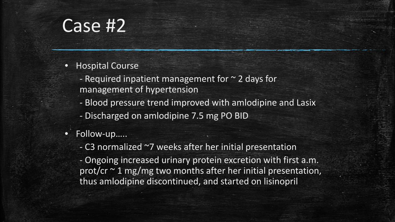
• Hospital Course- Required inpatient management for ~ 2 days for management of hypertension- Blood pressure trend improved with amlodipine and Lasix- Discharged on amlodipine 7.5 mg PO BID
• Follow-up…..- C3 normalized ~7 weeks after her initial presentation- Ongoing increased urinary protein excretion with first a.m. prot/cr ~ 1 mg/mg two months after her initial presentation, thus amlodipine discontinued, and started on lisinopril

Case #3
▪ 16 y/o male with recurrent gross hematuria; second episode clearly associated with illness
▪ Cutting out soda did not help
▪ UA with 3+ protein, Prot/Cr 1.8, BP 145/88, Cr 1 mg/dL
▪ Biopsy = IgA; started on ACEi

Proteinuria

Case #1▪ 17 year-old girl; found to have 3+ proteinuria at cheerleading
physical
▪ “Straight A” high school senior
▪ Healthy, very active, c/o some joint pain and fatigue x 6 months
▪ Remote history of UTI x 2, no hospitalizations, no fever, no edema
▪ Seen by adult nephrologist – Normal U/S and VCUG, ANA & Anti dsDNA neg x2, ANCA neg x2, complement studies neg x2, CT angiogram normal, 24 hour with 760 mg protein, course of steroids made no difference

Case #1
• Nephrology clinic:
• Mother exceedingly anxious
• Patient’s physical exam:-Healthy appearing athlete -Ht 50% Wt 25% BMI 23.6-HR 65 BP 100/72 T 36.5-Otherwise normal
• Screening test-Urine dip 2+ protein, no heme

Case #2
▪ 5 year-old boy with 3 month history of swelling
▪ Tried Benadryl, which didn’t help. Allergy testing negative.
▪ Swelling worse around eyes in a.m., then feet get swollen in p.m. Shoes hurt.
▪ Unremarkable PMH and FH
▪ Urine dip shows 4+ protein, trace blood.
▪ Labs done at InstaCare last week showed Na 131, Cr 0.4, alb 1.4, Ca 7.9, lytes otherwise normal

Proteinuria
▪ Proteinuria is well established as a marker of renal disease
▪ Even though proteinuria is more likely to be associated with renal disease than hematuria, the work-up is easier because of the smaller differential diagnosis
▪ Normal protein excretion is up to 100 mg/m2/day, or ~150mg/day in older children; higher in neonates
▪ Urinary albumin excretion is usually < 20 mg/day
Proteinuria: Epidemiology
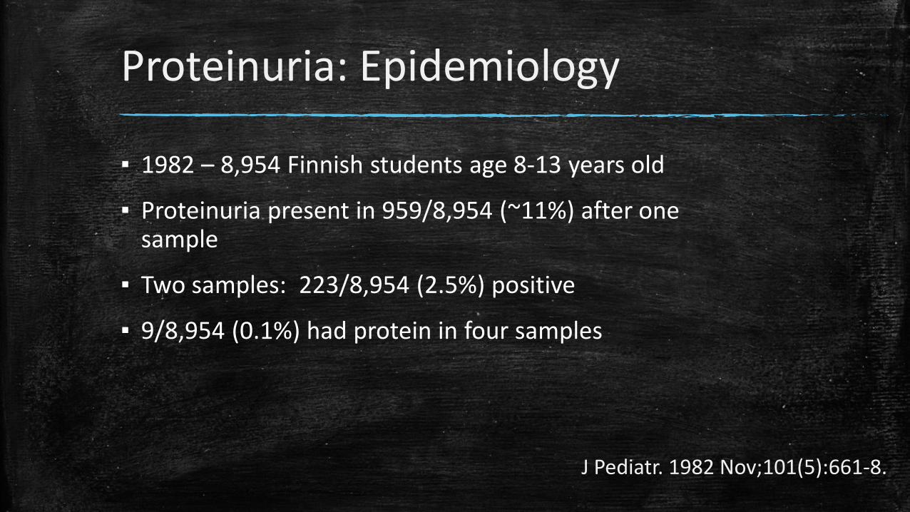
▪ 1982 – 8,954 Finnish students age 8-13 years old
▪ Proteinuria present in 959/8,954 (~11%) after one sample
▪ Two samples: 223/8,954 (2.5%) positive
▪ 9/8,954 (0.1%) had protein in four samples
J Pediatr. 1982 Nov;101(5):661-8.

Proteinuria: Mechanisms▪ Glomerular – Filtration of blood proteins across glomerular capillary wall– May result in edema, hypoalbuminemia and elevated cholesterol
(nephrotic syndrome)
▪ Tubular – Loss of low molecular weight proteins such as beta-2-microglobulin and
retinol binding protein due to inability of tubular cells to reabsorb the proteins (example, interstitial nephritis)
▪ Overflow– Excretion of proteins due to overproduction as in multiple myeloma

Proteinuria: Patterns▪ Transient:– May occur with stress or increased activity such as fever, seizure, acute illness,
dehydration, or vigorous exercise
▪ Orthostatic (Postural): – Blood flow to kidney decreases in recumbent and increases in standing position– Accounts for about 60-75% of childhood and adolescent proteinuria– Present in ~2-5% of older children and adolescents– Increased urinary protein excretion while upright– Typically benign, 80% resolve 20 years after diagnosis
▪ Persistent (6-12 months): Usually indicates underlying renal disease– Concomitant hematuria, may represent glomerulonephritis

Proteinuria: Detection▪ Urine dipstick measures albumin in a semi-quantitative
way– Albumin reacts with tetrabromophenol and turns blue– Does not measure other proteins, so low molecular
weight proteins not assessed

Proteinuria: Detection▪ Urine dipsticks measure urine protein concentration, but not
excretion rates– A very concentrated urine may have 1-2+ protein, but the patient
may not actually have increased protein excretion
▪ Proteinuria may be quantified with a spot urine pr/cr ratio– Removes the urine concentration from the equation – Normal is < 0.2 mg/mg– Nephrotic range is >2 mg/mg– Measures albumin and LMW proteins
▪ Spot pr/cr is comparable and much easier than 24 hour collection

Proteinuria: Initial Evaluation
▪ If greater than trace proteinuria, obtain a first morning urine sample for a protein to creatinine ratio (normal <0.2 mg/mg)– Obtain after a 24 hour period without vigorous excercise– Patient should empty bladder before bed
▪ Renal function panel (BMP + albumin + phosphorus)
▪ Blood pressure measurement

Orthostatic Proteinuria
▪ Blood flow to kidney decreases in recumbent and increases in standing position
▪ Affected individuals usually tall and thin, but not always
▪ Some individuals have mild hyperfiltration when standing – mild proteinuria
▪ Proteinuria disappears/greatly reduced when recumbent
▪ Diagnosis: First morning protein to creatinine ratio normal with elevated urinary protein excretion on clinic samples
▪ Considered benign
▪ Monitor annually with first morning prot/cr ratio

Fixed Proteinuria
▪ History: Febrile UTIs (renal scarring), edema, gross hematuria, family history
▪ Check BP
▪ Initial labs: CBC, RFP, lipid panel (if nephrotic range)
▪ Renal ultrasound, C3, C4, ANA, anti-dsDNA, hepatitis serologies
▪ Biopsy – MCD, FSGS, Membranous nephropathy, Lupus nephritis, MPGN

Case #1▪ 17 y/o female
▪ Seen by adult nephrologist – Normal US and VCUG, ANA & Anti dsDNA x2, ANCA x2, Complement studies x2, CT angiogram normal, 24 hour urine with 760 mg protein, course of steroids made no difference.– Cost ~ $10,000?
▪ Family expected a kidney biopsy that day, in clinic.
▪ 1st am urine negative, urine dipstick ~ 24-56 cents.– Cured. Mother still concerned about fatigue, malaise.
▪ Orthostatic proteinuria– “Hydrostatic” forces increase protein excretion typically in young, thin adolescents and children– Benign, 80% resolve within 20 years– 24 hour urine typically with 500-1200 mg/day, rarely nephrotic range– 50 year follow up of some cases showed no increase risk of CKD

Case #2
▪ 5 year-old boy with 3 month history of swelling
▪ Tried Benadryl, which didn’t help. Allergy testing negative.
▪ Swelling worse around eyes in a.m., then feet get swollen in p.m. Shoes hurt.
▪ Unremarkable PMH and FH
▪ Urine dip shows 4+ protein, trace blood.
▪ Labs done at InstaCare last week showed Na 131, Cr 0.4, alb1.4, Ca 7.9, lytes otherwise normal
▪ Up/c 5.8, cholesterol 720, iCa 1.25.

Case #2
▪ Nephrotic Syndrome
▪ Podocytopathy? Immune dysfunction
▪ Treatment: steroids
▪ ~85% in children will have minimal change disease
▪ 15-30% may have associated microscopic hematuria
▪ Hypocalcemia due to hypoalbuminemia, normal iCa
▪ Hypercholesterolemia due to increased hepatic synthetic activity

Summary: Hematuria
▪ Routine urine screening no longer recommended by AAP
▪ Asymptomatic hematuria:– Confirm hematuria with repeat dipsticks AND
microscopy– Microscopy to assess appearance of RBCs and for
presence of casts– Ultrasound– Urine culture, RFP, CBC– Other steps: C3, C4, antiDNase B, ANA, anti-dsDNA, 24
hour urine for stone panel, CT scan depending on history.

Summary: Proteinuria
▪ Proteinuria– Confirm presence of fixed proteinuria with first morning urine
for protein to creatinine ratio– If confirmed: Obtain RFP, CBC, C3, C4, ANA, anti-dsDNA,
hepatitis serologies– Ultrasound– History and physical- especially BP measurement and
assessment of any edema– HTN: To reinforce, if there are concerns for any renal disease in
children, BP must be obtained

Who Ya’ Gonna Call?▪ Pediatric Nephrology at
University of Utah/Primary Children’s Hospital– Dr. Raoul Nelson– Dr. Joseph Sherbotie– Dr. Matt Grinsell– Dr. Meredith Seamon– Debra Sandt, CPNP– Beth Esparza, RN– Cindy Terrill, RD
– Clinic scheduling: 801-213-3599– On call: 801-662-1000– Physician Access Line: