mesenchymal stem cells show functional defect and
TRANSCRIPT
RESEARCH Open Access
Mesenchymal stem cells show functionaldefect and decreased anti-cancer effectafter exposure to chemotherapeutic drugsChinnapaka Somaiah1†, Atul Kumar1†, Renu Sharma1†, Amit Sharma1, Trishna Anand1, Jina Bhattacharyya2,Damodar Das2, Sewali Deka Talukdar2 and Bithiah Grace Jaganathan1*
Abstract
Background: Mesenchymal stem cells (MSC) are used for several therapeutic applications to improve the functionsof bone, cardiac, nervous tissue as well as to facilitate the repopulation of hematopoietic stem cells. MSC give riseto the non-hematopoietic stromal cells of the bone marrow and are important for the maintenance of normalhematopoiesis. Chemotherapeutic drugs used for treatment of leukemia extensively damage the stromal cells andalter their gene expression profiles.
Methods: We determined the changes in adipogenic, osteogenic differentiation, phenotypic and gene expressionin MSC during treatment with chemotherapeutic drugs cytarabine, daunorubicin and vincristine. We also testedanti-cancer effects of drug treated MSC on leukemia cells.
Results: Treatment with the chemotherapeutic drugs resulted in functional defects in MSC, leading to reducedproliferation, osteogenic and adipogenic differentiation. The drug treated MSC also showed decreased expression ofcell surface receptors, and the changes in proliferation, phenotype and differentiation defect was partially reversibleafter withdrawing the drugs from the cells. The drug treated MSC showed increased expression of cytokines, IL6,FGF2 and TNFA but reduced levels of differentiation markers SOX9 and ACTC1. Drug treated MSC also contributedto reduced anti-cancer effects in leukemia cells.
Conclusions: Chemotherapeutic drug treatment altered the phenotype, osteogenic and adipogenic differentiationpotential of MSC and modified the gene expression profile of the cells to render them more chemoprotective ofthe leukemic cells. Thus, additional therapeutic efforts to target the stromal cell population will help in preventingchemoresistance, disease relapse in leukemia and to maintain a healthy bone marrow stroma.
Keywords: Bone marrow stroma, Chemotherapy, Chemoprotection, Leukemia, Osteoblasts, IL6, FGF2
BackgroundThe bone marrow derived MSC have the ability todifferentiate into several cell types and have gainedimportance in regenerative medicine, tissue engineeringand immune modulation [17, 24, 25]. MSC contribute tothe development of non-hematopoietic stromal cells inthe bone marrow. The stromal cells present in the bonemarrow are important for maintenance of normal and
malignant hematopoietic cells [31]. Chemotherapeuticdrugs used for leukemia therapy not only target thecancer cells, but also affect the cells of thehematopoietic microenvironment. Chemotherapeutictreatment for hematologic malignancies as well asother cancers have been shown to damage the bonemarrow microenvironment cells and MSC in vitro andin vivo [7, 11, 14, 20–23].When allogeneic MSC were utilized for co-injection with
bone marrow cells to improve the engraftment percentage,the allogeneic MSC did not engraft long-term, however therecipient MSC supported the hematopoietic cells [1, 9].Thus, defective stromal cells in the bone marrow might
* Correspondence: [email protected]†Equal contributors1Stem Cell and Cancer Biology Group, Department of Biosciences andBioengineering, Indian Institute of Technology Guwahati, Guwahati, IndiaFull list of author information is available at the end of the article
© The Author(s). 2018 Open Access This article is distributed under the terms of the Creative Commons Attribution 4.0International License (http://creativecommons.org/licenses/by/4.0/), which permits unrestricted use, distribution, andreproduction in any medium, provided you give appropriate credit to the original author(s) and the source, provide a link tothe Creative Commons license, and indicate if changes were made. The Creative Commons Public Domain Dedication waiver(http://creativecommons.org/publicdomain/zero/1.0/) applies to the data made available in this article, unless otherwise stated.
Somaiah et al. Journal of Biomedical Science (2018) 25:5 DOI 10.1186/s12929-018-0407-7
affect the long-term recovery of hematopoiesis afterchemotherapy or allogeneic hematopoietic stem celltransplantation where the recipient stromal cells have tosupport the hematopoietic stem cells and recovery ofhematopoiesis. When MSC are required for autologoustransplantation to mediate tissue repair or regeneration, itis important to understand the changes sustained by theMSC due to exposure to the chemotherapeutic drugs.Another important aspect that must be understood is,
whether the pre-exposure of MSC to chemotherapeuticdrugs affect their ability to support the leukemic cellsduring chemotherapy since MSC have been shown tohave functional aberrations and protect leukemia cellsduring chemotherapy [12, 13]. Chemotherapy treatmentfor leukemia is performed in several cycles and anychange in the gene expression profile of the stromal cellswhich render them more supportive of the leukemiccells will result in an unfavorable outcome. Thus, under-standing the effect of chemotherapeutic drugs on thefunctional and gene expression properties of MSC willhelp in improving the treatment strategies for leukemiaand to avoid relapse of the disease.In the current study, we determined the effect of che-
motherapeutic drugs cytarabine (CYT), daunorubicin(DAU) and vincristine (VIN) which are frequently uti-lized for leukemia treatment, on the properties of MSC.The cell surface marker expression, differentiation po-tential and gene expression profiles were determined forthe chemodrug treated MSC. We found that the chemo-therapeutic drugs significantly reduced the adipogenicand osteogenic differentiation ability of MSC, howeverthe cells partially recovered their differentiation potentialafter the removal of the drug. The drug pre-treated MSCwere more supportive of the leukemic cells during thedrug treatment which was accompanied by increased ex-pression of IL6, IL8, FGF2 or TNFA.
MethodsChemicals and reagentsDulbecco’s modified eagle’s medium (DMEM), Oil red O,alizarin red, dexamethasone, iso butyl methyl xanthine,indomethacin, insulin, β- glycerophosphate, ascorbic acidand basic fibroblast growth factor were purchased fromSigma Aldrich (Steinheim, Germany). Tissue culture plas-tic plates and flasks were from Eppendorf (Germany).Fluorescent conjugated anti-human antibodies were fromBD biosciences (Germany). Fetal bovine serum (FBS) andreal-time PCR reagents were purchased from ThermoFisher scientific (USA).
Bone marrow MSCMSC were isolated from the bone marrow of patientswho have been referred to the Hematology Departmentof Gauhati medical college hospital for diagnosis. The
samples were obtained after informed consent as perhospital ethical committee guidelines. The bone marrowcells were subjected to RBC lysis with ammonium chlor-ide solution (0.15 M, pH 7.3) on ice and the resultingcells were seeded in tissue culture plates for isolation ofMSC as described earlier [28].
Drug treatment of MSCMSC were seeded in tissue culture plates and treatedwith the chemotherapeutic drugs cytarabine (10μM),daunorubicin (0.1μM) or vincristine (0.1μM) for 48 hunless otherwise mentioned. The media containing thedrug was removed and live cell percentage was deter-mined by trypan blue exclusion counting. The cells wereseeded in fresh plates with complete media without thedrugs and allowed to proliferate. The recovered cellswere utilized for further experiments.
Adipogenic and osteogenic differentiationAdipogenic and osteogenic differentiation of MSC wasperformed as described earlier [18, 27]. For adipogenicdifferentiation, the cells were cultured in DMEM mediawith 10% serum supplemented with dexamethasone, iso-butylmethylxanthine, indomethacin and insulin. Forosteogenic differentiation, media containing 10% serumand dexamethasone, β-glycerophosphate, and ascorbicacid were added to the cells. The cells were differenti-ated for 21 days in the induction media and osteogenicdifferentiation was determined by staining for alizarinred and adipogenic differentiation by oil red-O staining.The alizarin red levels were quantified by eluting with10% (w/v) cetylpyridinium chloride in 10 mM sodiumphosphate, and absorbance measurement at 562 nm andOil red O stain was extracted from the cells with 100%isopropanol and quantified by absorbance measurementat 500 nm.
PhenotypingThe cell surface protein expression was analyzed by flowcytometry. MSC were trypsinized and stained with fluor-escent dye conjugated anti-human antibodies for 30 minat 4°C. The cells were washed, stained with propidiumiodide (PI) for live/dead discrimination and analyzedwith FACS caliber (BD Biosciences).
Co-culture of leukemia cells with stromal cellsHL60 or THP1 leukemic cell line (1x10⁵cells/ml) wasadded to drug pre-treated or untreated MSC seeded intissue culture plates. The cells were cultured for 48 hand indicated chemotherapeutic drugs were added tothe co-culture at the indicated concentrations. After48 h of treatment, the leukemic cells in suspension werecollected from the co-culture without disturbing theMSC layer. Adherent leukemia cells were collected by
Somaiah et al. Journal of Biomedical Science (2018) 25:5 Page 2 of 10
brief trypsinization (30s) while monitoring the detach-ment of leukemic cells from the stromal cells undermicroscope. Both the suspension and adherent leukemiacells were further processed for apoptosis analysis.
Apoptosis analysisApoptosis analysis was performed by staining the cellswith Annexin-V and PI (Thermo Fisher Scientific) accord-ing to the manufacturer’s instructions. The cells werewashed with ice-cold PBS, stained with anti -annexin Vantibody and propidium iodide and incubated in dark for15 mins at room temperature. The stained cells were ana-lyzed with FACS Calibur.
Gene expression analysisGene expression analysis was performed by real-timePCR. Total RNA was extracted using TriZol reagent(Thermo Fisher Scientific). RNA was reverse transcribedusing superscript III reverse transcriptase and Oligo dTprimers. The GAPDH, PPARG and OCN primersequences were as described earlier [28]. Other primersequences are given in supplementary information(Additional file 1: Table S1). Real-time PCR was per-formed with Power SyBr Green reagents in an ABI 7500real-time PCR machine (Thermo Fisher Scientific). Thegene expression levels in each sample was normalized totheir respective GAPDH expression level. The foldchange in the expression levels compared to the controlwas calculated using ΔΔCt method.
Data analysisFlow cytometric data was analyzed using FlowJosoftware (FlowJo, LLC). Geometric mean fluorescenceintensity (MFI) was calculated to detect the changes inthe expression levels. Statistical analysis was performedusing SPSS software and student T test was used tocompare the difference between treated and untreatedsamples. Variation between different MSC samples intheir gene expression profiles was determined by Mann-Whitney non-parametric variables test.
ResultsChemotherapeutic drug treatment affects themorphology and proliferation of MSCIn order to understand the effect of chemotherapeuticdrugs used for leukemia treatment on the bone marrowMSC, the cells were treated with the chemotherapeuticagents CYT, DAU and VIN. When MSC were treatedwith CYT, DAU or VIN, there was a significant decreasein the cell proliferation (Fig. 1a). VIN treated cells hadthe lowest viable cell number compared to the CYT orDAU treated cells (38.8 ± 2.4% in VIN treated cells, 59.7± 8.7% in CYT treated cells, 45.8 ± 4.2% in DAU treatedcells versus 95.8 ± 4.1% in control). VIN treatment
affected the cell morphology significantly where thespindle shaped cells assumed a flattened morphology.Reduced proliferation was observed even 7 days after thedrug removal in CYT and DAU treated MSC whereasVIN treated MSC recovered their proliferation capacityand proliferated at similar rates as that of the controlcells (Fig. 1b). Thus, the proliferation inhibition of VINwas reversible in MSC and effect of CYT and DAU werenot completely reversible although the cells partiallyregained their proliferation capacity. Moreover, after cul-turing the cells in the drug free media, the cells treatedwith all the drugs CYT, DAU and VIN assumed normalspindle shape (Fig. 1c).
Changes in osteogenic, adipogenic differentiation andphenotype was partially reversible afterchemotherapeutic drug treatmentMSC give rise to osteoblasts and adipocytes when in-duced with specific factors. MSC derived osteoblastsmaintain the hematopoietic stem cells in the bone mar-row [31], therefore, efficient osteoblastic differentiationof MSC treated with chemotherapeutic drug is import-ant for tissue engineering applications as well as for therecovery of the normal hematopoiesis in the patientduring remission. MSC treated with CYT, DAU and VINwere induced to differentiate into osteoblasts and adipo-cytes immediately after drug treatment or after culturingthe cells in the drug free media for 7 days. Both osteo-genic and adipogenic differentiation was significantly re-duced in drug treated MSC, where there was an averageten-fold reduction in osteogenic differentiation and five-fold reduction in adipogenic differentiation potential ofdrug treated MSC (Fig. 1d-f ). When MSC were allowedto proliferate in the drug free media post treatment, theyshowed higher osteogenic and adipogenic differentiationability compared to MSC induced to differentiate soonafter drug treatment (Fig. 1e, f ). Reduction in osteogenicand adipogenic differentiation was completely reversedin VIN treated MSC when they were cultured in VINfree media prior to induction of differentiation. CYTtreated MSC partially regained their osteogenic and adipo-genic differentiation potential whereas inhibition ofosteogenic and adipogenic differentiation due to DAUtreatment was not fully reversible (Fig. 1d-f).Further MSC treated with CYT, DAU and VIN were
analyzed for changes in cell surface expression of integ-rins CD13, CD29, CD49E, cell surface markers CD44,CD73, CD90 and death receptor CD95. There was a sig-nificant decrease in the expression of all cell surfacemarkers after DAU treatment whereas CYT and VINtreatment significantly reduced the expression of CD13and CD49E (Fig. 2a). VIN treatment also resulted insignificant reduction of CD90 and CD95 cell surface ex-pression (Fig. 2b). When MSC were cultured in the drug
Somaiah et al. Journal of Biomedical Science (2018) 25:5 Page 3 of 10
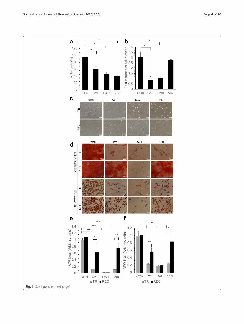
Fig. 1 (See legend on next page.)
Somaiah et al. Journal of Biomedical Science (2018) 25:5 Page 4 of 10
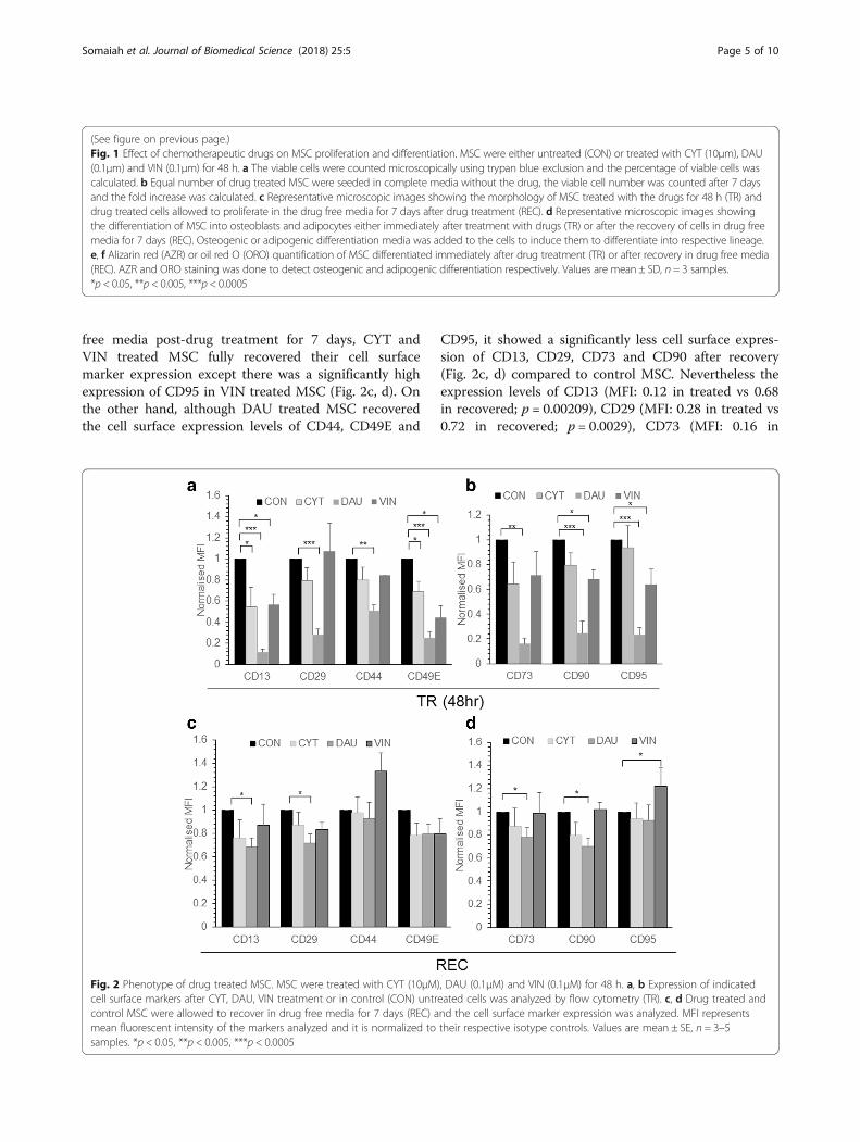
free media post-drug treatment for 7 days, CYT andVIN treated MSC fully recovered their cell surfacemarker expression except there was a significantly highexpression of CD95 in VIN treated MSC (Fig. 2c, d). Onthe other hand, although DAU treated MSC recoveredthe cell surface expression levels of CD44, CD49E and
CD95, it showed a significantly less cell surface expres-sion of CD13, CD29, CD73 and CD90 after recovery(Fig. 2c, d) compared to control MSC. Nevertheless theexpression levels of CD13 (MFI: 0.12 in treated vs 0.68in recovered; p = 0.00209), CD29 (MFI: 0.28 in treated vs0.72 in recovered; p = 0.0029), CD73 (MFI: 0.16 in
(See figure on previous page.)Fig. 1 Effect of chemotherapeutic drugs on MSC proliferation and differentiation. MSC were either untreated (CON) or treated with CYT (10μm), DAU(0.1μm) and VIN (0.1μm) for 48 h. a The viable cells were counted microscopically using trypan blue exclusion and the percentage of viable cells wascalculated. b Equal number of drug treated MSC were seeded in complete media without the drug, the viable cell number was counted after 7 daysand the fold increase was calculated. c Representative microscopic images showing the morphology of MSC treated with the drugs for 48 h (TR) anddrug treated cells allowed to proliferate in the drug free media for 7 days after drug treatment (REC). d Representative microscopic images showingthe differentiation of MSC into osteoblasts and adipocytes either immediately after treatment with drugs (TR) or after the recovery of cells in drug freemedia for 7 days (REC). Osteogenic or adipogenic differentiation media was added to the cells to induce them to differentiate into respective lineage.e, f Alizarin red (AZR) or oil red O (ORO) quantification of MSC differentiated immediately after drug treatment (TR) or after recovery in drug free media(REC). AZR and ORO staining was done to detect osteogenic and adipogenic differentiation respectively. Values are mean ± SD, n = 3 samples.*p < 0.05, **p < 0.005, ***p < 0.0005
Fig. 2 Phenotype of drug treated MSC. MSC were treated with CYT (10μM), DAU (0.1μM) and VIN (0.1μM) for 48 h. a, b Expression of indicatedcell surface markers after CYT, DAU, VIN treatment or in control (CON) untreated cells was analyzed by flow cytometry (TR). c, d Drug treated andcontrol MSC were allowed to recover in drug free media for 7 days (REC) and the cell surface marker expression was analyzed. MFI representsmean fluorescent intensity of the markers analyzed and it is normalized to their respective isotype controls. Values are mean ± SE, n = 3–5samples. *p < 0.05, **p < 0.005, ***p < 0.0005
Somaiah et al. Journal of Biomedical Science (2018) 25:5 Page 5 of 10

treated vs 0.78 in recovered; p = 0.00053) and CD90(MFI: 0.24 in treated vs 0.698 in recovered; p = 0.0119)in MSC cultured in drug free media post drug treatmentwere significantly higher than those observed in MSCimmediately after the DAU treatment (Fig. 2).
Drug treated MSC provided higher chemoprotection forleukemia cellsIn order to understand whether treatment with chemo-therapeutic drugs render the MSC more supportive ofthe leukemia cells, apoptosis of leukemia cells were ana-lyzed during co-culture with MSC. Leukemia cell linesHL60 was co-cultured with MSC pre-exposed to chemo-therapeutic drugs CYT, DAU or, VIN or in the presenceof untreated MSC. CYT, DAU or VIN was added to thecells in co-culture and apoptosis percentage of HL60was analyzed. HL60 in co-culture with MSC weretreated with the same drug to which MSC were pre-exposed, for instance, HL60 co-cultured with MSC weretreated with CYT if the MSC were pre-exposed to CYTand so on. CYT, DAU and VIN treatment significantlyinduced apoptosis in the control HL60 cells (Fig. 3). Asreported by others [5], co-culture with MSC significantlyreduced the chemosensitivity of HL60 to the chemother-apeutic drugs except during VIN treatment (Fig. 3).However, co-culture of HL60 or THP1 leukemic cellswith drug pre-treated MSC significantly reduced theirapoptosis percentage during chemotherapeutic drugtreatment suggesting a higher chemoprotective effect ofdrug treated MSC (Fig. 3, Additional file 2: Figure S1).Thus, pre-exposure of MSC to chemotherapeutic drugsrender them more supportive of the leukemia cells dur-ing chemotherapeutic treatment.To understand the chemoprotective effect of drug
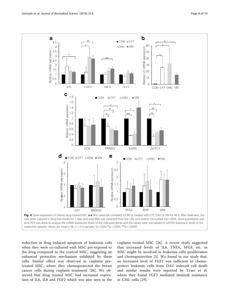
treated MSC further, the gene expression changes inCYT, DAU and VIN treated MSC were analyzed. Sincethe MSC after drug treatment were highly apoptotic(Fig. 1a), the gene expression changes was analyzed inMSC that were allowed to recover for 7 days after drugremoval. DAU treated MSC showed significantly highexpression of FGF2; VIN treated MSC had significantlyhigh IL6, FGF2 and TNFA mRNA expression levels(Fig. 4a). A significantly high IL8 expression was ob-served in MSC pre-treated with all the different drugswhereas no change in ILF2 levels were observed (Fig.4b). The expression of osteogenic specific gene OCNwas unaffected in all conditions whereas the adipogenicdifferentiation gene PPARG was downregulated in drugtreated cells (Fig. 4c). Chondrogenic differentiation geneSOX9 was significantly reduced in CYT and DAUtreated MSC whereas it was unaffected in VIN treatedcells (Fig. 4c). Cardiac actin gene (ACTC1) was signifi-cantly downregulated in MSC treated with all the differentdrugs (Fig. 4c). No significant difference in expression was
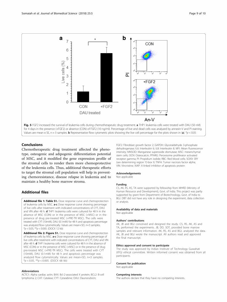
seen in oxidative stress response genes MNSOD, CAT(Fig. 4d) or apoptosis related genes BCL2, XIAP or BAX(Fig. 4e) was seen. To check whether the increased expres-sion of FGF2 observed in drug treated MSC might con-tribute to the chemoprotection of leukemia cells, THP1leukemia cell line was cultured and treated with DAU inthe presence of FGF2. We found a significant increase inthe percentage of live cells when FGF2 was present duringDAU treatment compared to the control (Fig. 5). Thus,chemotherapeutic drugs modify the MSC gene expressionprofile which in turn might chemoprotect leukemia cells.
DiscussionA healthy bone marrow microenvironment is necessaryfor repopulation of normal hematopoietic cells after che-motherapeutic treatment. Several studies have shownthat co-injection of MSC with the hematopoietic cellssignificantly increased the repopulation and reconstitu-tion of the recipient hematopoietic system [1, 9]. How-ever, the donor MSC failed to engraft long-term in therecipient and so the hematopoietic recovery and func-tioning were carried out by the host MSC [1, 9]. Thus, itis important to understand how chemotherapy affectsthe MSC characteristics, especially the osteogenic differ-entiation ability and expression of pro-survival cytokineswhich is critical to maintain the hematopoietic stemcells. Our study has shown that each chemotherapeuticagent had varied effect on the MSC phenotype, differen-tiation ability and gene expression. Although a combin-ation of drugs are used during chemotherapeutictreatment for leukemia, understanding the effect of indi-vidual drug at physiological concentration is important.All the chemotherapeutic drugs induced apoptosis ofMSC and decreased proliferation was observed evenafter the removal of the chemotherapeutic drugs. Similaranti-proliferative and apoptotic effects were reported inMSC after treatment with doxorubicin [22] and bleo-mycin [20]. However, adipose derived MSC were re-ported to be resistant to toxic effects of cisplatin,vincristine and camptothecin [3, 15] and bone marrowderived MSC were resistant to cisplatin [19] and pacli-taxel [4] induced apoptosis. In our study, we observed asignificant reduction in osteogenic and adipogenic differ-entiation of CYT, DAU and VIN treated MSC immedi-ately after treatment. However the differentiation abilitywas regained in CYT and VIN treated MSC when thecells were cultured in drug free media. Reduced adipo-genic differentiation of MSC was also reported afterbleomycin treatment [20]. Several studies observed atoxic effect of chemotherapy on bone marrow derivedstromal cells where decreased number of osteoprogeni-tors was reported in patients who underwent high dosechemotherapy with 5-fluorouracil, epidoxorubicin, andcyclophosphamide [2]. Bone marrow damage was seen
Somaiah et al. Journal of Biomedical Science (2018) 25:5 Page 6 of 10

in patients treated with 6-mercaptopurine and metho-trexate [6] but MSC differentiation ability was unaffectedduring cisplatin treatment [19]. However, MSC treatedwith dexamethasone and vincristine recovered their pro-liferation and differentiation ability faster than otherchemotherapeutic agents such as CYT [14] as seen alsoin our study. We found a significant downregulation ofseveral cell surface receptors including CD44 after DAUtreatment which the cells eventually recovered after the
removal of the drug. Similar downregulation of CD44expression in MSC was reported after cyclophosphamideand melphalan treatment [10].An important aspect of our study was to understand
how the MSC exposed to chemotherapeutic drugsprotected the leukemia cells during treatment with thechemotherapeutic drugs. The chemoprotective effect ofstromal cells in leukemia were reported by us and others[8, 13, 16, 30]. In the current study, we found a significant
Fig. 3 Chemoprotection of leukemia cells by drug treated MSC. a HL60 leukemia cells were cultured for 48 h in the absence of MSC (CON) or, in thepresence of MSC (+MSC) or in the presence of drug pre-treated MSC (+PRE-TR MSC). The cells were treated with CYT (10μM), DAU (0.1μM) and VIN(0.1μM) for 48 h and apoptosis percentage was analyzed flow cytometrically by staining with annexin-V and PI. Values are mean ± SD, n = 3 samples.b Representative flow cytometric plots showing the apoptosis percentage in drug-treated HL60 as shown in (a). *p < 0.05, **p < 0.005
Somaiah et al. Journal of Biomedical Science (2018) 25:5 Page 7 of 10
reduction in drug induced apoptosis of leukemia cellswhen they were co-cultured with MSC pre-exposed tothe drug compared to the control MSC, suggesting anenhanced protective mechanism exhibited by thesecells. Similar effect was observed in cisplatin pre-treated MSC, where they chemoprotected the breastcancer cells during cisplatin treatment [26]. We ob-served that drug treated MSC had increased expres-sion of IL6, IL8 and FGF2 which was also seen in the
cisplatin treated MSC [26]. A recent study suggestedthat increased levels of IL6, TNFA, bFGF, etc. inMSC might be involved in leukemia cells proliferationand chemoprotection [5]. We found in our study that,an increased level of FGF2 was sufficient to chemo-protect leukemic cells from DAU induced cell deathand similar results were reported by Traer et al.where they found FGF2 mediated imatinib resistancein CML cells [29].
Fig. 4 Gene expression of chemo drug treated MSC. a-e MSC were left untreated (CON) or treated with CYT, DAU or VIN for 48 h. After treatment, thecells were cultured in drug free media for 7 days and total RNA was extracted from the cells and reverse transcribed into cDNA. Semi-quantitative real-time PCR was done to analyze the mRNA expression levels of the indicated genes and the values were normalized to GAPDH expression levels in therespective samples. Values are mean ± SE, n = 3–4 samples. *p < 0.05, **p < 0.005, ***p < 0.0005
Somaiah et al. Journal of Biomedical Science (2018) 25:5 Page 8 of 10
ConclusionsChemotherapeutic drug treatment affected the pheno-type, osteogenic and adipogenic differentiation potentialof MSC, and it modified the gene expression profile ofthe stromal cells to render them more chemoprotectiveof the leukemia cells. Thus, additional therapeutic effortsto target the stromal cell population will help in prevent-ing chemoresistance, disease relapse in leukemia and tomaintain a healthy bone marrow stroma.
Additional files
Additional file 1: Table S1. Dose response curve and chemoprotectionof leukemia cells by MSC. a-c Dose response curve showing percentageof live cells after treatment with indicated concentrations of CYT, DAUand VIN after 48 h. d THP1 leukemia cells were cultured for 48 h in theabsence of MSC (CON) or in the presence of MSC (+MSC) or in thepresence of drug pre-treated MSC (+PRE-TR MSC). The cells weretreated with CYT (10mM), DAU (0.1mM) for 48 h and apoptosis percentagewas analyzed flow cytometrically. Values are mean+SD, n=3 samples.*p < 0.05, **p < 0.005. (DOCX 12 kb)
Additional file 2: Figure S1. Dose response curve and chemoprotectionof leukemia cells by MSC. a-c Dose response curve showing percentage oflive cells after treatment with indicated concentrations of CYT, DAU and VINafter 48 h. d THP1 leukemia cells were cultured for 48 h in the absence ofMSC (CON) or in the presence of MSC (+MSC) or in the presence of drugpre-treated MSC (+PRE-TR MSC). The cells were treated with CYT(10mM), DAU (0.1mM) for 48 h and apoptosis percentage wasanalyzed flow cytometrically. Values are mean+SD, n=3 samples.*p < 0.05, **p < 0.005. (DOCX 68 kb)
AbbreviationsACTC1: Alpha cardiac actin; BAX: Bcl-2-associated X protein; BCL2: B-celllymphoma 2; CAT: Catalase; CYT: Cytarabine; DAU: Daunorubicin;
FGF2: Fibroblast growth factor 2; GAPDH: Glyceraldehyde 3-phosphatedehydrogenase; IL6: Interleukin 6; IL8: Interleukin 8; MFI: Mean fluorescenceintensity; MNSOD: Manganese superoxide dismutase; MSC: mesenchymalstem cells; OCN: Osteocalcin; PPARG: Peroxisome proliferator activatedreceptor gamma; PI: Propidium iodide; RBC: Red blood cells; SOX9: SRY(sex determining region Y)-box 9; TNFA: Tumor necrosis factor alpha;VIN: Vincristine; XIAP: X-linked inhibitor of apoptosis protein
AcknowledgementsNot applicable
FundingCS, AK, RS, AS, TA were supported by fellowship from MHRD (Ministry ofHuman Resource and Development), Govt. of India. This project was partlysupported by grant from Department of Biotechnology, Govt. of India toBGJ. DBT did not have any role in designing the experiment, data collectionor analysis.
Availability of data and materialsNot applicable
Authors’ contributionsAK, JB and BGJ conceived and designed the study. CS, RS, AK, AS andTA, performed the experiments. JB, DD, SDT, provided bone marrowsamples and relevant information. AK, RS, AS and BGJ. analyzed the data.AK, JB and BGJ wrote the manuscript. All authors read and approvedthe final manuscript.
Ethics approval and consent to participateThe study was approved by Indian Institute of Technology Guwahati(IITG) ethical committee. Written informed consent was obtained from allparticipants.
Consent for publicationNot applicable
Competing interestsThe authors declare that they have no competing interests.
Fig. 5 FGF2 increased the survival of leukemia cells during chemotherapeutic drug treatment. a THP1 leukemia cells were treated with DAU (50 nM)for 4 days in the presence (+FGF2) or absence (CON) of FGF2 (10 ng/ml). Percentage of live and dead cells was analyzed by annexin-V and PI staining.Values are mean ± SE, n = 3 samples. b Representative flow cytometric plots showing the live cell percentage for the plots shown in (a). *p < 0.05
Somaiah et al. Journal of Biomedical Science (2018) 25:5 Page 9 of 10
Publisher’s NoteSpringer Nature remains neutral with regard to jurisdictional claims inpublished maps and institutional affiliations.
Author details1Stem Cell and Cancer Biology Group, Department of Biosciences andBioengineering, Indian Institute of Technology Guwahati, Guwahati, India.2Department of Hematology, Gauhati Medical College and Hospital,Guwahati, India.
Received: 17 August 2017 Accepted: 8 January 2018
References1. Ball LM, Bernardo ME, Roelofs H, Lankester A, Cometa A, Egeler RM, Locatelli
F, Fibbe WE. Cotransplantation of ex vivo-expanded mesenchymal stemcells accelerates lymphocyte recovery and may reduce the risk of graftfailure in haploidentical hematopoietic stem-cell transplantation. Blood.2007;110(7):2764–7.
2. Banfi A, Podesta M, Fazzuoli L, Sertoli MR, Venturini M, Santini G, CanceddaR, Quarto R. High-dose chemotherapy shows a dose-dependent toxicity tobone marrow osteoprogenitors - A mechanism for post-bone marrowtransplantation osteopenia. Cancer. 2001;92(9):2419–28.
3. Bellagamba BC, de Abreu BRR, Grivicich I, Markarian CF, Chem E, CamassolaM, Nardi NB, Dihl RR. Human mesenchymal stem cells are resistant tocytotoxic and genotoxic effects of cisplatin in vitro. Genet Mol Biol. 2016;39(1):129–34.
4. Bosco DB, Kenworthy R, Zorio DAR, Sang QXA. Human mesenchymal stemcells are resistant to paclitaxel by adopting a non-proliferative fibroblasticstate. Plos One. 2015;10(6):e0128511. https://doi.org/10.1371/journal.pone.0128511.
5. Brenner AK, Nepstad I, Bruserud O. Mesenchymal stem cells support survivaland Proliferation of Primary human acute Myeloid leukemia cells throughheterogeneous Molecular Mechanisms. Front Immunol. 2017;8
6. Corazza F, Hermans C, Ferster A, Fondu P, Demulder A, Sariban E. Bonemarrow stroma damage induced by chemotherapy for acute lymphoblasticleukemia in children. Pediatric Res. 2004;55(1):152–8.
7. Galotto M, Berisso G, Delfino L, Podesta M, Ottaggio L, Dallorso S, Dufour C,Ferrara GB, Abbondandolo A, Dini G, Bacigalupo A, Cancedda R, Quarto R.Stromal damage as consequence of high-dose chemo/radiotherapy in bonemarrow transplant recipients. Exp Hematol. 1999;27(9):1460–6.
8. Iwamoto S, Mihara K, Downing JR, Pui CH, Campana D. Mesenchymal cellsregulate the response of acute lymphoblastic leukemia cells to asparaginase.J Clin Invest. 2007;117(4):1049–57.
9. Jaganathan BG, Tisato V, Vulliamy T, Dokal I, Marsh J, Dazzi F, Bonnet D.Effects of MSC co-injection on the reconstitution of aplastic anemia patientfollowing hematopoietic stem cell transplantation. Leukemia. 2010;24(10):1791–5.
10. Kemp K, Morse R, Sanders K, Hows J, Donaldson C. Alkylatingchemotherapeutic agents cyclophosphamide and melphalan causefunctional injury to human bone marrow-derived mesenchymal stemcells. Ann Hematol. 2011;90(7):777–89.
11. Kemp K, Morse R, Wexler S, Cox C, Mallam E, Hows J, Donaldson C.Chemotherapy-induced mesenchymal stem cell damage in patients withhematological malignancy. Ann Hematol. 2010;89(7):701–13.
12. Kumar A, Anand T, Bhattacharyya J, Sharma A, Jaganathan BG. K562 chronicmyeloid leukemia cells modify osteogenic differentiation and geneexpression of bone marrow stromal cells. J Cell Commun Signal. 2017;
13. Kumar A, Bhattacharyya J, Jaganathan BG. Adhesion to stromal cellsmediates imatinib resistance in chronic myeloid leukemia through ERK andBMP signaling pathways. Sci Rep. 2017;7
14. Li J, Law HKW, Lau YL, Chan GCF. Differential damage and recovery ofhuman mesenchymal stem cells after exposure to chemotherapeuticagents. Br J Haematology. 2004;127(3):326–34.
15. Liang W, Xia HL, Li J, Zhao RC. Human adipose tissue derived mesenchymalstem cells are resistant to several chemotherapeutic agents. Cytotechnology.2011;63(5):523–30.
16. Lin YM, Zhang GZ, Leng ZX, Lu ZX, Bu LS, Gao S, Yang SJ. Study on thebone marrow mesenchymal stem cells induced drug resistance in the U937cells and its mechanism. Chin Med J. 2006;119(11):905–10.
17. Ma S, Xie N, Li W, Yuan B, Shi Y, Wang Y. Immunobiology of mesenchymalstem cells. Cell Death Diff. 2014;21(2):216–25.
18. Mawrie D, Kumar A, Magdalene D, Bhattacharyya J, Jaganathan BG.Mesenchymal stem cells from human extra ocular muscle harborneuroectodermal differentiation potential. Plos One. 2016;11(6):e0156697.https://doi.org/10.1371/journal.pone.0156697.
19. Nicolay NH, Perez RL, Ruhle A, Trinh T, Sisombath S, Weber KJ, Ho AD,Debus J, Saffrich R, Huber PE. Mesenchymal stem cells maintain theirdefining stem cell characteristics after treatment with cisplatin. ScientificReports. 2016;6
20. Nicolay NH, Ruhle A, Perez RL, Trinh T, Sisombath S, Weber KJ, Ho AD,Debus J, Saffrich R, Huber PE. Mesenchymal stem cells are sensitive tobleomycin treatment. Sci Rep. 2016;6
21. Nifontova I, Svinareva D, Petrova T, Drize N. Sensitivity of mesenchymalstem cells and their progeny to medicines used for the treatment ofhematoproliferative diseases. Acta Haematologica. 2008;119(2):98–103.
22. Oliveira MS, Carvalho JL, Campos ACD, Gomes DA, de Goes AM, Melo MM.Doxorubicin has in vivo toxicological effects on ex vivo culturedmesenchymal stem cells. Toxicol Lett. 2014;224(3):380–6.
23. Prata KD, Orellana MD, De Santis GC, Kashima S, Fontes AM, Carrara RDV,Palma PVB, Neder L, Covas DT. Effects of high-dose chemotherapy on bonemarrow multipotent mesenchymal stromal cells isolated from lymphomapatients. Exp Hematol. 2010;38(4):292–300.
24. Ratajczak J, Hilkens P, Gervois P, Wolfs E, Jacobs R, Lambrichts I, BronckaersA. Angiogenic capacity of periodontal ligament stem cells pretreated withdeferoxamine and/or fibroblast growth factor-2. Plos One. 2016;11(12)
25. Richardson SM, Kalamegam G, Pushparaj PN, Matta C, Memic A,Khademhosseini A, Mobasheri R, Poletti FL, Hoyland JA, Mobasheri A.Mesenchymal stem cells in regenerative medicine: Focus on articularcartilage and intervertebral disc regeneration. Methods. 2016;99:69–80.
26. Skolekova S, Matuskova M, Bohac M, Toro L, Durinikova E, Tyciakova S,Demkova L, Gursky J, Kucerova L. Cisplatin-induced mesenchymal stromalcells-mediated mechanism contributing to decreased antitumor effect inbreast cancer cells (vol 14, 4, 2016). Cell Commun Signaling. 2016;14
27. Somaiah C, Kumar A, Mawrie D, Sharma A, Patil SD, Bhattacharyya J,Swaminathan R, Jaganathan BG. Collagen promotes higher adhesion,survival and proliferation of mesenchymal stem cells. Plos One. 2015;10(12)
28. Sonowal H, Kumar A, Bhattacharyya J, Gogoi PK, Jaganathan BG. Inhibition ofactin polymerization decreases osteogeneic differentiation of mesenchymalstem cells through p38 MAPK pathway. J Biomed Sci. 2013;20:71. https://doi.org/10.1186/1423-0127-20-71.
29. Traer E, Javidi-Sharifi N, Agarwal A, Dunlap J, English I, Martinez J, Tyner JW,Wong M, Druker BJ. Ponatinib overcomes FGF2-mediated resistance in CMLpatients without kinase domain mutations. Blood. 2014;123(10):1516–24.
30. Weisberg E, Wright RD, McMillin DW, Mitsiades C, Ray A, Barrett R, Adamia S,Stone R, Galinsky I, Kung AL, Griffin JD. Stromal-mediated protection oftyrosine kinase inhibitor-treated BCR-ABL-expressing leukemia cells. MolCancer Ther. 2008;7(5):1121–9.
31. Zhang JW, Niu C, Ye L, Huang HY, He X, Tong WG, Ross J, Haug J, JohnsonT, Feng JQ, Harris S, Wiedemann LM, Mishina Y, Li LH. Identification of thehaematopoietic stem cell niche and control of the niche size. Nature. 2003;425(6960):836–41.
• We accept pre-submission inquiries
• Our selector tool helps you to find the most relevant journal
• We provide round the clock customer support
• Convenient online submission
• Thorough peer review
• Inclusion in PubMed and all major indexing services
• Maximum visibility for your research
Submit your manuscript atwww.biomedcentral.com/submit
Submit your next manuscript to BioMed Central and we will help you at every step:
Somaiah et al. Journal of Biomedical Science (2018) 25:5 Page 10 of 10