met amplification and exon 14 splice site fi non small cell … · nte clinical research ethnics...
TRANSCRIPT

Biology of Human Tumors
MET Amplification and Exon 14 Splice SiteMutation Define Unique Molecular Subgroups ofNon–Small Cell Lung Carcinoma with PoorPrognosisJoanna H. Tong1,2,3, Sai F. Yeung1,2,3, Anthony W.H. Chan1,2,3, Lau Y. Chung1,2,3,Shuk L. Chau1,2,3, Raymond Wai Ming Lung1,2,3, Carol Y. Tong1,2,3, Chit Chow1,2,3,Edith K.Y. Tin1,2,3, Yau H. Yu1,2,3, Hui Li1,2,3, Yi Pan1,2,3,Wing P. Chak1,2,3, Calvin S.H. Ng4,Tony S.K. Mok3,5, and Ka F. To1,2,3
Abstract
Purpose: Activation of MET oncogene as the result of ampli-fication or activation mutation represents an emerging moleculartarget for cancer treatment. We comprehensively studied METalterations and the clinicopathologic correlations in a large cohortof treatment-na€�ve non–small cell lung carcinoma (NSCLC).
Experimental Design: Six hundred eighty-seven NSCLCs weretested for MET exon 14 splicing site mutation (METD14), DNAcopy number alterations, and protein expression by Sangersequencing, FISH, and IHC, respectively.
Results:METD14 mutation was detected in 2.62% (18/687)of NSCLC. The mutation rates were 2.6% in adenocarcinoma,4.8% in adenosquamous carcinoma, and 31.8% in sarcoma-toid carcinoma. METD14 mutation was not detected insquamous cell carcinoma, large cell carcinoma, and lympho-epithelioma-like carcinoma but significantly enriched in sar-comatoid carcinoma (P < 0.001). METD14 occurred mutuallyexclusively with known driver mutations but tended to coexist
with MET amplification or copy number gain (P < 0.001).Low-levelMET amplification and polysomy might occur in thebackground of EGFR or KRAS mutation whereas high-levelamplification (MET/CEP7 ratio �5) was mutually exclusive tothe major driver genes except METD14. Oncogenic METD14mutation and/or high-level amplification occurred in a totalof 3.3% (23/687) of NSCLC and associated with higherMET protein expression. METD14 occurred more frequentlyin older patients whereas amplification was more commonin ever-smokers. Both METD14 and high-level amplificationwere independent prognostic factors that predicted poorersurvival by multivariable analysis.
Conclusions: The high incidence of METD14 mutation insarcomatoid carcinoma suggested that MET inhibition mightbenefit this specific subgroup of patients. Clin Cancer Res; 22(12);3048–56. �2016 AACR.
See related commentary by Drilon, p. 2832
IntroductionNon–small cell lung cancer (NSCLC) represents a paradigm
for the development of targeted cancer therapy. EGFR and ALKare well-known examples demonstrating that matched action-able oncogenic mutations with appropriate tyrosine kinase
inhibitor (TKI) therapies improve patients' life quality andsurvival. Recent genomic studies in lung adenocarcinoma havefound actionable oncogenic mutations involving RTK/RAS/RAF/PI3K axis such as EGFR, KRAS, HER2, BRAF, ARAF, CRAF,PIK3CA, MET, RIT1, MAP2K1, NRAS, HRAS mutations and ALK,NRG1, NTRK, ERBB4, RET, ROS1, and BRAF translocations,suggesting more than 70% of lung adenocarcinoma could bedefined by gene mutations (1, 2).
MET is a high-affinity receptor tyrosine kinase (RTK) that couldinitiate an array of pathways promoting cell proliferation, sur-vival, and metastasis upon stimulation. Gain-of-function altera-tions of MET by DNA amplification, mutation, and protein over-expression are driver events of oncogenesis in many cancer types.Dysregulation of MET enhances the malignant properties andpredict poor prognosis that represents a possible target for per-sonalized therapy (3). MET-directed anticancer strategies byblocking different MET pathway components are under preclin-ical and clinical trials. These include antibodies targeting MET orHGF, selective, and unselective small molecules targeting METRTK activity.
NSCLCs harboring MET DNA amplification are dependent onMET for growth and survival (4). NSCLC patients with de novoMET DNA amplification are responsive to crizotinib, suggesting
1Department of Anatomical and Cellular Pathology, The Chinese Uni-versity of Hong Kong, Hong Kong. 2State Key Laboratory in Oncologyin South China,The Chinese University of Hong Kong, Hong Kong. 3LiKa-Shing Institute of Health Sciences,The Chinese University of HongKong, HongKong. 4Division of Cardiothoracic Surgery, Department ofSurgery, The Chinese University of Hong Kong, Hong Kong. 5Depart-mentofClinicalOncology,TheChineseUniversityofHongKong,HongKong.
Note: Supplementary data for this article are available at Clinical CancerResearch Online (http://clincancerres.aacrjournals.org/).
Joanna H. Tong and Sai F. Yeung contributed equally to this article.
Corresponding Author: Ka F. To, The Chinese University of Hong Kong, 1F, LCWClinical ScienceBuilding, Prince ofWalesHospital, ShatinNT, HongKong. Phone:852-2632-3335; Fax: 852-2637-6274; E-mail: [email protected]
doi: 10.1158/1078-0432.CCR-15-2061
�2016 American Association for Cancer Research.
ClinicalCancerResearch
Clin Cancer Res; 22(12) June 15, 20163048
on February 20, 2020. © 2016 American Association for Cancer Research. clincancerres.aacrjournals.org Downloaded from
Published OnlineFirst February 4, 2016; DOI: 10.1158/1078-0432.CCR-15-2061

thatMET amplification is a primary oncogenic driver and a validclinical target (5, 6). MET mutations affecting exon 14 splicingelements occur in up to 5% of lung adenocarcinoma. Thesemutations result in a juxtamembrane domain lacking METprotein (METD14) with extended half-life after HGF stimula-tion that has been considered an oncogenic driver event. In vitroand in vivo studies have demonstrated that tumors harboringMET exon 14 mutation responded to MET inhibitors (1, 7, 8).Clinical response to MET inhibitors in patients with METD14þ
lung adenocarcinoma has been reported (9), further supportingMET as a novel therapeutic target. However, a recent phase IIIrandomized clinical trial failed to demonstrate additional ben-efit of onartuzumab, an anti-MET mAb, on advanced stageNSCLC patients treated with erlotinib whose tumors wereidentified as METþ by IHC (10). This underscores the impor-tance of appropriate predictive biomarker for patient stratifi-cation in the new era of personalized medicine.
We have reported the driver mutation profile of 154 lungadenocarcinoma and adenosquamous cell carcinoma (ADSQ)and demonstrated that MET DNA alterations defined a subgroupof patients with aggressive diseases that might potentially benefitfrom anti-MET targeted therapy (11). The clinical implication ofMET alterations in different histologic subsets of NSCLC remainsundefined. In the current study, we aimed to determine theprevalence of MET DNA alterations, including exon 14 skippingmutations (METD14) and amplifications, in a large cohort ofChinese patients, and define the clinicopathologic characteristicsof MET-positive tumors.
Materials and MethodsPatients and samples
Patients with primary NSCLC who underwent surgical resec-tion at Prince of Wales Hospital, Hong Kong, between 1995 and2011 were selected for the retrospective study. All availableformalin-fixed paraffin-embedded (FFPE) surgical resection spe-cimens were reviewed by two pathologists (K.F. To and A.W.Chan) to confirm the histologic diagnosis and select the repre-sentative tumor blocks with appropriate tumor content. Medicalrecords were reviewed to extract data on clinicopathologic para-
meters. The pathologic stages were determined according to the7th edition of American Joint Committee on Cancer tumor–node–metastasis classification system. Early stage referred tostage I to IIIA whereas advanced stage referred to stage IIIb to IV.Patients were categorized into either never-smoker (smoke lessthan 100 cigarettes in their lifetime) or ever-smoker (smokemore than 100 cigarettes in their lifetime; ref. 1). Patients whoreceived neoadjuvant chemotherapy or radiotherapy wereexcluded from the study. A total of 687 treatment-na€�ve NSCLCmet the selection criteria that were included in the currentstudy. The study protocol was approved by the Joint CUHK-NTE Clinical Research Ethnics Committee. The driver mutationprofile of 154 adenocarcinoma and ADSQ has been reported ina previous study (11).
Construction of tissue microarrayTissue microarrays (TMA) were constructed using a tissue
arrayer (Beecher Instruments). The location of tumor area onthe donor FFPE tissue block was first marked on the hematox-ylin and eosin–stained histologic section. Three representative1-mm cores were obtained from each tumor and were insertedto a recipient paraffin block. For FISH and IHC, 4-mm tissuesections were prepared and mounted onto Superfrost Plusmicroscope slides.
IHCIHCwas carried out using Benchmark XT autostainer (Ventana)
using Ultraview detection system. MET IHC was performed usingConfirm anti-Total c-MET (SP44) rabbit mAb (Ventana) accord-ing to the manufacturer's instruction. Expression level of METprotein was determined by a scoring system considering bothstaining intensity and prevalence of intensities in tumor cells. Thefour staining scores were defined as following: 3þ (�50% oftumor cells staining with strong intensity); 2þ (�50% of tumorcells with moderate or higher staining but < 50% with strongintensity); 1þ (�50% of tumor cells with weak or higher stainingbut < 50%withmoderate or higher intensity); or 0 (no staining or< 50% of tumor cells with any intensity; ref. 12). Tumors withmoderate to strong MET protein expression (score 2þ and 3þ)were considered IHCþ, whereas score 0 and 1þ were regarded asIHC� for MET expression.
Mutational analysisDNA was extracted from FFPE tissue using QIAamp DNAmini
kit (Qiagen) according to the manufacturer's protocol. Manualmicrodissection was performed to ensure more than 70% tumorcontent in each DNA sample for subsequent analysis. Sangersequencing was performed to screen for MET exon 14 splice sitemutations.
FISHMET gene copy number/amplification status were investigated
by MET/CEP7 FISH probe (Abbott Molecular) as reported previ-ously (11). Copy number per cell and MET/CEP7 ratio werecounted in at least 50 nonoverlapping tumor cell nuclei. As thereis no consensus approach in MET FISH scoring, we used threescoring systems for MET FISH assay:
1. Tumors with�5MET signals per cell were classified as FISHþ
according to Capuzzo scoring system (13).
Translational Relevance
MET oncogene is an emerging molecular target for non–small cell lung carcinoma (NSCLC). Multiple mechanismscontribute to MET activation. The incidence and clinicopath-ologic characteristics of tumors withMET alterations are yet tobe established. This study represents the first comprehensiveparallel screening of MET alterations including METD14mutation, DNA copy number alteration, and protein expres-sion in a large cohort of NSCLC. Oncogenic METD14 muta-tion and high-level amplification (MET/CEP7 ratio �5),which are mutually exclusive to other major driver mutations,occur in 3.3%ofNSCLC that define a distinct subset of NSCLCwith sarcomatoid histology and aggressive clinical course. Thefinding suggests a pivotal role of MET signaling in tumorigen-esis of sarcomatoid carcinoma and raises the possibility thatMET inhibition may aid in treating this highly aggressive andchemotherapy-resistant subtype of NSCLC.
MET DNA Alterations in NSCLC
www.aacrjournals.org Clin Cancer Res; 22(12) June 15, 2016 3049
on February 20, 2020. © 2016 American Association for Cancer Research. clincancerres.aacrjournals.org Downloaded from
Published OnlineFirst February 4, 2016; DOI: 10.1158/1078-0432.CCR-15-2061
2. Tumor with MET/CEP7 ratio �2 were defined as FISHþ byPathVysion (14, 15).
3. High-level amplification (H-Amp) was defined as clusteredMET signals or MET/CEP7 ratio �5 (6).
Screening for major driver eventsEGFR exons 18–21, KRAS exons 2 and 3were screened by PCR-
direct sequencing. ALK and ROS1 translocations were examinedby dual color break-apart FISH analysis as described previously(11).
Statistical analysisStatistical analysis was performed using SPSS 19.0 (IBMCorp.).
c2 and Fisher exact test were used to analyze associations ofmutational, protein expression, and gene copy number statuswith clinical characteristics. We compared MET status in eachhistologic subtype versus all other subtypes. For example, METstatus in adenocarcinomas was compared with all the non-ade-nocarcinomas and so on. Overall survival (OS) was defined as thetime from disease diagnosis to patient's death due to diseaseprogression. The Kaplan–Meier method was used to estimate thesurvival rates for different groups. The equivalences of the survivalcurves were tested by log-rank statistics. The Cox proportionalhazards model was employed for univariable and multivariablesurvival analyses. The variables found to be statistical significantin the univariable survival analysis were further evaluated in themultivariable survival analysis. A two-tailed P value of <0.05 wasconsidered to be statistically significant.
ResultsPatient characteristics
A total of 687 treatment-naive NSCLC, comprising 392(57.1%)adenocarcinoma, 180 (26.2%) squamous cell carcinoma(SCC), 45 (6.6%) large cell carcinoma (LCC), 21 (3.1%) ADSQ,27 (3.9%) lymphoepithelioma-like carcinoma (LELC), and 22(3.2%) pulmonary sarcomatoid carcinoma (PSC) were recruited.The median age of the patients was 66 years (range, 27–94 years)and male to female ratio was 2.1:1. Ever-smokers represented63.9% of all patients and were more common in SCC thanother histologies (P < 0.001). Adenocarcinoma was more com-mon in female (P < 0.001) and never-smokers (P < 0.001).LELC occurred more frequently in female (P ¼ 0.043), never-
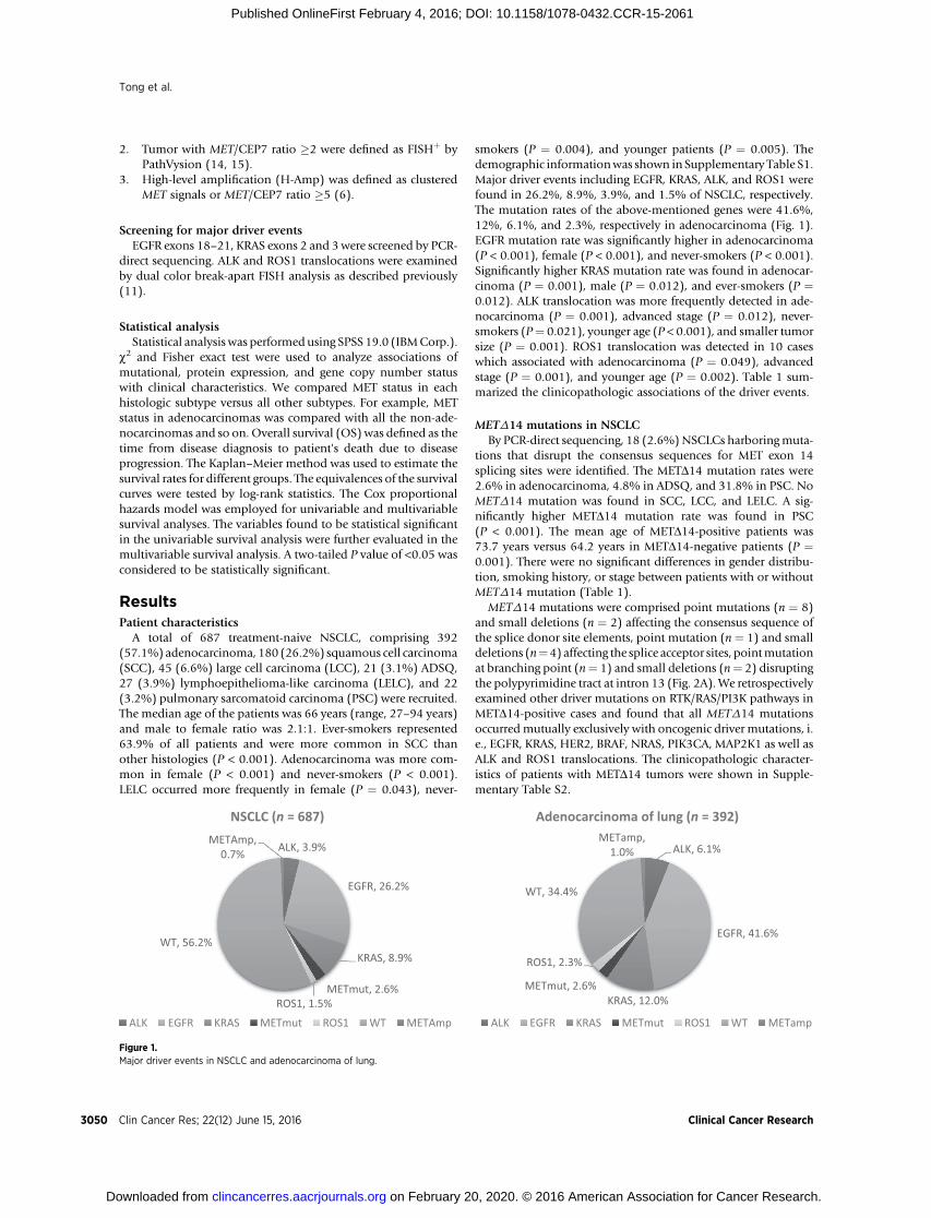
smokers (P ¼ 0.004), and younger patients (P ¼ 0.005). Thedemographic informationwas shown in Supplementary Table S1.Major driver events including EGFR, KRAS, ALK, and ROS1 werefound in 26.2%, 8.9%, 3.9%, and 1.5% of NSCLC, respectively.The mutation rates of the above-mentioned genes were 41.6%,12%, 6.1%, and 2.3%, respectively in adenocarcinoma (Fig. 1).EGFR mutation rate was significantly higher in adenocarcinoma(P < 0.001), female (P < 0.001), and never-smokers (P < 0.001).Significantly higher KRAS mutation rate was found in adenocar-cinoma (P ¼ 0.001), male (P ¼ 0.012), and ever-smokers (P ¼0.012). ALK translocation was more frequently detected in ade-nocarcinoma (P ¼ 0.001), advanced stage (P ¼ 0.012), never-smokers (P¼ 0.021), younger age (P < 0.001), and smaller tumorsize (P ¼ 0.001). ROS1 translocation was detected in 10 caseswhich associated with adenocarcinoma (P ¼ 0.049), advancedstage (P ¼ 0.001), and younger age (P ¼ 0.002). Table 1 sum-marized the clinicopathologic associations of the driver events.
METD14 mutations in NSCLCBy PCR-direct sequencing, 18 (2.6%)NSCLCs harboringmuta-
tions that disrupt the consensus sequences for MET exon 14splicing sites were identified. The METD14 mutation rates were2.6% in adenocarcinoma, 4.8% in ADSQ, and 31.8% in PSC. NoMETD14 mutation was found in SCC, LCC, and LELC. A sig-nificantly higher METD14 mutation rate was found in PSC(P < 0.001). The mean age of METD14-positive patients was73.7 years versus 64.2 years in METD14-negative patients (P ¼0.001). There were no significant differences in gender distribu-tion, smoking history, or stage between patients with or withoutMETD14 mutation (Table 1).
METD14 mutations were comprised point mutations (n ¼ 8)and small deletions (n ¼ 2) affecting the consensus sequence ofthe splice donor site elements, point mutation (n ¼ 1) and smalldeletions (n¼4) affecting the splice acceptor sites, pointmutationat branching point (n¼ 1) and small deletions (n¼ 2) disruptingthe polypyrimidine tract at intron 13 (Fig. 2A).We retrospectivelyexamined other driver mutations on RTK/RAS/PI3K pathways inMETD14-positive cases and found that all METD14 mutationsoccurredmutually exclusively with oncogenic driver mutations, i.e., EGFR, KRAS, HER2, BRAF, NRAS, PIK3CA, MAP2K1 as well asALK and ROS1 translocations. The clinicopathologic character-istics of patients with METD14 tumors were shown in Supple-mentary Table S2.
ALK, 3.9%
EGFR, 26.2%
KRAS, 8.9%
METmut, 2.6%ROS1, 1.5%
WT, 56.2%
METAmp,0.7%
NSCLC (n = 687)
ALK EGFR KRAS METmut ROS1 WT METAmp
ALK, 6.1%
EGFR, 41.6%
KRAS, 12.0%METmut, 2.6%
ROS1, 2.3%
WT, 34.4%
METamp,1.0%
Adenocarcinoma of lung (n = 392)
ALK EGFR KRAS METmut ROS1 WT METamp
Figure 1.Major driver events in NSCLC and adenocarcinoma of lung.
Tong et al.
Clin Cancer Res; 22(12) June 15, 2016 Clinical Cancer Research3050
on February 20, 2020. © 2016 American Association for Cancer Research. clincancerres.aacrjournals.org Downloaded from
Published OnlineFirst February 4, 2016; DOI: 10.1158/1078-0432.CCR-15-2061

MET gene copy number alterationFISH analysis was performed to investigate the copy number
alteration (CNA) of MET gene in NSCLC. As there is noconsensus scoring system for MET CNA, we employed threedifferent scoring systems for MET FISH analysis as described inmethodology. The results from the original scoring systemswere summarized in Supplementary Table S3. The final FISHstatus was the integration of three scoring systems according tothe patterns of DNA CNAs.
Twenty-nine cases were classified as FISHþ by at least onescoring system (Supplementary Table S4). Four distinct patternsofMET CNAwere identified (Fig. 2B and Supplementary Fig. S1):
1. H-Amp: ratio of MET/CEP7 � 5; n ¼ 8.2. Polysomy:MET signal� 5,without gene amplification; n¼9.3. Low-level amplification/high gene copy number (L-Amp/
H-GCN): 2 � MET/CEP7 < 5, MET signal � 5; n ¼ 7.4. Low-level amplification/low gene copy number (L-Amp/
L-GCN): 2 � MET/CEP7 < 5, MET signal < 5; n ¼ 5.
High-level amplification-Amp was more common in PSC(3/22, 13.6%, P < 0.001) than other histologic subtypes where-as polysomy was exclusively found in adenocarcinoma (9/392,2.3%, P < 0.001). A significant enrichment of SCC was observedin L-AMP/L-GCN group (4/5, 80%). MET gene amplificationbut not polysomy associated with positive smoking history(Supplementary Table S3).
There was a significant association betweenMETD14mutationandMET CNA (P < 0.001). Among 29 FISHþ cases, 20.7% (6/29)showed METD14 mutation (Supplementary Table S4). Althoughin FISH� group, only 1.8% (12/658) of the cases harboredMETD14mutation (Table 1).METD14mutations occurred more
frequently in old-age patients whereas DNA amplifications weremore commonly seen in ever-smokers.
MET CNA may occur in the background of other driver events.Almost all polysome (8/9) coexisted with other driver mutations:5 with EGFR mutation, 2 with METD14, and 1 with KRASmutation. Three of 12 L-Amp, including 1 L-Amp/L-GCN and2 L-Amp/H-GCN, cooccurred with EGFR, KRAS, orMETD14. In 8tumors with H-Amp, coexistingMETD14 mutation was found in3. Notably, H-Amp coexisted with METD14 only and was mutu-ally exclusive of other driver genes (Supplementary Table S4).
MET protein expression in NSCLCModerate to strong MET protein expression was detected in
33.5% (230/687) of NSCLC (Fig. 2C). MET IHC-positive rates were49.7% in adenocarcinoma, 42.9% in ADSQ, 40.9% in PSC, 15.6%in LCC, and 5.6% in SCC. All LELCs were negative for MET proteinexpression. Compared with other histologic subtypes, NSCLC withadenocarcinoma component (including adenocarcinoma andADSQ) had a significantly higher positive rate for MET IHC (P <0.001). This is in keeping with previous report that MET expressionwas more prevalent in adenocarcinoma than SCC (16; Table 2).
Association between MET DNA alterations and METprotein expression
METD14 mutation status significantly correlated with METIHC (P < 0.001). All METD14þ tumors, including 10 adenocar-cinoma, 1 ADSQ, and 7 PSC demonstrate strong MET immuno-reactivity. Overall, there was a good correlation between METFISH and IHC (P < 0.001, Table 2). Concordant results were seenin 478 (69.7%) cases, with IHC�/FISH� in 453 (65.9%) andIHCþ/FISHþ in 25 (3.6%) cases. IHCþ/FISH� and IHC�/FISHþ
were observed in 205 (29.8%) and4 (0.6%) samples, respectively.
Table 1. Clinicopathologic features of NSCLC patients with MET DNA alterations and other major driver events
Total METD14 MET H-Amp EGFRþve KRASþve ALKþve ROS1þveCharacteristics n ¼ 687 n ¼ 18 P n ¼ 8 P n ¼ 180 P n ¼ 61 P n ¼ 27 P n ¼ 10 P
HistologyAD 392 10 1a 4 0.73a 163 <0.001a 47 0.001a 24 0.001a 9 0.049a
SCC 180 0 0.006a 0 0.119a 10 <0.001a 2 <0.001a 0 0.002a 0 0.07a
LCC 45 0 0.623a 1 0.42a 0 <0.001a 4 0.998a 1 1a 1 0.498a
ADSQ 21 1 0.432a 0 1a 5 0.789a 4 0.096a 2 0.198a 0 1a
LELC 27 0 1a 0 1a 1 0.003a 1 0.501a 0 0.619a 0 1a
PSC 22 7 <0.001a 3 0.001a 1 0.014a 3 0.434a 0 1a 0 1a
GenderFemale 223 7 0.612 1 0.448 98 <0.001 11 0.012 13 0.077 6 0.083Male 464 11 7 82 50 14 4
StageIA–IIIA 583 15 0.723 4 0.056 149 0.306 51 0.989 19 0.012 4 0.001IIIB–IV 91 3 3 28 8 8 6
Smoking historyNS 223 9 0.222 0 0.053 112 <0.001 11 0.012 14 0.021 4 0.253ES 395 9 7 53 43 10 3
AgeMean � SD 687 73.7 � 11.6 <0.001 65.5 � 11.7 0.788 63.5 � 10.9 0.201 64.7 � 9.1 0.878 55.0 � 13.7 <0.001 53.9 � 16.2 0.002
Tumor sizeMean � SD 671 3.7 � 1.3 0.479 4.5 � 2.1 0.675 3.7 � 1.7 0.003 4.1 � 2.5 0.986 2.6 � 1.2 0.001 3.5 � 2.7 0.4
MET IHCPositive 230 18 <0.001 8 <0.001 79 0.001 35 <0.001 18 <0.001 4 0.738Negative 457 0 0 101 26 9 6
MET FISHPositive 29 6 <0.001 \ 6 0.49 2 1 0 0.622 0 1Negative 658 12 \ 174 59 27 10
Abbreviations: ES, ever smoker; NS, never smoker.aVersus all other histologic types.
MET DNA Alterations in NSCLC
www.aacrjournals.org Clin Cancer Res; 22(12) June 15, 2016 3051
on February 20, 2020. © 2016 American Association for Cancer Research. clincancerres.aacrjournals.org Downloaded from
Published OnlineFirst February 4, 2016; DOI: 10.1158/1078-0432.CCR-15-2061

A
B
C
3+ 2+
01+
a b
c d e
A PyPyPy….PyPy AG GTA-29 -24 -9
Intron 13 Intron 14
PolypyrimidineTractBranch Point 3’ splice site 5’ splice site
c. 2942 c. 3082
c. 3082+3_A>G
c. 3082+3_A>T
c. 2941-8_2967del36
c. 2941-23_2941-10del13
c. 3082+1_G>C
c. 3082_3082+16del16
c. 2941-28_2941-29_AC>GG
c. 3082+1_G>T c. 2941-18_2941-2del16
c. 2941-16_2972_del47
c. 2941-30_2949_del38
c. 3082_G>T x3
c. 3082+2_T>G
c.3082+1del1
c. 2942-1_G>A
c. 2941-33_2941-12del21
Figure 2.A, schematic illustration of the spectrum of MET exon 14 skipping mutations identified in this study (N ¼ 18). B, representative images showing MET DNACNAs determined by FISH analysis. H-Amp (a); disomy (b); polysomy (c); L-Amp/high gene copy number (d); L-Amp/low gene copy number (e). C, representativeimages of MET IHC showing tumors with MET IHC scores 0–3þ.
Tong et al.
Clin Cancer Res; 22(12) June 15, 2016 Clinical Cancer Research3052
on February 20, 2020. © 2016 American Association for Cancer Research. clincancerres.aacrjournals.org Downloaded from
Published OnlineFirst February 4, 2016; DOI: 10.1158/1078-0432.CCR-15-2061
All tumors with MET H-Amp (n ¼ 8) and polysomy (n ¼ 9)displayed strong protein expression. MET IHC thus had 100%sensitivity and negative predictive value for the detection ofMET
H-Amp and polysomy (Supplementary Table S5). Good correla-tion between IHC and FISH was also observed in L-AMP/H-GCNgroup. Six of 7 L-AMP/H-GCN (85.7%) tumors were IHCþ. On
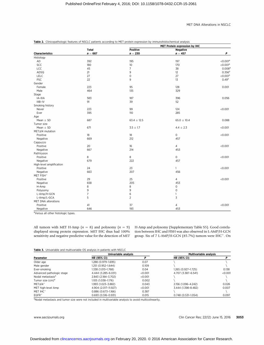
Table 2. Clinicopathologic features of NSCLC patients according to MET protein expression by immunohistochemical analysis
MET Protein expression by IHCTotal Positive Negative
Characteristics n ¼ 687 n ¼ 230 n ¼ 457 P
HistologyAD 392 195 197 <0.001a
SCC 180 10 170 <0.001a
LCC 45 7 38 0.008a
ADSQ 21 9 12 0.356a
LELC 27 0 27 <0.001a
PSC 22 9 13 0.49a
GenderFemale 223 95 128 0.001Male 464 135 329
StageIA–IIIA 583 187 396 0.056IIIB–IV 91 39 52
Smoking historyNever 223 99 124 <0.001Ever 395 110 285
AgeMean � SD 687 63.4 � 12.5 65.0 � 10.4 0.088
Tumor sizeMean � SD 671 3.5 � 1.7 4.4 � 2.3 <0.001
METD14 mutationPositive 18 18 0 <0.001Negative 669 212 457
CappuzzoPositive 20 16 4 <0.001Negative 667 214 453
PathVysionPositive 8 8 0 <0.001Negative 679 222 457
High-level amplificationPositive 24 23 1 <0.001Negative 663 207 456
MET FISHþ
Positive 29 25 4 <0.001Negative 658 205 453H-Amp 8 8 0Polysomy 9 9 0L-Amp/H-GCN 7 6 1L-Amp/L-GCA 5 2 3
MET DNA alterationsPositive 41 37 4 <0.001Negative 646 193 453
aVersus all other histologic types.
Table 3. Univariable and multivariable OS analysis in patients with NSCLC
Univariable analysis Multivariable analysisParameter HR (95% CI) P HR (95% CI) P
Older age 1.288 (0.979–1.695) 0.07 \ \Male gender 1.251 (0.952–1.644) 0.109 \ \Ever-smoking 1.338 (1.013–1.768) 0.04 1.265 (0.927–1.725) 0.138Advanced pathologic stage 4.444 (3.285–6.031) <0.001 4.707 (3.387–6.541) <0.001Nodal metastasisa 2.843 (2.184–3.702) <0.001 \ \Tumor size (cm)a 1.105 (1.038–1.176) 0.002 \ \METD14þ 1.993 (1.023–3.882) 0.043 2.156 (1.096–4.242) 0.026MET high-level Amp 4.904 (2.017–11.927) <0.001 3.444 (1.398–8.482) 0.007MET IHCþ 0.886 (0.673–1.166) 0.387 \ \EGFRþ 0.693 (0.516–0.931) 0.015 0.748 (0.531–1.054) 0.097aNodal metastasis and tumor size were not included in multivariable analysis to avoid multicolinearity.
MET DNA Alterations in NSCLC
www.aacrjournals.org Clin Cancer Res; 22(12) June 15, 2016 3053
on February 20, 2020. © 2016 American Association for Cancer Research. clincancerres.aacrjournals.org Downloaded from
Published OnlineFirst February 4, 2016; DOI: 10.1158/1078-0432.CCR-15-2061
the contrary, only 2 of 5 cases of L-Amp/L-GCN (40%)were IHCþ
(Supplementary Fig. S1). All 4 FISHþ/IHC� tumors,which includ-ed 3 SCC and 1 LELC, harbored L-Amp. The clinical significance ofFISHþ/IHC� SCC and LELC remained to be defined.
A total of 205 cases were IHC(þ)/FISH(�). Among them, 12cases had METD14 mutation. The remaining 193 cases wereheterogeneous in mutation status comprising EGFR mutation(n ¼ 73), KRAS mutation (n ¼ 33), ALK translocation (n ¼18), and ROS1 translocation (n ¼ 4). Mutation on these geneswas not detected in 65 IHC(þ)/FISH(�) cases.
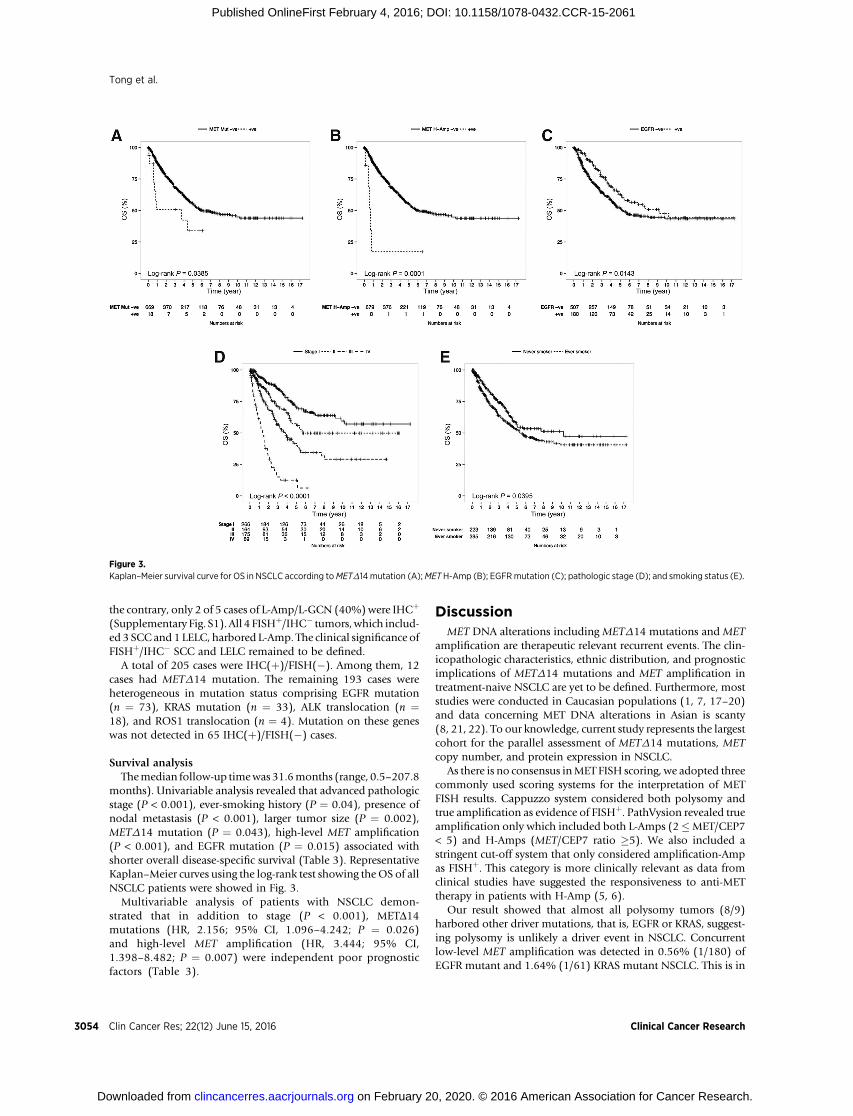
Survival analysisThemedian follow-up timewas31.6months (range, 0.5–207.8
months). Univariable analysis revealed that advanced pathologicstage (P < 0.001), ever-smoking history (P ¼ 0.04), presence ofnodal metastasis (P < 0.001), larger tumor size (P ¼ 0.002),METD14 mutation (P ¼ 0.043), high-level MET amplification(P < 0.001), and EGFR mutation (P ¼ 0.015) associated withshorter overall disease-specific survival (Table 3). RepresentativeKaplan–Meier curves using the log-rank test showing theOS of allNSCLC patients were showed in Fig. 3.
Multivariable analysis of patients with NSCLC demon-strated that in addition to stage (P < 0.001), METD14mutations (HR, 2.156; 95% CI, 1.096–4.242; P ¼ 0.026)and high-level MET amplification (HR, 3.444; 95% CI,1.398–8.482; P ¼ 0.007) were independent poor prognosticfactors (Table 3).
DiscussionMET DNA alterations including METD14 mutations and MET
amplification are therapeutic relevant recurrent events. The clin-icopathologic characteristics, ethnic distribution, and prognosticimplications of METD14 mutations and MET amplification intreatment-naive NSCLC are yet to be defined. Furthermore, moststudies were conducted in Caucasian populations (1, 7, 17–20)and data concerning MET DNA alterations in Asian is scanty(8, 21, 22). To our knowledge, current study represents the largestcohort for the parallel assessment of METD14 mutations, METcopy number, and protein expression in NSCLC.
As there is no consensus inMET FISH scoring, we adopted threecommonly used scoring systems for the interpretation of METFISH results. Cappuzzo system considered both polysomy andtrue amplification as evidence of FISHþ. PathVysion revealed trueamplification only which included both L-Amps (2�MET/CEP7< 5) and H-Amps (MET/CEP7 ratio �5). We also included astringent cut-off system that only considered amplification-Ampas FISHþ. This category is more clinically relevant as data fromclinical studies have suggested the responsiveness to anti-METtherapy in patients with H-Amp (5, 6).
Our result showed that almost all polysomy tumors (8/9)harbored other driver mutations, that is, EGFR or KRAS, suggest-ing polysomy is unlikely a driver event in NSCLC. Concurrentlow-level MET amplification was detected in 0.56% (1/180) ofEGFR mutant and 1.64% (1/61) KRAS mutant NSCLC. This is in
Figure 3.Kaplan–Meier survival curve for OS in NSCLC according toMETD14 mutation (A);METH-Amp (B); EGFRmutation (C); pathologic stage (D); and smoking status (E).
Tong et al.
Clin Cancer Res; 22(12) June 15, 2016 Clinical Cancer Research3054
on February 20, 2020. © 2016 American Association for Cancer Research. clincancerres.aacrjournals.org Downloaded from
Published OnlineFirst February 4, 2016; DOI: 10.1158/1078-0432.CCR-15-2061

keeping with previous report demonstrating MET amplificationwas not mutually exclusive to EGFR/KRAS mutations in treat-ment-na€�ve patients and thus did not fulfill the criteria of onco-genic driver (23). However, we found that high-levelMET ampli-fication (MET/CEP7 ratio�5)wasmutually exclusive to themajordriver events in RTK/RAS/PI3K axis except METD14. Our datashowed a significant association between MET DNA CNAs andMETD14 mutation. Such observations have been reported inEGFR, KRAS, and other oncogenes that activation mutationspositively correlated with gene copy number though the under-lying mechanisms remain to be elucidated. Mutant allele specificimbalance of oncogenes has been noted in human cancers (24).Although activating in one single allele of an oncogene is believedto be sufficient to drive tumorigenesis, concurrent mutation, andcopy number gain are frequently found in tumors harboringmutations. These genetic alterations may have synergistic effectplaying a greater role in development and maintenance of malig-nant phenotype. Although occurred in a low frequency, H-Ampassociated with strong MET protein expression and poorer prog-nosis. We further demonstrated that H-Amp was an independentprognostic factor by multivariable analysis. Our result suggestedthat H-Amps (MET/CEP7 ratio�5)might be a good criterion thatdefines a molecular subset with poorer prognosis and potentiallybenefit from MET inhibitors.
MET exon 14 skipping mutations are not common but havebeen reported in diverse cancer types including lung cancer,glioblastoma multiforme, head and neck SCC as well as incancer cell lines H596 (lung ADSQ; ref. 7), Hs746T (gastriccancer; ref. 25) and HCC2218 (breast cancer; ref. 26). AlthoughMETD14 mutations are less common than EGFR and KRAS, ithas been detected in up to 5% of lung adenocarcinoma, a figurethat is comparable with ALK translocations. This represents anadditional target with proven sensitivity toward MET mAbMETMab (7) and MET TKIs (crizotinib and carbozantinib;refs. 9, 17, 19). Given the high prevalence of NSCLC world-wide, anti-MET targeted therapy could potentially benefit thou-sands of patients each year.
The incidences of METD14 mutation and MET H-Amp were2.6% and 1.0%, respectively, in lung adenocarcinoma, and2.6% and 1.2% in NSCLC. The mutation rates are comparablewith previous reports (1, 7–9, 20–22) and no significant ethnicdifference across East Asian and Caucasian was found.METD14mutation was not found in SCC (n ¼ 180) in the current study.Surprisingly, we found a significantly higher frequency ofMETD14 mutation in PSC (31.8%) compared with other his-tologic subtypes. This is the first study parallel comparing METstatus across different histologic subtypes of NSCLC and dem-onstrating high METD14þ in PSC. We also found frequent high-level MET amplification in PSC (13.6%). The result is inkeeping with recent studies showing frequent MET alterationsin PSC (16, 27). PSC is a group of poorly differentiated NSCLCcontaining components of sarcoma or sarcoma-like elementsaccording to 2014 WHO classification. Five subtypes are rec-ognized: pleomorphic carcinoma, spindle cell carcinoma, giantcell carcinoma, carcinosarcoma, and pulmonary blastoma. It isconsidered a rare but distinct entity comprising approximately1% of all malignant neoplasms of lung. The biology of sarco-matoid carcinoma is poorly understood. They generally run anaggressive clinical course and are resistant to chemotherapy dueto heterogeneity (28–30). The genetics of sarcomatoid carci-noma is largely unexplorered. Identification of MET activating
mutation in NSCLC with sarcomatoid differentiation is encour-aging. As MET is implicated in the epithelial mesenchymaltransition process (3, 31), activation of MET might affect thedifferentiation state of the tumor cells. This raises a possibilitythat MET inhibition may aid in treating this specific subtype oflung cancer.
MET protein overexpression was detected in 33.5% of treat-ment-na€�ve NSCLC by IHC. However, only 16.1% of IHCþ
tumors harbor MET DNA alterations, that is, METD14 and/orCNA. Majority of the IHCþ tumors do not have MET geneticalteration in DNA level. This might be one of the reasons thatMET IHC failed to predict response to anti-MET mAb therapy ina recent phase III trial (32). Although MET protein overexpres-sion can be found in up to 65% of lung adenocarcinoma (11),most of them are driven by secondary events that promotetumor growth and progression but not oncogenic drivers forthe individual tumor. As a matter of fact, the most commonmechanism for MET activation in cancer is protein overexpres-sion as a consequence of transcriptional upregulation. Manyfactors include other oncogenes, hypoxia-induced factors, cyto-kines, or proangiogenic factors secreted by the reactive stromaor ligand-dependent autocrine or paracrine loop contribute tothe transcriptional upregulation of MET (33). According tooncogene addict model, such cases may have additional geneticlesions attenuating the dependence of tumor cells on METsignaling and therefore fail to respond to anti-MET targetedtherapy. This underscores the importance of identifying keydriver events that define specific subset of patients likely tobenefit from targeted therapy for patient management in per-sonalized medicine.
Nevertheless, our results demonstrated good correlationbetween MET IHC and DNA alterations. Especially for the detec-tion of high-level MET amplification and polysomy, IHC washighly sensitive andhada100%negative predictive value. IHC is aroutine technique inmost pathologic laboratory for sensitive andreliable detection of protein expression. The high negative pre-dictive value of MET IHC for the presence of MET DNA alterationallows for a fast screening for patients with NSCLC to join propermolecular test.
In conclusion, oncogenicMETDNAalterations defined3.3%ofNSCLC patients with aggressive diseases and older age. We foundsignificant enrichment of MET DNA alterations in PSC. METinhibition may aid in treating this specific subtype of lung cancer.
Disclosure of Potential Conflicts of InterestT.S.K. Mok has received speaker's bureau honoraria from ACEA Biosciences,
Amgen, Astrazeneca, BI, BioMarin, Clovis Oncology, Eli Lilly, GSK, Janssen,MSD, Novartis, Pfizer, Roche/Genentech, SFJ, and Vertex; and is a consultant/advisory boardmember for ACEABiosciences, Astrazeneca, BI, BioMarin, ClovisOncology, Eli Lilly, GSK, Janssen, Merck Serono, MSD, Novartis, Pfizer, Roche/Genentech, SFJ, and Vertex. No potential conflicts of interest were disclosed bythe other authors.
Authors' ContributionsConception and design: J.H. Tong, S.F. Yeung, T.S.K. Mok, K.F. ToDevelopment of methodology: J.H. Tong, C.Y. Tong, C.S.H. Ng, K.F. ToAcquisition of data (provided animals, acquired and managed patients,provided facilities, etc.): J.H. Tong, S.F. Yeung, C.S.H. Ng, T.S.K. Mok,K.F. ToAnalysis and interpretation of data (e.g., statistical analysis, biostatistics,computational analysis): J.H. Tong, S.F. Yeung, A.W.H. Chan, T.S.K. Mok,K.F. To
MET DNA Alterations in NSCLC
www.aacrjournals.org Clin Cancer Res; 22(12) June 15, 2016 3055
on February 20, 2020. © 2016 American Association for Cancer Research. clincancerres.aacrjournals.org Downloaded from
Published OnlineFirst February 4, 2016; DOI: 10.1158/1078-0432.CCR-15-2061

Writing, review, and/or revision of the manuscript: J.H. Tong, S.F. Yeung,A.W.H. Chan, T.S.K. Mok, K.F. ToAdministrative, technical, or material support (i.e., reporting or organizingdata, constructingdatabases): J.H. Tong, L.Y. Chung, S.L. Chau, E.K.Y. Tin, R.W.M. Lung, C.Y. Tong, C. Chow, Y.H. Yu,H. Li, Y. Pan,W.P. Chak, C.S.H.Ng, T.S.K.Mok, K.F. ToStudy supervision: J.H. Tong, R.W.M. Lung, K.F. To
The costs of publication of this articlewere defrayed in part by the payment ofpage charges. This article must therefore be hereby marked advertisement inaccordance with 18 U.S.C. Section 1734 solely to indicate this fact.
Received August 25, 2015; revised January 11, 2016; accepted January 16,2016; published OnlineFirst February 4, 2016.
References1. The Cancer Genome Atlas ResearchN. Comprehensivemolecular profiling
of lung adenocarcinoma. Nature 2014;511:543–50.2. Pao W, Girard N. New driver mutations in non-small-cell lung cancer.
Lancet Oncol 2011;12:175–80.3. Birchmeier C, BirchmeierW,Gherardi E, VandeWoudeGF.Met,metastasis,
motility and more. Nat Rev Mol Cell Biol 2003;4:915–25.4. Lutterbach B, Zeng Q, Davis LJ, Hatch H, Hang G, Kohl NE, et al. Lung
cancer cell lines harboring MET gene amplification are dependent on Metfor growth and survival. Cancer Res 2007;67:2081–8.
5. Schwab R, Petak I, Kollar M, Pinter F, Varkondi E, Kohanka A, et al.Major partial response to crizotinib, a dual MET/ALK inhibitor, in asquamous cell lung (SCC) carcinoma patient with de novo c-METamplification in the absence of ALK rearrangement. Lung Cancer2014;83:109–11.
6. Ou SH, Kwak EL, Siwak-Tapp C, Dy J, Bergethon K, Clark JW, et al. Activityof crizotinib (PF02341066), a dual mesenchymal-epithelial transition(MET) and anaplastic lymphoma kinase (ALK) inhibitor, in a non-smallcell lung cancer patient with de novo MET amplification. J Thorac Oncol2011;6:942–6.
7. Kong-Beltran M, Seshagiri S, Zha J, Zhu W, Bhawe K, Mendoza N, et al.Somatic mutations lead to an oncogenic deletion of met in lung cancer.Cancer Res 2006;66:283–9.
8. Onozato R, Kosaka T, Kuwano H, Sekido Y, Yatabe Y, Mitsudomi T.Activation of MET by gene amplification or by splice mutations deletingthe juxtamembrane domain in primary resected lung cancers. J ThoracOncol 2009;4:5–11.
9. Paik PK, Drilon A, Fan PD, Yu H, Rekhtman N, Ginsberg MS, et al.Response to MET inhibitors in patients with stage IV lung adenocarci-nomas harboring MET mutations causing exon 14 skipping. CancerDiscov 2015;5:842–9.
10. Spigel DR, Edelman MJ, O'Byrne K, Paz-Ares L, Shames DS, Yu W, et al.Onartuzumab plus erlotinib versus erlotinib in previously treated stage IIbor IV NSCLC: Results from the pivotal phase III randomized, multicenter,placebo-controlledMETLung (OAM4971g) global trial. J ClinOncol 32:5s,2014 (suppl; abstr 8000).
11. Yeung SF, Tong JH, LawPP, Chung LY, Lung RW, TongCY, et al. Profiling ofoncogenic driver events in lung adenocarcinoma revealedMETmutation asindependent prognostic factor. J Thorac Oncol 2015;10:1292–300.
12. Dziadziuszko R, Wynes MW, Singh S, Asuncion BR, Ranger-Moore J,Konopa K, et al. Correlation between MET gene copy number by silverin situ hybridization and protein expression by immunohistochemistry innon-small cell lung cancer. J Thorac Oncol 2012;7:340–7.
13. Cappuzzo F, Marchetti A, Skokan M, Rossi E, Gajapathy S, Felicioni L,et al. Increased MET gene copy number negatively affects survival ofsurgically resected non-small-cell lung cancer patients. J Clin Oncol2009;27:1667–74.
14. Tanaka A, Sueoka-Aragane N, Nakamura T, Takeda Y, Mitsuoka M, Yama-saki F, et al. Co-existence of positiveMET FISH status with EGFRmutationssignifies poor prognosis in lung adenocarcinoma patients. Lung Cancer2012;75:89–94.
15. Jurmeister P, Lenze D, Berg E, Mende S, Schaper F, Kellner U, et al.Parallel screening for ALK, MET and ROS1 alterations in non-small celllung cancer with implications for daily routine testing. Lung Cancer2015;87:122–9.
16. Tsuta K, Kozu Y, Mimae T, Yoshida A, Kohno T, Sekine I, et al. c-MET/phospho-MET protein expression and MET gene copy number in non-small cell lung carcinomas. J Thorac Oncol 2012;7:331–9.
17. Waqar SN,Morgensztern D, Sehn J. METmutation associated with respon-siveness to crizotinib. J Thorac Oncol 2015;10:e29–31.
18. Imielinski M, Berger AH, Hammerman PS, Hernandez B, Pugh TJ, Hodis E,et al. Mapping the hallmarks of lung adenocarcinoma with massivelyparallel sequencing. Cell 2012;150:1107–20.
19. Jenkins RW, Oxnard GR, Elkin S, Sullivan EK, Carter JL, Barbie DA.Response to crizotinib in a patient with lung adenocarcinoma harboringa MET splice site mutation. Clin Lung Cancer 2015;16:E101–4.
20. Azzato EM, Desphande C, Gabriel C, Langer C, Evans T, Gault CR, et al.MET mutations in non-small cell lung cancer: four case reports of muta-tions at or near the splice junction for exon 14. J OncoPathol 2014;2:65.
21. Seo JS, Ju YS, Lee WC, Shin JY, Lee JK, Bleazard T, et al. The transcriptionallandscape and mutational profile of lung adenocarcinoma. Genome Res2012;22:2109–19.
22. Okuda K, Sasaki H, Yukiue H, Yano M, Fujii Y. Met gene copy numberpredicts the prognosis for completely resected non-small cell lung cancer.Cancer Sci 2008;99:2280–5.
23. Schildhaus HU, Schultheis AM, Ruschoff J, Binot E, Merkelbach-Bruse S,Fassunke J, et al. MET amplification status in therapy-naive adeno- andsquamous cell carcinomas of the lung. Clin Cancer Res 2015;21:907–15.
24. Soh J,OkumuraN, LockwoodWW,YamamotoH, ShigematsuH,ZhangW,et al. Oncogene mutations, copy number gains and mutant allele specificimbalance (MASI) frequently occur together in tumor cells. PLoS One2009;4:e7464.
25. Asaoka Y, TadaM, Ikenoue T, Seto M, Imai M, Miyabayashi K, et al. Gastriccancer cell line Hs746T harbors a splice site mutation of c-Met causingjuxtamembrane domain deletion. Biochem Biophys Res Commun2010;394:1042–6.
26. Stephens P, Edkins S, Davies H, Greenman C, Cox C, Hunter C, et al. Ascreenof the complete protein kinase gene family identifiesdiversepatternsof somatic mutations in human breast cancer. Nat Genet 2005;37:590–2.
27. Liu X, Jia Y, Stoopler MB, Shen Y, Cheng H, Chen J, et al. Next-generationsequencing of pulmonary sarcomatoid carcinoma reveals high frequencyof actionable MET genemutations. J Clin Oncol . 2015Jul 27. [Epub aheadof print].
28. Huang SY, Shen SJ, Li XY. Pulmonary sarcomatoid carcinoma: a clinico-pathologic study and prognostic analysis of 51 cases. World J Surg Oncol2013;11:252.
29. Franks TJ, Galvin JR. Sarcomatoid carcinoma of the lung: histologic criteriaand common lesions in the differential diagnosis. Arch Pathol Lab Med2010;134:49–54.
30. Pelosi G, Sonzogni A, De Pas T, Galetta D, Veronesi G, Spaggiari L, et al.Review article: pulmonary sarcomatoid carcinomas: a practical overview.Int J Surg Pathol 2010;18:103–20.
31. Gherardi E, BirchmeierW, Birchmeier C, VandeWoudeG. TargetingMET incancer: rationale and progress. Nat Rev Cancer 2012;12:89–103.
32. Perol M. Negative results of METLung study: an opportunity to betterunderstand the role of MET pathway in advanced NSCLC. Transl LungCancer Res 2014;3:392–4.
33. Organ SL, TsaoMS. An overview of the c-MET signaling pathway. Ther AdvMed Oncol 2011;3:S7–19.
Clin Cancer Res; 22(12) June 15, 2016 Clinical Cancer Research3056
Tong et al.
on February 20, 2020. © 2016 American Association for Cancer Research. clincancerres.aacrjournals.org Downloaded from
Published OnlineFirst February 4, 2016; DOI: 10.1158/1078-0432.CCR-15-2061
2016;22:3048-3056. Published OnlineFirst February 4, 2016.Clin Cancer Res Joanna H. Tong, Sai F. Yeung, Anthony W.H. Chan, et al. Prognosis
Small Cell Lung Carcinoma with Poor−Molecular Subgroups of NonUnique Amplification and Exon 14 Splice Site Mutation Define MET
Updated version
10.1158/1078-0432.CCR-15-2061doi:
Access the most recent version of this article at:
Material
Supplementary
http://clincancerres.aacrjournals.org/content/suppl/2016/02/04/1078-0432.CCR-15-2061.DC1
Access the most recent supplemental material at:
Cited articles
http://clincancerres.aacrjournals.org/content/22/12/3048.full#ref-list-1
This article cites 32 articles, 6 of which you can access for free at:
Citing articles
http://clincancerres.aacrjournals.org/content/22/12/3048.full#related-urls
This article has been cited by 7 HighWire-hosted articles. Access the articles at:
E-mail alerts related to this article or journal.Sign up to receive free email-alerts
Subscriptions
Reprints and
To order reprints of this article or to subscribe to the journal, contact the AACR Publications Department at
Permissions
Rightslink site. Click on "Request Permissions" which will take you to the Copyright Clearance Center's (CCC)
.http://clincancerres.aacrjournals.org/content/22/12/3048To request permission to re-use all or part of this article, use this link
on February 20, 2020. © 2016 American Association for Cancer Research. clincancerres.aacrjournals.org Downloaded from
Published OnlineFirst February 4, 2016; DOI: 10.1158/1078-0432.CCR-15-2061