metabolic enzymes that bind rna yet another level of ... · metabolic enzymes that bind rna: yet...
TRANSCRIPT
Review
Metabolic enzymes that bind RNA: yet another level of cellular regulatory network?
Joanna Cieśla½
Nencki Institute of Experimental Biology, Polish Academy of Sciences, Warszawa, Poland; ½e-mail: [email protected]
Received: 03 October, 2005; revised: 15 November, 2005; accepted: 11 January, 2006 available on-line: 12 January, 2006
Several enzymes that were originally characterized to have one defined function in intermedia-tory metabolism are now shown to participate in a number of other cellular processes. Multi-functional proteins may be crucial for building of the highly complex networks that maintain the function and structure in the eukaryotic cell possessing a relatively low number of protein-encoding genes. One facet of this phenomenon, on which I will focus in this review, is the inter-action of metabolic enzymes with RNA. The list of such enzymes known to be associated with RNA is constantly expanding, but the most intriguing question remains unanswered: are the metabolic enzyme–RNA interactions relevant in the regulation of cell metabolism? It has been proposed that metabolic RNA-binding enzymes participate in general regulatory circuits linking a metabolic function to a regulatory mechanism, similar to the situation of the metabolic enzyme aconitase, which also functions as iron-responsive RNA-binding regulatory element. However, some authors have cautioned that some of such enzymes may merely represent “molecular fos-sils” of the transition from an RNA to a protein world and that the RNA-binding properties may not have a functional significance. Here I will describe enzymes that have been shown to interact with RNA (in several cases a newly discovered RNA-binding protein has been identified as a well-known metabolic enzyme) and particularly point out those whose ability to interact with RNA seems to have a proven physiological significance. I will also try to depict the molecular switch between an enzyme’s metabolic and regulatory functions in cases where such a mecha-nism has been elucidated. For most of these enzymes relations between their enzymatic functions and RNA metabolism are unclear or seem not to exist. All these enzymes are ancient, as judged
by their wide distribution, and participate in fundamental biochemical pathways.
Keywords: enzymes, mRNA binding, gene expression, translation regulation
One of the methods used most often in de-tecting direct RNA–protein interaction in vitro is electrophoretic mobility shift assay, in which a com-plex of radioactively labelled RNA with protein is electophoresed and visualized by autoradiography. The other is filter-binding assay, in which only pro-tein (and protein with bound labelled RNA) binds to the filter that is subsequently measured for ra-
dioactivity. To ensure specific RNA–protein interac-tion, an excess of unlabelled competitor RNA (for example yeast tRNA) is routinely added to the pro-tein prior to incubation with labelled RNA. In some cases to resolve the RNA–protein complex into spe-cific complexes a combination of heparin and RNase T1 is used. Additionally, to show the specificity of particular RNA–protein interaction, competition ex-
Abbreviations: ARE, AU-rich element; CKBB, creatine kinase brain form B; COLBP, cytochrome c oxidase L-form trans-cript-binding protein; COX, cytochrome c oxidase; DHFR, dihydrofolate reductase; eALAS, erythroid isoform of 5-amino-levulinate synthase; FdUMP, fluorodeoxyuridine monophosphate; GAPDH, glyceraldehyde-3-phosphate dehydrogenase; GDH, glutamate dehydrogenase; GDK, phosphoglycerate kinase; GM-CSF, granulocyte-macrophage colony-stimulating factor; gRNA, guide RNA; HADHB, hydroxyacyl coenzyme A dehydrogenase/3-ketoacyl-thiolase/enoyl-coenzyme A hy-dratase (trifunctional protein), β subunit; HAV, hepatitis A virus; HBV, hepatitis B virus; HCV, hepatitis C virus; HDV, he-patitis D virus; HPIV, human parainfluenza virus; IDH, isocitrate dehydrogenase; IRES, internal ribosomal entry site; IRE, iron-responsive element; IRF, iron-regulatory factor; IRP, iron-regulatory protein; LDH, lactate dehydrogenase; meTHF, N5,10-methylenetetrahydrofolate; MTX, methotrexate; MyHC, myosin heavy chain; RT-PCR, reverse transcription-polyme-rase chain reaction; SHMT, serine hydroxymethyltransferase; TS, thymidylate synthase; uPAR, urokinase-type plasmino-gen activator receptor; UTR, untranslated region.
Vol. 53 No. 1/2006, 11–32
on-line at: www.actabp.pl
12 2006J. Cieśla
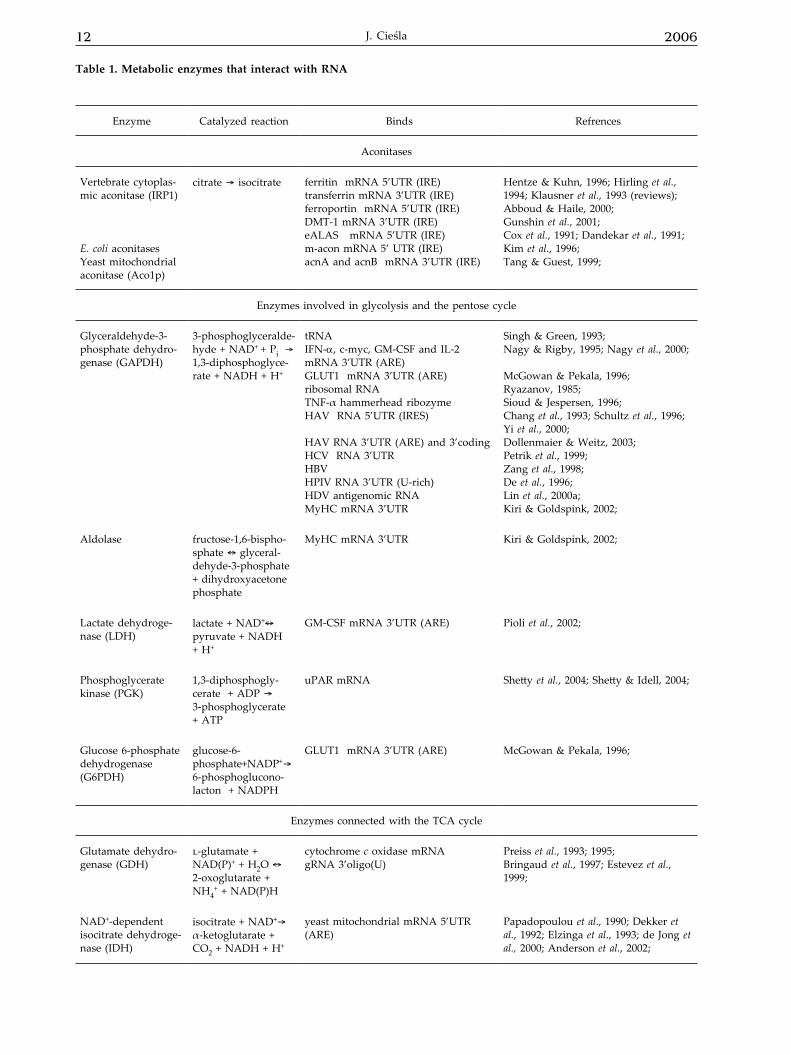
Enzyme Catalyzed reaction Binds Refrences
Aconitases
Vertebrate cytoplas-mic aconitase (IRP1)
E. coli aconitasesYeast mitochondrial aconitase (Aco1p)
citrate ® isocitrate ferritin mRNA 5’UTR (IRE)transferrin mRNA 3’UTR (IRE)ferroportin mRNA 5’UTR (IRE)DMT-1 mRNA 3’UTR (IRE)eALAS mRNA 5’UTR (IRE)m-acon mRNA 5’ UTR (IRE)acnA and acnB mRNA 3’UTR (IRE)
Hentze & Kuhn, 1996; Hirling et al., 1994; Klausner et al., 1993 (reviews); Abboud & Haile, 2000;Gunshin et al., 2001;Cox et al., 1991; Dandekar et al., 1991;Kim et al., 1996;Tang & Guest, 1999;
Enzymes involved in glycolysis and the pentose cycle
Glyceraldehyde-3-phosphate dehydro-genase (GAPDH)
3-phosphoglyceralde-hyde + NAD+ + Pi ® 1,3-diphosphoglyce-rate + NADH + H+
tRNAIFN-α, c-myc, GM-CSF and IL-2 mRNA 3’UTR (ARE)GLUT1 mRNA 3’UTR (ARE)ribosomal RNATNF-α hammerhead ribozymeHAV RNA 5’UTR (IRES)
HAV RNA 3’UTR (ARE) and 3’coding HCV RNA 3’UTRHBVHPIV RNA 3’UTR (U-rich)HDV antigenomic RNAMyHC mRNA 3’UTR
Singh & Green, 1993;Nagy & Rigby, 1995; Nagy et al., 2000;
McGowan & Pekala, 1996;Ryazanov, 1985;Sioud & Jespersen, 1996;Chang et al., 1993; Schultz et al., 1996; Yi et al., 2000;Dollenmaier & Weitz, 2003;Petrik et al., 1999; Zang et al., 1998;De et al., 1996;Lin et al., 2000a;Kiri & Goldspink, 2002;
Aldolase fructose-1,6-bispho-sphate « glyceral-dehyde-3-phosphate + dihydroxyacetone phosphate
MyHC mRNA 3’UTR Kiri & Goldspink, 2002;
Lactate dehydroge-nase (LDH)
lactate + NAD+« pyruvate + NADH + H+
GM-CSF mRNA 3’UTR (ARE) Pioli et al., 2002;
Phosphoglycerate kinase (PGK)
1,3-diphosphogly-cerate + ADP ® 3-phosphoglycerate + ATP
uPAR mRNA Shetty et al., 2004; Shetty & Idell, 2004;
Glucose 6-phosphate dehydrogenase (G6PDH)
glucose-6-phosphate+NADP+® 6-phosphoglucono-lacton + NADPH
GLUT1 mRNA 3’UTR (ARE) McGowan & Pekala, 1996;
Enzymes connected with the TCA cycle
Glutamate dehydro-genase (GDH)
l-glutamate + NAD(P)+ + H2O «2-oxoglutarate + NH4
+ + NAD(P)H
cytochrome c oxidase mRNA gRNA 3’oligo(U)
Preiss et al., 1993; 1995;Bringaud et al., 1997; Estevez et al., 1999;
NAD+-dependent isocitrate dehydroge-nase (IDH)
isocitrate + NAD+® α-ketoglutarate + CO2 + NADH + H+
yeast mitochondrial mRNA 5’UTR (ARE)
Papadopoulou et al., 1990; Dekker et al., 1992; Elzinga et al., 1993; de Jong et al., 2000; Anderson et al., 2002;
Table 1. Metabolic enzymes that interact with RNA
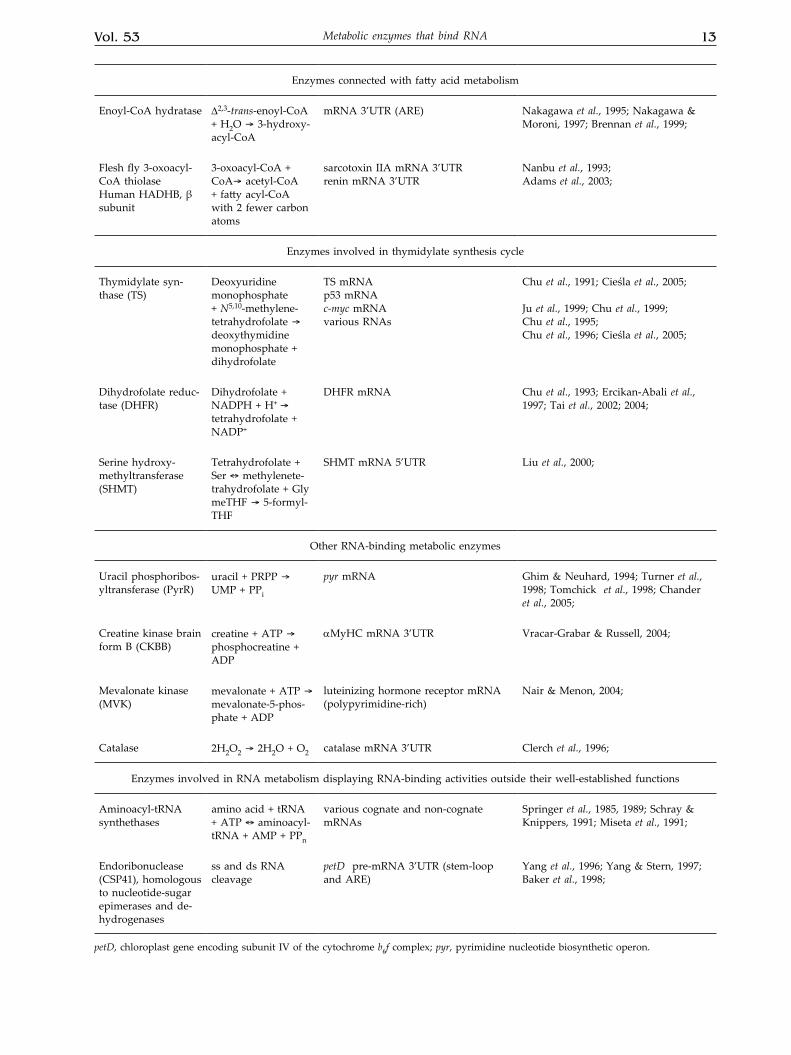
Vol. 53 13Metabolic enzymes that bind RNA
Enzymes connected with fatty acid metabolism
Enoyl-CoA hydratase ∆2,3-trans-enoyl-CoA + H2O ® 3-hydroxy-acyl-CoA
mRNA 3’UTR (ARE) Nakagawa et al., 1995; Nakagawa & Moroni, 1997; Brennan et al., 1999;
Flesh fly 3-oxoacyl-CoA thiolase Human HADHB, β subunit
3-oxoacyl-CoA + CoA® acetyl-CoA + fatty acyl-CoA with 2 fewer carbon atoms
sarcotoxin IIA mRNA 3’UTR renin mRNA 3’UTR
Nanbu et al., 1993;Adams et al., 2003;
Enzymes involved in thymidylate synthesis cycle
Thymidylate syn-thase (TS)
Deoxyuridine monophosphate + N5,10-methylene-tetrahydrofolate ® deoxythymidine monophosphate + dihydrofolate
TS mRNAp53 mRNAc-myc mRNAvarious RNAs
Chu et al., 1991; Cieśla et al., 2005;
Ju et al., 1999; Chu et al., 1999;Chu et al., 1995;Chu et al., 1996; Cieśla et al., 2005;
Dihydrofolate reduc-tase (DHFR)
Dihydrofolate + NADPH + H+ ® tetrahydrofolate + NADP+
DHFR mRNA Chu et al., 1993; Ercikan-Abali et al., 1997; Tai et al., 2002; 2004;
Serine hydroxy-methyltransferase (SHMT)
Tetrahydrofolate + Ser « methylenete-trahydrofolate + GlymeTHF ® 5-formyl-THF
SHMT mRNA 5’UTR Liu et al., 2000;
Other RNA-binding metabolic enzymes
Uracil phosphoribos-yltransferase (PyrR)
uracil + PRPP ® UMP + PPi
pyr mRNA Ghim & Neuhard, 1994; Turner et al., 1998; Tomchick et al., 1998; Chander et al., 2005;
Creatine kinase brain form B (CKBB)
creatine + ATP ® phosphocreatine + ADP
αMyHC mRNA 3’UTR Vracar-Grabar & Russell, 2004;
Mevalonate kinase (MVK)
mevalonate + ATP ® mevalonate-5-phos-phate + ADP
luteinizing hormone receptor mRNA (polypyrimidine-rich)
Nair & Menon, 2004;
Catalase 2H2O2 ® 2H2O + O2 catalase mRNA 3’UTR Clerch et al., 1996;
Enzymes involved in RNA metabolism displaying RNA-binding activities outside their well-established functions
Aminoacyl-tRNA synthethases
amino acid + tRNA + ATP « aminoacyl-tRNA + AMP + PPn
various cognate and non-cognate mRNAs
Springer et al., 1985, 1989; Schray & Knippers, 1991; Miseta et al., 1991;
Endoribonuclease (CSP41), homologous to nucleotide-sugar epimerases and de-hydrogenases
ss and ds RNA cleavage
petD pre-mRNA 3’UTR (stem-loop and ARE)
Yang et al., 1996; Yang & Stern, 1997; Baker et al., 1998;
petD, chloroplast gene encoding subunit IV of the cytochrome b6f complex; pyr, pyrimidine nucleotide biosynthetic operon.
14 2006J. Cieśla
periments are conducted with the use of unlabelled RNA that binds the protein and competes with la-belled RNA or various unlabelled unrelated RNAs that are unable to compete with the labelled RNA. Dissociation constants (Kd) for specific RNA-protein interaction are within low picomolar to high na-nomolar values, however, only for few RNA-bind-ing enzymes have they been measured.
IRP1/CYTOSOLIC ACONITASE
The best-studied example of a specific RNA-binding protein — whose interaction with its cog-nate binding site in mRNA (iron-responsive element, IRE) is regulated in response to metabolic signals — is iron-regulatory protein (IRP), formerly known as iron-regulatory factor (IRF) or IRE-binding pro-tein (IRE-BP) (reviewed by Klausner & Rouault, 1993; Hentze & Kuhn, 1996; Rouault, 2002; Cairo et al., 2002).
The iron-responsive element (IRE), forming a hairpin structure, is highly conserved in evolution for any given gene and is remarkably similar among different genes harboring such elements, suggesting precise structural constraints in the binding to IRPs (Henderson et al., 1994). Studies on plant and yeast cells, which lack endogenous IRP activity, indicate that the binding of IRP to the IRE is sufficient to reg-ulate translation without requirement for additional cis-acting sequences or trans-acting factors (Kim et al., 1995), although adjacent non-IRE sequences may affect the function of an IRE (Dickey et al., 1988; Dix et al., 1993).
Actually, two structurally and functionally similar cytoplasmic IRPs (IRP1 and IRP2) modulate stability and translation of mRNA encoding proteins involved in regulation of iron homeostasis, i.e. trans-ferrin receptor (TfR, receptor for cellular iron uptake) and ferritin (Ft, iron storage protein). IRPs bind with high affinity (Kd = 5–50 pM; Butt et al., 1996) to IRE in the 3’ UTR of TfR mRNA protecting it from deg-radation and leading to enhanced synthesis of TfR protein. On the other hand, IRPs bind to an IRE in 5’ UTR of Ft mRNA causing translational repression and leading to decreased synthesis of Ft protein.
IRP1 was purified from human liver (Rouault et al., 1989) and, after partial peptide sequencing, the cDNA encoding this protein was cloned (Rouault et al., 1990) making it possible to find a significant homology with two eukaryotic mitochondrial aconi-tases (Hentze & Argos, 1991). Then, bovine cytosolic aconitase was purified, demonstrated to possess an IRE-binding activity and shown by peptide sequenc-ing to be identical with IRP1 (Kennedy et al., 1992). The mitochondrial enzyme lacks the IRE-binding ac-tivity, but it also seems to have an additional func-
tion, independent of the enzyme’s catalytic activity — it has been found to be associated with mitochon-drial DNA and shown to be essential for its main-tenance (Chen et al., 2005). IRP1, but not IRP2, can switch between the mRNA-binding and enzymatic activities according to cell iron levels. In iron-replete cells the [4Fe-4S] catalytic cluster is assembled, the IRE-binding site is not accesible, and IRP1 displays aconitase activity; in iron-depleted cells the cluster is lacking and IRP1 apoprotein functions as an RNA-binding protein (Constable et al., 1992; Klausner & Rouault, 1993; Hentze & Kuhn, 1996). This revers-ible switch allows aconitase/IRP1 to constantly sense iron levels and adapt them to cell requirements without any appreciable change in protein levels. The state of the [4Fe-4S] cluster is also changed by oxygen species and by •NO generated by endog-enous synthases or exogenous donors (reviewed by Drapier, 1997; Hanson & Leibold, 1999; Cairo & Pie-trangelo, 2000), providing a direct regulatory linkage between iron metabolism and oxidative stress. The cluster’s stability also depends on the phosphoryla-tion state of a serine residue required for RNA bind-ing (Brown et al., 2002). The enzymatic function of cytosolic aconitase, which is the product of a gene distinct from that encoding the mitochondrial TCA cycle aconitase, has been well characterised, but its role in the metabolism of the cell is not well under-stood and remains to be established. According to studies in yeast (Narahari et al., 2000), an iron-me-diated conversion of IRP1 to aconitase would have the advantage of increasing the NADPH level, thus providing reducing equivalents helpful in maintain-ing the redox balance.
In addition to modulating ferritin and trans-ferrin receptor levels, IRP can regulate other IRE-containing mRNAs, encoding proteins closely related to iron utilization (erythroid 5-aminolevulinate syn-thase, eALAS), uptake (proton-coupled iron trans-porter, DMT1) and release (ferroportin) (Cox et al., 1991; Dandekar et al., 1991; Abboud & Haile, 2000; Gunshin et al., 2001). Studies of the aconitase family of proteins in prokaryotes and eukaryotes support the suggestion that IRP may also control the synthe-sis of proteins that are not direct regulators of iron metabolism, such as mammalian mitochondrial aco-nitase (Kim et al., 1996; Chen et al., 1997), Escherichia coli aconitases A and B (Tang & Guest, 1999), Dro-sophila melanogaster succinate dehydrogenase (Koh-ler et al., 1995; Gray et al., 1996), and Bacillus subtilis cytochrome oxidase (Alen & Sonenshein, 1999), all of which contain an IRE or IRE-like element in the mRNA. The results of these studies point to regu-latory link between iron and energy metabolism in vertebrates, invertebrates and bacteria.
Recently, Lin and coleagues (2001) discovered that protein levels of the 75-kDa Fe-S subunit of mi-
Vol. 53 15Metabolic enzymes that bind RNA
tochondrial respiratory chain Complex I were mod-ulated by levels of iron in the cell, whereas mRNA levels were minimally changed. They found that the 5’ UTR of the 75-kDa subunit mRNA contained a novel IRE-like stem-loop sequence. RNA-protein gel shift assays demonstrated that a specific cytoplasmic protein, being neither IRP1 nor IRP2 (IRP3?), bound the novel IRE and that the binding of the protein was affected by the iron status. In addition, ferritin IRE was able to compete for binding with this puta-tive IRP. These results suggest that the 75-kDa Fe-S subunit of mitochondrial Complex I may be regulat-ed by a novel IRE-IRP system.
As the numbers of target IRE-containing mRNAs and iron-regulatory proteins seem to be growing, novel IRP–IRE interactions regulating and connecting other essential metabolic pathways are awaiting discovery.
ENZYMES INVOLVED IN GLYCOLYSIS AND THE PENTOSE CYCLE
Several NAD+-dependent dehydrogenases and kinases, among them the glycolytic enzymes glyc-eraldehyde-3-phosphate dehydrogenase (GAPDH), lactate dehydrogenase (LDH) and phosphoglycer-ate kinase (PGK), have been shown to bind RNA. Although they were found to bind different RNAs, they share common features associated with this activity. They possess coenzyme binding domains, remarkably similar in three-dimensional structure, consisting of two identical βαβ-folds each associ-ated with a mononucleotide-binding surface (Ross-mann fold; Rossmann et al., 1975), whose similarity is not due to a highly conserved primary structure, but rather to homology at crucial positions in order to ensure similar folding patterns (Wierenga et al., 1986). It has been shown that (di)nucleotides (NAD+, NADH, ATP) can compete with RNA binding. Re-sults of mutational studies showed that the N-termi-nal 43 amino-acid residues of GAPDH, which cor-respond to the first mononucleotide-binding domain of the NAD-binding fold, are sufficient to confer RNA-binding (Nagy et al., 2000). Studies on RNA-binding combined with peptide mapping localized RNA-binding to the predicted N-terminal 6.8-kDa peptide, known to be involved in the formation of the NAD+-binding domain of GAPDH (Nagy & Rigby, 1995). These results suggest an involvement of the (di)nucleotide binding fold in RNA bind-ing. Nagy and coworkers (2000) proposed adding the NAD-binding domain to the list of RNA-bind-ing domains/motifs. Most of RNA-binding proteins that contain a Rossmann fold interact with AU-rich elements (ARE) of mRNA. The ARE elements con-fer message instability as well as translational sup-
pression (Shaw & Kamen, 1986; reviewed by Espel, 2005). The combination of functionally and structur-ally different sequence motifs, including AU pen-tamers and nonamers and other U-rich stretches, determines the ultimate destabilizing ability of each particular ARE. Numerous proteins have been re-ported to specifically recognize these elements and suggested to play a role in the regulation of stability and translatability of various lymphokine, cytokine and proto-oncogene mRNAs.
Glyceraldehyde-3-phosphate dehydrogenase (GAPDH), lactate dehydrogenase (LDH), aldolase, glucose-6-phosphate dehydrogenase (G6PDH), phosphoglycerate kinase (PGK)
Glyceraldehyde-3-phosphate dehydrogenase has generally been considered to be a housekeeping protein involved mainly in glycolysis, but a num-ber of studies have indicated that it displays diverse activities (reviewed by Sirover, 1999; Ishitani et al., 2003). The protein can act as a kinase, a tubulin- and actin-binding protein, play a role in membrane fusion, and act as a target for nitric oxide (Modun et al., 2000; Mazzola & Sirover, 2002). It has been shown that GAPDH is overexpressed and translo-cated to the nucleus during apoptosis (Sunaga et al., 1995). In the growing cells GAPDH can act as uracil-DNA-glycosylase and helicase (Meyer-Sigler et al., 1991) and participates in the control of transcription and DNA repair (Ronai, 1993). The enzyme has been shown to cleave RNA (Evguenieva-Hackenberg et al., 2002). GAPDH has been found to bind specifi-cally to sequences critical in the regulation of sev-eral mRNAs (see Table 1). These include cis-acting elements important in the stability and translation of mRNA, i.e. AU-rich elements in 3’UTR sequences of lymphokine mRNAs, regions involved in viral tran-scription and replication, i.e. 3’UTR sequences in-volved in RNA-dependent RNA polymerase binding for mRNA synthesis, and 5’ UTR sequences involved in HAV replication and functioning as ribosomal recognition sites (IRES, internal ribosome entry site) enabling cap-independent translation initiation (with Kd = 0.2 nM; Yi et al., 2000). The enzyme has also been shown to bind with high affinity (Kd = 18 nM) to several tRNAs in a manner that most likely in-volves both sequence and structure (Singh & Green, 1993). The physiological significance of the in vitro studies was confirmed by studies in vivo, including identification of GAPDH/lymphokine mRNA asso-ciation in polysomes (Nagy & Rigby, 1995) or the presence of GAPDH in RNP particles in human pa-rainfluenza virus-infected cells (De et al., 1996). The activity of GAPDH as either a glycolytic enzyme or an ARE-binding protein may be regulated by the lo-cal concentration of NAD+, NADH, and ATP in dif-
16 2006J. Cieśla
ferent subcellular compartments (polysomes, cytosol, and nucleus). The Rossmann fold of GAPDH ap-pears to be reciprocally regulated between its RNA-binding (inactive in glycolysis) and NAD+-binding (active in glycolysis) states in vivo (Nagy & Rigby, 1995). Alternatively, post-translational modifications (redox or otherwise) might influence the intracellu-lar location of GAPDH as well as differentially regu-late its RNA- or NAD+-binding activity. Nagy and Rigby (1995) have found that incubation of GAPDH with the reducing agent 2-mercaptoethanol enhances the binding of both lymphokine 3’ UTR and tRNA probes, whereas treatment with an oxidizing agent, diamide, markedly decreases the binding of GAPDH to the IFN-α 3' UTR. On the other hand, Arutyuno-va and coworkers (2003) have shown that binding of total tRNA to reduced apo-GAPDH is much lower than to the oxidized enzyme form.
Recently, GAPDH and another glycolytic enzyme, aldolase, were identified as proteins com-plexed with the 3' UTR sequence of myosin heavy chain (MyHC) mRNA (Kiri & Goldspink, 2002). The authors suggest that both enzymes could be in-volved in transporting the MyHC mRNA to specific regions and/or anchoring of the message in regions of myosin synthesis by association with the cyto-skeletal framework. This hypothesis is supported by studies showing that glycolytic enzymes interact with microtubules (Walsh et al., 1989).
Another glycolytic enzyme, lactate dehydroge-nase, is a tetramer that may exist in five forms each consisting of combinations of subunits designated H (for heart) and M (for skeletal muscle). LDH-M (the isoenzyme consisting of four identical skeletal mus-cle type subunits) has been identified as an ARE-binding protein interacting with granulocyte-mac-rophage colony-stimulating factor (GM-CSF) mRNA 3’ UTR and mediating ARE-dependent translation (Pioli et al., 2002). The RNA–protein binding in vitro and competition with NAD+ was demonstrated us-ing UV cross-linking and nitrocellulose filter binding assays. The latter assay indicated that the binding was ARE-specific, as the affinity of purified LDH for mutant AUGUA GM-CSF mRNA was markedly diminished in comparison with wild-type GM-CSF mRNA (Kd = 1879 nM and 501.9 nM, respective-ly). Immunoprecipitation, sucrose density gradient analysis and other data collectively demonstrated that LDH is a polysome-associated protein localized to this compartment by RNA–protein interactions. Moreover, direct association of LDH with AUF1, another ARE-binding protein, that may play mul-tiple roles in the regulation of mRNA turnover, was demonstrated (reviewed by Dean et al., 2004). One potential model for LDH function is that it provides further specificity for the translation and turnover of certain ARE-containing mRNAs through its direct
interaction with both ARE and AUF1 protein. There is also a possibility that LDH may act independently of AUF1 and play a distinct role in mRNA transla-tion and turnover. Although both GAPDH and LDH bind to ARE of GM-CSF mRNA, their roles in post-transcriptional gene regulation seem to be different. LDH, but not GAPDH, intracts with AUF1 and ap-pears to be more specific in its interaction with ARE. In addition, LDH binds to GM-CSF mRNA with greater affinity compared to GAPDH (Pioli et al., 2002).
GAPDH, LDH and an enzyme involved in the pentose cycle, glucose-6-phosphate dehydroge-nase (G6PDH), have been shown to specifically bind to various regions of the basal glucose transporter (GLUT1) mRNA 3’ UTR (McGowan & Pekala, 1996). The authors hypothesized that binding of one of the dehydrogenases involved in glucose metabolism could control the GLUT1 mRNA half-life and would provide a rapid mechanism for controlling glucose transport, the rate-limiting step of glucose utilization. Comparison of gel mobility shifts obtained with pu-rified enzymes and GLUT1 mRNA 3’ UTR to those obtained using authentic cell extracts indicates that GAPDH and G6PDH may play a role in intact cells.
Phosphoglycerate kinase, like several other glycolytic enzymes, has been shown to possess a wide range of biological activities in addition to its well-characterized role in glycolysis, among them possible regulation of virus transcription through interaction of PGK (and another glycolytic enzyme, enolase) with tubulin (Ogino et al., 2001). Recently, the protein purified from cultured human bronchial epithelial cells that specifically reacted with uroki-nase-type plasminogen activator receptor (uPAR) mRNA has been identified as PGK (Shetty et al., 2004). Expression of uPAR controls several cellular functions, including epithelial cell adhesion, signal-ling and mitogenesis, because most of the biological activities of urokinase plasminogen activator (uPA) are dependent on its association with uPAR (Vassali et al., 1991; Chapman, 1997). The uPA action yields plasmin, a serine protease, which has been implicat-ed in the pathogenesis of several lung diseases. PGK expressed in both eukaryotic and prokaryotic cells bound in vitro to the coding region of uPAR mRNA (but not to the ARE of uPAR 3’ UTR) as shown by gel mobility and Northwestern assays, and a direct in vivo interaction was confirmed by combined im-munoprecipitation-RT-PCR (Shetty et al., 2004). PGK overexpression in cultured cells inhibited cell sur-face uPAR expression, decreased rate of prolifera-tion and migration of cells most likely through de-stabilization of uPAR mRNA. Evidence indicate that phosphorylation of PGK may play a major role in regulating its ability to bind uPAR mRNA (Shetty & Idell, 2004). The physiologic relevance of the con-
Vol. 53 17Metabolic enzymes that bind RNA
nection between uPAR mRNA decay and glycolysis remains unknown.
ENZYMES CONNECTED WITH THE TCA CYCLE
Glutamate dehydrogenase (GDH)
Several subunits of mammalian cytochrome c oxidase (COX) have two tissue-specific isoforms re-ferred to as the L-form (for liver) and H-form (for heart). The H-isotypes are only found in the heart and skeletal muscle and are regulated at the level of transcription, whereas the L-isotypes are present in all tissues, and several observations have suggested that their regulation is likely to occur at the level of mRNA expression. A protein termed COLBP (cyto-chrome c oxidase L-form transcript-binding protein) has been shown to recognize sequences or structures within the 3’ UTRs of transcripts encoding the L-, but not H-forms of COX (subunits VIII and VIIa). Moreover, tissue-specific distribution of COLBP ac-tivity paralleled the presence of the liver isopeptides in the mature oxidase complex (Preiss et al., 1993), which indicated that COLBP may mediate the tis-sue-specific regulation of COX liver isoform mRNA expression, possibly by increasing mRNA stability. COLBP has been identified as mitochondrial matrix glutamate dehydrogenase (GDH) (Preiss & Light-owlers, 1993; Preiss et al., 1995). Binding of mRNAs for COX subunits by GDH in vitro was inhibited by GTP and NADH in combination, but not by either alone, and partial inhibition of complex formation was noted in the presence of ADP. Mammalian l-glutamate dehydrogenase, catalyzing the reversible oxidative deamination of glutamate to α-ketoglutar-ate and ammonia, utilizing either NAD+ or NADP+, has potential importance in linking several meta-bolic pathways. It possesses many complex proper-ties, being allosterically regulated by numerous ef-fectors and exhibiting concentration-dependent po-lymerization from a basic hexameric unit to massive polymers (Fisher, 1985). Immunohistochemical data showed two distinct mitochondrial matrix locations for GDH, one soluble and one membrane-associated (de Duve et al., 1962; Knecht et al., 1986). Scatchard analysis and surface plasmon resonance techniques have shown that only a subset of GDH is able to bind RNA, and this subset was shown to be phos-phorylated (Preiss et al., 1995). The mutually exclu-sive subcellular locations for the nuclear-encoded COX subunit mRNA and GDH seem incompatible with an in vivo interaction and consequently with any role in transcript protection or expression. How-ever, it has been hypothesized that the RNA-binding domain of inner-mitochondrial-membrane-associated GDH may span both membranes at contact sites
and function to localize several species of cytosolic mRNA at the mitochondrial periphery, anchoring the translation complex to the mitochondrial sur-face (Preiss et al., 1995; Lightowlers et al., 1996). In accord, a radiolabelled mRNA transcript was found bound to intact mitochondria (Preiss et al., 1995). However, this interesting hypothesis has not been, to my knowledge, verified by further studies.
Studies in trypanosomes provided two facts confirming the affinity between GDH protein and certain sequences or structures in COX mRNA 3’ UTR: i) the 3’ UTR of cytochrome oxidase subunit II (COX II) mRNA serves exclusively as a cis guide RNA for the coding region of the COX II mRNA (Golden & Hajduk, 2005), and ii) mitochondrial GDH from Leishmania tarentolae has been identified as a protein that binds to 3’ oligo(U) of guide RNA (gRNA) (Bringaud et al., 1997). Guide RNAs are small RNAs (40–70 nt), typically transcribed from maxicircle DNA independent of their own cognate mRNA. They are complementary to fragments of mature RNA (Blum et al., 1990; Pollard et al., 1990), whose editing involves the insertion and deletion of uridine residues at multiple sites mainly within their coding regions (reviewed by Benne, 1994; Es-tevez & Simpson, 1999; Stuart et al., 2005). Taking into account that GDH may bind UTP in competi-tion with NADP(H) and that L. tarentolae in vitro ed-iting activity was also inhibited by high concentra-tions of NADP(H), it was proposed that GDH might be involved in editing reaction and could represent a link between mitochondrial metabolism and RNA editing (Bringaud et al., 1997). However, the results of GDH-knockout experiments in the homologous bloodstream T. brucei suggest that the gRNA-bind-ing properties of GDH may not play a role in RNA editing (Estevez et al., 1999).
Despite the very interesting hypotheses re-garding the RNA-binding activities of GDH in mam-mals and trypanosomes, there is no conclusive proof for a physiological role of these interactions.
NAD+-specific isocitrate dehydrogenase (IDH)
Another mitochondrial dehydrogenase, NAD+-specific isocitrate dehydrogenase, that is proposed to regulate flux through the TCA cycle primarily via a positive allosteric response to AMP (Kornberg & Pricer, 1951; Hathaway & Atkinson, 1963), has been demonstrated to bind specifically to 5’ UTRs of the eight major yeast mitochondrial mRNAs, encoding inner membrane-localized components of electron transport chain, including three subunits of cyto-chrome c oxidase, three subunits of ATP synthase and apocytochrome b (Dekker et al., 1992). Most leader sequences of these transcripts are very long and AU-rich, and are not capped at their 5’ ends.
18 2006J. Cieśla
Specific protein binding sites are localized in dif-ferent positions with respect to the start codon, but they share secondary structural features that may di-rect binding of IDH. Unlike for the dehydrogenases described above, IDH–mRNA complex formation in gel shift assays appeared not to be affected by addi-tion of either substrate, cofactor or allosteric effectors of the enzyme (Elzinga et al., 1993; Anderson et al., 2002); sedimentation experiments, however, have shown that the binding of RNA by IDH is affected by AMP in the presence of isocitrate. The enzymatic activity of IDH has been found to be allosterically inhibited by mRNA (Anderson, et al., 2000) and this inhibition is not prevented by the presence of NAD+ (Elzinga et al., 1993; Anderson et al., 2002). In addition, the amino-acid residues identified by site-directed mutagenesis as necessary for RNA bind-ing by IDH were not important for enzyme activity (Elzinga, 2001). Thus the catalytic and RNA-binding sites seem to be distinct. It is not suprising, since crystallographic analyses of related enzymes (Hur-ley et al., 1991; Imada et al., 1991) have revealed that the NAD+-binding sites of decarboxylating dehydro-genases lack the classical Rossmann fold.
The relevance of the IDH–RNA interaction in vivo is suggested by studies of de Jong et al. (2000), who showed the rates of mitochondrial translation to be increased 1.5–3-fold in yeast strains lacking IDH as compared to normal yeast strain, in accord with a similar increase of the translation product’s turnover. They suggested that IDH may supress translation of mRNAs in the mitochondrial matrix until the transcripts are in the vicinity of mRNA-specific membrane-bound translational activators (Grivell, 1995) and thus ensure correct localization of electron transport chain components. Since AMP modulates IDH enzymatic activity and apparently also IDH association with RNA, mitochondrial levels of AMP may be a primary determinant of both the catalytic function in the tricarboxylic acid cycle and the rates of translation of mitochondrial mRNAs. It seems possible that the interaction of IDH with mi-tochondrial mRNAs could provide a mechanism for coordinate control of metabolite flux through the TCA cycle and of mitochondrial gene expression at the level of translation.
ENZYMES CONNECTED WITH METABOLISM OF FATTY ACIDS
Enoyl-CoA hydratase
The protein termed AUH was originally puri-fied from human brain using affinity chromatography on the AU-rich element (ARE) of interleukin 3, and was found to exhibit enoyl-CoA hydratase activity
(Nakagawa et al., 1995). AUH amino-acid sequence exhibited significant homology to both long- and short-chain rat mitochondrial enoyl-CoA hydratases (Kamijo et al., 1993). The protein was able to bind ARE-containing 3’ UTR of GM-CSF mRNA, as shown by Northwestern blot analysis. Recombinant AUH has been shown to bind specifically to ARE-contain-ing mRNA 3’ UTRs in GM-CSF, IL-3, c-fos, c-myc and adenoviral AdIVwt transcripts. However, the primary structure of AUH does not contain either a Rossmann fold-forming sequence or any previously described RNA-binding motifs. RNA-binding analy-sis of deletion mutants revealed a 20 amino-acid se-quence that was necessary and sufficient for RNA binding. A distinctive feature of this segment is that it contains multiple lysine residues which provide a positive charge (Nakagawa & Moroni, 1997). The crystal structure of human AUH protein revealed the distances between these Lys residues to be similar to those between RNA phosphate groups, suggesting that the so called “lysine comb” may bind to ssRNA (Kurimoto et al., 2001). AUH protein was found to bind to a solid ARE-displaying matrix with retained enzymatic activity, suggesting that the RNA binding domain is different from the catalytic domain (Na-kagawa et al., 1995). The 20-amino-acid RNA-binding domain of human AUH, as well as sequences thought to constitute the enzyme’s active site, are 100% con-served in murine AUH. Murine AUH has also been shown to be a bifunctional protein exhibiting enzy-matic and RNA-binding activities (Brennan et al., 1999). It has been shown, using Western blot analysis and immunoelectron microscopy, that AUH is locat-ed in the mitochondria of mouse cells and therefore allows the possibility that ARE-mediated RNA decay and/or processing mechanisms may also function in this organelle. As an ARE-binding protein that con-tains intrinsic enoyl-CoA hydratase activity, AUH may provide a link between mitochondrial metabolic pathways and RNA stability.
Thiolase
In a search for factors that bind to the ARE of flesh fly (Sarcophaga peregrina) defense protein, sarcotoxin IIA mRNA, Nanbu et al. (1993) purified a protein that also had a strong affinity for poly(A), poly(U), and poly(C) and some affinity for poly(G) and poly(I). These results suggested that the protein may interact with poly-pyrimidine rich regions or the poly(A) tract of RNAs. From the partial amino-acid sequence it was shown that this protein has a strik-ing similarity to rat mitochondrial and peroxisomal thiolases and yeast cytoplasmic thiolase. The protein has been demonstrated to have 3-oxoacyl-thiolase activity (see Table 1) similar to rat mitochondrial thiolase. Moreover, purified rat mitochondria thio-
Vol. 53 19Metabolic enzymes that bind RNA
lase also had an RNA-binding activity, which sug-gests that this activity is an intrinsic feature of the thiolase (Nanbu et al., 1993). The effect of the bind-ing of sarcotoxin IIA mRNA by flesh fly thiolase on mRNA stability has not been tested and the possible physiologic role of this interaction has not been, to my knowledge, elucidated.
Hydroxyacyl coenzyme A dehydrogenase/3-ketoacyl-thiolase/enoyl-coenzyme A hydratase (trifunctional protein), β subunit (HADHB)
Renin, secreted by renal juxtaglomerular cells, is rate-limiting in angiotensin II generation and tightly regulated in order to ensure expression appropriate to need. One of the levels of this con-trol is renin mRNA half-life. Although renin mRNA does not contain a classical ARE, an AU-rich region at the very end of the 3’ UTR has high homology to a region of Sarcophaga peregrina sarcotoxin IIA mRNA. This 34-nucleotide sequence had a destabi-lizing effect on a linked reporter gene. Yeast three-hybrid screening of a fetal kidney cDNA library, with the renin 3’ UTR as a bait, isolated hydroxya-cyl coenzyme A dehydrogenase/3-ketoacyl-thiolase/enoyl-coenzyme A hydratase (trifunctional protein), β subunit (HADHB) as a novel mRNA binding protein. Various gel separation and antibody tech-niques, as well as use of recombinant HADHB, confirmed this result (Adams et al., 2003). Immuno-precipitation and RT-PCR confirmed association of HADHB with renin mRNA in vivo. Upon decreas-ing HADHB mRNA with RNAi in renin-express-ing human pulmonary adenocarcinoma cells, renin protein and mRNA stability increased, pointing to destabilization of renin mRNA by HADHB. Also the thiolase activity may be important in renin mRNA turnover, since 4-bromocrotonic acid (4-BCA), a specific thiolase inhibitor, reduced binding of cell extract proteins, among them HADHB, to renin 3’ UTR in vitro, and cultured cells treated with 4-BCA had elevated renin protein levels over the course of several hours (Adams et al., 2003). The cleavage of disulphide bonds, that are now regarded rather as switches for protein function than mere structural motifs (Hogg, 2003), might affect the function of certain renin mRNA-binding proteins. Intracellular imaging revealed distinct localization of HADHB to the inner mitochondrial membrane, yet similarly to GDH that binds nuclear-encoded COX subunit mRNA, the data support an ability of HADHB to bind renin mRNA.
Since several other mitochondrial proteins, among them AUH and 3-oxoacyl-CoA thiolase from S. peregrina, can bind mRNA, a novel potential link between mitochondrial processes (such as β-oxida-tion) and mRNA metabolism is emerging.
ENZYMES INVOLVED IN THYMIDYLATE SYNTHESIS CYCLE
All three enzymes involved in thymidylate synthesis cycle, thymidylate synthase (TS), dihydro-folate reductase (DHFR) and serine hydroxymethyl-transferase (SHMT) have been shown to bind their own mRNA within the 5’ UTR or/and the coding region of the message and to inhibit the cognate mRNA translation.
TS catalyzes the formation of thymidine monophosphate using the N5,10-methylenetetrahy-drofolate cofactor (meTHF) as the reductant and do-nor of one-carbon group. The second product of TS reaction, dihydrofolate, is reduced by an NADPH-utilizing enzyme, DHFR, and the resulting tetrahy-drofolate is then a substrate for the third member of the cycle, pyridoxal-5’-phosphate-dependent enzyme SHMT which converts it back to meTHF using the serine as the donor of one-carbon group.
The reaction catalyzed by TS is the only de novo source of TMP, a substrate for DNA synthesis. DHFR activity provides the key intermediate in one-carbon transfer reactions and is required for the de novo synthesis of dTMP, purines and certain amino acids, whereas SHMT plays a pivotal role in chan-neling metabolites between amino-acid and nucle-otide metabolism. The levels of all three enzymes increase in proliferating cells, including a variety of tumours, and this increase appears to be regulated at both transcriptional and posttranscriptional levels. ST and DHFR are targets in anticancer therapies, in which analogues of folate or pyrimidine are used to inhibit enzyme activity. Development of resistance to drugs has necessitated the identification of alter-nate targets and SHMT is strongly considered for the design of specific drugs (Rao et al., 2000).
Thymidylate synthase (TS)
The RNA-binding activity of thymidylate synthase has been discovered by Chu and cowork-ers, who showed, using gel mobility shift assay, that human TS formed a complex with its own mRNA (Chu et al., 1991). Using cell-free rabbit reticulocyte model system they also demonstrated the inhibi-tion of TS mRNA translation by exogenously added TS protein. Addition of a TS substrate (deoxyurid-ine monophosphate, dUMP) or inhibitor (5-fluoro-deoxyuridine monophosphate, FdUMP) prevented both the complex formation and the inhibition of translation by human TS (Chu et al., 1994b). These results and several reports describing elevation in TS enzyme level (not connected with mRNA level increase) after short-term exposure to fluoropyrimi-dines on in vitro and in vivo models as well as in clinical tumour specimens (Spears et al., 1982; Wash-
20 2006J. Cieśla
tien, 1984; Swain et al., 1989) prompted the authors to formulate the hypothesis that TS mRNA transla-tion is autoregulated by its own protein end product and that accumulation of FdUMP and dUMP after fluoropyrimidine treatment results in a decrease of the intracellular level of unbound TS, which then results in enhanced TS mRNA translation. This hy-pothesis has been elaborated in following papers from the same laboratory and summarized in sev-eral review articles (Chu & Allegra, 1996; Schmitz et al., 2001; Liu et al., 2002; Tai et al., 2004b).
Using a series of truncated TS RNAs in gel mobility shift and UV cross-linking assays the sites in human and E. coli TS mRNA molecules respon-sible for TS protein binding were identified. In hu-man TS mRNA one binding site contained a puta-tive stem-loop structure that incorporated the trans-lational start sequence and another was contained within a 200-nt sequence of the protein-coding re-gion (Chu et al., 1993b; Lin et al., 2000b). In bacterial TS mRNA three sequences (each 200–300 nucleo-tides long) were indicated as responsible for binding (Voeller at el., 1995).
As far as the RNA-binding site on the enzyme molecule is concerned, the responsible domain(s) has not been fully recognized yet. Site-directed mu-tagenesis of cysteins on human TS protein together with RNA gel shift and in vitro translation assays have determined that Cys-180 plays a critical role in mediating RNA recognition (but not in the TS enzymatic activity, since the mutated TS expressed over 80% of the wild-type enzyme catalytic activity), whereas TS protein mutated in Cys-195 (active site cysteine, crucial for TS catalytic activity) retained the RNA-binding activity (Lin et al., 2003). On the other hand, mutation of homologous active site Cys-146 in bacterial thymidylate synthase made the enzyme un-able to bind its own mRNA (Voeller et al., 1995). Re-cently Voeller et al. (2002) studied the interaction of mRNA with synthetic 17-mer peptides, correspond-ing to successive sequences of TS, and showed that, for the human enzyme, short peptides at the dimer interface are responsible for mRNA binding. The lat-ter would indicate that mRNA could be bound only by the monomer, and not by the dimer. To prove it, the authors demonstrated that a 50-h dialysis, with the use of a membrane letting only monomers through, allowed about 50% of the enzyme activ-ity to appear on the other side of the membrane. It should be mentioned that mammalian thymidylate synthase is active as a dimer, as a result of a non-symmetric arrangement of its identical subunits, both participating (although unequally) in the structure of each of the two active centers (Carreras & Santi, 1995). That is why the above result should probably be interpreted in terms of a monomer-dimer equilib-rium in solution, with great excess of the dimer (we
have tested that electrophoretic separation of a large amount of recombinant rat thymidylate synthase un-der non-denaturing conditions in a polyacrylamide gradient gel did not allow identification of a free monomer; Cieśla and Rode, unpublished).
The results of other studies by the Chu’s group showed that repression of thymidylate syn-thase mRNA translation by exogenously added TS protein in rabbit reticulocyte lysate may be mim-icked by an antisense oligonucleotide (Schmitz et al., 1998). However, in parallel, results of other authors were published, concerning the in vivo influence of antisense oligoribonucleotides complementary to the part of human thymidylate synthase mRNA that contains the translation initiation site and appears to be one of the critical elements for the binding by the human enzyme of its own mRNA. Transfection of cells of two human tumour lines (MCF-7 and HeLa) with vectors expressing those antisense oligoribonu-cleotides did not cause lowering of TS cellular level (DeMoor et al., 1998). Other data, indicating also that results of in vitro studies do not necessarily corre-spond to in vivo conditions, were published (Kitch-ens et al., 1999). The results of that work indicated that prolongation of the enzyme protein half-life, resulting from enzyme stabilization, rather than au-toregulation by the thymidylate synthase protein of its own translation, is the primary mechanism of TS induction by fluoropyrimidines.
Zhang and Rathod (2002) have recently shown that bifunctional DHFR-TS protein of the protozoan parasite that causes malaria, Plasmodium falciparum, also binds to its own mRNA and inhibits DHFR-TS translation. However, in contrast to the correspond-ing individual human enzymes, DHFR and TS (Er-cikan et al., 1993; Chu et al., 1993a; 1994), this interac-tion is not impaired by substrates or antimetabolites of either enzyme. Considering a direct correlation between the latter difference in translational regula-tion of the target enzymes in human and parasitic cells and the selectivity of the antimalarial, antifolate drug WR99210, the authors suggested the antifolate’s inability to relieve translational inhibition in para-sites as co-responsible for the selectivity. However, this correlation has been questioned recently, since on challenging the parasite’s cultures with fluoropy-rimidines targeted to thymidylate synthase, as well as with antifolates targeted to DHFR, among them WR99210, at levels close to their respective IC50 val-ues, DHFR-TS was found to be upregulated (Nir-malan et al., 2004). Thus, the demonstrated in vitro insensitivity of the DHFR-TS-invoked translational repression to substrates/inhibitors does not seem to affect the expected result in intact cells. Interesting-ly, also our studies on recombinant thymidylate syn-thase of another parasite, the nematode Trichinella spiralis, showed the enzyme to bind its own mRNA

Vol. 53 21Metabolic enzymes that bind RNA
and repress in vitro translation of the mRNA, with neither activity being impaired by dUMP, meTHF or FdUMP (Cieśla et al., 2005). These results prompted our further comparative studies that showed that thymidylate synthases from T. spiralis, rat and hu-man bind and inhibit translation of their cognate mRNAs, with both phenomena being not abrogated by the presence of ligands (for comparison of the pri-mary structures of the TSs see: Takeishi et al., 1985; Cieśla et al., 1995; Dąbrowska et al., 2004). Howev-er, while the T. spiralis thymidylate synthase is ca-pable of both binding and inhibiting translation of its own mRNA, the same enzyme preparation used in the two assays revealed that translation was af-fected at a considerably lower protein/mRNA molar ratio (in the range 5–20) than was binding (≥ 1000). This finding suggested either the presence in the re-ticulocyte preparation of a factor strengthening the mRNA–protein interaction or a mechanism whereby thymidylate synthase impaired translation differ-ently than by mRNA binding (e.g. interaction of the enzyme with the ribosome). The fact that TS mRNA binding by TS protein exhibits saturation at a very high protein/mRNA molar ratio (evident also in the Chu’s papers, although the authors did not com-ment on that) indicated that only a small fraction of the enzyme is capable of RNA binding. This points to a possibility of the binding being dependent on some posttranslational modification (Fu et al., 1985; Knirsch & Clerch, 2001; Haegebarth, 2004) and a lack of a mechanistic relation between the two (mRNA binding and translational repression) phenomena. It should be noted that in view of our results being in agreement with those of Zhang and Rathod (2002), but in striking contrast to those of Chu and cowork-ers, the model of translational autoregulation of TS expression (Chu et al., 1991) may require a correc-tion.
Further studies on the thymidylate synthase potential to bind RNA, employing immunoprecipi-tation-RT-PCR technique on an extract from colon cancer cells overexpressing TS protein demonstrated that the protein may in vivo form complexes (with similar affinity) not only with its own mRNA, but also with several other RNAs such as those for c-myc, p53, IFN-induced 15 kDa protein, zinc finger protein 8, kinesin heavy chain and mitochondrial complete RNA (Chu et al., 1994a; 1996; 1999). TS protein binding to 28S and 18S ribosomal RNA, al-beit with a much lower affinity, may be a result of a physical interaction at the level of polysome (Chu et al., 1996). Much effort has been devoted to investi-gate two of these cross-interactions, namely the bind-ing of TS protein to c-myc and p53 mRNAs. Using immunoprecipitation-RT-PCR and gel mobility shift assay a 165-nt sequence on c-myc RNA that bound the TS protein was located in the 3’ part of the pro-
tein-coding region (Chu et al., 1995). This sequence, similarly to full-length c-myc mRNA, repressed the inhibition of TS mRNA translation by TS protein in rabbit reticulocyte lysate. Similar experiments in-volving TS protein and p53 mRNA have pointed to a sequence contained within 498 nucleotides in the protein-coding region of the p53 message as the TS protein-binding region (Chu et al., 1999). In in vitro translation system TS protein inhibited p53 mRNA translation. To address the role of this interaction in vivo Chu and coworkers examined the level of p53 in cells overexpressing TS. They found that the p53 protein was not detectable in human colon cancer and rat hepatoma cell lines overexpressing TS pro-tein. The expression of p53 mRNA was the same in parental and resistant cell lines suggesting that the suppression of p53 expression in TS overexpressing cells was a direct result of a decreased translational efficiency of p53 mRNA (Chu et al., 1999; Ju et al., 1999). The authors suggest that a direct interaction between TS protein and p53 mRNA controls p53 ex-pression in vivo (Ju et al., 1999). On the other hand, Longley et al. (2003) have not observed decreased expression of the p53 protein in any of the studied human cell lines overexpressing TS, which indicates lack of direct regulation of p53 synthesis by TS.
Dihydrofolate reductase (DHFR)
DHFR, the second enzyme involved in the thymidylate synthesis cycle is critical in maintaining the cellular pool of reduced folates. The intracellu-lar level of DHFR is regulated by concentrations of serum factors, changes in cAMP levels, viral infec-tions, exposure to antimetabolites such as MTX and fluoropyrimidines (Bertino et al., 1963; Kellems et al., 1979; Gudewicz et al., 1981; Schuetz et al., 1988). Al-terations in DHFR expression result from gene am-plification (Hanggi & Littlefield, 1976; Kaufman et al., 1979) as well as from both transcriptional (San-tiago et al., 1984) and posttranscriptional (Bertino et al., 1965; Cowan et al., 1986) regulatory mechanisms. DHFR levels acutely increase after exposure of ma-lignant cells to the antifolate methotrexate, and are independent of DHFR mRNA level and gene copy number (Domin et al., 1982; Bastow et al., 1984). Af-ter transcriptional as well as mRNA stability changes and inhibition of enzyme degradation by MTX were ruled out (Domin et al., 1982), it has been proposed that this phenomenon may result from a transla-tional control mechanism (Bastow et al., 1984). It has been shown using gel mobility shift assay, UV cross-linking and nitrocellulose filter-binding methods that DHFR can bind specifically to its own mRNA (Chu et al., 1993a; Ercikan et al., 1993). Studies with the use of rabbit reticulocyte in vitro translation system have shown that DHFR protein inhibits the trans-
22 2006J. Cieśla
lation of its own mRNA (Ercikan et al., 1993). Sub-strates (dihydrofolate and NADPH) or MTX mark-edly diminished the binding of DHFR protein to its mRNA and released the translation arrest (Chu et al., 1993a; Ercikan-Abali et al., 1997). A model of the autoregulation was proposed: DHFR binds to its cognate mRNA when the metabolic needs of the cell are satisfied or the cell is in a quiescent state, whereas when substrate levels are high and/or an inhibitor is present, DHFR is no longer able to bind to its own mRNA, allowing for translation of new DHFR protein.
To characterize the cis-acting element(s) within DHFR mRNA, a series of truncated tran-scripts were used in binding experiments. A 100-nt sequence within the coding region, containing two putative stem-loop structures, was identified as a binding site (Ercikan-Abali et al., 1997). In vitro and in vivo studies further localized this cis-acting element to an 82-nucleotide sequence (Tai et al., 2004a). In parallel, studies aiming at identification of amino-acid residues involved in the interaction with mRNA were carried out. A site-directed mu-tagenesis study has pointed to Cys-6 as being es-sential, and the Ile-7, Arg-28 and Phe-34 residues as being important for RNA binding. The fact that catalytically inactive mutant proteins retain their RNA-binding activity has indicated that the cata-lytic domain is distinct from the RNA-binding one (Tai et al., 2002). It remains unclear as to whether these amino acids interact directly with the RNA or whether they maintain the protein in a con-formational state that allows the proper binding domain of the protein to interact with the target mRNA. More recently Skacel et al. (2005) used Chi-nese hamster ovary cells lacking DHFR, in which wild-type and mutant human DHFR–EGFP fusion proteins were expressed, to functionally identify amino acids essential for the upregulation of DHFR caused by antifolates. The results indicated that the upregulation appeared to be independent of the catalytic activity and of the enzyme’s affinity for MTX. Since two of the critical residues (Leu-22 and Ser-118) reside in the NADPH binding pocket and the third (Glu-30) is involved in conformational changes upon the binding of NADPH to DHFR, the NADPH-binding site of the enzyme may be in-volved in mRNA binding. Skacel et al. (2005) have proposed a new model for translational regulation of DHFR by MTX in which the enzyme exists as at least two conformers: one with bound NADPH, and the other bound to DHFR mRNA. The conformers are in equilibrium and can interconvert. Binding of MTX or dihydrofolate to the DHFR–NADPH com-plex shifts the equilibrium towards the catalytic function of DHFR, whereas MTX or dihydrofolate binding to the DHFR–mRNA complex leads to a
conformational change releasing the mRNA, result-ing in resumption of DHFR synthesis.
This model assumes that only fraction of the enzyme, the one being in proper conformation, is able to interact with mRNA. Furthermore, other data indicate that mRNA binding by DHFR is not a sole determinant of the translational upregulation in response to antifolate treatment. Raltitrexed, a relatively weak DHFR inhibitor, is significantly less effective than MTX in abolishing the RNA binding activity (Tai et al., 2002), but it is almost equally ef-fective in causing translational upregulation in cells expressing DHFR–EGFP fusion protein from a trans-gene (Skacel et al., 2005).
To document in vivo activity, various DHFR sequences contained within the coding region were cloned in the 5’ end of luciferase reporter gene, and transient transfection experiments were performed using human colon cancer RKO cells. In cells trans-fected with a plasmid containg DHFR mRNA cis-ac-iting element, the luciferase activity was decreased by 50% in comparison with cells transfected with umodified luciferase reporter plasmid, but could be restored by treatment of these cells with MTX or siRNA targeting DHFR mRNA (Tai et al., 2004).
It should be noted that DHFR protein may also interact with the translation machinery, as we have found recently that chicken liver DHFR repress-es the translation of thymidylate synthase mRNA in vitro, although it does not bind to TS mRNA in gel shift assay (Cieśla et al., 2005).
Serine hydroxymethyltransferase (SHMT)
SHMT is an enzyme that in human exists both in the cytosol (cSHMT) and in mitochondria (mSHMT). Sequence analysis of the respective genes (Stover et al., 1997; Girgis et al., 1998) revealed con-siderable similarity between the two isoenzymes, although the cytosolic gene was longer than its mitochondrial counterpart. Complete knockout of mSHMT in Saccharomyces cerevisiae leads to gly-cine auxotrophy, but knocking out of cSHMT does not render the organism auxotrophic for glycine (Kastanos et al., 1997), suggesting different metabolic roles for the two isoenzymes. cSHMT is positioned to play key roles in regulating folate-activated one-carbon metabolism (Girgis et al., 1997). The 5’ UTR of cSHMT message is alternatively spliced, creat-ing a full-length 5’ UTR composed of exons 1 and 2 and a shorter UTR that results from excision of exon 2. Both spliced forms have been predicted to form stable stem-loop structures (Girgis et al., 1998), which may indicate tightly regulated protein ex-pression (Byrne et al., 1995). Human cSHMT protein caused inhibition of in vitro translation of luciferase gene with the long or short form of cSHMT 5’ UTR
Vol. 53 23Metabolic enzymes that bind RNA
fused to the 5’ end. The inhibition was independent of amino acid and folate substrate binding to the cSHMT enzyme. In gel shift experiments the cSH-MT protein bound to the 5’ UTR of cSHMT mRNA and this interaction was not disturbed by enzyme substrates, either. These results, together with the results of ternary complex absorbance studies and Scatchard analysis, suggest that the catalytic domain of cSHMT and the binding site for mRNA are in-dependent. Additionally, the short form of cSHMT mRNA 5’ UTR could effectively inhibit cleavage of allothreonine catalyzed by cSHMT by decreas-ing Km, which showed that induction of conforma-tional changes in the enzyme by mRNA binding influenced its catalytic properties (Liu et al., 2000). 5-Formyltetrahydrofolate (5-formyl-THF), a product of the second reaction catalyzed by cSHMT (see Table 1) is a slow, tight-binding inhibitor of the enzyme (Stover & Schirch, 1991). 5-Formyl-THF binds to and dissociates quickly from the cSHMT–glycine binary complex when pyridoxal-5’-phosphate is in its ex-ternal aldimine form. The conversion of the external aldimine to the glycine quinonoid is slow and this step is associated with a conformational change in the enzyme. The off-rate of generation of 5-formyl-THF from the cSHMT–glycine-5-formyl-THF ter-nary complex is rate-limited by the slow conversion of the quinonoid ternary complex to the external aldimine ternary complex. The binding of cSHMT mRNA 5’ UTR shifts the equilibrium from the qui-nonoid ternary complex to the external aldimine ternary complex and thereby permits rapid release of the inhibitor from the enzyme. Therefore, mRNA binding may also play a role in limiting the inhibi-tory effects of 5-formyl-THF (Liu et al., 2000). How-ever, additional studies are required to determine if cSHMT can regulate its translation in vivo.
Recently Yang and Meier (2003) suggested a novel noncatalytic function for yeast cytosolic hy-droxymethyltransferase in cell size control, possibly through the regulation of ribosome biogenesis via a chaperone of small nucleolar ribonucleoprotein par-ticles, Srp40p.
Of interest is the possible connection between folate-dependent one-carbon metabolism and iron metabolism that has been elucidated recently by Op-penheim et al. (2001). The authors have found that human MCF-7 cells expressing rat heavy (but not light) chain ferritin (which is an endogenous chela-tor influencing free iron regulatory pool) exhibits markedly increased expression of cSHMT. Increased heavy-chain ferritin expression did not alter the cSHMT mRNA level, but it did increase its trans-lation rate and this was mediated, at least in part, through the cSHMT transcript 5’ UTR. It is not clear if heavy-chain ferritin interacts directly with the cSHMT 5’ UTR, or if it influences the activity of
other mRNA-binding proteins that may be sensitive to decreased intracellular iron concentrations. MCF-7 cells with increased expression of cSHMT displayed increased efficiency of de novo thymidylate biosyn-thesis, pointing to cSHMT activity as the rate-limit-ing step of the thymidylate cycle in these cells.
OTHER RNA-BINDING METABOLIC ENZYMES
PyrR/uracil phosphoribosyltransferase
The bacterial pyrimidine biosynthetic operon (pyr) encodes all of the enzymes for the de novo bio-synthesis of UMP, and two additional cistrons encod-ing uracil permease and the regulatory protein PyrR (Lerner et al., 1987; Kahler & Switzer, 1996). In many bacteria PyrR acts by binding to specific sequences on pyr mRNA and causing transcriptional attenuation (Switzer et al., 1999) when intracellular levels of urid-ine nucleotides are elevated (Kd = 10.2 nM at saturat-ing UMP and 216 nM when dUMP was omitted from the binding mixture (Turner et al., 1998)
The uracil phosphoribosyltransferase (UP-RTase) activity of PyrR was first discovered by Ghim and Nuehardt (1994), but the protein has lit-tle amino-acid sequence resemblance to other bacte-rial uracil phosphoribosyltransferases and its cata-lytic activity (see Table 1) is likely to be extremely low under physiological conditions (Turner et al., 1998). The role of the enzymatic function of PyrR is not known; Bacillus subtilis possesses an additional UPRTase that has been shown to be quantitatively more important than PyrR (Martinussen et al., 1995). The crystal structures of B. caldolyticus PyrR revealed binding of Mg2+, UMP and GMP and these ligands affected the binding of PyrR to pyr RNA. The bind-ing was enhanced by UMP or UTP, and Mg2+ acted synergistically with either nucleotide to increase the affinity of PyrR for RNA. GMP bound in the UP-RTase active site reduced the affinity of the protein for RNA (Chander et al., 2005).
Creatine kinase (CKBB)
Altered cardiac workload regulates the trans-lation and localization of the α myosin heavy chain (αMyHC) messenger RNA through the 3’ UTR by protein–RNA interactions. Gel shift analysis and mi-crosequencing techniques were used to identify the proteins binding to αMyHC mRNA 3’ UTR from neonatal rat heart (Vracar-Grabar & Russell, 2004). The proteins were identified as creatine kinase brain B isoform (CKBB), phosphoglycerate kinase (PGK), lactate dehydrogenase (LDH) and fatty acid binding protein (FABP), but Western blotting of the purified proteins and the cytosolic fraction from whole heart
24 2006J. Cieśla
of neonatal rat preincubated with MyHC mRNA 3’ UTR confirmed only CKBB. LDH, PGK and FABP might be involved in an interaction with αMyHC mRNA 3’ UTR but not directly as binding proteins. They might have a more supportive role than a loosely associated complex with CKBB, further pro-viding a link between different pathways of energet-ic metabolism and contractile protein synthesis. The authors (Vracar-Grabar & Russell, 2004) have not found any known consensus RNA binding domains within the CKBB protein sequence, nonetheless the secondary structure of αMyHC mRNA 3’ UTR re-vealed a hairpin loop with several bulges that might be potential sites for protein binding. Increasing concentrations of ATP enhanced the interaction of CKBB with αMyHC mRNA 3’ UTR, suggesting an involvement of CKBB autophosphorylation. The autophosphorylation may depend on pH and the available levels of ATP, ADP and creatine phosphate important for the primary function of “energy shuf-fling” in the cell. The finding that CKBB, an energy transduction enzyme, binds to the mRNA 3’ UTR of the faster ATP-consuming αMyHC suggests a pos-sible regulatory linkage between the metabolic state of the cell and myosin isoform expression. Removal of calcium and magnesium ions had a potentiating effect on the formation of CKBB–αMyHC mRNA 3’ UTR complex in vitro, but since CKBB is not itself a calcium binding protein, a simple calcium regula-tory mechanism is unlikely.
Mevalonate kinase (MK)
Luteinizing hormone receptor (LHR) mRNA-binding protein, termed LRBP, has been identified in rat and human ovary (Kash & Menon, 1999). It bound with high affinity to a polypyrimidine-rich bipartite sequence in the coding region of LHR mRNA. Further studies have demonstrated that in an in vitro reconstituted mRNA decay system, the addition of partially purified LRBP causes acceler-ated degradation of LHR mRNA (Nair et al., 2002). N-terminal microsequencing, followed by homolo-gy search revealed the LRBP identity as mevalonate kinase (MVK). Mevalonate kinase is an enzyme in-volved in de novo synthesis of cholesterol. In the ovary, the interaction of LH with LH receptor is a key event in the regulation of synthesis of steroid hormones from cholesterol (Dufau, 1998). Increased cellular cholesterol inhibits sterol-responsive genes, including key enzymes in cholesterol synthesis (Brown & Goldstein, 1990). Hence, it is conceiv-able that under this condition MVK functions as an mRNA-binding protein for the regulation of LHR mRNA levels, leading to the down-regulation of LHR expression. This type of regulation appears to be cell-specific (Kash & Menon, 1998). The me-
valonate kinase LHR mRNA-binding activity was inhibited in the presence of ATP and mevalonate, which indicated that the RNA recognition site of mevalonate kinase might involve the ATP/meval-onate binding region of the protein, or that bind-ing of substrates, ATP and mevalonate, might in-duce a conformational change in MVK, making it fully functional as a kinase enzyme and rendering it unable to bind to LHR mRNA. Treatment of hu-man embryonic kidney cells with mevastatin (an inhibitor of hydroxymethylglutaryl-CoA reductase that converts hydroxymethylglutaryl-CoA to me-valonate) to deplete cellular mevalonate resulted in an increase in LHR mRNA binding activity of me-valonate kinase. The data support a novel function of rat mevalonate kinase as an LHR mRNA-bind-ing protein in the posttranscriptional regulation of LH-receptor expression in the ovary.
Catalase
Clerch et al. (1996) have found that NADPH-depleted bovine liver catalase (from Sigma, but not from Calbiochem or Boehringer) binds rat catalase mRNA 3’ UTR.
The catalase mRNA 3’UTR cis element in-volved in the binding is composed of a stem-loop structure and a series of linear CA dinucleotide re-peats (Clerch & Massaro, 1992). The catalase protein/catalase RNA complex formation was redox-sensi-tive, inhibited by NADPH and competitively elimi-nated by a CA dinucleotide repeat (Clerch, 1995). These and other results have indicated that binding of catalase protein to its mRNA involves the dinu-cleotide binding domain of catalase. Since NADPH is not required for catalase enzymatic activity but serves to protect catalase from inactivation, the au-thors speculated that NADPH binding to catalase performed an additional function unrelated to its catalytic activity: that is, to regulate the binding of catalase to RNA. Studies aimed at elucidating if these observations have any physiological impor-tance or whether catalase is only a molecular fos-sil of the RNA world (Kyrpides & Ouzounis, 1995), have not been, to my knowledge, undertaken.
ENZYMES INVOLVED IN RNA METABOLISM DISPLAYING RNA-BINDING ACTIVITIES
OUTSIDE THEIR WELL-ESTABLISHED FUNCTIONS
Aminoacyl-tRNA synthetases
Aminoacyl-tRNA synthetases are another ex-ample of multifunctional enzymes which, beside
Vol. 53 25Metabolic enzymes that bind RNA
their primary function of activation of amino acids and transfer to the cognate tRNAs, are involved in many processes such as apoptosis, embryogenesis, synthesis of chlorophil, alarmons AP4A, histidine, and also in regulation of gene expression on the level of transcription and translation (reviewed by Ivanov et al., 2000, and by Kamińska et al., 2002).
E. coli threonyl-tRNA synthetase (ThrRS) reg-ulates the translation of its own mRNA by binding to it in a region called the operator, located in front of the ribosomal binding site within the 5’ UTR. The primary and secondary structures of the opera-tor resemble those of the anticodon arm of several tRNAThr isoacceptor species and are recognized by ThrRS in an analogous way (Springer et al., 1989). The enzyme competes with the 30S ribosomal subu-nit which binds to adjacent translation initiation site. Thus, when the cellular concentration of free syn-thetase is high, the enzyme binds to threonine syn-thetase mRNA and competes with and exludes ribos-ome binding. On the contrary, if the synthetase con-centration is low, the ribosome competes effectively with the enzyme and threonine synthetase mRNA is efficiently translated (Springer et al., 1985). Two re-gions within the synthetase have been identified ge-netically that are essential to the autoregulation and also are involved in aminoacylation (Springer et al., 1989)
Human glutaminyl-tRNA synthetase (GlnRS) is also able to bind to its own mRNA with affinity comparable to the quantitative estimates for the in-teraction of several synthetases with their cognate tRNAs (Kd = 200 nM; Schray & Knippers, 1991). The enzyme contains two binding regions, one unspecif-ic and the other, responsible for specific binding to a secondary structure RNA element in the 3’ UTR, located in the core region defined as the part of the human enzyme showing high degree of amino-acid sequence similarity with bacterial and yeast syn-thetases, and therefore considered to be important for the basic enzymatic function. An obvious possi-bility is that GlnRS recognizes structural features in the mRNA binding site that also occur in tRNAGln. The physiological role of this interaction is unclear. One possibility would be that bound GlnRS induces degradation of mRNA (Schumperli, 1988; Marzluff & Pandey, 1988).
A protein isolated from polysomal messenger ribonucleoprotein (mRNP) particles from cultured murine erythroleukemia cells was identified as ser-yl-tRNA synthetase (SerRS). It contains a sequence motif that is shared by both EF-1α and GlnRS (Mise-ta et al., 1991). This region in EF-1α is located be-tween two sequences that are belived to be involved in GTP binding (Brands et al., 1986). Sequences cor-responding to the well described RNA-binding pro-tein consensus (Adam et al., 1986) were not found in
SerRS. The results of UV-crosslinking after treatment of cell lysates with edeine, which inhibits the asso-ciation of 50S ribosomal subunits with 48S initiation complex, suggest that SerRS may participate directly in the initiation phase of protein synthesis. Given the dimeric structure of SerRS, it could be involved in the association of mRNA with 18S rRNA (Miseta et al., 1991).
It is very likely that more aminoacyl-tRNA synthetases are involved in regulation of gene ex-pression. A role in the initiation phase of protein synthesis has also been suggested for leucyl-tRNA synthetase (Clemens, 1990) and some evidence in-dicates that Neurospora mitochondrial tyrosyl-tRNA synthetase functions in splicing group I introns (Cherniak et al., 1990).
CSP41/endoribonuclease
Most chloroplast pre-mRNAs and mature mRNAs contain an inverted repeat sequence in their 3’ UTR that can fold into a stable stem-loop struc-ture, which are required for correct 3’-processing of the pre-mRNAs and for stabilization of mature mRNAs, by impeding processive 3’ to 5’ exonucleo-lytic degradation. Spinach CSP41 protein is part of a protein complex (containing also stem-loop-binding proteins CSP55 and CSP29) that binds to the 3’ UTR of petD precursor mRNA, a chloroplast gene encod-ing subunit IV of the cytochrome b6/f complex (Chen et al., 1995). The RNA–protein complex formation requires not only the stem-loop structure, but also an AU-rich element (box II) immediately upstream of the stem-loop. The CSP41 protein has been puri-fied and nuclear csp41 gene cloned and expressed in bacteria (Yang et al., 1996). Although the amino-acid sequence of CSP41 protein did not show a sig-nificant overall similarity to any nuclease in the data bases, it had a substantial nonspecific endoribonu-clease activity, degrading both single-stranded and double-stranded RNAs (but not DNAs), with prefer-ence for RNAs containing a stable stem-loop struc-ture (Yang & Stern, 1997). Experimental data suggest that the function of CSP41 could be altered upon in-teractions with CSP51 and CSP29 (Chen et al., 1995; Yang et al., 1996) by supression of its RNase activ-ity. CSP41 could have two functions in chloroplast mRNA metabolism. First, free CSP41 could directly cleave chloroplast mRNA and pre-mRNA in the 3’ stem-loop structure to initiate bulk mRNA degrada-tion, and second, as CSP41, along with CSP51 and CSP29, forms a stable mRNA–protein complex with the 3’ stem-loop structure of pre-mRNA, its RNase activity could be suppressed and the entire complex could serve as a steric hindrance to 3’ to 5’ proces-sive exoribonucleases, and thus ensure efficient 3’ end maturation. Although some RNases have RNA-
26 2006J. Cieśla
binding activities to target their substrates (Court, 1993), the RNA-binding activity of CSP41 does not enhance the specificity or efficiency of its RNase ac-tivity in vitro (Yang & Stern, 1997), indicating that this RNA-binding activity could be involved in a second function.
CONCLUDING REMARKS
It is obvious that the list of RNA-binding metabolic enzymes or RNA-binding proteins with intrinsic enzymatic activity is far from being com-plete. Higher and higher degree of complexity of the metabolic regulatory network is being revealed and well-known proteins with metabolic function established long ago, are now being found to be multifunctional. The relationship between enzymatic functions of proteins described in this paper and their role in gene expression is in most cases unclear and it is likely that it may not exist at all. The switch between those two functions may be triggered by many phenomena, among them the metabolic state of the cell, posttranslational modifications, localiza-tion of proteins to different cellular compartments, or intermolecular interactions. All those novel meta-bolic connections, regulations and mechanisms are awaiting discovery and elucidation.
Acknowledgments
Supported by the Ministry of Education and Science, Poland (grant number 2 P05A 118 26).
REFERENCES
Abboud S, Haile DJ (2000) A novel mammalian iron-regu-lated protein involved in intracellular iron metabolism. J Biol Chem 275: 19906–19912.
Adam SA, Nakagawa T, Swanson MS, Woodruff TK, Drey-fuss G (1986) mRNA polyadenylate-binding protein: gene isolation and sequencing and identification of a ribonucleoprotein consensus sequence. Mol Cell Biol 6: 2932–2943.
Adams DJ, Beveridge DJ, van der Weyden L, Mangs H, Leedman PJ, Morris BJ (2003) HADHB, HuR, and CP1 bind to the distal 3’-untranslated region of human ren-in mRNA and differentially modulate renin expression. J Biol Chem 278: 44894–44903.
Alen C, Sonenshein AL (1999) Bacillus subtilis aconitase is an RNA-binding protein. Proc Natl Acad Sci USA 96: 10412–10417.
Anderson SL, Minard KI, McAlister-Henn L (2000) Allos-teric inhibition of NAD+-specific isocitrate dehydroge-nase by a mitochondrial mRNA. Biochemistry 39: 5623–5629.
Anderson SL, Schirf V, McAlister-Henn L (2002) Effect of AMP on mRNA binding by yeast NAD+-specific isoci-trate dehydrogenase. Biochemistry 41: 7065–7073.
Arutyunova EI, Danshina PV, Domnina LV, Pleten AP, Muronetz VI (2003) Oxidation of glyceraldehyde-3-
phosphate dehydrogenase enhances its binding to nu-cleic acids. Biochem Biophys Res Commun 307: 547–552.
Baker ME, Grundy WN, Elkan CP (1998) Spinach CSP41, an mRNA-binding protein and ribonuclease, is homol-ogous to nucleotide-sugar epimerases and hydroxyster-oid dehydrogenases. Biochem Biophys Res Commun 248: 250–254.
Bastow KF, Prabhu R, Cheng YC (1984) The intracellular content of dihydrofolate reductase: possibilities for control and implications for chemotherapy. Adv En-zyme Regul 22: 15–26.
Benne R (1994) RNA editing in trypanosomes. Eur J Bio-chem 221: 9–23.
Bertino JR, Silber R, Freeman M, Alenty A, Albrecht M, Gabrio BW, Huennekens F (1963) Studies on normal and leukemic leukocytes. IV. Tetrahydrofolate-depen-dent enzyme systems and dihydrofolic reductase. J Clin Invest 42: 1899–1907.
Bertino JR, Cashmore A, Fink M, Calabresi P, Lefkowitz E (1965) The “induction” of leukocyte and erythrocyte dihydrofolate reductase by methotrexate. II. Clinical and pharmacologic studies. Clin Pharmacol Therap 6: 763–770.
Blum B, Bakalara N, Simpson L (1990) A model for RNA editing in kinetoplastid mitochondria: “guide” RNA molecules transcribed from maxicircle DNA provide the edited information. Cell 60: 189–198.
Brands JH, Maassen JA, van Hemert FJ, Amons R, Moller W (1986) The primary structure of the alpha subunit of human elongation factor 1. Structural aspects of guanine-nucleotide-binding sites. Eur J Biochem 155: 167–171.
Brennan LE, Nakagawa J, Egger D, Bienz K, Moroni C (1999) Characterisation and mitochondrial localisation of AUH, an AU-specific RNA-binding enoyl-CoA hy-dratase. Gene 228: 85–91.
Bringaud F, Stripecke R, Frech GC, Freedland S, Turck C, Byrne EM, Simpson L (1997) Mitochondrial glutamate dehydrogenase from Leishmania tarentolae is a guide RNA-binding protein. Mol Cell Biol 17: 3915–3923.
Brown NM, Anderson SA, Steffen DW, Carpenter TB, Kennedy MC, Walden WE, Eisenstein RS (1998) Nov-el role of phosphorylation in Fe-S cluster stability re-vealed by phosphomimetic mutations at Ser-138 of iron regulatory protein-1. Proc Natl Acad Sci USA 98: 15235–15240.
Brown MS, Goldstein JL (1990) Regulation of the meval-onate pathway. Nature 343: 425–430.
Butt J, Kim HY, Basilion JP, Cohen S, Iwai K, Philpott CC, Altschul S, Klausner RD, Rouault TA (1996) Differenc-es in the RNA binding sites of iron regulatory proteins and potential target diversity. Proc Natl Acad Sci USA 93: 4345–4349.
Byrne PC, Sanders PG, Snell K (1995) Translational control of mammalian serine hydroxymethyltransferase expres-sion. Biochem Biophys Res Commun 214: 496–502.
Cairo G, Pietrangelo A (2000) Iron regulatory proteins in pathobiology. Biochem J 352: 241–250.
Cairo G, Recalcati S, Pietrangelo A, Minoti G (2002) The iron regulatory proteins: targets and modulators of free radical reactions and oxidative damage. Free Radic Biol Med 32: 1237–1243.
Carreras CW, Santi DV (1995) The catalytic mechanism and structure of thymidylate synthase. Annu Rev Bio-chem 64: 721–762
Chander P, Halbig KM, Miller JK, Fields CJ, Bonner HK, Grabner GK, Switzer RL, Smith JL (2005) Structure of the nucleotide complex of PyrR, the pyr attenuation protein from Bacillus caldolyticus, suggests dual regula-
Vol. 53 27Metabolic enzymes that bind RNA
tion by pyrimidine and purine nucleotides. J Bacteriol 187: 1773–1782.
Chang KH, Brown EA, Lemon SM (1993) Cell type-specific proteins which interact with the 5’ nontranslated re-gion of hepatitis A virus RNA. J Virol 67: 6716–6725.
Chapman HA (1997) Plasminogen activators, integrins, and coordinated regulation of cell adhesion and migration. Curr Opin Cell Biol 9: 712–724.
Chen Q, Adams CC, Usack L, Yang J, Monde RA, Stern DB (1995) An AU-rich element in the 3’ untranslated region of the spinach chloroplast petD gene participates in sequence-specific RNA–protein complex formation. Mol Cell Biol 15: 2010–2018.
Chen OS, Schalinske KL, Eisenstein RS (1997) Dietary iron intake modulates the activity of iron regulating protei-ns (IRPs) and the abundance of ferritin and mitochon-drial aconitase in rat liver. J Nutr 127: 238–248.
Chen XJ, Wang X, Kaufman BA, Butow R (2005) Aconitase couples metabolic regulation to mitochondrial DNA maintenance. Science 307: 714–717.
Cherniack AD, Garriga G, Kittle JD Jr, Akins RA, Lambow-itz AM (1990) Function of Neurospora mitochondrial tyrosyl-tRNA synthetase in RNA splicing requires an idiosyncratic domain not found in other synthetases. Cell 62: 745–755.
Chu E, Allegra CJ (1996) The role of thymidylate synthase as an RNA binding protein. Bioessays 18: 191–198.
Chu E, Koeller DM, Casey JL, Drake JC, Chabner BA, El-wood PC, Zinn S, Allegra CJ (1991) Autoregulation of human thymidylate synthase messenger RNA transla-tion by thymidylate synthase. Proc Natl Acad Sci USA 88: 8977–8981.
Chu E, Takimoto CH, Voeller D, Grem JL, Allegra CJ (1993a) Specific binding of human dihydrofolate re-ductase protein to dihydrofolate reductase messenger RNA in vitro. Biochemistry 32: 4756–4760.
Chu E, Voeller D, Koeller DM, Drake JC, Takimoto CH, Maley GF, Maley F, Allegra CJ (1993b) Identification of an RNA binding site for human thymidylate synthase. Proc Natl Acad Sci USA 90: 517–521.
Chu E, Voeller DM, Jones KL, Takechi T, Maley GF, Maley F, Segal S, Allegra CJ (1994a) Identification of a thymi-dylate synthase ribonucleoprotein complex in human colon cancer cells. Mol Cell Biol 14: 207–213.
Chu E, Voeller DM, Morrison PF, Jones KL, Takechi T, Maley GF, Maley F, Allegra CJ (1994b) The effect of reducing reagents on binding of thymidylate synthase protein to thymidylate synthase messenger RNA. J Biol Chem 269: 20289–20293.
Chu E, Takechi T, Jones KL, Voeller DM, Copur SM, Ma-ley GF, Maley F, Segal S, Allegra CJ (1995) Thymidyla-te synthase binds to c-myc RNA in human colon can-cer cells and in vitro. Mol Cell Biol 15: 179–185.
Chu E, Cogliati T, Copur SM, Borre A, Voeller DM, Al-legra CJ, Segal S (1996) Identification of in vivo target RNA sequences bound by thymidylate synthase. Nu-cleic Acids Res 24: 3222–3228.
Chu E, Copur SM, Ju J, Chen TM, Khleif S, Voeller DM, Mizunuma N, Patel M, Maley GF, Maley F, Allegra CJ (1999) Thymidylate synthase protein and p53 mRNA form an in vivo ribonucleoprotein complex. Mol Cell Biol 19: 1582–1594.
Cieśla J, Weiner KX, Weiner RS, Reston JT, Maley GF, Ma-ley F (1995) Isolation and expression of rat thymidylate synthase cDNA: phylogenetic comparison with human and mouse thymidylate synthases. Biochim Biophys Acta 1261: 233–242.
Cieśla J, Jagielska E, Skopiński T, Dąbrowska M, Maley F, Rode W (2005) Binding and repression of translation of
the cognate mRNA by Trichinella spiralis thymidylate synthase differ from the corresponding interactions of the human enzyme. Biochem J 390: 681–688.
Clemens MJ (1990) Does protein phosphorylation play a role in translational control by eukaryotic aminoacyl-tRNA synthetases? Trends Biochem Sci 15: 172–175.
Clerch LB (1995) A 3’ untranslated region of catalase mRNA composed of a stem-loop and dinucleotide re-peat elements binds a 69 kDa redox-sensitive protein. Arch Biochem Biophys 317: 267–274.
Clerch LB, Massaro D (1992) Oxidation-reduction-sensitive binding of lung protein to rat catalase mRNA. J Biol Chem 267: 2853–2855.
Clerch LB, Wright A, Massaro D (1996) Dinucleotide-binding site of bovine liver catalase mimics a cata-lase mRNA-binding protein domain. Am J Physiol 270: L790–L794.
Constable A, Quick S, Gray NK, Hentze MW (1992) Mod-ulation of the RNA-binding activity of a regulatory protein by iron in vitro: switching between enzymatic and genetic function? Proc Natl Acad Sci USA 89: 4554–4558.
Court D (1993) RNA processing and degradation by RNase III. In Control of Messenger RNA Stability (Belasco JG, Brawerman G, eds) pp 71–116, Academic Press, San Diego.
Cowan KH, Goldsmith ME, Ricciardone MD, Levine R, Ru-balcaba E, Jolivet J (1986) Regulation of dihydrofolate reductase in human breast cancer cells and in mutant hamster cells transfected with a human dihydrofolate reductase minigene. Mol Pharmacol 30: 69–76.
Cox TC, Bawden MJ, Martin A, May BK (1991) Human erythroid 5-aminolevulinate synthase: promoter analy-sis and identification of an iron-responsive element in the mRNA. EMBO J 10: 1891–1902.
Dąbrowska M, Jagielska E, Cieśla J, Płucienniczak A, Kwiatowski J, Wranicz M, Boireau P, Rode W (2004) Trichinella spiralis thymidylate synthase: cDNA cloning and sequencing, and developmental pattern of mRNA expression. Parasitology 128: 209–221.
Dandekar T, Stripecke R, Gray NK, Goossen B, Constable A, Johansson HE, Hentze MW (1991) Identification of a novel iron-responsive element in murine and human erythroid delta-aminolevulinic acid synthase mRNA. EMBO J 10: 1903–1909.
De BP, Gupta S, Zhao H, Drazba JA, Banerjee AK (1996) Specific interaction in vitro and in vivo of glyceralde-hyde-3-phosphate dehydrogenase and LA protein with cis-acting RNAs of human parainfluenza virus type 3. J Biol Chem 271: 24728–24735.
de Duve C, Wattiaux R, Baudhuin P (1962) Distribution of enzymes between subcellular fractions in animal tis-sues. Adv Enzymol 24: 291–358.
de Jong L, Elzinga SD, McCammon MT, Grivell LA, van der Spek H (2000) Increased synthesis and decreased stability of mitochondrial translation products in yeast as a result of loss of mitochondrial (NAD(+))-depend-ent isocitrate dehydrogenase. FEBS Lett 483: 62–66.
Dean JL, Sully G, Clark AR, Saklatvala J (2004) The in-volvement of AU-rich element-binding proteins in p38 mitogen-activated protein kinase pathway-mediated mRNA stabilisation. Cell Signal 16: 1113–11321.
Dekker PJ, Stuurman J, van Oosterum K, Grivell LA (1992) Determinants for binding of a 40 kDa protein to the leaders of yeast mitochondrial mRNAs. Nucleic Acids Res 20: 2647–2655.
DeMoor JM, Vincent MD, Collins OM, Koropatnick J (1998) Antisense nucleic acids targeted to the thymidylate
28 2006J. Cieśla
synthase (TS) mRNA translation start site stimulate TS gene transcription. Exp Cell Res 243: 11–21.
Dickey LF, Wang YH, Shull GE, Wortman IA 3rd, Theil EC (1988) The importance of the 3’-untranslated region in the translational control of ferritin mRNA. J Biol Chem 263: 3071–3074.
Dix DJ, Lin PN, McKenzie AR, Walden WE, Theil EC (1993) The influence of the base-paired flanking region on structure and function of the ferritin mRNA iron regulatory element. J Mol Biol 231: 230–240.
Dollenmaier G, Weitz M (2003) Interaction of glyceralde-hyde-3-phosphate dehydrogenase with secondary and tertiary RNA structural elements of the hepatitis A vi-rus 3’ translated and non-translated regions. J Gen Virol 84: 403–414.
Domin BA, Grill SP, Bastow KF, Cheng Y-C (1982) Effect of methotrexate on dihydrofolate reductase activity in methotrexate-resistant human KB cells. Mol Pharmacol 21: 478–482.
Drapier JC (1997) Interplay between NO and [Fe-S] clus-ters: relevance to biological systems. Methods 11: 319–329.
Dufau ML (1998) The luteinizing hormone receptor. Annu Rev Physiol 60: 461–496.
Elzinga SDJ (2001) PhD Dissertation. University of Amster-dam, The Netherlands.
Elzinga SD, Bednarz AL, van Oosterum K, Dekker PJ, Grivell LA (1993) Yeast mitochondrial NAD(+)-de-pendent isocitrate dehydrogenase is an RNA-binding protein. Nucleic Acids Res 21: 5328–5331.
Ercikan E, Banerjee D, Waltham M, Schnieders B, Scotto KW, Bertino JR (1993) Translational regulation of the synthesis of dihydrofolate reductase. Adv Exp Med Biol 338: 537–540.
Ercikan-Abali EA, Banerjee D, Waltham MC, Skacel N, Scotto KW, Bertino JR (1997) Dihydrofolate reductase protein inhibits its own translation by binding to dihy-drofolate reductase mRNA sequences within the cod-ing region. Biochemistry 36: 12317–12322.
Espel E (2005) The role of AU-rich elements of mRNAs in controlling translation. Semin Cell Dev Biol 16: 59–67.
Estevez AM, Simpson L (1999) Uridine insertion/deletion RNA editing in trypanosome mitochondria. Gene 240: 247–260.
Estevez AM, Kirszenbaum F, Wirtz E, Bringaud F, Grunstein J, Simpson L (1999) Knockout of the gluta-mate dehydrogenase gene in bloodstream Trypanosoma brucei cells in culture has no effect on editing of mito-chondrial mRNAs. Mol Biochem Parasitol 100: 5–17.
Evguenieva-Hackenberg, Schiltz E, Klug G (2002) Dehy-drogenases from all three domains of life cleave RNA. J Biol Chem 277: 46145–46150.
Fisher HF (1985) l-Glutamate dehydrogenase from bovine liver. Methods Enzymol 113: 16–27.
Fu X, Philips N, Jentoft J, Tauzon PT, Traught JA, Leis J (1985) Site-specific phosphorylation of avian retrovirus nucleocapsid protein pp12 regulates binding to viral RNA. Evidence for different protein conformations. J Biol Chem 260: 9941–9947.
Ghim SY, Neuhard J (1994) The pyrimidine biosynthesis operon of the thermophile Bacillus caldolyticus includes genes for uracil phosphoribosyltransferase and uracil permease. J Bacteriol 176: 3698–3707.
Girgis S, Suh JR, Jolivet J, Stover PJ (1997) 5-Formyltetra-hydrofolate regulates homocysteine remethylation in human neuroblastoma. J Biol Chem 272: 4729–4734.
Girgis S, Nasrallah IM, Suh JR, Oppenheim E, Zanetti KA, Mastri MG, Stover PJ (1998) Molecular cloning, char-acterization and alternative splicing of the human cy-
toplasmic serine hydroxymethyltransferase gene. Gene 210: 315–324.
Golden DE, Hajduk SL (2005) The 3’-untranslated region of cytochrome oxidase II mRNA functions in RNA ed-iting of African trypanosomes exclusively as a cis guide RNA. RNA 11: 29–37.
Gray NK, Pantopoulos K, Dandekar T, Ackrell B, Hentze MW (1996) Translational regulation of mammalian and Drosophila citric acid cycle enzymes via iron-responsive elements. Proc Natl Acad Sci USA 93: 4925–4930.
Grivell LA (1995) Nucleo-mitochondrial interactions in mi-tochondrial gene expression. Crit Rev Biochem Mol Biol 30: 121–164.
Gudewicz TM, Morhenn VB, Kellems RE (1981) The effect of polyoma virus, serum factors, and dibutyryl cyclic AMP on dihydrofolate reductase synthesis, and the en-try of quiescent cells into S phase. J Cell Physiol 108: 1–8.
Gunshin H, Allerson CR, Polycarpou-Schwarz M, Rofts A, Rogers JT, Kishi F, Hentze MW, Rouault TA, Andrews NC, Hediger MA (2001) Iron-dependent regulation of the divalent metal ion transporter. FEBS Lett 509: 309–316.
Haegebarth A, Heap D, Bie W, Derry JJ, Richard S, Tyner AL (2004) the nuclear tyrosine kinase Brk/Sik phos-phorylates and inhibits the RNA-binding activities of the Sam68-like mammalian proteins SLM-1 and SLM-2. J Biol Chem 279: 54398–54404.
Hanggi UJ, Littlefield JW (1976) Altered regulation of the rate of synthesis of dihydrofolate reductase in meth-otrexate-resistant hamster cells. J Biol Chem 251: 3075–3080.
Hanson ES, Leibold EA (1999) Regulation of the iron regu-latory proteins by reactive nitrogen and oxygen spe-cies. Gene Expr 7: 369–376.
Hathaway JA, Atkinson DE (1963) The effect of adenylic acid on yeast nicotinamide adenine dinucleotide iso-citrate dehydrogenase, a possible metabolic control mechanism. J Biol Chem 238: 2875–2881.
Henderson BR, Menotti E, Bonnard C, Kuhn LC (1994) Optimal sequence and structure of iron-responsive el-ements. Selection of RNA stem-loops with high affin-ity for iron regulatory factor. J Biol Chem 269: 17481–17489.
Hentze MW, Argos P (1991) Homology between IRE-BP, a regulatory RNA-binding protein, aconitase, and isopro-pylmalate isomerase. Nucleic Acids Res 19: 1739–1740.
Hentze MW, Kuhn LC. (1996) Molecular control of verte-brate iron metabolism: mRNA-based regulatory circuits operated by iron, nitric oxide, and oxidative stress. Proc Natl Acad Sci USA 93: 8175–8182.
Hirling H, Henderson BR, Kuhn LC (1994) Mutational analysis of the [4Fe-4S]-cluster converting iron regula-tory factor from its RNA-binding form to cytoplasmic aconitase. EMBO J 13: 453–461.
Hogg PJ (2003) Disulfide bonds as switches for protein function. Trends Biochem Sci 28: 210–214.
Hurley JH, Dean AM, Koshland DE Jr, Stroud RM (1991) Catalytic mechanism of NADP(+)-dependent isocitrate dehydrogenase: implications from the structures of magnesium-isocitrate and NADP+ complexes. Biochem-istry 30: 8671–8678.
Imada K, Sato M, Tanaka N, Katsube Y, Matsuura Y, Os-hima T (1991) Three-dimensional structure of a highly thermostable enzyme, 3-isopropylmalate dehydroge-nase of Thermus thermophilus at 2.2 Å resolution. J Mol Biol 222: 725–738.
Ishitani R, Tajima H, Takata H, Tsuchiya K, Kuwae T, Ya-mada M, Takahashi H, Tatton NA, Katsube N (2003)
Vol. 53 29Metabolic enzymes that bind RNA
Proapoptotic protein glyceraldehyde-3-phosphate de-hydrogenase: a possible site of action of antiapoptotic drugs. Prog Neuropsychopharmacol Biol Psychiatry 27: 291–301.
Ivanov KA, Moor NA, Lavrik OI (2000) Non-canonical functions of aminoacyl-tRNA synthetases. Biochemistry (Mosc) 65: 888–897.
Ju J, Pedersen-Lane J, Maley F, Chu E (1999) Regulation of p53 expression by thymidylate synthase. Proc Natl Acad Sci USA 96: 3769–3774.
Kahler AE, Switzer RL (1996) Identification of a novel gene of pyrimidine nucleotide biosynthesis, pyrDII, that is required for dihydroorotate dehydrogenase activity in Bacillus subtilis. J Bacteriol 178: 5013–5016.
Kamijo T, Aoyama T, Miyazaki J, Hashimoto T (1993) Mo-lecular cloning of the cDNAs for the subunits of rat mitochondrial fatty acid beta-oxidation multienzyme complex. Structural and functional relationships to other mitochondrial and peroxisomal beta-oxidation enzymes. J Biol Chem 268: 26452–26460.
Kamińska M, Zakrzewska A, Madajka M, Barciszewski J (2002) Participation of aminoacyl-tRNA synthetases in cellular processes. Post Biochem 48: 189–199 (in Polish).
Kash JC, Menon KMJ (1998) Identification of a hormonally regulated luteinizing hormone/human chorionic gona-dotropin receptor mRNA binding protein. Increased mRNA binding during receptor down-regulation. J Biol Chem 273: 10658–10664.
Kash JC, Menon KMJ (1999) Sequence-specific binding of a hormonally regulated mRNA binding protein to cyti-dine-rich sequences in the lutropin receptor open read-ing frame. Biochemistry 38: 16889–16897.
Kastanos EK, Woldman YY, Appling DR (1997) Role of mitochondrial and cytoplasmic serine hydroxymethyl-transferase isozymes in de novo purine synthesis in Sac-charomyces cerevisiae. Biochemistry 36: 14956–14964.
Kaufman RJ, Brown PC, Schimke RT (1979) Amplified di-hydrofolate reductase genes in unstably methotrexate-resistant cells are associated with double minute chro-mosomes. Proc Natl Acad Sci USA 76: 5669–5673.
Kellems RE, Morhenn VB, Pfendt EA, Alt FW, Schimke RT (1979) Polyoma virus and cyclic AMP-mediated con-trol of dihydrofolate reductase mRNA abundance in methotrexate-resistant mouse fibroblasts. J Biol Chem 254: 309–318.
Kennedy MC, Mende-Mueller L, Blondin GA, Beinert H (1992) Purification and characterization of cytosolic ac-onitase from beef liver and its relationship to the iron-responsive element-binding protein (IRE-BP). Proc Natl Acad Sci USA 89: 11730–11734.
Kim HY, Klausner RD, Rouault TA (1995) Translational re-pressor activity is equivalent and is quantitatively pre-dicted by in vitro RNA binding for two iron-responsive element-binding proteins, IRP1 and IRP2. J Biol Chem 270: 4983–4986.
Kim H-Y, LaVaute T, Iwai K, Klausner RD, Rouault TA (1996) Identification of conserved and functional iron-responsive element in the 5’-untranslated region of mammalian mitochondrial aconitase. J Biol Chem 271: 24226–24230.
Kiri A, Goldspink G (2002) RNA-protein interactions of the 3’ untranslated regions of myosin heavy chain tran-scripts. J Muscle Res Cell Motil 23: 119–129.
Kitchens ME, Forsthoefel AM, Rafique Z, Spencer HT, Berger FG (1999) Ligand-mediated induction of thymi-dylate synthase occurs by enzyme stabilization. Impli-cations for autoregulation of translation. J Biol Chem 274: 12544–1257.
Klausner RD, Rouault TA (1993) A double life: cytosolic aconitase as a regulatory RNA binding protein. Mol Biol Cell 4: 1–5.
Klausner RD, Rouault TA, Harford JB (1993) Regulating the fate of mRNA: the control of cellular iron metabo-lism. Cell 72: 19–28.
Knecht E, Martinez-Ramon A, Grisolia S (1986) Electron microscopic localization of glutamate dehydrogenase in rat liver mitochondria by an immunogold procedure and monoclonal and polyclonal antibodies. J Histochem Cytochem 34: 913–922.
Knirsch L, Clerch LB (2001) Tyrosine phosphorylation regulates manganese superoxide dismutase (MnSOD) RNA-binding protein activity and MnSOD protein ex-pression. Biochemistry 40: 7890–7895.
Kohler SA, Henderson BR, Kuhn LC (1995) Succinate de-hydrogenase B mRNA of Drosophila melanogaster has a functional iron-responsive element in its 5’-untrans-lated region. J Biol Chem 270: 30781–3076.
Kornberg A, Pricer WE (1951) Di- and triphosphopyrid-ine nucleotide isocitric dehydrogenases in yeast. J Biol Chem 189: 123–136.
Kurimoto K, Fukai S, Nureki O, Muto Y, Yokoyama S (2001) Crystal structure of human AUH protein, a sin-gle-stranded RNA binding homolog of enoyl-CoA hy-dratase. Structure (Camb) 9: 1253–1263.
Kyrpides NC, Ouzounis CA (1995) Nucleic acid-binding metabolic enzymes: living fossils of stereochemical in-teractions. J Mol Evol 40: 564–569.
Lerner CG, Stephenson BT, Switzer RL (1987) Structure of the Bacillus subtilis pyrimidine biosynthetic (pyr) gene cluster. J Bacteriol 169: 2202–2206.
Lestienne G, Blanquet P, Grunberg-Manago M (1985) Au-togenous control of Escherichia coli threonyl-tRNA syn-thetase expression in vivo. J Mol Biol 185: 93–104.
Lightowlers RN, Sang AE, Preiss T, Chrzanowska-Light-owlers ZM (1996) Targeting proteins to mitochondria: is there a role for mRNA localization? Biochem Soc Trans 24: 527–531.
Lin SS, Chang SC, Wang YH, Sun CY, Chang MF (2000a) Specific interaction between the hepatitis delta virus RNA and glyceraldehyde 3-phosphate dehydrogenase: an enhancement on ribozyme catalysis. Virology 271: 46–57.
Lin X, Parsels LA, Voeller DM, Allegra CJ, Maley GF, Ma-ley F, Chu E (2000b) Characterization of a cis-acting regulatory element in the protein coding region of thy-midylate synthase mRNA. Nucleic Acids Res 28: 1381–1389.
Lin E, Graziano JH, Freyer GA (2001) Regulation of the 75-kDa subunit of mitochondrial complex I by iron. J Biol Chem 276: 27685–27692.
Lin X, Liu J, Maley F, Chu E (2003) Role of cysteine amino acid residues on the RNA binding activity of human thymidylate synthase. Nucleic Acids Res 31: 4882–4887.
Liu X, Reig B, Nasrallah IM, Stover PJ (2000) Human cyto-plasmic serine hydroxymethyltransferase is an mRNA binding protein. Biochemistry 39: 11523–11531.
Liu J, Schmitz JC, Lin X, Tai N, Yan W, Farrell M, Bailly M, Chen T, Chu E (2002) Thymidylate synthase as a translational regulator of cellular gene expression. Bio-chim Biophys Acta 1587: 174–182.
Longley DB, Latif T, Boyer J, Allen WL, Maxwell PJ, John-ston PG (2003) The interaction of thymidylate synthase expression with p53-regulated signaling pathways in tumor cells. Semin Oncol 30 (Suppl 6): 3–9.
Martinussen J, Glaser P, Andersen PS, Saxild HH (1995) Two genes encoding uracil phosphoribosyltransferase are present in Bacillus subtilis. J Bacteriol 177: 271–274.
30 2006J. Cieśla
Marzluff WF, Pandey NB (1988) Multiple regulatory steps control histone mRNA concentrations. Trends Biochem Sci 13: 49–52.
Mazzola JL, Sirover MA (2002) Alteration of intracellular structure and function glyceraldehyde-3-phosphate de-hydrogenase: a common phenotype of neurodegenera-tive disorders? Neurotoxicology 23: 603–609.
McGowan K, Pekala PH (1996) Dehydrogenase binding to the 3’-untranslated region of GLUT1 mRNA. Biochem Biophys Res Commun 221: 42–45.
Meyer-Siegler K, Mauro DJ, Seal G, Wurzer J, deRiel JK, Sirover MA (1991) A human nuclear uracil DNA gly-cosylase is the 37-kDa subunit of glyceraldehyde-3-phosphate dehydrogenase. Proc Natl Acad Sci USA 88: 8460–8464.
Miseta A, Woodley CL, Greenberg JR, Slobin LI (1991) Mammalian seryl-tRNA synthetase associates with mRNA in vivo and has homology to elongation factor 1 alpha. J Biol Chem 266: 19158–19161.
Modun B, Morrissey J, Williams P (2000) The staphylococ-cal transferrin receptor: a glycolytic enzyme with novel functions. Trends Micobiol 8: 231–237.
Nagy E, Rigby WF (1995) Glyceraldehyde-3-phosphate dehydrogenase selectively binds AU-rich RNA in the NAD(+)-binding region (Rossmann fold). J Biol Chem 270: 2755–2763.
Nagy E, Henics T, Eckert M, Miseta A, Lightowlers RN, Kellermayer M (2000) Identification of the NAD(+)-binding fold of glyceraldehyde-3-phosphate dehydro-genase as a novel RNA-binding domain. Biochem Bio-phys Res Commun 275: 253–260.
Nair AK, Menon KMJ (2004) Isolation and characterization of a novel trans-factor for luteinizing hormone receptor mRNA from ovary. J Biol Chem 279: 14937–14944.
Nair AK, Kash JC, Peegel H, Menon KMJ (2002) Post-tran-scriptional regulation of luteinizing hormone receptor mRNA in the ovary by a novel mRNA-binding pro-tein. J Biol Chem 277: 21468–21473.
Nakagawa J, Moroni C (1997) A 20-amino-acid autono-mous RNA-binding domain contained in an enoyl-CoA hydratase. Eur J Biochem 244: 890–899.
Nakagawa J, Waldner H, Meyer-Monard S, Hofsteenge J, Jeno P, Moroni C (1995) AUH, a gene encoding an AU-specific RNA binding protein with intrinsic enoyl-CoA hydratase activity. Proc Natl Acad Sci USA 92: 2051–2055.
Nanbu R, Kubo T, Hashimoto T, Natori S (1993) Purifica-tion of an AU-rich RNA binding protein from Sarcoph-aga peregrina (flesh fly) and its identification as a Thio-lase. J Biochem (Tokyo) 114: 432–437.
Narahari J, Ma R, Wang M, Walden WE (2000) The aconi-tase function of iron regulatory protein 1. Genetic stud-ies in yeast implicate its role in iron-mediated redox regulation. J Biol Chem 275: 16227–16234.
Nirmalan N, Sims PF, Hyde JE (2004) Translational up-reg-ulation of antifolate drug targets in the human malaria parasite Plasmodium falciparum upon challenge with in-hibitors. Mol Biochem Parasitol 136: 63–70.
Ogino T, Yamadera T, Nonaka T, Imajoh-Omi S, Mizu-moto K (2001) Enolase, a cellular glycolytic enzyme, is required for efficient transcription of Sendai virus ge-nome. Biochem Biophys Res Commun 285: 447–455.
Oppenheim EW, Adelman C, Liu X, Stover PJ (2001) Heavy chain ferritin enhances serine hydroxymethyl-transferase expression and de novo thymidine biosyn-thesis. J Biol Chem 276: 19855–19861.
Papadopoulou B, Dekker P, Blom J, Grivell LA (1990) A 40 kd protein binds specifically to the 5’-untranslated
regions of yeast mitochondrial mRNAs. EMBO J 9: 4135–4143.
Petrik J, Parker H, Alexander GJ (1999) Human hepatic glyceraldehyde 3-phosphate dehydrogenase binds to the poly(U) tract of the 3’ non-coding region of hepati-tis C genomic RNA. J Gen Virol 80: 3109–3113.
Pioli PA, Hamilton BJ, Connolly JE, Brewer G, Rigby WF (2002) Lactate dehydrogenase is an AU-rich element-binding protein that directly interacts with AUF1. J Biol Chem 277: 35738–35745.
Pollard VW, Rohrer SP, Michelotti EF, Hancock K, Hajduk SL (1990) Organization of minicircle genes for guide RNAs in Trypanosoma brucei. Cell 63: 783–790.
Preiss T, Lightowlers RN (1993) Post-transcriptional regu-lation of tissue-specific isoforms. A bovine cytosolic RNA-binding protein, COLBP, associates with messen-ger RNA encoding the liver-form isopeptides of cyto-chrome c oxidase. J Biol Chem 268: 10659–10667.
Preiss T, Hall GA, Lightowlers RN (1993) Identification of bovine glutamate dehydrogenase as an RNA binding protein. J Biol Chem 268: 24523–24526.
Preiss T, Sang AE, Chrzanowska-Lightowlers ZM (1995) The mRNA-binding protein COLBP is glutamate dehy-drogenase. FEBS Lett 367: 291–296.
Rao NA, Talwar R, Savithri HS (2000) Molecular organi-zation, catalytic mechanism and function of serine hy-droxymethyltransferase — a potential target for cancer chemotherapy. Int J Biochem Cell Biol 32: 405–416.
Rode W, Cieśla J, Jagielska E, Dąbrowska M, Maley F (2005) Binding and repression of translation of the cog-nate mRNAs by rat and Trichinella spiralis thymidylate synthases differ from the corresponding interactions of the human enzyme. Chemistry and Biology of Pteridines and Folates. Proceedings of the 13th International Sym-posium on Pteridines and Folates, Egmond aan Zee, The Netherlands, June 20–24, 2005 (G.J. Peters and G. Jansen eds.) Abstracts p. 62.
Ronai Z (1993) Glycolytic enzymes as DNA binding pro-teins. Int J Biochem 25: 1073–1076.
Rossmann MG, Liljas A, Brändén C-I, Banaszak LJ. (1975) Evolutionary and structural relationships among dehy-drogenases. In The Enzymes. 3rd edn (Boyer PD ed) vol 11A, pp 61–101, Academic Press, London.
Rouault TA, Hentze MW, Haile DJ, Harford JB, Klausner RD (1989) The iron-responsive element binding pro-tein: a method for the affinity purification of a regula-tory RNA-binding protein. Proc Natl Acad Sci USA 86: 5768–5772.
Rouault TA, Tang CK, Kaptain S, Burgess WH, Haile DJ, Samaniego F, McBride OW, Harford JB, Klausner RD (1990) Cloning of the cDNA encoding an RNA regu-latory protein — the human iron-responsive element-binding protein. Proc Natl Acad Sci USA 87: 7958–7962.
Rouault TA (2002) Post-transcriptional regulation of hu-man iron metabolism by iron regulatory proteins. Blood Cells Mol Dis 29: 309–314.
Ryazanov AG (1985) Glyceraldehyde-3-phosphate dehy-drogenase is one of the three major RNA-binding pro-teins of rabbit reticulocytes. FEBS Lett 192: 131–134.
Santiago C, Collins M, Johnson LF (1984) In vitro and in vivo analysis of the control of dihydrofolate reductase gene transcription in serum-stimulated mouse fibro-blasts. J Cell Physiol 118: 79–86.
Schmitz JC, Agrawal S, Chu E (1998) Repression of human thymidylate synthase mRNA translation by antisense 2’-O-methyl oligoribonucleotides. Antisense Nucleic Acid Drug Development 8: 371–378.
Schmitz JC, Liu J, Lin X, Chen TM, Yan W, Tai N, Gollerkeri A, Chu E (2001) Translational regulation
Vol. 53 31Metabolic enzymes that bind RNA
as a novel mechanism for the development of cellular drug resistance. Cancer Metastasis Rev 20: 33–41.
Schray B, Knippers R (1991) Binding of human glutaminyl-tRNA synthetase to a specific site of its mRNA. Nucleic Acids Res 19: 5307–5312.
Schuetz JD, Gorse KM, Goldman ID, Westin EH (1988) Transient inhibition of DNA synthesis by 5-fluorode-oxyuridine leads to overexpression of dihydrofolate reductase with increased frequency of methotrexate re-sistance. J Biol Chem 263: 7708–7712.
Schultz DE, Hardin CC, Lemon SM (1996) Specific inter-action of glyceraldehyde 3-phosphate dehydrogenase with the 5’-nontranslated RNA of hepatitis A virus. J Biol Chem 271: 14134–14142.
Schumperli D (1988) Multilevel regulation of replication-dependent histone genes. Trends Genet 4: 187–191.
Shaw G, Kamen R (1986) A conserved AU sequence from the 3’ untranslated region of GM-CSF mRNA mediates selective mRNA degradation. Cell 46: 659–667.
Shetty S, Idell S (2004) Urokinase receptor mRNA stabil-ity involves tyrosine phosphorylation in lung epithelial cells. Am J Respir Cell Mol Biol 30: 69–75.
Shetty S, Muniyappa H, Halady PK, Idell S (2004) Regula-tion of urokinase receptor expression by phosphoglyc-erate kinase. Am J Respir Cell Mol Biol 31: 100–106.
Singh R, Green MR (1993) Sequence-specific binding of transfer RNA by glyceraldehyde-3-phosphate dehydro-genase. Science 259: 365–368.
Sioud M, Jespersen L (1996) Enhancement of hammerhead ribozyme catalysis by glyceraldehyde-3-phosphate de-hydrogenase. J Mol Biol 257: 775–789.
Sirover MA (1999) New insights into an old protein: the functional diversity of mammalian glyceraldehyde-3-phosphate dehydrogenase. Biochim Biophys Acta 1432: 159–184.
Skacel N, Menon LG, Mishra PJ, Peters R, Banerjee D, Ber-tino JR, Abali EE (2005) Identification of amino acids required for the functional up-regulation of human di-hydrofolate reductase protein in response to antifolate treatment. J Biol Chem 280: 22721–22731.
Spears CP, Shahinian AH, Moran RG, Heidelberger C, Corbett TH (1982) In vivo kinetics of thymidylate syn-thetase inhibition of 5-fluorouracil-sensitive and -re-sistant murine colon adenocarcinomas. Cancer Res 42: 450–456.
Springer M, Plumbridge JA, Butler JS, Graffe M, Dondon J, Mayaux JF, Fayat G, Lestienne P, Blanquet S, Grun-berg-Manago M (1985) Autogenous control of Escheri-chia coli threonyl-tRNA synthetase expression in vivo. J Mol Biol 185: 93–104.
Springer M, Graffe M, Dondon J, Grunberg-Manago M (1989) tRNA-like structures and gene regulation at the translational level: a case of molecular mimicry in Es-cherichia coli. EMBO J 8: 2417–2424.
Stover P, Schirch V (1991) 5-Formyltetrahydrofolate poly-glutamates are slow tight binding inhibitors of serine hydroxymethyltransferase. J Biol Chem 266: 1543–1550.
Stover PJ, Chen LH, Suh JR, Stover DM, Keyomarsi K, Shane B (1997) Molecular cloning, characterization, and regulation of the human mitochondrial serine hy-droxymethyltransferase gene. J Biol Chem 272: 1842–1848.
Stuart KD, Schnaufer A, Ernst NL, Panigrahi AK (2005) Complex management: RNA editing in trypanosomes. Trends Biochem Sci 30: 97–105.
Sunaga K, Takahashi H, Chuang DM, Ishitani R (1995) Glyceraldehyde-3-phosphate dehydrogenase is over-expressed during apoptotic death of neuronal cultures and is recognized by a monoclonal antibodies against
amyloid plagues from Alzheimer’s brain. Neurosci Lett 200: 133–136.
Swain SM, Lippman ME, Egan EF, Drake JC, Steinberg SM, Allegra CJ (1989) Fluorouracil and high-dose leu-covorin in previously treated patients with metastatic breast cancer. J Clin Oncol 7: 890–899.
Switzer RL, Turner RJ, Lu Y (1999) Regulation of the Ba-cillus subtilis pyrimidine biosynthetic operon by tran-scriptional attenuation: control of gene expression by an mRNA-binding protein. Prog Nucleic Acids Res Mol Biol 62: 329–367.
Tai N, Ding Y, Schmitz JC, Chu E (2002) Identification of critical amino acid residues on human dihydrofolate reductase protein that mediate RNA recognition. Nu-cleic Acids Res 30: 4481–4488.
Tai N, Schmitz JC, Chen TM, Chu E (2004a) Character-ization of a cis-acting regulatory element in the pro-tein-coding region of human dihydrofolate reductase mRNA. Biochem J 378: 999–1006.
Tai N, Schmitz JC, Liu J, Lin X, Bailly M, Chen TM, Chu E (2004b) Translational autoregulation of thymidylate synthase and dihydrofolate reductase. Front Biosci 9: 2521–2526.
Takeishi K, Kaneda S, Ayusawa D, Shimizu K, Gotoh O, Seno T (1985) Nucleotide sequence of a functional cDNA for human thymidylate synthase. Nucleic Acids Res 13: 2035–2043.
Tang Y, Guest JR (1999) Direct evidence for mRNA bind-ing and post-transcriptional regulation by Escherichia coli aconitases. Microbiology 145: 3069–3079.
Tomchick DR, Turner RJ, Switzer RL, Smith JL (1998) Ad-aptation of an enzyme to regulatory function: structure of Bacillus subtilis PyrR, a pyr RNA-binding attenuation protein and uracil phosphoribosyltransferase. Structure 6: 337–350.
Turner RJ, Bonner ER, Grabner GK, Switzer RL (1998) Pu-rification and characterization of Bacillus subtilis PyrR, a bifunctional pyr mRNA-binding attenuation pro-tein/uracil phosphoribosyltransferase. J Biol Chem 273: 5932–5938.
Vassalli JD, Sappino AP, Belin D (1991) The plasminogen activator/plasmin system. J Clin Invest 88: 1067–1072.
Voeller DM, Changchien L, Maley GF, Maley F, Takechi T, Turner RE, Montfort WR, Allegra CJ, Chu E (1995) Characterization of a specific interaction between Esch-erichia coli thymidylate synthase and Escherichia coli thymidylate synthase mRNA. Nucleic Acids Res 23: 869–875.
Voeller DM, Zajac-Kaye M, Fisher RJ, Allegra CJ (2002) The identification of thymidylate synthase peptide do-mains located in the interface region that bind thymi-dylate synthase mRNA. Biochem Biophys Res Commun 297: 24–31.
Vracar-Grabar M, Russell B (2004) Creatine kinase is an alpha myosin heavy chain 3’UTR mRNA binding pro-tein. J Muscle Res Cell Motil 25: 397–404.
Walsh JL, Keith TJ, Knull HR (1989) Glycolytic enzyme interactions with tubulin and micotubules. Biochim Bio-phys Acta 999: 64–70.
Washtien WL (1984) Increased levels of thymidylate syn-thetase in cells exposed to 5-fluorouracil. Mol Pharmacol 25: 171–177.
Wierenga RK, Terpstra P, Hol WGJ (1986) Prediction of the occurrence of the ADP-binding βαβ-fold in pro-teins, using an amino acid sequence fingerprint. J Mol Biol 187: 101–107.
Yang J, Stern DB (1997) The spinach chloroplast endoribo-nuclease CSP41 cleaves the 3’-untranslated region of
32 2006J. Cieśla
petD mRNA primarily within its terminal stem-loop structure. J Biol Chem 272: 12874–12880.
Yang J, Schuster G, Stern DB (1996) CSP41, a sequence-specific chloroplast mRNA binding protein, is an endo-ribonuclease. Plant Cell 8: 1409–1420.
Yang Y, Meier UT (2003) Genetic interaction between a chaperone of small nucleolar ribonucleoprotein parti-cles and cytosolic serine hydroxymethyltransferase. J Biol Chem 278: 23553–23560.
Yi M, Schultz DE, Lemon SM (2000) Functional signifi-cance of the interaction of hepatitis A virus RNA with glyceraldehyde 3-phosphate dehydrogenase (GAPDH): opposing effects of GAPDH and polypyrimidine tract
binding protein on internal ribosome entry site func-tion. J Gen Virol 74: 6459–6468.
Zang WQ, Fieno AM, Grant RA, Yen TS (1998) Identifica-tion of glyceraldehyde 3-phosphate dehydrogenase as a cellular protein that binds to the hepatitis B virus post-transcriptional regulatory element. Virology 248: 46–52.
Zhang K, Rathod PK (2002) Divergent regulation of di-hydrofolate reductase between malaria parasite and human host. Science 296: 545–547. Erratum in: Science (2002), 297: 1278.