metabolic interactions between drugs and renal … of the toxicant at the target site, and the...
TRANSCRIPT
Kidney International, Vol. 39 (1991), pp. 531—540
Metabolic interactions between drugs and renal tubulo-interstitial cells: Role in nephrotoxicity
GEORGE J. KALOYANIDES
Division of Nephrology and Hypertension, Department of Medicine, State University of New York at Stony Brook, and Research Service,Department of Veterans Affairs Medical Center, Northport, New York, USA
A growing number of drugs exhibit selective toxicity for thekidney, the clinical expression of which may range from a milddisturbance of a discrete function to a generalized depression ofall functions. The magnitude of the problem has stimulatedresearch into the mechanisms underlying the susceptibility ofthe kidney to drug-induced alterations of function and struc-ture. From such studies has emerged new insights into themetabolic interactions between specific drugs and renal tubulo-interstitial cells that underlie the development of toxic injury.This paper will review current concepts of the selective suscep-tibility of the renal tubulointerstitium to drug-induced toxicity.Space does not permit a comprehensive treatment of thiscomplex subject; therefore a limited number of drugs or otherxenobiotics to serve as examples of generic mechanisms havebeen selected for discussion.
Determinants of drug cytotoxicityThe toxic as well as the therapeutic activity of a drug is
critically dependent on the concentration of the active moietyreaching sensitive target sites. In the case of the kidney,exposure to increased concentration of a drug or its metabolitetypically derives from the fact that the kidney is the major routeby which the offending agent is eliminated from the body.Depending on the specific mechanisms mediating drug elimina-tion by the kidney, different portions of the nephron may beexposed to the drug or its metabolite. For example, the renalexcretion of certain drugs is accomplished by tubular secretionvia the organic anion transport system of proximal tubular cells.Consequently these cells may be exposed to high intracellularconcentrations of potentially toxic agents. In other instancesactive tubular absorption may generate toxic concentrations ofthe offending agent. Any time the fractional absorption of waterexceeds the fractional absorption of drug the lumenal concen-tration of drug will rise. This pattern is commonly seen withpolar drugs or their derivatives, and during antidiuresis highconcentrations of these compounds may be generated in distaltubular fluid, so that despite low permeability of the lumenalmembrane, collecting duct cells may be exposed to toxicintracellular concentrations of drug.
In other instances the susceptibility of the kidney to the toxiceffect of a drug derives from the fact that during the process of
© 1991 by the International Society of Nephrology
531
renal excretion the drug undergoes bioactivation to a reactivemetabolite El]. Xenobiotics are metabolized by a wide variety ofenzymes and these have been classified under two broadcategories [2, 31. Phase I reactions include oxidations, reduc-tions and hydrolyses, the products of which may be highlyreactive and potentially toxic because of their ability to induceoxidative stress and/or to bind to macromolecules. Phase Ireactions are commonly coupled to phase II reactions, which ingeneral are conjugation or synthetic mechanisms such as gluc-uronidation, sulfation, acylation, methylation and glutathion-ation. Phase II reactions usually generate highly polar, watersoluble, and biologically inactive products which can be readilyexcreted in the urine. However, in some cases phase II reactionproducts prove to be toxic. The optimal arrangement forprotecting the cell against injury from phase I reactions is tohave the metabolic machinery for phase I and phase II reactionsin the same cell. As will become apparent this arrangement doesnot exist for all regions of the kidney. Moreover, even whenpresent these pathways may become saturated or critical com-ponents may be depleted so that toxic intermediates mayaccumulate.
The largest capacity for the metabolic transformation ofdrugs resides in the liver. The same enzymatic pathwayspresent in the liver are also found in the kidney, although thespecific activity of these pathways in the kidney are, as ageneral rule, substantially lower than those in the liver [2, 31. Oftoxicological significance these metabolic pathways are notuniformly distributed throughout the kidney; rather they arelocalized to specific nephron segments or regions of the kidney(Table 1). This heterogenous metabolic profile of the kidneyexplains the susceptibility of certain areas of the kidney toinjury from crtain xenobiotics.
Figure 1 summarizes the major pathways by which a drug(X), a stable metabolite (Mr) or a reactive metabolite (Mi) mayinteract adversely with a cell. In general the toxic effect of adrug on an intracellular target is determined by the concentra-tionz of the toxicant at the target site, and the duration ofexposure of the target to the offending drug or metabolite.Major determinants for pathways 1 and 2 (Fig. 1) include: thequantity of drug ingested; the rate of drug metabolism at anextrarenal site, typically the liver; the rate at which drug ormetabolite is delivered to the kidney; and the rate at which drugor metabolite is transported into and out of the cell. The activityof pathway 6(Fig. I), in addition to thefactors just mentioned,

532 Kaloyanides: Role of drugs in nephrotoxicity
Table 1. Intrarenal distribution of enzymes that participate in thebiotransformation of xenobiotics
RC or PT OM IM Reference
Cytochrome P-450 MFO 4+ 2+ tr [34—36]NADPH cytochrome 4+ 2+ 2+ [34, 36]
reductaseUDP-glucuronosyl transferase 4+ 2+ — [95]Sulfotransferase 4+ 2+ — [95]GSSG reductase 4+ 2+ 2+ [80])GT 4+ 2+ tr [80, 84, 86]GPx-I 4+ 2+ tr [80]GPx-lI 4+ 2+ 1+ [80]PGES tr 2+ 4+ [70, 72—741
The highest activity for a specific enzyme among the three regions ofthe kidney was designated 4+ irrespective of the absolute activity.Abbreviations are: RC, renal cortex; PT, proximal tubule; OM, outermedulla; IM, inner medulla; yGT, y-glutamyltranspeptidase; GPx-l,glutathione peroxidase-I; GPx-II, glutathione peroxidase-Il; PGES,prostaglandin endoperoxide synthetase; tr, trace.
will be determined by the rate at which the reactive metabolite(Mi) is generated (pathways 3 and 4) and the rate at which thereactive metabolite is inactivated (pathway 5). The activity ofeach pathway may be influenced by genetic factors, by drugswhich induce or inhibit the pathway, and by the availability ofco-factors required for optimal function of the pathway. Insubsequent sections representative examples of these differentpathways will be discussed.
Role of active transport
Aminoglycoside antibiotics (AGs)AGs exhibit the capacity to disrupt the metabolism, function
and structure of renal proximal tubular cells [4, 5]. In humanseven therapeutic doses of these drugs commonly cause proxi-mal tubular cell necrosis. The toxicity of AGs is determined bythe interaction of two processes: the extent to which thesedrugs are accumulated by proximal tubular cells and the poten-tial of these drugs to react with intracellular targets [4, 6]. AGsare excreted unchanged almost exclusively via the kidneys, andthe dominant mechanism of renal excretion is glomerular filtra-tion [7]. Of relevance to the toxicology of AGs is the fact that asmall fraction of filtered drug is transported by a low affinity buthigh capacity system into proximal tubular cells, and due to theslow rate at which drug is released from these cells, toxicintracellular concentrations of drug can be attained even duringstandard therapy [7]. The transport of AGs across the apicalmembrane of proximal tubular cells is mediated, at least in part,by adsorptive endocytosis, followingwhich the drug is translo-cated to the lysosomal compartment where it may reach mconcentrations [7]. Indirect evidence also implicates a smallcomponent of uptake across the basolateral membrane by an asyet undefined mechanism [7].
The relevance of cellular transport and accumulation of drugto the pathogenesis of toxicity is supported by the following: 1)necrosis is confined to cells of the nephron segment where thedrug is absorbed [4, 61; 2) for a given antibiotic the risk oftoxicity increases as the renal cortical concentration of drugincreases [4, 6, 71; 3) rats with diabetes mellitus are resistant toaminoglycoside nephrotoxicity due to the fact that these ratsaccumulate less drug in the renal cortex than do sensitive
non-diabetic rats [8, 9]; and 4) this resistance can be overcomeby augmenting the renal cortical concentration of drug [9].However, the ranking of individual AGs according to the extentto which they are transported and accumulated by renal prox-imal tubular cells does not predict the clinical nephrotoxicitypotentials of these drugs because this classification method failsto take into consideration the second major determinant AGtoxicity, that is, the potential of these drugs to interact with anddisrupt one or more critical intracellular targets.
Insight into the fundamental mechanism underlying AG tox-icity has been gleaned from studies of the interaction of thesedrugs with phospholipids. It has been established that AGs,which are organic polycations and carry a net cationic chargeranging from +3.10 for tobramycin to +4.37 for neomycin [10],preferentially bind to anionic phospholipids, especially phos-phatidylinositol-4-5'-bisphosphate (PIP2), whereas little or nobinding to anionic non-lipid compounds is observed [11, 12].This has lead to the hypothesis that anionic phospholipidsfunction as membrane receptors for these drugs [5, 7, 11]. Italso has been established that AGs induce a phosphatidylinosi-tol (P1)-enriched phospholipidosis in the renal cortex and incells grown in culture [5, 13] due primarily to impaired degra-dation of phospholipid at the level of the lysosome, the subcel-lular site where AGs accumulate in high concentration inassociation with phospholipid in the form of myeloid bodies, thetypical ultrastructural lesion of AG toxicity. The impaireddegradation of phospholipids is due to AG-induced inhibition oflysosomal phospholipases, presumably consequent to drug-substrate complexation [14]. Binding of the cationic AGs toanionic PIP2 is probably responsible for the disruption of the P1cascade by these agents [15].
These perturbations of phospholipid metabolism have stimu-lated investigations into the molecular mechanisms by whichAGs bind to anionic phospholipids of model membranes and theeffects of such binding on the biophysical properties of themembranes [11, 16—18]. We recently reported that binding ofAGs to anionic phospholipid is mediated by an electrostaticinteraction between the positively-charged amino groups of thedrug and the negatively charged phosphate of the lipid as well asby hydrogen bonding between the amino groups of the drug andthe carbonyl esters linked to glycerol [17]. Furthermore, bind-ing of drug to anionic P1 altered the biophysical properties ofthe liposome; for example, it depressed the permeability toglycerol and stimulated membrane aggregation [17, 18]. Ofparticular interest is that the rank order of AGs, with respect totheir ability to depress glycerol permeability and stimulateaggregation of P1-containing liposomes (neomycin > gentami-cm > tdbramycin > netilmicin), coincides precisely with theirestablished clinical nephrotoxicity potentials [18]. The toxico-logical significance of AG binding to membrane phospholipid issupported by other experiments involving polyaspartic acid(PAA), a polymer of the anionic aspartic acid, which protectsrats against experimental AG nephrotoxicity without inhibitingthe renal accumulation of drug [19]. We have shown recentlythat PAA completely inhibits gentamicin-induced perturbationsof phospholipid metabolism in cultured renal tubular cells andprevents gentamicin from altering the biophysical properties ofP1-containing liposomes [20]. The mechanism of protectioninvolves inactivation of the AG by electrostatic complexationwith PAA.

Fig. 1. Major pathways by which drugs ordrug metabolites interact with cells to causetoxic injury. Drug X or a stable metabolite(Mx) may interact directly with sensitiveintracellular targets after attaining thresholdconcentrations (pathways I and 4). X and Mxmay be enzymatically converted to a toxicmetabolite (Mx) which if it forms at a rate thatexceeds the detoxication pathway (5) reactswith sensitive targets to cause injury.(Adapted with permission from figure 1 inHOOK JB; SMITH JA: Biochemicalmechanisms of nephrotoxicity. TransplantProc 17 (Suppl 1l):41—50, 1985).
Thus AGs provide an example of toxicity due to the intra-cellular transport and accumulation of a drug that requires nometabolic transformation for expression of toxicity (Fig. 1,pathway 1). Although these drugs bind to and disrupt thefunction and metabolism of multiple intracellular membranesand organelles including plasma membranes, lysosomes, mi-crosomes and mitochondria [5], the primary membrane target ofAGs has not been unequivocally established. The leadinghypothesis states that toxicity begins at the level of the lyso-some where drug and phospholipid accumulate until a criticalconcentration is reached that causes labilization of the lysosom-al membrane [21]. Presumably the resulting redistribution ofdrug and possibly potent lysosomal hydrolases to other cellmembranes and organelles propagates the injury cascade. In-creased oxidative stress accompanies AG nephrotoxicity, but itis a consequence rather than a causal mechanism of toxicity [22,23].
Beta-lactam antibioticsThe beta-lactam antibiotics include the penicillins, the ceph-
alosponns and the thienamycins. Most of these drugs, beingorganic anions, are actively transported by a carrier mediatedprocess across the basolateral membrane of renal proximaltubular cells where they may attain high intracellular concen-trations, and several have the potential to cause selective injuryand necrosis of these transporting cells [24]. The importance ofthe organic anion transport system to the nephrotoxicity ofbeta-lactam antibiotics is supported by the following: 1) toxicitycan be prevented by inhibitors of organic anion transport; 2)toxicity is restricted to those cells of the proximal tubule wherethe organic anion transport system resides; and 3) toxicity is notobserved with beta-lactams which are not secreted by theorganic anion transport system [24].
Cephaloridine was the first cephalosporin to be recognized asnephrotoxic in humans. Detailed studies of the renal transportof this drug in experimental animals have established that it isreadily taken up across the basolateral membrane by theorganic anion transport system; however, its rate of egressacross the lumenal membrane is significantly slower than that ofother secreted cephalosporins due to the fact that being a
zwitter ion, the cationic moiety impedes its passage across themembrane [24]. Consequently at equivalent doses cephalori-dine reaches proximal tubular cell concentrations substantiallyhigher than do other secreted cephalosporins. While highintracellular concentration is a requirement for the expressionof toxicity, it may not be the most important determinant.Comparative studies have established that cephaloglycin isslightly more nephrotoxic than cephaloridine, yet peak andsustained renal cortical concentrations of cephaloglycin aresignificantly lower than those observed with equivalent doses ofcephaloridine and only slightly higher than those obtained withnon-nephrotoxic cephalosporins [24]. Clearly cephaloglycinmust have a greater potential than cephaloridine for interactingat the molecular level with its intracellular target(s).
Two theories have been proposed to explair the moleculartoxicity of these agents: I) lipid peroxidation of cell membranesand 2) mitocondrial injury. Cephaloridine has been shown toinduce lipid peroxidation accompanied by depletion of reducedglutathione (GSH) and increased levels of oxidized glutathione(GSSG) in the kidney of the rat and rabbit, and the nephrotox-icity is potentiated by selenium deficiency and vitamin Edeficiency which augment lipid peroxidation [25]. The pyridin-ium side chain of cephaloridine has been postulated to generatesuperoxide anion via a redox reaction involving NADPH andcatalyzed by cytochrome P-450 reductase. The observation thatcephaloridine-induced stimulation of MDA formation is con-centration and time dependent and precedes the nephrotoxicrelated depression of transport function in renal cortical slicessuggests that lipid peroxidation is causally linked to the patho-genesis of toxicity. However, this mechanism does not explainthe toxicity of cephaloglycin as a nephrotoxic dose of this agentfailed to induce in vivo the changes of lipid peroxidation andoxidative stress observed with cephaloridine [26].
A growing body of evidence implicates mitochondnal respi-ratory depression in the pathogenesis of cephalosporin nephro-toxicity. Nephrotoxic cephalosporins inhibit mitochondrial res-piration in vivo, the extent of which is proportional to theirnephrotoxicity potential [26—281. The onset of mitochondrialrespiratory depression can be detected 0.5 to 1 hour afteradministration of a toxic dose of drug, it is irreversible, and it
Kaloyanides: Role of drugs in nephrozoxicity 533
x —
Mx-conjugate
15
2< $6______ ______________ Membrane ____________ Mx 4
targets i
i ToxicI
i injury
— Mx
Cell

534 Kaloyanides: Role of drugs in nephrotoxicity
precedes by 5 to 10 hours the appearance of ultrastructuralmitochondrial damage which resembles that seen with ischemicinjury [26—28]. Many cephalosporins, both non-toxic as well astoxic, have the potential to inhibit succinate-dependent mito-chondrial respiration in vitro [29] by blocking succinate uptakeinto the inner mitochondrial matrix [30]. In contrast to thedelayed onset after in vivo exposure, inhibition of mitochon-dna! respiration after in vitro exposure to drug occurs withinfive minutes and can be completely reversed during the earlyphase by increasing the concentration of substrate. Tune [24]proposed a model, derived from a synthesis of these observa-tions, to explain the mitochondrial toxicity of cephalosporins.The initial reversible depression of succinate-dependent mito-chondrial respiration after in vitro exposure to cephalosporinsis mediated by competitive inhibition of the mitochondnialcarrier of anionic substrate. Irreversible depression is postu-lated to be due to acylation of these carriers by reactivecephalosporins. Beta-lactam antibiotics are known to formcovalent complexes with bacterial penicillin-binding proteins(PBPs), and analysis of molecular structure indicates thatreactivity is determined by the stability of the bond linking theR substituent to the 3-carbon position of the cephalosponin ring[24]. In vitro studies have established the rank order of variousbeta-lactam antibiotics with respect to their relative potenciesto acylate PBPs: cefaclor > cephaloglycin � cephaloridine >cephalothin � cefazolin > several penicillins > cephalexin.This order is at variance with the nephrotoxicity potentials ofbeta-lactam antibiotics established from in vivo studies: ceph-aloglycin > cephaloridine >> cefaclor> cefazolin >cephalo-thin >> penicillin and cephalexin. The differences between therank order for reactivity and that for nephrotoxicity potentialcan be explained by differences in the concentrative uptake ofthese agents by renal proximal tubular cells. Although cefacloris a highly reactive acylator, the product of renal cortical drugconcentration and time, defined as the area under the curve(AUC), is only 7% of that of cephaloridine and 37% of that ofcephaloglycin [24]. The nephrotoxicity potentials of beta-lac-tam antibiotics, therefore, is determined both by their relativereactivity as acylators of target proteins and by the amount ofdrug (AUC) available to react with target proteins. This modelaccurately predicts the rank order of the nephrotoxic cephalo-sporins, Recent studies indicate that a similar mechanism ofmitochondrjal injury is a major factor in the pathogenesis ofimipenem nephrotoxicity [31]. Thus, the toxicity of theseagents, similar to AGs, is mediated by pathway 1 in Figure 1.However, the mechanism of concentrative uptake, the primaryintracellular target and the mechanism of drug-target interactionobviously differ from those associated with AG toxicity.
Biotransformation of drugs to toxic metabolites
Role of microsomal cytochrome P450-dependent mixedfunction oxidases (MFO)
Cytochrome P-450-dependent MFO comprise a family ofmicrosomal enzymes of different substrate specificity that cat-alyze a variety of oxidative reactions such as aromatic andaliphatic hydroxylation, N-, 0- and S-dealkylations, sulfoxida-tion, N-oxidation and epoxidation [1—3, 32, 33]. In addition tocytochrome P-450, the oxidative system includes a reductase,usually NADPH-cytochrome P-450 reductase (identical to
NADPH-cytochrome c reductase) but occasionally NADH-cytochrome b5 reductase, and phosphatidyicholine. In thekidney the cytochrome P-450 MFO system is found primarily inthe renal cortex; significantly less activity is located in the outermedulla and no activity is detected in the inner medulla [34-361.Within the renal cortex cytochrome P-450 MFO activity isconfined to cells of the proximal tubule, principally S2 and S3segments (Table 1). NADPHcytochrome c reductase is alsodistributed asymmetrically within the kidney and the highestconcentration is found in proximal tubular cells (Table 1) [36].Endogenous substrates for the microsomal cytochrome P-450system include fatty acids, steroids, fat soluble vitamins andeicosanoids. This enzyme system also catalyzes the oxidationof a wide variety of drugs as well as other xenobiotics and maygenerate highly reactive and toxic metabolites [1—3, 32, 33]. Themetabolism of acetaminophen will be discussed to illustratebioactivation by the cytochrome P-450 system.
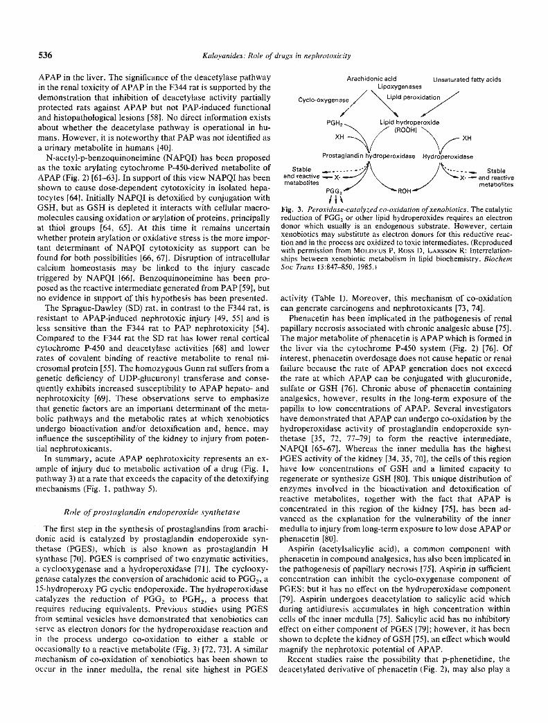
Acetaminophen, acetyl-p-aminophenol or APAP, is an effec-tive and safe antipyretic and analgesic drug when used intherapeutically recommended doses. In the clinical setting ofdrug overdose APAP can cause hepatic injury often accompa-nied by acute renal failure [37—39]. The risk of hepatic necrosisand renal failure increases as a function of the magnitude of theingested dose of drug. In therapeutic doses APAP is readilymetabolized in the liver to glucuronyl and sulfate conjugateswhich subsequently are eliminated in the urine; however, asmall fraction of APAP is metabolized by cytochrome P-450MFO to a reactive intermediate that is inactivated by GSH andis excreted as a conjugate of cysteine or mercapturic acid[40—42]. Usually less than 2% of APAP is excreted unchanged inthe urine [40, 41]. When an overdose (typically greater than 15grams) is ingested, the capacity of the liver to metabolize APAPto glucuronyl and sulfate conjugates is exceeded and a largerfraction is metabolized via the cytochrome P-450 system to areactive metabolite which is detoxified by reacting with GSH(Fig. 2) This is reflected by the increased urinary excretion ofcysteine and mercapturic acid conjugates [41]. However, asGSH levels become depleted, the reactive metabolite bindscovalently to macromolecules (Fig. 2) [43] and it is the arylationof critical intracellular proteins that appears to be causallylinked to the induction of cellular injury [44].
Another consequence of ingesting an overdose of APAP isthat a larger quantity of unmetabolized drug is delivered to thekidney as can be inferred from the elevated plasma concentra-tion of APAP and from the increased excretion of unchangeddrug [41, 42]. The renal handling of APAP involves glomerularfiltration—only 13% is bound to plasma protein—followed bypassive tubular absorption which, depending on the diureticstate, ranges from 60% to 74% of the filtered load in the dog[45]. The kidney can metabolize APAP to glucuronyl andsulfate conjugates but its capacity to do so is substantially lessthan that of the liver [46]. Similar to the liver the kidney can alsometabolize APAP to an arylating intermediate via the cy-tochrome P-450 system [47, 48]. The intrarenal distribution ofthis enzyme system (Table 1) explains the finding that tubularcell necrosis from acute APAP toxicity is restricted to theproximal tubule [39, 471.
The conclusion that metabolism of APAP to a toxic metabo-lite by cytochrome P-450-dependent MFO mediates acute tox-icity is based primarily on animal studies of hepatotoxicity in

Kaloyanides: Role ofdrugs in nephrotoxicity 535
Fig. 2. Pathways for the metabolism ofphenacerin and its major metabolites,acetaminophen (APAP) and phenetidine, totoxic intermediates. The dominant pathway isindicated by the size of the arrows. PAP,para-aminophenol; NAPQI, N-acetyl-p-benzoquinoneimine; P-450, cytochrome P-450mixed function oxidase; PGES, prostaglandinendoperoxide synthetase. Consult text fordetails.
which it was shown that: 1) induction of cytochrome P-450augments toxicity and is accompanied by a dose-dependentincrease in covalent binding of radiolabeled APAP to macro-molecules [43, 44, 49]; 2) inhibition of cytochrome P-450-dependent MFO decreases covalent binding of radiolabeledAPAP to protein and prevents cellular necrosis [43, 44, 49]; and3) maneuvers which promote GSH depletion augment covalentbinding of APAP and toxicity whereas maneuvers which main-tain GSH levels have the opposite effects [50—53]. That a similarcytochrome p-450 dependent mechanism mediates the acuterenal toxicity of APAP was inferred from the following obser-vations in the Fischer (F) 344 rat: 1) APAP causes dose-dependent depletion of renal GSH accompanied by reciprocalincreases in covalent binding of ['4C]APAP to renal proteins; 2)similar to the findings in liver the covalent binding of ['4C]A-PAP to renal protein is enzyme dependent and requiresNADPH and 02; 3) pretreatment with cobalt chloride, aninhibitor of cytochrome P-450-dependent MFO activity, pro-tected rats against APAP induced acute tubular necrosis, pre-
vented depletion of GSH, and greatly reduced APAP binding torenal proteins in vivo and in vitro [471.
Recent studies by Newton et al [54—58] support the view thatan additional metabolic pathway involving para-aminophenol(PAP) formation participates in the bioactivation of APAP. PAPis the deacetylated derivative of APAP (Fig. 2) and whenadministered to experimental animals causes renal functionaland histopathologic lesions identical to those induced by APAP[54]. Of interest, PAP is 5 to 10 times as potent a nephrotoxicantas APAP whereas it is has little toxicity for the liver. PAP,similar to APAP, depletes renal cortical GSH and arylates renalcortical proteins [55, 59], but the metabolic pathway for gener-ating the reactive intermediate appears to be independent of themicrosomal cytochrome P-450 MFO system [56, 60]. Impor-tantly, in the F344 rat PAP has been identified as a urinarymetabolite of APAP and is derived at least in part from thedeacetylation of APAP in the kidney [54, 551. Newton, Pasinoand Hook [57] have presented evidence that the deacetylasepathway plays no appreciable role in the metabolic activation of
NCOCH3
PGES
G!ucuronjde
Sulfate
NHCOCH3 NHCOCH3
I l I ji* P-450
OH OCH2CH3
HENACETIND
NH2__-I
OCH2CH3
(PHENETIDI
Glucuronide
Sulfate
P-450/NH2 HONCOCH3
OH OH
PAP ?NHCOCH3 NCOCH3
(ii 'SG GSH PROTEIN
OH 0
(PQI )
NHCOCH3
S-PROTEIN
OH
@ELL INJU)
536 Kaloyanides: Role of drugs in nephrotoxicity
APAP in the liver. The significance of the deacetylase pathwayin the renal toxicity of APAP in the F344 rat is supported by thedemonstration that inhibition of deacetylase activity partiallyprotected rats against APAP but not PAP-induced functionaland histopathological lesions [58]. No direct information existsabout whether the deacetylase pathway is operational in hu-mans. However, it is noteworthy that PAP was not identified asa urinary metabolite in humans [40].
N-acetyl-p-benzoquinoneimine (NAPQI) has been proposedas the toxic arylating cytochrome P-450-derived metabolite ofAPAP (Fig. 2) [61—631. In support of this view NAPQI has beenshown to cause dose-dependent cytotoxicity in isolated hepa-tocytes [641. Initially NAPQI is detoxified by conjugation withGSH, but as GSH is depleted it interacts with cellular macro-molecules causing oxidation or arylation of proteins, principallyat thiol groups [64, 65]. At this time it remains uncertainwhether protein arylation or oxidative stress is the more impor-tant determinant of NAPQI cytotoxicity as support can befound for both possibilities [66, 67]. Disruption of intracellularcalcium homeostasis may be linked to the injury cascadetriggered by NAPQI [66]. Benzoquinoneimine has been pro-posed as the reactive intermediate generated from PAP [59], butno evidence in support of this hypothesis has been presented.
The Sprague-Dawley (SD) rat, in contrast to the F344 rat, isresistant to APAP-induced nephrotoxic injury [49, 551 and isless sensitive than the F344 rat to PAP nephrotoxicity [54].Compared to the F344 rat the SD rat has lower renal corticalcytochrome P-450 and deacetylase activities [681 and lowerrates of covalent binding of reactive metabolite to renal mi-crosomal protein [55]. The homozygous Gunn rat suffers from agenetic deficiency of UDP-glucuronyl transferase and conse-quently exhibits increased susceptibility to APAP hepato- andnephrotoxicity [69]. These observations serve to emphasizethat genetic factors are an important determinant of the meta-bolic pathways and the metabolic rates at which xenobioticsundergo bioactivation and/or detoxification and, hence, mayinfluence the susceptibility of the kidney to injury from poten-tial nephrotoxicants.
In summary, acute APAP nephrotoxicity represents an ex-ample of injury due to metabolic activation of a drug (Fig. 1,pathway 3) at a rate that exceeds the capacity of the detoxifyingmechanisms (Fig. 1, pathway 5).
Role ofprostaglandin endoperoxide synthetase
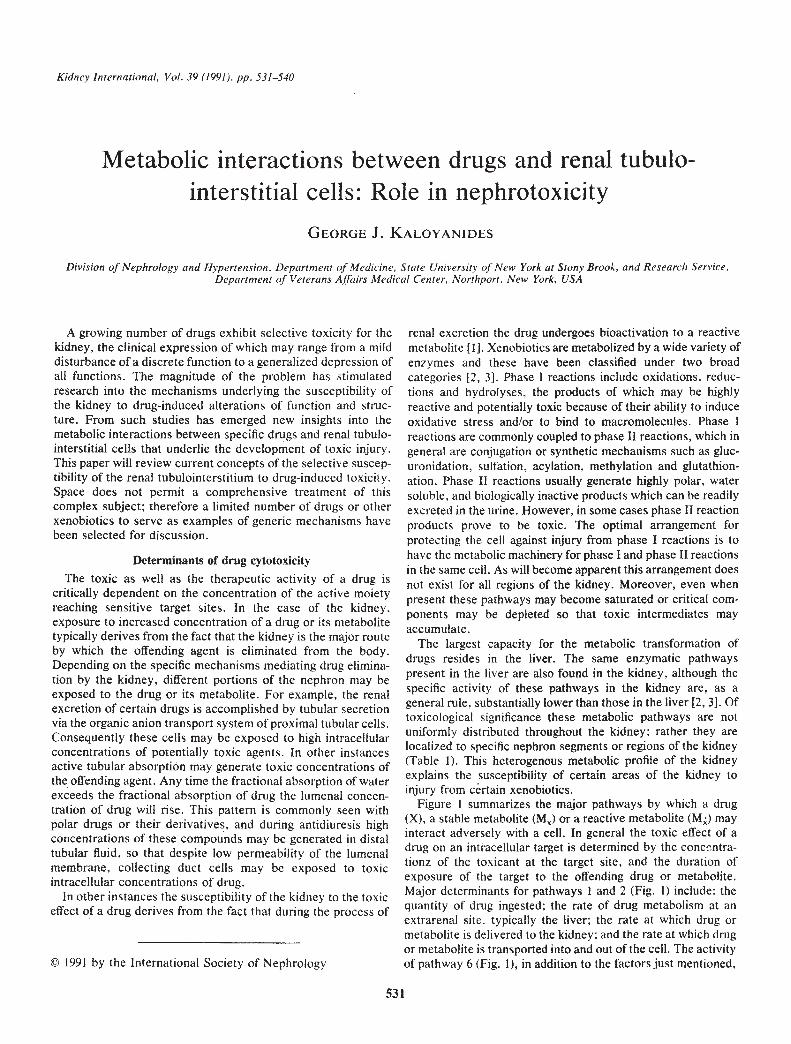
The first step in the synthesis of prostaglandins from arachi-donic acid is catalyzed by prostaglandin endoperoxide syn-thetase (PGES), which is also known as prostaglandin Hsynthase [70]. PGES is comprised of two enzymatic activities,a cyclooxygenase and a hydroperoxidase [71]. The cyclooxy-genase catalyzes the conversion of arachidonic acid to PGG2, a15-hydroperoxy PG cyclic endoperoxide. The hydroperoxidasecatalyzes the reduction of PGG2 to PGH2, a process thatrequires reducing equivalents. Previous studies using PGESfrom seminal vesicles have demonstrated that xenobiotics canserve as electron donors for the hydroperoxidase reaction andin the process undergo co-oxidation to either a stable oroccasionally to a reactive metabolite (Fig. 3) [72, 73]. A similarmechanism of co-oxidation of xenobiotics has been shown tooccur in the inner medulla, the renal site highest in PGES
Arachidonic acid Unsaturated fatty acidsLipoxygenases
Cyclo-oxygena,/" '\\iPd Peroxio,77PGH2 Lipid hydroperoxide
(ROOH)XH
Prostaglandin hydroperoxidase Hydroperoxidase
Stable Stableand reactive — X. .—/ \ / X• - and reactivemetabolites
pGG2—" "NROH___"metabolites
Fig. 3. Peroxidase-catalyzed co-oxidation of xenobiotics. The catalyticreduction of PGG2 or other lipid hydroperoxides requires an electrondonor which usually is an endogenous substrate. However, certainxenobiotics may substitute as electron donors for this reductive reac-tion and in the process are oxidized to toxic intermediates. (Reproducedwith permission from MOLDEUS P, Ross D, LARSSON R: Interrelation-ships between xenobiotic metabolism in lipid biochemistry. BiochemSoc Trans 13:847—850, 1985.)
activity (Table 1). Moreover, this mechanism of co-oxidationcan generate carcinogens and nephrotoxicants [73, 74].
Phenacetin has been implicated in the pathogenesis of renalpapillary necrosis associated with chronic analgesic abuse [75].The major metabolite of phenacetin is APAP which is formed inthe liver via the cytochrome P-450 system (Fig. 2) [76]. Ofinterest, phenacetin overdosage does not cause hepatic or renalfailure because the rate of APAP generation does not exceedthe rate at which APAP can be conjugated with glucuronide,sulfate or GSH [76]. Chronic abuse of phenacetin containinganalgesics, however, results in the long-term exposure of thepapilla to low concentrations of APAP. Several investigatorshave demonstrated that APAP can undergo co-oxidation by thehydroperoxidase activity of prostaglandin endoperoxide syn-thetase [35, 72, 77—79] to form the reactive intermediate,NAPQI [65—67]. Whereas the inner medulla has the highestPGES activity of the kidney [34, 35, 70], the cells of this regionhave low concentrations of GSH and a limited capacity toregenerate or synthesize GSH [80]. This unique distribution ofenzymes involved in the bioactivation and detoxification ofreactive metabolites, together with the fact that APAP isconcentrated in this region of the kidney [75], has been ad-vanced as the explanation for the vulnerability of the innermedulla to injury from long-term exposure to low dose APAP orphenacetin [80].
Aspil:in (acetylsalicylic acid), a common component withphenacetin in compound analgesics, has also been implicated inthe pathogenesis of papillary necrosis [751. Aspirin in sufficientconcentration can inhibit the cyclo-oxygenase component ofPGES; but it has no effect on the hydroperoxidase component[79]. Aspirin undergoes deacetylation to salicylic acid whichduring antidiuresis accumulates in high concentration withincells of the inner medulla [75]. Salicylic acid has no inhibitoryeffect on either component of PGES [79]; however, it has beenshown to deplete the kidney of GSH [75], an effect which wouldmagnify the nephrotoxic potential of APAP.
Recent studies raise the possibility that p-phenetidine, thedeacetylated derivative of phenacetin (Fig. 2), may also play a

Kaloyanides: Role of drugs in nephrotoxicity 537
role in the pathogenesis of papillary necrosis associated withchronic phenacetin abuse. Like APAP, p-phenetidine has beenshown to be an excellent substrate for co-oxidation by PGESfrom the renal medulla, the products of which are N-(4-ethoxyphenyl)p-benzoquinoneimine (NEPBQI) and N-(4-ethoxyphenyl)p-benzoquinonedi-imine (Fig. 2) [81]. NEPBQIbinds GSH, arylates microsomal proteins [81, 82] and is cyto-toxic [831. In addition, an unidentified primary oxidation prod-uct of p-phenetidine is genotoxic [82]. Genetic or drug-inducedvariation in the rate of p-phenetidine formation in the liver orpossibly within cells of the renal medulla may be an importantdeterminant of phenacetin nephrotoxicity.
In summary, the nephrotoxicity associated with phenacetinabuse represents an example of injury mediated by the meta-bolic activation in the renal inner medulla of a stable metaboliteformed in the liver (Fig. I, pathway 4).
Role of y glutamyltranspeptidase and the formation ofcysteine S-conjugates
Glutathione (GSH) is a ubiquitous tripeptide (L-y-glutamyl-L-cysteinylglycine) that participates in many biological processesincluding the synthesis of proteins and DNA, transport of aminoacids, modulation of enzyme activity, metabolism of endogenousand exogenous substrates and the detoxication of reactive metab-olites [84]. The first step in the metabolism of xenobiotics and theirmetabolites involves the formation of GSH S-conjugates. Thisreaction can occur spontaneously in the case of highly electro-philic compounds or it can be catalyzed by GSH S-transferases. afamily of enzymes with broad and overlapping substrate specific-ities [85]. GSH S-transferases are present in many cells; however,impressively high levels are found in the liver. Sequential steps inthe metabolism of GSH S-conjugates include splitting of the
.-glutamyl bond by y.glutamyltranspeptidase ()GT) to formS-cysteinylglycine conjugates, followed by dipeptidase degrada-tion to cysteine S-conjugates which can be N-acetylated to mer-capturic acids or further metabolized by 13-lyase to thiol deriva-tives (Fig. 4) [84—86]. As a general rule the formation of GSHS-conjugates represents a detoxication pathway for many reactivexenobiotics and metabolites. However, for certain substrates,mainly halogenated hydrocarbons, formation of GSH S-conju-gates represents a bioactivation pathway that generates nephro-toxic, hepatotoxic, mutagenic and/or carcinogenic metabolites[85—88]. The susceptibility of the kidney to toxic injury by thispathway relates to the major role the kidney plays in the formationand processing of toxic cysteine S-conjugates. Several reviews ofthis subject have appeared recently [86—88].
Halogenated alkanes and alkenes are substrates for liverGSH S-transferases which catalyze the formation of GSHS-conjugates. For example, the reaction of trichioroethylenewith GSH generates S-(1,2 dichlorovinyl)glutathione (DCVG),the precursor of a nephrotoxic derivative [89]. The formation inthe liver of S-(pentachloro-l,3-butadienyl) glutathione (PCBG)from the GSH S-transferase catalyzed reaction of GSH withhexachloro-l,3-butadiene represents another example [90].GSH S-conjugates are transported from hepatocytes intoplasma and bile, following which they are degraded sequentiallyby )GT and dipeptidases to form the corresponding cysteineS-conjugates. yGT is present on the surface membranes ofmany cells including biliary tract epithelium, jejunal epitheliumand renal proximal tubular epithelium, both lumenal and baso-lateral membranes 184, 86]. Of toxicological significance thekidney exhibits the highest >43T activity of any organ or tissue[84, 86]. GSH S-conjugates transported into plasma are deliv-ered to the kidney, where after glomerular filtration they are
Gly
Cys — R
GIu
y-Glutamyltranspeptidase
Gly
Cys — R
Gly 4—'j Cysteinyl glycinedipeptidase
R — S — Glucuronide
COOH UDPG-glucuronylCysteine conjugate transferase
R — S — CH2 — CH13-Lyase
'R — SH + Pyruvate + AmmoniaI S-methylNH2 transferase
(Cysteine conjugate)R — S — CH3
COOH
R—S—CH2—CH 0F UNH—C— CH3
(Mercapturate)
N-acetyttransferase
Deacetylase
(Methykhio conjugate)
Fig. 4. Pathways for the metabolism of glutathione conjugates. Some cysteine conjugates are directly toxic following uptake by renal proximaltubules or are metabolized by cysteine conjugate /3-lyase to toxic thiol metabolites. (Reproduced with permission from PIcKErr B, Lu AYH:Glutathione S—transferases: Gene structure, regulation and biological function. Ann Rev Biochem 58:743—764, 1989.)

538 Kaloyanides: Role of drugs in nephrotoxicity
catabolized by yGT and cysteinyiglycine dipeptidase to formthe cysteine S-conjugates which are transported across theapical membrane [85]. An unknown quantity of the GSHS-conjugates may be degraded by GT on the basolateralmembrane or transported directly into the cell by a sodium-dependent probenecid inhibitable pathway [91]. This pathwayappears to be distinct from that which mediates the activeuptake of cysteine S-conjugates [91, 92]. Conflicting data existsconcerning the role of organic anion transport in mediating theactive uptake of these conjugates [92, 93]. The fate of GSHS-conjugates which are transported from hepatocytes into bilehas not been studied in detail, but it seems that an unknownquantity is returned to the circulation and processed by renalproximal tubular cells [86].
Nephrotoxic cysteine S-conjugates can cause proximal tubu-lar cell injury by two major pathways. Certain nephrotoxicconjugates, such as S-(2-chloroethyl)cysteine, undergo sponta-neous rearrangement to form an episulfonium ion which cancause toxic injury by interacting with nucleophilic groups ofcritical macromolecules [87, 88]. In other cases the cysteineS-conjugate is the penultimate nephrotoxin that becomes acti-vated by renal /3-lyase [87, 88]. S-(1,2 dichlorovinyl)cysteineexemplifies this mechanism [89]. Mitochondria appear to be amajor target of the toxic metabolites generated by renal J3-lyase[94]. In vitro studies have demonstrated that incubation ofisolated renal cortical mitochondria with known toxic cysteineS-conjugates results in disruption of the function, metabolismand structural integrity of these organelles. The susceptibility ofmitochondria to injury from cysteine S-conjugates can beexplained by the observation that most of the /3-lyase activity inrenal tubular cells is found in the mitochondrial fraction [89].Thus, the nephrotoxicity associated with cysteine S-conjugatesof halogenated hydrocarbons represent examples of injurymediated by pathway 2 and pathway 4 in Figure 1.
Conclusion
In this brief review the generic pathways by which drugsinteract with cells of the tubulo-interstitium to cause toxicityhave been emphasized. Although the potential number oftoxicants is large and rapidly expanding, the basic pathways bywhich they exert their toxic effect are few in number. Theselective vulnerability of specific cells of the kidney, for exam-ple, proximal tubular cells, to certain drugs and xenobiotics is apredictable consequence of the unique transport and/or meta-bolic profile of such cells that results in the generation and/oraccumulation of the offending toxicant. At the present timehuge gaps exist in our knowledge of the ultimate toxic species ofmany agents, the specific targets of the toxicant and how theinteraction of the toxicant with its target eventuates in cellularinjury or death. As our understanding of specific molecularmechanisms mediating toxicity expands, it should be possibleto devise strategies for protecting the kidney from a major causeof injury.
Acknowledgments
The author expresses his appreciation to Dr. Bruce Tune for provid-ing access to work in press and to Pamela Geller for secretarialassistance in the preparation of this manuscript.
Reprint requests to George J. Kaloyanides, M.D., Division ofNephrology and Hypertension, Health Sciences Center, State Univer-sity ofNew York at Stony Brook, Stony Brook, New York 11794-8152,USA.
References
1. RUSH GF, SMITH JH, NEWTON iF, HOOK JB: Chemically inducednephrotoxicity: Role of metabolic activation. CRC Crit Rev Toxicol13:99—160, 1984
2. ANDERS MW: Metabolism of drugs by the kidney. Kidney mt18:636-647, 1980
3. TARLOFF JB, GOLDSTEIN RS, HooK JB: Xenobiotic metabolism inthe mammalian kidney, in Nephrotoxicity in the Experimental andClinical Situation. Part I, edited by BACH PH, LOCK EA, London,Martinus Nijhoff, 1987, pp 371—404
4. KALOYANIDES GJ, PASTORIZA-MUNOZ E: Aminoglycoside nephro-toxicity. Kidney mt 18:571—582, 1980
5. KALOYANIDES GJ: Aminoglycoside-induced functional and bio-chemical defects in the renal cortex. Fund Appi Toxicol 4:930—943,1984
6. SOBERON L, BOWMAN RL, PASTORIZA-MUNOZ E, KALOYANIDESGJ: Comparative nephrotoxicities of gentamicin, netilmicin andtobramycin in the rat. J Pharmacol Exp Ther 210:334—343, 1979
7. KALOYANIDES GJ: Renal Pharmacology of aminoglycoside antibi-otics, in Contributions to Nephrology, 42, Drug-Induced Nephro-toxicity, edited by BLANCH! C, BERTELLI A, DUARTE CO. Basel,Karger, 1984, pp 148—167
8. PASTOR!ZA-MUNOZ E, JOSEPOVITZ C, RAMSAMMY L, KALOYA-NIDES GJ: Renal handling of netilmicin in the rat with streptozoto-cm-induced diabetes mellitus. J Pharmacol Exp Ther 241: 166—173,1987
9. RAMSAMMY LS, JosEPovITz C, JONES D, LING KY, LANE BP,KALOYANIDES Gi: Induction of nephrotoxicity by high doses ofgentamicin in diabetic rats. Proc Soc Exp Biol Med 186:306—312,1987
10. JosEpovITz C, PASTORIZA-MUNOZ E, TIMMERMAN D, Scorr M,FELDMAN S, KALOYANIDES GJ: Inhibition of gentamicin uptake inrat renal cortex in vivo by aminoglycosides and organic polycat-ions.JPharmacolExp Ther223:314—32l, 1982
11. Au S, WEINER N, SCHACHT J: Aminoglycoside antibiotics prefer-entially increase permeability in phosphóinositide-containing mem-branes: A study with carboxyfluorescein liposomes. BiochimBiophys Acta 902:80—86, 1987
12. WILLIAMS SE, SCHACHT J: Binding of neomycin and calcium tophospholipids and other anionic componds. J Antibiotics 39:457—462, 1986
13. RAMSAMMY LS, JOSEPOVITZ C, LANE B, KALOYANIDES GJ: Effectof gentamicin on phospholipid metabolism in cultured rabbit prox-imal tubular cells. Am J Physiol 256 (Cell Physiol 25):C204—C213,1989
14. MINGEOT-LECLERCQ MP, LAURENT G, TULKENS PM: Biochemicalmechanism of aminoglycoside-induced inhibition of phosphatidyl-choline hydrolysis by lysosomal phospholipases. Biochem Pharma-col 37:591—599, 1988
15. RAMSAMMY LS, JOsEPOV!Tz C, KALOYANIDES GJ: Gentamicininhibits agonist stimulation of the phosphatidylinositol cascade inprimary cultures of rabbit proximal tubular cells and in rat renalcortex. J Pharmacol Exp TIter 247:989—996, 1988
16. MINGEOT-LECLERQ MP, SCHANCK AN, RONVAUX MF, DELEERSM, BRASSEUR R, RUYSSCHAERT JM, TULKENS PM: Ultrastructural,physico-chemical and conformational study of the interactions ofgentamicin and bis (beta-diethylaminoethylether) hexestrol withnegatively charged phospholipid bilayers. Biochem Pharmacol38:729—741, 1989
17. RAMSAMMY LS, KALOYANIDES GJ: The effect of gentamicin on thebiophysical properties of phosphatidic acid liposomes is influencedby the 0-C=0 group of the lipid. Biochem 27:8249—8254, 1988
18. KALOYANIDES GJ, RAMSAMMY L: Alterations of biophysical prop-erties of liposomes predict aminoglycoside nephrotoxicity: Inhibi-tory effect of polyaspartic acid, in Proceedings of the FourthInternational Symposium on Nephrotoxicity, edited by BACH PH,New York, Marcel Dekker (in press)

Kaloyanides: Role of drugs in nephrotoxicity 539
19. RAMSAMMY LS, JOSEPOVITZ C, LANE BP, KALOYANIDES Gi:Polyaspartic acid protects against gentamicin nephrotoxicity in therat. J Pharmacol Exp Pher 250:149—153, 1989
20. RAMSAMMY L, JosEPovITz C, LANE B, KALOYANIDES GJ: Polyas-partic acid inhibits gentamicin-induced perturbations of phospho-lipid metabolism. Am J Physiol (in press)
21. TULKENS PM: Nephrotoxicity of aminoglycoside antibiotics. Tox-icol Let! 46:107—123, 1989
22. RAMSAMMY LS, JOSEPOVITZ C, LING KY, LANE BP, KALOYA-NIDES GJ: Failure of inhibition of lipid peroxidation by vitamin E toprotect against gentamicin nephrotoxicity in the rat. BiochemPharmacol 36:2125—2132, 1987
23. KALOYANIDES GJ, RAMSAMMY L, JosEPovITz C: Assessment ofthree therapeutic interventions for modifying gentamicin nephro-toxicity in the rat, in Proceedings of the Fourth InternationalSymposium on Nephrotoxicity, edited by BACH PH, New York,Marcel Dekker (in press)
24. TUNE BM: The nephrotoxicity of cephalosporin antibiotics—struc-ture-activity relationships. Comments Toxicol 1:145—170, 1986
25. GOLDSTEIN RS, SMITH PF, TARLOFF JB, CONTARDI L, RUSH GF,HOOK JB; Biochemical mechanisms of cephaloxidine nephrotoxic-ity. Life Sci 42:1809—1816, 1988
26. TUNE BM, FRAVERT D, Hsu C-Y: Oxidative and mitochondrial
toxic effects of cephalosporin antibiotics in the kidney. Bioche,nPharmacol 38:795—802, 1989
27. TUNE BM, FRAVERT D: Cephalosporin nephrotoxicity. Transport,cytotoxicity and mitochondrial toxicity of cephaloglycin. J Phar-macolExp Ther2lS:186.-190, 1980
28. TUNE BM, HSU CY: The renal mitochondrial toxicity of cephalo-spOrins: Specificity of the effect on anionic substrate uptake. JPharmacol Exp Ther (in press)
29. BENDIRDJIAN JP, PRIME DJ, BROWNING MC, TUNE BM: Themitochondrial respiratory toxicity of cephalosporins. Molecularproperties and pathogenic significance in Nephrotoxicity, Ototox-icity of Drug, edited by FILLASTRE JP, Rouen, INSERM, 1982, pp303—319
30. TUNE BM, SIBLEY RK, Hsu CY: The mitochondrial respiratorytoxicity of cephalosporin antibiotics. An inhibitory effect on sub-strate uptake. J Pharmacol Exp Ther 245:1054—1059, 1988
31. TUNE BM, FRAVERT D, Hsu CY: Thienamycin nephrotoxicity:Mitochondrial injury and oxidative effects of imipenem in the rabbitkidney. Biochem Pharmacol (in press)
32. GUENGERICH FP: Characterization of human microsomal cy-tochrome P-450 enzymes. Ann Rev Pharmacol Toxicol 29:241—264,1989
33. GUENGERICHFP: Cytochrome P-450 enzymes and drug metabo-lism, in Progress in Drug Metabolism (vol 10), edited by BRIDGESJW, CHASSEAUD LF, GIBSON GG; London, Taylor Francis, 1987,
pp 1—5434. ZENSER TV, MATTAMMAL MB, DAVIS BB: Differential distribution
of the mixed-function oxidase activities in rabbit kidney. J Phar-macolExp Ther2O7:7l9—725, 1978
35. MOHANDAS J, DUGGIN GG, HORVATH JS, TILLER DJ: Regionaldifferences in peroxidatic activation of paracetamol (acetamin-ophen) mediated by cytochrome P-450 and prostaglandin endoper-oxide synthetase in rabbit kidney. Res Commun Chem PatholPharmacol 34:69—80, 1981
36. ENDOU H: Cytochrome P-450 monooxygenase system in the kid-
ney: Its intranephron localization and its induction. J Pharmaco!33:423—429, 1983
37. BROWN R: Hepatic and renal damage with paracetamol overdose. J
C/in Pathol 21:793—794, 196838. HAMLYN AN, DOUGLAS AP, JAMES OFW: The spectrum of para-
cetamol (acetaminophen) overdose: Clinical and epidemiologicalstudies. Postgrad Med J 54:400—404, 1978
39. KLEINMAN JG, BREITENFIELD RV. ROTH DA: Acute renal failureassociated with acetaminophen ingestion: Report of a case andreview of the literature. C/in Nephro! 14:201—205. 1980
40. MROCHEK JE, KATZ S, CHRISTIE WH, DINSMORE SR: Acetamin-ophen metabolism in man, as determined by high-resolution liquidchromatography. C/in Chem 20:1086—1096, 1974
41. DAVIS M, SIMMONS CJ, HARRISON NG, WILLIAMS R: Paracetamol
overdose in man: Relationship between pattern of urinary metabo-lites and severity of liver damage. Quart J Med 45:181—191, 1976
42. PRESCOTT LF, WRIGHT N: The effects of hepatic and renal damageon paracetamol metabolism and excretion following overdosage. Apharmacokinetic study. Br J Pharmacol 49:602—613, 1973
43. JOLLOW Di, MITCHELL JR. POTTER WZ, DAVIS DC, GILLETrE JR,BRODIE BB: Acetaminophen-induced hepatic necrosis. II. Role ofcovalent binding in vivo. J Pharmacol Exp Tiler 187:195—202, 1973
44. POTTER WZ, THORGEIRSSON SS, JOLLOW Di, MITCHELL JR:Acetaminophen-induced hepatic necrosis. V. Correlation of hepaticnecrosis, covalent binding and glutathione depletion in hamsters.Pharmaco! 12:129—143, 1974
45. DUGGIN GG, MUDGE GH: Renal tubular transport of paracetamoland its conjugates in the dog. Br J Pharmacol 54:359—366, 1974
46. JONES DP, SUNDBY GB, ORMSTAD K, ORRENIUS S: Use of isolatedkidney cells for study of drug metabolism. Biochem Pharmaco!28:929—935, 1979
47. MCMURTRY Ri, SNODGRASS WR, MITCHELL JR: Renal necrosis,glutathione depletion and covalent binding after acetaminophen. JToxicol App! Pharmacol 46:87—100, 1978
48. MUDGE GH, GEMBORYS MW, DUGGIN GG: Covalent binding ofmetabolites of acetaminophen to kidney protein and depletion of
renal glutathione. J Pharmaco! Exp Titer 206:218—226, 1978
49. MITCHELL JR, JOLLOW DJ, POTTER WZ, DAVIS DC, GILLETTE JR.BRODIE BB: Acetaminophen-induced hepatic necrosis. I. Role ofdrug metabolism. J Pharmacol Exp Ther 187:185—194, 1973
50. MITCHELL JR, JALLOW Di, POTTER WZ, GILLETTE JR. BRODIEBB: Acetaminophen induced hepatic necrosis. IV. Protective roleof glutathione. J Pharmacol Exp Ther 187:211—217, 1973
51. MCLEAN AEM, DAY OA: The effect of diet on the toxicity ofparacetamol and the safety of paracetamol-methionine mixtures.Biochem Pharmaco! 22:37—42, 1975
52. MASSEY TE, RACZ Wi: Effects of N-acetylcysteine on metabolism,covalent binding and toxicity of acetaminophen in isolated mousehepatocytes. Toxicol Appi Pharmaco! 60:220—228, 1981
53. LAUTERBURG BH, CONCORAN GB, MITCHELL JR: Mechanism ofaction of N-acetylcysteine in the protection against the hepatotox-icity of acetaminophen in rats in vivo. J C/in Invest 7 1:980-991,1983
54. NEWTON Ji, KUS CH, GEMBURYS MW, MUDGE GH, HooK JB:Nephrotoxicity of p-aminophenol, a metabolite of acetaminophenin the Fischer 344 rat. Toxico! App! Pharmacol 65:446—344, 1982
55. NEWTON JF, YOSHIMOTO M, BERNSTEIN J. RUSH GF, HOOK JB:Acetaminophen nephrotoxicity in the rat. 1. Strain differences innephrotoxicity and metabolism of p-aminophenol, a metabolite ofacetaminophen. Toxicol Appi Pharmaco! 69:307—3 18, 1983
56. NEWTON iF, BAILIE MB, HOOK JB: Acetaminophen nephrotoxic-ity in the rat. Renal metabolic activation in vitro. Toxicol App!Pharmacol 70:433—444, 1983
57. NEWTON iF, PASINO DA, HOOK JB: Acetaminophen nephrotoxic-ity in the rat: Quantitation of renal metabolic activation in vivo.Toxicol App! Pharmacol 78:39—46, 1985
58. NEWTON iF, Kuo CH, DESHONE GM, HOEFLE D, BERNSTEIN J,HOOK JB: The role of p-aminophenol in actaminophen-inducednephrotoxicity: Effect of bis(p-nitrophenyl)phosphate on acetamin-ophen and p-aminophenol nephrotoxicity and metabolism in Fisher344 rats. ToxicoAppl Pharmacol 81:416—480, 1985
59. CROWE CA, YONG AC, CALDER IC, HAM KN, TANGE JD: Thenephrotoxicity of p-aminophenol. 1. The effect on microsomalcytochromes, glutathione and covalent binding in kidney and liver.Chem Biol Interact 27:235—243, 1979
60. CALDER IC, YONG AC, WOODS RA, CROW CA, HAM KN, TANGEJD: The nephrotoxicity of p-aminopherol. II. The effect of meta-
bolic inhibitors and inducers. Chem Biol Interact 27:245—254, 197961. HINSON JA, NELSON SD, MITCHELL JR: Studies on the microsomal
formation of arylating metabolites of acetaminophen and phenace-
tin. Mo! Pharmaco! 13:625—633, 197762. MINER DJ, KISSINGER PT: Evidence for involvement of N-acetyl-
p-quinoneimine in acetominophen metabolism. Biochem Pharma-cal 28:3285—3290, 1979
63. HARVISON PJ, GUENGERICH FP, RASHED MS, NELSON SD: Cy-lochrome P-450 isozyme selectivity in the oxidation of acetamin-
ophen. Chem Res Toxico! 1:47—52. 1988

540 Kaloyanides: Role ofdrugs in nephrotoxicity
64. HOLME JA, DAHLIN DC, NELSON SD, DYSING E: Cytotic effects ofN-acetyl-p-benzoquinone imine, a common arylating intermediateof paracetamol and N-hydroxyparacetamol. Biochem Pharmaco!33:401—406, 1984
65. ALBANO A, RUNDGREN M, HARVISON PJ, NELSON SD, MOLDEUSP: Mechanisms of N-acetyl-p-benzoquinone imine cytotoxicity.Mol Pharmacol 28:306—311, 1985
66. MOORE M, THoR H, MOORE G, NELSON P, ORRENIUS S: Thetoxicity of acetaminophen and N-acetyl-p-benzoquinone imine inisolated hepatocytes is associated with thiol depletion and in-creased cytosolic Ca2. J Biol Chem 260:13035—13040, 1985
67. RUNDGREN M, PORUBEK DJ, HARVISON PJ, COTGREAVE IA, M0L-DEuS P, NELSON SD: Comparative cytotoxic effects of N-acetyl-p-benzoquinone imine and two dimethylated analogues. Mo! Phar-macol 34:566—672, 1988
68. NEWTON JF, YosHIMoTo M, BERNSTEIN J, RUSH GF, HooK JB:Acetaminophen nephrotoxicity in the rat. I. Strain differences innephrotoxicity and metabolism. Toxicol App! Pharmaco! 69:291—306, 1983
69. DEMORAIS SMF, WELLS PG: Enhanced acetaminophen toxicity inrats with glucuronyl transferase deficiency. Hepatol 10:163—167,1989
70. SCHLONDORFF D, ARDAILLOU R: Prostaglandins and other arachi-donic acid metabolites in the kidney. Kidney mt 29:108—119, 1986
71. MARNETT U, CHEN YP, MADDIPATI KR, LABEQUE R, PLE P:Localization of the peroxidase active site of PGH synthase, inAdvances in Prostaglandin, Thromboxane and Leukotriene Re-search (vol. 19), edited by SAMUELSSON B, W0NG PY, SUN FF,New York, Raven Press, 1989, pp 458—461
72. MOLDEUS P, Ross D, LARSSON R: Inter-relationships betweenxenobiotic metabolism and lipid biochemistry. Biochem Soc Trans-act 13:847—850, 1985
73. ELING 1, BOYD J, REED G, MASON R, SIVARAJAH K: Xenobioticmetabolism by prostaglandin endoperoxide synthetase. DrugMetab Rev 14:1023—1053, 1983
74. DAVIS BB, MATTAMMAL MB, ZENSER TV: Renal metabolism ofdrugs and xenobiotics. Nephron 27:187—196, 1981
75. DUGGIN GO: Mechanisms in the development of analgesicnephropathy. Kidney In! 18:553—561, 1980
76. MARGETTS 0: Phenacetin and paracetamol. J mt Med Res 4:(Suppl4):55—70, 1976
77. MOLDEUS P, ANDERSSON B, RAHIMTULA A, BERGGREN M: Pros-taglandin synthetase catalyzed activation of paracetamol. BiochemPharmaco! 31:1363—1368, 1982
78. MOHANDAS J, DUGGIN GG, HORVATH JS, TILLER Di: Metabolicoxidation of acetaminophen (paracetamol) mediated by cytochromeP-450 mixed function oxidase and prostaglandin endoperoxidesynthetase in rabbit kidney. Toxicol App! Pharmacol 61:252—259,
198179. ZENSER TV, MATTAMMAL MB, RAPP NS, DAVIS BB: Effect of
aspirin and metaboism of acetaminiphen and benzidine by renalinner medulla prostaglandin hydroperoxidase. J Lab Clin Med101:58—65, 1983
80. MOHANDAS I, MARSAHLL JJ, DUGGIN GO, HORVATH iS, TILLERDi: Differential distribution of glutathione and glutathione-relatedenzymes in rabbit kidney. Biochem Pharmacol 33:1801—1807, 1984
81. LARSSON R, Ross D, BERLIN T, INGE 0, MOKLEUS P: Prostaglan-din synthase catalyzed metabolic activation of p-phenatidine andacetarninophen by microsomes isolated from rabbit and humankidney. J Pharmacol Exp Ther 235:475—480, 1985
82. LARSSON R, Ross D, NORDENSKJOLD M, LINDEKE B, OLSSON LI,MOLDEUS P: Reactive products formed by peroxidase catalyzedoxidation of p-phenetidine. Chem Biol Interact 52: 1—14, 1984
83. LARSSON R, LINDQUIST T, LINDEKE B, M0LDEU5 P: Cellulareffects of N(4-ethoxyphenyl) p-benzoquinone imine, a p-pheneti-dine metabolite formed during peroxidase reactions. Chem Biol
Interact 60:317—330, 198684. MEISTER A, ANDERSON ME: Glutathione. Ann Rev Biochem 52:
711—760, 198385. PICKETF CB, Lu AYH: Glutathione S-transferases: Gene struc-
ture, regulation and biological function. Ann Rev Biochem 58:743—764, 1989
86. ORMSTAD K: Metabolism of glutathione in the kidney, in Nephro-toxicity in the Experimental and Clinical Situation, Part!, edited byBACH PH, LOCK EA, London, Martinus Nijhoff, 1987, pp 405—428
87. ANDERS MW, LASH L, DEKANT W, ELFARRA AA, DOHN DR:Biosynthesis and biotransformation of glutathione S-conjugates totoxic metabolites. CRC Grit Rev Toxicol 18:311—341, 1988
88. LOCK EA: Metabolic activation of halogenated chemicals and itsrelevance to nephrotoxicity, in Nephrotoxicity in the Experimentaland Clinical Situation, Part I, edited by BACH PH, LocK EA,London, Martinas Nijhoff, 1987, pp 429—461
89. ELFARRA AA, JACKOBSON I, ANDERS MN: Mechanism of 3-(1,2dichlorovinyl) glutathione-induced nephrotoxicity. Biochem Phar-macol 35:283—288, 1986
90. DEKANT W, SCHRENK D, VAMVAKAS S, HENSCHLER D: Metabo-lism of hexachloro-1,3-betadiene in mice: In vivo and in vitroevidence for activation by glutathione conjugation. Xenobiotica18:803—816, 1988
91. KRAMER RA, FOUREMAN G, GREENE KA, REED DJ: Nephrotox.icity of S-(2-chloroethyl)glutathione in the Fischer rat: Evidence fory-glutamyl transpeptidase-independent uptake by the kidney. JPharmacol Exp Ther 242:741—748, 1987
92. LASH LH, ANDERS MW: Uptake of nephrotoxic S-conjugates byisolated rat renal proximal tubular cells. J Pharmacol Exp Ther248:531—537, 1989
93. ZHANG 0, STEVENS JL: Transport and activation of S(l,2 dichlo-rovinyl)-L-cysteine and N-acetyl-S-( 1 ,2-dichlorovinyl)-L-cystein inrat kidney proximal tubules. Toxicol App! Pharmacol 100:51—61,1989
94. LASH LH, ANDERS MW: Mechanism of S-(1,2-dichlorovinyl)-L-homcystein-induced renal mitochondrial toxicity. Mo! Pharmacol32:549—556, 1987
95. HJILLE iT, HAXELTON GA, KLAASSEN CD, HJILLE ii: Glucuron-idation and sulfation in rabbit kidney. J Pharmaco! Exp Ther236:150—156, 1986