metabolic sensor governing bacterial virulence in ... · metabolic sensor governing bacterial...
TRANSCRIPT

Metabolic sensor governing bacterial virulence inStaphylococcus aureusYue Dinga, Xing Liub, Feifei Chena, Hongxia Dia, Bin Xua, Lu Zhouc, Xin Dengd,e, Min Wuf, Cai-Guang Yangb,1,and Lefu Lana,1
aDepartment of Molecular Pharmacology and bChinese Academy of Sciences Key Laboratory of Receptor Research, Shanghai Institute of Materia Medica,Chinese Academy of Sciences, Shanghai 201203, China; cDepartment of Medicinal Chemistry, School of Pharmacy, Fudan University, Shanghai 201203,China; dDepartment of Chemistry and eInstitute for Biophysical Dynamics, The University of Chicago, Chicago, IL 60637; and fDepartment of Basic SciencesUniversity of North Dakota School of Medicine and Health Sciences, Grand Forks, ND 58203
Edited by Richard P. Novick, New York University School of Medicine, New York, NY, and approved October 14, 2014 (received for review June 13, 2014)
An effective metabolism is essential to all living organisms, in-cluding the important human pathogen Staphylococcus aureus. Toestablish successful infection, S. aureus must scavenge nutrientsand coordinate its metabolism for proliferation. Meanwhile, it alsomust produce an array of virulence factors to interfere with hostdefenses. However, the ways in which S. aureus ties its metabolicstate to its virulence regulation remain largely unknown. Here weshow that citrate, the first intermediate of the tricarboxylic acid(TCA) cycle, binds to and activates the catabolite control proteinE (CcpE) of S. aureus. Using structural and site-directedmutagenesisstudies, we demonstrate that two arginine residues (Arg145 andArg256) within the putative inducer-binding cavity of CcpE are im-portant for its allosteric activation by citrate. Microarray analysisreveals that CcpE tunes the expression of 126 genes that compriseabout 4.7% of the S. aureus genome. Intriguingly, although CcpEis a major positive regulator of the TCA-cycle activity, its regulonconsists predominantly of genes involved in the pathogenesis ofS. aureus. Moreover, inactivation of CcpE results in increased staph-yloxanthin production, improved ability to acquire iron, increasedresistance to whole-blood–mediated killing, and enhanced bacterialvirulence in a mouse model of systemic infection. This study revealsCcpE as an important metabolic sensor that allows S. aureus tosense and adjust its metabolic state and subsequently to coordinatethe expression of virulence factors and bacterial virulence.
Staphylococcus aureus | metabolism | iron acquisition |virulence gene expression | bacterial virulence
One of the most adaptable human pathogens is Staphylococ-cus aureus (1, 2). Causing more deaths than AIDS in the
United States (3), S. aureus owes its success largely to its antibioticresistance and its ability to produce a wide array of virulencefactors that interfere with host defenses (4, 5). Even without an-tibiotic resistance, S. aureus has the effective means to causeinfections in almost every tissue of the human body (1). Thisversatility is thought to result from the remarkable capacity of thispathogen to adapt rapidly to changes in environmental conditionsand to regulate the expression of a large array of virulence factorsin a coordinated manner (1, 2, 4–7).Like other living organisms, the ability of S. aureus to sense and
adapt to changes in its environment is paramount to its survival.Some external signals can be sensed and responded to directly bytwo-component systems (8); however, other external signals thatregulate the expression of virulence factors require transductioninto intracellular signals, which subsequently are sensed by regula-tory proteins such as transcriptional regulators CodY, CcpA, andMgrA (6). For instance, when nutrients are limited, a decrease inintracellular levels of GTP and branched-chain amino acids causesan allosteric deactivation of CodY, which leads to activated tran-scription of the CodY regulon that consists predominantly of genesinvolved in amino acid biosynthesis, transport of macromolecules,and virulence (9, 10).
To survive and replicate efficiently in the host, S. aureus hasdeveloped exquisite mechanisms for scavenging nutrients andadjusting its metabolism to maintain growth while also coping withstress (6, 11). On the other hand, S. aureus produces a wide arrayof virulence factors to evade host immune defenses and to derivenutrition either parasitically or destructively from the host duringinfections (6). Indeed, pathogen exploitation of host nutrients isone of the most fundamental aspects of host–pathogen inter-actions and infectious diseases (11–16). For example, in vertebratehosts one of the first lines of defense against S. aureus infection iswithholding iron to prevent the growth of S. aureus; thereforeS. aureus has evolved highly efficient nutrient-retrieval strategiesto counteract this nutritional deprivation (11, 17, 18). However,although it is well known that nutrients modulate intracellularmetabolic status, as reflected by the concentration of key meta-bolic intermediates (19–22), the links between the metabolicintermediates and the expression of S. aureus virulence factorsremain largely unknown (6).Here we show that citrate, the first intermediate of the tri-
carboxylic acid (TCA) cycle, binds to and activates catabolitecontrol protein E (CcpE) (23). We found that CcpE acts as amaster regulator for the expression of virulence factors, centralmetabolism, iron acquisition, and bacterial virulence of S. aureus.This study establishes an intimate link between central metabo-lism and bacterial virulence in S. aureus.
Significance
Staphylococcus aureus is one of the most successful and adapt-able human pathogens and is a major cause of hospital-acquiredinfections. Here we provide insight into how S. aureus uses thecatabolite control protein E (CcpE) to sense its intracellularmetabolic status and to regulate its virulence-associated prop-erties. We define a key circuit of the virulence regulatory net-work of S. aureus and emphasize that metabolic status may bea critical element governing the virulence of this pathogen.Understanding the role of metabolites in virulence factor ex-pression ultimately may contribute to the development of novelstrategies to combat this dreaded infectious disease.
Author contributions: Y.D., C.-G.Y., and L.L. designed research; Y.D., X.L., F.C., H.D., B.X.,and L.L. performed research; L.Z., X.D., M.W., and L.L. contributed new reagents/analytictools; Y.D., X.L., C.-G.Y., and L.L. analyzed data; and Y.D., C.-G.Y., and L.L. wrotethe paper.
The authors declare no conflict of interest.
This article is a PNAS Direct Submission.
Data deposition: The atomic coordinates have been deposited in the Protein Data Bank,www.pdb.org (PDB ID code 4QBA). The microarray data reported in this paper have beendeposited in the Gene Expression Omnibus (GEO) database, www.ncbi.nlm.nih.gov/geo(accession no. GSE57260).1To whom correspondence may be addressed. Email: [email protected] or [email protected].
This article contains supporting information online at www.pnas.org/lookup/suppl/doi:10.1073/pnas.1411077111/-/DCSupplemental.
www.pnas.org/cgi/doi/10.1073/pnas.1411077111 PNAS Early Edition | 1 of 10
MICRO
BIOLO
GY
PNASPL
US

ResultsDeletion of ccpE Led to Enhanced Staphyloxanthin ProductionMediated by the Down-Regulation of citB. Previously, we showedthat the transposon insertion in the ccpE gene (SAV0672 inS. aureus Mu50) of the S. aureus Newman strain enhances theproduction of staphyloxanthin (24), a virulence factor used to evadehost oxidative killing (25–27). To verify further that the effect of theccpE transposon insertion was specific to ccpE disruption and wasnot an artifact of transposition, we generated a ccpE-deletion mu-tant in the S. aureus Newman strain as described in SI Appendix,Experimental Procedures. The resulting mutant, ΔccpE, was phe-notypically similar to the ccpE mutant with the transposon in-sertion, displaying an increase in staphyloxanthin production ascompared with either the wild-type Newman strain or its com-plemented strain (ΔccpE/p-ccpE) (Fig. 1A and SI Appendix, Fig.S1A). When all experiments described above were repeated for theUSA300 strain JE2 and its isogenic ccpE-deletion mutant (JE2-ΔccpE), similar results were observed (SI Appendix, Fig. S1 B andC), demonstrating that the effect of ccpE deletion on staph-yloxanthin production is not limited to the Newman strain andthat CcpE negatively controls the production of staphyloxanthinin S. aureus.Given that the deletion of ccpE causes reduced expression of
citB (the gene encoding aconitase, the second enzyme of theTCA cycle) (23) and that disruption of TCA-cycle genes causesenhanced staphyloxanthin production (24), we next sought todetermine if the constitutive expression of citB could suppressthe effect of ccpE deletion on the production of staphyloxanthin.
As shown in Fig. 1A and SI Appendix, Fig. S1A, the introductionof p-citB (SI Appendix, Table S1) into the ΔccpE strain restoredstaphyloxanthin production to wild-type levels. To demonstratefurther that citB is involved in the production of staphyloxanthin,we created a markerless deletion of citB in the S. aureus Newmanand JE2 strains, resulting in ΔcitB and JE2-ΔcitB (SI Appendix,Table S1), respectively. Both ΔcitB and JE2-ΔcitB strains exhib-ited enhanced staphyloxanthin production compared with theparent strain (Fig. 1A and SI Appendix, Fig. S1). In addition, whencomplemented with a plasmid carrying a wild-type citB gene(p-citB), staphyloxanthin production was restored to wild-typelevels in both ΔcitB and JE2-ΔcitB mutants (Fig. 1A and SI Ap-pendix, Fig. S1). We also observed that inactivation of either ccpEor citB caused an increase in the promoter activity of thecrtOPQMN operon containing staphyloxanthin biosynthesis genes(SI Appendix, Fig. S1D). Taken together, these results clearlysuggest that CcpE negatively controls the production of staph-yloxanthin by activating the transcription of citB.
CcpE Activates the Expression of citB in a Direct Manner. A recentstudy has shown that CcpE binds to the promoter of citB andactivates its expression (23). To delineate further the function ofCcpE on the regulation of citB, we searched for potential CcpE-binding sites in the promoter region of citB using a dye-basedDNase I footprinting analysis. As shown in Fig. 1B, in the presenceof 6His-CcpE, two regions of the citB promoter apparently wereprotected against DNase I digestion. Protected region I extendedfrom nucleotides −153 to −128 relative to the citB start codon, and
Fig. 1. CcpE negatively modulates pigment production of S. aureus by activating the expression of citB. (A) Pigmentation of S. aureus Newman strain and itsderivatives grown on TSA without glucose supplement, at 37 °C for 24 h. The wild-type Newman, ΔccpE, and ΔcitB strains harbor plasmid pYJ335. (B)Electropherograms show the protection pattern of the citB promoter after digestion with DNaseI following incubation in the absence and the presence of2 μM 6His-CcpE. There are two CcpE-protected regions (I and II) in the promoter of the citB. CcpE-protected region I harbors a box I-like sequence (shown inbold letters) [ATAA-N7-TTAT, where N is any nucleotide; the potential LysR-type transcriptional regulator (LTTR) box is underlined]. Protected region IIcontains a box II-like sequence (AATA, shown in bold letters), which can be found in the CcpC-binding sites of citB in B. subtilis. (C) Assessment of mutations ordeletion of the protected region I on CcpE binding. EMSA assays were performed using a wild-type fragment of the citB promoter DNA (citB-p, fromnucleotides −151 to +88 of the start codon of citB) or the same fragment with ATAAGTTTTGCTTAT mutated to CGCCACTTTGCTTAT (citB-M-p). Test DNA wascitB-p or citB-M-p, as indicted. citB-U, a DNA fragment of the citB promoter DNA (from nucleotides −128 to +88 of the start codon of citB) containing noCcpE-protected region. (D) Effects of mutations to the protected region I on the promoter activity of citB. ATAAGTTTTGCTTAT of the citB promoter(nucleotides −767 to +78 of the start codon of citB) was mutated to CGCCACTTTGCTTAT. Bacteria were grown in TSB at 37 °C with shaking, 250 rpm ofaeration, and sampled at 6 h. Values are relative to the wild-type Newman strain (set to 1). The expression level of transcriptional fusion citB-lacZ is highenough that the introduction of ccpE-null could have a detectable reducing effect. Results represent means ± SD, and data are representative of three in-dependent experiments.
2 of 10 | www.pnas.org/cgi/doi/10.1073/pnas.1411077111 Ding et al.
protected region II spanned nucleotides −287 to −269. In addition,a certain degree of protection also was seen in the region betweenprotected regions I and II. We noted that protected region I con-tains a box I-like sequence (ATAA-N7-TTAT, where N is anynucleotide) and that protected region II contains a box II-likesequence (AATA or TTAT), as found in the binding sites of CcpC(28), a citrate-responsive regulator in Bacillus subtilis showing 61%similarity and 35% identity to CcpE (28, 29).Protein-sequence analysis predicted that CcpE is a LysR-type
transcriptional regulator (LTTR) that binds to the LTTR boxT-N11-A (where N is any nucleotide) (30). Given that the box I-likesequence contains a potential LTTR box (ATAA-N7-TTAT, shownin bold letters in Fig. 1B), we next sought to determine if the boxI-like sequence is important for the binding of CcpE to the DNAfragment of the citB promoter. We performed an EMSA using an∼0.24-kb citB-p DNA fragment (from nucleotides −151 to +88 ofthe start codon of citB, containing an intact box I-like sequence),a citB-U DNA fragment (from nucleotides −128 to +88 of the startcodon of citB, lacking the box I-like sequence), and a citB-M-pDNAfragment in which the box I-like sequence ATAAGTTTTGCTTATwas mutated to CGCCACTTTGCTTAT). As shown in Fig. 1C,neither the citB-U nor citB-M-p DNA fragment binds to CcpE, butcitB-p does, demonstrating that the box I-like sequence is critical forbinding to 6His-CcpE. Moreover, the DNA fragment citB-p12(from nucleotides −321 to +66), which contains both CcpE-pro-tected regions I and II, showed a higher affinity for 6His-CcpE thandid the citB-L-p DNA fragment (from nucleotides −194 to +88),which contains only the protected region I (SI Appendix, Fig. S2A),indicating that DNA sequence outside the protected region I alsomay contribute to the interaction between CcpE and the citB pro-moter DNA. This result is consistent with the footprinting datashown in Fig. 1B. Additionally, the dissociation constant for thebinding of 6His-CcpE to a citB promoter DNA fragment is in thelow micromolar range (Fig. 1C and SI Appendix, Fig. S2), indicatinga relatively weak interaction between the CcpE and its target DNAsequences. The precise mechanisms of CcpE–DNA interactionsrequire further investigation. Nonetheless, these results suggest thatthe box I-like sequence, which contains a potential LTTR box, isimportant for the citB promoter to interact with CcpE.As is consistent with a recent study showing that CcpE is a pos-
itive regulator of citB (23), the promoter activity of citB (citB-lacZ,nucleotides −767 to +78 of the start codon) was approximatelyeight times lower in the ΔccpE mutant than in the wild-type strain(Fig. 1D). In addition, the mutation of the box I-like sequence inthe citB promoter abolished the promoter activity of citB-M-lacZ(Fig. 1D), suggesting that an intact box I-like sequence is requiredfor the full activity of the citB promoter. As expected, the mutantcitB promoter (i.e., citB-M) did not respond to the deletion of ccpE(Fig. 1D). Taken together, these results indicate that the box I-likesequence is likely involved in the CcpE-mediated activation of thecitB promoter. Based on these data, we conclude that CcpE mayactivate the expression of citB in a direct manner.
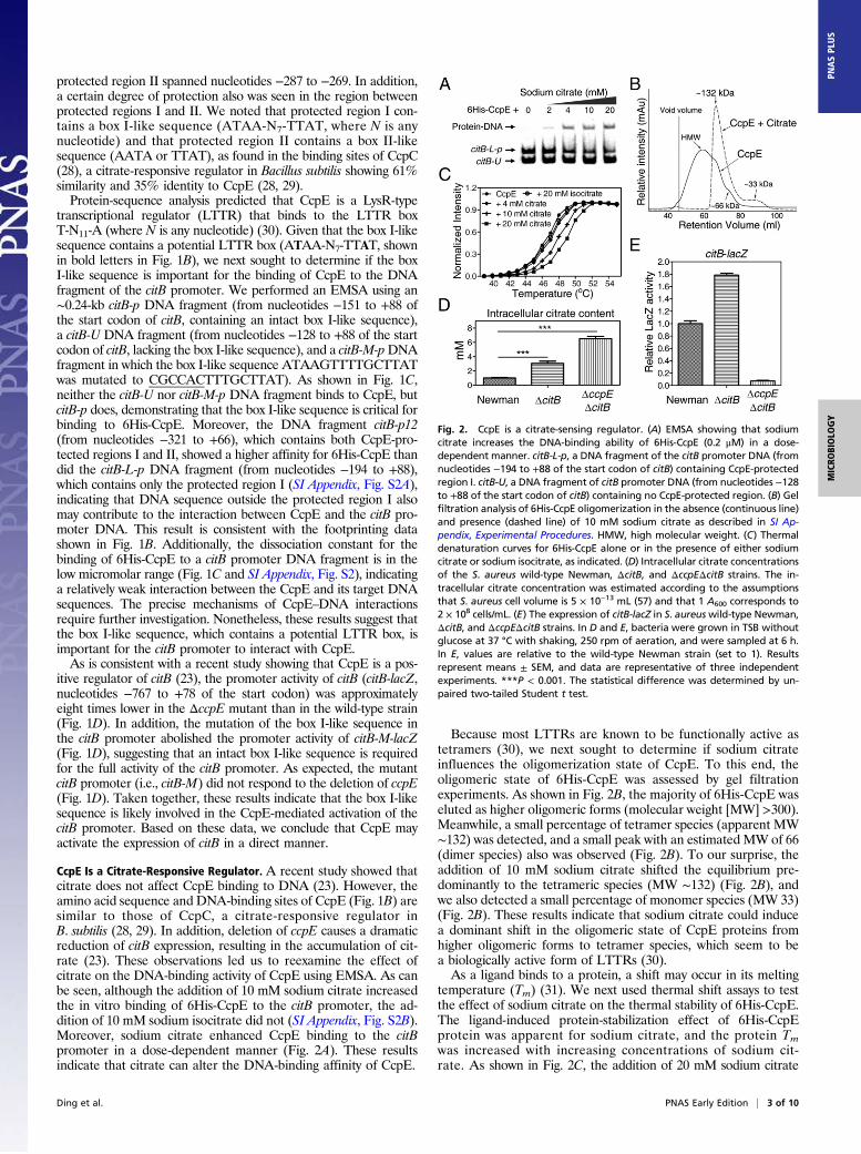
CcpE Is a Citrate-Responsive Regulator. A recent study showed thatcitrate does not affect CcpE binding to DNA (23). However, theamino acid sequence and DNA-binding sites of CcpE (Fig. 1B) aresimilar to those of CcpC, a citrate-responsive regulator inB. subtilis (28, 29). In addition, deletion of ccpE causes a dramaticreduction of citB expression, resulting in the accumulation of cit-rate (23). These observations led us to reexamine the effect ofcitrate on the DNA-binding activity of CcpE using EMSA. As canbe seen, although the addition of 10 mM sodium citrate increasedthe in vitro binding of 6His-CcpE to the citB promoter, the ad-dition of 10 mM sodium isocitrate did not (SI Appendix, Fig. S2B).Moreover, sodium citrate enhanced CcpE binding to the citBpromoter in a dose-dependent manner (Fig. 2A). These resultsindicate that citrate can alter the DNA-binding affinity of CcpE.
Because most LTTRs are known to be functionally active astetramers (30), we next sought to determine if sodium citrateinfluences the oligomerization state of CcpE. To this end, theoligomeric state of 6His-CcpE was assessed by gel filtrationexperiments. As shown in Fig. 2B, the majority of 6His-CcpE waseluted as higher oligomeric forms (molecular weight [MW] >300).Meanwhile, a small percentage of tetramer species (apparent MW∼132) was detected, and a small peak with an estimated MW of 66(dimer species) also was observed (Fig. 2B). To our surprise, theaddition of 10 mM sodium citrate shifted the equilibrium pre-dominantly to the tetrameric species (MW ∼132) (Fig. 2B), andwe also detected a small percentage of monomer species (MW 33)(Fig. 2B). These results indicate that sodium citrate could inducea dominant shift in the oligomeric state of CcpE proteins fromhigher oligomeric forms to tetramer species, which seem to bea biologically active form of LTTRs (30).As a ligand binds to a protein, a shift may occur in its melting
temperature (Tm) (31). We next used thermal shift assays to testthe effect of sodium citrate on the thermal stability of 6His-CcpE.The ligand-induced protein-stabilization effect of 6His-CcpEprotein was apparent for sodium citrate, and the protein Tmwas increased with increasing concentrations of sodium cit-rate. As shown in Fig. 2C, the addition of 20 mM sodium citrate
Fig. 2. CcpE is a citrate-sensing regulator. (A) EMSA showing that sodiumcitrate increases the DNA-binding ability of 6His-CcpE (0.2 μM) in a dose-dependent manner. citB-L-p, a DNA fragment of the citB promoter DNA (fromnucleotides −194 to +88 of the start codon of citB) containing CcpE-protectedregion I. citB-U, a DNA fragment of citB promoter DNA (from nucleotides −128to +88 of the start codon of citB) containing no CcpE-protected region. (B) Gelfiltration analysis of 6His-CcpE oligomerization in the absence (continuous line)and presence (dashed line) of 10 mM sodium citrate as described in SI Ap-pendix, Experimental Procedures. HMW, high molecular weight. (C) Thermaldenaturation curves for 6His-CcpE alone or in the presence of either sodiumcitrate or sodium isocitrate, as indicated. (D) Intracellular citrate concentrationsof the S. aureus wild-type Newman, ΔcitB, and ΔccpEΔcitB strains. The in-tracellular citrate concentration was estimated according to the assumptionsthat S. aureus cell volume is 5 × 10⁻¹³ mL (57) and that 1 A600 corresponds to2 × 108 cells/mL. (E) The expression of citB-lacZ in S. aureuswild-type Newman,ΔcitB, and ΔccpEΔcitB strains. In D and E, bacteria were grown in TSB withoutglucose at 37 °C with shaking, 250 rpm of aeration, and were sampled at 6 h.In E, values are relative to the wild-type Newman strain (set to 1). Resultsrepresent means ± SEM, and data are representative of three independentexperiments. ***P < 0.001. The statistical difference was determined by un-paired two-tailed Student t test.
Ding et al. PNAS Early Edition | 3 of 10
MICRO
BIOLO
GY
PNASPL
US

increased the Tm by ∼3 °C. In contrast, the addition of 20 mMsodium isocitrate had no significant effect on the Tm value of 6His-CcpE (Fig. 2C). These results suggest that citrate directly bindsto CcpE.Noting that citB deletion results in the accumulation of in-
tracellular citrate (Fig. 2D), we next examined whether citB de-letion affects the regulatory function of CcpE in vivo. Wemeasured the regulatory activities of CcpE in the wild-typeNewman strain and in a citB-deletion mutant (ΔcitB) using thecitB-lacZ reporter gene as a readout. As shown in Fig. 2E, theexpression level of citB-lacZ was about 1.8 times higher inthe ΔcitB mutant than in the wild-type Newman strain. To ex-amine if CcpE is required for increased expression of citB-lacZ(Fig. 2E), we measured the citB-lacZ expression in a citB andccpE double-deletion mutant (ΔcitBΔccpE). The intracellularcitrate concentration was approximately two times higher but theexpression of citB-lacZ was ∼20 times lower in the ΔcitBΔccpEmutant than in the ΔcitB strain, (Fig. 2E), demonstrating that thehigh citrate pool alone does not increase citB expression.Therefore, the up-regulation of citB-lacZ in the ΔcitB mutantmay result from the activation of CcpE by the elevated con-centration of intracellular citrate (Fig. 2 A and B).
Crystal Structure of the Inducer-Binding Domain of CcpE. Membersof LTTRs have a conserved structure with an N-terminal DNA-binding domain and a C-terminal inducer-binding domain (IBD)(30). To explore the ligand-binding characteristics of CcpE andto gain structural insights into the transcriptional regulation byCcpE, we decided to crystallize CcpE. Although we could notcrystallize full-length CcpE, we succeeded in crystallizing theIBD of CcpE (CcpEIBD, which comprises amino acid residues63–288 of CcpE). The structure was solved by single-wavelengthanomalous dispersion from a SeMet-substituted crystal form andwas refined at 2.21-Å resolution (SI Appendix, Table S3). Thefinal Rwork and Rfree were 20.2% and 24.3%, respectively (SIAppendix, Table S3).As shown in Fig. 3A, the crystallized CcpEIBD is a homodimer
with a head-to-tail arrangement of monomers in the asymmetricunit. In accordance with our expectations, each chain of theCcpEIBD comprises two potential regulatory domains (RD1 andRD2) by analogy with other LTTRs, and a hinge formed fromthe central regions of two antiparallel β-stands (β4 and β9) linksthese two regulatory domains of each IBD (Fig. 3A and SI Ap-pendix, Fig. S3). These observations are highly similar to thoseobserved for the IBDs of LTTRs (30). Because the inducer-binding cavity (IBC) of LTTRs appears to be located at the
Fig. 3. Arg145 and Arg256, located within the putative inducer-binding cavity of CcpE, are critical for the activation of CcpE by citrate. (A, Left) Overallprotein folding of the inducer-binding fragment of CcpE is presented in a cartoon. One monomer is colored in magenta and the other in cyan. Each monomerpossesses two domains separated by a hinge formed from the central regions of β4 and β9. (Right) A local view of the putative inducer-binding cavity of CcpE.Protein is shown in gray, amino acids in magenta sticks, water as red spheres, chloride ions as green spheres, and hydrogen bonds as black dashed lines. (B andC) Gel filtration analysis of 6His-CcpER145A (B) and 6His-CcpER256A (C) oligomerization in the absence (continuous line) and presence (dashed line) of 10 mMsodium citrate. (D and E) Thermal denaturation curves for 6His-CcpER145A (D) and 6His-CcpER256A (E) in the absence and presence of 20 mM sodium citrate. (F)Effect of the amino acid substitutions in CcpE on its ability to promote the expression of citB-lacZ in the ΔccpE mutant. ccpER145A, arginine 145 of ccpEmutated to alanine; ccpER256A, arginine 256 of ccpE mutated to alanine. The wild-type Newman and ΔccpE strains harbor the control plasmid pYJ335-Tc.S. aureus was grown in TSB at 37 °C with shaking, 250 rpm of aeration, and was sampled at 6 h. Values are relative to the wild-type Newman strain (set to 1).Results represent means ± SEM, and data are representative of three independent experiments.
4 of 10 | www.pnas.org/cgi/doi/10.1073/pnas.1411077111 Ding et al.
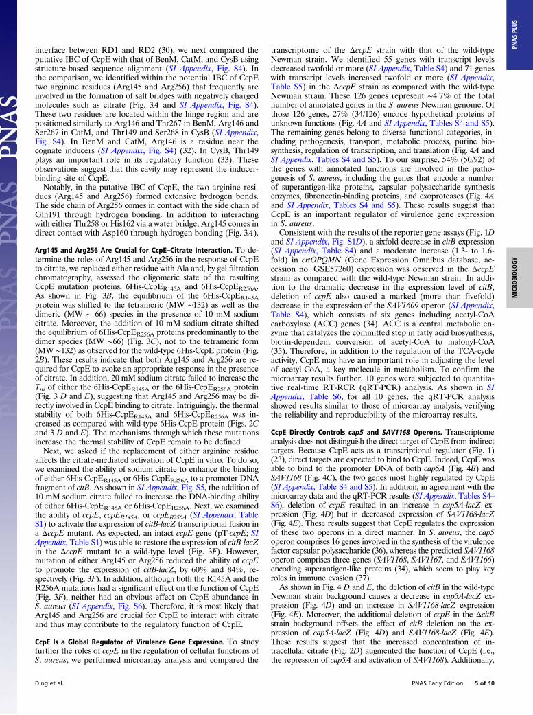
interface between RD1 and RD2 (30), we next compared theputative IBC of CcpE with that of BenM, CatM, and CysB usingstructure-based sequence alignment (SI Appendix, Fig. S4). Inthe comparison, we identified within the potential IBC of CcpEtwo arginine residues (Arg145 and Arg256) that frequently areinvolved in the formation of salt bridges with negatively chargedmolecules such as citrate (Fig. 3A and SI Appendix, Fig. S4).These two residues are located within the hinge region and arepositioned similarly to Arg146 and Thr267 in BenM, Arg146 andSer267 in CatM, and Thr149 and Ser268 in CysB (SI Appendix,Fig. S4). In BenM and CatM, Arg146 is a residue near thecognate inducers (SI Appendix, Fig. S4) (32). In CysB, Thr149plays an important role in its regulatory function (33). Theseobservations suggest that this cavity may represent the inducer-binding site of CcpE.Notably, in the putative IBC of CcpE, the two arginine resi-
dues (Arg145 and Arg256) formed extensive hydrogen bonds.The side chain of Arg256 comes in contact with the side chain ofGln191 through hydrogen bonding. In addition to interactingwith either Thr258 or His162 via a water bridge, Arg145 comes indirect contact with Asp160 through hydrogen bonding (Fig. 3A).
Arg145 and Arg256 Are Crucial for CcpE–Citrate Interaction. To de-termine the roles of Arg145 and Arg256 in the response of CcpEto citrate, we replaced either residue with Ala and, by gel filtrationchromatography, assessed the oligomeric state of the resultingCcpE mutation proteins, 6His-CcpER145A and 6His-CcpER256A.As shown in Fig. 3B, the equilibrium of the 6His-CcpER145Aprotein was shifted to the tetrameric (MW ∼132) as well as thedimeric (MW ∼ 66) species in the presence of 10 mM sodiumcitrate. Moreover, the addition of 10 mM sodium citrate shiftedthe equilibrium of 6His-CcpER256A proteins predominantly to thedimer species (MW ∼66) (Fig. 3C), not to the tetrameric form(MW ∼132) as observed for the wild-type 6His-CcpE protein (Fig.2B). These results indicate that both Arg145 and Arg256 are re-quired for CcpE to evoke an appropriate response in the presenceof citrate. In addition, 20 mM sodium citrate failed to increase theTm of either the 6His-CcpER145A or the 6His-CcpER256A protein(Fig. 3 D and E), suggesting that Arg145 and Arg256 may be di-rectly involved in CcpE binding to citrate. Intriguingly, the thermalstability of both 6His-CcpER145A and 6His-CcpER256A was in-creased as compared with wild-type 6His-CcpE protein (Figs. 2Cand 3 D and E). The mechanisms through which these mutationsincrease the thermal stability of CcpE remain to be defined.Next, we asked if the replacement of either arginine residue
affects the citrate-mediated activation of CcpE in vitro. To do so,we examined the ability of sodium citrate to enhance the bindingof either 6His-CcpER145A or 6His-CcpER256A to a promoter DNAfragment of citB. As shown in SI Appendix, Fig. S5, the addition of10 mM sodium citrate failed to increase the DNA-binding abilityof either 6His-CcpER145A or 6His-CcpER256A. Next, we examinedthe ability of ccpE, ccpER145A, or ccpER256A (SI Appendix, TableS1) to activate the expression of citB-lacZ transcriptional fusion ina ΔccpE mutant. As expected, an intact ccpE gene (pT-ccpE; SIAppendix, Table S1) was able to restore the expression of citB-lacZin the ΔccpE mutant to a wild-type level (Fig. 3F). However,mutation of either Arg145 or Arg256 reduced the ability of ccpEto promote the expression of citB-lacZ, by 60% and 84%, re-spectively (Fig. 3F). In addition, although both the R145A and theR256A mutations had a significant effect on the function of CcpE(Fig. 3F), neither had an obvious effect on CcpE abundance inS. aureus (SI Appendix, Fig. S6). Therefore, it is most likely thatArg145 and Arg256 are crucial for CcpE to interact with citrateand thus may contribute to the regulatory function of CcpE.
CcpE Is a Global Regulator of Virulence Gene Expression. To studyfurther the roles of ccpE in the regulation of cellular functions ofS. aureus, we performed microarray analysis and compared the
transcriptome of the ΔccpE strain with that of the wild-typeNewman strain. We identified 55 genes with transcript levelsdecreased twofold or more (SI Appendix, Table S4) and 71 geneswith transcript levels increased twofold or more (SI Appendix,Table S5) in the ΔccpE strain as compared with the wild-typeNewman strain. These 126 genes represent ∼4.7% of the totalnumber of annotated genes in the S. aureus Newman genome. Ofthose 126 genes, 27% (34/126) encode hypothetical proteins ofunknown functions (Fig. 4A and SI Appendix, Tables S4 and S5).The remaining genes belong to diverse functional categories, in-cluding pathogenesis, transport, metabolic process, purine bio-synthesis, regulation of transcription, and translation (Fig. 4A andSI Appendix, Tables S4 and S5). To our surprise, 54% (50/92) ofthe genes with annotated functions are involved in the patho-genesis of S. aureus, including the genes that encode a numberof superantigen-like proteins, capsular polysaccharide synthesisenzymes, fibronectin-binding proteins, and exoproteases (Fig. 4Aand SI Appendix, Tables S4 and S5). These results suggest thatCcpE is an important regulator of virulence gene expressionin S. aureus.Consistent with the results of the reporter gene assays (Fig. 1D
and SI Appendix, Fig. S1D), a sixfold decrease in citB expression(SI Appendix, Table S4) and a moderate increase (1.3- to 1.6-fold) in crtOPQMN (Gene Expression Omnibus database, ac-cession no. GSE57260) expression was observed in the ΔccpEstrain as compared with the wild-type Newman strain. In addi-tion to the dramatic decrease in the expression level of citB,deletion of ccpE also caused a marked (more than fivefold)decrease in the expression of the SAV1609 operon (SI Appendix,Table S4), which consists of six genes including acetyl-CoAcarboxylase (ACC) genes (34). ACC is a central metabolic en-zyme that catalyzes the committed step in fatty acid biosynthesis,biotin-dependent conversion of acetyl-CoA to malonyl-CoA(35). Therefore, in addition to the regulation of the TCA-cycleactivity, CcpE may have an important role in adjusting the levelof acetyl-CoA, a key molecule in metabolism. To confirm themicroarray results further, 10 genes were subjected to quantita-tive real-time RT-RCR (qRT-PCR) analysis. As shown in SIAppendix, Table S6, for all 10 genes, the qRT-PCR analysisshowed results similar to those of microarray analysis, verifyingthe reliability and reproducibility of the microarray results.
CcpE Directly Controls cap5 and SAV1168 Operons. Transcriptomeanalysis does not distinguish the direct target of CcpE from indirecttargets. Because CcpE acts as a transcriptional regulator (Fig. 1)(23), direct targets are expected to bind to CcpE. Indeed, CcpE wasable to bind to the promoter DNA of both cap5A (Fig. 4B) andSAV1168 (Fig. 4C), the two genes most highly regulated by CcpE(SI Appendix, Table S4 and S5). In addition, in agreement with themicroarray data and the qRT-PCR results (SI Appendix, Tables S4–S6), deletion of ccpE resulted in an increase in cap5A-lacZ ex-pression (Fig. 4D) but in decreased expression of SAV1168-lacZ(Fig. 4E). These results suggest that CcpE regulates the expressionof these two operons in a direct manner. In S. aureus, the cap5operon comprises 16 genes involved in the synthesis of the virulencefactor capsular polysaccharide (36), whereas the predicted SAV1168operon comprises three genes (SAV1168, SAV1167, and SAV1166)encoding superantigen-like proteins (34), which seem to play keyroles in immune evasion (37).As shown in Fig. 4 D and E, the deletion of citB in the wild-type
Newman strain background causes a decrease in cap5A-lacZ ex-pression (Fig. 4D) and an increase in SAV1168-lacZ expression(Fig. 4E). Moreover, the additional deletion of ccpE in the ΔcitBstrain background offsets the effect of citB deletion on the ex-pression of cap5A-lacZ (Fig. 4D) and SAV1168-lacZ (Fig. 4E).These results suggest that the increased concentration of in-tracellular citrate (Fig. 2D) augmented the function of CcpE (i.e.,the repression of cap5A and activation of SAV1168). Additionally,
Ding et al. PNAS Early Edition | 5 of 10
MICRO
BIOLO
GY
PNASPL
US

mutation of either Arg145 or Arg256 reduced the ability of ccpEto modulate the expression of either cap5A-lacZ (Fig. 4F) orSAV1168-lacZ (Fig. 4G), indicating that these two potential citrate-binding residues are important for CcpE-dependent expression ofthe cap5 and SAV1168 operons.Using a dye-based DNase I footprinting analysis, we found
that two regions of the SAV1168 promoter apparently wereprotected against DNase I digestion by 6His-CcpE (SI Appendix,Fig. S7A). Protected region I contains a potential LTTR box(T-N11-A), and protected region II contains two box II-like sequences(AATA and TTAT) (SI Appendix, Fig. S7A). Interestingly, CcpE-protected region I of SAV1168 promoter DNA contains a nucle-otide sequence (ATGATAAGTTTTGCTTAaATA) similar tothat of citB promoter DNA (ATGATAAGTTTTGCTTAtATA).Mutations in this region resulted in a ninefold decrease in pro-moter activity of SAV1168 in the wild-type Newman strain(SAV1168-M-lacZ; SI Appendix, Fig. S7B). More importantly, themutant promoter of SAV1168 was insensitive to the deletion ofccpE (SI Appendix, Fig. S7B), confirming the critical role of theprotected region I in the CcpE-mediated regulation of SAV1168.Additionally, like the Newman ΔccpE mutant (Figs. 1D and
4E), the JE2-ΔccpE mutant exhibits decreased expression ofcitB-lacZ and SAV1168-lacZ in relation to its parent strain (SIAppendix, Fig. S8), indicating a conserved function of CcpE in
the S. aureus Newman and JE2 strains. Taken together, theseresults suggest that CcpE directly controls the expression of thecap5 and SAV1168 operons.
CcpE Regulates Iron Acquisition in S. aureus. Because inactivation ofCcpE results in the accumulation of intracellular citrate, we nextexamined if CcpE modulates the production of the citrate-containing siderophores, which are used by S. aureus to overcomeiron limitation during infections (11, 17, 18). When bacteria weregrown in RPMI medium without supplemental iron, the deletionof ccpE resulted in increased production of siderophore (Fig. 5 Aand B). The introduction of p-ccpE (SI Appendix, Table S1) intothe ΔccpE strain abolished the increase in siderophore pro-duction (Fig. 5B), suggesting that CcpE represses the productionof siderophore in S. aureus. Moreover, consistent with the resultsof CAS (chrom azurol S)-based analysis (Fig. 5 A and B), theΔccpE strain exhibited an increased growth rate in RPMI me-dium without supplemental iron, as compared with wild-type andthe complemented strains (Fig. 5C). When the growth mediumwas supplemented with iron, the growth advantage of the ΔccpEstrain disappeared (Fig. 5D). These results suggest that thedown-regulation of CcpE may provide an advantage, allowingthe S. aureus Newman strain to adapt to an iron-limiting
Fig. 4. CcpE is a global regulator of virulence gene expression in S. aureus. (A) Grouping CcpE-regulated genes according to their annotated function showsthat the CcpE regulon consists predominantly of genes involved in the pathogenesis of S. aureus. Numbers of genes whose expressions are down-regulated orup-regulated in the ΔccpE strain compared with the wild-type Newman strain are shown. (B) EMSA showing that 6His-CcpE binds to the promoter DNA(cap5A-p) of cap5A (NWMN_0095) but not to a DNA fragment (ccpE-O) amplified from the ccpE (NWMN_0641) gene in the absence of citrate. (C) EMSAshowing that 6His-CcpE binds to the promoter DNA (SAV1168-p) of NWMN_1077 but not to a DNA fragment (citB-U) of the citB promoter (from nucleotides−128 to +88 of the start codon of citB) containing no CcpE-protected region in the absence of citrate. (D–G) The expression of transcriptional cap5A-lacZ andSAV1168-lacZ fusions in the wild-type Newman strain and its derivatives, as indicated. In F and G the wild-type Newman and ΔccpE strains harbor the controlplasmid pYJ335-Tc. S. aureus was grown in TSB at 37 °C with shaking, 250 rpm of aeration, and sampled at 6 h. Values are relative to the wild-type Newmanstrain (set to 1). Results represent means ± SEM, and data are representative of three independent experiments. The statistical difference was determined byunpaired two-tailed Student t test (**P < 0.01, ***P < 0.001). NWMN_1077 gene of the S. aureus Newman strain is corresponding to the SAV1168 locus ofS. aureus Mu50.
6 of 10 | www.pnas.org/cgi/doi/10.1073/pnas.1411077111 Ding et al.

condition. Similar results also were obtained with the S. aureusJE2 strain (SI Appendix, Fig. S9).To examine if CcpE is involved in iron uptake by S. aureus, we
measured intracellular iron pools of the wild-type Newman,ΔccpE, and ΔccpE/p-ccpE strains using atomic absorptionspectroscopy. After cultivation in RPMI medium for 12 h, theiron pool of the ΔccpE strain was about 24% larger than that ofwild-type Newman strain, but the introduction of ccpE (p-ccpE;SI Appendix, Table S1) decreased the intracellular iron pool ofthe ΔccpE strain by ∼50% (Fig. 5E), demonstrating that CcpEnegatively modulates iron uptake by S. aureus.In addition to the increased siderophore production, the de-
letion of ccpE also caused a decrease in pH in the culture me-dium when bacteria reached the postexponential growth phase(23). Because both siderophore and acidic pH promote the re-lease of iron from transferrin, we measured the capacity of theculture supernatants to release iron from transferrin. As shownin Fig. 5F, spent culture supernatant from the ΔccpE strainincreases the rate of iron release from transferrin, but in-troduction of the ccpE gene completely abolished the phenotype,suggesting that inactivation of CcpE facilitates the release of ironfrom the host iron-sequestering protein. In sum, these results
clearly suggest that CcpE regulates iron acquisition in S. aureus.This notion is substantiated further by the observation that thedeletion of ccpE decreased the expression of the iron-regulatedsirABC operon genes (38) in either tryptic soy broth (TSB)medium (without glucose) or RPMI medium (SI Appendix, TablesS4 and S6).
CcpE Modulates Virulence of S. aureus. As mentioned above, thedeletion of ccpE caused increased staphyloxanthin production(Fig. 1A and SI Appendix, Fig. S1), improved the ability ofS. aureus to acquire iron (Fig. 5 and SI Appendix, Fig. S9), andaltered the expression of a large number of genes associated withthe pathogenesis of S. aureus (Fig. 4A and SI Appendix, Tables S4and S5). Therefore we next sought to determine if ccpE con-tributes to the staphylococcal resistance of host innate immuneresponses. We mixed the wild-type strain, ΔccpE, and its com-plemented strain (ΔccpE/p-ccpE) with blood collected fromhuman volunteers and measured the survival of the bacteria. Asshown in Fig. 6A, the relative survival of the ΔccpE strain wasincreased as compared with the wild-type Newman strain(∼1.5% vs. ∼0.5%). Introduction of the ccpE gene reduced thebacterial survival to the level seen in the wild-type strain (Fig.6A), indicating that the inactivation of CcpE increases thestaphylococcal resistance to whole-blood–mediated killing.To test further the role of ccpE in invasive staphylococcal
disease, we subjected the test strains—wild type, ΔccpE, andΔccpE-C—to a murine model of abscess formation (24, 39) andmeasured the bacterial survival in host organs. As shown in Fig.6B, the lack of ccpE increased the bacterial survival in bothkidney and liver. Again, the introduction of the ccpE gene low-ered the bacterial survival (ΔccpE-C in Fig. 6B). Interestingly, inthe liver, the bacterial load of the complemented strain (ΔccpE-C)was two-log lower than that of the wild-type Newman strain (Fig.6B), suggesting that the appropriate expression of ccpE is im-portant for S. aureus virulence. In sum, these results suggest thatCcpE plays a key role in the pathogenesis of S. aureus in a mu-rine model of abscess formation.
DiscussionIncreasingly, metabolic potential is considered a critical elementgoverning a pathogen’s virulence as well as its ability to survive inits host (6, 13, 16, 40–44). Here we show that the S. aureus CcpEprotein captures changes in citrate levels and transforms theminto various cellular responses. Intriguingly, although CcpE isa positive regulator of TCA-cycle activity, its regulon consistspredominantly of virulence-associated genes. In addition to di-rectly controlling the promoter activity of citB, CcpE also directlycontrols the expression of virulence-related genes such as cap5Aand SAV1168. By binding to and activating CcpE, citrate appearsto be a key catabolite for coordinating the S. aureus metabolicstate with bacterial virulence. A model for a global regulatoryrole of CcpE in S. aureus is shown in Fig. 7.A recent study showed that CcpE is a major positive regulator
of citB, a TCA-cycle gene, and that the DNA binding of CcpE isnot affected by citrate (23). In this study we also found that CcpEbinds specifically to an LTTR box of the citB promoter and acti-vates its promoter activity (Fig. 1 B–D). However, in contrast tothe previous report, we observed that citrate alters the DNA-binding affinity of CcpE (Fig. 2A and SI Appendix, Fig. S2B). Thediscrepancy seems to be caused by the distinct buffer composition.When we used an EMSA binding buffer similar to that describedin the previous study, we also failed to observe the citrate-medi-ated enhancement of DNA binding by CcpE (SI Appendix, Fig.S10A). WhenMg2+ was eliminated from our buffer, sodium citratestill was able to enhance the DNA-binding activity of CcpE (SIAppendix, Fig. S10B). However, when the nonionic detergentNonidet P-40 was omitted, the citrate-mediated effect was notobserved (SI Appendix, Fig. S10C), showing that the nonionic
Fig. 5. Deletion of ccpE results in improved ability of S. aureus to acquireiron. In all panels, the wild-type Newman and ΔccpE strains harbor theplasmid pYJ335. (A) Assessment of the siderophore production using achrome azurol S agar diffusion assay as described in SI Appendix, Experi-mental Procedures. The orange halos formed around the wells correspond tothe iron-chelating activity of the siderophores. (B) Siderophore levels inspent culture supernatants of the Newman strain and its derivatives, as in-dicated. Siderophore units were calculated as described in SI Appendix, Ex-perimental Procedures. (C and D) Representative growth curves for S. aureusgrown in iron-limited (C) and in iron-sufficient (D) medium. (E) De-termination of intracellular iron content of the wild-type Newman strainand its derivatives. Bacteria were grown in RPMI medium for 24 h withshaking, 250 rpm of aeration. Then the cells were collected, prepared for,and run on atomic absorption spectroscopy. Results show iron content asa percentage of the dry weight. Values represent means ± SEM. (F) Ironrelease from transferrin mediated by various spent media from the wild-typeNewman strain and its derivatives. A decrease in optical density signifiesa release of iron from transferrin. Data are representative of three inde-pendent experiments.
Ding et al. PNAS Early Edition | 7 of 10
MICRO
BIOLO
GY
PNASPL
US

detergent plays a key role in the CcpE response to citrate. How-ever, the mechanism by which Nonidet P-40 affects the citrate-mediated activation of CcpE requires further investigation, whichmay help shed light on the mode of action of CcpE.In addition to affecting the DNA-binding activity of CcpE
(Fig. 2A and SI Appendix, Fig. S2B), citrate can shift the equi-librium of CcpE proteins to a predominantly tetrameric species(Fig. 2B), which seems to be the biologically active form ofLTTRs (30). Moreover, citrate is able to increase the thermalstability of CcpE (Fig. 2C), indicating that it probably binds toCcpE directly. These results also suggest that citrate likely is ableto cause an allosteric activation of CcpE. Consistent with thishypothesis, we found that the deletion of citB, which leads to anaccumulation of intracellular citrate (Fig. 2D), resulted in CcpE-dependent alteration of promoter activities of citB (Fig. 2E),SAV1168 (Fig. 4E), and cap5A (Fig. 4D). Structural and site-directed mutagenesis studies further revealed that Arg145 andArg256 in the potential inducer-binding cavity (Fig. 3A and SIAppendix, Fig. S4) of CcpE are crucial for CcpE–citrate in-teraction and the activation of CcpE by citrate (Fig. 3 and SIAppendix, Fig. S5). Based on these data, we concluded that citratecan function as an upstream signal to determine the gene-regulatory activity of CcpE (Fig. 7). This notion is consistent withthe observation that LTTRs usually require the binding of an in-ducer to activate transcription, and the inducers are intermediatesin the degradation pathway regulated by the LTTR (30).Inactivation of ccpE resulted in increased intracellular con-
centrations of citrate (23). This effect may be caused in part bythe down-regulation of citB (Fig. 1D and Fig. 2D). Additionally,the increase in citrate accumulation that occurs when a ccpE-nullmutation is introduced into a citB-deletion mutant (Fig. 2D)suggests that CcpE is capable of modulating intracellular citratelevels independently of aconitase (CitB). Thus, we speculate thatCcpE negatively regulates a citrate-producing enzyme or posi-tively regulates a citrate-consuming enzyme or alters a pathwaythat diverts acetyl-CoA from citrate production (e.g., ACC). Infact, CcpE is a positive regulator of ACC (SI Appendix, TableS4). Taken together, these findings indicate that CcpE maycontrol the intracellular concentrations of citrate via multiplepathways.Citrate is the first intermediate of the TCA cycle, which is
highly conserved in all organisms. Citrate provides organismswith energy, reducing potential and biosynthetic intermediates(6, 45). A high level of citrate means that biosynthetic precursors
are abundant and indicates a sufficient energy supply (6, 45). Inorganisms, intracellular concentrations of citrate are in the mil-limolar range (46–49). A similar intracellular level of citrate wasobserved for S. aureus (Fig. 2D). In addition to being an im-portant intermediate of the TCA cycle, citrate allostericallyactivates ACC (50), which catalyzes the committed step in fattyacid synthesis in most organisms (35). In addition, citrate caninhibit a number of enzymes, such as phosphofructokinase, pyru-vate kinase, and succinate dehydrogenase, and these inhibitionsallow an immediate adjustment of glycolysis and TCA-cycle fluxesto ATP production (50). In this study we have demonstrated thatcitrate is able to activate CcpE, which acts as an important tran-scriptional regulator in S. aureus (Figs. 1 and 4). Therefore therole of citrate on cellular functions is much more profound thanpreviously thought (Fig. 7).Inactivation of CcpE increased the production of staphylo-
xanthin, and this effect likely is mediated by the down-regulationof citB (Fig. 1). In addition, we found that the ccpE-deletionmutant (ΔccpE) and the citB-deletion mutant (ΔcitB) have
Fig. 6. Deletion of ccpE causes increased resistance to whole-blood–mediated killing and enhanced bacterial virulence. (A) Whole-blood survival in the wild-type Newman (harboring pYJ335) and ΔccpE strains complemented with vector (pYJ335) alone or with p-ccpE. Values represent means ± SEM, and data arerepresentative of three independent experiments. (B) Virulence of the S. aureus wild-type Newman strain (harboring pCL-lacZ) and the ΔccpE strain com-plemented with vector (pCL-lacZ) alone or with pCL::ccpE (ΔccpE-C) (SI Appendix, Table S1). BALB/c mice were infected by retroorbital injection of staphy-lococcal suspensions. Inocula of 3 × 10⁶ cfu staphylococci per mouse were used. S. aureus colonization in murine kidney or liver was measured by tissuehomogenization, dilution, and colony formation on TSA plates after 5 d of infection. Each circle, triangle, and square represents data from one experimentalanimal. Horizontal bars indicate observation means, and dashed lines mark limits of detection. *P < 0.05; **P < 0.01; n.s., not significant; statistical differencewas determined by unpaired two-tailed Student t test.
Fig. 7. Proposed role of CcpE in metabolite sensing and the information ittransfers. Through the use of citrate as a key inducer, S. aureus CcpE is involvedin the regulation of TCA-cycle activity, the production of staphyloxanthin, ironacquisition, virulence gene expression, and bacterial virulence, as described inthe main text. CitZ, citrate synthase; CitB, aconitase; CitC, isocitrate de-hydrogenase. CitB is the second enzyme of the TCA cycle and is responsible forthe interconversion of citrate to isocitrate. Acetyl-CoA is the precursor forstaphyloxanthin production via the mevalonate pathway (58). Lactate facilitatesthe release of iron from the host iron-sequestering protein transferrin (51).
8 of 10 | www.pnas.org/cgi/doi/10.1073/pnas.1411077111 Ding et al.

similar phenotypes with respect to the improved ability ofS. aureus to acquire iron, increased growth rate under iron-limitingcondition, and increased total intracellular iron content, as com-pared with the wild-type Newman strain (Fig. 5 and SI Appendix,Fig. S11). Therefore, the down-regulation of citB may contributeto the modulation of iron acquisition in S. aureus by CcpE. It hasbeen reported that S. aureus can redirect its central metabolism toincrease iron availability (51). In iron-starved S. aureus, excesslactate is produced as a result of the down-regulation of the TCAcycle and the up-regulation of the glycolytic pathway (51). Theincrease in lactate production in turn facilitates the release of ironfrom host transferrin (51). In addition, it has been reported thatcitrate synthase, which catalyzes the first reaction in the TCAcycle, is important for iron-regulated synthesis of staphyloferrin A,a citrate-containing siderophore (52). Therefore, given that theability of pathogens to acquire iron in a host is an important de-terminant of both their virulence and the nature of the infectionproduced, it is likely that changes in TCA-cycle activity couldprovide a survival advantage for S. aureus during infection (11, 17,18, 53, 54).In S. aureus, TCA-cycle activity also was found to be critical
for the elaboration of the capsule (55) and staphyloxanthin (24).In addition, down-regulation of the TCA cycle through aconitase(CitB) inactivation prevents the maximal expression of the vir-ulence factors and therefore alters the interaction betweenS. aureus and the host (56). Because CcpE increases the ex-pression of citB (Fig. 1), it is likely that CcpE will affect theexpression of some virulence factors via citB. Moreover, CcpEis able to control the expression of the SAV1168 and cap5Aoperons in a direct manner (Fig. 4). Although the precise modeof action of CcpE requires further investigation, it is clear thatCcpE controls important virulence-associated traits (Figs. 1Aand 5) and the expression of a number of virulence-associatedgenes (Fig. 4 and SI Appendix, Tables S4 and S5). Importantly,the down-regulation of CcpE seems to protect the bacteriumfrom the human whole-blood–mediated killing (Fig. 6A) andfacilitates the bacterial survival in the host (Fig. 6B). Thus, theaccumulation of citrate, rather than the elimination of aconitase,is likely to be the critical factor that determines the interaction
between the S. aureus citB mutant and the host (56). However,this hypothesis awaits further investigation.In summary, this study has shown that CcpE is a citrate-sensing
regulator that acts as a master regulator for the virulence-associ-ated properties of S. aureus (Fig. 7). The significance of this workextends beyond S. aureus. For instance, CcpE homologs arepresent in many other Gram-positive bacteria, including the im-portant human pathogens Bacillus anthracis, Listeria mono-cytogenes, and Staphylococcus epidermidis. Moreover, the twoarginine residues (Arg145 and Arg256) are conserved in CcpEhomologs (SI Appendix, Fig. S12), indicating a conserved func-tion. Understanding the role of metabolites in virulence factorexpression ultimately may provide new insight into bacterialpathogenesis as well as therapeutic strategies to combat thisdreaded infectious disease.
Experimental ProceduresSI Appendix, Table S1 lists the bacterial strains and plasmids used in thisstudy. Unless otherwise noted, S. aureus strains were grown in TSB (Difco286220) or on tryptic soy agar (TSA) plates without glucose supplement.Escherichia coli strains were grown in LB broth (Difco) or on LB agar plates.For plasmid maintenance, antibiotics were used at the following concen-trations where appropriate: for S. aureus, erythromycin at 10 μg/mL, chlor-amphenicol at 10 μg/mL, and tetracyclines at 5 μg/mL; for E. coli, ampicillin at100 μg/mL and kanamycin at 50 μg/mL. Unless otherwise noted, cultureswere incubated at 37 °C in a shaker (IKA KS 4000i Control Orbital Shaker)with 250 rpm of aeration. Details of other procedures are available in SIAppendix, Experimental Procedures. Experiments with blood from humanvolunteers involved protocols that were reviewed, approved, and performedunder regulatory supervision of Shanghai Institute of Materia Medica’s In-stitutional Review Board. Informed consent was obtained.
ACKNOWLEDGMENTS. We thank Taeok Bae (Indiana University School ofMedicine Northwest) for helpful discussions and S. F. Reichard for editingthe manuscript. This work was supported financially by National NaturalScience Foundation of China Grants 21472207 (to L.L.), 31270126 (to L.L.),and 91313303 (to C.-G.Y.), Shanghai Committee of Science and TechnologyGrants 12JC1410200 (to L.L.) and 12ZR1453200 (to F.C.), the National Scienceand Technology Major Project “Key New Drug Creation and ManufacturingProgram” Grants 2014ZX09507009-015 (to L.L.) and 2013ZX09507-004 (toC.-G.Y.), and the Hundred Talents Program of the Chinese Academy ofSciences (L.L.).
1. Lowy FD (1998) Staphylococcus aureus infections. N Engl J Med 339(8):520–532.2. DeLeo FR, Chambers HF (2009) Reemergence of antibiotic-resistant Staphylococcus
aureus in the genomics era. J Clin Invest 119(9):2464–2474.3. Klein E, Smith DL, Laxminarayan R (2007) Hospitalizations and deaths caused by
methicillin-resistant Staphylococcus aureus, United States, 1999-2005. Emerg Infect
Dis 13(12):1840–1846.4. Cheung AL, Bayer AS, Zhang G, Gresham H, Xiong YQ (2004) Regulation of virulence
determinants in vitro and in vivo in Staphylococcus aureus. FEMS Immunol Med Mi-
crobiol 40(1):1–9.5. Bronner S, Monteil H, Prévost G (2004) Regulation of virulence determinants in
Staphylococcus aureus: Complexity and applications. FEMS Microbiol Rev 28(2):
183–200.6. Somerville GA, Proctor RA (2009) At the crossroads of bacterial metabolism and vir-
ulence factor synthesis in Staphylococci. Microbiol Mol Biol Rev 73(2):233–248.7. Novick RP, Geisinger E (2008) Quorum sensing in staphylococci. Annu Rev Genet 42:
541–564.8. Beier D, Gross R (2006) Regulation of bacterial virulence by two-component systems.
Curr Opin Microbiol 9(2):143–152.9. Stenz L, et al. (2011) The CodY pleiotropic repressor controls virulence in gram-pos-
itive pathogens. FEMS Immunol Med Microbiol 62(2):123–139.10. Sonenshein AL (2005) CodY, a global regulator of stationary phase and virulence in
Gram-positive bacteria. Curr Opin Microbiol 8(2):203–207.11. Skaar EP (2010) The battle for iron between bacterial pathogens and their vertebrate
hosts. PLoS Pathog 6(8):e1000949.12. Rohmer L, Hocquet D, Miller SI (2011) Are pathogenic bacteria just looking for food?
Metabolism and microbial pathogenesis. Trends Microbiol 19(7):341–348.13. Eisenreich W, Dandekar T, Heesemann J, Goebel W (2010) Carbon metabolism of
intracellular bacterial pathogens and possible links to virulence. Nat Rev Microbiol
8(6):401–412.14. Fuchs TM, Eisenreich W, Heesemann J, Goebel W (2012) Metabolic adaptation of
human pathogenic and related nonpathogenic bacteria to extra- and intracellular
habitats. FEMS Microbiol Rev 36(2):435–462.
15. JungWH, Sham A, White R, Kronstad JW (2006) Iron regulation of the major virulencefactors in the AIDS-associated pathogen Cryptococcus neoformans. PLoS Biol 4(12):e410.
16. Brown SA, Palmer KL, Whiteley M (2008) Revisiting the host as a growth medium. NatRev Microbiol 6(9):657–666.
17. Hammer ND, Skaar EP (2011) Molecular mechanisms of Staphylococcus aureus ironacquisition. Annu Rev Microbiol 65:129–147.
18. Beasley FC, Heinrichs DE (2010) Siderophore-mediated iron acquisition in the staphylococci.J Inorg Biochem 104(3):282–288.
19. Chubukov V, Gerosa L, Kochanowski K, Sauer U (2014) Coordination of microbialmetabolism. Nat Rev Microbiol 12(5):327–340.
20. Rabinowitz JD, Silhavy TJ (2013) Systems biology: Metabolite turns master regulator.Nature 500(7462):283–284.
21. Mouchiroud L, Eichner LJ, Shaw RJ, Auwerx J (2014) Transcriptional coregulators:Fine-tuning metabolism. Cell Metab 20(1):26–40.
22. Yuan HX, Xiong Y, Guan KL (2013) Nutrient sensing, metabolism, and cell growthcontrol. Mol Cell 49(3):379–387.
23. Hartmann T, et al. (2013) Catabolite control protein E (CcpE) is a LysR-type tran-scriptional regulator of tricarboxylic acid cycle activity in Staphylococcus aureus. J BiolChem 288(50):36116–36128.
24. Lan L, Cheng A, Dunman PM, Missiakas D, He C (2010) Golden pigment productionand virulence gene expression are affected by metabolisms in Staphylococcus aureus.J Bacteriol 192(12):3068–3077.
25. Liu GY, et al. (2005) Staphylococcus aureus golden pigment impairs neutrophil killingand promotes virulence through its antioxidant activity. J Exp Med 202(2):209–215.
26. Liu CI, et al. (2008) A cholesterol biosynthesis inhibitor blocks Staphylococcus aureusvirulence. Science 319(5868):1391–1394.
27. Clauditz A, Resch A, Wieland KP, Peschel A, Götz F (2006) Staphyloxanthin plays a rolein the fitness of Staphylococcus aureus and its ability to cope with oxidative stress.Infect Immun 74(8):4950–4953.
28. Jourlin-Castelli C, Mani N, Nakano MM, Sonenshein AL (2000) CcpC, a novel regulatorof the LysR family required for glucose repression of the citB gene in Bacillus subtilis.J Mol Biol 295(4):865–878.
Ding et al. PNAS Early Edition | 9 of 10
MICRO
BIOLO
GY
PNASPL
US
29. Mittal M, et al. (2013) Dual role of CcpC protein in regulation of aconitase gene ex-pression in Listeria monocytogenes and Bacillus subtilis.Microbiology 159(Pt 1):68–76.
30. Maddocks SE, Oyston PC (2008) Structure and function of the LysR-type transcrip-tional regulator (LTTR) family proteins. Microbiology 154(Pt 12):3609–3623.
31. Pantoliano MW, et al. (2001) High-density miniaturized thermal shift assays asa general strategy for drug discovery. J Biomol Screen 6(6):429–440.
32. Clark T, Haddad S, Neidle E, Momany C (2004) Crystallization of the effector-bindingdomains of BenM and CatM, LysR-type transcriptional regulators from Acinetobactersp. ADP1. Acta Crystallogr D Biol Crystallogr 60(Pt 1):105–108.
33. Colyer TE, Kredich NM (1996) In vitro characterization of constitutive CysB proteinsfrom Salmonella typhimurium. Mol Microbiol 21(2):247–256.
34. Wang L, Trawick JD, Yamamoto R, Zamudio C (2004) Genome-wide operon predictionin Staphylococcus aureus. Nucleic Acids Res 32(12):3689–3702.
35. Polyak SW, Abell AD, Wilce MC, Zhang L, Booker GW (2012) Structure, function andselective inhibition of bacterial acetyl-coa carboxylase. Appl Microbiol Biotechnol93(3):983–992.
36. O’Riordan K, Lee JC (2004) Staphylococcus aureus capsular polysaccharides. Clin Mi-crobiol Rev 17(1):218–234.
37. Fraser JD, Proft T (2008) The bacterial superantigen and superantigen-like proteins.Immunol Rev 225:226–243.
38. Dale SE, Sebulsky MT, Heinrichs DE (2004) Involvement of SirABC in iron-siderophoreimport in Staphylococcus aureus. J Bacteriol 186(24):8356–8362.
39. Sun F, et al. (2012) Protein cysteine phosphorylation of SarA/MgrA family transcrip-tional regulators mediates bacterial virulence and antibiotic resistance. Proc NatlAcad Sci USA 109(38):15461–15466.
40. Song C, et al. (2013) Metabolic reconstruction identifies strain-specific regulation ofvirulence in Toxoplasma gondii. Mol Syst Biol 9:708.
41. Abu Kwaik Y, Bumann D (2013) Microbial quest for food in vivo: ‘Nutritional viru-lence’ as an emerging paradigm. Cell Microbiol 15(6):882–890.
42. Poncet S, et al. (2009) Correlations between carbon metabolism and virulence inbacteria. Contrib Microbiol 16:88–102.
43. Görke B, Stülke J (2008) Carbon catabolite repression in bacteria: Many ways to makethe most out of nutrients. Nat Rev Microbiol 6(8):613–624.
44. Barbier T, Nicolas C, Letesson JJ (2011) Brucella adaptation and survival at thecrossroad of metabolism and virulence. FEBS Lett 585(19):2929–2934.
45. Meléndez-Hevia E, Waddell TG, Cascante M (1996) The puzzle of the Krebs citric acidcycle: Assembling the pieces of chemically feasible reactions, and opportunism in thedesign of metabolic pathways during evolution. J Mol Evol 43(3):293–303.
46. Wise DR, et al. (2011) Hypoxia promotes isocitrate dehydrogenase-dependent car-boxylation of α-ketoglutarate to citrate to support cell growth and viability. Proc NatlAcad Sci USA 108(49):19611–19616.
47. Bennett BD, et al. (2009) Absolute metabolite concentrations and implied enzymeactive site occupancy in Escherichia coli. Nat Chem Biol 5(8):593–599.
48. Nurmohamed S, et al. (2011) Polynucleotide phosphorylase activity may be modu-lated by metabolites in Escherichia coli. J Biol Chem 286(16):14315–14323.
49. Promper C, Schneider R, Weiss H (1993) The role of the proton-pumping and alter-native respiratory chain NADH:ubiquinone oxidoreductases in overflow catabolismof Aspergillus niger. Eur J Biochem 216(1):223–230.
50. Icard P, Poulain L, Lincet H (2012) Understanding the central role of citrate in themetabolism of cancer cells. Biochim Biophys Acta 1825(1):111–116.
51. Friedman DB, et al. (2006) Staphylococcus aureus redirects central metabolism toincrease iron availability. PLoS Pathog 2(8):e87.
52. Sheldon JR, Marolda CL, Heinrichs DE (2014) TCA cycle activity in Staphylococcusaureus is essential for iron-regulated synthesis of staphyloferrin A, but not staph-yloferrin B: The benefit of a second citrate synthase. Mol Microbiol 92(4):824–839.
53. Ratledge C, Dover LG (2000) Iron metabolism in pathogenic bacteria. Annu Rev Mi-crobiol 54:881–941.
54. Schaible UE, Kaufmann SH (2004) Iron and microbial infection. Nat Rev Microbiol2(12):946–953.
55. Sadykov MR, et al. (2010) Tricarboxylic acid cycle-dependent synthesis of Staphylo-coccus aureus Type 5 and 8 capsular polysaccharides. J Bacteriol 192(5):1459–1462.
56. Somerville GA, et al. (2002) Staphylococcus aureus aconitase inactivation un-expectedly inhibits post-exponential-phase growth and enhances stationary-phasesurvival. Infect Immun 70(11):6373–6382.
57. Vilhelmsson O, Miller KJ (2002) Humectant permeability influences growth andcompatible solute uptake by Staphylococcus aureus subjected to osmotic stress.J Food Prot 65(6):1008–1015.
58. Pelz A, et al. (2005) Structure and biosynthesis of staphyloxanthin from Staphylo-coccus aureus. J Biol Chem 280(37):32493–32498.
10 of 10 | www.pnas.org/cgi/doi/10.1073/pnas.1411077111 Ding et al.