metabolism of 9fe-sorbitol complex in man
TRANSCRIPT
1796 JuNE 30, 1962 LUMBAR SPINE DENSITOMETRY BI&TISN_ ~~~~~~~~~~~~MEDICALJOURNAL
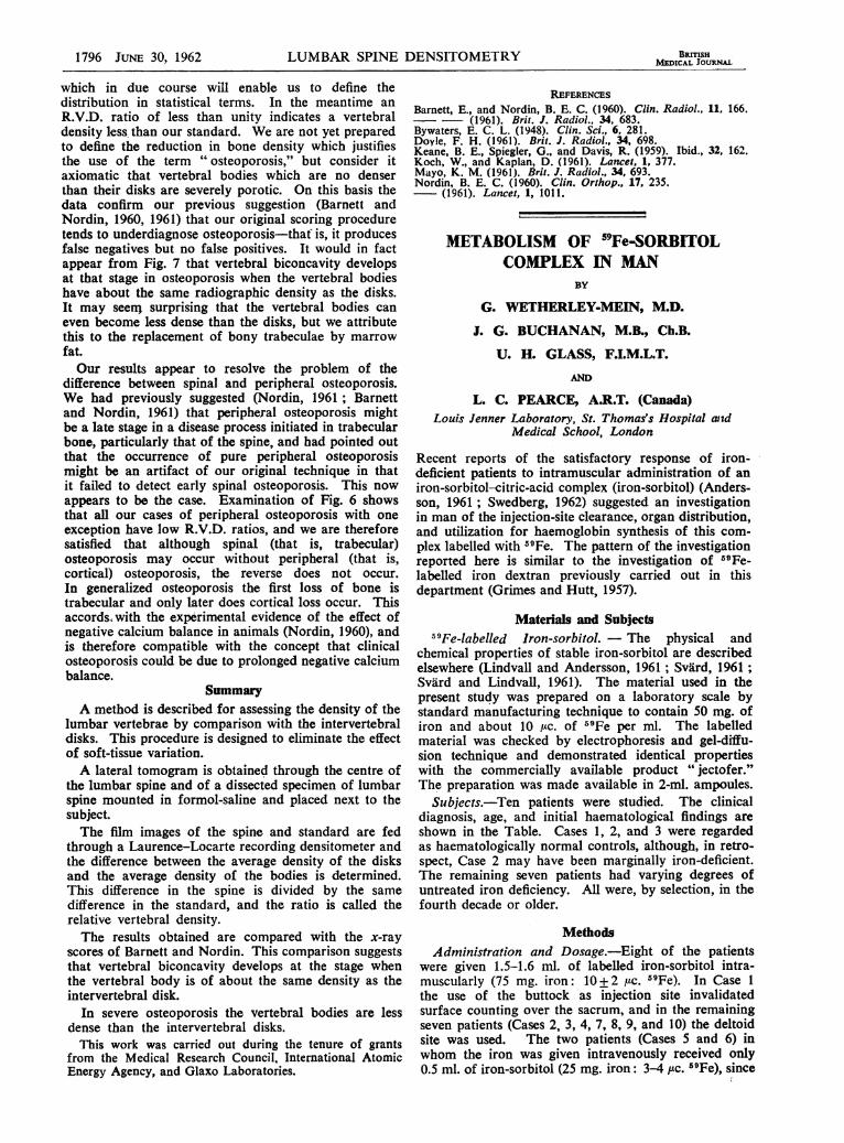
which in due course will enable us to define thedistribution in statistical -terms. In the meantime anR.V.D. ratio of less than unity indicates a vertebraldensity less than our standard. We are not yet preparedto define the reduction in bone density which justifiesthe use of the term " osteoporosis," but consider itaxiomatic that vertebral bodies which are no denserthan their disks are severely porotic. On this basis thedata confirm our previous suggestion (Barnett andNordin, 1960, 1961) that our original scoring proceduretends to underdiagnose osteoporosis-that is, it producesfalse negatives but no false positives. It would in factappear from Fig. 7 that vertebral biconcavity developsat that stage in osteoporosis when the vertebral bodieshave about the same radiographic density as the disks.It may seerm surprising that the vertebral bodies caneven become less dense than the disks, but we attributethis to the replacement of bony trabeculae by marrowfat.Our results appear to resolve the problem of the
difference between spinal and peripheral osteoporosis.We had previously suggested (Nordin, 1961 ; Barnettand Nordin, 1961) that peripheral osteoporosis mightbe a late stage in a disease process initiated in trabecularbone, particularly that of the spine, and had pointed outthat the occurrence of pure peripheral osteoporosismight be an artifact of our original technique in thatit failed to detect early spinal osteoporosis. This nowappears to be the case. Examination of Fig. 6 showsthat all our cases of peripheral osteoporosis with oneexception have low R.V.D. ratios, and we are thereforesatisfied that although spinal (that is, trabecular)osteoporosis may occur without peripheral (that is,cortical) osteoporosis, the reverse does not occur.In generalized osteoporosis the first loss of bone istrabecular and only later does cortical loss occur. Thisaccords.with the experimental evidence of the effect ofnegative calcium balance in animals (Nordin, 1960), andis therefore compatible with the concept that clinicalosteoporosis could be due to prolonged negative calciumbalance.
SummaryA method is described for assessing the density of the
lumbar vertebrae by comparison with the intervertebraldisks. This procedure is designed to eliminate the effectof soft-tissue variation.A lateral tomogram is obtained through the centre of
the lumbar spine and of a dissected specimen of lumbarspine mounted in formol-saline and placed next to thesubject.The film images of the spine and standard are fed
through a Laurence-Locarte recording densitometer andthe difference between the average density of the disksand the average density of the bodies is determined.This difference in the spine is divided by the samedifference in the standard, and the ratio is called therelative vertebral density.The results obtained are compared with the x-ray
scores of Barnett and Nordin. This comparison suggeststhat vertebral biconcavity develops at the stage whenthe vertebral body is of about the same density as theintervertebral disk.
In severe osteoporosis the vertebral bodies are lessdense than the intervertebral disks.This work was carried out during the tenure of grants
from the Medical Research Council, International AtomicEnergy Agency, and Glaxo Laboratories.
REFERENCES
Barnett, E., and Nordin, B. E. C. (1960). Clin. Radiol., 11, 166.-~- (1961). Brit. J. Radiol., 34, 683.
Bywaters, E. C. L. (1948). Clin. Sci., 6, 281.Doyle, F. H. (1961). Brit. J. Radiol., 34, 698.Keane, B. E., Spiegler, G., and Davis, R. (1959). Ibid., 32, 162.Koch, W., and Kaplan, D. (1961). Lancet, 1, 377.Mayo, K. M. (1961). Brit. J. Radio!., 34, 693.Nordin, B. E. C. (1960). Clin. Orihop., 17, 235.
(1961). Lancet, 1, 1011.
METABOLISM OF 9Fe-SORBITOLCOMPLEX IN MAN
BY
G. WETHERLEY MEIN, M.D.
L. G. BUCHANAN, M.B., Ch.B.
U. H. GLASS, F.I.M.L.T.AND
L. C. PEARCE, A.R.T. (Canada)Louis Jenner Laboratory, St. Thomas's Hospital and
Medical School, London
Recent reports of the satisfactory response of iron-deficient patients to intramuscular administration of aniron-sorbitol-citric-acid complex (iron-sorbitol) (Anders-son, 1961; Swedberg, 1962) suggested an investigationin man of the injection-site clearance, organ distribution,and utilization for haemoglobin synthesis of this com-plex labelled with 59Fe. The pattern of the investigationreported here is similar to the investigation of 5"Fe-labelled iron dextran previously carried out in thisdepartment (Grimes and Hutt, 1957).
Materials and Subjects59Fe-labelled Iron-sorbitol. - The physical and
chemical properties of stable iron-sorbitol are describedelsewhere (Lindvall and Andersson, 1961; Svard, 1961;Svard and Lindvall, 1961). The material used in thepresent study was prepared on a laboratory scale bystandard manufacturing technique to contain 50 mg. ofiron and about 10 Mc. of 59Fe per ml. The labelledmaterial was checked by electrophoresis and gel-diffu-sion technique and demonstrated identical propertieswith the commercially available product "jectofer."The preparation was made available in 2-ml. ampoules.
Subjects.-Ten patients were studied. The clinicaldiagnosis, age, and initial haematological findings areshown in the Table. Cases 1, 2, and 3 were regardedas haematologically normal controls, although, in retro-spect, Case 2 may have been marginally iron-deficient.The remaining seven patients had varying degrees ofuntreated iron deficiency. All were, by selection, in thefourth decade or older.
MethodsAdministration and Dosage.-Eight of the patients
were given 1.5-1.6 ml. of labelled iron-sorbitol intra-muscularly (75 mg. iron: 10+2 /c. 5"Fe). In Case 1the use of the buttock as injection site invalidatedsurface counting over the sacrum, and in the remainingseven patients (Cases 2, 3, 4, 7, 8, 9, and 10) the deltoidsite was used. The two patients (Cases 5 and 6) inwhom the iron was given intravenously received only0.5 ml. of iron-sorbitol (25 mg. iron: 3-4 ,uc. 59Fe), since
METABOLISM OF IRON-SORBITOL COMPLEX BRmSH 1797MEDICAL JOURNAL
Summary of Results in 10 Patients Receiving "Fe Sorbitol
%I.D.Case A] Dia|oh JHb Seru Injec- | %I.D. at .% I.D. in Utilization UtilizationNo. Age Diagnosis g./ M.C.H.C. FSem tson Excreted Injection Plasma of Retained /% I.D.No. ~~~~~100 ml. Fe Route in Urine Site T+ lODays I.D. Dose Unaccounted
_____________ ____ _____ _____ T+lODays T+lODays T+lODays
1 58 Myocardial infarct 14-9 33 _ I.M. 42-0 3-6 0 33 0 61 0 21-42 71 Angina .. .. 12-6 31 53 ., 34-5 6-3 0 27-8 46-9 31-43 49 Myocardial infarct .. 15*3 38 - , 23-0 4-4 2-1 26-9 38-1 40-64 36 Menorrhagia .. 10-3 26 66 ,, 31-1 4-0 0 41-7 64-3 23-25 76 Carcinoma of stomach 7-4 27 66 I.V. 10-6* - 0 40-8 *6 44 Menorrhagia .. .. 7-7 27 42 99 53 0 - 0 510 108-0 07 50 , .. 93 25 34 I.M. 33-4 10 0 49-6 75 5 16-08 45 Polyarthritis .. .. 12-6 30-7 66 32-2 3-5 0 37-0 57-5 27-39 82 Carcinoma of stomach 6-4 25 15 18.5* 2-0 2-3 48-0 _* _10 38 Haematuria .. . 8 0 29 54 ,. 183 0 0 66 5 74-9 15 2
* Incomplete urinary collection.
the toxicity of larger doses of the preparation whengiven in this way is not known. In neither patient wasthere any subjective or objective reaction. In all casesaliquots were retained for determining the activity ofthe injected dose.
Surface Counting.-Changes in activity over injectionsite, control site (opposite arm or buttock), heart, liver,spleen, and marrow (sacrum) were recorded at selectedtime intervals from T+ 2 minutes to T+10 days. Theapparatus and technique used have been previouslydescribed (Wetherley-Mein, Hutt, Langmead, and Hill,1956). The observed activities of each organ werecorrected for decay and counter variation and finallyexpressed as a percentage of the activity of that organat T+2 minutes.Plasma and Red-cell Activity.-Blood samples were
obtained at each surface-count determination throughoutthe period T+2 minutes to T+10 days. For eachsampling time 5 ml. of plasma and 5 ml. of haemolysedwhole blood were counted separately in a well-typescintillation counter. Blood volume was determined bya 5'Cr dilution technique (Mollison, 1960). Total plasmaand red-cell activities at each sampling-time were thencalculated and expressed as the percentage of theinjected dose circulating at that time.
Urinary Excretion.-All urinary samples were collec-ted during the period T + 0 to T + 48 hours. Time ofvoiding and volume of each sample were recorded and5-ml. aliquots were counted in the well-type scintillationcounter. The percentage of the injected dose excretedin each sample was determined. In two patients (CasesS and 9) urinary collection was incomplete.Plasma Iron.-This was determined by the method of
Bothwell and Mallett (1955).Other Haematological Data.-These were obtained
by standard techniques (Dacie, 1956).
ResultsClearance from Injection Site.-In Case 1 the use
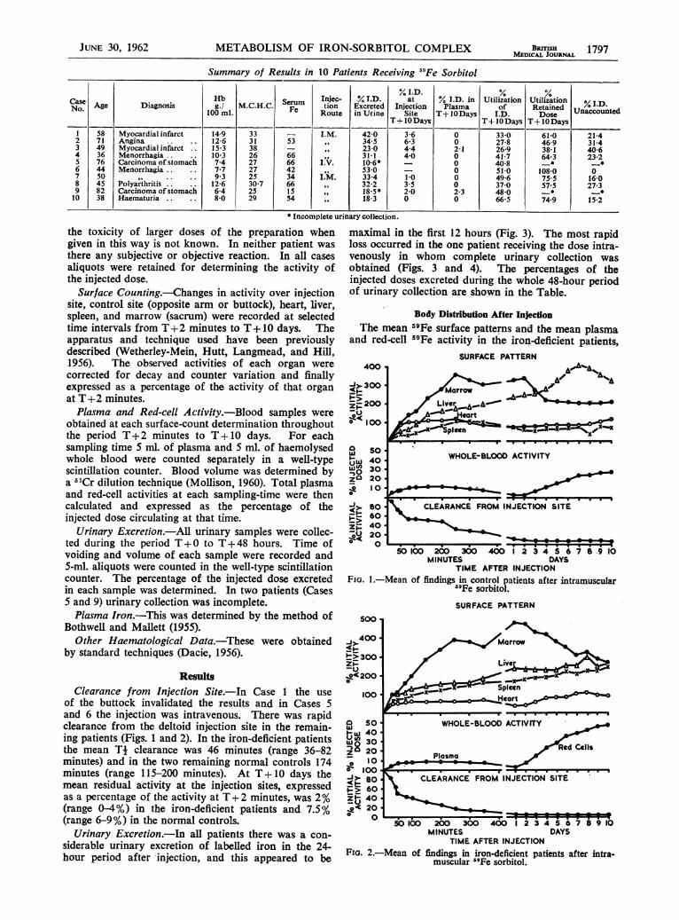
of the buttock invalidated the results and in Cases 5and 6 the injection was intravenous. There was rapidclearance from the deltoid injection site in the remain-ing patients (Figs. 1 and 2). In the iron-deficient patientsthe mean Ti clearance was 46 minutes (range 36-82minutes) and in the two remaining normal controls 174minutes (range 115-200 minutes). At T +10 days themean residual activity at the injection sites, expressedas a percentage of the activity at T + 2 minutes, was 2%(range 0-4%) in the iron-deficient patients and 7.5%(range 6-9%) in the normal controls.
Urinary Excretion.-In all patients there was a con-siderable urinary excretion of labelled iron in the 24-hour period after injection, and this appeared to be
maximal in the first 12 hours (Fig. 3). The most rapidloss occurred in the one patient receiving the dose intra-venously in whom complete urinary collection wasobtained (Figs. 3 and 4). The percentages of theinjected doses excreted during the whole 48-hour periodof urinary collection are shown in the Table.
Body Distribution After InjectionThe mean 59Fe surface patterns and the mean plasma
and red-cell 59Fe activity in the iron-deficient patients,SURFACE PATTERN
400 -
)-< 300 -
i>200zo 100
0
noI1-zo
zpBSC 0 l .
v -VI * @@50100 20o 300 400 1 2i3 45 6 7 8.9 10
MINUTES DAYSTIME AFTER INJECTION
FIG. 1.-Mean of findings in control patients after intramuscular"Fe sorbitol.
SURFACE PATTERN
Hea
WHOLE-BLOOD ACTIVITY
ed CellsPlasma
CLEARANCE FROM INJECTION SITE
50 100 200 300 400 2 3 4 5678910MINUTES DAYS
TIME AFTER INJECTIONFIG. 2.-Mean of findings in iron-deficient patients after intra-
muscular ""Fe sorbitol.
JUNE 30, 1962

1798 JUNE 30, 1962 METABOLISM OF IRON-SORBITOL COMPLEX
the control patients, and the patients who receivedintravenous iron-sorbitol are shown in Figs. 1, 2, and 5.Marrow Uptake.-In the patients receiving intra-
muscular doses (Figs. 1 and 2) the marrow curves arevery similar to those obtained in normal and iron-deficient patients receiving intravenous tracer doses of"Fe citrate (Ledlie and Baxter, 1954; Wetherley-Meinet al., 1956) and indicate an initial marrow accusnula-tion of iron as it is cleared from the plasma and asubsequent decrease in marrow concentration as thelabel enters the circulation incorporated in redcellhaemoglobin. Although quantitative interpretation ofthese surface patterns is difficult the findings suggest a
Lu
z
z
LAS
8
a0
I-
z
-0.0
42
40
38
36
34.
32
30'
28
26 -
24
22 o
20 -
I a8
16
14
12
10
84
2
I
I I
I II II'II
I
I II
I
I I'
I.
I ;I.,
, . )
URINARY EXCRETION
Iron-deificient Intramuscular o-opatients
Intravenous *- -_
Controls x.x
u
z
0O
IIIIIII
TIME IN HOURSFio. 3.-Urinary excretion of ""Fe sorbitol in all patients.
I NTRAMUSCULAR5 (CASE 4)
85ao57 S INTRAVENOUS70 (CASE 6)65 .
w
z
0
55 .50 .45 ..40 x35 .,30 -
25 -
205
105
0-0
TIME IN HOURS
FiG. 4.-Comparison of urinary excretion and plasma activity inpatients receiving "'Fe sorbitol.
greater deviation of iron to the marrow in the iron-deficient patients and to the liver in the controls.Plasma and Red-cell Activity.-Measurement of
activity in plasma and whole-blood samples showedlittle overlap of plasma and red-cell activity. Whole-blood activity in the first 24 hours after injection was
due essentially to isotope carried in the plasma andsubsequently to isotope in red cells. The curves repre-
senting plasma activity in the intravenous group (Fig. 5),presumably the resultant of urinary excretion andclearance to marrow and other sites, are virtuallyexponential and resemble the clearances observed afterintravenous 59Fe citrate. In both the intramusculargroups (Figs. 1 and 2) the plasma curves over the firstfive hours are modified by continued entry of label fromthe injection site and from T +1 hour to T+ 4 hours a
steady state obtained with constant plasma activity. Thered-cell-utilization curves (Figs. 1, 2, and 5) show, in all
groups, entry of labelled cells to the circulation at aboutT+ 24 hours, an initial phase of relatively rapid increasein activity, and, particularly in the control group,flattening of the curves by T+ 7 to T +10 days. Thefractions of the injected doses used for haemoglobin
120 1_ 2
100Z
80
60
40
20
0
SURFACE PATTERN
70 WHOLE-BLOOD ACTIVITY
60
50
wU 40I.-
30
Plasma Red Cells20
000
50 o00 200 300350 2 3 4 5 6 7 8 9
MINUTES i DAYSTIME AFTER INJECTION
FIG. 5.-Mean findings in iron-deficient patients receiving intra-venous 9Fe sorbitol.
synthesis (% utilization) by T+10 days are shown inthe Table. It can be seen that the percentage utilizationsin the iron-deficient group are al greater than any inthe control group. The t-test showed that the differencebetween the means of these groups was significant atthe 5% level.
Distribution to Lver
It was hoped that the surface counting data wouldgive some indication of the role of the liver in thehandling of the iron-sorbitol complex. The findings inthe intravenous group (Fig. 5) are unhelpful, since thesurface data are obviously dominated by the changesin plasma activity. In the intramuscular groups (Figs. 1and 2) there is, particularly in the normal controls, a
steady rise in liver activity during the period T+1 toT+6 hours. Since during this phase heart and plasma
BRmSHMEDICAL JOURNAL

Jur.113 30, 1962 METABOLISM OF IRON-SORBITOL COMPLEX
activity remains constant, it is probable that this repre-sents accumulation of isotope in the liver. Since wedid not feel that valid results could be obtained bycorrection of surface data for organ blood-low, nointerpretation of the liver curves during the period ofincreasing red-cell activity (T+2 to T+10 days) canbe made. However, in the Table it will be seen thatvarying proportions of the injected dose cannot beaccounted for in terms of urinary excretion, residue atinjection site, or appearance in red cells. It seemspossible that this moiety may be held in the liver orother reticulo-endothelial sites.
DiscussionAlthough the metabolism of injected 59Fe sorbitol
in general resembles that of 59Fe dextran and59Fe citrate there are certain important differenceswhich are probably determined by the low molecularweight and the initial stability of the iron-sorbitol-citric-acid complex in the plasma.After intramuscular injection the rates of removal of
the 59Fe-sorbitol complex and 59Fe dextran are strikinglydifferent. Grimes and Hutt (1957) found Ti buttockclearance times ranging from 10 to 40 hours as com-pared with the mean T- deltoid clearance times for 59Fesorbitol of 46 minutes (iron-deficient group) and 174minutes (controls) observed in this study. Grimes andHutt, unlike ourselves, found no difference in clearancerates between iron-deficient and control patients, but thenumbers of patients in each group in their series andours are probably too small for this difference to besignificant. Clearance of the 59Fe sorbitol from theinjection site is almost complete by 10 days (Table,Figs. 1 and 2), whereas Grimes and Hutt found that17-40% of the 59Fe dextran remained at the site after450-1,224 hours. Use and movement of the muscleinjected certainly affects absorption from it, and in ourpatients, at bed rest, the injection of a relatively smallvolume (1.5 ml.) into the deltoid might be expected toproduce more rapid absorption than the larger volumes(5 ml.) given in the buttock in the 59Fe dextran study.However, the findings in one patient (Case 1) whoreceived a buttock injection suggested that the use of thedeltoid site did not obviously modify the clearancerates, and it seems most probable that the differences inclearance of 59Fe sorbitol and 59Fe dextran are afunction of the different molecular weights of the twosubstances.
This view is supported by the results of studies onmechanisms of absorption from muscle in animals.Barnes and Trueta (1941) showed that toxins and snakevenoms with molecular weights of 5,000-20,000 weredirectly absorbed by the blood-stream while withmolecular weights greater than 20,000 absorption wasby lymphatics. The molecular weights of the iron-sorbitol complex are of the order of 5,000 (Eriksson,1961), and it has been established that this substance isprimarily absorbed by the capillaries (Svaird andLindvall, 1961), while the 59Fe dextran, with a molecularweight certainly greater than 20,000, is absorbedprimarily by lymphatics (Beresford, Golberg, and Smith,1957).Although after its absorption into the blood-stream
it seems that some fraction of the iron of the 59Fesorbitol complex becomes bound to transferrin (Svardand Lindvall, 1961), the greater part of it, like 59Fe
dextran, is stable and is cleared from the plasmaunchanged. In this respect both these substances differfrom the iron of "Fe citrate, virtually all, of which isimmediately bound to transferrin if this is available(Loeffler, Rappoport, and Coffins, 1955).Both the dextran and the citrate iron thus come to
exist in the plasma in high-molecular-weight complexesand, as such, are completely available for clearance fromthe plasma to marrow and reticulo-endothelial storagesites such as the liver. In contrast, although varyingproportions of the 59Fe-sorbitol complex are also clearedto marrow and liver, its existence in the plasmapredominantly as a substance of low molecular weightpermits its excretion in the urine. The relationshipbetween plasma activity and urinary loss (Fig. 4),particularly in the patient receiving it intravenously(Case 6), suggests that the rate of urinary excretion is adirect function of the plasma level. This is obviouslyof some importance in terms of optimal route andtherapeutic dose, since animal studies suggest thatincreasing doses are associated with increased plasmalevels (Lindvall and Andersson, 1961) and presumably,therefore, with increased urinary loss.The findings in the present study demonstrate
that, like 59Fe dextran and 59Fe citrate, the59Fe sorbitol not excreted in the urine moves fromplasma to marrow and probably to liver as well, itspartition between marrow and liver being possibly afunction of the degree of iron deficiency. Theutilization curves of all these preparations are similarand, in all, labelled iron appears in erythrocytes about24 hours after injection. The 10-day utilization oftracer doses of 59Fe citrate is between 70% and 85% incontrol patients and 100% in iron-deficient patients. Inthe previous iron-dextran and the present iron-sorbitolstudies, where therapeutic rather than tracer doses wereused, the 10-day utilization of the available fractionsof the injected dose was, not surprisingly, almostinvariably lower than this (see Table). The findingsin the present study and in animal studies suggest thatthe fractions of iron-sorbitol unaccounted for by T+ 10days are stored in the liver and possibly diffuse intotissue fluids (Lindvall and Andersson, 1961; S.Wahlqvist, 1962, personal communication).
Longer-term studies in untreated patients will benecessary to determine whether this stored fraction isreadily available for haemoglobin synthesis, but com-parison of the utilization curves of the iron-deficient andcontrol patients (Figs. 1 and 2) suggests that in theiron-deficient group maximal utilization has not occurredby T+ 10 days and that there is further utilization ofthe stored fraction.
SumnmayStudies with 59Fe-labelled iron-sorbitol complex in
man showed that after intramuscular injection there wasrapid clearance from the site of injection.The mean T{ deltoid clearance for 59Fe sorbitol vas
46 minutes in the iron-deficient group and 174 minutesin the controls. Clearance from the injection site wasalmost complete by ten days. These 59Fe sorbitolclearance times from the injection site are strikinglyshorter than those for iron dextran.
It seems probable that the differences in clearance of59Fe sorbitol and 59Fe dextran are a function of thedifferent molecular weights of the two substances.
JUNE 30, 1962 METABOLISM OF IRON-SORBITOL COMPLEX BRrrisH 1799MMICAL JOURNAL
1800 JUNE 30, 1962 METABOLISM OF IRON-SORBITOL COMPLEX
The low molecular weight of iron-sorbitol complexresulted in a rapid excretion in the urine. This wasmaximal in the,first twelve hours. The urinary excretionfor the 48-hour period after injection ranged from 18%to 53%.The red-cell utilization at ten days ranged from 27%
to 66 %. Utilization in the iron-deficient group wasgreater than in the control group. The utilization curvein the iron-deficient group showed that maximalutilization had not occurred by T+ 10 days.The surface counting data suggested that the retained
fraction of iron not utilized for haemoglobin synthesisby T+ 10 days (16% to 40%) was stored predominantlyin the liver.
We are grateful to Dr. S. Wahlqvist, of Astra, Sweden,for providing the "9Fe sorbitol. The expenses of theinvestigation were partly borne by the Endowment Fund,St. Thomas's Hospital.
REFERCBEsAnderssotn, N. S. E. (1961). Brit. med. J., 2, 275.Barnes, J. M., and Trueta, J. (1941). Lancet, 1, 623.Beresford, C. R., Golberg, L., and Smith, J. P. (1957). Brit. J.
Pharmacol., 12, 107.Bothwell, T. H., and Mallett, B. (1955). Biochem. J., 59, 599.Dacie, J. V. (1956). Practical Haematology, 2nd ed. Churchill,
London.Eriksson, F. R. (1961). Quoted by Svard and Lindvall (1961).Grimes, A. J., and Hutt, M. S. R. (1957). Brit. med. J., 2, 1074.Ledlie, E. M., and Baxter, C. F. (1954). Proceedings of 2nd
Radio-active Isotope Conference, p. 97. Butterworths,London.
Lindvall, S., and Andersson, N. S. E. (1961). Brit. J. Pharmacol.,17, 358.
Loeffler, R. K., Rappoport, D. A., and Collins, V. P. (1955).Proc. Soc. exp. Biol. (N.Y.), 88, 441.
Mollison, P. L. (1960). Recent Advances in Clinical Pathology,edited by S. C. Dyke, p. 223. Churchill, London.
Svard, P. O. (1961). J. Pharm. Pharmacol., 13, 641.- and Lindvall, S. (1961). Ibid., 13, 650.
Swedberg, B. (1962). Proceedings of 8th Congress of EuropeanSociety of Haematology, p. 254. Karger, Basel.
Wetherley-Mein, G., Hutt, M. S. R., Langmead, W. A., andHill. M. J. (1956). Brit. med. J., 1, 1445.
SARCOMA INDUCrION BY IRON-CARBOHYDRATE COMPLEXESBY
J. FIELDING, M.R.C.P., D.P.H1Consultant Haematologist, Paddington General Hospital, London
[WITH SPECIAL PLATE]
In order to administer iron parenterally withoutincurring the toxic effects of the ionized metal, ferrichydroxide has been bound to carbohydrates to formcomplexes of fairly high molecular weight. Thesecomplexes simulate the binding of iron by the beta-globulin transferrin, the physiological carrier of iron inthe circulation. The first clinically useful carbohydratecomplex was made with saccharose, later with dextransand dextrins, and more recently with sorbitol. In 1959Richmond reported that iron-dextran (" imferon ")induced sarcomas in rats after intramuscular injection inmassive doses. Haddow and Horning (1960) confirmedthis in both rats and mice. This unexpected observationrestimulated interest in the role which metals may playin carcinogenesis; it also demanded reconsideration ofthe use of iron-carbohydrate complexes in man for theparenteral treatment of iron deficiency.The discussion has centred mainly on the massive dose
of iron complex used to induce sarcomas in experimentalanimals. Golberg and his co-workers emphasized thesystemic effects of massive iron overload, which includereticulo-endothelial stimulation, altered tissue enzymeactivity, increased tissue peroxide and lipofuscin polymerformation, indicative of defective vitamin-E metabolism.They propose that the iron dosage given to experimentalanimals should be compared to the dosage used intreatment on a body-weight basis in order to assess thepossible tumour risk in man. They point out thatsimilar biochemical changes occur at the site of injection,and postulate that the bulk of tissue available forinjection relative to the amount injected is also relevant(Golberg et al., 1960; Baker et al., 1961).Haddow and Horning (1960) concluded that sarcoma
induction by iron-dextran is essentially a local action.This implies that the absolute amount of iron injectedis the determinate factor rather than the body-weight/dose ratio (Haddow, 1960).
Most experimental work so far has been with miceand rats, which are evidently highly susceptible species.Haddow and Horning record a single induced tumourin a hamster; they failed to produce tumours in guinea-pigs, but of three surviving rabbits in a group of sixtreated with massive iron-dextran one has produced atumour after a latent period of 39 months (Haddow,1961). Golberg and his co-workers (1960) failed toinduce tumours in dogs.
Previously published experimental findings in rats aresummarized in Table I. Total dosages of 250 mg. ormore of iron as iron-dextran given intramuscularly orsubcutaneously in divided doses into a single site pro-duce high yields of sarcomas varying from 55% to 92%of animals at risk. At such dose levels the sarcogenicaction of iron-dextran is evidently easily reproduciblein different laboratories with different strains ofanimal. The results of Golberg and his co-workers(1960) are of particular interest, since their experimentsdiffered in design from others: the doses were givenalternately into two limbs instead of into a single site.Injecting 116 mg. of iron as iron-dextran into each limb-that is, a total of 232 mg. Fe-they obtained only a5% yield of tumours, compared with the much higheryields obtained when a similar total dose is given into asingle site. There is perhaps evidence here which favoursthe " local action " hypothesis rather than the systemiceffect of iron overload as the significant factor in ironsarcogenesis. Table I also shows a comparison madeby Lundin (1962) of three iron complexes. He foundthat both iron-dextrin arid iron-dextran in high dosageproduced high yields of tumours in rats, whereas iron-sorbitol produced a single tumour, which he describedas a fibroma rather than a sarcoma. The significance ofthese results is discussed below.The object of the experiment described here was to
test the effect in mice of subcutaneous injection of a