metabolism of steroids tumop by - cancer research
TRANSCRIPT
[CANCER RESEARCH 29, 1647--1652, September 1969]
Metabolism of Steroids TumoP
by Transplantable Mouse Interstitial Cell
O. J. Lucis and R. Lucis
Department of Pathology, Dalhousie University, Halifax, Nova Scotia, Canada
SUMMARY
Biotransformation of steroid hormones by a spontaneous interstitial cell tumor originating from a BALB/cJ mouse has been studied in vitro. Tumor tissue grown in a female host converted estrone predominantly to estradiol-17~. The same tissue transformed estradiol-17/~ to estrone to a considerably lesser extent. Incubation of the tumor tissue with androstene- dione yielded testosterone as the principal conversion prod- uct and several other metabolites with chromatographic charac- teristics of l lB-hydroxytestosterone, 11~-hydroxyandrostene- dione, and 11-ketotestosterone. Interstitial cell tumors grown in male and female hosts transformed progesterone to testos- terone, 20a-hydroxypregn-4-en-3-one, androstenedione, and 11-deoxycorticosterone. The tumor tissue in vitro showed the presence of enzyme systems which are commonly found in testis tissue as well as steroid 11~/- and 21-hydroxylases which are normally localized in adrenocortical cells. Testis tissue from animals bearing interstitial cell tumor formed in vitro less testosterone from progesterone than testes from normal animals.
INTRODUCTION
Clifton et al. (2) have observed that spontaneous interstitial cell tumors arising from mouse testis on isologous transplanta- tion induced in the recipients effects which could be ascribed to androgen and adrenocortical steroid hypersecretion. It has been estimated by Andervont et at. (1) that the incidence of spontaneous interstitial cell tumors in BALB/c strain of mice is less than 1%. This strain, however, was found to be highly susceptible to the development of estrogen-induced interstitial cell tumors (1, 5). Huseby et al. (5) have shown that estrogen- induced interstitial cell tumors in BALB/c mice exhibit, in vitro, the presence of enzyme systems necessary for the forma- t ion of androgens and 11-deoxycorticosterone. In the present study, the biotransformation of steroids by a transplantable, spontaneous mouse interstitial cell tumor was investigated.
1 This study was supported by grants from the National Cancer Insti- tute of Canada and the Medical Research Council of Canada.
Received October 30, 1968; accepted April 24, 1969.
MATERIALS AND METHODS
Mouse interstitial cell Tumor D12344, originating from an animal of BALB/cJ strain, was obtained as a subcutaneous transplant in female hosts from the Jackson Laboratories, Bar Harbor, Maine. The tumor in the host animals became palpable 6 to 8 weeks after transplantation. The histologic structure of the tumor is shown in Fig. 1. In our laboratory, the tumor was further transplanted into 7- to 8-week-old male recipients of BALB/cJ strain. For transplantation, the tumor tissue was minced with scissors under sterile conditions and suspended in 0.9% sodium chloride. The suspended tumor mince was in- jected subcutaneously over the right scapular region of the recipient. Palpable tumors developed 7 - 8 weeks after the transplantation.
Tumors weighing 1 to 3 grams were used for incubation studies. For purposes of comparison, testes from normal BALB/cJ mice and from tumor-bearing animals of the same age were also incubated in vitro.
Immediately after dissection of the tumor and testis from ether-anesthetized animals, the tissues were minced with scis- sors. Weighed portions of tissue mince were suspended in flasks containing 10 ml of Krebs-Ringer bicarbonate buffer (pH 7.4) + 200 mg % of glucose and radioactive steroid precur- sor dissolved in 0.05 ml of propylene glycol. The tissue mince was added to the incubation medium which had been pre- heated to 36~ These preparations were incubated under at- mospheric air in a Dubnoff metabolic shaking incubator for three hours at 36~ After incubation the buffer solution was filtered through glass wool. The flask and the tissue were washed twice with 2.5 ml of Krebs-Ringer solution, and the washings were combined with the incubation medium. Radio- active steroids from the incubation medium were extracted three times with 30 ml of ethyl acetate, and the tissue was extracted with 50 ml of acetone overnight. The solvents were evaporated in a rotating flash evaporator under reduced pres- sure at temperatures below 50~ Obtained residue was redis- solved in 10.0 ml of ethanol, and aliquots of 0.1 ml were used for liquid scintillation counting. Total recoveries of the radio- activity after incubation ranged from 82 to 90%. The remain- ing extract was evaporated to dryness, and the residue was separated by paper chromatography using Whatman #1 chro- matography paper according to quantitative technics described by Neher (11).
Steroid transformation products were separated on the following paper chromatographic systems: (LPG) ligroine/
SEPTEMBER 1969 1647
Research. on January 5, 2019. © 1969 American Association for Cancercancerres.aacrjournals.org Downloaded from

O. J. Lucis and R. Lucis
propylene glycol; (TPG) toluene/propylene glycol; (E4) isooctane:tertiary butanol/methanol:water (100:45/45:10) volume ratios); (Bush A2) petroleum ether/methanol:water (100/70:307 ; (D) methylcyclohexane:toluene/methanol:water (100:25/80:20); (HBMe) hexane:benzene/methanol:water (20:80/70:30); and (MTMe) methylcyclohexane:toluene/ methanohwater (225:275/500:100). For further character- ization of the isolated products, thin-layer chromatography (TLC) was performed on silica gel plates (Eastman Kodak #6060) using the following solvent systems: A/B (acetone 50:benzene 50); H (benzene 80:ethanol 20); K (benzene 90:ethanol 10); N (benzene 95:ethanol 5). Labeled com- pounds on the chromatograms were located by autoradiog- raphy on X-ray frlm and also by strip scanning (8). Nonradio- active steroids with A4-3-keto grouping used as pilots or car- tiers were visualized under ultraviolet light. Estrogens were detected by the ferric chloride-potassium ferricyanide color reaction.
Radioactive areas from the chromatograms were cut out, hung on a bent 22-gauge hypodermic needle attached to a syringe, and eluted twice with 20 ml of ethanol. The eluate was collected in 250-ml round-bottom flasks, and the solvent was evaporated under reduced pressure in a rotating flash evap- orator. Residue obtained was dissolved in ethanol and aliquots ranging from 1 to 10% were used for liquid scintillation count- ing (10). Acetylation of isolated steroids was done with acetic anhydride in pyridine solution at room temperature for 18-24 hours (10). Chromatographically isolated radioactive steroids were further identified by crystallization to constant specific activity. Authentic nonradioactive crystalline steroid (10 to 29 mg) was combined with isolated radioactive compound and dissolved in either acetone, ethanol, or methanol (0.5 to 1.5 ml). To this solution hexane or water was added to obtain a saturated solution with respect to steroid. The solution was kept at 4~ until crystals were obtained. The mother liquor was separated from the crystals, and the crystals were dried in vacuum. Radioactivity associated with the crystals was deter- mined by liquid scintillation counting using accurately weighed portions (1.00 to 3.00 mg).
Radioactive steroids used for incubation studies were pur- chased from the New England Nuclear Corporation, Boston, Mass., and from the Radiochemical Center, Amersham, England. Prior to use, the radiochemical homogeneity of the precursors was verified by paper chromatography. The follow- ing radioactive steriods were used in this study: androstene- dione-4-14C (sp. act. 1 Vc = 5.7/ag), ll-deoxycorticosterone- 1,2-3H (sp. act. 1/~c = 0.015/.tg), estrone-16-vcC (sp. act. i pc = 26 #g), estradiol-17/3-4-14C (sp. act. 1/~c - 33 #g), progester- one-4-14C (sp. act. 1 #c = 5.6/.tg).
RESULTS
Conversion of Estrogens by Tumor Tissue
Tissue mince from two interstitial cell tumors, weighing 1.5 and 2.3 gm and grown in female hosts, was used for incuba- tion. The tissue preparation was well mixed, and 0.4-gm por- tions were added to incubation flasks containing radioactive
Table 1
Incubation medium Tissue
Chromatographically separated products I II I II
Polar compounds (%) 2.5 2.3 0.9 1.3 Estradiol-17/3 (%) 86.8 87.9 83.4 84.0 Estrone (%) 10.7 9.8 15.7 14.6 Total recovered radioactivity after
chromatography (dpm x 10 -4) 105.8 100.8 63.2 67.6 Radioactivity recovered in crude
extracts (dpm x 10 -4 ) 107.6 107.6 81.6 87.0
Conversion of estrone-16-14C by interstitial cell tumor tissue in vitro. Tissue weight, 0.4 gm; estrone-16-14C, 26 ~g (1 ~c). Time of incubation, 3 hours. I and II are duplicate tissue specimens.
precursor. In order to verify the homogeneity of the tissue enzymatic activity, the incubations were done in duplicate. After incubation of the tissue preparation for 3 hours with 26 /lg (1 /.tc) of labeled estrone, the tisstte showed a marked up- take of the radioactivity. Table 1. On chromatography in the MTMe system for 4.5 hours, the extracts were separated into three components. The polar material remained on the starting line and extended 2 cm below the starting line. Radioactive material corresponding to estradiol-17/3 had an R F of 0.21 and estrone had an R F of 0.55. As shown in Table 1, estradiol-17/3 appeared as the principal conversion product within the tissue as well as in the incubation medium. Chromatographically iso- lated estradiol-17/3 was further chromatographed in System D for 28 hours and the radioactive material travelled 9.0 cm from the starting line.
To confirm the identity of radioactive estradiol-17/3, an ali- quot was combined with 18.88 mg of nonradioactive estradiol- 17/3 and crystallized successively three times using acetone: hexane, acetone:water,,and methanol:water as solvents. The specific activity of the obtained crystals remained constant (18,100 dpm/mg; 17,800 dpm/mg, and 17,900 dpm/mg). The chemical nature of the polar compounds formed from estrone was not established.
Using tissue samples from the same tumor, incubation with radioactive estradiol-17/3 resulted in the formation of estrone as well as unidentified polar compounds which, in the MTMe system, remained on the starting line (Table 2). The extent of
Table 2
Incubation medium Tissue
Chromatographically separated products I II I II
Polar compounds (%) 2.4 2.3 0.6 0.6 Estradiol-17/~ (%) 93.6 93.9 93.2 93.9 Estrone (%) 4.0 3.8 6.2 5.5 Total recovered radioactivity after
chromatography (dpm x 10 -4) 62.1 64.4 48.2 51.0 Radioactivity recovered in crude
extracts (dpm x 10 -4) 95.3 99.7 72.2 75.2
Conversion of estradiol-17/g4-14 C by interstitial cell tumor tissue in vitro. Tissue weight, 0.4 gm; estradiol-17//-4A4C, 30.0/.tg (0.9 /.tc). Time of in- cubation, 3 hours.
1648 CANCER RESEARCH VOL. 29
Research. on January 5, 2019. © 1969 American Association for Cancercancerres.aacrjournals.org Downloaded from

Interstitial Cell Tumor and Steroid Hormones
estradiol-1713 dehydrogenation to estrone was 15 to 20 times lower than the conversion of estrone to estradiol-17~.
Radioactive estrone formed from estradiol-17~ was rechro- matographed in System D for 6 hours, where it had an R F of 0.28. After elution, an aliquot was combined with 19.22 mg of nonradioactive estrone and crystallized successively from ace- tone:hexane, acetone:water, and methanol:water. Specific ac- tivities of estrone crystals remained constant (1,580 dpm/mg; 1,590 dpm/mg, and 1,570 dpm/mg).
Conversion of Androstenedione by Tumor Tissue
Transformation of C 19 steroids was investigated in vitro us- ing tissue from a single tumor grown in a female host. Tissue mince (0.4 gin) was incubated with 5.7/2g of labeled andro- stenedione. The crude extracts from the incubation medium and from the tissue, on chromatography in the Bush A 2 sys- tem, revealed that testosterone was the major conversion prod- uct (R F 0.437 (Table 37. Radioactive testosterone formed from androstenedione was identified by crystallization with authentic carrier using three different solvent mixtures (ace- tone:hexane; acetone:water, and methanol:water). Specific ac- tivities of the crystals obtained remained constant (12,500 dpm/mg, 12,510 dpm/mg, and 12,560 dpm/mg).
Radioactive material more polar than testosterone in the Bush A 2 system remained on the starting line. After elution this material was combined with 50 /~g of 11~-hydroxy- testosterone (11~-OH-T), 1113-hydroxyandrostenedione (11~- OH-A), and 11-ketotestosterone (11-K-T). On chromatography in the HBMe system, radioactivity coincided with the carriers; the R F was 0.53 for 11/~-OH-T, 0.75 for 11-keto-T, and 0.89 for 11~-OH-A. For further characterization, ll/3-OH-T was re- chromatographed on two-dimensional TLC using systems K and A/B where the RF's were 0.34 and 0.36 and radioactivity coincided with the carrier. This material was eluted and acety- lated to 1113-OH-testosterone acetate. When rechromato- graphed two dimensionally on the TLC systems H and K, the radioactivity remained with the acetylated carrier having RF'S of 0.55 and 0.37. Radioactive substance with chromatographic characteristics of 11/3-OH-A on two dimensional TLC systems K and A/B had the same mobility as the authentic carrier (RF'S of 0.47 and 0.61). Also the radioactive 11-K-T on two- dimensional TLC in system K and system N moved together with the carrier (RF'S of 0.34 and 0.22). Shortage of authen-
Table 3
Chromatographically separated products Incubation medium Tissue
Polar compounds (%) Testosterone (%) Androstenedione (%) Total recovered radioactivity after
chromatography (dpm x 10 -4 ) Radioactivity recovered in crude
extracts (dpm x 10 -4)
29.4 11.0 60.2 77.8 10.4 11.2
106.3 51.0
141.0 64.2
Conversion of androstenedione-4 -14 C by interstitial cell tumor tissue in vitro. Tissue weight, 0.4 gm; radioactive substrate, 5.7 btg (1 btc). Time of incubation, 3 hours.
tic steroids precluded further identification of the l l-oxygen- ated derivatives.
Conversion of Progesterone by Tumor Tissue
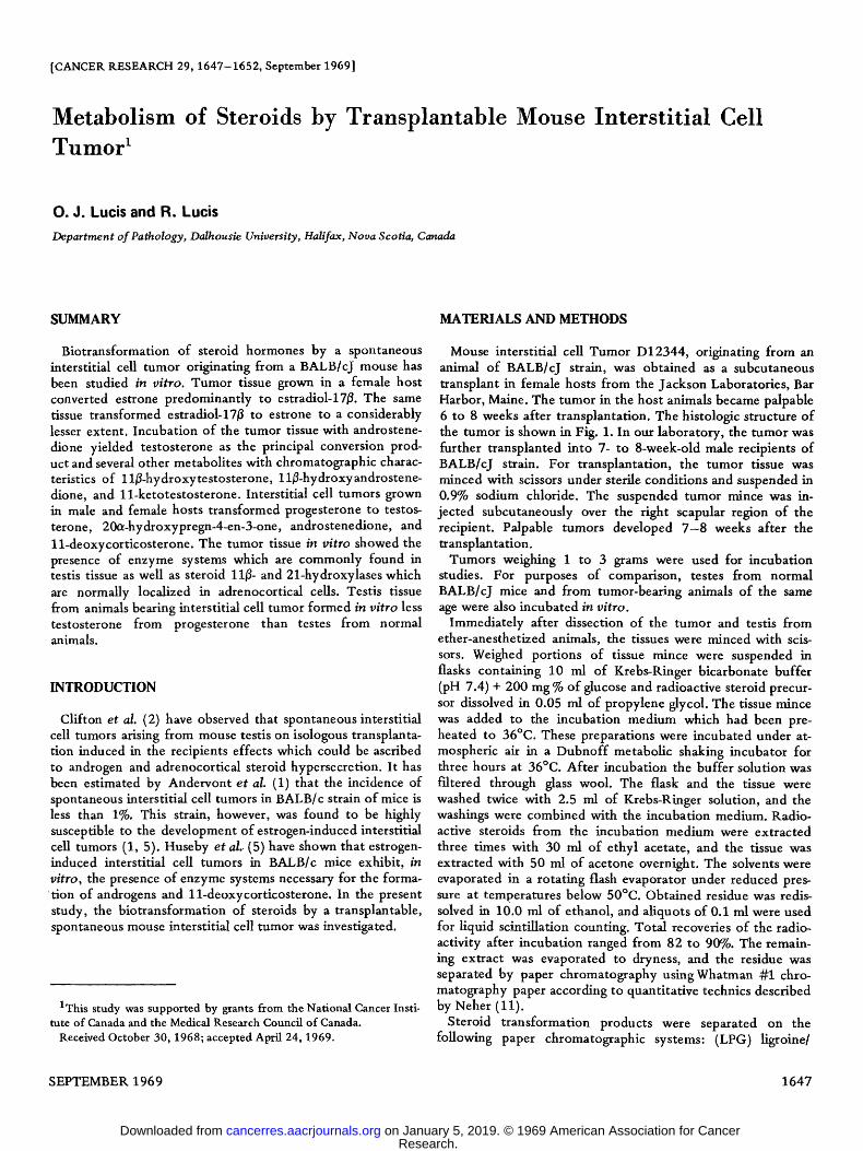
Duplicate tissue specimens from tumors grown in male and female hosts were each incubated with 1/.tc of progesterone. After incubation and extraction the total recovered radioactiv- ity in the case of tumor samples from male hosts was 82.4 and 84.4%; in the case of tumor specimens from female hosts, it was 87.0 and 87.7%. The crude extracts were combined with 50 gg of nonradioactive testosterone, DOC, 17whydroxypro- gesterone (17a-OH-P), 20~-hydroxypregn-4-en-3-one, and an- drostenedione. On chromatography in the LPG system for 28 hours, the extracts showed a dense radioactive area on the starting line. Testosterone and 174-OH-P carriers moved to- gether 10.1 cm from the starting line, and the carriers were associated with radioactivity. DOC travelled 14.7 cm and 20a-hydroxypregn-4-en-3-one 24.0 cm. Both carriers coincided with the radioactivity. Overflows collected from these chroma- tograms were rechromatographed on the LPG system for 5 hours when androstenedione travelled 10.8 cm and progester- one 24.0 cm. From the original chromatogram the testoster- one fraction containing 174-OH-P was acetylated to form testosterone acetate. After acetylation 17c~-OH-P was sepa- rated from testosterone acetate on the Bush A 2 system where 174-OH-P had an R F of 0.41 and testosterone acetate an R F of 0.92. The distribution of radioactivity in the transformation products recovered after chromatography in LPG and Bush A 2 systems is shown in Chart 1. Tumor tissues obtained from female as well as male hosts metabolized progesterone in a remarkably similar way. From the transformation products, DOC appeared as the principal steroid followed by testoster- one, 20a-hydroxypregn-4-en-3-one, androstenedione, and 174-hydroxyprogesterone. These conversion products were further characterized by repeated chromatography and crystal- lization with authentic carriers. Isolated 14C-labeled DOC was combined with 3H-labeled DOC and acetylated to DOC ace- tate. On repeated chromatography in Systems D, E4, and TPG, the respective R F values were 0.81, 0.75, and 0.87. The 3H/14C ratios remained constant (4.6, 4.6, and 4.7). Testos- terone was identified as testosterone acetate by crystallization with authentic carrier using acetone:hexane, acetone:water, and methanol:water as solvents. Specific activities of the crystals remained constant (5,470 dpm/mg, 5,470 dpm/mg, and 5,500 dpm/mg). Isolated radioactive 20a-hydroxypregn- 4-en-3-one was repeatedly chromatographed with authentic cartier in Systems D, TPG, and E 4 where this compound re- mained together with the carrier and had the corresponding R F values 0.40, 0.60, and 0.55. An aliquot of isolated 20a-hydroxypregn-4-en-3-one was combined with 29.7 mg of authentic nonradioactive carrier and crystallized using metha- nol:water, acetone:water, and ethanol:water as solvents. The specific activities of the crystals remained constant (7,500 dpm/mg, 7,700 dpm/mg, and 7,700 dpm/mg). Androstenedi- one was characterized by chromatography in LPG and Bush A 2 systems only.
The radioactive products more polar than testosterone sepa- rated from the crude extracts in the LPG system were further
SEPTEMBER 1969 1649
Research. on January 5, 2019. © 1969 American Association for Cancercancerres.aacrjournals.org Downloaded from
O. J. Lucis and R. Lucis
"POLAR"
DOC
MALE HOST
20a - OH - P
17 a - OH - P l
TESTOSTERONE ' ~
ANDROSTENEDIONE I
PROGESTERONE
FEMALE HOST
1
i0 o io /o RADIOACTIVE PRODUCTS
Chart 1. Conversion of progesterone-4-14C by mouse interstitial cell tumor tissue from male and female hosts. Tissue weight, 0.32 gm; pro- gesterone-4-14C, 5.6 /ag (1 /~c). Time of incubation, 3 hours. Each bar represents the mean of duplicate incubations. See Appendix for abbrev- iations.
TESTES TESTES NORMAL ANIMALS TUMOR-BEARING ANIMALS
"POLAR" L
20a-OH- P
17a-OH- P [
TESTOSTERONE [
ANDROSTENEDIONE F
PROGESTERONE !
1
~ : . > : i : ~ " .::.".'.:'i:~
RADI OACTIVE PRODUCTS
Chart 2. Conversion of progesterone-4-14C by mouse testes from normal and interstitial cell, tumor-bearing animals. Tissue weight, 0.3
14 gin; progesterone-4- C, 5.6 #g (1 #c). Time of incubation, 3 hours. See Appendix for abbreviations.
chromatographed on HBMe system. Four radioactive areas were obtained RF'S of 0.17, 0.54, 0.71, and 0.83. The chemi- cal identity of these products was not established.
Conversion of Progesterone by Mouse Testis Tissue
Tissue mince was prepared from testes of normal and tumor- bearing animals and incubated with 1 /~c of progesterone- 4A4C. After incubation and extraction, 90.7% of the radioac- tivity was recovered from the normal mouse testis sample and 84.5% from the other specimen. The crude extracts were sepa- rated by the same procedure as described in the interstitial cell tumor experiments. The distribution of radioactivity in the transformation products obtained after chromatography in LPG and Bush A 2 systems is shown in Chart 2. Testis tissue from tumor-bearing animals exhibited a lower activity in pro- gesterone utilization and testosterone formation than testes from normal mice. Apart from testosterone the testis tissue formed androstenedione, 170~-OH-P, and a very small amount of 200~-hydroxypregn-4-en-3-one (20~-OH-P). The polar frac- tion derived from progesterone precursor which remained on the starting line in the LPG system was not further character- ized. The presence of DOC on the chromatograms was not detected.
DISCUSSION
Mouse interstitial cell tumor tissue in vi tro transforms ster- oid hormones in a pattern which partly exhibits characteristics of the testis tissue and partly resembles the features of the adrenal cortex. Such dual characteristics of the spontaneous mouse interstitial cell tumor are very similar to those of estro- gen-induced interstitial cell tumors described by Dominguez et al. (3) and Huseby et al. (5). The similarity of this interstitial cell tumor tissue with the testis tissue lies in the presence of enzyme systems which transform progesterone to testosterone. Apart from this biosynthetic pathway the tumor tissue exhi- bits active steroid 11/~-and 21-hydroxylase activities. The lat- ter enzyme systems are normally found in the cells of the adrenal cortex. Recently Inano et al. (6) have demonstrated that a spontaneous mouse interstitial cell tumor originating from an animal of ddN/KF strain contained, in addition to 113- and 21-hydroxylase activities, also a steroid 18-hy- droxylase enzyme system. It has been observed by Dominguez et al. (3) that homogenates from testes of BALB/c mice, upon incubation with radioactive progesterone, yield small quanti- fies of labeled DOC. Similar results have been obtained with rat testis tissue homogenates (3). These observations sug- gest that normal testis tissue contains a detectable steroid 21-hydroxylase activity. In the present study, using mouse testis tissue mince, a conversion of progesterone to DOC could not be demonstrated. This discrepancy could have resulted from the preparations that were used for incubation. In a tis- sue mince, the cellular integrity remains largely preserved, and the interaction of 21-hydroxylase with progesterone in such a system might be restricted. In a tissue homogenate, on the other hand, cellular structure is disrupted and the intracellular enzyme systems disorganized. This, as well as an addition of
1650 CANCER RESEARCH VOL. 29
Research. on January 5, 2019. © 1969 American Association for Cancercancerres.aacrjournals.org Downloaded from

In ters t i t ia l Cell T u m o r a n d S t e r o i d H o r m o n e s
exogenous cofactors, would allow the steroid 21-hydroxylase to act upon progesterone and form DOC. As far as it is known, the steroid 11/~-hydroxylase activity has not been demon- strated in normal mammalian testis tissue (7). The presence of this enzyme system has been detected in the tumor tissue described in the present study. Previous investigations of ster- oid biosynthesis by estrogen-induced mouse interstitial cell tu- mors have also revealed that the tissue contains steroid 11~-hydroxylase activity (3, 5). In common with mouse tu- mors, several human interstitial cell neoplasms studied in vi tro have shown the presence of steroid 11~hydroxylase (12, 13).
The mechanism by which the steroid 11/Lhydroxylase ap- pears in interstitial cell neoplasms is not clearly understood. It is possible that during the process of tumorigenesis a disorgan- ization of the enzyme-synthesizing process has taken place, inducing the production of 11~- and 18-hydroxylases. The appearance of active steroid 21-hydroxylase in mouse intersti- tial cell tumors may also be due to an increased synthesis of this enzyme. It has been pointed out by Dominguez and Huseby (4) that, in estrogen-induced testicular interstitial cell tumors, the activity of 20c~-hydroxydehydrogenase enzyme is increased. Prominence of this enzyme is also demonstrable in the tumor tissue used in the present study. In the mouse testis tissue the reduction of progesterone to 20a-hydroxypregn- 4-en-3-one was found to be very low. The functional signifi- cance of 20~-hydroxydehydrogenase in the testis and in the neoplastic tissue is still obscure.
Studies on the patterns of estrogen metabolism by mouse interstitial cell tumor have revealed that this tissue possesses enzyme systems which favor a reduction of estrone to estradi- o1-17~. This pattern of estrogen transformation is similar to that observed with rat and human testis tissue (9). Testis tissue from mice bearing transplanted interstitial cell tumor utilized, in vitro, less progesterone for the formation of testosterone than testes from normal mice. This suggests that the steroids secreted by the tumor in vivo may have suppressed the testicu- lar steroidogenesis.
ACKNOWLEDGMENTS
The authors wish to thank Dr. J. A. Embil, Jr., for his help in tumor transplantations and Mr. F. Stefani for the preparation of the photo- micrograph.
APPENDIX
The following trivial names and abbreviations are used in this paper: estradiol-17#, 3,17/~-dihydroxyestra-l,3,5(10)-triene; estrone, 3-hydrox- yestra-l,3,5(10)-trien-17-one; testosterone, 17#-hydroxyandrost-4-en- 3-one; 11#-hydroxytestosterone (ll#-OH-T), 11#,17/3-dihydroxyan- drost-4-en-3-one; 11-ketote stosterone (11-K-T), 17#-hydroxyandrost-4-
ene-3,11-dione; androstenedione, androst-4-ene-3,17-dione; 11#-hy- droxyandrostenedione (11#-OH-A), 11#-hydroxyandrost-4-ene-3,17- dione; progesterone, pregn-4-ene-3,20-dione; 170~hydroxyprogesterone (1701-OH-P), 170~-hydroxy-pregn-4-ene-3,20-dione; 2001-OH-P, 200l-hy- droxypregn-4-en-3-one; ll-deoxycorticosterone (DOC), 21-hydroxy- pregn-4-en-3,20.dione.
REFERENCES
1. Andervont, H. B., Shimkin, M. B., and Canter, H. Y. Susceptibility of Seven Inbred Strains and the F 1 Hybrids to Estrogen-Induced Testicular Tumors and Occurrence of Spontaneous Testicular Tu- mors in Strain BALB/c Mice. J. Natl. Cancer Inst., 25: 1069-1081, 1960.
2. Clifton, K. H., Bloch, E., Upton, A. C., and Furth, J. Transplant- able Leydig-Cell Tumors in Mice. Arch. Pathol., 62: 354--368, 1956.
3. Dominguez, O. V., Acevedo, F., Huseby, R. A., and Samuels, L. T. Steroid 21-Hydroxylase in Normal Testes and Malignant Interstitial Cell Tumors. J. Biol. Chem., 235: 2608--2612, 1960.
4. Dominguez, O. V., and Huseby, R. A. Heterogeneity of Induced Testicular Interstitial Cell Tumors of Mice as Evident by Steroid Biosynthetic Enzyme Activities. Cancer Res., 28: 348--353, 1968.
5. Huseby, R. A., Dominguez, O. V., and Samuels, L. T. Testes Func- tion. Function of Normal and Abnormal Testicular Interstitial Cells in the Mouse. Recent Progr. Hormone Res., 17: 1--51, 1961.
6. Inano, H., Machino, A., and Tamaoki, B. I. Steroid Biosynthesis in vitro by Transplantable Cell Tumor of Mice. I. Identification and Quantitative Determination of the Metabolites and Intracellular Distribution of the Enzymes Related to Testosterone Formation. Endocrinology, 83: 659-670, 1968.
7. Lipsett, M. B., Sarfaty, G. A., Wilson, H., Bardin, C. W., and Fish- man, L. M. Metabolism of Testosterone and Related Steroids in Metastatic Interstitial Cell Carcinoma of the Testis. J. Clin. Invest., 45: 1700-1708, 1966.
8. Lucis, O. J., McFarlane, E. S., Embil, J. A., Jr., and Lucis R. Transformation in Cell Culture of Sex Hormones by Adeno-virus- 12-induced Tumor Cells and Normal Hamster Lung Fibroblasts. Cancer Res., 28: 1513--1519, 1968.
9. Lucis, O. J., and Lucis, R. Metabolism ofC18 and C19 Steroids by Testis Tissue. Gen. Comp. Endocrinol., 12: 63--71, 1969.
10. Lucis, R., Carballeira, A., and Venning, E. H. Biotransformation of Progesterone-4-14C and 11-deoxy-corticosterone-4-14C by Rat Adrenal Glands in vitro. Steroids, 6: 737-756, 1965.
11. Neher, R. Paper Chromatography. In: Steroid Chromatography, Chap. 4, pp. 87-238. Amsterdam: Elsevier Publishing Company, 1964.
12. Savard, K., Dorfman, R. I., Baggett, B., Fielding, L. L., Engels, L. L., McPherson, H. T., Lister, L. M., Johnson, D. S., Hamblen, E. C., and Engel, F. L. Clinical, Morphological and Biochemical Studies of a Virilizing Tumor in the Testis. J. Clin. Invest., 39: 534-553, 1960.
13. Smith, E. R., Breuer, H., and Schriefers, H. A Study of the Steroid Metabolism of an Interstitial-Cell Tumor of the Testis. Biochem. J., 93: 583-587, 1964.
SEPTEMBER 1969 1651
Research. on January 5, 2019. © 1969 American Association for Cancercancerres.aacrjournals.org Downloaded from

O. J. Lucis and R. Lucis
Fig. 1. Mouse interstitial cell tumor grown in female host. Hematoxylin and eosin, x 400.
1652 C A N C E R R E S E A R C H V O L . 29
Research. on January 5, 2019. © 1969 American Association for Cancercancerres.aacrjournals.org Downloaded from
1969;29:1647-1652. Cancer Res O. J. Lucis and R. Lucis Cell TumorMetabolism of Steroids by Transplantable Mouse Interstitial
Updated version
http://cancerres.aacrjournals.org/content/29/9/1647
Access the most recent version of this article at:
E-mail alerts related to this article or journal.Sign up to receive free email-alerts
Subscriptions
Reprints and
To order reprints of this article or to subscribe to the journal, contact the AACR Publications
Permissions
Rightslink site. Click on "Request Permissions" which will take you to the Copyright Clearance Center's (CCC)
.http://cancerres.aacrjournals.org/content/29/9/1647To request permission to re-use all or part of this article, use this link
Research. on January 5, 2019. © 1969 American Association for Cancercancerres.aacrjournals.org Downloaded from