metal ions in biological system and potential...
TRANSCRIPT

Metal ions in biological system
and Potential Medicine
Avinash Kumbhar
University of Pune,Pune

Physics
BioinorganicChemistry
Pharm
aco
logy
Bioch
emistry
Bioinorganic Chemistry is a very difficult subject
Bioinorganic Chemistry is a highly interdisciplinary research field

What is Bioinorganic Chemistry?
It’s a study of the role of naturally occurring metal ions in biological systems
as well as the role of externally introduced metal ions.
To search the answers for the following questions-
Which metal ions are used in biological systems?
How nature chose these elements?
How do these elements get into the cells?
How the concentration of these elements are regulated?
How these metals bind to biopolymers?
How these metals help in folding the biopolymers?

How these metals help in folding the biopolymers?
How are these metals inserted into the active site?
What are the major roles of metal ions in biological
systems?
Which metal ions play a role in medicinal chemistry?
Which metal ions are toxic to biological systems?
The study of metal ions with the aim of understanding
the life processes.

Conclusions derived from the above periodic table:
1. “Chemistry of life “ is the chemistry of lighter elements”
2. Biological elements have been selected from practically all
groups and
sub groups except IIIA and IVA and inert gases.

Chemical elements essential to life forms can be divided into the following
(i) Bulk elements: C, H, N, O, P, S
(ii) Macro minerals and ions: Na, K, Mg, Ca, Cl, PO43-, SO4
2-
(iii) Trace elements: Fe, Zn, Cu
(iv) Ultratrace elements comprises of
(a) non-metals: F, I, Se, Si, As, B
(b) metals: Mn, Mo, Co, Cr, V, Ni, Cd, Sn, Pb, Li

Essentiality of elements is defined by,
(1) A physiological deficiency appears when the element is removed from
the diet
(2) The deficiency is relieved by the addition of that element to the diet
(3) A specific biological function is associated with the element

Why are metal ions important in biology ?
Catalysing reactions via:
– Hydrolytic e.g. carbonic anhydrase, carboxypeptidase
– Substrate transfer e.g. haemoglobin, myoglobin
– Electron transfer e.g. cytochrome C oxidase
– Thermodynamic and kinetic considerations
Stabilising structure:
– Protein
– DNA
– Skeletal
Charge balancing
– Osmotic balance
– Nerve function
Replication and information encoding


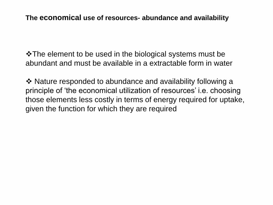
The economical use of resources- abundance and availability
The element to be used in the biological systems must be
abundant and must be available in a extractable form in water
Nature responded to abundance and availability following a
principle of ‘the economical utilization of resources’ i.e. choosing
those elements less costly in terms of energy required for uptake,
given the function for which they are required


Subdivisions of Bioinorganic Chemistry
Natural selection of elements-related to evolution
Economical use of resources.
Biological environment and elemental ability.
Homeostasis.
Biomimetic Chemistry
Structural and functional modeling of enzymes and proteins.
Medicinal Inorganic Chemistry
Synthesis and application of inorganic compounds as
drugs and diagnostic agents.
Toxicology of metals and metalloids.
Chemical Nucleases
Biomineralization
Controlled assembly of advanced materials in biology

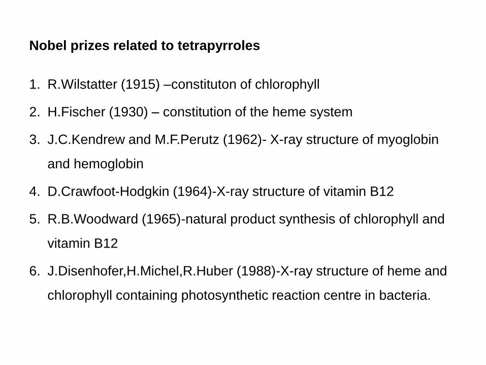
Nobel prizes related to tetrapyrroles
1. R.Wilstatter (1915) –constituton of chlorophyll
2. H.Fischer (1930) – constitution of the heme system
3. J.C.Kendrew and M.F.Perutz (1962)- X-ray structure of myoglobin
and hemoglobin
4. D.Crawfoot-Hodgkin (1964)-X-ray structure of vitamin B12
5. R.B.Woodward (1965)-natural product synthesis of chlorophyll and
vitamin B12
6. J.Disenhofer,H.Michel,R.Huber (1988)-X-ray structure of heme and
chlorophyll containing photosynthetic reaction centre in bacteria.


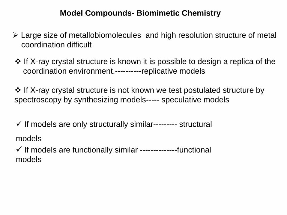
Model Compounds- Biomimetic Chemistry
Large size of metallobiomolecules and high resolution structure of metal
coordination difficult
If X-ray crystal structure is known it is possible to design a replica of the
coordination environment.----------replicative models
If X-ray crystal structure is not known we test postulated structure by
spectroscopy by synthesizing models----- speculative models
If models are only structurally similar--------- structural
models
If models are functionally similar --------------functional
models

• Biomimetic approach has helped in the study of
1. Assignment or verification of the metal oxidation states
2. effects of distance and medium on electron transfer rates
3. role of steric and electronic factors
4. Identity of likely intermediates of enzyme catalyzed reactions
Strategy for models complexes- spontaneous self assembly
Nature adopted the a similar strategy based on available
chemistry in the geosphere during evolution .

[1] Catalysis
Oxidoreductases
Amine oxidase
Ammonia monooxygenase
Ascorbate oxidase
Ceruloplasmin
Cu,Znsuperoxide dismutase
Cytochrome c oxidase
Diamine oxidase
Dopamine ßhydroxylase
Galactose oxidase
Laccase
Lysyl oxidase
Methane monooxygenase
N2O reductase
Nitrite reductase
Peptidylglycinehydroxylating monooxygenase
Phenylalanine hydroxylase
Tyrosinase
Ubiquinone oxidase
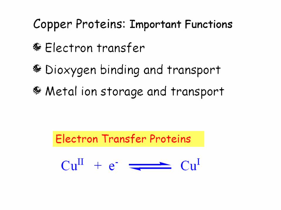
[2] Electron transfer
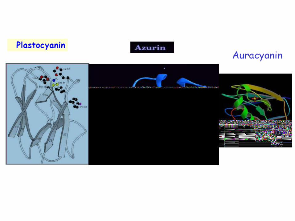
Auracyanin
Azurin
Phytocyanin family
Plastocyanin family
Rusticyanin
Copper proteins by function
Biomimetic chemistry

Copper –An alternative to biological iron
Function Fe protein Cu protein
O2 transport Hemoglobin (h)
Hemerythrin (nh)
Hemocyanin
oxygenation Cytochrome P-450(h)
Methane monooxygenase(nh)
Catechol dioxygenase nh)
tyrosinase
oxidase Peroxidases(h) Amine oxidases
laccase
Electron transfer Cytochromes (h) Blue copper
proteins
Antioxidative Peroxidases (h)
Bacterial SOD (nh)
SOD
NO2- reduction Nitrite reductase(h) Nitrite reductase


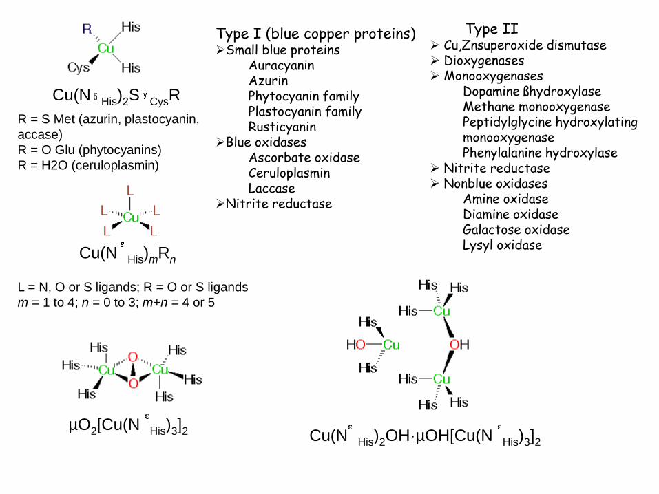
Cu(N His)2S CysR
R = S Met (azurin, plastocyanin,
accase)
R = O Glu (phytocyanins)
R = H2O (ceruloplasmin)
Cu(N His)mRn
L = N, O or S ligands; R = O or S ligands
m = 1 to 4; n = 0 to 3; m+n = 4 or 5
µO2[Cu(N His)3]2 Cu(N His)2OH·µOH[Cu(N His)3]2
Type I (blue copper proteins)Small blue proteins
Auracyanin Azurin Phytocyanin family Plastocyanin family Rusticyanin
Blue oxidases Ascorbate oxidase Ceruloplasmin Laccase
Nitrite reductase
Type II Cu,Znsuperoxide dismutase Dioxygenases Monooxygenases
Dopamine ßhydroxylase Methane monooxygenase Peptidylglycine hydroxylating monooxygenase Phenylalanine hydroxylase
Nitrite reductase Nonblue oxidases
Amine oxidase Diamine oxidase Galactose oxidase Lysyl oxidase


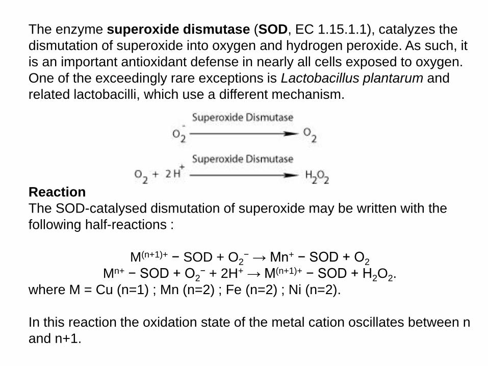
The enzyme superoxide dismutase (SOD, EC 1.15.1.1), catalyzes the
dismutation of superoxide into oxygen and hydrogen peroxide. As such, it
is an important antioxidant defense in nearly all cells exposed to oxygen.
One of the exceedingly rare exceptions is Lactobacillus plantarum and
related lactobacilli, which use a different mechanism.
Reaction
The SOD-catalysed dismutation of superoxide may be written with the
following half-reactions :
M(n+1)+ − SOD + O2− → Mn+ − SOD + O2
Mn+ − SOD + O2− + 2H+ → M(n+1)+ − SOD + H2O2.
where M = Cu (n=1) ; Mn (n=2) ; Fe (n=2) ; Ni (n=2).
In this reaction the oxidation state of the metal cation oscillates between n
and n+1.


Cu2+ + O2-· → Cu+ + O2
Cu+ + O2-· + 2H+ → Cu2+ + H2O2
Net Reaction: 2 O2-· + 2H+ → H2O2 + O2
K=(~105 M-1 s-1 at pH 7)

Manganese(II) ions function as cofactors for a number of enzymes and the
element is thus a required trace mineral for all known living organisms.
Biological role:
The classes of enzymes that have manganese cofactors are very broad
and include such classes as oxidoreductases, transferase, hydrolases,
lyases, isomerases, ligases, lectins and integrins.
Mn-SOD is the type of SOD present in eukaryotic mitochondria, and
also in most bacteria. The Mn-SOD enzyme is probably one of the most
ancient, for nearly all organisms living in the presence of oxygen use it to
deal with the toxic effects of superoxide, formed from the 1-electron
reduction of dioxygen.
Manganese in Biology

Manganese is also important in photosynthetic oxygen evolution in
Chloroplasts in plants, which are also evolutionarily of bacterial
origin.
The Oxygen evolving complex (OEC), a water-oxidizing enzyme
contained in chloroplast membrane, and which is involved in the
terminal photo oxidation of water during the light reactions of
photosynthesis, has a metalloenzyme core containing four atoms of
manganese.

Mechanistic cycle (commonly referred
to as the Kok catalytic cycle) for water
oxidation in the photosystem II (PSII)
active site.
H2O + CO2 1/n(CH2O)n + 3O2 H = + 470 KJ/mole
Photosystem II

Proposed PSII Mn4 Structures


Zinc and filamentous structures
1. Collagens Zinc collagenase
2. Proteoglycans Zinc stromelysin
3. Denatured collagens Zinc gelatinase
4. Keratins Zinc cross links
Zinc enzymes in synthesis
1. RNA RNA polymerase
2. DNA synthesis Reverse transcriptase
3. Viral synthesis terminal dNT transferase
4. Transfer RNA tRNA synthetase
5. Essential amino acids dehydroquinate synthase
6. Essential amino acids aspartate transcarbamylase
Zinc proteins related to peptide hormonal action
1. Insulin Zinc associated with hormone storage
2. Angiotensin Zinc in angiotensin conversion enzyme
3. Enkephalin Zinc in enzyme enkephalinase
4. Neurotensin degradation by zinc enzyme

Zinc in Degradation
1. Pancreatic juice Carboxypeptidase
2. extracellular digestion Thermolysin
3. Breakdown of DNA Nucleotidase and RNA


Peroxidases are haemcontaining enzymes that use hydrogen peroxide (H2O2)
as the electron acceptor to catalyse a number of oxidative reactions.
Most haem peroxidases follow the reaction scheme-
Haem proteins by function
Catalysis
Electron transfer
Oxygen transport and storage
Nitric oxide transport
Peroxidases typically catalyze a reaction of the form:
ROOR' + electron donor (2 e-) + 2H+ → ROH + R'OH
Iron in Biology
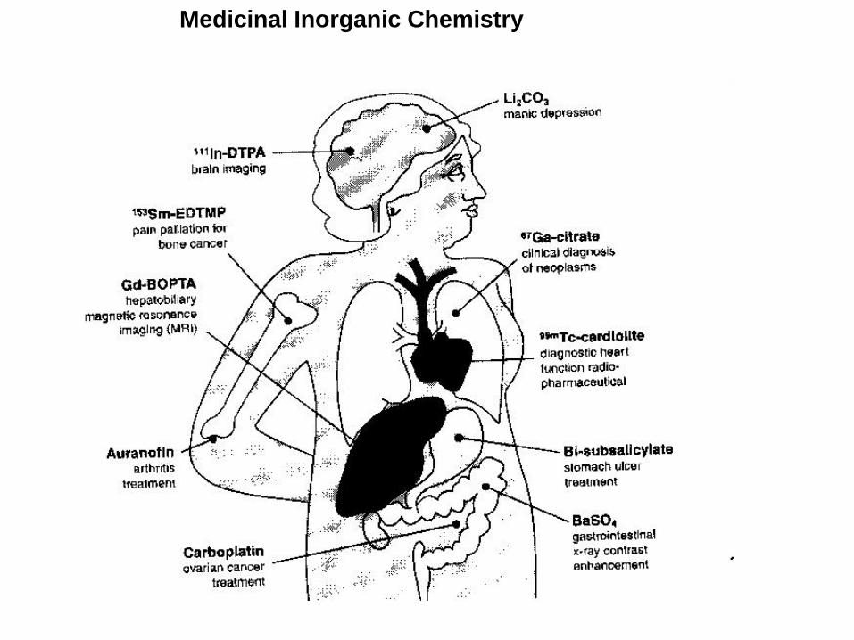
Medicinal Inorganic Chemistry

Application of Metals in Medicine
Li+: Treatment of depression (Li2CO3, low doses)
Gd3+: Contrast agent (NMR)
BaSO4: Contrast agent (radiography)
99mTc: radio diagnostics (thyroid)
Au(I): Rheumatism

Sb(III): Eczema
Bi(III): Gastric ulcer
Cd: Carboanhydrase(Thalassiosira
weissflogii)

Metal based Anti-Cancer Drugs
In 1965 Rosenberg discovered antiproliferative effect of a cisplatin whilst
conducting studies on bacteria under in an electric field produced by
platinum complexes
He was able to show that the compound cisplatin was responsible for the
effect and this was found to be effective against treating some cancers.
Cisplatin is now THE MOST used anti-cancer drug
**BUT CONTAINS NO CARBON ATOMS** !!
HOW DOES CIS-PLATIN TARGET CANCER ?
By reaction with DNA?
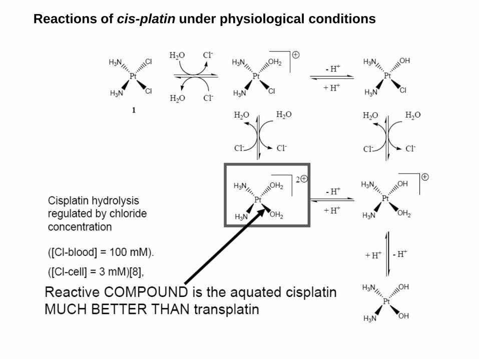
Reactions of cis-platin under physiological conditions

Applicable in relatively narrow range of tumors.
Limited solubility in Aqueous solution.
Intravenous administration, inconvenience to
outpatient treatment.
Nephrotoxicity,neurotoxicity and nausia.
Higher toxicity leading to lower doses of 100 mg/day.
Disadvantages of cis-platin

cis-platin and new drugs
From a clinical point–of-view the current challenges in drug development
include:
(i) addressing the poor solubility of cisplatin and analogues in water
(ii) cellular resistance of cancer cells to cisplatin
(iii) toxic side effects of cisplatin
(e.g. nausea, neurotoxicity, kidney damage)
(iv) use of platinum-based therapeutics to treat cisplatin resistant cell
lines
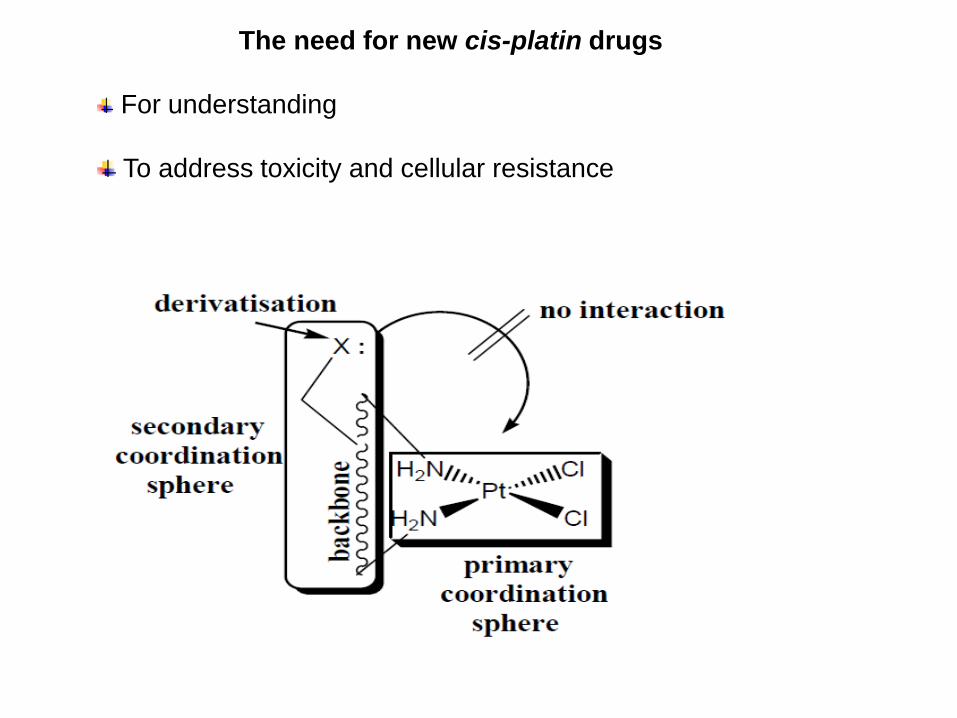
The need for new cis-platin drugs
For understanding
To address toxicity and cellular resistance

Second line Anticancer Drugs
Carboplatin is less toxic than cisplatin and can be given in
higher doses (2000 mg/dose). Routinely used in clinics.
Oxaplatin is approved for secondary treatment of metastatic
colorectal cancer in France and other European countries and
Nedaplatin has received approval for use in Japan.
The search continues for an improved Pt-anticancer agents
which are less toxic, orally active and non-cross-resistant with
cis-platin and trans-platin.
PtOH3N
H3N O
O
O
PtOH3N
H3N O
O
PtOHN
HN O
O
Carboplatin Nedaplatin Oxaliplatin

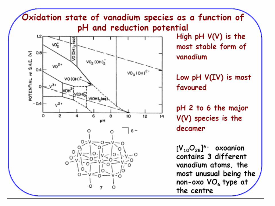
Vanadium-based drugs for diabetes
Insulin is the mainstay of treatment of Type-I (insulin dependent-10%) and
Type-II (insulin-independent –90%) diabetic patients.
Daily subcutaneous injections of insulin to insulin-defecient patients
lowered the blood glucose to normal values and interrupts a fatal metabolic
disorder.
Oral administration is ineffective in mammals.
Insulin stimulates the uptake of glucose (glycogen in liver and muscle) fatty
acids (triglycerides in adipose tissue) and amino acids (proteins in muscle)
from blood circulation for further storage and utilization.
Insulin also inhibits the action of other hormones that trigger the breakdown
of glycogen,fatty acids and proteins.

Need and search for insulin substitutes
Development of Insulin resistance.
Development of methods for preparation on insulin responsive cells
in 1970 facilitated investigation of mechanism of insulin action as
well as identification of several agents that mimic the insulin action.
Proteins (trypsin, lectins, and antibodies) H2O2,Zn, Mn ions effective
in rat adipocytes but FAIL in animal model.


Schematic representation of the activation of glucose intake by insulin
(a & b) activation of glucose
intake by insulin
(c) Blockage of glucose intake in
absence of insulin
Counteractions by vanadium
compounds
(d) Activation of a
phosphatase
(e) Activation of a non-
membrane kinase
(f) Vanadylation of the
insulin receptor tyrosine D. Rehder et al. Coordination Chemistry
Review, 237 (2003) 53/63

Insulin-mimetic Vanadium compounds in various stages of clinical
tests
Vanadium(IV) maltalato complex (28) has been introduced in clinical
tests in humans.
V(V)-bispicolinato complex (29) has been successful in curing
diabetic cats
Slow development of Vanadium compounds in pharmaceutical industry1. Toxicity of vanadate
2. Heavy metals not accepted by the market3. Vanadium is retained in the bone, Half-life of VO2+ is one month
4. Market logistics and competing interference

Gold in treatment of rheumatoid arthritis
(1) Sodium aurothiomalate (Myocrisin)
(2) Aurthioglucose (Solganol)
(3) Auranofin (Radaura)
Nucleases (Restriction Enzymes)
Enzymes which carry out hydrolysis of internucleotide linkages in
nucleic acids at relatively specific points.
A] Endonucleases:-Hydrolysis at internal position in DNA at
nucleotide or RNA strand.
B] Exonucleases:-Hydrolysis only at terminal linkage,some at 5` and
others at 3`end.
Required for controlled fragmentation of DNA and RNA into smaller
pieces at specific points.This property has opened a new area in the
Biochemistry of genes systematic dissection and mapping of
chromosomes.Therefore possible to splice or recombine genes from
one organism into the genome of another.
Werner Arber(Switzerland),Daniel Nathans(USA) and Hamilton Smith
(USA) – Nobel Prize in Medicine in 1978 for discovery of Restriction
Endonucleases.

Transition metal complexes as chemical Nucleases
Basics of Nucleic acid interactions:-
A] Nucleic acid structures
B] Nucleases
C] Fundamental interactions of metal complexes with
nucleic acids
i] Coordination
ii] Intercalation
D] Fundamental reactions of metal complexes with
nucleic acids
i] Redox
ii] Hydrolysis
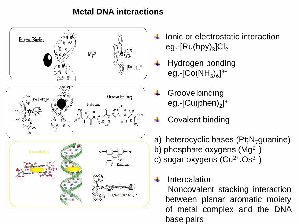
Metal DNA interactions
Ionic or electrostatic interaction
eg.-[Ru(bpy)3]Cl2
Hydrogen bonding
eg.-[Co(NH3)6]3+
Groove binding
eg.-[Cu(phen)2]+
Covalent binding
a) heterocyclic bases (Pt;N7guanine)
b) phosphate oxygens (Mg2+)
c) sugar oxygens (Cu2+,Os3+)
Intercalation
Noncovalent stacking interaction
between planar aromatic moiety
of metal complex and the DNA
base pairs


Narrow and deep
Narrow and quite deep
Broad and shallow
Minor groove
FlatWide and quite deep
Narrow and very deep
Major groove
45.6 Å35.4 Å25.3 ÅPitch per turn of helix
1210.411Base pair per turn of helix
Anti for C,T and Syn for G
AntiAntiGlycosidic bond
3.8 Å3.4 Å2.3 ÅRise per base pair
Most elongatedIntermediateBroadestShape
Left handedRight handedRight handed Screw sense
Z-DNAB-DNAA-DNA


Families of well studied Coordination complexes
A] Cis-platin as anticancer agent
B] Cu(Phen)2+ as chemical nuclease
C] Fe(EDTA) 2- as footprinting agent
D] M(phen)3 (M=Ru,Rh,Os) as spectroscopic probes
N
N N
N
Ru
N
N
N
N
2+
cis-platin [Ru(phen)2dppz]2+ [Cu(phen)2+
Why study Metal-DNA interactions?
For evolving molecular biological tools (Synthetic
restriction enzymes)
The genetic basis of several major diseases has been
recognized and therefore necessary to target aberrant
DNA by direct binding or chemical excision
To develop novel DNA-sequence reading and cleavage
systems that are amenable to synthetic manipulation and
have suitable biocompatibility (stability,cellular
penetration and recycling)
To understand DNA reactivity and detect DNA structures
Techniques used to study metal nucleic acid interactions
A] Viscosity measurements
B] 1H NMR Studies
C] UV-Visible studies
D] Emission spectroscopy
E] Cyclic voltammetric studies
F] Circular Dichroism
G] Gel electrophoresis
H] Resonance Raman studies
I] Equilibrium dialysis
J] Covalent Binding assay

Ionic or electrostatic interaction
eg.-[Ru(bpy)3]Cl2
Hydrogen bonding
eg.-[Co(NH3)6]3+
Groove binding (major and minor)
eg.-[Cu(phen)2]+
Covalent binding
a) heterocyclic bases ( Pt ; N7 guanine)
b) phosphate oxygens ( Mg2+)
c) sugar oxygens ( Cu2+,Os3+)
Oxidation
eg.-[Ru(bpy)3]2+
Possible Metal-DNA interactions

Intercalation
Ru
N
N
N
N
2+
Non covalent stacking interaction between planar aromatic moiety
of metal complex and the DNA base pairs
Modification of the ancillary ligand
influences the optical and DNA
binding properties of the
complexes.

Following changes occur upon intercalation
A] Unwinding and lengthening of the DNA helix
B] Electronic interaction of the intercalator within the helix
C] DNA Rigidity and orientation of the intercalator within the helix
Experimental criteria that establish intercalation can be classified
as follows:
A] Experiments that evaluate structural changes in the DNA helix
1.Changes in solution viscosity of bulk DNA.
2.Changes in sedimentation coefficient.
3.Downfield shifts in the 31P NMR spectrum

B] Experiments that indicate an electronic interaction between
the
intercalator and DNA bases.
1.Hypochromism
2.Bathochromic shift
3.Emission enhancement
4.1H NMR up field shifts in the aromatic protons of the
intercalated
molecule
C] Experiments that demonstrate molecular orientation or rigidity
1.Dichroic technique
2.Changes in luminescence polarization

[A] Viscosity Measurements

[B] 1H NMR Studies
Shifts in 1H NMR resonance's of both DNA binding complex and the
oligonucleotide are evidence of increased association
These shifts can be used empirically to gain structural insights into
binding modes of complexes such as M(phen)32+ where (M=Ru,Rh)

[C] UV-Visible Studies
DNA / ea- ef = DNA / eb- ef + 1 / Kb (ea- ef )
Hypochromism and red shift are observed on binding with DNA
These spectroscopic perturbations can be used to define equilibrium
binding affinities and chiral preferences as well as extent of intercalation

[D] Emission spectroscopy
Increase in Fluorescence
intensity and lifetime of excited
state due to intercalation
Figure-Emission spectrum of free
[Ru(phen)3]2+(………) [-Ru(phen)3]
2+ in
the presence of DNA (--------), and -
[Ru(phen)3]2+( -) in the presence of DNA
showing the enantioselective binding of
the complexes to the helix.

[E] Voltammetric studies of the interaction of tris(phen) complexes
with DNA
Coordination complexes of 1,10-phenanthroline and bipyridine with
Co3+,Ru3+,Fe3+ are known to intercalate between base pairs of DNA
Interaction of M-DNA interaction with reduced and oxidised metal
Differentiates betwwen intercalation and electrostatic binding
Estimates binding parameters (binding site sizes and binding constant)
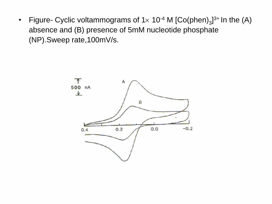
• Figure- Cyclic voltammograms of 1 10-4 M [Co(phen)3]3+ In the (A)
absence and (B) presence of 5mM nucleotide phosphate
(NP).Sweep rate,100mV/s.

[F] Circular dichroism
240 260 280 300 320-8
-6
-4
-2
0
2
4
6 ab
Figure - Circular Dichroism spectra
of calf thymus DNA (20 M) in the
absence (a) and presence
(b) of [Co(dppz)2Cl2]Cl (10 M)
complex.

G] Covalent binding assay for
bis-phenantroline dichloro
Ruthenium (II) complexes to B-
DNA

Applications of different metal complexes that bind nucleic acids are
[A] Spectroscopic probes
Tris (phenanthroline) Ru (II) complexes offer a novel spectroscopicprobe of nucleic acids
Derivatives of the tris(phenanthroline) metal complexes that maybecome exceedingly useful as spectroscopic probes
e.g.-[Ru(bpy)2dppz]2+ and [Ru(phen)2dppz]2+ (dppz=dipyridophenazine).
Quite novel luminescent phenanthroline and diphenylphenanthroilinecomplexes of copper (I) are extremely valuable as cleavage probes.

[B] Metallofootprinting agents
Derivative of a tris (phenanthroline) metal complexes e.g.[Rh(phi)2bpy]3+ currently being applied in footprinting experiments
[Cu(phen)2]+ and manganese porphyrins have been used to footprint
DNA-binding proteins
[C] Conformational probes
Wide application in probing the local variations in conformation thatarise along nucleic acid polymers
Used in probing the structural variations in nucleic acids

Conclusion
Study, understand and teach Bioinorganic Chemistry –
it is closest to life

Vitamin B12
Vitamin B12 is the only known
essential biomolecule with a
stable metal-carbon bond, that is,
it is an organometallic
compound.
a methyl group - as in methylcobalamin
a 5'-deoxyadenosine at the the 5' positon - as in adenosylcobalamin
(coenzyme B12)
a cyanide group - as in Vitamin B12 - as supplied from drug companies
Cobalt in Biology

Homeostasis
• All living species have two different ‘environments ‘ one the external surroundings the
‘habitat’ and the other ‘milieu’ made up of the biological fluids e.g. the blood plasma.
• This conservation of conditions in a living system, homeostasis, does not correspond
to a state of equilibrium in thermodynamic terms ( which would be equivalent to
death) but to a series of related and controlled states in a dynamic process of
continuous material, charge, and energy flows through the cells, with forced and fixed
directions.
DNA/RNA
(Plan genetics)
Proteins
Membranes
Saccharides
(Machinery)
Living system
Electrons
Ions
Energy
(Bio-energetics
Compartments)

Scheme showing the close connections in a prokaryote cell between a variety of elements
Scheme showing involvement of phosphorus

DNA binding
Major adducts of platinum drugs with DNA are the 1,2-GpG and 1,2-ApG
intrastrand crosslinks – 90%
Structural studies show that the Pt cross links induce bending and
unwinding of DNA and cause destacking of the purine bases.
cis-{Pt(NH3)2}2+- d(CCTG*G*TCC)·d(GGACCAGG) indicates that the B
DNA backbone conformation is significantly altered to accommodate the
platinated lesion
Spectroscopic and calorimetric studies on the major adduct of the cis-
platin with a 20-mer DNA duplex containing a GG intrastrand-crosslink have
suggested that platination induces a conformational shift from an B-like to
an A-like form – may be important in HMG recognition.

Spectroscopic and calorimetric studies on the major adduct of the
cis-platin with a 20-mer DNA duplex containing a GG intrastrand-
crosslink have suggested that platination induces a conformational
shift from an B-like to an A-like form – may be important in HMG
recognition.
It is known that platinum forms bifunctional DNA adducts with the
following order of sequence preference: -GG- > -AG- >> -GA and
platination is kinetically controlled.
Inter strand cross links can also be generated between DNA and
cis-platin between to G’s on opposite sides of the duplex
Monofunctional adducts can also form and can be long lived (t(1/2)
= 80hrs)

DNA binding – GpG INTRA STRAND
cis-platin binds to DNA and causes a critical structural change in the
DNA – a bend of 45 degrees
cis-platin binds to two
Adjacent G’s at N7
on the DNA in an
INTRA strand
cross-link


Model Compounds- Biomimetic Chemistry
Large size of metallobiomolecules and high resolution structure of metal
coordination difficult
If X-ray crystal structure is known it is possible to design a replica of the
coordination environment.----------replicative models
If X-ray crystal structure is not known we test postulated structure by
spectroscopy by synthesizing models-----speculative models
If models are only structurally similar--------- structural models
If models are functionally similar --------------functional models

Biomimetic approach has helped in the study of
1. Assignment or verification of the metal oxidation states
2. effects of distance and medium on electron transfer
rates
3. role of steric and electronic factors
4. Identity of likely intermediates of enzyme catalyzed
reactions
Strategy for models complexes- spontaneous self assembly
Nature adopted the a similar strategy based on available chemistry in
the
geosphere during evolution .

Manganese(II) ions function as cofactors for a number of enzymes and the element
is thus a required trace mineral for all known living organisms.
Biological role
The classes of enzymes that have manganese cofactors are very
broad and include such classes as oxidoreductases, transferases,
hydrolases, lyases, isomerases, ligases, lectins, and integrins.
Mn-SOD is the type of SOD present in eukaryotic mitochondria, and
also in most bacteria. The Mn-SOD enzyme is probably one of the most
ancient, for nearly all organisms living in the presence of oxygen use it to
deal with the toxic effects of superoxide, formed from the 1-electron
reduction of dioxygen.
Manganese in Biology

Manganese is also important in photosynthetic oxygen evolution in
chloroplasts in plants, which are also evolutionarily of bacterial origin.
The oxygen evolving complex (OEC), a water-oxidizing enzyme
contained in chloroplast membrane, and which is involved in the
terminal photooxidation of water during the light reactions of
photosynthesis, has a metalloenzyme core containing four atoms of
manganese.

Mechanistic cycle (commonly referred
to as the Kok catalytic cycle) for water
oxidation in the photosystem II (PSII)
active site.
H2O + CO2 1/n(CH2O)n + 3O2 H = + 470 KJ/mole
Photosystem II


Proposed PSII Mn4 Structures
