metal levels in the liver, muscle, gill, intestine, and gonad of lake van fish (chalcalburnus...
TRANSCRIPT

Metal Levels in the Liver, Muscle, Gill, Intestine, and Gonadof Lake Van Fish (Chalcalburnus tarichi) with Abnormal Gonad
Ahmet R. Oğuz & Aslı Yeltekin
Received: 25 March 2014 /Accepted: 10 April 2014 /Published online: 26 April 2014# Springer Science+Business Media New York 2014
Abstract Recently, an increasing number of studies haveshown that Lake Van—the biggest soda lake in the world—is polluted due to an increasing population. Studies haveshown abnormalities in the Lake Van fish (Chalcalburnustarichi), the sole fish species that inhabits the lake. Unlikethe vitellogenic and mature oocytes in normal gonads, abnor-mal gonads show large amounts of connective tissue andyoung oocytes. In this study, metal levels (nickel [Ni], copper[Cu], cobalt [Co], iron [Fe], zinc [Zn], cadmium [Cd], lead[Pb], and manganese [Mn]) in the muscle, liver, gill, intestine,and gonad of Lake Van fish with normal and abnormal gonadswere assessed. Further, the metal contents in the wastewaterfrom the wastewater treatment facility situated near Lake Vanin Van City were assessed. All the metal levels, except that ofZn, were high in the Lake Van environment (P<0.05). Thehighest metal content in the tissues was for Fe, while thelowest level was for Co. The Pb level was found to be veryhigh in both fish groups. Cd was not found in the tissues ofboth fish groups. The levels of Fe, Cu, Pb, and Mn were notsignificant in the tissues of both normal and abnormal fishgroups. Zn level was significantly high in the livers andgonads of fish with abnormal gonads, and Co level wassignificantly high only in the livers (P<0.05). Consequently,high levels of Zn in the liver and gonads and high levels of Coin the liver may be factors causing the abnormal gonads in theLake Van fish.
Keywords Heavymetal . Abnormal gonad . LakeVan fish .
Chalcalburnus tarichi . LakeVan
Introduction
The physiological functions of metals are widespread andnecessary in biological systems. Zinc (Zn), copper (Cu), co-balt (Co), iron (Fe), manganese (Mn), magnesium (Mg), mo-lybdenum (Mo), selenium (Se), and nickel (Ni) are essentialmetals, while cadmium (Cd), mercury (Hg), lead (Pb), andarsenic (As) are nonessential elements. Metals play importantroles in oxygen transport [1], DNA and protein syntheses [2],prevention of apoptosis [3], and the structure of enzymes andbone tissue [4]. Additionally, metals ensure stabilization ofcell and organelle membranes and macromolecules such asDNA, RNA, and enzymes. Excessive metal levels have toxiceffects and cause disorders in the histologic, metabolic, andendocrine functions in animals. Further, excessive metallevels cause oxidative stress [5], which adversely affects thereproductive cells [6].
Lake Van is the biggest lake in Turkey and the fourthbiggest lake in the world. Because of the high pH and alka-linity levels, to a great extent, life forms are restricted in thelake [7]. The only fish species to adapt to the conditions in thelake is the Lake Van fish (Chalcalburnus tarichi, Pallas 1811).Although the Lake Van fish is the only species, it constitutesone third of the domestic fish production (10.000 t/year) ofTurkey. The fish offers a great and cheap protein resource forthe local community.
In recent years, abnormalities in the Lake Van fish causedby pollution in Lake Van have been observed. Studies haveshown that abnormalities in the gonad might be caused byendocrine-disrupting chemicals [8]. In the preliminary studiesby Oğuz and Kankaya [9], some localities of the lake wereshown to be contaminated by endocrine-disrupting chemicals.
A. R. Oğuz (*)Department of Biology, Faculty of Science, Yüzüncü Yıl University,65080 Van, Turkeye-mail: [email protected]
A. YeltekinDepartment of Chemistry, Faculty of Science, Yüzüncü YılUniversity, 65080 Van, Turkey
Biol Trace Elem Res (2014) 159:219–223DOI 10.1007/s12011-014-9980-0

Endocrine-disrupting chemicals cause endocrine system dis-orders by displaying agonist or antagonist functions in thehormones of animals and human beings. Such endocrine-disrupting chemicals are found in insecticides, herbicides,detergents, resins, and plasticizers. In addition to thechemicals, some heavy metals show endocrine-disrupting-chemical characteristics and are defined as “endocrine-disrupting metals” [10, 11]. Such metals not only influencethe endocrine system pathways but also directly impart nega-tive effects on the gametes in the gonad [12, 13].
In this study, some somatic index values and the histologyof the Lake Van fish with normal and abnormal gonads wereevaluated. The levels of Ni, Cu, Co, Fe, Zn, Cd, Pb, andMn inthe surface water of Lake Van and wastewater discharged intothe lake from a wastewater treatment plant were assessed.Furthermore, the metal levels in different tissues of fish withnormal and abnormal gonads were determined, and the corre-lation between the metal levels and abnormal gonad wasexamined.
Material and Methods
Fish
Freshly caught fish (C. tarichi) were purchased from a localfisherman who captured them from Lake Van. The fish werethen immediately transported on ice to the laboratory. Thegonad tissue samples were collected and fixed for histologicaluse and gender identification. The fork lengths and total andgonad weights of the fish were measured. Tissue samples(muscle, gill, liver, intestine, and gonad) were collected andstored at −80 °C until analysis. Age determination was per-formed through the operculum samples belonging to the fish.All the procedures were carried out according to the NationalAnimal Care regulations.
The gonadosomatic index (GSI) was measured using thefollowing formula: GSI=gonad weight/total weight×100.
Histology
Pieces of gonad tissue were fixed in Bouin’s solution for 12 h.Afterward, the tissues were dehydrated through a gradedseries of ethanol, cleared in xylene, and embedded in paraffinwax. Histologic sections of the paraffin-embedded tissue werecut at a thickness of 5–7 μm by using a rotary microtome (HM325 Manual Microtome, MICROM International GmbH,Walldorf, Germany). Slide-mounted sections weredeparaffinized in xylene and rehydrated through graded seriesof methanol washes. Sections were then stained using hema-toxylin and eosin. All slides were mounted with coverslipsand examined under a light microscope (Leica Microsystems,Wetzlar, Germany) attached with a digital camera system.
Metal Analysis
For metal analysis, all the chemicals used were of analyticalgrade and obtained from Merck (Germany). Freeze-driedtissue samples (liver, muscle, gill, intestine, and gonad) wereweighed (approximately 0.5 g) and digested in nitric acid(65 %) at 120 °C in an oven. After the solution cooled toroom temperature, perchloric acid (65 %) was added. Afterdigestion, the residues were diluted with deionized water andfiltered using a filter paper. The residues were maintained at4 °C until analysis [14]. Metal levels (Ni, Cu, Co, Fe, Zn, Cd,Pb, and Mn) were determined by atomic absorption spectro-photometry (AAS; ICE3000 series, Thermo, USA). Metalswere determined at the following wavelengths (λ): Ni,232.0 nm; Cu, 324.8 nm; Co, 240.0 nm; Fe, 248.3 nm; Zn,213.9 nm; Cd, 228.8 nm; Pb, 217.0 nm; and Mn, 279.5 nm.All tissue samples were analyzed in duplicate. The heavymetal concentrations were expressed as micrograms per gramof wet tissue.
Statistical Analysis
Data were expressed as means±standard deviation (SD), andthe differences between sample groups were evaluated usingtheMann–WhitneyU test. Significant differences between thetwo fish groups were considered when P was ≤0.05.
Results
After the morphologic examination of the fish gonads,the abnormal gonads were noted to be smaller than thenormal gonads. Consequently, the total weight, gonadweight, and GSI of fish with abnormal gonads werelesser than those of normal fish (Table 1, P<0.05).After histologic examination of the tissues, normal fishgonads were observed to have vitellogenic oocytes,whereas the abnormal fish gonads had younger, imma-ture oocytes (Fig. 1). Additionally, the abnormal gonadshad remarkably dense connective tissue. Age determina-tion showed that all the fish were aged more than
Table 1 Morphological indexes in the Lake Van Fish having normal andabnormal gonads
Normal Abnormal
Fork length (cm) 19.20±1.55 18.76±0.72
Weight (g) 100.92±10.34 76.08±6.39*
Gonad (g) 6.97±0.87 3.07±0.63*
GSI (%) 6.93±0.70 4.09±1.07*
Fish sampled on March 2013. All values are means±SD, N=10
*P<0.001
220 Oğuz and Yeltekin

3 years (data not shown) and sufficiently mature toreproduce.
The mean metal concentrations of the Lake Van water andwastewater are shown in Table 2. Cd was not detected in the
wastewater. Further, all the measured metal levels, except thatof Zn, were high in the lake water. The highest level was notedfor Pb, and the lowest was for Cd.
Metal (Ni, Cu, Co, Fe, Zn, Cd, Pb, and Mn) levels in themuscle, liver, gill, intestine, and gonad tissues of fish withnormal and abnormal gonads are shown in Table 3 as micro-grams per gram wet tissue. The highest metal level in thetissues was noted for Fe, and the lowest was for Co. Cd wasnot detected in any of the tissue samples. The Zn and Co levelsin the liver and gonad tissues were found to significantly differ(P<0.05) between fish with normal and abnormal gonads.
Discussion
Fish are highly sensitive to aquatic pollution. Studies haveshown metal accumulation in the gonads of fish exposed tometals [15–17], which results in reductions in the productionand viability of gametes. Further, the fertilization of sperm andoocyte and embryonic development in fish are adverselyaffected by metal contamination [18].
The examination of the somatic indexes of the fish showedthat reductions in the total weight, gonad weight, and GSI offish with abnormal gonads were because of immature ovaries(Fig. 1). Previous studies of the abnormal gonad in fish haveshown a low plasma 17-β-estradiol level [8]. Hence, similar-ities are present in the symptoms of abnormal fish in Lake Vanand those of fish in aquatic areas contaminated with heavymetals. Hence, similarities are present in the symptoms ofabnormal fish in Lake Van and those of fish in aquatic areascontaminated with heavy metals, such as decrease in GSI and17-β-estradiol level and presence of immature and atreticfollicles in the ovaries [13, 19].
The wastewater discharged into the lakewas shown to havelow metal levels (Table 2). This could be because the waste-water that is treated at the treatment plant is domestic waste-water, since there are no developed industries situated near thelake. Therefore, the heavy metal concentration in the lake andconsequently accumulated in the Lake Van fish is caused bythe geochemical structure of the region and partially by thedomestic wastewater discharged into the lake. In addition, thedischarge of untreated effluents from nearby towns and agri-cultural activities may have contributed to the accumulation ofmetals in the lake.
The Pb level in the Lake Van fish was higher than that inseveral other fish species [20–23]. In a previous study relatedto the fish, the Pb level was shown to be high [24]. Afterexamination of the lake water metal levels (Table 2), the Pblevel was confirmed to be very high, which suggests that thePb accumulation in the tissues is because of the high Pb levelin the water. No significant differences were noted for the Ni,Cu, Fe, Cd, and Mn levels between the tissues of fish withnormal and abnormal gonads. Since the levels of essential
Fig. 1 Gonad tissues of normal (a) and abnormal (b) gonad samples ofLake Van fish (asterisks connective tissue, arrowheads vitellogenic oo-cytes, arrow young oocytes; scale bar 200 μm)
Table 2 Metal concentrations (mg/L) in Lake Van and the efflux of thewastewater treatment plant
Lake Van Wastewater
Ni 0.186±0.015 (9) 0.008±0.006 (4)**
Cu 0.042±0.014 (10) 0.0262±0.007 (9)**
Co 0.122±0.044 (10) 0.0150±0.013 (8)***
Fe 0.211±0.067 (10) 0.0664±0.024 (9)**
Zn 0.040±0.019 (10) 0.036±0.013 (9)
Cd 0.0033±0.002 (7) <LOD
Pb 0.214±0.050 (10) 0.0614±0.016 (9)***
Mn 0.043±0.005 (10) 0.055±0.009 (9)*
Data are expressed as mean±SD. N=10. Numbers in parentheses repre-sent the number of determinations
LOD limit of detection
*P<0.05; **P<0.01; ***P<0.001
Metal levels in the tissues of Lake Van Fish with abnormal gonad 221
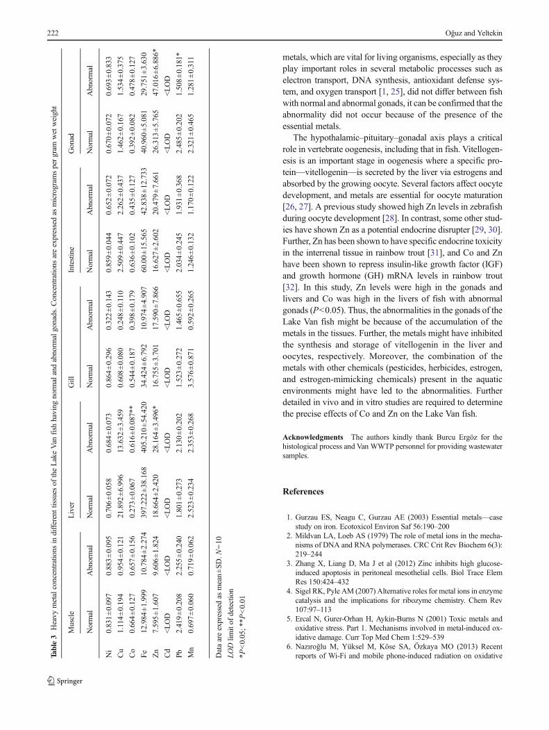
metals, which are vital for living organisms, especially as theyplay important roles in several metabolic processes such aselectron transport, DNA synthesis, antioxidant defense sys-tem, and oxygen transport [1, 25], did not differ between fishwith normal and abnormal gonads, it can be confirmed that theabnormality did not occur because of the presence of theessential metals.
The hypothalamic–pituitary–gonadal axis plays a criticalrole in vertebrate oogenesis, including that in fish. Vitellogen-esis is an important stage in oogenesis where a specific pro-tein—vitellogenin—is secreted by the liver via estrogens andabsorbed by the growing oocyte. Several factors affect oocytedevelopment, and metals are essential for oocyte maturation[26, 27]. A previous study showed high Zn levels in zebrafishduring oocyte development [28]. In contrast, some other stud-ies have shown Zn as a potential endocrine disrupter [29, 30].Further, Zn has been shown to have specific endocrine toxicityin the interrenal tissue in rainbow trout [31], and Co and Znhave been shown to repress insulin-like growth factor (IGF)and growth hormone (GH) mRNA levels in rainbow trout[32]. In this study, Zn levels were high in the gonads andlivers and Co was high in the livers of fish with abnormalgonads (P<0.05). Thus, the abnormalities in the gonads of theLake Van fish might be because of the accumulation of themetals in the tissues. Further, the metals might have inhibitedthe synthesis and storage of vitellogenin in the liver andoocytes, respectively. Moreover, the combination of themetals with other chemicals (pesticides, herbicides, estrogen,and estrogen-mimicking chemicals) present in the aquaticenvironments might have led to the abnormalities. Furtherdetailed in vivo and in vitro studies are required to determinethe precise effects of Co and Zn on the Lake Van fish.
Acknowledgments The authors kindly thank Burcu Ergöz for thehistological process and Van WWTP personnel for providing wastewatersamples.
References
1. Gurzau ES, Neagu C, Gurzau AE (2003) Essential metals—casestudy on iron. Ecotoxicol Environ Saf 56:190–200
2. Mildvan LA, Loeb AS (1979) The role of metal ions in the mecha-nisms of DNA and RNA polymerases. CRC Crit Rev Biochem 6(3):219–244
3. Zhang X, Liang D, Ma J et al (2012) Zinc inhibits high glucose-induced apoptosis in peritoneal mesothelial cells. Biol Trace ElemRes 150:424–432
4. Sigel RK, Pyle AM (2007) Alternative roles for metal ions in enzymecatalysis and the implications for ribozyme chemistry. Chem Rev107:97–113
5. Ercal N, Gurer-Orhan H, Aykin-Burns N (2001) Toxic metals andoxidative stress. Part 1. Mechanisms involved in metal-induced ox-idative damage. Curr Top Med Chem 1:529–539
6. Nazıroğlu M, Yüksel M, Köse SA, Özkaya MO (2013) Recentreports of Wi-Fi and mobile phone-induced radiation on oxidativeTa
ble3
Heavy
metalconcentrations
indifferenttissues
oftheLakeVan
fish
having
norm
alandabnorm
algonads.C
oncentratio
nsareexpressedas
microgram
spergram
wetweight
Muscle
Liver
Gill
Intestine
Gonad
Normal
Abnormal
Normal
Abnormal
Normal
Abnormal
Normal
Abnormal
Normal
Abnormal
Ni
0.831±0.097
0.883±0.095
0.706±0.058
0.684±0.073
0.864±0.296
0.322±0.143
0.859±0.044
0.652±0.072
0.670±0.072
0.693±0.833
Cu
1.114±0.194
0.954±0.121
21.892
±6.996
13.632
±3.459
0.608±0.080
0.248±0.110
2.509±0.447
2.262±0.437
1.462±0.167
1.534±0.375
Co
0.664±0.127
0.657±0.156
0.273±0.067
0.616±0.087**
0.544±0.187
0.398±0.179
0.636±0.102
0.435±0.127
0.392±0.082
0.478±0.127
Fe12.984
±1.999
10.784
±2.274
397.222±38.168
405.210±54.420
34.424
±6.792
10.974
±4.907
60.00±15.565
42.838
±12.733
40.960
±5.081
29.751
±3.630
Zn
7.595±1.607
9.606±1.824
18.664
±2.420
28.164
±3.496*
16.755
±3.701
17.590
±7.866
16.627
±2.602
20.479
±7.661
26.313
±5.765
47.016
±6.886*
Cd
<LOD
<LOD
<LOD
<LOD
<LOD
<LOD
<LOD
<LOD
<LOD
<LOD
Pb2.419±0.208
2.255±0.240
1.801±0.273
2.130±0.202
1.523±0.272
1.465±0.655
2.034±0.245
1.931±0.368
2.485±0.202
1.508±0.181*
Mn
0.697±0.060
0.719±0.062
2.523±0.234
2.353±0.268
3.576±0.871
0.592±0.265
1.246±0.132
1.170±0.122
2.321±0.465
1.281±0.311
Dataareexpressedas
mean±SD
.N=10
LODlim
itof
detection
*P<0.05;*
*P<0.01
222 Oğuz and Yeltekin

stress and reproductive signaling pathways in females and males. JMembr Biol 246(12):869–75
7. Reimer A, Landmann G, Kempe S (2009) Lake Van, EasternAnatolia, hydrochemistry and history. Aquat Geochem 15:195–222
8. Ünal G, Türkoğlu V, Oğuz AR, Kaptaner B (2007) Gonadal histol-ogy and some biochemical characteristics of Chalcalburnus tarichi(Pallas, 1811) having abnormal gonads. Fish Physiol Biochem 33:153–165
9. Oğuz AR, Kankaya E (2013) Determination of selected endocrinedisrupting chemicals in Lake Van, Turkey. Bull Environ ContamToxicol 91(3):283–286
10. Georgescu B, Georgescu C, Dărăban S, Bouaru A, Paşcalău S (2011)Heavy metals acting as endocrine disrupters. J Anim Sci Biotechnol44(2):89–93
11. Iavicoli I, Fontana L, Bergamaschi A (2009) The effects of metals asendocrine disruptors. J Toxicol Environ Health Part B Crit Rev 12:206–223
12. Kumar S, Pant SC (1984) Comparative effects of the sublethalpoisoning of zinc, copper and lead on the gonad of teleost, Puntiusconchonius Hamilton. Toxicol Lett 23:189–194
13. Levesque HM, Dorval J, Hontela A, Van Der Kraak GJ, PeterCampbell GC (2003) Hormonal, morphological, and physiologicalresponses of yellow perch (Perca flavescens) to chronic environmen-tal metal exposures. J Toxicol Environ Health 7(66):657–676
14. Brown AA, Taylor A (1985) Applications of a slotted quartz tube andflame atomic-absorption spectrometry to the analysis of biologicalsamples. Analyst 110:579–582
15. Ellenberger SA, Baumann PC, May TW (1994) Evaluation of effectscaused by high copper concentrations in Torch lake, Michigan, onreproduction of yellow perch. J Great Lakes Res 20:531–536
16. Allen P (1995) Accumulation profiles of lead and cadmium in theedible tissues of Oreochromis aureus during acute exposure. J FishBiol 47:559–568
17. Pelgrom SMGJ, Lamers LPM, Lock RAC et al (1995) Interactionsbetween copper and cadmium modify metal organ distribution inmature tilapia, Oreochromis mossambicus. Environ Pollut 90:415–423
18. Jezierska B, Lugowska K, Witeska M (2009) The effects of heavymetals on embryonic development of fish (a review). Fish PhysiolBiochem 35:625–640
19. Mousa HA, Mousa MA (1999) Immunocytochemical and histolog-ical studies on the hypophyseal-gonadal system in the freshwater Niletilapia, Oreochromis niloticus (L.), during sexual maturation andspawning in different habitats. J Exp Zool 284:343–354
20. Uluozlu OD, Tuzen M, Mendil D, Soylak M (2007) Trace metalcontent in nine species of fish from the Black and Aegean Seas,Turkey. Food Chem 104:835–840
21. Has-Schön E, Bogut I, Rajkovic V, Bogut S, Cacic M, Horvati J(2008) Heavy metal distribution in tissues of six fish species includedin human diet, inhabiting freshwaters of the nature park “HutovoBlato” (Bosnia and Herzegovina). Arch Environ Contam Toxicol 54:75–83
22. Tuzen M (2009) Toxic and essential trace elemental contents in fishspecies from the Black Sea, Turkey. Food Chem Toxicol 47:1785–1790
23. Hao Y, Chen L, Zhang X, Zhang D, Zhang X, Yu Y (2013) Traceelements in fish from Taihu Lake, China: levels, associated risks, andtrophic transfer. Ecotoxicol Environ Saf 90:89–97
24. Bilgili A, Sağmanlıgil H, Çetinkaya N, Yarsan E, Türel İ (1995) Vangölü suyunun doğal kalitesi ve buradan avlanan İnci kefali(Calcalburnus tarichii, Pallas 1811) örrneklerinde bazı ağır metaldüzeyleri. A Ü Vet Fak Derg 42:445–450
25. Bleackley MR, Macgillivray RT (2011) Transition metal homeosta-sis: from yeast to human disease. Biometals 24:785–809
26. Ozkaya MO, Nazıroğlu M (2010) Multivitamin and mineral supple-mentation modulates oxidative stress and antioxidant vitamin levelsin serum and follicular fluid of women undergoing in vitro fertiliza-tion. Fertil Steril 94(6):2465–6
27. Özkaya MO, Nazıroğlu M, Barak C, Berkkanoglu M (2011) Effectsof multivitamin/mineral supplementation on trace element levels inserum and follicular fluid of women undergoing in vitro fertilization(IVF). Biol Trace Elem Res 139:1–9
28. Riggio M, Filosa S, Parisi E, Scudiero R (2003) Changes in zinc,copper and metallothionein contents during oocyte growth and earlydevelopment of the teleost Danio rerio (zebrafish). Comp BiochemPhysiol 135:191–196
29. Choe SY, Kim SJ, KimHG et al (2003) Evaluation of estrogenicity ofmajor heavy metals. Sci Total Environ 312:15–21
30. Denier X, Hill EM, Rotchell J, Minier C (2009) Estrogenic activity ofcadmium, copper and zinc in the yeast estrogen screen. Toxicol inVitro 23:569–573
31. Leblond V, Hontela A (1999) Effects of in vitro exposures to cadmi-um, mercury, zinc, and 1-(2-chlorophenyl)-1-(4-chlorophenyl)-2,2-dichloroethane on steroidogenesis by dispersed interrenal cells ofrainbow trout (O. mykiss). Toxicol Appl Pharmacol 157:16–22
32. Ekinci D, Ceyhun SB, Aksakal E, Erdoğan O (2011) IGF and GHmRNA levels are suppressed upon exposure to micromolar concen-trations of cobalt and zinc in rainbow trout white muscle. CompBiochem Physiol C Toxicol Pharmacol 153(3):336–341
Metal levels in the tissues of Lake Van Fish with abnormal gonad 223