metatarsalgia: diagnosis and management · metatarsalgia: diagnosis and management etiologies and...
TRANSCRIPT

MARCH 2002 • PODIATRY MANAGEMENTwww.podiatrym.com 77
forms the subject of this ContinuingPodiatric Medical Education Article.The isolated complaint ofmetatarsalgia, with pain under themetatarsal head, has been called“primary” metatarsalgia,1,2 pressuremetatarsalgia,3 or structural
metatarsalgia. Although there arenumerous etiologies of metatarsal-gia,4,5 very few patients present withFreiberg’s disease, let alone Tetralo-gy of Fallot as a cause of plantarforefoot pain. This CME article will
Continued on page 79
Welcome to Podiatry Management’s CME Instructional program. Our journal has been approved as a sponsor of Continu-ing Medical Education by the Council on Podiatric Medical Education.
You may enroll: 1) on a per issue basis (at $15 per topic) or 2) per year, for the special introductory rate of $99 (you save $51).You may submit the answer sheet, along with the other information requested, via mail, fax, or phone. In the near future, you maybe able to submit via the Internet.
If you correctly answer seventy (70%) of the questions correctly, you will receive a certificate attesting to your earned credits. You willalso receive a record of any incorrectly answered questions. If you score less than 70%, you can retake the test at no additional cost. Alist of states currently honoring CPME approved credits is listed on pg. 136. Other than those entities currently accepting CPME-approvedcredit, Podiatry Management cannot guarantee that these CME credits will be acceptable by any state licensing agency, hospital, man-aged care organization or other entity. PM will, however, use its best efforts to ensure the widest acceptance of this program possible.
This instructional CME program is designed to supplement, NOT replace, existing CME seminars. Thegoal of this program is to advance the knowledge of practicing podiatrists. We will endeavor to publish high quality manuscripts bynoted authors and researchers. If you have any questions or comments about this program, you can write or call us at: PodiatryManagement, P.O. Box 490, East Islip, NY 11730, (631) 563-1604 or e-mail us at [email protected].
Following this article, an answer sheet and full set of instructions are provided (p. 136).—Editor
Pain under metatarsal headswith callus formation is an ex-tremely common reason for
seeking podiatric treatment and
By Ellen Sobel, D.P.M., Ph.D., C.PED. &Steven Levitz, D.P.M.
Objectives1) To determine the etiology of plantar
keratosis, diffuse callosities, callus underthe first, second, and fifth metatarsalheads as well as to distinguish betweendiffuse and discrete plantar keratosis.
2) To describe the symptoms experi-enced by the patient with structural or me-chanically induced metatarsalgia.
3) To be aware of the nature of asso-ciated digital deformities.
4) To review the biomechanics ofmetatarsalgia.
5) To be aware of common condi-tions resulting in pain in the forefootwhich may be in the differential diagno-sis for structural metatarsalgia.
6) To update the recent research on theetiology of structural metatarsalgia.
7) To update recent research on theeffectiveness of metatarsal pads, insolesand custom foot orthoses in the treat-ment of metatarsalgia.
8) To be aware of the variety of pre-fabricated commercial paddings, in-soles, and foot orthoses in the manage-ment of metatarsalgia.
9) To know the pedorthic manage-ment for patients with metatarsalgia.
Continuing
Medical Education
Metatarsalgia:Diagnosis
andManagement
Etiologies and differential diagnoses.
C L I N I C A L P O D I A T R YC L I N I C A L P O D I A T R Y

78 www.podiatrym.comPODIATRY MANAGEMENT • MARCH 2002
FIGURE 1. The normal plantar fat pad with fibrous septa.FIGURE 2A. Second metatarsal head shows sharp plantarlateral condyle responsible for metatarsal callosity.
Contin
uing
Medica
l Edu
catio
n

be thought of as a pinched nerve (Figure 2B).Intractable plantar keratoses tend to occur
under metatarsal heads two, three, and four, whichmay or may not be associated with hallux valgus. Theyare deep and painful, however, when an individual hasmultiple IPK’s and they may not all hurt. Debridementreduces pressure to the ball of the foot 30%. After de-bridement moleskin padding can be placed over thecallus for two to three days. Some patients will even
bathe with the moleskin in place.Normally, the dorsal angle of the
metatarsophalangeal joint is about160°.16 In a hammertoe deformity thisangle may be reduced to 90°, at whichangle the base of the proximal pha-lanx articulates with the dorsum ofthe head of the metatarsal. A hammer-toe deformity causes the proximalphalanx to push down on the dorsalaspect of the metatarsal head, causingthe metatarsophalangeal joint to
stretch and the glenoid plate to degenerate. This pro-cess can occur very quickly.
Fifth Metatarsal Head CallusCallus on the fibular side of the fifth metatarsal
head occurs because the head of the fifth metatarsal is
Continuing
Medical Educationfocus on the most common structural causes ofmetatarsalgia, and provide several examples of fre-quently occurring differential diagnoses and the practi-cal management of this common problem.
Plantar Keratosis CallositiesPlantar callus or tyloma is the most common cause
of metatarsalgia. Pain limited to thehead of the metatarsophalangeal ar-ticulation with callus formation be-neath the metatarsal head is a sign ofabnormal weight bearing pressure.The stress of the body weight resultsin an inflammation on the plantarsurface of the head of the metatarsalbone as well as at the metatarsopha-langeal articulation.6 The plantar fatpad has been thought to atrophy withaging in some people and fails to pro-vide adequate cushioning, producing generalized dis-comfort beneath the metatarsal heads (Figure 1).7
Hyperkeratosis or callus is a thickening of the skincaused by hyperplasia of the keratin layer, histological-ly similar to a corn.8,9,10,11 It is found most frequentlyunder one or more of the lesser metatarsal heads in theforefoot12,13 especially under subcutaneous tissuethinned by continuous and excessive pressure.14
Metatarsal callosities are divided into large diffusekeratosis and well localized intractable plantar keratosis(IPK). DIFFUSE PLANTAR KERATOSES lack a discretecentral core and are usually one to two centimeters indiameter.15 They may be caused by a relatively long orplantar-flexed second metatarsal. The Morton’s foot,consisting of a short first metatarsal, causes increasedstress under the second metatarsal and subsequently alarge, diffuse keratosis. In patients with significant hal-lux valgus deformity, the stress-absorbing function ofthe first metatarsal diminishes and a so-called transferlesion develops under the second metatarsal.
Intractable Plantar Keratosis (IPK)The cause of the discrete IPK is an enlargement of
the plantar lateral condyle of the metatarsal head (Fig-ure 2A).10 The condylar process on the fibular side is al-
ways thelarger ofthe two.Pain pro-duced byan IPK iscaused byt r a p p e dnerves andcapillaries(redipegs)r e su l t ingin neuriticp a i n .Thereforean IPK can
Metatarsalgia...
Continued on page 80
MARCH 2002 • PODIATRY MANAGEMENTwww.podiatrym.com 79
FIGURE 2B. Entrapment of capillary andnerve within the IPK. The enlarged plantarlateral condyle is protruding.
Plantar callus or tyloma is the most
common cause of metatarsalgia.

tibial sesamoid becomes a weight-bearing focus andcauses a keratotic lesion. The keratotic lesion producedby the tibial sesamoid is a discrete, localized keratosiswith a dense keratotic center. When this lesion is de-brided, a punctate keratotic focus is identified.
Biomechanics of the Metatarsal HeadsAll of the metatarsals sustain the body’s weight.17,18
One-half of the body weight passes through each ankleminus the weight of the foot. Half of the force on thefoot passes to the five metatarsal heads and the remain-ing half passes to the heel. If plantar weight bearing isdivided into 12 units, 6 units will pass to the heel and 6units will pass to the forefoot. Of the six units underthe metatarsal head, each of the lesser metatarsals takesone unit and the first metatarsal head takes 2 units.Therefore the first metatarsal normally carries approxi-mately twice as much weight as each lesser metatarsal.
Viladot18 describes a first ray insufficiency syndromein which the first metatarsal cannot bear its share ofthe weight. Conditions such as hallux valgus, short firstmetatarsal, metatarsus adductus, and proximal place-ment of the sesamoids result in reduced weightbearingfor the first ray and place increased pressure under thelesser metatarsals. Flatfoot with resultant forefootsupinatus indirectly reduces the weightbearing underthe first metatarsal head. Relaxation of the capsuloliga-mentous structures prevents the firm tight contact ofthe first metatarsal to the ground, resulting in an up-ward or dorsal tilt of the first metatarsal. Conversely,first ray overload syndrome, chiefly exemplified by hal-lux rigidus and sesamoiditis, places too much pressureon the first ray with possible clinical symptoms (callosi-ty and pain) under the first metatarsal head.
Morton18 proposed that the cause of metatarsalgiawas a structural shortness of the first metatarsal whichhad to be compensated via lateral weight distribution.A functional shortness of the first metatarsal manifest-ed with hypertrophy of the second metatarsal head andcortex, metatarsal-cuneiform split, and proximally dis-
the most prominent point on the outer border of theforefoot.16
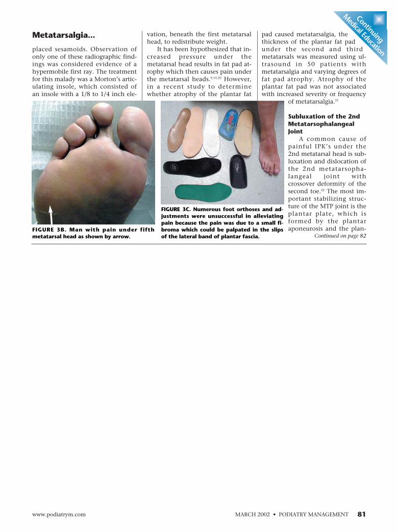
Case Presentation 1: This 50 year old male maintenanceworker had severe pain under the fifth metatarsal head forthe past several years. Physical examination revealed a scarover the dorsum of the foot from removal of Morton’sNeuroma (Figure 3A), with moderate callus formationunder the fifth metatarsal head (Figure 3B). Numerous footorthoses were of no help, despite the careful dispersionunder the callus (Figure 3C). Careful palpation revealed asmall fibroma on the slips of the lateral band of plantar fas-cia inserting into the fifth metatarsal (Figure 3B). One of
the authors (SL)has removed threeof these fibromasin patients whocould not get re-lief with debride-ment and foot or-thoses.
First MetatarsalHead Callus
A special caseis that of the cal-losity under thefirst metatarsal
head, which most commonly occurs under the tibialsesamoid. Hyperkeratotic lesions beneath the firstmetatarsal head are caused by an enlarged or malformedtibial sesamoid, or excessive plantar flexion of the firstray. The tibial sesamoid normally assumes more of theweight-bearing function transmitted to the head of thefirst metatarsal. During weightbearing the first metatarsalassumes a valgus torque and the lesser metatarsal headsassume a varus torque. This is another reason why thetibial sesamoid is more subject to excessive weightbearingunder the first metatarsal head, and the lateral plantarcondyles are more subject to weightbearing under thelesser metatarsal head.
Hallux valgus deformity frequently results in thefirst metatarsal head slipping off the sesamoid and the
Metatarsalgia...Con
tinuin
g
Medica
l Edu
catio
n
Continued on page 81
80 www.podiatrym.comPODIATRY MANAGEMENT • MARCH 2002
The first metatarsalnormally carriesapproximately twice as much
weight as each lessermetatarsal.
FIGURE 3A. Dorsum of the foot showing old surgical scarfrom removal of Morton’s Neuroma.
placed sesamoids. Observation ofonly one of these radiographic find-ings was considered evidence of ahypermobile first ray. The treatmentfor this malady was a Morton’s artic-ulating insole, which consisted ofan insole with a 1/8 to 1/4 inch ele-
Metatarsalgia... pad caused metatarsalgia, thethickness of the plantar fat padunder the second and thirdmetatarsals was measured using ul-trasound in 50 patients withmetatarsalgia and varying degrees offat pad atrophy. Atrophy of theplantar fat pad was not associatedwith increased severity or frequency
of metatarsalgia.21
Subluxation of the 2ndMetatarsophalangealJoint
A common cause ofpainful IPK’s under the2nd metatarsal head is sub-luxation and dislocation ofthe 2nd metatarsopha-langeal joint withcrossover deformity of thesecond toe.22 The most im-portant stabilizing struc-ture of the MTP joint is theplantar plate, which isformed by the plantaraponeurosis and the plan-
vation, beneath the first metatarsalhead, to redistribute weight.
It has been hypothesized that in-creased pressure under themetatarsal head results in fat pad at-rophy which then causes pain underthe metatarsal heads.9,19,20 However,in a recent study to determinewhether atrophy of the plantar fat
Continuing
Medical Education
Continued on page 82
MARCH 2002 • PODIATRY MANAGEMENTwww.podiatrym.com 81
FIGURE 3B. Man with pain under fifthmetatarsal head as shown by arrow.
FIGURE 3C. Numerous foot orthoses and ad-justments were unsuccessful in alleviatingpain because the pain was due to a small fi-broma which could be palpated in the slipsof the lateral band of plantar fascia.

tar capsule.23 Metatarsophalangealjoint instability can be produced bydamage of the joint capsule, collat-eral ligaments, articular cartilage, orsubchondral bone as a result ofrepetitive microtrauma (i.e., walk-ing), or inflammatory, metabolic,and infectious diseases.24 The use ofhigh-heeled shoes may producechronic hyperextension forces at themetatarsophalangeal joint that maycause stretching of the plantaraponeurosis and capsule with even-tual instability of the metatarsopha-langeal joint.23 Chronic repetitivemicrotrauma disrupts the plantarplate and collateral ligaments. Syn-ovitis from rheumatoid arthritis canalso disrupt the plantar metarsopha-langeal joint ligaments and capsularstructures.24
Once this joint stability is com-promised, the intrinsic interosseousand lumbrical muscle, which flexthe MPJ and extend the PIPJ and
Metatarsalgia...Con
tinuin
g
Medica
l Edu
catio
n
82 www.podiatrym.comPODIATRY MANAGEMENT • MARCH 2002
FIGURE 4A (left photo). 39-year-oldmale with a 2.5 inch short left leg sec-ondary to trauma when he waspushed off train tracks. He suffered se-vere muscle loss of the left lower ex-tremity with weakness of all left ex-tensor muscles and a fused right knee.
FIGURE 4B. 2.5 inch short left leg re-sults in deep IPK’s submetatarsals 1and 5 on the left.Continued on page 82

DIPJ, are overcome by the strongerextrinsic muscles (EDL, EHL andFDL). This results in hyperextensionof the MPJ and flexion of the PIPJand DIPJ, producing hammertoe de-formity.
Cavus FootStructural deformities such as
cavus foot place increased pressureon the first and fifth metatarsalheads.25,26 Contracted digits, atrophyof the fat pad, and rigidity tend tooccur in the cavus foot type and ex-acerbate plantar pressures under themetatarsal heads. The plantar flexedmetatarsals of the cavus foot also re-sult in increased weightbearing pres-sure on the metatarsal heads.
Limb Length DifferenceAfter walking for prolonged peri-
ods of time with significant limblength discrepancy, the shorter leg willcompensate with rigid ankle equines,
Metatarsalgia...Con
tinuin
g
Medica
l Edu
catio
n
84 www.podiatrym.comPODIATRY MANAGEMENT • MARCH 2002
FIGURE 5B. Plain radiographs showhealing of stress fractures of the 3rdand 4th metatarsal necks. The 4thmetatarsal fracture is impacted.
FIGURE 5A. Clinical presentation ofmetatarsal fracture—pain andswelling over the dorsum of the rightfoot localized to the shaft of the sec-ond metatarsal.Continued on page 84
Metatarsalgia... therefore increasing weight-bearing stress on the forefoot andthe metatarsal heads. This results incallus formation (Figure 4A/B).
TraumaMetatarsal fractures are a com-
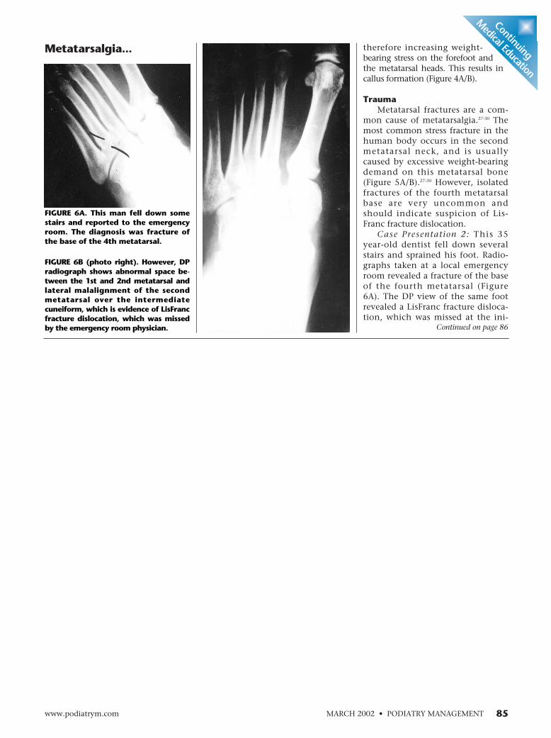
mon cause of metatarsalgia.27-30 Themost common stress fracture in thehuman body occurs in the secondmetatarsal neck, and is usuallycaused by excessive weight-bearingdemand on this metatarsal bone(Figure 5A/B).27-30 However, isolatedfractures of the fourth metatarsalbase are very uncommon andshould indicate suspicion of Lis-Franc fracture dislocation.
Case Presentation 2: This 35year-old dentist fell down severalstairs and sprained his foot. Radio-graphs taken at a local emergencyroom revealed a fracture of the baseof the fourth metatarsal (Figure6A). The DP view of the same footrevealed a LisFranc fracture disloca-tion, which was missed at the ini-
Continuing
Medical Education
Continued on page 86
MARCH 2002 • PODIATRY MANAGEMENTwww.podiatrym.com 85
FIGURE 6A. This man fell down somestairs and reported to the emergencyroom. The diagnosis was fracture ofthe base of the 4th metatarsal.
FIGURE 6B (photo right). However, DPradiograph shows abnormal space be-tween the 1st and 2nd metatarsal andlateral malalignment of the secondmetatarsal over the intermediatecuneiform, which is evidence of LisFrancfracture dislocation, which was missedby the emergency room physician.

quela of a probable third metatarsalfracture, which healed with a splitmetatarsal head (Figure 7).
The incidence of stress fractures
to the metatarsals have been shownto be reduced in military recruits whowore soft custom foot orthoses.31,32
Prior surgeryPatients who have had previous
surgery on a metatarsal, especiallyremoval of a metatarsal head, arelikely to have painful callus on theadjacent metatarsal head.
Case Presentation 4. This 55 year-old female presents with chief com-plaint of painful callus locatedunder the second metatarsal head(Figure 8A). Physical examination
tial presentation (Figure 6B).Metatarsal fractures may result
in healing in such a way that themetatarsals are misshapen, moreplantar flexed, creating additionalpressure and callus formation in theforefoot.
Case Presentation 3: This 65 year-old female had painful callus underthe third metatarsal head. She remem-bered previously injuring her footabout a year ago, but claimed that herfoot had healed. Radiographs revealeda split third metatarsal head as a se-
Metatarsalgia...Con
tinuin
g
Medica
l Edu
catio
n
Continued on page 88
86 www.podiatrym.comPODIATRY MANAGEMENT • MARCH 2002
TABLE 1EFFECTIVENESS OF MECHANICAL TREATMENT OFMETATARSALGIA-REVIEW OF THE LITERATURE
STUDY N TREATMENT MEASUREMENT RESULTS
Holmes & Timmerman, 10 healthy volunteers soft metatarsal pads Pedograph Reduction in plantar pressure for1990 5 females; 5 males (no other details) woman 12% to 60%
men 14% to 40%
Chang, Faraj, Harris, et al., 10 healthy male Plastazote insoles Interlink (Santa Barbara, Significant increases in peak1994 subjects (6.4 mm thick) CA) pressure sensors pressures, contact durations,
rubber metatarsal in portable in-shoe pressure-time integrals atpads-6 cm long data acquisition system. metatarsal shaft region with5.2 cm wide, metatarsal pad use..8 cm thick Mild decrease in mean peakDistal margins 5 mm pressure under 1st & 2nd metproximal to metatarsal head. Contact durationheads. decreased at all met heads;worn in P.W. Minor pressure-time integralsExtradepth shoes decreased at mets 1-4.
Poon & Love, 14 patients with Custom “metatarsal F-scan 13% reduction in forefoot1997 metatarsalgia dome” foot orthoses Visual analog pain scale plantar pressure
71% reduction in pain asmeasured by visual analogscale.
Postema, Burm, Zande, et al 42 patients with Custom molded insole EMED System Rockerbar decreased peak1998 metatarsalgia & rockerbar added pressure under forefoot 15%
41 females depth shoe Custom molded insole decrease1 male (Patient casted full pressure under forefoot by 18%
weightbearing)Metatarsal pad was5 mm thick and 40mmlong
Kelly & Winson, 1998 33 patients Viscoped Insole Musgrave Footprint Viscoped-6/18 patients rated(Bauerfeind, UK) system. much improvedLanger Blueline Visual analogue pain Langer Blue group 12/15 ratedorthosis (Langer, score. much improvedDeerpark, NY) Langer Blue Line group reducedfor 8 weeks forefoot plantar pressures
significantly better than Viscopedinsoles
Atrophy of the plantarfat pad was not
associated with increasedseverity or frequency of
metatarsalgia.

88 www.podiatrym.comPODIATRY MANAGEMENT • MARCH 2002
reveals a short third toe (Figure 8B). X-rays revealedsurgical removal of the 3rd metatarsal head (Figure 8C).
Neuromuscular DiseaseNeuromuscular disease may result in pronounced
muscle imbalance leading to particularly thickenedpainful metatarsal calluses (Figure 9A/B).33
BrachymetatarsiaBrachymetatarsia disrupts the smooth metatarsal
parabola resulting in uneven weightbearing with pain-ful metatarsalgia.
Case Presentation 5: This 20 year-old female had ahistory of removal of a pituitary gland tumor severalyears previously, which caused her to require takinglarge doses of hormone therapy. This resulted in prema-ture closure of the growth plates with brachymetatarsal-gia (Figure 10).
Clinical PresentationWhen a patient complains of forefoot pain the first
Metatarsalgia...Con
tinuin
g
Medica
l Edu
catio
n
thing is to observe whether a callus is present.34 The sub-jective findings include pain under the metatarsals of aburning and cramping nature.6 The objective findings in-clude a plantar callus under the head of the metatarsal,
often with indi-vidual or multiplecontracted digits.There may betenderness overthe plantar sur-face of them e t a t a r s a l s .There may alsobe a depressionon the dorsumover the head ofthe metatarsal
bone. There may be decreased passive range of motion ofthe involved metatarsophalangeal joint.
Associated DeformitiesThe contracted digital deformities are hammertoes,
mallet toes, and claw toes. A hammertoe is a flexioncontracture at the proximal interphalangeal joint. Acorn frequently develops on the head of the proximalphalanx. Extrinsic pressure of the hallux against thesecond toe owing to restrictive shoe gear results inhammertoe deformity or subluxation or dislocation ofthe second metatarsal phalangeal (MTP) joint(crossover deformity).22 A mallet toe is a flexion con-tracture of the distal interphalangeal joint making thedistal aspect of the toe point toward the ground. A hardcorn develops on the tip of the toe. A clawtoe is a flex-ion contracture at both the proximal and distal inter-phlangeal joints.35 Clawed digits may be associated withneuromuscular disease.36
Hard corns (heloma durum) are an accumulation ofseveral layers of epidermis over a bony prominencegenerally found on the lateral side of the fifth toe andon the dorsum of the toes as the skin rubs against theshoe.37 They are the body’s attempt to protect the skinover the bony prominence. Because there is no mois-ture in these locations, the corn remains hard. Softcorns (heloma molle) occur intertriginously most fre-quently in the fourth interspace in either the web spaceor the medial or lateral borders of the lesser digits. Thesoft corn retains the moisture of the interspace, whichis responsible for its macerated soft texture.38
Differential DiagnosisA study of metatarsalgia in 98 patients revealed
23 distinct diagnoses.1 Scranton1 divided metatarsal-gia into structural, systemic, and miscellaneous fore-foot pain categories. Structural and postoperative eti-ologies were the most common causes of forefootpain; however, rheumatoid arthritis, Morton’s neuro-ma, and sesamoiditis were also relatively common.Although the great percentage of pain in the fore-foot, especially under the metatarsal heads, is causedby callosities, the most common of these diagnoseswill be considered.
Continued on page 89
FIGURE 7. Patient clinically had 3rd metatarsal callus. X-raysshowed that a metatarsal fracture sustained about a yearago healed with split metatarsal heads. One of themetatarsal heads was directed plantarly, causing pain andcallosity with ambulation.
The most common stress fracture
in the human bodyoccurs in the second
metatarsal neck.

Verruca PlantarisIt is important to distinguish
verruca plantaris from plantar ker-atosis. The keratosis is mistakenlytreated with anti-wart chemical ap-plications until keratosis breaksdown and ulcerates or leaves a per-manent scar. A plantar wart is ten-
Metatarsalgia... The third common digitalnerve is vulnerable to damagedue to excessive motion between
der when it is squeezed from medialto lateral and bleeds easily when de-brided. Plantar keratosis can be easi-ly pared down fairly deep and willnot bleed if debrided carefully.
Morton’s NeuromaMorton’s neuroma is a well-doc-
umented clinical entity which ismost probably a mechanically-in-duced degenerative neuropathy thatpredilects for the third common dig-ital nerve in middle-aged women.39-43
Continuing
Medical EducationContinued on page 90
MARCH 2002 • PODIATRY MANAGEMENTwww.podiatrym.com 89
FIGURE 8A. Patient presents with chiefcomplaint of painful 2nd metatarsalhead callus.
FIGURE 8B. Clinically there is a short3rd toe.
FIGURE 8C. X-rays reveal surgical re-moval of the 3rd metatarsal head.

the third and fourth metatarsals,compression by the large overlying
Metatarsalgia...Con
tinuin
g
Medica
l Edu
catio
n
90 www.podiatrym.comPODIATRY MANAGEMENT • MARCH 2002
transverse intermetatarsal ligamentand weightbearing forces from highheeled shoes. Over long periods oftime, nerve fiber degeneration, ex-
cessive intraneural and juxtaneuralreparative fibrosis significantly en-large the nerve, making it evenmore vulnerable to compression.
The metatarsal squeezetest is a clinical diagnos-tic test for this disorder.
Systemic ArthritisThe metatarsopha-
langeal joints are thefirst areas of the foot tobe involved in rheuma-toid arthritis.44 An x-rayof the foot with estab-lished and advancedrheumatoid arthritisshows characteristic sub-luxations at the metatar-sophalanageal joints, butx-rays in the early diag-nosis of rheumatoidarthritis are not helpful.
TreatmentThe treatment for
plantar callosities be-
FIGURE 9A (photo left). IPK under the firstmetatarsal head with contracted digits. Patient hada gunshot wound to the spine result in muscle im-balance, which led to the digital contractures.
FIGURE 9B. AFO padded with felt and a special footplate with cut out for the first metatarsal was nec-essary to alleviate this patient’s symptoms from theexcess pressure of the callus.
Continued on page 92

92 www.podiatrym.comPODIATRY MANAGEMENT • MARCH 2002
gins with debridement of diffuse callosities withenucleation of intractable plantar keratoses (IPK’s). Asurgical scalpel with a number ten surgical knife isused for debridement. A number fifteen surgical knifemay be used for enucleation. Lubrication and hydra-tion to maximize skin tone is essential. Callus debride-ment in one recent study found no significant differ-ence in peak forefoot pressure before and after callusremoval;45 however, in another report, plantar forefootpressures were reduced after debridement of callusesin diabetic patients.46
A hyperkeratolytic agent such as Carmol®-40% UreaCream (DoakDermatologics,Fairfield, N.J),or Amlactin®
12%-(12% am-monium lac-tate), may behelpful whenhyperkeratosisis particularlyt h i c k e n e d .These agentsprovide enzy-matic debride-ment of callus-es. Urea gentlydissolves the in-tercellular ma-trix, which re-sults in loosen-ing the hornylayer of the skinand sheddingscaly skin atregular inter-vals, softeninghyperkeratoses.Carmol ®- 40%Urea Cream istopically ap-
Metatarsalgia...Con
tinuin
g
Medica
l Edu
catio
n
plied to affected skin twice per day and rubbing thecream into the skin until completely absorbed.
Metatarsal PadsMetatarsal pads are pear-shaped pads which are
placed just proximal to the metatarsal heads to transferand distributebody weightto necks andshafts of themetatarsals.47
M e t a t a r s a lpads havebeen found tobe effective inreducing pres-sure under them e t a t a r s a lheads. Softm e t a t a r s a lpads were ef-fective in re-ducing pres-
sure under the metatarsal heads in ten asymptomaticvolunteers as measured by pedobarograph.48 Subjectswalked barefoot with the metatarsal pad taped to thefoot. This study found that metatarsal pads were moreeffective in reducing plantar pressures in females thanin males possibly because of previous use of high-heeled shoes or because of the smaller size of the fe-male foot. The authors concluded that simple inexpen-sive metatarsal pads were an effective treatment formetatarsalgia, especially in female patients.
In a more recent but similar study, ten male asymp-tomatic volunteers walked in 6.4mm thick plastazoteinsoles with rubber metatarsal pads worn in P.W. Minorshoes.49 Metatarsal pad use resulted in peak load in-creases in the midfoot region and mild decreases in theforefoot region under the 1st and 2nd metatarsal headsand slight increases laterally. Contact durations de-creased at all metatarsal head locations. Thereforemetatarsal pads seemed to redistribute pressure awayfrom the metatarsal head more proximally into themetatarsal shaft. The authors concluded that insoleswith metatarsal pads were an effective treatment for
metatarsalgia.Prefabricated wool felt
metatarsal pads are availablefrom Hapad, Inc. (Bethel Park,PA) (Figure 11A). Thesemetatarsal pads have adhesivebacking and are designed to beplaced directly inside the shoe(Figure 11B). When a shoe isworn, the inside of the shoe can
FIGURE 10. This 20 year old female hada history of removal of a pituitary glandtumor several years previously whichcaused her to require taking large dosesof hormone therapy, which resulted inpremature closure of the growth plateswith brachymetatarsalgia.
FIGURE 11A. Prefabricated wool feltmetatarsal pads (Hapad, Inc., BethelPark, PA) with adhesive backing.
FIGURE 11B. Prefabricated woolfelt metatarsal pads (Hapad, Inc.Bethel Park, PA) with adhesivebacking are designed to be placeddirectly inside the shoe and lastthe life of the shoe.
Continued on page 93

be examined for pressure areas inthe metatarsal region usually indi-cated by dark spots. The front of thepad is placed just proximal to thedark spot pressure area. While fit-ting the pad in the shoe, only asmall portion of the adhesive on theback of the pad should be shown sothat the pad can be placed in differ-ent spots until the patient feelscomfortable in the shoe.
Special prefabricated metatarsalbandages are available from ApexFoot Health Industries (Hackensack,New Jersey), which slide easily on thefoot and do not take up as much
room as an insole or foot orthosis andcan be worn in virtually any shoewith complete comfort (Figure 12).
Insoles & Foot OrthosesInsoles or accommodative foot
orthoses assist in rebalancing to re-distribute and disperse calluses andare made of soft materials such asplastazote, PPT, felt, foam rubber,sponge rubber, plastics, leather,Spenco®, and Sorbothane. Insolesand custom foot orthoses have beenshown to reduce forefoot pressureand pain in patients withmetatarsalgia (Figure 13A/B).
A number of studies have com-pared the effectiveness of variouskinds of insoles on the relief ofmetatarsalgia and reduction of fore-foot pressure. In a comparativestudy on the effectiveness of plasta-zote/PPT versus silicone insoles,plantar forefoot pressure was low-ered in all patients. However, reduc-tion in forefoot plantar pressure was84% for those wearing the plasta-zote/PPT insoles for eight weeks ver-sus 34% forefoot pressure reductionfor those wearing silicone insoles forthe same time period.3
Metatarsalgia... tion in plantar pres-sure of 7% to 9% underthe metatarsal heads.51
In one study with sev-eral interesting conclusionsit was found that custommolded insoles and arockerbar were found to beeffective in reducing centralforefoot pressure in 42 pa-tients with metatarsalgia.52
It was determined that thecustom molded insole re-duced central forefoot pres-sure by 18% and the rockerbar contributed to reduc-
tion in forefoot pressure by approxi-mately 15%. In this study, custommolded insoles reduced forefootpressure better than prefabricated in-soles. However, reduction in painwas not related to a reduction inpressure. Similarly, walking speedand body mass were found to be un-related to plantar forefoot pressurein this study. The authors recom-mended prescribing the custom
In another study, fourteen pa-tients with metatarsalgia woremetatarsal dome orthoses with a fol-low-up of 15.5 weeks.50 At the timeof follow-up there was found to be a13% decrease in mean forefoot plan-tar pressure and a 71% reduction inpain as measured by a visual analogscale, with 90% of patients wearingtheir orthoses most of the time. Andin yet another similar study, custommoulded inserts resulted in reduc-
Continuing
Medical Education
Continued on page 94
MARCH 2002 • PODIATRY MANAGEMENTwww.podiatrym.com 93
TABLE 2TYPES OF ROCKER BOTTOM SOLES
(Adapted from Janesse D: Introduction to Pedorthics, Pedorthic Foot Wear
Association, Columbia, Maryland, 1998)
TYPE ROCKER INDICATION
Mild Rocker Sole Relieve metatarsal pressureTypically found on running shoes
Heel-to-toe Rocker Sole Digitial deformities such as hammer toes& claw toesCalcaneal ulcersMidfoot amputation
Toe-only rocker sole Hallux rigidus
Severe angle rocker For extreme relief of ulceratedmetatarsal heads
Negative-heel rocker Rigid calcaneus deformityPainful metatarsal heads
Double rocker sole Midfoot charcot foot
FIGURE 12. Metatarsal bandage (Apex Foot HealthIndustries, Hackensack, New Jersey).
It is important todistinguish
verruca plantaris from plantar
keratosis.

ly more than flat inserts.53
Twelve rheumatoid arthritis pa-tients with second metatarsal headpain were treated with four differentfoot orthoses which included prefab-ricated, standard custom molded,
custom moldedwith metatarsaldome, custommolded withmetatarsal bar anda shoe-only con-trol.54 Results re-vealed that all or-thoses significant-ly reduced pres-sure beneath thefirst and secondmetatarsal headcompared to theshoe-only controlas measured bythe EMED Pedarsystem. However,the custom mold-ed orthosis with ametatarsal dome
rheumatoid arthritis, foot orthoseshave been effective in reducing fore-foot pressure and relievingmetatarsal pain. In diabetic patients,custom foot inserts loweredmetatarsal head pressure significant-
molded insole and rocker bottomshoe together.52
In patients with foot problemsdue to diabetes mellitus and
Metatarsalgia...Con
tinuin
g
Medica
l Edu
catio
n
94 www.podiatrym.comPODIATRY MANAGEMENT • MARCH 2002
FIGURE 13A. A variety of insoles for relief ofmetatarsalgia. Top left 3/4 Comf-Orthotic® of woolfelt with metatarsal lift and arch support (Hapad,Bethel Park, PA). Bottom left Soft Plastazote PPTcombination insoles (Langer Biomechanics Group,DeerPark, New York). Top right Sorbolite shock ab-sorbing Comfort Insole (IEM Medical Technologies,Inc, Ravenna, Ohio). Bottom right Hapad full lengthComf-Orthotic® (Hapad, Inc., Bethel Park, PA).
FIGURE 13B. Variety of metatarsal pads and P.Q.Lady’s dress shoe comfort inserts with special ballof the foot cushion. The insole is made of a spe-cial 3-layer material consisting of a top cover ofsmooth nylon, midlayer of SbR for shock absorp-tion and a bottom layer of 1/8 inch pure PQ Viscoelastic polymer for extra shock absorption (Rieck-en’s Orthotic Laboratory, Evansville, Indiana). Continued on page 95

was the most effective designfor rheumatoid arthritis pa-tients with painful secondmetatarsal heads.54
FootwearShoes should be well-
padded, have rubber soles tocushion and absorb shockand have a relatively lowheel. Depth inlay orthope-dic shoes have a large upperto accommodate hammerdigits and are large enoughto fit an insole. A shoe witha rocker bottom sole has theanterior half of the sole curved up-ward toward the end of the shoewith the apex of the curve just prox-imal to the metatarsal heads allow-ing for a smooth transmission fromheel to toe during stance phase ofgait. Shoes with rocker-bottom soleshave been shown to reduce pressureunder the metatarsal heads and area frequently recommended external
shoe modification for metatarsal-gia.55,56 Of the six types of rocker-bot-tom soles (Table 2), the mild rockersole is the most commonly used andis effective in relieving metatarsalpressure.57 Since the mild rocker isfrequently found on running shoes,the heavy cushioning of the bettermen’s running shoes in conjunctionwith the rocker sole makes this anexcellent choice for patients withmetatarsalgia.
A metatarsal bar, not to be con-fused with a rockerbar, is anotherexternal shoe modification whichworks as a fulcrum to reducemetatarsophalangeal extensionforces creating a negative heel ef-fect.57 The bars are made fromleather or crepe and should be no
Metatarsalgia... Use of ready-made insolesin the treatment of lessermettarsalgia: A prospective ran-domized controlled trial. FootAnkle 19: 217-220, 1998.
4 Albert SF, Jahnigen DW:Treating common foot disordersin older patients. Geriatrics 38:42-55, 1983, June.
5 Pack LG, Julien PH: Differ-ential diagnosis of lessermetatarsalgia. Clin Podiatr MedSurg 7(4): 573-7, 1990.
6 Hauser EDW: Diseases of thefoot. W.B. Saunders Company,Philadelphia, 1941. pp. 311-314.
7 Mann RA: MetatarsalgiaCommon causes and conserva-tive treatment. PostgraduateMedicine 75:150-67, 1984.
8 Helfand A: Nail and hyperkeratoticproblems in the elderly foot. AFP: 39(2)101-110, 1989.
9 Helfand AE Lesser metatarsalgia in thegeriatric patient. Clin Podiatr Med. Surg.7(4): 743-749, 1990.
10 Mann RA, DuVries HL: Intractableplantar keratosis. Orthop Clin North Am41: 67, 1973.
11 Mann RA: Keratotic disorders of theplantar skin. In Surgery of the Foot andAnkle, ed by RA Mann, St. Louis, CVMosby Company, 1986. Chapter 7. pp.180-198.
12 Calliet R: Foot and Ankle Pain. F.A.Davis Company, Philadelphia, 1982, edi-tion 2.
13 Friedman SL: “Palliative Care,” In JMRobbins: Primary Podiatric Medicine. W.B.Saunders Company, Philadelphia, 1994,Chapter 13, 167-82.
14 Klenerman L, Nissen KI, Baker H: TheFoot and Its Disorders, Blackwell Scientific
thicker than 3/8 of an inch. Themetatarsal bar should be locatedproximal to the metatarsal heads ofthe foot.
Treatment of associated ham-merdigits consists of observation ifnot symptomatic. Shoe wear withhigher toe box, low heel, depthinlay shoe is recommended. De-bridement of the associated cornand plantar callosity is always help-ful. Silicone toe sleeve may beplaced directly on the affected ham-mered digit. A Budin splint (figure14), or silicone gel metatarsal cush-ion (Apex Foot Health Industries,Hackensack, NJ (Figure 15) may beapplied to extend a flexible ham-merdigit. Surgical referral is rec-ommended if the problem is ex-tremely painful and does not re-spond to conservative treatment.
For high-heeleddress shoes thinlightweight prefabri-cated foot orthoseswith a spring archdesign are availablefrom the EneslowFoot Comfort Cen-ter (New York, NewYork) (Figure 16). �
References1 Scranton PE:
Metatarsalgia: Diagnosisand Treatment. J BoneJoint Surg. 62A: 723-32,1980.
2 Scranton PE:Metatarsalgia: A clinicalreview of diagnosis andmanagement. FootAnkle 1: 229, 1981.
3 Kelly A, Winson I:
Continuing
Medical Education
Continued on page 96
MARCH 2002 • PODIATRY MANAGEMENTwww.podiatrym.com 95
Metatarsal pads have been found to be effective in reducing pressure
under the metatarsal heads.
FIGURE 15. Anti-Shox® Gel Metatarsal Cushion (Apex FootHealth Industries, Hackensack, New Jersey.)
FIGURE 14. Budin splint to stretch toe and pad metatarsalheads.

Drs. Levitz and Sobel are professors inthe Department of Orthopedics,NYCPM.
Prevention of stress fractures using custombiomechanical shoe orthoses. Clin OrthopRel Res. 360: 182-190, 1999.
32 Milgrom C, Giladi M, Kashtan H, etal: A prospective study of the effect of ashock-absorbing orthotic device on the in-cidence of stress fractures in military re-cruits. Foot Ankle 6: 101-104, 1985.
33 Sobel E, Giorgini R: Problems andmanagement of the rearfoot in neuromus-cular disease. A report of ten cases. J AmerPodiatr Med Assoc 89 (1): 24-38, 1999.
34 Coughlin MJ; Common causes ofpain in the forefoot in adults. Review arti-cle. J Bone Joint Surg 82B: 781-9, 2000.
35 Sands AK, Byck DC: Idiopathicclawed toes. Foot Akle Clin 3(2): 245-58,1998.
36 Teasdall RD: Neuropathic clawedtoes. Foot ankle Clin 3(2): 229-43, 1998.
37 Astion DJ: The fifth toe hard corn.Foot Ankle Clin 3(2): 305-11, 1998.
38 Donley BG, Gates NT: Interdigitialcorns. Foot Ankle Clin 3(2): 293-303, 1998.
39 Oliver TB, Beggs I: Ultrasound in theassessment of metatarsalgia: A Surgical andHistological Correlation. Clin Radiol 53:287-9, 1998.
40 Quirk R: Morton’s neuroma. Aus-tralian Fam Phys 16(8): 1117-20, 1987.
41 Williams JW, Meaney J, WhitehouseGH, et al: MRI in the Investigation of Mor-ton’s Neuroma: Which Sequences? Clin Ra-diol 52: 46-9, 1997.
42 Younger ASE, Claridge RJ: The role ofdiagnostic block in the management ofMorton’s neuroma. Can J Surg 41(2): 127-30, 1998.
43 Wu KK: Morton neuroma andmetatarsalgia. Curr Opin Rheumatol 12(2):131-42, 2000. (Published posthumously,Dr. Wu died November 25, 1999.)
44 Sobel E, Caselli MA, McHale K: PedalManifestations of Musculoskeletal Disease.Clin Podiat Med Surg. 15: 435-480, 1998.
45 Potter J, Potter MJ: Effect of callus re-moval on peak plantar pressures. The Foot10: 23-26, 2000.
46 Young MJ, CavanaghPR, Johnson TG, MurrayMM, Boulton AJM: The ef-fect of callus removal onplantar foot pressures in dia-betic patients. Diabetic Med9: 55-7, 1992.
47 Milgram JE: Officemeasures for relief of thepainful foot. J Bone JointSurg 46A 1095-1116, 1964.
48 Holmes GB: Timmer-man L: A quantitative assess-ment of the effect ofmetarsal pads on plantarpressures. Foot Ankle 11:141-145, 1990.
49 Chang A, Abu-FarajZU, Harris GF, Nery J, ShereffMJ: Multistep measurement
of plantar pressure alterations using metatrsalpads. Foot Ankle Intern 15: 654-660, 1994.
50 Poon C, Love B: Efficacy of foot or-thotics for metatarsalgia. The Foot, 202-204, 1997.
51 Bennett P, Miskewitch V, Duplock L:Analysis of the effects of custom mouldedfoot orthotics. Gait Posutre 3, 183, 1994.
52 Postema K, Burm PET, Zande ME,Limbeek JV: Primary metatarsalgia: Influ-ence of a custom moulded insole and arockerbar on plantar pressure. Pros Orthotintern 22: 35-44, 1998.
53 Lord M, Hosein R: Pressure redistributionby moded inserts in diabetic footwear A pilotstudy. J Rehab Res Dev 31: 214-221, 1994.
54 Hodge MC, Bach TM, Carter GM: Or-thotic management of plantar pressure andpain in rheumatoid arthritis. Clin Biomech14(8): 567-575, 1999.
55 Nawoczenski DA, Birke JA, ColemanWC: Effect of rocker sole design on plantarforefoot pressures. JAPMA 78: 455-60, 1988.
56 Schaff PS, Cavanagh PR: Shoes for theinsensitive foot: The effect of a “rocker bot-tom’ shoe modification on plantar pressuredistribution. Foot Ankle 11: 129-140, 1990.
57 Janisse D: Introduction to Pedor-thics. Pedorthic Footwear Association,Columbia, Maryland, 1998.
Publications, 1976, pp. 131-63.15 Silfverskiold JP: Common foot prob-
lems. Postgrad 89(5) 183-8, 1991.16 DuVries HL: Surgery of the Foot. C.V.
Mosby Company, St. Louis, 1959.17 Morton DJ: The Human Foot, New
York, Columbia University Press, 1948.18 Viladot A: Metatarsalgia due to
biomechanical alterations of the forefoot.Orthop Clin NA 4: 165-178, 1973.
19 Chairman EL: Restoration of theplantar fat pad with autoliptransplantation.J Foot Ankle Surg. 33: 373-379, 1994.
20 Hlavac H: The plantar fat pad andsome related problems. J Am. Podiatr.Assoc. 60: 151-155, 1970.
21 Waldecker U: Plantar fat pad atro-phy: A cause of metatarsalgia? J Foot AnkleSurg 40(1): 21-27, 2001.
22 Weinfeld SB: Evaluation and man-agement of crossover second toe deformity.Foot Ankle Clin 3(2) 215-228, 1998.
23 Coughlin MJ: Subluxation and dislo-
cation of the second metatarsophalangealjoint. Orthop Clin North Am 29: 535-551,1989.
24 Mann RA, Mizel MA: Monoarticularnontraumatic synovitis of the metatrsopha-langeal joint: A new diagnosis. Foot Ankle6: 18-21, 1985.
25 Jimenez AL, Martin DE, Phillips AJ:Lesser metatarsalgia evaluation and treat-ment. Clin Podiatr Med Surg 7(4): 597-618,1990.
26 Subotnick SI: The Cavus Foot PhysSport Med 8(7): 53-5, 1980.
27 Childers RL, Meyers DH, Turner PR:Lesser metatarsal stress fractures: A study of37 cases. Clin Podiatr Med Surg 7(4): 633-44, 1990.
28 Kaye Ra: Insufficiency stress fractureof the foot and ankle in postmenopausalwomen. Foot Ankle Int 19: 221-224, 1998.
29 Shereff MJ: Complex fractures of themetatarsals Review Article: Foot and AnkleSeries. Orthopedics 13: 875-82, 1990.
30 Spector FC, Karlin JM, Scurran BL, Sil-vani SL: Lesser metatarsal fractures Inci-dence, management, and review. J Am Po-diatr Med Assoc 74(6): 259-64, 1984.
31 Finestone A, Giladi M, Elad H, et al:
Metatarsalgia...Con
tinuin
g
Medica
l Edu
catio
n
96 www.podiatrym.comPODIATRY MANAGEMENT • MARCH 2002
Shoes with rocker-bottom soles have been shown to reduce pressure
under the metatarsal heads.
FIGURE 16. Walking Balance Orthotics especially de-signed to wear with high heeled shoes (Eneslow FootComfort Center, New York, NY.)
CME Exam on page 100

MARCH 2002 • PODIATRY MANAGEMENTwww.podiatrym.com 99
1) What is the most common cause of forefootpain?
A) Morton’s neuromaB) Rheumatoid arthritisC) OsteoarthritisD) Callus
2) The nature of the pain in intractable plantar ker-atosis can be characterized as:
A) VascularB) PsychologicalC) NeuriticD) Endocrinological
3) What is the most common cause of intractableplantar keratosis?
A) Enlargement of the plantar medial condyle ofthe metatarsal headB) Enlargement of the plantar lateral condyle ofthe metatarsal headC) Clawtoe deformity of the corresponding digitD) Plantar flexion of the metatarsal head
4) What is the most common etiology of a callusunder the first metatarsal head?
A) Enlargement of the tibial sesamoidB) Enlargement of the fibular sesamoidC) Cavus footD) Forefoot valgus deformity
5) Which of the following results in first ray over-load syndrome?
A) Relaxation of the capsuloligamentous struc-tures of the first metatarsalB) Hallux valgusC) SesamoiditisD) Flatfoot
6) What is the torque of the metatarsals?A) Neutral torqueB) Valgus torque of the first metatarsal andvarus torque of the lesser metatarsals.C) Varus torque of all five metatarsalsD) Valgus torque of the lesser metatarsals andneutral torque of the first metatarsal
7) The findings of recent research on metatarsalpads and foot orthoses show that:
A) They have been found to reduce pressure,but only in asymptomatic subjects.B) They have been shown to reduce pressure inasymptomatic subjects and patients withmetatarsalgia.C) They have been shown to reduce pressure in
Continued on page 100
See instructions and answer sheet on pages 136-138.
E X A M I N A T I O N
ContinuingMedical Education
EARN CME CREDITSFROM
PODIATRY MANAGEMENT
Now you can earn CME credits by care-fully reading articles and answeringquestions.
You have two methods to enroll: 1) on a perissue basis (at $15 per topic) or 2) per year forthe special introductory rate of $99 (you save$51). You may submit the answer sheet, alongwith your check (payable to Podiatry Manage-ment) and your state license number(s).
If you correctly answer seventy percent (70%)of the questions correctly, you will receive a cer-tificate attesting to your earned credits. Thisprogram is approved by the Council on Podia-tric Medical Education (CPME). PM’s CME pro-gram is valid in all states except Kentucky,Pennsylvania, and Texas. Podiatry Managementcannot guarantee that these CME credits will beacceptable by any state licensing agency, hospi-tal, managed care organization, or other entity.PM will, however, use its best efforts to ensurethe widest acceptance of this program possible.
This instructional CME program is designedto supplement, NOT replace, existing CMEseminars. The goal of this program is to ad-vance the knowledge of practicing podiatrists. Wewill endeavor to publish high-quality manuscriptsby noted authors and researchers. If you have anyquestions or comments about this program, youcan write us at: Podiatry Management, P.O.Box 490, E. Islip, NY 11730, or e-mail us [email protected] or call us at 1-631-563-1604.
Simply go to page 77, read the article by Drs.Sobel and Levitz, and/or go to page 123 andread the article by Dr. Rehm, then answer the20 multiple choice questions that correspond toeach article. You then have the choice to mail,fax, or call in your answers.
Circle #25

B) Sesamoiditis and traumaC) Metatarsophalangeal sub-luxationD) Structural and postoperative
12) Which is NOT TRUE pertain-ing to metatarsal pads?
A) Metatarsal pads can beplaced directly in the shoe.B) Metatarsal pads increasepressure under the metatarsalnecks.C) Metatarsal pads are placeddirectly under the metatarsalheads.D) Metatarsal pads can bemade out of wool felt.
13) Summarizing the literatureon insoles/foot orthoses used totreat metatarsalgia, which of thefollowing insoles would have thebest pain and pressure-reducingfeatures?
A) Prefabricated, soft and lam-inatedB) Prefabricated, soft, laminat-ed and containing metatarsalpadC) Custom molded, semirigidfoot orthosisD) Custom molded, soft, withmetatarsal pad
14) Which shoe feature or modifi-cation would be LEAST helpful fora patient with metatarsalgia?
A) Rocker barB) High toe boxC) Rubber soleD) SACH heel
15) Which type of insert/foot or-thosis has been most effective inpreventing stress fractures?
A) Semi-rigid custom foot or-thosesB) Soft accommodative footorthosesC) Prefabricated laminated in-solesD) None of these
16) All of the following may re-sult in excess plantar metatarsal
asymptomatic subjects andsymptomatic patients as wellas re duce pain in symp-tomatic patientsD) While they are effectiveclinically, they have not beendemonstrated to reduce pressure in asymptomatic individuals or patients withmetatarsalgia
8) Recent research on patientswith plantar metatarsal calluseshave shown that when callusesare debrided:
A) Plantar pressure is reducedB) Plantar pressure is not reducedC) Plantar pressure may ormay not be reducedD) Pain is not reduced
9) What has recent researchfound pertaining to the relation-ship between plantar fat pad at-rophy and metatarsalgia?
A) Atrophy of the plantar fatpad has been found to be oneetiology of metatarsalgiaB) Atrophy of the plantar fatpad has been found to be asso-ciated with metatarsalgia, butdoes not in a causal mannerC) Plantar fat pad atrophy isassociated with increasedpressure under the plantar fatpad, but does not cause painD) There is no relationship be-tween atrophy of the plantarfat pad and metatarsalgia
10) What is the main stabilizingstructure of the MTP joint?
A) Plantar plateB) Flexor digitorum longusC) The metatarsal lateral col-lateral ligamentD) The metatarsal phalangealplantar ligament
11) In Scranton’s study ofmetatarsalgia, what were themost common diagnoses?
A) Rheumatoid arthritis andMorton’s neuroma
callus formation EXCEPT:A) Short leg with equinus con-tracturesB) Removal of metatarsal headC) Calcaneus foot typeD) Fracture of a metatarsal
17) Plantar metatarsal callus is athickening of the skin caused byhyperplasia of the keratin layer,histologically similar to a dorsaldigital heloma durum.
A) TrueB) False
18) Why must fractures of the 4thmetatarsal be observed very care-fully?
A) Because they are difficult toheal.B) Because they are easilymissed and more commonthan once thought.C) Because they are associatedwith osteoporosis inmenopausal females.D) Because they are associatedwith LisFranc fracture disloca-tion.
19) The best use of prefabricatedwool felt metatarsal pads avail-able from Hapad, Inc. (BethelPark, PA) is to:
A) Place them directly on theskinB) Place them on an insoleC) Place them directly in theshoeD) Place them on a customfoot orthosis
20) What type of rocker sole ismost frequently found on run-ning shoes?
A) There usually is no rockersole on running shoes.B) Double rockerC) Sharp angle rockerD) Mild rocker
E X A M I N A T I O N(cont’d)
Contin
uing
Medica
l Edu
catio
n
100 www.podiatrym.comPODIATRY MANAGEMENT • MARCH 2002
SEE INSTRUCTIONS AND ANSWER SHEET ON PAGES 136-138.